REVIEW ARTICLE published: 04 October 2013 doi: 10.3389/fncel.2013.00172 MicroRNA regulation and dysregulation in epilepsy Danyella B. Dogini, Simoni H. Avansini, Andre S.Vieira and Iscia Lopes-Cendes* Department of Medical Genetics, School of Medical Sciences, University of Campinas, Campinas, São Paulo, Brazil Edited by: Laure Bally-Cuif, Centre National de la Recherche Scientifique, France Reviewed by: Hermona Soreq, The Hebrew University of Jerusalem, Israel Alexander K. Murashov, East Carolina University, USA *Correspondence: Iscia Lopes-Cendes, Department of Medical Genetics, School of Medical Sciences, University of Campinas, TessáliaVieira de Camargo, 126, Campinas, São Paulo 13083-887, Brazil e-mail:[email protected] Epilepsy, one of the most frequent neurological disorders, represents a group of diseases that have in common the clinical occurrence of seizures. The pathogenesis of different types of epilepsy involves many important biological pathways; some of which have been shown to be regulated by microRNAs (miRNAs). In this paper, we will critically review relevant studies regarding the role of miRNAs in epilepsy. Overall, the most common type of epilepsy in the adult population is temporal lobe epilepsy (TLE), and the form associated with mesial temporal sclerosis (MTS), called mesial TLE, is particularly relevant due to the high frequency of resistance to clinical treatment. There are several target studies, as well few genome-wide miRNA expression profiling studies reporting abnormal miRNA expression in tissue with MTS, both in patients and in animal models. Overall, these studies show a fine correlation between miRNA regulation/dysregulation and inflammation, seizure-induced neuronal death and other relevant biological pathways. Furthermore, expression of many miRNAs is dynamically regulated during neurogenesis and its dysregulation may play a role in the process of cerebral corticogenesis leading to malformations of cortical development (MCD), which represent one of the major causes of drug-resistant epilepsy. In addition, there are reports of miRNAs involved in cell proliferation, fate specification, and neuronal maturation and these processes are tightly linked to the pathogenesis of MCD. Large-scale analyzes of miRNA expression in animal models with induced status epilepticus have demonstrated changes in a selected group of miRNAs thought to be involved in the regulation of cell death, synaptic reorganization, neuroinflammation, and neural excitability. In addition, knocking-down specific miRNAs in these animals have demonstrated that this may consist in a promising therapeutic intervention. Keywords: microRNAs, epilepsy, temporal lobe, cortical malformations, animal models MicroRNAs IN HUMAN MESIAL TEMPORAL LOBE EPILEPSY Epileptic seizures are the clinical manifestations that reflect a tem- porary dysfunction of a set of neurons in the brain (Engel, 2001). Epilepsy has a high prevalence in the population, about 1.5–2% and it is considered a public health problem since it has important social and economic impact (Annegers et al., 1996; Borges et al., 2004). Because of its high prevalence and severity, temporal lobe epilepsy (TLE) is one of the most studied types of epilepsy. In TLE complete seizure control with drug treatment is achieved in less than 50% of patients (Sander, 1993; Mattson, 1994). The most common form of TLE is mesial TLE (MTLE), which has the symp- toms generated by the involvement of the medial temporal lobe structures (Engel, 2001). Resistance to drug treatment is a cru- cial problem for patients with MTLE and surgery to remove the affected brain area is, in many cases, a successful therapeutic strat- egy (Engel, 2001). Surgical specimens in MTLE most frequently show mesial temporal sclerosis (MTS), which is a pathological condition with specific features, including selective neural loss and gliosis in the CA1 hippocampal region (Wieser, 2004). Other changes may include dispersion of the granule cells in the dentate gyrus, neurogenesis of granule cell and synaptic reorganization of the mossy fibers (Thom, 2004). Focal lesions and malformations of cortical development (MCD; cortical dysplasia) may represent other findings in patients with drug refractory MTLE (Blumcke et al., 2002; Thom, 2004). It has been demonstrated that different microRNAs (miR- NAs) may have different expression pattern in different brain regions, and these differences in distribution may be related to the preferential concentration of synaptically localized mRNA tar- geted by these miRNAs (Pichardo-Casas et al., 2012). Furthermore, these differences in concentration could be modulated by epilep- togenic activity (Pichardo-Casas et al., 2012). McKiernan et al. (2012a) detected a significant expression of about 200 miRNAs in healthy human hippocampus. However, when working with tissue obtained from patients with MTLE and using TaqMan® low-density arrays (TLDAs) they found a large-scale reduction of miRNA expression, with 51% of miRNAs tested expressed at lower levels than in controls and about 24% not detectable in epileptic tissue. In addition, these authors showed that a possi- ble mechanism involved in failure of mature miRNA expression was a significant decreased expression of DICER, an enzyme required for the generation of mature miRNAs (McKiernan et al., 2012a). MicroRNA may also have a significant role in inflammation pathways which have been shown to be involved in MTLE (Vez- zani et al., 2013). MiR-146a is significantly up-regulated in tissue Frontiers in Cellular Neuroscience www.frontiersin.org October 2013 | Volume 7 | Article 172 | 1

Welcome message from author
This document is posted to help you gain knowledge. Please leave a comment to let me know what you think about it! Share it to your friends and learn new things together.
Transcript
REVIEW ARTICLEpublished: 04 October 2013
doi: 10.3389/fncel.2013.00172
MicroRNA regulation and dysregulation in epilepsy
Danyella B. Dogini, Simoni H. Avansini, Andre S. Vieira and Iscia Lopes-Cendes*
Department of Medical Genetics, School of Medical Sciences, University of Campinas, Campinas, São Paulo, Brazil
Edited by:
Laure Bally-Cuif, Centre National de la
Recherche Scientifique, France
Reviewed by:
Hermona Soreq, The Hebrew
University of Jerusalem, Israel
Alexander K. Murashov, East Carolina
University, USA
*Correspondence:
Iscia Lopes-Cendes, Department of
Medical Genetics, School of Medical
Sciences, University of Campinas,
Tessália Vieira de Camargo, 126,
Campinas, São Paulo 13083-887, Brazil
e-mail:[email protected]
Epilepsy, one of the most frequent neurological disorders, represents a group of diseases
that have in common the clinical occurrence of seizures. The pathogenesis of different
types of epilepsy involves many important biological pathways; some of which have
been shown to be regulated by microRNAs (miRNAs). In this paper, we will critically
review relevant studies regarding the role of miRNAs in epilepsy. Overall, the most
common type of epilepsy in the adult population is temporal lobe epilepsy (TLE), and
the form associated with mesial temporal sclerosis (MTS), called mesialTLE, is particularly
relevant due to the high frequency of resistance to clinical treatment. There are several
target studies, as well few genome-wide miRNA expression profiling studies reporting
abnormal miRNA expression in tissue with MTS, both in patients and in animal models.
Overall, these studies show a fine correlation between miRNA regulation/dysregulation
and inflammation, seizure-induced neuronal death and other relevant biological pathways.
Furthermore, expression of many miRNAs is dynamically regulated during neurogenesis
and its dysregulation may play a role in the process of cerebral corticogenesis leading
to malformations of cortical development (MCD), which represent one of the major
causes of drug-resistant epilepsy. In addition, there are reports of miRNAs involved in cell
proliferation, fate specification, and neuronal maturation and these processes are tightly
linked to the pathogenesis of MCD. Large-scale analyzes of miRNA expression in animal
models with induced status epilepticus have demonstrated changes in a selected group
of miRNAs thought to be involved in the regulation of cell death, synaptic reorganization,
neuroinflammation, and neural excitability. In addition, knocking-down specific miRNAs
in these animals have demonstrated that this may consist in a promising therapeutic
intervention.
Keywords: microRNAs, epilepsy, temporal lobe, cortical malformations, animal models
MicroRNAs IN HUMAN MESIAL TEMPORAL LOBE EPILEPSY
Epileptic seizures are the clinical manifestations that reflect a tem-
porary dysfunction of a set of neurons in the brain (Engel, 2001).
Epilepsy has a high prevalence in the population, about 1.5–2%
and it is considered a public health problem since it has important
social and economic impact (Annegers et al., 1996; Borges et al.,
2004). Because of its high prevalence and severity, temporal lobe
epilepsy (TLE) is one of the most studied types of epilepsy. In
TLE complete seizure control with drug treatment is achieved in
less than 50% of patients (Sander, 1993; Mattson, 1994). The most
common form of TLE is mesial TLE (MTLE), which has the symp-
toms generated by the involvement of the medial temporal lobe
structures (Engel, 2001). Resistance to drug treatment is a cru-
cial problem for patients with MTLE and surgery to remove the
affected brain area is, in many cases, a successful therapeutic strat-
egy (Engel, 2001). Surgical specimens in MTLE most frequently
show mesial temporal sclerosis (MTS), which is a pathological
condition with specific features, including selective neural loss
and gliosis in the CA1 hippocampal region (Wieser, 2004). Other
changes may include dispersion of the granule cells in the dentate
gyrus, neurogenesis of granule cell and synaptic reorganization of
the mossy fibers (Thom, 2004). Focal lesions and malformations
of cortical development (MCD; cortical dysplasia) may represent
other findings in patients with drug refractory MTLE (Blumcke
et al., 2002; Thom, 2004).
It has been demonstrated that different microRNAs (miR-
NAs) may have different expression pattern in different brain
regions, and these differences in distribution may be related to
the preferential concentration of synaptically localized mRNA tar-
geted by these miRNAs (Pichardo-Casas et al.,2012). Furthermore,
these differences in concentration could be modulated by epilep-
togenic activity (Pichardo-Casas et al., 2012). McKiernan et al.
(2012a) detected a significant expression of about 200 miRNAs
in healthy human hippocampus. However, when working with
tissue obtained from patients with MTLE and using TaqMan®
low-density arrays (TLDAs) they found a large-scale reduction
of miRNA expression, with 51% of miRNAs tested expressed at
lower levels than in controls and about 24% not detectable in
epileptic tissue. In addition, these authors showed that a possi-
ble mechanism involved in failure of mature miRNA expression
was a significant decreased expression of DICER, an enzyme
required for the generation of mature miRNAs (McKiernan et al.,
2012a).
MicroRNA may also have a significant role in inflammation
pathways which have been shown to be involved in MTLE (Vez-
zani et al., 2013). MiR-146a is significantly up-regulated in tissue
Frontiers in Cellular Neuroscience www.frontiersin.org October 2013 | Volume 7 | Article 172 | 1
Dogini et al. MicroRNA in epilepsy
obtained from patients with MTLE (Aronica et al., 2010; Omran
et al., 2012). MiR-146a has been implicated in regulation of
astrocyte-mediated inflammatory response (Iyer et al., 2012). In
addition, in vitro experiments showed a significant up-regulation
of miR-146a in astrocytes when exposed to interleukin-1 beta
(IL-1b) stimulation, which is known to be up-regulated in the
acute phase of some animal models of MTLE (Aronica and Crino,
2011). Another miRNA that has been associated with inflamma-
tory pathways in MTLE is miR-155 (Ashhab et al., 2013). It has
been demonstrated an increase in the expression of miR-155 in
hippocampal tissue from children with MTLE, as well as in an
immature rat epilepsy model. Moreover, the observed increase in
miR-155 expression correlates with an increase in TNF-α in the
nervous tissue (Ashhab et al., 2013).
It is well known that neuronal death related to seizures involves
direct glutamate-driven excitotoxic necrosis. MiR-34a, which
belongs to a conserved miRNA family, appears to have a direct
pro-apoptotic effect in cells and regulates p53 (Hermeking, 2010).
In addition, up-regulation or overexpression of this miR-34a pro-
motes apoptosis in a variety of non-neuronal cell (Chang et al.,
2007). Therefore, it has been suggested recently, that miR-34a
could represent a key player in the mechanism underlying neu-
ronal death induced by seizures (Hu et al., 2012; Sano et al.,
2012).
MicroRNAs may also be involved in enzyme-related epileptic
pathology. It is known that adenosine is an endogenous regulator
of hippocampal activity and that it has a potent anti-ictogenic and
neuroprotective properties (Bjorklund et al., 2008), as well as it is
crucial for astrocyte physiology (Boison, 2009). Synaptic levels of
adenosine in adult brain are largely regulated by an astrocyte-based
adenosine-cycle (Boison, 2009). Adenosine is rapidly phospho-
rylated by adenosine kinase (ADK), which is almost exclusively
expressed in astrocytes (Studer et al., 2006). According to the ADK
hypothesis of epileptogenesis (Boison, 2009), any type of brain
injury can produce astrogliosis, which leads to the up-regulation
of ADK, creating focal adenosine deficiency as a direct cause of
seizures. Using lentiviral vectors in human mesenchymal stem cells
coexpressing miRNA against ADK transduction, Ren and Boison
(2010) found about 80% of ADK down-regulation. These results
suggest that miRNAs are important regulators of seizure-induced
neuronal death and that these molecules might be used as novel
therapeutic targets in the treatment of epilepsy. Some other miR-
NAs, such as miR-124, miR-134, miR-132, miR-196b (You et al.,
2012; Peng et al., 2013) have also been reported to be involved in
epilepsy (Table 1).
MicroRNAs AND MALFORMATIONS OF CORTICAL
DEVELOPMENT
Malformations of cortical development are a frequent cause of
medically intractable epilepsy. It has been estimated that 25–40%
of drug-resistant epilepsies are caused by MCD (Guerrini et al.,
2003). The development of the human cerebral cortex is a dynamic
and complex process. These processes are orchestrated by inter-
actions between extracellular and intracellular signaling cues and
any disruption of these cellular processes can result in cortical mal-
formations (Sisodiya, 2004; Guillemot et al., 2006; Guerrini et al.,
2008; McLoughlin et al., 2012).
Molecular biology and genetic studies have greatly expanded
knowledge on cortical neurogenesis so that several disorders of
cortical development have been recognized and, for some of them,
specific causative genetic defects have been identified (Aronica
et al., 2012). Furthermore, recent data support a major role for
miRNAs in fine-tuning of signaling pathways that control the
concomitant phases of corticogenesis. Supporting this notion, we
have previously shown that groups of miRNAs are differentially
regulated during normal mouse brain development (Dogini et al.,
2008). Small alterations of their expression have been associated
with a variety of neurological disorders (Volvert et al., 2012). Nev-
ertheless, few studies have investigated the possible role of miRNAs
in the pathogenesis and/or epileptogenesis of MCDs. Therefore,
we aim in the next few paragraphs to summarize current knowl-
edge about miRNAs and cerebral corticogenesis (Figure 1) and
how its dysregulation may play a role in the process leading to
MCDs and ultimately to epileptogenesis as seen in some of these
lesions (Table 1).
MicroRNAs IN NEURONAL AND GLIAL PROLIFERATION AND
DIFFERENTIATION
The first step of cortical development is cellular proliferation and
differentiation, which takes place between the 5th week and 20th
week of gestation (Sidman and Rakic, 1973; Guerrini and Barba,
2010). Microcephaly, tuberous sclerosis, and focal cortical dys-
plasia (FCD) have been considered to be malformations of these
phases. MiR-9, miR-124, miR-137, miR-184, and let-7b were
shown to control cell proliferation in the cortex (Krichevsky et al.,
2006; Makeyev et al., 2007; Silber et al., 2008; Liu et al., 2010a;
Zhao et al., 2010). In addition, loss of miR-9 expression, a brain-
specific miRNA, suppresses the proliferation and promotes the
migration of human embryonic neural progenitors, cultured in
vitro, by targeting stathmin, which increases microtubule instabil-
ity in migrating neuroblasts (Delaloy et al., 2010). In the mouse
embryonic brain, miR-9 suppressed TLX expression, resulting in
a reduction of neural stem cell proliferation and an acceleration
of neural differentiation (Zhao et al., 2009).
The cellular complexity of the cerebral cortex emerges through
specification of cortical progenitors into distinct subtypes of
neurons and glia that reach cortical layers (Kriegstein and Alvarez-
Buylla, 2009). Changes in gene expression underlie the transition
from progenitors to neurons (Guillemot et al., 2006). Conditional
removal of Dicer in the cortex affects this process. Kawase-Koga
et al. (2009) reported that the cerebral cortex of deficient Dicer-
mice showed a significant reduction in cortical thickness, caused
by a reduction in neural stem cells and neural progenitors with
increased apoptosis and impaired neuronal differentiation. In the
same way, it has been observed an inability to generate both neu-
rons and glial cells in the embryonic cerebral cortex of a Dicer-null
mouse, and that this enzyme plays a role in maintaining the
phenotype of neural stem cells during neuronal differentiation
(Andersson et al., 2010). Other miRNAs have also been reported as
critical for neural differentiation. These include miR-137, miR34a,
miR-153, miR-324, and miR-181a (Smrt et al., 2010; Agostini et al.,
2011; Stappert et al., 2013).
Focal cortical dysplasia is characterized by a spectrum of abnor-
malities in the development of the laminar structure of the human
Frontiers in Cellular Neuroscience www.frontiersin.org October 2013 | Volume 7 | Article 172 | 2

Dogini et al. MicroRNA in epilepsy
Table 1 | MicroRNAs potentially involved in epilepsy.
MicroRNA Human studies/
experimental models
Potential role in epilepsy Reference
miR-124 Human; immature rat Potential role in mesial temporal lobe epilepsy; control
cell proliferation
Makeyev et al. (2007); Peng et al. (2013)
miR-132 Human; mouse kainic acid Associated to neuronal activation and synaptic
plasticity
Vo et al. (2005)
miR-134 Human (in vitro experiments);
mouse kainic acid
Suppresses evoked seizures; regulates cell migration Jimenez-Mateos et al. (2012)
miR-137 Human; rat Regulates cell proliferation; critical for neural
differentiation
Krichevsky et al. (2006); Smrt et al. (2010).
miR-146 Human; mouse; rat Regulation of astrocyte-mediated inflammatory
response; neural inflammation
Lukiw et al. (2008); Nakasa et al. (2008), Pauley
et al. (2008); Sonkoly et al. (2008), Aronica and
Crino (2011); Iyer et al. (2012), Cheng et al.
(2013)
miR-153,
miR-324,
miR-181a
Human; rat Critical role in neural differentiation Smrt et al. (2010); Agostini et al. (2011),
Stappert et al. (2013)
miR-184 Human; mouse kainic acid Regulates cell proliferation; neuroprotective effect Krichevsky et al. (2006); Makeyev et al. (2007),
Silber et al. (2008); Liu et al. (2010a), Zhao et al.
(2010); McKiernan et al. (2012b)
miR-196b Human Associated with the occurrence of seizures You et al. (2012)
miR-21 Rat pilocarpine Possible associated with increased neuronal loss
following status epilepticus
Risbud and Porter (2013)
miR-34a Human; rat pilocarpine; mouse
kainic acid
Involved in seizure-induced neuronal death; critical for
neural differentiation
Agostini et al. (2011); Hu et al. (2012), Sano et al.
(2012)
miR-9 Human (in vitro experiments) Regulates cell proliferation; promotes cell migration;
accelerates neural differentiation
Krichevsky et al. (2006); Delaloy et al. (2010)
let-7b Human; rat kainic acid Regulates cell proliferation Krichevsky et al. (2006); Makeyev et al. (2007),
Silber et al. (2008); Liu et al. (2010b), Zhao et al.
(2010)
cerebral cortex. Microscopically, FCD is usually associated with
cell abnormalities, giant/dysmorphic neurons and balloon cells
(Palmini et al., 2004; Guerrini et al., 2008; Sisodiya et al., 2009;
Blumcke et al., 2011). As FCDs are the most frequent epilepto-
genic malformation, susceptible to surgical treatment, it is of great
importance to understand the mechanisms underlying epileptoge-
nesis in FCDs (Aronica et al., 2012; Hauptman and Mathern, 2012;
Sakakibara et al., 2012). In this context, Iyer et al. (2012) evaluated
function of miR-146a in response to pro-inflammatory stimuli
and found, by using in situ hybridization, increased expression
of miR-146a in reactive astrocytes which are abundantly present
within the dysplastic cortex in FCD IIb. This observation sug-
gests a role for miR-146a in an astrocyte-mediated mechanism
predisposing to seizure in FCDs.
MicroRNAs IN NEURONAL MIGRATION
In humans, neuronal migration occurs from 6th–7th weeks
till approximately 20th–24th weeks of gestation (Sidman and
Rakic, 1973; Guerrini and Barba, 2010). Abnormalities disrupt-
ing neuronal migration result in highly epileptogenic lesions,
causing severe neurological impairment, such as those found
in periventricular nodular heterotopia, subcortical heterotopias,
and lissencephaly (Guerrini and Parrini, 2010). Doublecortin
(Dcx) regulates tangential and radial neuron migration and has
been implicated in the pathogenesis of lissencephaly and sub-
cortical heterotopias (Reiner et al., 2006). Gaughwin et al. (2011)
demonstrate that miR-134 regulates cell migration in vitro and
down-regulates Dcx protein in vivo, thereby attenuating neuronal
migration.
Experiments using neural stem cells of embryonic mouse
brains suggest that miR-137 triggered premature differentiation
and outward migration through regulation of a lysine-specific
histone demethylase (LSD1; Sun et al., 2011). Moreover, the trans-
fection of exogenous miR-125b increased migration of neural
stem/progenitor cells compared to a control group (Cui et al.,
2012).
Frontiers in Cellular Neuroscience www.frontiersin.org October 2013 | Volume 7 | Article 172 | 3
Dogini et al. MicroRNA in epilepsy
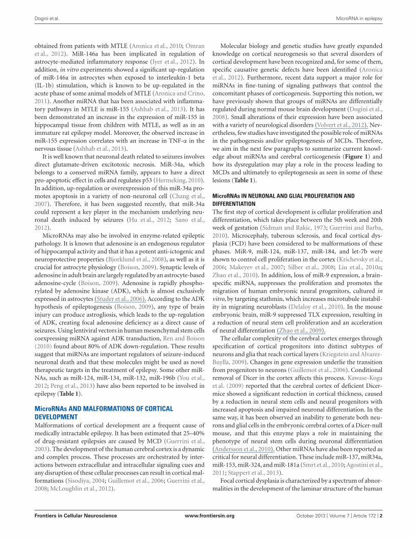
FIGURE 1 | MicroRNAs involved in the regulation of cerebral cortex
development. The figure demonstrates microRNAs that have been
associated with the three main phases of cortical development. In the
first stage, stem cells generate progenitors that are not yet committed
to differentiation and can produce neurons, astrocytes, and oligoden-
drocytes; the concomitant steps of proliferation and differentiation (5th–
20th weeks of gestation) are regulated by a set of microRNAs: miR-9,
miR-124, miR-137, miR-184, let-7b and miR-34a, miR-153, miR-324, miR-
181a. Successive waves of neurons migrate (6th–24th weeks of ges-
tation) from the ventricular regions, along radial glial cells, toward the
more external areas of the cortex, these processes are regulated by
miR-9, miR-134, and miR-137. Finally, the organization of cortical layers
(16th–40th, weeks of gestation) is regulated at this stage through
miR-137 and miR-125b.
A mice model constructed with Dicer depletion, by the Nestin-
Cre system revealed a critical role for Dicer in cortical migration
(McLoughlin et al., 2012). There was a sevenfold increase in Dcx
expression that may have contributed to the premature matu-
ration of neurons in inappropriate regions, which in turn may
led to complete cortical disorganization (McLoughlin et al., 2012).
Shibata et al. (2011) observed, after reduction of miR-9 expression,
that cortical layers were reduced and that the tangential migration
of interneurons from basal forebrain was impaired.
MicroRNAs IN NEURONAL ORGANIZATION
The third stage in cortical development is cortical organization.
When migration is complete, the cortex is a six-layered structure,
with each layer containing different types of neurons (Guerrini
and Barba, 2010). Polymicrogyria and schizencephaly have been
considered to be malformations of this post-migrational cortical
organization stage. Two miRNAs have been shown to regulate key
processes at this stage. MiR-137 which regulates neuronal mat-
uration by inhibiting dendrite formation through binding Mind
bomb 1 (Mibl; Smrt et al., 2010), and miR-125b which seems to
have a similar role, since overexpression of miR-125b leads to
longer and thinner dendritic spines (Edbauer et al., 2010).
MicroRNAs AND ANIMAL MODELS OF EPILEPSY
Induced animal models are one of the most used tools to study
the pathophysiology of different types of epilepsy and they have
been most frequently used in MTLE. In these models, animals
present behavioral, electroencephalographic, and neuropatholog-
ical features in the limbic structures similar to those observed in
patients with MTLE (Avanzini et al., 1993; Lothman et al., 1995;
Engel, 1996).
One of the first miRNAs shown to be differentially expressed
in the hippocampus in an induced animal model was miR-132
(Nudelman et al., 2010). These authors observed an increase in
the expression of miR-132 in the hippocampus 8 h after the
administration of the convulsant drug pilocarpine in mice. In neu-
rons, miR-132 expression is induced by electrical activity and the
action of neurotrophins, consequently its proposed role would be
the regulation of synaptic plasticity-related genes (Vo et al., 2005;
Wayman et al., 2008). Another miRNA that was initially explored
in epilepsy experimental models was miR-146a (Aronica et al.,
2010). This miRNA can be induced by pro-inflammatory
cytokines, such as IL-1b, and it is up-regulated in various human
disorders associated with inflammatory response (Lukiw et al.,
2008; Nakasa et al., 2008; Pauley et al., 2008; Sonkoly et al., 2008;
Frontiers in Cellular Neuroscience www.frontiersin.org October 2013 | Volume 7 | Article 172 | 4

Dogini et al. MicroRNA in epilepsy
Cheng et al., 2013). In a rat model of MTLE induced by repetitive
electrical stimulation of the perforant pathway, it was observed
that miR-146a was up-regulated in the CA3 hippocampus sub-
field 1 week (latent phase) and 3 months (chronic phase) after the
episode of status epilepticus. In these experiments, the observation
by in situ hybridization of miR-146 expression in hippocampus
reactive astrocytes further indicated a possible role for this miRNA
in neural inflammation. However, the exact genes regulated by
miR-146 in the hippocampus remains to be determined.
Subsequently, with an increasing interest in the possible role
of regulatory RNAs in epilepsy, large-scale analyzes of miRNA
expression profile by either hybridization or TaqMan® arrays were
undertaken in the hippocampus of animals with induced epilepsy
(Liu et al., 2010b; Jimenez-Mateos et al., 2011; Song et al., 2011;
Hu et al., 2012; McKiernan et al., 2012b; Pichardo-Casas et al.,
2012; Peng et al., 2013; Risbud and Porter, 2013). Analyzes were
performed on the lithium-pilocarpine model (Song et al., 2011;
Hu et al., 2012), systemic pilocarpine (Risbud and Porter, 2013),
systemic kainic acid (Liu et al., 2010b; McKiernan et al., 2012b;
Pichardo-Casas et al., 2012), intra-amygdala kainic acid (Jimenez-
Mateos et al., 2011), with time points ranging from a few hours
(McKiernan et al., 2012b) to months after status epilepticus (Song
et al., 2011; Hu et al., 2012). All studies found a significant number
of miRNAs differentially regulated in the epileptic state when com-
pared to control animals, indicating a tight regulation of miRNAs
associated with the events observed in induced epilepsy models.
Some miRNAs were found to be differentially expressed, such as
miR-34a (Hu et al., 2012; Sano et al., 2012) or miR-132 (Nudelman
et al., 2010; Jimenez-Mateos et al., 2011). However, a coherent
interpretation of the results produced by the above mentioned
experiments is hindered by the still incomplete knowledge of miR-
NAs regulated genes in the hippocampus and by the heterogeneity
of findings obtained by different studies.
The apparent lack of reproducibility in the miRNA expression
profile experiments may be explained by the diversity in animal
models, time points, and even hippocampal structures analyzed.
Moreover, miRNA expression was profiled employing microar-
rays (Song et al., 2011; Hu et al., 2012; Pichardo-Casas et al., 2012;
Risbud and Porter, 2013) or TLDAs (Liu et al., 2010b; Eacker
et al., 2011; Jimenez-Mateos et al., 2011; McKiernan et al., 2012b).
As a consequence, differences on the sensibility and specificity
of both techniques may be responsible for part of the diver-
sity observed in the published literature. In addition, a critical
point to be considered is that some studies analyzed whole hip-
pocampus homogenates (Liu et al., 2010b; Song et al., 2011; Hu
et al., 2012; Pichardo-Casas et al., 2012; Peng et al., 2013; Risbud
and Porter, 2013) and others were restricted to the CA3 subfield
(Jimenez-Mateos et al., 2011; McKiernan et al., 2012b). It is known
that the different hippocampus subfields are molecularly diverse
(Lein, 2004; Greene et al., 2009). Therefore, analyzes of whole hip-
pocampus homogenates certainly dilutes subfield-specific changes
that may take place in these epilepsy models. Strategies such as
laser capture microdissection of different hippocampus subfields
could circumvent the exposed shortcomings of whole homogenate
strategies, improving the ability of an experiment to detect more
subtle and spatially restricted changes in miRNA regulation. Fur-
thermore, since different hippocampus subfields have different
functional characteristics, sensibility to neurodegeneration and
contributions to the establishment of an epileptic state (Becker
et al., 2003; Majores et al., 2004), a separate analyzes of miRNA
profile in each structure certainly would facilitate data interpre-
tation. Another point to be considered is that the translation of
these animal models miRNA expression findings to human MTLE
could be hindered by the fact that many patients do not present an
initial precipitating event (Van Paesschen et al., 1997). Moreover
the occurrence of an episode of status epilepticus is uncommon in
human MTLE. Such a diversity of models and analyzes strategies
present in the literature poses an advantage, since the differen-
tially regulated miRNAs common to all studies may indicate the
presence of a common mechanism underlying the epileptogenic
process. However, care should be taken when employing rodent
data in the effort of understanding human MTLE miRNA asso-
ciated mechanisms due to the existence of many primate-specific
miRNAs (Bentwich et al., 2005). Therefore some mechanisms may
only be found with the direct analysis of tissue from patients that
undergo epilepsy surgery.
As already noted, many of the functional implications of the
identified differentially expressed miRNAs in the hippocampus
of animals with induced epilepsy are still unknown. Antagomirs
are stable, locked nucleic acids, engineered RNA oligonucleotides
that can recognize, based on sequence complementarity, specific
miRNAs, inducing its degradation (Krutzfeldt et al., 2005, 2007).
These engineered molecules consist in valuable tools for prob-
ing miRNAs function in vivo, and indeed, functional studies were
undertaken in some epilepsy animal models. The induction of low
intensity seizures renders animals resistant to subsequent induc-
tion of an epileptic state, a phenomena termed epileptic tolerance
(for a review see Jimenez-Mateos and Henshall, 2009). It was
observed that miR-132 was down-regulated in mice CA3 subfield
after seizure preconditioning (Jimenez-Mateos et al., 2011). In the
same study, the authors observed that the reduction in expres-
sion of miR-132, by the intracerebroventricular administration
of an antagomir directed to this miRNA, reduced neuronal loss
in the hippocampus after the induction of status epilepticus in
mice. In the hippocampus miR-132 regulates mRNAs such as
acetylcholinesterase or the GTPase activator p250GAP (Hanin
and Soreq, 2011; Shaltiel et al., 2013). Furthermore, miR-132
has been previously associated with synaptic plasticity (Vo et al.,
2005; Wayman et al., 2008). However, the miR-132 gene targets
responsible for the facilitation of neuronal death remain to be
determined. Yet another study exploring the role of miRNAs in
epileptic tolerance, found an increase in the expression of miR-
184 after preconditioning by systemic administration of a low
dose of kainic acid (McKiernan et al., 2012b). Subsequently, these
authors demonstrated that reduction of miR-184 by intracere-
broventricular administration of an antagomir directed to this
miRNA reduced the neuroprotective effect of preconditioning
on hippocampal neurons, restoring the levels of neuronal death
observed when status epilepticus was induced without precon-
ditioning. The mRNAs that may interact in vivo with miR-184
in the hippocampus are not determined and the mechanism
responsible for this miRNA-mediated neuroprotection in the hip-
pocampal CA3 subfield is also unknown. Finally, miR-34a was
shown to be up-regulated in different epilepsy animal models
Frontiers in Cellular Neuroscience www.frontiersin.org October 2013 | Volume 7 | Article 172 | 5

Dogini et al. MicroRNA in epilepsy
and its involvement in neuronal death in the hippocampus was
probed with the use of antagomirs (Hu et al., 2012; Sano et al.,
2012). The down-regulation of miR-34a by intracerebroventric-
ular injection of antagomirs reduced neuronal death observed in
the hippocampus in a lithium-pilocarpine epilepsy model (Hu
et al., 2012), but it had no effect on an intra-amygdala kainic
acid injection model in mice (Sano et al., 2012). The difference in
the experiments outcome may be related to the different models,
species and time points analyzed. It is believed that miR-34a may
regulate expression of apoptosis-related genes in the hippocam-
pus; however, further experiments are needed to confirm these
observations.
Among the functional studies involving miRNAs, the one that
explored the role of miR-134 in experimental epilepsy is note-
worthy. In an intra-amygdala kainic acid injection epilepsy model
in mice, it was observed an increase in the expression level of
miR-134 following status epilepticus. Furthermore, this miRNA
was shown to be expressed by pyramidal neurons in CA3, by
interneurons in the hilus and by neocortical as well as amyg-
dala neurons (Jimenez-Mateos et al., 2012). In the same study,
the reduction of miR-134 expression by intracerebroventricular
injection of antagomirs induced a decrease in CA3 pyramidal
neurons spine density and, remarkably, it significantly reduced
the severity of the induced seizures following intra-amygdala
kainic acid injection. The authors also demonstrated that the
induced down-regulation of this single miRNA enhanced resis-
tance to evoked seizures resulting in reduction in all events associ-
ated with experimental induction of epilepsy, namely neuronal
loss, gliosis, sprouting, and subsequent spontaneous recurrent
seizures.
In conclusion, miRNAs are emerging as key regulators of sets
of genes involved in the events that take place during epileptogen-
esis and chronic epilepsy states. Additionally, functional studies
employing antagomirs indicate that these regulatory RNAs as
promising targets for new possible strategies in the treatment of
epilepsy.
ACKNOWLEDGMENTS
We are grateful to Mrs. Mercedes de Fátima Santos for her tech-
nical assistance with the art work. This work was supported by
FAPESP (Fundação de Amparo à Pesquisa do Estado de São Paulo,
BRAZIL), grant # CEPID 2013/07559-3.
REFERENCES
Agostini, M., Tucci, P., Killick, R.,
Candi, E., Sayan, B. S., Rivetti di
Val Cervo, P., et al. (2011). Neu-
ronal differentiation by TAp73 is
mediated by microRNA-34a regu-
lation of synaptic protein targets.
Proc. Natl. Acad. Sci. U.S.A. 108,
21093–21098. doi: 10.1073/pnas.
1112061109
Andersson, T., Rahman, S., Sansom,
S. N., Alsio, J. M., Kaneda, M.,
Smith, J., et al. (2010). Reversible
block of mouse neural stem cell
differentiation in the absence of
dicer and microRNAs. PLoS ONE
5:e13453. doi: 10.1371/journal.
pone.0013453
Annegers, J. F., Rocca, W. A., and Hauser,
W. A. (1996). Causes of epilepsy:
contributions of the Rochester epi-
demiology project. Mayo Clin. Proc.
71, 570–575. doi: 10.1016/S0025-
6196(11)64114-1
Aronica, E., Becker, A. J., and Spreafico,
R. (2012). Malformations of cor-
tical development. Brain Pathol.
22, 380–401. doi: 10.1111/j.1750-
3639.2012.00581.x
Aronica, E., and Crino, P. B. (2011).
Inflammation in epilepsy: clini-
cal observations. Epilepsia 52(Suppl.
3), 26–32. doi: 10.1111/j.1528-
1167.2011.03033.x
Aronica, E., Fluiter, K., Iyer, A., Zurolo,
E., Vreijling, J., van Vliet, E. A., et al.
(2010). Expression pattern of miR-
146a, an inflammation-associated
microRNA, in experimental and
human temporal lobe epilepsy. Eur.
J. Neurosci. 31, 1100–1107. doi:
10.1111/j.1460-9568.2010.07122.x
Ashhab, M. U., Omran, A., Kong, H.,
Gan, N., He, F., Peng, J., et al.
(2013). Expressions of tumor necro-
sis factor alpha and microRNA-155
in immature rat model of status
epilepticus and children with mesial
temporal lobe epilepsy. J. Mol. Neu-
rosci. doi: 10.1007/s12031-013-0013-
9 [Epub ahead of print].
Avanzini, G., Vergnes, M., Spreafico, R.,
and Marescaux, C. (1993). Calcium-
dependent regulation of geneti-
cally determined spike and waves
by the reticular thalamic nucleus
of rats. Epilepsia 34, 1–7. doi:
10.1111/j.1528-1157.1993.tb02369.x
Becker, A. J., Chen, J., Zien,
A., Sochivko, D., Normann, S.,
Schramm, J., et al. (2003). Correlated
stage- and subfield-associated hip-
pocampal gene expression patterns
in experimental and human tempo-
ral lobe epilepsy. Eur. J. Neurosci.
18, 2792–2802. doi: 10.1111/j.1460-
9568.2003.02993.x
Bentwich, I., Avniel, A., Karov, Y.,
Aharonov, R., Gilad, S., Barad, O.,
et al. (2005). Identification of hun-
dreds of conserved and nonconserved
human microRNAs. Nat. Genet. 37,
766–770. doi: 10.1038/ng1590
Bjorklund, O., Shang, M., Tonazzini, I.,
Dare, E., and Fredholm, B. B. (2008).
Adenosine A1 and A3 receptors pro-
tect astrocytes from hypoxic damage.
Eur. J. Pharmacol. 596, 6–13. doi:
10.1016/j.ejphar.2008.08.002
Blumcke, I., Thom, M., Aronica,
E., Armstrong, D. D., Vinters,
H. V., Palmini, A., et al. (2011).
The clinicopathologic spectrum of
focal cortical dysplasias: a consensus
classification proposed by an ad hoc
Task Force of the ILAE Diagnostic
Methods Commission. Epilepsia 52,
158–174. doi: 10.1111/j.1528-1167.
2010.02777.x
Blumcke, I., Thom, M., and Wiestler, O.
D. (2002). Ammon’s horn sclerosis:
a maldevelopmental disorder asso-
ciated with temporal lobe epilepsy.
Brain Pathol. 12, 199–211. doi:
10.1111/j.1750-3639.2002.tb00436.x
Boison, D. (2009). Engineered
adenosine-releasing cells for epilepsy
therapy: human mesenchymal stem
cells and human embryonic stem
cells. Neurotherapeutics 6, 278–283.
doi: 10.1016/j.nurt.2008.12.001
Borges, M. A., Min, L. L., Guerreiro,
C. A., Yacubian, E. M., Cordeiro,
J. A., Tognola, W. A., et al. (2004).
Urban prevalence of epilepsy: pop-
ulational study in Sao Jose do Rio
Preto, a medium-sized city in Brazil.
Arq. Neuropsiquiatr. 62, 199–204. doi:
10.1590/S0004-282X2004000200002
Chang, T. C., Wentzel, E. A., Kent,
O. A., Ramachandran, K., Mullen-
dore, M., Lee, K. H., et al. (2007).
Transactivation of miR-34a by p53
broadly influences gene expression
and promotes apoptosis. Mol. Cell 26,
745–752. doi: 10.1016/j.molcel.2007.
05.010
Cheng, H. S., Sivachandran, N., Lau, A.,
Boudreau, E., Zhao, J. L., Baltimore,
D., et al. (2013). MicroRNA-146
represses endothelial activation by
inhibiting pro-inflammatory path-
ways. EMBO Mol. Med. 5, 949–966.
doi: 10.1002/emmm.201202318
Cui, Y., Xiao, Z., Han, J., Sun, J.,
Ding, W., Zhao, Y., et al. (2012).
MiR-125b orchestrates cell prolifera-
tion, differentiation and migration in
neural stem/progenitor cells by tar-
geting Nestin. BMC Neurosci. 13:116.
doi: 10.1186/1471-2202-13-116
Delaloy, C., Liu, L., Lee, J. A.,
Su, H., Shen, F., Yang, G. Y.,
et al. (2010). MicroRNA-9 coordi-
nates proliferation and migration of
human embryonic stem cell-derived
neural progenitors. Cell Stem Cell 6,
323–335. doi: 10.1016/j.stem.2010.
02.015
Dogini, D. B., Ribeiro, P. A., Rocha, C.,
Pereira, T. C., and Lopes-Cendes, I.
(2008). MicroRNA expression pro-
file in murine central nervous system
development. J. Mol. Neurosci. 35,
331–337. doi: 10.1007/s12031-008-
9068-4
Eacker, S. M., Keuss, M. J., Berezikov,
E., Dawson, V. L., and Dawson, T.
M. (2011). Neuronal activity reg-
ulates hippocampal miRNA expres-
sion. PloS ONE 6:e25068. doi:
10.1371/journal.pone.0025068
Edbauer, D., Neilson, J. R., Fos-
ter, K. A., Wang, C. F., See-
burg, D. P., Batterton, M. N.,
et al. (2010). Regulation of synaptic
structure and function by FMRP-
associated microRNAs miR-125b and
miR-132. Neuron 65, 373–384. doi:
10.1016/j.neuron.2010.01.005
Engel, J. Jr. (1996). Introduction to
temporal lobe epilepsy. Epilepsy Res.
26, 141–150. doi: 10.1016/S0920-
1211(96)00043-5
Engel, J. Jr. (2001). Mesial tempo-
ral lobe epilepsy: what have we
learned? Neuroscientist 7, 340–352.
doi: 10.1177/107385840100700410
Frontiers in Cellular Neuroscience www.frontiersin.org October 2013 | Volume 7 | Article 172 | 6

Dogini et al. MicroRNA in epilepsy
Gaughwin, P., Ciesla, M., Yang, H.,
Lim, B., and Brundin, P. (2011).
Stage-specific modulation of cortical
neuronal development by Mmu-
miR-134. Cereb. Cortex 21, 1857–
1869. doi: 10.1093/cercor/bhq262
Greene, J. G., Borges, K., and Dingle-
dine, R. (2009). Quantitative tran-
scriptional neuroanatomy of the rat
hippocampus: evidence for wide-
ranging, pathway-specific hetero-
geneity among three principal cell
layers. Hippocampus 19, 253–264.
doi: 10.1002/hipo.20502
Guerrini, R., and Barba, C. (2010).
Malformations of cortical devel-
opment and aberrant cortical
networks: epileptogenesis and
functional organization. J. Clin.
Neurophysiol. 27, 372–379. doi:
10.1097/WNP.0b013e3181fe0585
Guerrini, R., Dobyns, W. B., and
Barkovich, A. J. (2008). Abnormal
development of the human cere-
bral cortex: genetics, functional con-
sequences and treatment options.
Trends Neurosci. 31, 154–162. doi:
10.1016/j.tins.2007.12.004
Guerrini, R., and Parrini, E. (2010).
Neuronal migration disorders. Neu-
robiol. Dis. 38, 154–166. doi:
10.1016/j.nbd.2009.02.008
Guerrini, R., Sicca, F., and Parmeggiani,
L. (2003). Epilepsy and malforma-
tions of the cerebral cortex. Epileptic
Disord. 5(Suppl. 2), S9–S26.
Guillemot, F., Molnar, Z., Tarabykin,
V., and Stoykova, A. (2006).
Molecular mechanisms of cortical
differentiation. Eur. J. Neurosci.
23, 857–868. doi: 10.1111/j.1460-
9568.2006.04626.x
Hanin, G., and Soreq, H. (2011).
Cholinesterase-targeting microR-
NAs identified in silico affect
specific biological processes.
Front. Mol. Neurosci. 4:28. doi:
10.3389/fnmol.2011.00028
Hauptman, J. S., and Mathern, G. W.
(2012). Surgical treatment of epilepsy
associated with cortical dysplasia:
2012 update. Epilepsia 53(Suppl.
4), 98–104. doi: 10.1111/j.1528-
1167.2012.03619.x
Hermeking, H. (2010). The miR-34
family in cancer and apoptosis. Cell
Death Differ. 17, 193–199. doi:
10.1038/cdd.2009.56
Hu, K., Xie, Y. Y., Zhang, C., Ouyang,
D. S., Long, H. Y., Sun, D. N., et al.
(2012). MicroRNA expression profile
of the hippocampus in a rat model
of temporal lobe epilepsy and miR-
34a-targeted neuroprotection against
hippocampal neurone cell apoptosis
post-status epilepticus. BMC Neu-
rosci. 13:115. doi: 10.1186/1471-
2202-13-115
Iyer, A., Zurolo, E., Prabowo,
A., Fluiter, K., Spliet, W. G.,
van Rijen, P. C., et al. (2012).
MicroRNA-146a: a key regulator
of astrocyte-mediated inflammatory
response. PloS ONE 7:e44789. doi:
10.1371/journal.pone.0044789
Jimenez-Mateos, E. M., Bray, I., Sanz-
Rodriguez, A., Engel, T., McK-
iernan, R. C., Mouri, G., et al.
(2011). miRNA Expression profile
after status epilepticus and hip-
pocampal neuroprotection by target-
ing miR-132. Am. J. Pathol. 179,
2519–2532. doi: 10.1016/j.ajpath.
2011.07.036
Jimenez-Mateos, E. M., Engel, T.,
Merino-Serrais, P., McKiernan, R.
C., Tanaka, K., Mouri, G., et al.
(2012). Silencing microRNA-134
produces neuroprotective and pro-
longed seizure-suppressive effects.
Nat. Med. 18, 1087–1094. doi:
10.1038/nm.2834
Jimenez-Mateos, E. M., and Henshall,
D. C. (2009). Seizure preconditioning
and epileptic tolerance: models and
mechanisms. Int. J. Physiol. Patho-
physiol. Pharmacol. 1, 180–191.
Kawase-Koga, Y., Otaegi, G., and Sun,
T. (2009). Different timings of Dicer
deletion affect neurogenesis and glio-
genesis in the developing mouse cen-
tral nervous system. Dev. Dyn. 238,
2800–2812. doi: 10.1002/dvdy.22109
Krichevsky, A. M., Sonntag, K. C.,
Isacson, O., and Kosik, K. S.
(2006). Specific microRNAs modu-
late embryonic stem cell-derived neu-
rogenesis. Stem Cells 24, 857–864.
doi: 10.1634/stemcells.2005-0441
Kriegstein, A., and Alvarez-Buylla, A.
(2009). The glial nature of embryonic
and adult neural stem cells. Annu.
Rev. Neurosci. 32, 149–184. doi: 10.
1146/annurev.neuro.051508.135600
Krutzfeldt, J., Kuwajima, S., Braich,
R., Rajeev, K. G., Pena, J.,
Tuschl, T., et al. (2007). Speci-
ficity, duplex degradation and sub-
cellular localization of antagomirs.
Nucleic Acids Res. 35, 2885–2892. doi:
10.1093/nar/gkm024
Krutzfeldt, J., Rajewsky, N., Braich,
R., Rajeev, K. G., Tuschl, T.,
Manoharan, M., et al. (2005). Silenc-
ing of microRNAs in vivo with
‘antagomirs’. Nature 438, 685–689.
doi: 10.1038/nature04303
Lein, E. S. (2004). Defining a
molecular atlas of the hippocam-
pus using DNA microarrays and
high-throughput in situ hybridiza-
tion. J. Neurosci. 24, 3879–3889. doi:
10.1523/JNEUROSCI.4710-03.2004
Liu, C., Teng, Z. Q., Santistevan, N.
J., Szulwach, K. E., Guo, W., Jin, P.,
et al. (2010a). Epigenetic regulation
of miR-184 by MBD1 governs neural
stem cell proliferation and differenti-
ation. Cell Stem Cell 6, 433–444. doi:
10.1016/j.stem.2010.02.017
Liu, D. Z., Tian, Y., Ander, B. P.,
Xu, H., Stamova, B. S., Zhan,
X., et al. (2010b). Brain and blood
microRNA expression profiling of
ischemic stroke, intracerebral hemor-
rhage, and kainate seizures. J. Cereb.
Blood Flow Metab. 30, 92–101. doi:
10.1038/jcbfm.2009.186
Lothman, E. W., Rempe, D. A., and
Mangan, P. S. (1995). Changes in
excitatory neurotransmission in the
CA1 region and dentate gyrus in
a chronic model of temporal lobe
epilepsy. J. Neurophysiol. 74, 841–848.
Lukiw, W. J., Zhao, Y., and Cui, J.
G. (2008). An NF-kappaB-sensitive
micro RNA-146a-mediated inflam-
matory circuit in Alzheimer disease
and in stressed human brain cells. J.
Biol. Chem. 283, 31315–31322. doi:
10.1074/jbc.M805371200
Majores, M., Eils, J., Wiestler, O. D.,
and Becker, A. J. (2004). Molecular
profiling of temporal lobe epilepsy:
comparison of data from human
tissue samples and animal mod-
els. Epilepsy Res. 60, 173–178. doi:
10.1016/j.eplepsyres.2004.07.002
Makeyev, E. V., Zhang, J., Carrasco,
M. A., and Maniatis, T. (2007).
The microRNA miR-124 promotes
neuronal differentiation by triggering
brain-specific alternative pre-mRNA
splicing. Mol. Cell 27, 435–448. doi:
10.1016/j.molcel.2007.07.015
Mattson, R. H. (1994). Current chal-
lenges in the treatment of epilepsy.
Neurology 44(Suppl. 5), S4–S9.
McKiernan, R. C., Jimenez-Mateos,
E. M., Bray, I., Engel, T., Bren-
nan, G. P., Sano, T., et al. (2012a).
Reduced mature microRNA levels
in association with dicer loss in
human temporal lobe epilepsy with
hippocampal sclerosis. PloS ONE
7:e35921. doi: 10.1371/journal.pone.
0035921
McKiernan, R. C., Jimenez-Mateos, E.
M., Sano, T., Bray, I., Stallings,
R. L., Simon, R. P., et al. (2012b).
Expression profiling the microRNA
response to epileptic precondition-
ing identifies miR-184 as a mod-
ulator of seizure-induced neuronal
death. Exp. Neurol. 237, 346–354. doi:
10.1016/j.expneurol.2012.06.029
McLoughlin, H. S., Fineberg, S. K.,
Ghosh, L. L., Tecedor, L., and David-
son, B. L. (2012). Dicer is required
for proliferation, viability, migration
and differentiation in corticoneu-
rogenesis. Neuroscience 223, 285–
295. doi: 10.1016/j.neuroscience.
2012.08.009
Nakasa, T., Miyaki, S., Okubo,
A., Hashimoto, M., Nishida, K.,
Ochi, M., et al. (2008). Expression
of microRNA-146 in rheumatoid
arthritis synovial tissue. Arthri-
tis Rheum. 58, 1284–1292. doi:
10.1002/art.23429
Nudelman, A. S., DiRocco, D. P.,
Lambert, T. J., Garelick, M. G.,
Le, J., Nathanson, N. M., et al.
(2010). Neuronal activity rapidly
induces transcription of the CREB-
regulated microRNA-132, in vivo.
Hippocampus 20, 492–498. doi:
10.1002/hipo.20646
Omran, A., Peng, J., Zhang, C.,
Xiang, Q. L., Xue, J., Gan, N.,
et al. (2012). Interleukin-1beta and
microRNA-146a in an immature rat
model and children with mesial
temporal lobe epilepsy. Epilepsia
53, 1215–1224. doi: 10.1111/j.1528-
1167.2012.03540.x
Palmini, A., Najm, I., Avanzini, G.,
Babb, T., Guerrini, R., Foldvary-
Schaefer, N., et al. (2004). Terminol-
ogy and classification of the cortical
dysplasias. Neurology 62, S2–S8.
Pauley, K. M., Satoh, M., Chan, A. L.,
Bubb, M. R., Reeves, W. H., and
Chan, E. K. (2008). Upregulated miR-
146a expression in peripheral blood
mononuclear cells from rheumatoid
arthritis patients. Arthritis Res. Ther.
10, R101. doi: 10.1186/ar2493
Peng, J., Omran, A., Ashhab, M. U.,
Kong, H., Gan, N., He, F., et al.
(2013). Expression Patterns of miR-
124, miR-134, miR-132, and miR-
21 in an immature rat model and
children with mesial temporal lobe
epilepsy. J. Mol. Neurosci. 50, 291–
297. doi: 10.1007/s12031-013-9953-3
Pichardo-Casas, I., Goff, L. A.,
Swerdel, M. R., Athie, A., Davila,
J., Ramos-Brossier, M., et al. (2012).
Expression profiling of synaptic
microRNAs from the adult rat
brain identifies regional differ-
ences and seizure-induced dynamic
modulation. Brain Res. 1436,
20–33. doi: 10.1016/j.brainres.
2011.12.001
Reiner, O., Coquelle, F. M., Peter, B.,
Levy, T., Kaplan, A., Sapir, T., et al.
(2006). The evolving doublecortin
(DCX) superfamily. BMC Genomics
7:188. doi: 10.1186/1471-2164-7-188
Ren, G., and Boison, D. (2010).
Engineering human mesenchymal
stem cells to release adenosine using
miRNA technology. Methods Mol.
Biol. 650, 225–240. doi: 10.1007/978-
1-60761-769-3_17
Risbud, R. M., and Porter, B. E. (2013).
Changes in microRNA expression
in the whole hippocampus and
hippocampal synaptoneurosome
Frontiers in Cellular Neuroscience www.frontiersin.org October 2013 | Volume 7 | Article 172 | 7

Dogini et al. MicroRNA in epilepsy
fraction following pilocarpine
induced status epilepticus.
PloS ONE 8:e53464. doi:
10.1371/journal.pone.0053464
Sakakibara, T., Sukigara, S., Saito, T.,
Otsuki, T., Takahashi, A., Kaneko,
Y., et al. (2012). Delayed matura-
tion and differentiation of neurons in
focal cortical dysplasia with the trans-
mantle sign: analysis of layer-specific
marker expression. J. Neuropathol.
Exp. Neurol. 71, 741–749. doi:
10.1097/NEN.0b013e318262e41a
Sander, J. W. (1993). Some aspects
of prognosis in the epilepsies: a
review. Epilepsia 34, 1007–1016. doi:
10.1111/j.1528-1157.1993.tb02126.x
Sano, T., Reynolds, J. P., Jimenez-
Mateos, E. M., Matsushima, S.,
Taki, W., and Henshall, D. C.
(2012). MicroRNA-34a upregula-
tion during seizure-induced neuronal
death. Cell Death Dis. 3:e287. doi:
10.1038/cddis.2012.23
Shaltiel, G., Hanan, M., Wolf,
Y., Barbash, S., Kovalev, E.,
Shoham, S., et al. (2013). Hippocam-
pal microRNA-132 mediates stress-
inducible cognitive deficits through
its acetylcholinesterase target. Brain
Struct. Funct. 218, 59–72. doi:
10.1007/s00429-011-0376-z
Shibata, M., Nakao, H., Kiyonari, H.,
Abe, T., and Aizawa, S. (2011).
MicroRNA-9 regulates neurogenesis
in mouse telencephalon by target-
ing multiple transcription factors.
J. Neurosci. 31, 3407–3422. doi:
10.1523/JNEUROSCI.5085-10.2011
Sidman, R. L., and Rakic, P. (1973).
Neuronal migration, with special ref-
erence to developing human brain:
a review. Brain Res. 62, 1–35. doi:
10.1016/0006-8993(73)90617-3
Silber, J., Lim, D. A., Petritsch, C., Pers-
son, A. I., Maunakea, A. K., Yu, M.,
et al. (2008). miR-124 and miR-137
inhibit proliferation of glioblastoma
multiforme cells and induce differ-
entiation of brain tumor stem cells.
BMC Med. 6:14. doi: 10.1186/1741-
7015-6-14
Sisodiya, S. M. (2004). Malforma-
tions of cortical development: bur-
dens and insights from important
causes of human epilepsy. Lancet
Neurol. 3, 29–38. doi: 10.1016/S1474-
4422(03)00620-3
Sisodiya, S. M., Fauser, S., Cross, J. H.,
and Thom, M. (2009). Focal cortical
dysplasia type II: biological features
and clinical perspectives. Lancet Neu-
rol. 8, 830–843. doi: 10.1016/S1474-
4422(09)70201-7
Smrt, R. D., Szulwach, K. E., Pfeif-
fer, R. L., Li, X., Guo, W., Patha-
nia, M., et al. (2010). MicroRNA
miR-137 regulates neuronal matu-
ration by targeting ubiquitin lig-
ase mind bomb-1. Stem Cells
28, 1060–1070. doi: 10.1002/stem.
431
Song, Y. J., Tian, X. B., Zhang, S., Zhang,
Y. X., Li, X., Li, D., et al. (2011).
Temporal lobe epilepsy induces dif-
ferential expression of hippocam-
pal miRNAs including let-7e and
miR-23a/b. Brain Res. 1387, 134–
140. doi: 10.1016/j.brainres.2011.
02.073
Sonkoly, E., Stahle, M., and Pivarcsi,
A. (2008). MicroRNAs and immu-
nity: novel players in the regula-
tion of normal immune function
and inflammation. Semin. Cancer
Biol. 18, 131–140. doi: 10.1016/
j.semcancer.2008.01.005
Stappert, L., Borghese, L., Roese-
Koerner, B., Weinhold, S., Koch,
P., Terstegge, S., et al. (2013).
MicroRNA-based promotion of
human neuronal differentiation and
subtype specification. PloS ONE
8:e59011. doi: 10.1371/journal.
pone.0059011
Studer, F. E., Fedele, D. E., Marowsky,
A., Schwerdel, C., Wernli, K.,
Vogt, K., et al. (2006). Shift
of adenosine kinase expression
from neurons to astrocytes dur-
ing postnatal development suggests
dual functionality of the enzyme.
Neuroscience 142, 125–137. doi:
10.1016/j.neuroscience.2006.06.016
Sun, G., Ye, P., Murai, K., Lang, M.
F., Li, S., Zhang, H., et al. (2011).
miR-137 forms a regulatory loop with
nuclear receptor TLX and LSD1 in
neural stem cells. Nat. Commun. 2,
529. doi: 10.1038/ncomms1532
Thom, M. (2004). Recent advances
in the neuropathology of focal
lesions in epilepsy. Expert Rev.
Neurother. 4, 973–984. doi:
10.1586/14737175.4.6.973
Van Paesschen, W., Duncan, J. S.,
Stevens, J. M., and Connelly, A.
(1997). Etiology and early progno-
sis of newly diagnosed partial seizures
in adults: a quantitative hippocampal
MRI study. Neurology 49, 753–757.
doi: 10.1212/WNL.49.3.753
Vezzani, A., Friedman, A., and Din-
gledine, R. J. (2013). The role
of inflammation in epileptogenesis.
Neuropharmacology 69, 16–24. doi:
10.1016/j.neuropharm.2012.04.004
Vo, N., Klein, M. E., Varlamova,
O., Keller, D. M., Yamamoto, T.,
Goodman, R. H., et al. (2005).
A cAMP-response element bind-
ing protein-induced microRNA reg-
ulates neuronal morphogenesis. Proc.
Natl. Acad. Sci. U.S.A. 102,
16426–16431. doi: 10.1073/pnas.
0508448102
Volvert, M. L., Rogister, F., Moo-
nen, G., Malgrange, B., and
Nguyen, L. (2012). MicroRNAs tune
cerebral cortical neurogenesis. Cell
Death Differ. 19, 1573–1581. doi:
10.1038/cdd.2012.96
Wayman, G. A., Davare, M., Ando,
H., Fortin, D., Varlamova, O.,
Cheng, H. Y., et al. (2008). An
activity-regulated microRNA con-
trols dendritic plasticity by down-
regulating p250GAP. Proc. Natl. Acad.
Sci. U.S.A. 105, 9093–9098. doi:
10.1073/pnas.0803072105
Wieser, H. G. (2004). Epilepsy ICoNo.
ILAE Commission Report. Mesial
temporal lobe epilepsy with hip-
pocampal sclerosis. Epilepsia 45, 695–
714. doi: 10.1111/j.0013-9580.2004.
09004.x
You, G., Yan, W., Zhang, W., Wang,
Y., Bao, Z., Li, S., et al. (2012).
Significance of miR-196b in tumor-
related epilepsy of patients with
gliomas. PloS ONE 7:e46218. doi:
10.1371/journal.pone.0046218
Zhao, C., Sun, G., Li, S., Lang, M.
F., Yang, S., Li, W., et al. (2010).
MicroRNA let-7b regulates neural
stem cell proliferation and differen-
tiation by targeting nuclear recep-
tor TLX signaling. Proc. Natl. Acad.
Sci. U.S.A. 107, 1876–1881. doi:
10.1073/pnas.0908750107
Zhao, C., Sun, G., Li, S., and Shi, Y.
(2009). A feedback regulatory loop
involving microRNA-9 and nuclear
receptor TLX in neural stem cell
fate determination. Nat. Struct. Mol.
Biol. 16, 365–371. doi: 10.1038/
nsmb.1576
Conflict of Interest Statement: The
authors declare that the research was
conducted in the absence of any com-
mercial or financial relationships that
could be construed as a potential con-
flict of interest.
Received: 03 July 2013; accepted: 14
September 2013; published online: 04
October 2013.
Citation: Dogini DB, Avansini SH, Vieira
AS and Lopes-Cendes I (2013) MicroRNA
regulation and dysregulation in epilepsy.
Front. Cell. Neurosci. 7:172. doi:
10.3389/fncel.2013.00172
This article was submitted to the journal
Frontiers in Cellular Neuroscience.
Copyright © 2013 Dogini, Avansini,
Vieira and Lopes-Cendes. This is an open-
access article distributed under the terms
of the Creative Commons Attribution
License (CC BY). The use, distribution
or reproduction in other forums is per-
mitted, provided the original author(s)
or licensor are credited and that the orig-
inal publication in this journal is cited, in
accordance with accepted academic prac-
tice. No use, distribution or reproduction
is permitted which does not comply with
these terms.
Frontiers in Cellular Neuroscience www.frontiersin.org October 2013 | Volume 7 | Article 172 | 8
Related Documents