Review Principles of demineralization: Modern strategies for the isolation of organic frameworks Part II. Decalcification Hermann Ehrlich a, *, Petros G. Koutsoukos b , Konstantinos D. Demadis c , Oleg S. Pokrovsky d a Max Bergmann Center of Biomaterials, Institute of Materials Science, Dresden University of Technology, Budapester Str. 27, D-01069 Dresden, Germany b Laboratory of Inorganic and Analytical Chemistry, Department of Chemical Engineering, University of Patras, GR-265 04 Patras, Greece c Crystal Engineering, Growth and Design Laboratory, Department of Chemistry, University of Crete, Voutes Campus, GR-71003 Heraklion, Crete, Greece d Laboratory of Mechanisms and Transfer in Geology, Observatory Midi-Pyrenees (OMP), UMR 5563, CNRS, 14 Avenue Edouard Belin, 31400 Toulouse, France Contents 1. Introduction ..................................................................................................... 170 2. Biological decalcification ........................................................................................... 170 3. Enzymes and their role in decalcification .............................................................................. 173 3.1. Carbonic anhydrase (CA) ..................................................................................... 173 3.2. Phosphoprotein phosphatase .................................................................................. 174 3.3. Vacuolar-type H + -ATPase ..................................................................................... 174 4. Mechanisms and kinetics of the demineralization of Ca-containing biomaterials .............................................. 174 4.1. Calcium phosphates ......................................................................................... 174 4.2. Mechanism and kinetics of the dissolution of Ca-containing biomaterials .............................................. 176 4.3. Models for the dissolution process ............................................................................. 176 4.3.1. Diffusion-reaction theory ............................................................................. 176 4.3.2. Surface-reaction controlled morphology-based theories ..................................................... 177 4.3.3. Phenomenological surface coordination models: case studies of effect of organic ligands on calcium and magnesium carbonates dissolution...................................................................... 179 5. Demineralization of naturally occurring Ca-containing biocomposites ....................................................... 180 5.1. Bone ..................................................................................................... 180 Micron 40 (2009) 169–193 ARTICLE INFO Article history: Received 8 May 2008 Accepted 30 June 2008 Keywords: Biomineralization Demineralization Decalcification Mechanisms Calcium release Bioerosion Dissolution ABSTRACT This is the second paper on principles of demineralization. The initial paper is dedicated to the common definitions and the history of demineralization. In present work we review the principles and mechanisms of decalcification, i.e., removing the mineral Ca-containing compounds (phosphates and carbonates) from the organic matrix in its two main aspects: natural and artificial. Natural chemical erosion of biominerals (cavitation of biogenic calcareous substrata by bacteria, fungi, algae, foraminifera, sponges, polychaetes, and mollusks) is driven by production of mineral and organic acids, acidic polysaccharides, and enzymes (cabonic anhydrase, alkaline and phosphoprotein phosphataes, and H + - ATPase). Examples of artifical decalcification includes demineralization of bone, dentin and enamel, and skeletal formations of corals and crustacean. The mechanism and kinetics of Ca-containing biomineral dissolution is analyzed within the framework of (i) diffusion-reaction theory; (ii) surface-reaction controlled, morphology-based theories, and (iii) phenomenological surface coordination models. The application of surface complexation model for describing and predicting the effect of organic ligands on calcium and magnesium dissolution kinetics is also described. Use of the electron microscopy-based methods for observation and visualization of the decalcification phenomenon is discussed. ß 2008 Elsevier Ltd. All rights reserved. * Corresponding author. E-mail address: [email protected] (H. Ehrlich). Contents lists available at ScienceDirect Micron journal homepage: www.elsevier.com/locate/micron 0968-4328/$ – see front matter ß 2008 Elsevier Ltd. All rights reserved. doi:10.1016/j.micron.2008.06.004

Welcome message from author
This document is posted to help you gain knowledge. Please leave a comment to let me know what you think about it! Share it to your friends and learn new things together.
Transcript

Micron 40 (2009) 169–193
Review
Principles of demineralization: Modern strategies for the isolation of organicframeworksPart II. Decalcification
Hermann Ehrlich a,*, Petros G. Koutsoukos b, Konstantinos D. Demadis c, Oleg S. Pokrovsky d
a Max Bergmann Center of Biomaterials, Institute of Materials Science, Dresden University of Technology, Budapester Str. 27, D-01069 Dresden, Germanyb Laboratory of Inorganic and Analytical Chemistry, Department of Chemical Engineering, University of Patras, GR-265 04 Patras, Greecec Crystal Engineering, Growth and Design Laboratory, Department of Chemistry, University of Crete, Voutes Campus, GR-71003 Heraklion, Crete, Greeced Laboratory of Mechanisms and Transfer in Geology, Observatory Midi-Pyrenees (OMP), UMR 5563, CNRS, 14 Avenue Edouard Belin, 31400 Toulouse, France
A R T I C L E I N F O
Article history:
Received 8 May 2008
Accepted 30 June 2008
Keywords:
Biomineralization
Demineralization
Decalcification
Mechanisms
Calcium release
Bioerosion
Dissolution
A B S T R A C T
This is the second paper on principles of demineralization. The initial paper is dedicated to the common
definitions and the history of demineralization. In present work we review the principles and
mechanisms of decalcification, i.e., removing the mineral Ca-containing compounds (phosphates and
carbonates) from the organic matrix in its two main aspects: natural and artificial. Natural chemical
erosion of biominerals (cavitation of biogenic calcareous substrata by bacteria, fungi, algae, foraminifera,
sponges, polychaetes, and mollusks) is driven by production of mineral and organic acids, acidic
polysaccharides, and enzymes (cabonic anhydrase, alkaline and phosphoprotein phosphataes, and H+-
ATPase). Examples of artifical decalcification includes demineralization of bone, dentin and enamel, and
skeletal formations of corals and crustacean. The mechanism and kinetics of Ca-containing biomineral
dissolution is analyzed within the framework of (i) diffusion-reaction theory; (ii) surface-reaction
controlled, morphology-based theories, and (iii) phenomenological surface coordination models. The
application of surface complexation model for describing and predicting the effect of organic ligands on
calcium and magnesium dissolution kinetics is also described. Use of the electron microscopy-based
methods for observation and visualization of the decalcification phenomenon is discussed.
� 2008 Elsevier Ltd. All rights reserved.
Contents lists available at ScienceDirect
Micron
journal homepage: www.e lsev ier .com/ locate /micron
Contents
1. Introduction . . . . . . . . . . . . . . . . . . . . . . . . . . . . . . . . . . . . . . . . . . . . . . . . . . . . . . . . . . . . . . . . . . . . . . . . . . . . . . . . . . . . . . . . . . . . . . . . . . . . . 170
2. Biological decalcification . . . . . . . . . . . . . . . . . . . . . . . . . . . . . . . . . . . . . . . . . . . . . . . . . . . . . . . . . . . . . . . . . . . . . . . . . . . . . . . . . . . . . . . . . . . 170
3. Enzymes and their role in decalcification. . . . . . . . . . . . . . . . . . . . . . . . . . . . . . . . . . . . . . . . . . . . . . . . . . . . . . . . . . . . . . . . . . . . . . . . . . . . . . 173
3.1. Carbonic anhydrase (CA) . . . . . . . . . . . . . . . . . . . . . . . . . . . . . . . . . . . . . . . . . . . . . . . . . . . . . . . . . . . . . . . . . . . . . . . . . . . . . . . . . . . . . 173
3.2. Phosphoprotein phosphatase . . . . . . . . . . . . . . . . . . . . . . . . . . . . . . . . . . . . . . . . . . . . . . . . . . . . . . . . . . . . . . . . . . . . . . . . . . . . . . . . . . 174
3.3. Vacuolar-type H+-ATPase . . . . . . . . . . . . . . . . . . . . . . . . . . . . . . . . . . . . . . . . . . . . . . . . . . . . . . . . . . . . . . . . . . . . . . . . . . . . . . . . . . . . . 174
4. Mechanisms and kinetics of the demineralization of Ca-containing biomaterials . . . . . . . . . . . . . . . . . . . . . . . . . . . . . . . . . . . . . . . . . . . . . . 174
4.1. Calcium phosphates . . . . . . . . . . . . . . . . . . . . . . . . . . . . . . . . . . . . . . . . . . . . . . . . . . . . . . . . . . . . . . . . . . . . . . . . . . . . . . . . . . . . . . . . . 174
4.2. Mechanism and kinetics of the dissolution of Ca-containing biomaterials . . . . . . . . . . . . . . . . . . . . . . . . . . . . . . . . . . . . . . . . . . . . . . 176
4.3. Models for the dissolution process . . . . . . . . . . . . . . . . . . . . . . . . . . . . . . . . . . . . . . . . . . . . . . . . . . . . . . . . . . . . . . . . . . . . . . . . . . . . . 176
4.3.1. Diffusion-reaction theory . . . . . . . . . . . . . . . . . . . . . . . . . . . . . . . . . . . . . . . . . . . . . . . . . . . . . . . . . . . . . . . . . . . . . . . . . . . . . 176
4.3.2. Surface-reaction controlled morphology-based theories . . . . . . . . . . . . . . . . . . . . . . . . . . . . . . . . . . . . . . . . . . . . . . . . . . . . . 177
4.3.3. Phenomenological surface coordination models: case studies of effect of organic ligands on calcium and
magnesium carbonates dissolution. . . . . . . . . . . . . . . . . . . . . . . . . . . . . . . . . . . . . . . . . . . . . . . . . . . . . . . . . . . . . . . . . . . . . . 179
5. Demineralization of naturally occurring Ca-containing biocomposites . . . . . . . . . . . . . . . . . . . . . . . . . . . . . . . . . . . . . . . . . . . . . . . . . . . . . . . 180
5.1. Bone . . . . . . . . . . . . . . . . . . . . . . . . . . . . . . . . . . . . . . . . . . . . . . . . . . . . . . . . . . . . . . . . . . . . . . . . . . . . . . . . . . . . . . . . . . . . . . . . . . . . . 180
* Corresponding author.
E-mail address: [email protected] (H. Ehrlich).
0968-4328/$ – see front matter � 2008 Elsevier Ltd. All rights reserved.
doi:10.1016/j.micron.2008.06.004
H. Ehrlich et al. / Micron 40 (2009) 169–193170
5.2. Dentin and enamel. . . . . . . . . . . . . . . . . . . . . . . . . . . . . . . . . . . . . . . . . . . . . . . . . . . . . . . . . . . . . . . . . . . . . . . . . . . . . . . . . . . . . . . . . . 181
5.3. Corals . . . . . . . . . . . . . . . . . . . . . . . . . . . . . . . . . . . . . . . . . . . . . . . . . . . . . . . . . . . . . . . . . . . . . . . . . . . . . . . . . . . . . . . . . . . . . . . . . . . . 183
5.4. Crustacea . . . . . . . . . . . . . . . . . . . . . . . . . . . . . . . . . . . . . . . . . . . . . . . . . . . . . . . . . . . . . . . . . . . . . . . . . . . . . . . . . . . . . . . . . . . . . . . . . 184
6. Epilogue . . . . . . . . . . . . . . . . . . . . . . . . . . . . . . . . . . . . . . . . . . . . . . . . . . . . . . . . . . . . . . . . . . . . . . . . . . . . . . . . . . . . . . . . . . . . . . . . . . . . . . . . 188
Fig. 1. D
vermife
Acknowledgements. . . . . . . . . . . . . . . . . . . . . . . . . . . . . . . . . . . . . . . . . . . . . . . . . . . . . . . . . . . . . . . . . . . . . . . . . . . . . . . . . . . . . . . . . . . . . . . 189
References . . . . . . . . . . . . . . . . . . . . . . . . . . . . . . . . . . . . . . . . . . . . . . . . . . . . . . . . . . . . . . . . . . . . . . . . . . . . . . . . . . . . . . . . . . . . . . . . . . . . . . 189
1. Introduction
Demineralization is the process of loss of the mineral phase ofthe hard tissues of living organisms. Hard tissues are composed ofinsoluble calcium salts of carbonate, silicate and phosphate ions.Organisms like fungi, algae and cyanobacteria encountered inaquatic systems are capable of growing on calcareous substrateswhich they dissolve possible through the development of a micro-environment in which the conditions (saturation) are favourablefor the dissolution of calcium carbonate. On the other hand the roleof microorganisms is known to be detrimental for the dissolutionof the apatitic minerals present in tooth and bone. Calciumcarbonate minerals may be encountered in nature in threedifferent polymorphs in the order of decreasing solubility: vaterite,aragonite and calcite (Plummer and Busenberg, 1982; Kitano,1962). The presence of these minerals depends on the chemistry ofthe environment in which they are formed and on factors such asthe temperature of the aquatic environment and the presence ofvarious foreign ions. Because of the fact that the three polymorphsof calcium carbonate have different arrangement of theirconstituent calcium and carbonate ions, their interaction andresistance to the activity of living organisms varies. The calciumphosphate phases found in hard tissues which include calciumphosphate dihydrate (CaHPO4�2H2O, DCPD), octacalcium phos-phate (Ca8H2(PO4)6�5H2O, OCP), tricalcium phosphate (b-Ca3(PO4)2, b-TCP) and hydroxyapatite (Ca5(PO4)3OH, HAP) makethe demineralization picture complicated (Nancollas and Wu,2000; Spanos et al., 2006a,b). HAP is the thermodynamically moststable phase of calcium phosphates and is the model inorganiccompound for hard tissues (Ackerman et al., 1992). Besides themarkedly different solubilities of these mineral phases, theirstability may be significantly affected not only by the compositionof the micro-environment in contact with the solid phase but alsoby the presence of various foreign compounds. Understandingthermodynamics and accurate measurements of the kinetics isneeded for drawing mechanistic conclusions concerning deminer-alization. Both thermodynamics the kinetics of mineral dissolutionduring biological demineralization depend very much on theactivity of micro-organisms and/or enzymes. It is important to note
rilling traces performed by (A) snails boring into the limestone via exopolysac
ra (image courtesy W.E.G. Muller, bar 0.5 cm).
that in the demineralization of tooth enamel, although themicrobial film is necessary for the demineralization, it is notsufficient. This underlines the fact that the mechanism of biologicaldemineralization is complex and efforts are needed towardsobtaining a thorough understanding of the process. The principaltask of this paper is to review the literature concerned with themechanism of biological demineralization focusing both inbiological and physico-chemical parameters associated with theprocess. This contribution is a follow-up of previous paper in whichthe principles of demineralization over very wide spectrum ofdisciplines were reviewed. In the present paper we have reviewedbiological decalcification processes in aquatic environmentsmediated by micro-organisms and enzymic activity, involvingthe dissolution of calcitic substrates. Next, thermodynamics andkinetics based mechanistic analyses of the demineralizationprocess are surveyed presenting the case of another importantclass of biominerals, calcium phosphates. This part of funda-mentals is followed by the review of demineralization processes inthe hard tissues of higher mammals, i.e. bone and teeth (dentin andenamel) and also demineralization involving calcium carbonatetypically encountered in corals and crustacean. Different kinds ofelectron microscopy tecniques and their use in investigations ofdecalcification phenomenon are discussed.
2. Biological decalcification
The phenomena of biological demineralization, includingbiologically induced decalcification, are widely distributed innature. The calcibiocavicole activity (Carriker and Smith, 1969) is afact well investigated and described for different unicellular andmetazoan organisms. That the nature of the environment may be afactor in the development of the penetrating habit, is suggested bythe fact that the great majority of calcibiocavitic species are marine(indeed, cariogenic microorganisms in oral cavities of vertebratesfunction in a ‘‘marine-like’’ environment). Grazing on surfacebiofilms by hard-toothed higher animals and invertebrates, as wellas by the growth of chasmolitic and cryptoendolithic microbes canresult in significant physical and chemical erosion of biominerals(Garcia-Pichel, 2006) (Fig. 1). Cavitation of biogenic calcareous
charide/mineral acids excretions (bar 20 cm), and (B) by the boring sponge Cliona
H. Ehrlich et al. / Micron 40 (2009) 169–193 171
substrata may provide bacteria, fungi, and algae with nutrientsfrom the organic matrix which are not otherwise available(Carriker and Smith, 1969). Aerobic heterotrophic bacteria,fermenting, sulphide-oxidizing and nitrifying bacteria, can dis-solve acid-labile minerals due to the production of acids as by-products of metabolism (carbonic, organic, sulphuric and nitricacids, respectively) (Ehrlich, 1996). Also lichens and fungi in soilsproduce organic acids such as lactic, succinic, oxalic, citric, aceticand a-keto acids (rewieved in Kalinowski et al., 2000). Thesedissolved acids and other organic exudates can affect pH inweathering solutions and thereby promote or inhibit etching.Organic ligands can complex cations in solution, inhibit precipita-tion or lower the saturation index in solution and enhancedissolution indirectly. Simple and complex organic ligands are ableto adsorb on the mineral surface and thus modify their dissolutionrates via polarizing the metal–oxygen bonds or bridging severalreactive surface centers together as described for the application toligand-promoted or inhibited dissolution of carbonates (Pokrovskyand Schott, 2001; Jordan et al., 2007; Golubev et al., submitted),oxides (Pokrovsky et al., 2005) and silicates (Golubev et al., 2006;Golubev and Pokrovsky, 2006). Studies by Welsh and Vandevivere(1994) have also shown that insoluble extracellular polysacchar-ides can both increase or decrease dissolution of minerals underdifferent conditions. Fungi, microalgae and cyanobacteria thatactively dissolve carbonate substrates, excavate microscopicgalleries as they grow within them. While the boring mechanismof any of these organisms remains unknown, the production of acidequivalents has often been suggested (Golubic et al., 1984).
Three alternative mechanistic models for better understandingof cyanobacterial boring mechanisms were recently proposed byGarcia-Pichel (2006). They are based on either temporal or spatialseparation of photosynthesis and respiration, and on the activeextrusion of calcium ions through an active cellular uptake andtransport process. From the three models, the latter is shown to bemost appropriate in describing and explaining the boringphenomenon. Briefly, the thermodynamic equilibrium for metalcarbonate dissolution–precipitation is known as follow:
CaCO3ðsÞ þHþ@Ca2þ þHCO�3 ;K0s (1)
towards the solid phase. The free energy of dissolution is given by
DGdiss ¼ �RgT lnIAP
K0s
(2)
where Rg is the gas constant, T the absolute temperature, IAP theion activity product and K0
s is the thermodynamic solubilityproduct of the respective salt (Stumm and Morgan, 1996).Dissolution of crystals present in solutions takes place providedthat the condition IAP<K0
s is met. In cyanobacteria, as in allorganisms, intracellular calcium is tightly regulated at the cost ofenergy through independent processes of calcium uptake andefflux. The normal levels of intracellular calcium are maintained atabout 0.1–0.2 mM, to prevent toxicity to the cell metabolism, buttransient levels may rise to 5 mM in cyanobacteria (Torrecilla et al.,2001). If external Ca2+ concentrations exceed these concentrationlevels, as is typically the case, calcium uptake may involve lowpassive permeability and/or Ca2+-sensitive trans-membranechannels. Efflux can be mediated by Ca2+/H+ antiportes or byCa2+-ATPases, powered by proton motive force of energizedmembranes or by intracellular coupling with ATP hydrolysis,respectively. Thus, there is evidence for the presence of thebuilding blocks needed for the calcium pump mechanism. Garcia-Pichel proposed a mechanism based on the active transport of Ca2+
through the cyanobacterial filament so that low concentration offree Ca2+ develop in the interstitial space at the end of the tunnel.
As a result, IAP values are reduced below levels that would favourthermodynamically the dissolution of the mineral phase. Thismodel is consistent with the coincidence in microetching evidencefound by Alexanderson (1975) and Fredd and Fogler (1998a,b),which points to a dissolution mode by cation removal in a similarway as the addition of chelators at high pH. Moreover, the model isconsistent with the known range of bored substrates, which shareonly Ca2+ as the common ion constituent (as, for example, inhydroxyapatite and calcite). An additional feature of this model isthat the problem of re-precipitation of carbonate is by-passed,because transport of the cation occurs intracellularly, effectivelyisolated from the interstitial space, where the local IAP would notbe increased above the background levels, even though pH andHCO3
� concentrations would (Garcia-Pichel, 2006).A large variety of organic and inorganic substrates such as
wood, mollusc shells, bryozoan skeletons, crustacean carapaces,corals have been found infested with boring foraminiferans(Venec-Peyre, 1996; Wisshak and Ruggeberg, 2006). They act asbioeroders of skeletal grains and contribute to the production offine and very fine particles (Venec-Peyre, 1987). The Foraminiferarepresent one of the most ecologically important groups of marineheterotrophic ptotists, the evolutionary history of which is wellknown for biomineralized lineages, and many of these are keyindices in biostratigraphic, paleoceanographic, and paleoclimaticreconstructions (Pawlowski et al., 2003).
Venec-Peyre (1987) suggests that the boring processes in caseof Foraminifera are chemical in nature. Due to the unicellularnature of the borers, the relevant borings cannot result from theactivity of differentiated organs (rasps or another adapted system)as with other borers. Regarding the perforated calcareous species,the protoplasm itself secretes calcium carbonate required for testelaboration. The cell probably acts by gradually and completelydissolving the substratum, resulting in the fragile looking aspectsof the cavity outline, and by removal of ions. For agglutinatedforaminiferan species, the process is somewhat different. Thedissolution of the substrate seems to be incomplete and leads tothe weakening of the carbonate framework into minute aggre-gates. The latter, as the various wastes on the surface of thesubstrate, are moved and accumulated near the Foraminifera bypseudopodia. Secondarily, the aggregates are gathered on theorganic lining and joined by a cement secreted by the cell. Thepurpose of such penetration may be to protect themselves againstwater turbulence and to provide material for test construction(Venec-Peyre, 1987).
Sponges (Porifera) are known as the first multicellularorganisms in earth. Whereas many sponges are chemically ormechanically defended, some that have no such defences and mayhave the competitive advantage of using a substrate that otherorganisms cannot use. One such strategy is to bore into thecarbonate substrate that is out of reach for most predators, withthe additional advantage of using a space unavailable to theircompetitors (Zundelevich et al., 2007). Thus, boring sponges(Fig. 1B), mostly from the family Clionidae, generally dominate thebioeroders community (Risk et al., 1995; Calcinai et al., 2000).These sponges inhabit cavities which they excavate in coral, thevalves of living molluscs, dead shells, and calcareous rock. Duringpenetration the substrate is gradually destroyed as the spongehollows out an extensive system of cavities and tunnels.Preliminary studies revealed that these excavations are producedas small fragments of calcareous material are removed by a specialtype of amoebocyte which exhibits an etching activity (Cobb,1969). Cellular penetration occurs along the interface where thesecells contact the substrate and is characterized by a unique patternof cell–substrate relationships. Each active cell releases asubstance which dissolves the substrate around its edge, forming

H. Ehrlich et al. / Micron 40 (2009) 169–193172
a linear etching which corresponds in size and shape to thecontours of the cell. Deeper etching occurs at the cell edge, movinggradually downward through the initial etching, sinks into thesubstrate in a noose-like fashion. During this movement the cellborder is drawn down through the slit-like crevice cut by the celledge, while the nucleus remains in position on the surface of thesubstrate within the original etched outline. Eventually, theundercutting action is completed and a small chip is freed fromthe substrate. Thus, penetration is achieved by the precise cellularrelease of a chemical agent which dissolves the calcareoussubstrate along restricted zones of contact between cell andsubstrate (Cobb, 1969). Carbonic anhydrase and acid phosphataseare probably involved in this process (Pomponi, 1980).
Rates of bioeroson induced by sponges depend on several bioticand abiotic factors, including nutrient and food availability,temperature, physiological state of organism, density and typeof substrate and the presence of etching occurs at the cell edge(Zundelevich et al., 2007; Schonberg, 2002a,b). Previously, Vacelet(1981) proposed that sponge zooxanthellae have an influence onthe calcium carbonate solubility and a general stimulatory effecton the host metabolism. Recently, Schonberg (2006) showed thatgrowth and erosion of the zooxanthellate Australian bioerodingsponge Cliona orientalis are enhanced in light. However, Zunde-levich et al. (2007) reported, that the measured bioerosion rate incase of sponge Pione cf. vastifica was 2.3 g m�2 sponge day�1,showing seasonal but not diurnal variations, suggesting that thezooxanthellae harbouring the sponge have no effect on its boringrate.
Specific behaviour of Pione lampa, a bioeroding sponge commonin sabellariid worm reef in Florida, with respect to its reproductionis described by Schonberg (2002a,b). This sponge species containedasexual reproductive elements: superficial buds and internalgemmules. Whereas buds are interpreted to function as dispersalelements, gemmules will primarly ensure survival under adverseconditions such as smothering, exposure to air and hightemperatures. Bioerosion activity of the sponge increases thechance to free gemmules, as the sponge not only etches intocalcareous particles cemented into the matrix produced by theworms, but also into the matrix itself. This ability enables thesponge to utilize the reef as substrate. Investigations on a differenttype of a burrowing organism found in the C. lampa Laubenfelsspecies of sponges (Bermuda) revealed that specific cells areresponsible for the removal of calcium carbonate chips allowingpenetration of sponges in calcareous stones (Rutzler and Rieger,1975). In higher invertebrate penetrants like polychaetes (Polydora
species), and molluscs (Urosalpinx species), dissolved productsmay be flushed out of the penetration with seawater by move-ments of the body or of the boring organs between periods ofchemical activity, as well as transported across plasma membranesinto the organism in exchange for other ions (Carriker and Smith,1969). Polychaetes are probably the most frequent and abundantmarine metazoans in benthic environments (Martin and Britayev,1998). Among the polychaetous annelids, which for the most partare free-living, crawling, burrowing and tube-dwelling, thesetting-up of close associations with other marine invertebratesis a rather common phenomenon. Four kinds of probable worm-borings are known from the Paleozoic Era (600–225 million yearsago) (Cameron, 1969).
The ‘‘mud worm’’, Polydora websteri, is a small polychaete borerwhich lives in the shells of oysters and other molluscs, and has longbeen considered a pest of bivalves (Lunz, 1940). Its presence maystimulate the mollusc to secrete extra layers of shell around theworm’s burrow. In this manner, Polydora causes its host to divertenergy to shell deposition, and perhaps leaving its weakened hostprey to other enemies and diseases. In terms of the boring
processes, Blake and Evans (1973) summarize three mechanismsin Polydora: a chemical mechanism, where special glands secreteacid solutions to dissolve substrates; a mechanical mechanism,where the enlarged modified setae on the 5th setiger abrade thesubstrate; and a combined chemical and mechanical mechanism.P. websteri penetrate all layers of the oyster shell, includingprismatic, calcite-ostracum, hypostracum, periostracum, andinternal conchiolin layers of visible thickness (Haigler, 1969).Worms induced to settle directly on test substrates at roomtemperature bored chalky deposits within 24 h, calcite-ostracumand hypostracum within 1 week, and conchiolin layers within amonth. The author reported that the ability of adult worms andtheir larvae to bore hard substrates is determined by a chemicalagent. This chemical is not conchiolinase, for Iceland spar iscomposed of pure calcite and lacks the conchiolin matrix whichbinds calcium carbonate crystals together in most oyster shelllayers. The adult and larvae of P. websteri did produce acid inseawater agar medium with phenol red indicator, and pieces ofIceland spar introduced into the medium after the worms wereremoved were etched in a manner indistinguishable from etchedareas in artificial blisters (Haigler, 1969).
Another Polydora species, Polydora villosa is found only in livingcorals colonies, where the infection rates range from 15% to 100%(Liu and Hsieh, 2000). The fine architecture on the inner surface ofthe U-sharped passages exhibits characteristics of abrasion madeby P. villosa. The rough characteristics, such as abrasion pitting andetching seen in the inner surface of the U-sharped passages in P.
villosa are similar to those found in coral skeletal crystals that havebeen dissolved by hydrochloric acid, acetic acid, or EDTA (Williamsand Margolis, 1974). Therefore Liu and Hsieh (2000) suggested thatthis polychaeta secretes acids to erode the coral skeleton. Duringthe boring period, P. villosa might curve its body, allowingminimum exposure to seawater, thus preventing the acid frombeing diluted.
Chughtai and Knight-Jones (1988) reported about sabellidpolychaetes burrowing into limestones. It was shown thatPseudopotamilla reniformis and Perkinsiana rubra have irregularwinding burrows, which penetrate to a distance of about 5 cm intohard limestone and are probably formed mostly by chemicalmeans. Acid mucopolysaccharides are produced by parapodialglands and by the ventral gland shields, as in other sabellids. Byproducing tubes of such substances, which readily bind calcium,sabellids are pre-adapted for boring into calcareous substrata.
The oldest mollusc bore holes which was identified by Carriker(1961) are some 400 million years old from the Middle Ordovician,and suggest the antiquity of the origin of the boring mechanism.Recently, Harper (2005) reported that 16% of 248 museumspecimens of the large Pliocene terebratulid Apletosia maxima
show evidence of having been attacked by drilling muricidgastropods. For many years investigators have been studyingthe mechanisms involved in the penetration of hard substrates bythe Mollusca. Most of the work done on the Bivalvia concerns themechanical aspects of penetration, while investigations of theGastropoda have been concerned more recently with the chemicalaspects of penetration (reviewed in Smith, 1969). It was reportedby the same author about differences in the rate of boring betweenrepresentatives of Bivalvia and Gastropoda. This phenomenoncould be responsible for the ecological reason of biologicallyinduced demineralization in different groups of Mollusca. Thus,the rate of boring by bivalvia mollusc Penitella conradi,19 mm day�1, is much slower than that found for gastropodUrosalpinx, 400 mm day�1. This large difference is not surprising,since the gastropod is boring for food while the bivalve is boring forenlargement of its burrow to accommodate increased body size.Carriker and Smith (1969) noted that the time spent by the

H. Ehrlich et al. / Micron 40 (2009) 169–193 173
gastropod, Urosalpinx, in mechanical abrasion by the radula ismuch shorter (40–60 s) than the period of chemical dissolution(25–30 min) during each mechanochemical cycle. This differenceis even greater in the case of P. conradi where chemical dissolutiongoes on for very long periods (6–8 h) before any mechanicalabrasion takes place.
Carriker et al. (1967, 1974), Carriker (1961) and Carriker andWilliams (1978) determined that the boring of holes in the shell ofbivalve prey by predatory muricid and naticid gastropods, toobtain food, consists of two alternating phases: (1) chemical, inwhich an accessory boring organ (ABO) or demineralization gland,secretes an uncharacterized substance that etches and weakensthe shell at the site of penetration, and (ii) mechanical, duringwhich the radula rasps off and swallows some of the weakenedshell as minute flakes. However, it is generally believed thatchemical boring has evolved as a specialisation of mechanicalboring (Morton and Scott, 1988).
Development of a microelectrode has enabled in 1967 the firstcontinuous recording of the pH of the secretion of the normallyfunctioning ABO of the shell-boring predatory snail Urosalpinx. Therecording was made in an incomplete borehole in a glass-shellmodel. The minimum pH recorded was 3.8; hitherto the secretionhad been considered neutral (Carriker et al., 1967).
The ABO was first described in Dolium galea (Naticidae) byTroschel (1854). Schiemenz (1891) first suggested that this ABOsecretes an acid. Ankel (1937), by placing freshly cut naticid ABOsagainst the shell, obtained shallow dissolution in a few hours andpostulated the presence of a calcase. In 1978 Carriker and Williamshypothesized that a combination of HCl, chelating agents, andenzymes in a hypertonic mucoid secretion released by the ABOdissolve shell during hole boring. The similarity of patterns ofdissolution etched by the ABO secretion and those producedartificially by HCl and EDTA as reported by Carriker (1978) supportthe hypothesis that these chemicals, or chemicals similar to them,are constituents of the ABO secretion. Lactic and succinic acids anda chitinase-like enzyme were also suggested as possible agents inshell dissolution (Carriker and Williams, 1978).
The fine structure of shell etched by the secretion wascontrasted in these experiments with normal shell and shellsolubilized artificially. A synoptic series of scanning electronmicrographs of representative regions of the normal shell ofMytilus edulis was prepared to serve as a standard for ultra-structural interpretations of the pattern of dissolution (Carriker,1978). It was suggested that preferential dissolution of shell matrixby the ABO secretion is functionally advantageous to boringgastropods because it increases the surface area of mineral crystalsexposed to solubilization and facilitates removal of shell units fromthe surface of the borehole by the radula.
Day (as reported in Carriker and Smith, 1969) in a study of theshell-dissolving secretion of the snail, Agrobuccinum, found thatH2SO4 is present and accounts for 67% of the CaCO3 dissolvingactivity of the secretion; the remaining solubilization of the shell isachieved by some other unidentified component which may be achelating agent.
Boring molluscs, rather than rasping organisms, have asignificant effect on the mechanical stability of the coral reefframework, since they remove material from the interior. In hisimpact study, Lazar (1991) measured total alkalinity changes as adirect clue to the rate and mechanism of boring of the bivalveLithophaga lessepsiana in colonies of the coral Stylophora pistillata.His experiments included comparison between total alkalinitymeasurements of seawater surrounding colonies of S. pistillata freeof L. lessepsiana and colonies infected with it. It is suggested that L.lessepsiana is able to redissolve chemically up to 40% of the CaCO3
deposited by coral.
Another example of biological demineralization is the case ofhard tissues of higher mammals. Bone and teeth suffer formmineral loss due to the bacterial activity which results in thedevelopment of an acidic environment followed by dissolution(Bryers and Ratner, 2006; Amaechi et al., 1999; Arends andJongebloed, 1977).
3. Enzymes and their role in decalcification
It is generally agreed that enzymes play a role in biominer-alization/demineralization/remineralization phenomena. Theprincipal enzymes of those mentioned in the literature arecarbonic anhydrase, alkaline phosphatase (Chave, 1984), phos-phoprotein phosphatase (Kreitzman et al., 1969; Kreitzman andFritz, 1970) and vacuolar-type H+-ATPase (Ziegler et al., 2004a,b).Carbonic anhydrase speeds equilibrium reactions in the CO2–H2Osystem, whereas alkaline phosphatase decouples inorganic phos-phorous from organophosphorous compounds. Both enzymescommonly occur at sites of carbonate and phosphate mineraliza-tion, and also at many noncalcifying sites and in noncalcifyingorganisms.
3.1. Carbonic anhydrase (CA)
A related milestone in the evolution of complex life was theevolution of the capacity to catalyze the hydration of CO2 (Jacksonet al., 2007). The generation of carbon dioxide in processingmetabolic wastes and its concomitant equilibration in the aqueousmedium:
CO2 þH2O@HCO�3þHþ (3)
is important in regulating pH, fixing carbon, and transporting ionsacross organic membrane (Chave, 1984). The metalloenzyme CA ispivotal to these processes by catalyzing reaction (3) approximatelyone million fold (Lindskog, 1997). Using a paleogenomic approach,including gene and protein expression techniques and phyloge-netic reconstruction, Worheide and co-workers (Jackson et al.,2007) recently showed that a molecular component of this toolkitwas the precursor to the a-CA, a gene family used by extantanimals in a variety of fundamental physiological processes. Theauthors used coralline demosponge Astrosclera willeyana, a ‘‘livingfossil’’ that has survived from the Mesozoic, to provide insight intothe evolution of the ability to biocalcify, and show that the a-CAfamily expanded from a single ancestral gene through severalindependent gene duplication events in sponges and eumetazoans.
The issue however is: what is the role of carbonic anhydrases inbiological decalcification?
The participation of the CA in shifting solubility equilibrium,resulting in the deposition of calcium carbonate by invertebrates,is well established. CA has been implicated in the dissolution ofcarbonate as well. The enzyme has been found in boring sponges,in the accessory boring organ of the shell-boring muricidgatropods, and its activity has been related to the process ofexcavating calcium carbonate substrata by acrothoraccian cirri-peds and muricid gastropods (reviewed by Hatch, 1980).
Concentration of Ca2+ in tissues of boring sponge Cliona celata isrelated to the excavating activity of the sponge. It suggests thatenzyme, which appears to be associated with mitochondrial-sizedparticles, is involved in the physiological mechanism of penetra-tion. This conclusion is supported by evidence that CA inhibitionresults in inhibition of the excavating ability of the sponge. Hatch(1980) proposed several plausible mechanisms for the participa-tion of CA in the penetration of both organic and inorganiccomponents of shell substrate. First, CA, within the filopodial

H. Ehrlich et al. / Micron 40 (2009) 169–193174
basket, could accomplish the dissolution of CaCO3 by simplyproviding hydrogen ions for transport across the membrane.Second, in clionid etching cells the exchange of hydrogen forbicarbonate ions would result in both the dissolution of thesubstratum and a lowering of the pH, with possible optimizationfor the enzymes responsible for the dissolution of the organicmatrix. It is also possible that the H+ ions resulting from the activityof CA participate only indirectly in the demineralization of thesubstratum by proving pH optimization for the activity of chelatingagents and/or the enzyme responsible for the breakdown of theorganic matrix.
Measurement of CA activity in homogenates of ABO of themuricid gastropod, Purpura lapillus showed that enzyme is alwayspresent in boring as well as in inactive ABOs, but in variableamounts (Chetal and Fournie, 1969). Experiments in vivo oninhibition and activation demonstrated clearly that CA remainsintracellularly and is responsible for demineralization of the valvesof lamellibranches by Purpura. It was shown that action of pure CO2
or mixtures of CO2 and O2 accelerated boring: in the optimalmixture, three times more boreholes were produced by snails thanin the controls, and in about half time. Under these conditions thereaction catalyzed by CA goes to the right with hydration of CO2,the reaction is intensified and results in an additional release of H+
ions. Consequently, destruction of CaCO3 by the ABO of Purpura inseawater enriched with CO2 is accelerated (Chetal and Fournie,1969).
It was reported that CA could be also located in differentcompartments of octocorals (Lucas and Knapp, 1996) andcrustaceans (Meyran et al., 1987) to carry out different aspectsof the processes of mineralization and demineralization.
3.2. Phosphoprotein phosphatase
In 1969 Kreitzman et al. reported that the enzyme phospho-protein phosphatase can catalyze the rapid demineralization oftooth enamel. This enzyme, which has no apparent proteolyticactivity and does not appear to hydrolyze synthetic hydroxyapa-tite, acts by dephosphorylating the mineralized phosphoproteinmatrix of the enamel. Moreover, Kreitzman and Fritz (1970)presented evidence indicating that phosphoprotein phosphatase,which has been implicated in the destruction of enamel and dentinmay also be operative in the destruction of bone. This suggests thatthe enzyme has a role in the resorption of bone mineral that issimilar to its role under physiologic and pathologic conditions(Kreitzman et al., 1970). Moreover the release of species fromenzymes has been shown to stimulate bone resorption (Gustafsonand Lerner, 1984).
3.3. Vacuolar-type H+-ATPase
Vacuolar-type H+-ATPase plays also a significant role inbiological decalcification. Demineralization of the bone matrixrequires acidification of this extracellular compartment. Thus,proton extrusion into an extracellular resorption compartment isan essential component of bone degradation by osteoclasts(Nordstrom et al., 1997). Osteoclasts as the major cellular agentsspecialized for bone resorption (Inoue et al., 1999) generate amassive acid flux to mobilize bone calcium (Carano et al., 1993).Along bone surfaces, active osteoclasts form the resorptivelacunae, where they can form an enclosed microenvironmentcomposed of highly convoluted membrane infoldings, i.e., ‘‘ruffledborder’’, and a clear zone tightly sealed against the bone surface.Within the enclosed resorption lacuna, osteoclasts secrete acidsand various hydrolytic lysosomal enzymes, metalloproteinases,cathepsins, and others for the destruction of inorganic and organic
components of the bone matrix (Inoue et al., 1999). Localextracellular acidification by polarized vacuolar-type H+ATPase,balanced by contralateral HCO3
�–Cl� exchange to maintainphysiological intracellular pH, is theorized to drive this process(Carano et al., 1993). This phenomenon is much like the extrusionof protons from the accessory boring organ (ABO) to dissolvemolluscan shells and other calcareous substrata.
Clelland and Saleuddin (2000) investigated vacuolar-typeATPase in the ABO of mollusc Nucella lamellosa (Gastropoda) withrespect to its role in shell demineralization and penetration. Theactive gap region of the ABO is composed of tall, mitochondria-richcells with distinct brush borders at their apicies, surrounding ahemolymph-containing central sinus. Using electron immunohis-tochemical methods it was unambiguously shown that a vacuolar-type proton transporting ATPase is present in the brush border ofthe accessory boring organ of N. lamellosa, and is responsible foracidifying secretion from this gland. On the basis of their findingsClelland and Saleuddin proposed model of the mechanism ofproton transport in the muricid ABO (Fig. 2).
Recently, Ziegler et al. (2004a,b) investigated expression andpolarity reversal of vacuolar-type H+-ATPase during the miner-alization–demineralization cycle in terrestrial isopod crustaceanPorcellio scaber sternal epithelial cells. Isopods molt by sheddingfirst the posterior and then the anterior half of their mineralizedcuticle and replace it by a new larger one to allow for growth.About 1 week before the molt, terrestrial species resorb Ca2+ andHCO3
� from the posterior cuticle and store it between the oldcuticle of the first four anterior sternites and the anterior aternalepithelium (ASE) as large CaCO3 deposits in the form of amorphouscalcium carbonate. Between the posterior and anterior molts thesternal these deposits are entirely resorbed within less than 24 hand used for the rapid mineralization of the newposterior cuticle toregain full support and protection. Resorbtion of the depositsrequires the transport of protons across the ASE to mobilize Ca2+
and HCO3� ions, which are then transported into the hemolymph.
During CaCO3 deposit formation and resorption the ASE isdifferentiated for ion transport. These differentiations include anincreased expression of the plasma membrane Ca2+-ATPase and ofthe Na+/Ca2+-exchanger, increased volume density of plasmamembrane proteins, and an increased surface area of thebasolateral plasma membrane by a system of ramifying invagina-tions (Ziegler et al., 2004a,b). Results obtained in these experi-ments indicate a contribution of a vacuolar-type H+-ATPase toCaCO3 deposition and a reversal of its polarity from the basolateralto the apical plasma membrane compartment within the samecells. Thus, similarities between the ABO, osteoclasts, the mantle offreshwater bivalves (Clelland and Saleuddin, 2000) and sternalepithelias cells of crustaceans additionally suggest that themechanism for decalcification of calcareous substrates in vivo isconserved in nature.
4. Mechanisms and kinetics of the demineralizationof Ca-containing biomaterials
4.1. Calcium phosphates
Calcium phosphates are the most significant inorganic con-stituents of the hard tissues of higher vertebrates. Despite the factthat the main inorganic salt encountered in the calciumphosphates of the biominerals is hydroxyapatite (Ca5(PO4)3OH,HAP), thermodynamically less stable calcium phosphates includ-ing octacalcium phosphate (Ca8H2(PO4)6�5H2O, OCP) and evendicalcium phosphate dihydrate (CaHPO4�2H2O, DCPD) have beenreported to be present (Nancollas and Bareham, 1975; Tomazicet al., 1994; Nancollas, 1992). HAP present in biominerals is not

Fig. 2. Revised model of the mechanism of proton transport in the muricid accessory boring organ (ABO). Carbonic anhydrase (CA) catalyzes the production of H+ and HCO3�
via the carbonic acid pathway. Bicarbonate is removed from the mitochondria-rich (MR) epithelial cell via basal HCO3�/Cl� antiporters, while protons are extruded from the
cell into the bore hole by V-ATPase pumps located in the microvilli. Mitochondria generate ATP to power the extrusion process, generate metabolic CO2 for the carbonic acid
reaction, and provide a reducing environment to stabilize the V-ATPase molecules. Chloride ions exit the cell via apical ion channels, and possibly by paracellular routes. The
protons and chloride ions (HCl) act to dissolve the mineralized component (CaCO3) of the shell, while degradative enzymes also present in the secretions of the ABO break
down the organic matrix. Carbon dioxide liberated from the dissolving shell may diffuse into the cell to enhance the carbonic acid reaction. The presence of HCO3�/Cl�
antiporters and, as speculated here, of chloride ion channels is based on comparable studies of mitochondria-rich cells in other animal epithelia.
H. Ehrlich et al. / Micron 40 (2009) 169–193 175
stoichiometric as it is able to incorporate various ions which do notessentially change its structure (Kibby and Hall, 1972). It has beenreported that sodium, potassium and magnesium ions exchangefor calcium fluoride and chloride for the hydroxyls and carbonatefor the phosphate (Trautz, 1967; Weatherell and Robinson, 1973;Young, 1975; Koutsoukos, 1998). Also, the presence also of non-stoichiometric HAP has been documented and a formula corre-
sponding to the compound: Ca10�x(PO4)6�x(HPO4)x(OH)2�x�xH2Owith x � 2 has been proposed (Ten Cate, 1979). The deviation of themolar Ca/P ratio found in biological apatites from the value of 1.67corresponding to the stoichiometric HAP has been ascribed to thepresence of HPO4
2� ions on the surface or to the cationicsubstitution into the apatitic lattice (Jenkins, 1978). The solubilityof the various calcium phosphates is quite different and depends

Fig. 3. Solubility isotherms of calcium phosphates calculated for 37 8C, 0.15 M NaCl.
H. Ehrlich et al. / Micron 40 (2009) 169–193176
strongly on the solution pH. Solubility isotherms calculated forcalcium phosphates which demonstrate the strong pH dependenceare shown in Fig. 3.
The de-calcification process of biominerals may be consideredas a dissolution process taking place at conditions in which thecalcium phosphate phases are undersaturated with respect to therespective fluid with which they are in contact. Interestingly, ithas been reported that the dissolution of calcium phosphatephases depends on the preparation method (Dehbi and Thomas,1987). This observation may have important implications for thedissolution of biominerals, which may form at different sites andunder different conditions (physiological or pathological). It isalso interesting to note that the dissolution of biological apatiteshas been shown to differ from the respective process in syntheticmaterials (Daculsi et al., 1989). More specifically, biologicalapatite crystallites dissolve from their core as contrasted to thenon-specific dissolution of the synthetic crystals. The differencewas ascribed to the protein–calcium phosphate interaction. Ingeneral, biominerals formation is the result of interactionsbetween mineral growth and biological organization. Bone andteeth are composite-like tissues in which the organic matrix formsa continuum into which the mineral crystallites are dispersed(Veis and Sabsay, 1983). Decalcification, should therefore beenvisioned as the interaction of a composite material with anundersaturated fluid environment which is developed amongothers through the function of cellular material or hormonesactivity of the various tissues mineralized (Tauchmanova et al.,2006).
4.2. Mechanism and kinetics of the dissolution of Ca-containing
biomaterials
From the thermodynamics point of view, a biomineral dissolvesin contact with a solution undersaturated with respect to thespecific salt. Undersaturation in the microenvironment of thebiominerals is developed primarily by pH changes. Quantitatively,undersaturation may be expressed in several types of units(Mullin, 1993a,b). For sparingly soluble salts MnþAn� the under-saturation ratio is defined as
S ¼ ðaMmþ Þnþs ðaAa� Þn�s
ðaMmþ Þnþ1 ðaAa� Þn�1¼ IP
K0s
� �1=v
(4)
where subscripts s and 1 refer to solution and equilibriumconditions, respectively, a the activities of the respective ions and
n+ + n� = n. IP and K0s are the ion products in the undersaturated
solution and at equilibrium, respectively.The fundamental driving force for the formation of a salt from a
supersaturated solution is the difference in chemical potential ofthe solute in the undersaturated solution from the respective valueat equilibrium:
Dm ¼ m1 �ms (5)
Since the chemical potential is expressed in terms of thestandard potential and the activity, a, of the solute:
m ¼ m0 þ RgT ln a (6)
Substitution of Eq. (6) to Eq. (5) gives for the driving force for themineral dissolution:
DmRgT¼ �ln
as
a1
� �¼ �ln S (7)
For electrolyte solutions the mean ionic activity is taken:
a ¼ an� ðn ¼ nþ þ n�Þ (8)
and
DmRgT¼ �ln
a�;sa�;1
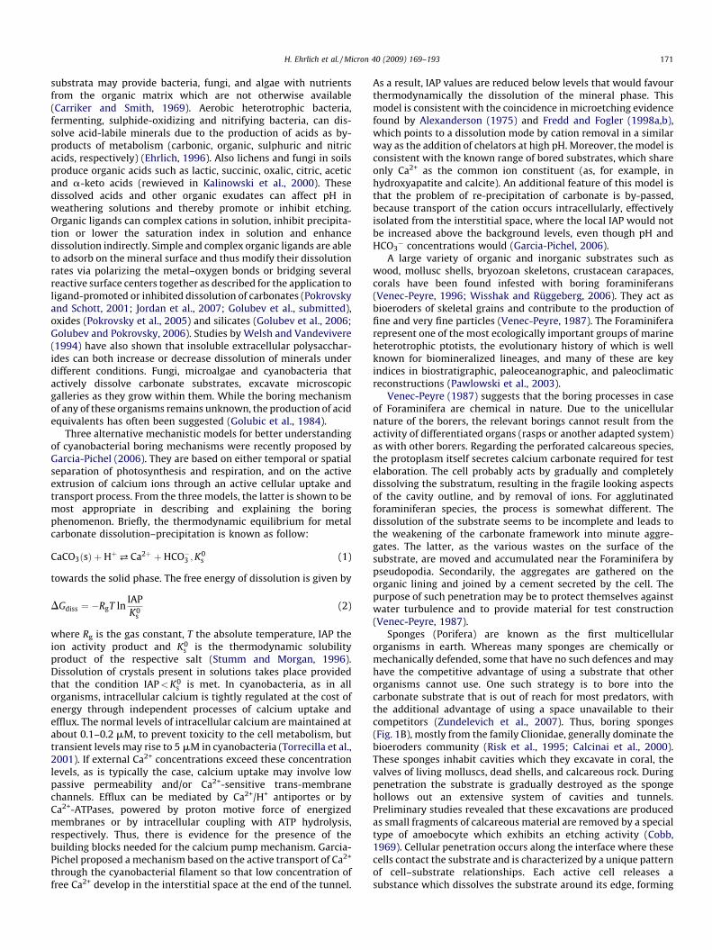
� �1=n
¼ �1
nln S (9)
The rates of dissolution may be defined as the velocity ofrecession of a crystal face relative to a fixed point of the crystal.This definition however cannot be easily applied to the formationof polycrystalline deposits such as those encountered in thebiomineral composites. In this case, experimentally the rates ofdissolution may be expressed by equation:
Rd ¼ðdm=dtÞ
A(10)
where m is the number of moles of the mineral in contact with theundersaturated solution and A is the surface area of the substrate(Wang et al., 2006a,b,c).
4.3. Models for the dissolution process
The models developed to describe quantitatively crystal growthand dissolution have followed two major approaches: diffusion-reaction and surface layer dissolution. A thorough review on thedissolution mechanisms involved in the dissolution of calciumphosphates has been presented by Dorozhkin (2002).
4.3.1. Diffusion-reaction theory
According to the Noyes–Whitney theory crystals may beconsidered to dissolve by the building units and their subsequenttransfer into the bulk solution. The rate of disintegration of thecrystals by this growth unit removal depends on the concentrationdifference between the crystal surface and the bulk solution. Theprocess of dissolution takes place in two stages: (i) detachment ofthe crystal building units from the crystal network and (ii) transferof the units to the bulk solution by diffusion. Step (i) is thus areaction step and step (ii) is diffusion. The mathematicaldescription of the two steps is respectively:
dc
dt
����R
¼ kRðc1 � csÞ ðreactionÞ
dc
dt
����D
¼ kDðcs � cbÞ ðdiffusionÞ(11)
where (dc/dt)jR, (dc/dt)jD are the rates corresponding to reactionand diffusion, respectively, kR and kD the respective rate constantsand c is the solute concentrations. The subscripts1, s and b refer to

Fig. 4. The dissolution process according to the reaction-diffusion theory.
Fig. 5. Surface of a crystal according to Kossel. The various types of dislocations are
shown.
H. Ehrlich et al. / Micron 40 (2009) 169–193 177
equilibrium, surface and bulk, respectively, and n is the reactionorder. The rate constants kR, kD cannot be experimentallymeasured in a polycrystalline system as they are different fordifferent crystal faces. The prevalence of reaction or diffusiondepends on the relative magnitudes of kR and kD. The two steps areschematically shown in Fig. 4. When steady state conditions areestablished the two rates are equal and the slowest stepdetermines the prevalent mechanism (Sjoberg and Rickard, 1983).
The value of the reaction order has in several cases beendetermined from experimental results that it is n = 1. Since thedissolution rate constant is
kD ¼D
d(12)
where D is the molar diffusion coefficient and d is the diffusionlayer thickness at the steady-state, Eq. (11) yield:
kgðc1 � csÞ ¼ kDðc1 � cbÞ (13)
from which
C1 ¼kRc1 þ kDcb
kR þ KD(14)
Substitution of Eq. (14) into one of Eq. (11) yields:
Rate�R ¼ dc
dt¼ k0ðc1 � cbÞ (15)
where
k0 ¼kgKP
kR þ kD(16)
Eq. (15) is a mathematical expression correlating the rates ofdissolution measured experimentally (e.g. from calcium increasein undersaturated solutions as a function of time) with the solutionundersaturation which is the deviation of the solute concentrationin the solution from equilibrium. Eq. (15) for salts of electrolytes,the quantity for the analytical concentration, c should be replacedby mean ion activities. A typical value for D, the molar diffusion is10�9 m2 s�1. The diffusion layer thickness may be calculatedaccording to (Mullin, 1993a,b):
d ¼ 3L
2
rsuL
h
� �1=2 hrsD
� ��1=3
(17)
where L is the diameter of the dissolving crystallites in apolycrystalline system, rs the density of the aqueous solution inwhich dissolution takes place (103 kg/m3), u the linear velocity ofthe dissolving particles and h is the viscosity of the undersaturated
aqueous solution (0.8904 � 10�3 kg 5�1 m�1 at 25 8C). It is thuspossible for an experiment to calculate the diffusion rate constantfrom Eqs. (12) and (17).
4.3.2. Surface-reaction controlled morphology-based theories
All crystals contain imperfections which includes steps, kinks,terraces, ledges and holes or vacancies. The schematic depiction ofa crystal surface is shown in Fig. 5. This model is known as the‘‘Kossel model’’ (Kossel, 1934). The dissolution proceeds by thedetachment of the crystal units in the following steps:
(i) D
etachment from an active site (e.g. kink); (ii) P artial hydration of the detached unit;(iii) H
ydration of the vacant site; (iv) D iffusion of the (partially) hydrated unit along the edge of astep;
(v) D iffusion of the unit along the step and completion ofhydration;
(vi) T he unit is transferred to the bulk solution through thediffusion boundary layer on the crystal surface.
The slowest of the steps (i)–(vi) is the rate-determining step.Since hydration reactions are in general very fast, the mechanism isdetermined either by step (iv) or (vi). In case diffusion to the bulksolution is the rate determining step the treatment is similar tothat described before for diffusion. The surface diffusion may bedescribed by three different models shown schematically in Fig. 6.As may be seen in Fig. 6A it is possible to start dissolution from onecenter from which originates the units detachment leading to stepdisintegration.

Fig. 6. (A) Mononuclear model. (B) Polynuclear in one crystal step. (C) Polynuclear in multiple steps: birth and spread. (D) Spiral model.
H. Ehrlich et al. / Micron 40 (2009) 169–193178
It is also possible that detachment takes place simultaneouslyfrom multiple centers (Fig. 6B and C) which may be developed inone or more, steps on the crystal surface. Alternatively, it has beensuggested that the crystal units’ detachment takes place alongspirals originating from an active site on the crystal surfacefollowing an Archimedian spiral (Fig. 6D). The unit detachmentalong the spirals takes place at constant velocity. The dissolutionmechanisms presented describe quantitatively the dissolution byequations relating the experimentally measured rates of dissolu-tion as a function of the solution undersaturation which is thenecessary condition for dissolution. The rate expressions for thevarious models are as follows:
For the mononuclear model (Fig. 6A):
R ¼ A exp�phg2y
K2T2ln S
� �(18)
where A a constant including the total crystal surface areaavailable, h a crystal step height, g the surface energy, y themolecular volume of the dissolving crystal, k the Boltzmannconstant, T the absolute temperature and S is the solutionundersaturation.
According to the polynuclear model (Fig. 6B and C). The rateexpression is
R ¼ AgykT
� �2
ðln SÞ�3=2 exp�B
T2ln S
� �(19)
where B is a constant. A similar expression was given by Zhang andNancollas (1991):
R ¼ kds2=3ð�ln SÞ1=6 expð�A=ln SÞ (20)
where A is a constant:
A ¼ p3
� � gkT
� �2
(21)
Transformation of Eq. (20) in a linear form yields:
lnR
s2=3ð�ln SÞ1=6¼ ln kd �
A
ln S(22)
For the spiral dissolution model the dependence of the rates onthe solution undersaturation is
R ¼ Cs2
s1tanh
s1
s(23)
where C and s1 are constants involving step size and crystal unitjump frequencies related with their migration along the spirals. Forlow undersaturation values (close to equilibrium) Eq. (23) isreduced to:
R ¼ Cs2
s1(24)
which predicts a parabolic dependence of the rates of dissolutionon the solution undersaturation. For large deviations from
equilibrium (s� s1) Eq. (23) yields:
R ¼ Cs (25)
As may be seen therefore it is possible to deduce the mechanismof dissolution of crystals according to the above-described models,depending on the fit of the rates measured on the solutionundersaturation. It should however be kept in mind that it ispossible that more than one mechanisms are operative, dependingon the solution undersaturation.
The possibility of a calcium phosphate biomineral to dissolvedepends on the value of the Gibbs free energy change for thetransition undersaturated! saturated solution. For the biologi-cally important minerals, HAP, OCP and DCPD the expressions forthis free energy change, which is the respective thermodynamicdrive force are as follows:
For DCPD:
DGDCPD ¼RT
2lnðCa2þÞðHPO4
2�ÞK0
s;DCPD
(26)
for OCP
DGOCP ¼RT
16lnðCa2þÞ8ðHþÞ2ðPO4
3�Þ6
K0s;OCP
(27)
and for HAP
DGHAP ¼RT
9lnðCa2þÞ5ðPO4
3�Þ3ðOH�Þ
K0s;HAP
(28)
In the logarithmic term, the ratio of the ionic product to therespective thermodynamic solubility product is the saturationratio, S. For undersaturated solutions in all cases S < 1 andtherefore the respective. Change in the thermodynamic drivingforce is <0, i.e. dissolution takes place upon contact of the crystalswith the undersaturated solutions. In the kinetics studies, it isoften the relative undersaturation, s, which is used defined as
s ¼ S� 1 (29)
In Eqs. (26)–(28) the quantities in parentheses are the activitiesof the respective ions. The thermodynamic solubility products forthe biominerals considered are: 2.32 � 10�7 M2 for DCPD (Tanget al., 2001), 2.51 � 10�99 M16 for OCP (Shyu et al., 1983) and5.52 � 10�118 M18 for HAP (McDowell et al., 1977). The computa-tion of the activities of free ions in complex solutions requirestaking into account all equilibria involved, together with the massand charge balance equations. Moreover, suitable expressions forthe ion activity coefficients are needed. Activity coefficients may beexpressed as a function of the solution ionic strength, I, usingvarious semi-empirical equations. Davies equation:
log f z ¼ �Az2
ffiffiIp
1þffiffiIp � 0:3I
!(30)

H. Ehrlich et al. / Micron 40 (2009) 169–193 179
has been suggested as a reasonably good expression for the ionicstrength of 0.15 M, typical for most of the biological fluids(Nancollas, 1966).
The kinetic expression often used to describe dissolution ofsparingly soluble salts is a first order relationship between the rateof dissolution, R and the solution undersaturation:
R ¼ kdissðCe � CuÞ (31)
where kdiss is the rate constant and Ce, Cu are the concentrations ofthe solute at equilibrium and in the undersaturated solution,respectively. Eq. (28) is not always valid (Patel et al., 1987;Gramain et al., 1989; Christoffersen and Christoffersen, 1992;Melikhov et al., 1990).
In general the overall dissolution process is interpreted by apower law of the type:
R ¼ kdisssn (32)
where n is a power indicative of the mechanism. For HAPdissolution in the pH range between 5.0 and 7.2 the rates werefound to be slower by four orders of magnitude with respect to therates corresponding to diffusion according to Fick’s law (Nancollas,1989). Concerning the mechanism of dissolution of calciumphosphates it seems that there is no consensus. For HAP, e.g.,the thermodynamically most stable phase, both surface diffusion(Christoffersen and Christoffersen, 1979; Margolis and Moreno,1992; Valsami-Jones et al., 1998) and mass transport controlledmechanisms (Brown and Chow, 1981; Lower et al., 1998), havebeen proposed. Considerable help in understanding mechanismsin dispute is the development of modern experimental techniques(e.g. the potentiometric techniques combined with SEM (Brownand Chow, 1981; Lower et al., 1998) and AFM (Valsami-Jones et al.,1998; Wang et al., 2006a,b,c; Koutsoukos and Valsami-Jones, 2003)for the investigation of the kinetics of these processes. DCPDdissolution was found to be controlled by surface diffusion and thedissolution data fitted to a spiral dissolution mechanism (Nan-collas, 1989). A significant advance in understanding the mechan-ism of dissolution came with the development of a methodology toperform dissolution studies at constant saturation (Budz andNancollas, 1988; Tang and Nancollas, 2002; Tang et al., 2001; Chowet al., 2003; Paschalis et al., 1996). The methodology was applied totooth enamel studies (Chen and Nancollas, 1986.) and it wasconcluded that the mechanism of dissolution was surface diffusioncontrolled. Further studies on tooth demineralization at conditionsof constant undersaturation led to the conclusion that the rates ofdissolution are higher inside the lesions in comparison with therespective values on the surface of the tooth enamel (de Rooij andNancollas, 1984). A modification of the constant compositionmethodology, using two different probes to monitor the dissolu-tion process was also successfully applied for mixed calciumphosphate phases (Tang et al., 2003). Maintenance of constantundersaturation during the course of dissolution provides infor-mation for the dissolving biominerals at pseudo steady stateconditions, mimicking physiological conditions. It is particularlyinformative for monitoring dissolution at the initial stages. It is atthis critical point that methods relying on variable saturation fail,because of the fast changes in the solution saturation with respectto the mineral phase investigated. At constant undersaturation notonly the rates of dissolution can be precisely measured thusobtaining reliable mechanistic information (through rate-under-saturation correlation) but also the effect of the presence ofimpurities and/or additives may be investigated. It was thus foundthat the rates of dissolution of biological apatites containing ionicimpurities decreased as a function of time despite the fact thatundersaturation, the driving force for dissolution, was kept
constant (Nancollas, 1989; Chen and Nancollas, 1986.). Thedissolution of carbonated apatites, considered as models forbiominerals has revealed that carbonate was preferentiallyreleased (Mayer et al., 1988; Budz et al., 1988a,b). In most caseshowever, it may be concluded that the mechanism of dissolution ofbiominerals is surface diffusion controlled. The rates therefore ofdemineralization of biological phosphate salts are controlled bythe surface, which means that interactions at the surface eitherwith macromolecules or with ions should be carefully consideredin the design of experiments in vitro. Eight different dissolutionmodels of calcium apatites (both fluorapatite and hydroxyapatite)were also analyzed in review by Dorozhkin (2002).
4.3.3. Phenomenological surface coordination models: case studies of
effect of organic ligands on calcium and magnesium carbonates
dissolution
Carbonate mineral dissolution kinetics has been an issue ofactive research efforts in relation to biomineralization (Boquetet al., 1973; Morita, 1980; Monger et al., 1991; Pokrovsky andSavenko, 1994, 1995; Ferris et al., 1994; Fujita et al., 2000; Warrenet al., 2001; Ferris et al., 2003; Dittrich and Obst, 2004; Lian et al.,2006; Mitchell and Ferris, 2006; Rodriguez-Navarro et al., 2007).Numerous laboratory experiments demonstrated that the dissolu-tion of carbonates (calcite and magnesite) is controlled by pH,concentration of HCO3
�/CO32� ions, and temperature. Despite a
number of studies on calcite dissolution in the presence of organicligands (Perry et al., 2004, 2005; Fredd and Fogler, 1998a,b; Wuand Grant, 2002; Frye and Thomas, 1993; Hoch et al., 2000;Thomas et al., 1993; Compton and Sanders, 1993; Compton andBrown, 1995; Spanos et al., 2006a,b), rarely the effect of variableligand concentration on the dissolution rate has been rigorouslyquantified. There are a few data on magnesite dissolution in thepresence of ligands at some technologically relevant solutionconditions (Hamdona et al., 1995; Demir et al., 2003; Lacin et al.,2005; Bayrak et al., 2006) and Jordan et al. (2007) studied ligand-controlled magnesite dissolution at 100 8C and low pCO2 in thepresence of a single concentration (0.01 M) of organic andinorganic ligands via a combination of macroscopic rate measure-ments and hydrothermal AFM. In order to extend the range ofligand concentrations to broader environmental conditions,Pokrovsky et al. (2008) performed detailed study on calcite andmagnesite dissolution in the presence of variable (10�5 to 10�2 M)ligand concentrations at conditions pertinent to CO2 storage basins(60 8C, 30 atm pCO2, pH 4.5–5.5). Recently, Golubev et al.(submitted) studied calcite dissolution in the presence of 16organic ligands ranging in the concentration from 10�6 M to0.03 M at otherwise constant solution parameters (25 8C, 0.1 MNaCl, pH 8.5). These organic ligands represent simple low-molecular weight organic acids (carboxylates, chelates, andaromatic compounds) and analogs of bacterial exometabolites,external cell envelopes, and natural polymers (humic acids). Thefollowing order of ligand effect on calcite dissolution at 0.03 Mligand concentration has been established: Gum xanthan <gluconate < alginate < fumarate ffi NaCl < malonate succinate� acetate < L-glutamate < 2,4-dihydroxybenzoate (DHBA) glucosamine phthalate gallate � pectin < 3,4-DHBA �citrate� EDTA (Fig. 7).
The effect of organic ligands on calcium carbonate dissolutioncan be rationalized within the framework of the surfacecoordination approach assuming the overall dissolution rate iscontrolled by reactions promoted at Ca centers by various ligandswhich compete for available surface sites (Stumm, 1992). Theeffectiveness of ligands depends on the molecular structure andthermodynamic stability of the surface complexes they form. Forexample, especially efficient are ligands whose functional groups

Fig. 7. The ratio of calcite dissolution rate in the presence of 0.03 M ligand to that in
the absence of ligand. Gum xanthan, gluconate, and alginate act as inhibitors of
calcite dissolution and citrate and EDTA are the strongest enhancers of dissolution
(Golubev et al., submitted).
H. Ehrlich et al. / Micron 40 (2009) 169–193180
contain two or more oxygen donors and which can form bi- ormultidentate mononuclear surface chelates (Stumm, 1992). Incontrast, ligands forming bi- or polynuclear complexes, that canbridge two or more metal centers at the surface lattice, are knownto retard dissolution.
In order to quantitatively model the effect of ligand oncarbonate dissolution rate, the simplest, bimolecular surfacereaction between positively charged surface groups (>CaOH2
+)and negatively charged ligands (Ln�) can be considered:
>CaOH2þ þ Ln� ¼ >Ca-L1�n þH2O (33)
According to this scheme, the rate of ligand-controlleddissolution is proportional to the concentration of the surfacecenter–ligand complex >Ca-L1�n which can be deduced from thereaction (3) stability constant (K�Ca-L):
K�Me-L ¼f>Ca-L1�ng
f>CaOH2þg � ½Ln�
(34)
It is reasonable to assume that, in the presence of a ligand, thecalcite forward dissolution rate is thus the sum of H2O-(RH2O) andligand-controlled dissolution, similar to that of brucite (Pokrovskyet al., 2005), dolomite (Pokrovsky and Schott, 2001), smectite(Golubev et al., 2006), and diopside (Golubev and Pokrovsky,2006):
R ¼ k>CoOH2
þ � f>CaOH2þg þ k>Me-L � f>Ca-L1�ng (35)
In Eq. (35), k>CaOH2
þ is the rate constant for water-promoteddissolution reaction measured in the absence of ligand at given pHand pCO2 and k>Ca-L is the empirical kinetic constant pertinent toeach ligand. The simplified phenomenological equation for calciteand magnesite dissolution in the presence of ligands can beobtained by combining Eqs. (34) and (35):
R ¼ k>CaOH2
þ 1� K�Ca-L½Ln�
1þ K�Ca-L½Ln�
!þ k>Ca-L
K�Ca-L½Ln�
1þ K�Ca-L½Ln�
(36)
Note that the stability constants for surface adsorptionreactions (K�Ca-L) correlate with corresponding values for associa-tion reactions in homogeneous aqueous solution as it is the case forother simple oxides (Schindler and Stumm, 1987; Ludwig et al.,1995; Pokrovsky et al., 2005).
5. Demineralization of naturally occurring Ca-containingbiocomposites
5.1. Bone
Bone is a hierarchically structured composite material in theengineering sense and its properties may differ significantly fromthose of the individual components. The function of the compositematerial depends not only on the chemistry of the components butalso on their relative orientation in the structure. Modern views onthe bone structure and formation including such aspects astransient precursor strategy in mineral formation of bone (Craneet al., 2006; Weiner, 2006; Grynpas and Omelon, 2007), mechan-isms of intrafibrillar (Olszta et al., 2007) and self-assembly (Cuiet al., 2007) mineralization of collagen as main organic templateare recently actively discussed.
Reports on bone demineralization date back to 1889 uponexploration of using demineralized bone for surgical implantation(Senn, 1889). This approach was successful in rats and resulted inligament reconstruction (Yamada, 2004). The history of bonedemineralization is represented in detail in the first part of ourreview (Ehrlich et al., 2008). The major problems in obtaining theorganic matrix of bone is, first, that very few of the proteins or thecomplex proteoglycans can be extracted without first solubilizingthe calcium-phosphate (Ca-P) crystals. Secondly, the majorstructural protein of the bone is collagen. While one can solubilizethe Ca-P crystals relatively easily (dilute strong acids, EDTA, weakacids such as acetic and citric acids), the fibrous collagen isinsoluble after decalcification, especially in its undenatured state(Glimcher et al., 1965). A reasonable number of the very many non-collagenous proteins, such as the phosphoproteins (Gotoh et al.,1990, 1995) including bone sialoprotein (Wang et al., 2006a,b,c)can be solubilized during the decalcification of the bone by avariety of solvents and to varying extents, depending on whichsolvent is used. Thus, it was reported (Gerstenfeld et al., 1994) thatchelation of calcium ions by EDTA and dissolution of the mineralphase by acid extraction released 95% or more of the total calciumcontent of the bone powder by 48 h, guanidine-HCl released lessthan 20% or less of the total calcium content even when extractionwas carried out by 168 h. Moreover, although guanidine-HClsolubilized a significant amount of collagen as gelatin, essentiallynone of phosphoproteins, osteocalcin, or the proteoglycan decorinwere solubilized, as detected by immunological techniques. Incontrast, extraction of the mineralized bone powder by HCl andformic acid was very efficient in selectively solubilizing osteocalcinand osteopontin, while bone sialoprotein was selectively releasedby EDTA, and solubilized to a lesser extent by formic acid. Similarly,EDTA selectively removed decorin compared with HCl, formic,acetic, or citric acids. These data provide some insight into theintrinsic solubility characteristics of collagen, the specific non-collagenous proteins, and their potential association with eachother and the mineral phase.
The distinct differences in the patterns of attack on bone byweakly ionized acids and their buffers as opposed to EDTA helpedto define some characteristics of ‘‘good’’ demineralizing agents.Weakly dissociated acids and their buffers produced a rapidlypenetrating diffuse attack on mineral of bone (Eggert and Germain,1979). In contrast, EDTA generated a well-defined front ofdemineralization. This was a pattern also observed with thestrongly ionized trichloroacetic acid. With these latter agents abarrier appears to be established at the front of demineralizationthat prevents diffusion of calcium salts from the core of thespecimen. Such a barrier is not established by acidic buffers withthe result that demineralization is more generalized at an earlystage. Rapid demineralizing buffers were those producing minimal

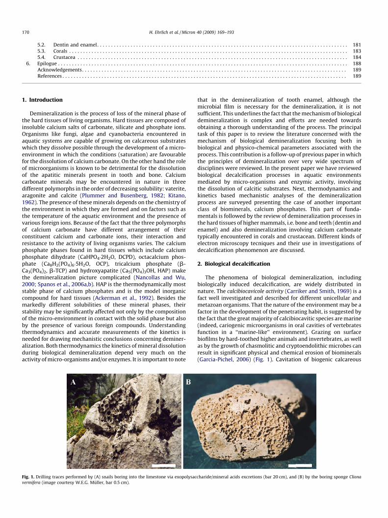
Fig. 8. SEM image: cariogenic bacteria on the work.
H. Ehrlich et al. / Micron 40 (2009) 169–193 181
secondary reprecipitation of nodules of calcium salts that wererapidly redissolved. Potassium formate and lactate-containingbuffers possessed this property and were described therefore asthe most rapid agents (Eggert and Germain, 1979). From a kineticpoint of view, acid demineralization of the bone specimensconsists of the following processes (according to Birkedal-Hansen,1974): (a) diffusion of acid in the bath toward the specimensurface; (b) diffusion of acid in the organic matrix of the decalcifiedpart of the specimen toward the decalcification front; (c) reactionwith hydroxyapatite at the decalcification front; and (d) outwarddiffusion of reaction products. The exact kinetic treatment of thisseries of events is complicated and elaborate.
In an attempt to reduce the commonly encountered artifacts oftissue shrinkage obtained with rapid decalcification with strongmineral acids many laboratories have utilized the much slowerdecalcification achieved with EDTA. This is a gentle, non-aciddecalcyfing reagent which has the advantage of not damaging thetissue but is slow acting. EDTA demineralization is generallyconsidered to be practically applicable only to small fragments ofbone (Belanger et al., 1965).
Demineralization of biological materials has generally beenconducted in aqueous media as reported above, resulting inconsiderable loss of water-soluble material, gross disruption ofmacromolecular organization and chemical breakdown of manycomponents of biological tissues (Scott and Kyffin, 1978). Byworking in organic solvents, loss of water-soluble materials shouldbe much less, and preservation of structures should be better. Boneis efficiently demineralized in ethanolic trimethylammoniumEDTA. Retention in the matrix of water-soluble materials (e.g.proteoglycan) was much better than in standard aqueous EDTAdemineralization procedures (Scott and Kyffin, 1978).
More detailed decalcification of bone including variousdecalcifying agents, methods and their effects on bone is describedand widely discussed in an excellent review by Callis and Sterchi(1998). Several novel technical approaches to the scanningelectron microscopy (SEM) of bone are considered in a paperfrom Boyde and Jones (1996). These include low kV imaging for thedetection of new surface bone packets (and residual demineralizedmatrix after resorption), low backscattered electron (BSE) imagingof uncoated, embedded, and unembedded samples, environmentalSEM for the study of wet tissue, low distortion, very lowmagnification imaging for the study of cancellous bone architec-ture, the use of multiple detectors for fast electrons in improvingthe imaging of porous samples, and high resolution, low voltageimaging for the study of collagen degradation during boneresorption. An SEM study on the process of acid demineralizationof cortical bone showed that the interface between the deminer-alized and mineralized sections of the bone specimens wasextremely sharp (Lewandrowski et al., 1997). This study suggeststhat cortical bone demineralization can be best described using anadvancing reaction front theory, and this explanation can be usedfor implementation of the concept of controlled demineralization(Lewandrowski et al., 1996).
High-resolution AFM imaging of bone (Hassenkam et al., 2004)and electron holography of organic matrixes (Simon et al., 2008)are recently also the subjects of detailed studies.
5.2. Dentin and enamel
In the history of demineralization studies, the driving force wasbased on problems of dental caries (Ehrlich et al., 2008). Dentinalcaries consists of demineralization of the mineral phase, followedby proteolytic degradation of the organic phase (Fig. 8). Initiationof the root caries in the cementum involves sub-surfacedemineralization (Schupbach et al., 1989), starting at several
points over a wide area of exposed cemental surface. Becausecementum is thin and porous, caries rapidly extends to theunderlying dentin.
Dentin is a mineralized connective tissue which constitutes themain bulk of the tooth. As in the case of bone, the major structuralprotein of dentin is type I collagen, the molecules of which are alsoaggregated into collagen fibrils with an axial period of67 nm. Theapatite crystallites are present in the form of fibrils and theyexceed (by weight and volume) the respective bone content(Glimcher, 2006). It is significant to note that dentin containsseveral highly phosphorylated proteins which, as suggested by in
vitro experiments with synthetic polymers containing variableamounts of phosphate groups, they are able to induce theformation of hydroxyapatite (Dalas et al., 1990, 1991). Toothenamel on the other hand is an outer layer of 1–3 mm thicknesswhich covers and protects dentine and pulp cavity. Approximately,96 wt% of enamel consists of crystalline HAP, the rest ofcomponents being the organic matter (ca. 0.6%) and water (ca.3.5% by weight) for the mature enamel. The mineral component isreduced upon enamel maturation and may vary in a manner thatrelates to local conditions and dietary habits (Ten Cate, 1979;Brudevold, 1962). It is interesting to note that the composition ofthe outer, reactive surface of enamel is different than the bulkcomposition. Sodium, magnesium, carbonate and fluoride con-centrations at the surface exceed those in the bulk enamel (Miles,1967; Peterson, 1975). In accordance the solubility of the enameldiffers significantly, being higher than the corresponding forstoichiometric HAP. The solubility of the outer enamel surface iseven lower, a fact which is certainly associated with the differentcomposition and with the presence of HPO4
2� (Patel and Brown,1975). Recent investigations using in situ AFM measurements andSEM observation of the apatitic crystallites have shown thatenamel demineralization takes place anisotropically along the caxis, resulting in hollow enamel cores and nanosized remainingcrystallites, resistant to further dissolution (Finke et al., 2000; Lijunet al., 2005). The fact that saliva contains sufficiently high calciumand phosphate concentrations make enamel resistant to deminer-alization (Dawes and Jenkins, 1962; Schmidt-Nielsen, 1946). Itshould be noted that saliva is the main ‘‘protector’’ of the teeth inthat it is supersaturated with respect to tooth mineral. There areseveral salivary proteins which inhibit the rate of precipitation ofmineral from the supersaturated saliva. Thus there is always atendency for remineralization to occur because of this super-saturation. In fact, the only reason why the teeth do not enlargecontinuously in the mouth is because of the presence of theacquired enamel pellicle on the surface of the teeth. This thin

H. Ehrlich et al. / Micron 40 (2009) 169–193182
deposit contains 130 different salivary proteins and provides abarrier to continued growth of enamel crystals. On pellicle removalby abrasion or attrition, it is rapidly replaced, thus forming arenewable lubricant. When teeth have been subjected to aciderosion, the acid dissolves the surface enamel if the pH of the fluidis less than that at which the fluid becomes unsaturated withrespect to tooth mineral. The surface enamel which has beendissolved cannot be regrown because of the blocking action by therapid deposition of a new acquired enamel pellicle on exposure ofthe eroded enamel to saliva.
The accumulation of microbial plaque on one hand lowers thepH of the environment in which enamel is exposed and on theother hand causes fermentation of the carbohydrates which resultsto pH values lower than 5 (Stephan, 1940). The dental plaque or‘‘pellicle’’ establishes a diffusion barrier for the flux of ions both tothe inside and the outside environment. It has been suggested thatit is possible for the surface layer to be retained while thesubsurface enamel dissolves (Moreno and Zahradnik, 1974;Zahradnik et al., 1976). Investigations on the effect of ionicstrength on enamel dissolution have shown that the rates ofdemineralization were enhanced while the formation of thesurface layer was reduced, suggesting diffusive coupling betweenthe acid and the dissolving ions (Anderson et al., 2004). In the oralenvironment, acid attack may be brought by acids present infoodstuff and drinks (Barbour et al., 2003). Dental caries beginslargely as a subsurface lesion underneath a biofilm (dental plaque).While the surface zone of the lesion is intact, the inside of thelesion is sterile and, if the lesion is kept free of plaque, there is thepossibility of remineralization, particularly in the presence offluoride. Dental caries is caused by microbially formed acid indental plaque. There is still no consensus on the factorsmaintaining the surface zone of the early lesion. It may, forinstance, be due to the deposition of more acidic forms of calciumphosphate, such as dicalcium phosphate dihydrate, as the calciumand phosphate ions leave the enamel. Because of the presence ofdental plaque, the calcium and phosphate ions which leave theenamel are not immediately washed away by the oral fluids, asoccurs in the process of acid erosion. The dical may then reconvertto hydroxyapatite when the plaque pH rises above the so-called‘‘critical pH’’. The latter is defined as the pH at which the plaquefluid (the fluid phase of dental plaque) is just saturated withrespect to tooth mineral. It is somewhat lower (about pH 5.1) thanthe critical pH of saliva, as plaque fluid contains higherconcentrations of calcium and phosphate than does saliva.
The mechanism of acidic interaction with HAP was found toinvolve two phases (Fig. 9) (Yoshioka et al., 2002). In the first phase,carboxylic acids bond to calcium of HAP. Depending on the
Fig. 9. Schematic presentation of the adhesion/decalcification concept adapted
from Yoshida et al. (2001).
diffusion rate of the calcium–acid complexes into solution, the acidwill in the second phase either remain attached to the HAP surfacewith only limited decalcification involved, or the calcium–acidcomplex will debond, resulting in a substantial decalcificationeffect. HAP crystallinity was found not to affect the adhesion/decalcification (AD) behavior of different acids when interactingwith apatitic substrates, so that the AD-concept can be applied toall human hard tissues with varying HAP crystallinity (Yoshiokaet al., 2002).
There are two reasons for the increased solubility of enamel inacid (Dawes, 2003). First, the hydrogen ions remove hydroxyl ionsto form water, as follows: H+ + OH� = H2O. The product of[H+][OH�] in water is always equals 10�14 (mol/L). Therefore, asthe [H+] increase in an acid solution, the [OH�] must decrease in areciprocal manner. Second, the inorganic phosphate in any fluidsuch as saliva or plaque fluid is present in four different forms,namely H3PO4, H2PO4
�, HPO42� and PO4
3�, and the proportionsdepend entirely on the pH. Fig. 10 illustrates how the proportionsof the four phosphate species vary with pH when the totalphosphate concentration is 5 � 10�3 mol/L, as is typical for saliva.The lower the pH, the lower the concentration of PO4
3�, the onlyspecies that contributes to the ionic product of HAP (Dawes, 2003).Thus, as any solution is acidified, the calcium concentration isunaffected but the concentrations of both OH� and PO4
3� arereduced and so, therefore, is the ion product, often to a value lessthan the solubility product. Dawes (2003) proposed the concept ofcritical pH for dissolution of enamel in oral fluids. The critical pH isthe pH at which a solution is just saturated with respect to aparticular mineral, such as tooth enamel. Saliva and plaque fluidare normally supersaturated with respect to tooth enamel becausethe pH is higher than the critical pH, so our teeth do not dissolve inour saliva or under plaque. Fig. 11 illustrates that above the criticalpH, plaque fluid will be supersaturated with respect to HAP, whilebelow the critical level it will be unsaturated. Thus, when plaquepH is below the critical level, caries will tend to occur (Dawes,2007).
It is established that enamel layers at tooth surfaces are amongthe hardest biological tissues and have highly mineralizedstructures exhibiting features that are close to pure syntheticapatites with about 97% mineral phase (Dorozhkin, 2007).
Fig. 10. The effect of pH on the concentrations of the various inorganic phosphate
species in saliva containing a total phosphate concentration of 5 � 10�3 mol/L.
there is a marked fall in the concentration of PO43� (thick line) as the pH is reduced
(adapted from Dawes, 2003).

Fig. 11. The effect of plaque pH, which may range from about 4 to 9, on
hydroxyapatite solubility and on the tendency for mineral deposition or
dissolution. At the critical pH, which varies slightly among individuals, plaque
fluid as just saturated with respect to hydroxyapatite and there is no tendency for
mineral to deposit or dissolve (adapted from Dawes, 2006).
H. Ehrlich et al. / Micron 40 (2009) 169–193 183
However, carbonate is always present and although the amount isrelatively low, the substitution of carbonate to phosphate ions inthe apatite crystal lattice results in a contraction of the a-axis andexpansion of the c-axis dimensions of the unit cell, as well as adecrease in crystallinity and increase in solubility and acidreactivity. Under physiological conditions, the suppression oftooth enamel surface dissolution is much more marked and thedemineralization only takes place at relatively high undersatura-tions (s > 0.4) and at lower pH (<5.5) (Tang et al., 2004a). Even athigher saturations of s = 0.90, the demineralization rate of toothenamel surfaces is extremely slow and complete dissolutionrequires about 4 weeks. It was reported, that the surface of enamelprior to dissolution is composed of numerous needle-like apatitesat an approximately micrometer scale (Tang et al., 2004a).However, complete dissolution of the inorganic particles doesnot take place, rather, the apatite sizes decrease and nano particlesare formed. This phenomenon is due to the formation andspreading of active etch pits on the dissolving surfaces. Onlywhen the pits are larger than a critical size, do they contribute todissolution. It is found by Nancollas and co-workers that thedissolution can be self-inhibited and even suppressed due todecreasing crystallite size (Wang et al., 2005). Biomaterialsbecome insensitive to dissolution in undersaturated solutionsand may be stabilized in the fluctuating biological milieux. Anexcellent example is proposed by Tang et al. (2004b) in studies ofcaries lesion formation. Enamel surfaces remain undissolved inwater (pH 5.5–5.8) in spite of the undersaturation. Caries(dissolution) is only induced at localized sites where bacteriahave produced acidic conditions. The mineral crystallites can bestabilized in the fluctuating physiological fluids, thus exercising adegree of self-presevation as the result of this kinetic size-effect atthe nanoscale level.
The chemistry of enamel caries, kinetics and mechanisms ofdental enamel demineralization were discussed in detail in specialreviews and experimental papers (e.g. Fosdick and Hutchinson,1965; Thylstrup et al., 1994; Robinson et al., 2000; Oliveira et al.,2002; Anderson et al., 2004; Hannig et al., 2005).
Various electron microscopy techniques were used for detailedobservation of dentinal demineralization on nano- and micro-levels. Comparative SEM studies focused on the effect of differentdemineralization methods on dentin surfaces (Isik et al., 1997), andon microbial contamination of noncavitated caries lesions (Paroloand Maltz, 2006). High-resolution electron microscopy (HR-TEM)was used in investigations on mechanism of the dimensionalincrease in demineralized dentine crystals (Hayashi et al., 1997).More recently Yanasigawa and Miake (2003) employed HR-TEM to
demonstrate demineralization and remineralization of enamelcrystals obtained from cross-cut sections of the c-axis of crystalsfrom carious lesions. Atomic force microscopy (AFM) was widelyused in the examination of the early stages of acid treatment ofdentin (Marshall et al., 1993), and of food-induced demineraliza-tion of the tooth enamel (Finke et al., 2000). AFM basednanoindentation has been used to study the nanomechanicalproperties and the ultrastructure of enamel samples as affected bydemineralization/remineralization cycles (Lippert et al., 2004).High-resolution AFM was also successfully used in studies onsequential demineralization and deproteinization of dentin withrespect to investigations of partially demineralized human dentincollagen fibrils (Habelitz et al., 2002).
5.3. Corals
Corals are phylogenetically basal metazoans, possess a simpleanatomy, can be easily manipulated for physiological studies, andpossess a majority of genes in common with vertebrates (Tambutteet al., 2007). They are therefore good models for biochemical,physiological, and evolutionary studies concerning either thetransport of ions or synthesis of organic matrix involved inbiomineralization processes. Demineralization of coral skeletonsusing different techniques and chemical agents leads to isolation ofthe following biomacromolecules as reported in literature: chitin(Wainwright, 1963), collagens (Kingsley et al., 1995; Franc et al.,1985), gorgonin (Ehrlich et al., 2006; Noe and Dullo, 2006;Sherwood et al., 2006), canthaxanthin (Cvejic et al., 2007) and awide diversity of proteins (reviewed in Tambutte et al., 2007).
Scleractinians (stony corals) are a group of calcified anthozoancorals, many of which populate shallow-water tropical tosubtropical reefs. Most of these corals calcify rapidly and theirabundancy on reefs is related to a symbiotic association withzooxanthellae. These one-celled algal symbiots live in theendodermal tissues of their coral host and are thought responsiblefor promoting rapid calcification (Stanley, 2003). Recently, Fineand Tchernov (2007) presented an experimental approachdocumenting how coral skeletons dissolve as a physiologicalresponse to increased atmospheric CO2. Thirty coral fragmentsfrom the five coral colonies of the scleractinian Mediterraneanspecies were subjected to pH values of 7.3–7.6 and 8.0–8.3(ambient) for 12 months. The corals were maintained in an indoorflow-through system under ambient seawater temperatures andphotoperiod. After 1 month in acidic conditions, morphologicalchanges were seen, initially polyp elongation, followed bydissociation of the colony form and complete skeleton dissolution.Surprisingly, after 12 months, when transferred back to ambientpH conditions, the experimental soft-bodied polyps calcified andreformed colonies.
It should be noted that the organic matrix of scleractinian coralscan be divided into ‘‘soluble’’ and ‘‘insoluble’’ components (Weiner,1984) depending of the extraction method used, and consequentlycare must be taken when comparing and interpreting results.Moreover, in 1980, Johnston (Johnston, 1980) warned that adifferentiation should be made between ‘‘skeletal organic matrix’’and ‘‘skeletal organic material’’, and suggested that the latterinvolved the skeletal organic matrix plus all other contaminatingcomponents such as endoliths and trapped tissues. From that time,in order to avoid such contamination, the chemical treatment ofcorals was carefully performed on the powders of skeletons(usually created using sodium hydroxide and/or bleach) in order toobtain only the skeletal organic matrix. Typical procedure isdescribed by Cuif et al. (1999) in their study on 24 species of coralscollected in very various environments, ranging from cold and/ordeep seas to tropical lagoons of Polynesian atolls. Coral samples

H. Ehrlich et al. / Micron 40 (2009) 169–193184
were thoroughly cleaned with sodium hypochlorite, rinsed, andoven dried (40 8C) overnight. Dried skeletal fragments werepowdered. The resulting powder was calibrated to regulate thedecalcification process made in very standardized conditions. Tocarry out the full sequence of mineralizing matrix characteriza-tions, 3 g of the calibrated powder were dispersed in 25 ml of Milli-Q water (magnetic stirring), then decalcification was made byaddition of ultra-pure acetic acid under permanent control of thepH. After the complete dissolution of the mineral phase, a low-speed centrifugation allowed soluble and insoluble matrices to beseparated. Desalting of soluble components was done on SephadexG25 gel-based low-pressure standard system, allowing salt andorganic compounds of lower molecular weights to be removed. Toobtain reliable biochemical information, these two successivesteps of preparation play a major role, allowing removal of most ofthe external contaminants, both insoluble (i.e. possible remains offungal or algal cell membranes) and low-weight soluble com-pounds that could have been introduced by seawater flow throughcoral skeletons (Cuif et al., 1999). Similar decalcification methodwas described recently for scleractinian coral S. pistillata (Puverelet al., 2005a). Microcolonies of this coral species were cleaned byremoving soft tissues with 1 M NaOH at 70 8C. The skeletons werethan rinsed with ultrapure water, dried at 60 8C overnight, andground to a fine powder with a mortar and pestle. The powder wasresuspended in ultrapure water and decalcified by addingultrapure acetic acid up to pH 4.0 in the presence of proteaseinhibitors. After complete dissolution of aragonite, soluble andinsoluble matrices were separated by centrifugation (10 min,10,000 � g). To desalt soluble components, the supernatantcontaining the soluble organic matrix was ultrafiltered and thenlyophilized. Using antibodies raised against soluble organic matrixisolated from coral S. pistillata as described above, the authorspresented direct evidence for the role of calicoblastic cells inorganic matrix synthesis and secretion. The same decalcificationmethod was used in comparative study on S. pistillata, known asbranched robust coral and Pavona cactus, a leafy complex coral(Puverel et al., 2005b). Soluble organic matrix of both coral specieswere shown to contain high amounts of potentially acidic aminoacids and glycine. However, proportions of glycosaminoglycansand SDS–PAGE analysis of soluble organic matrix proteins werevery different. Internal peptide sequences of two matrix proteins(one from each species) were obtained. One sequence of S. pistillata
is unusual because it contains a long poly-aspartate domain, asdescribed in proteins belonging to the calsequestrin family and inproteins from molluscan species.
In contrast to the usual view of an aggregate of purely mineralunits (the coral fibres) independently growing by a simplechemical precipitation (Perrin, 2003), coral skeletons appear tobe biochemically controlled structures. Different kinds of proteinswhich are responsible for biomineralization phenomenon wereisolated from scleractinian corals. Thus, Cuif and Dauphin (2004)reported on isolation of sulfated acidic proteoglycans which wereable to create polymeric frameworks. Atomic force microscopyconfirms the close relationship of this organic phase and mineralphase at the nanometric scale. Whereas acidic macromolecules areknown to be associated with the mineral phase of Scleractiniancoral skeletons (reviewed in Tambutte et al., 2007), galaxin, amatrix protein from the exoskeleton of the zooxanthellate coral G.fascicularis, is rich in cystine, serine, glycine and alanine and doesnot contain high fractions of acidic amino acids (Fukuda et al.,2003).
Previous studies of coral biomineralization strongly focused onscleractinians (see references above), whereas the organic matrix-mediated calcification mode of the soft corals, or Octocorallia,including different alcionarian and gorgonian families is largely
unknown. In contrast to deep-water scleractinians, the slow-growing skeletons of gorgonian octocorals with a longevitypotential on the order of hundreds of years form on sub-annualto centennial time scales and hence represent potential archives ofpast oceanic and climatic dynamics in intermediate and deep-water masses (Noe and Dullo, 2006).
Recently, Rahman and Isa (2005) documented demineralizationprocedure for spicules of soft coral Lobophytum crassum (Rahmanand Isa, 2005). The colony of the corals was cut using sharp scissorsinto small pieces. The pieces were ground five to six times with amixer machine and washed with tap water until the spicules wereobtained. The collected spicules were stirred vigorously in 1 MNaOH for 2 h to remove the fleshy tissues and debris. Themechanically and chemically cleaned spicules were extensivelywashed in distilled water and decalcified in 0.5 M EDTA (pH 7.8)overnight. The decalcifying solution was centrifuged with4000 rpm (15 min) to remove any insoluble materials followedby filtration with filter paper. The authors reported that solubleorganic matrix comprised 0.03% of the spicule weight. The SDS–PAGE analysis of the preparation showed four protein bands. The67-kDa protein appears to be glycosylated. Moreover, the isolatedorganic matrix possesses carbonic anhydrase activity whichfunctions in calcium carbonate crystal formations, indicating thatorganic matrix is not only structural protein but also a catalyst.
The scleroprotein gorgonin plays a crucial role in the formationof organic nodes and the secretion of calcitic internodes byproviding a structural framework in the biomineralizationprocesses (Noe and Dullo, 2006). The decalcification procedureby OsteosoftTM (Merck) treatment was used by Ehrlich et al. (2006)to gain understanding of the nature and nanostructure of thegorgonin-containing organic matrix of octocoral Isidella sp. Onthe 7th day of decalcification at 37 8C authors observed only thepresence of a transparent gelatinous pellicle after completedissolution of the calcite based axis internode of the coral(Fig. 12A and B). Gorgonin layers appear brownish in lightmicroscopy (Fig. 12B) and differ drastically from the transparentorganic matrix. The identification of the gorgonin-free organicmatrix by means of SEM, TEM (Fig. 12C and D) and AFM (Fig. 13)shows fibrillar protein behaviour. The results of amino acidanalysis of this organic matrix show glutamine and proline (28.9and 24.0%, respectively) as the dominant components among otheramino acids detected. The very low content of glycine (2.5%) rulesout the possibility of the fibrillar matrix being of a collagenousnature. It was concluded (Ehrlich et al., 2006) that isolatedstructural protein is an example of an acidic fibrillar protein. Thisprotein was responsible for calcification phenomenon in Isididaecorals in contrast to gorgonin-based structural layers on which nomineral phase formation were observed.
Unfortunately, the properties of coral as implants havegenerally been studied with non-demineralized skeletons ratherthan with whole extracts of organic matrix or isolated molecules.Since in coral larvae and adults, the molecules involved in mammalosteogenesis such as bone morphogenetic proteins (BMPs) arepresent in tissues (Hayward et al., 2002), the study of suchancestral molecules and their potential implications in coralskeleton formations, as well as in mammalian osteogenesis, mightopen new pathways not only in medicine but also in evolutionarystudies of biomineralization.
5.4. Crustacea
Crustaceans are a remarkable group of animals because of theirability to elaborate cyclically two kinds of calcified biomineraliza-tions: an unstretchable exoskeleton (or cuticle) and also, for manyspecies, depending of the way of life of the considered animal,

Fig. 12. Light microscopy image of organic matrix isolated from Isidella sp. octocoral using Osteosoft-based demineralization technique at 37 8C (A). Brownish coloured layers
of gorgonin which is resistant to calcification could be visualized under magnification of the same sample (B). Nanoorganised structures made by acidic fibrillar protein
responsible for mineralization of octocoral skeletal internodes are observed using TEM (C and D).
H. Ehrlich et al. / Micron 40 (2009) 169–193 185
transitory calcium deposits (Luquet and Marin, 2004). They serveas an ideal model for the study of calcium homeostasis due to theirnatural molting cycle. Demineralization and remineralization ofthe calcified cuticle is accompanied by bidirectional Ca2+ transferacross the primary Ca2+-transporting epithelia: gills, antennalgland (kidney), digestive system, and cuticular hypodermis(Wheatly, 1999).
The major sources of Ca2+ for crustaceans are water and food,with water being the most important in zooplankton. Develop-
ment and associated growth force crustaceans to replace theircuticle frequently. During ecdysis, most of the Ca2+ is lost with theresidual exuvia and must be replaced by absorption of the mineralfrom the environment (Fabritius and Ziegler, 2003). Most of theCa2+ is probably associated with carbonate and phosphateminerals in the carapace (Stevenson, 1985), because calcificationis the primary cause of hardening in crustaceans cuticles(Chockalingam, 1974). However, freshwater decapods may storesome 20–30% of Ca2+ on the form of gastroliths (Wheatly and

Fig. 13. AFM imaging of the Isidella sp. octocoral fibrillar organic matrix isolated after decalcification.
H. Ehrlich et al. / Micron 40 (2009) 169–193186
Ayers, 1995). While it is easy for marine, and to a lesser degree forlimnic species to replace Ca2+ by taking it up from the water viatheir gills, terrestrial crustaceans depend on Ca2+ taken up withtheir diet, which contains little Ca2+ (Fabritius et al., 2005).Terrestrial isopods store Ca2+ in a variety of storage sites, the mostcommon among these are the sternal CaCO3 deposits locatedwithin the ecdysial gap between the epithelium and the old cuticleof the first four anterior sternites (Ziegler and Miller, 1997). Thestorage amounts of Ca2+ are highly variable in different crusta-ceans. For example, semiterrestrial Ligia spp. seems to have thehighest Ca2+-storage capability (about 75%) among crustaceans ingeneral (Ziegler et al., 2007). Terrestrial amphipods store 67% oftheir Ca2+, the freshwater/land crab Holthuisana transversa 64%,various land crabs 5–14%, and freshwater crayfishes about 17%(reviewed in Ziegler et al., 2007).
The majority of the skeletal structures of crustaceans arecomposed of calcium carbonate in the form of calcite (Romanoet al., 2007), francolite, a carbonate containing F-apatite (Low-enstam, 1971; Watabe and Pan, 1984), and amorphous calciumcarbonate (ACC) (Fabritius et al., 2005), or amorphous calciumphosphate (Raz et al., 2002). In all cases mineral phases areassociated with an organic matrix (chitin and/or proteins),suggesting that the organic components function as a templatefor biomineralization in crustaceans.
In contrast to studies on demineralization of skeletons in otherinvertebrates, the main driving force in the case of crustaceans isdetermined by the practical applications of chitin. Recovery ofchitin from shells of crustacean species is desirable in manylocations throughout the world where shrimps, prawns, lobsters,crab, crayfish, etc. are harvested. One of the difficulties inprocessing crustacean shells to recover chitin, is the removal ofthe large amounts of mineral matter, primarily calcium carbonate.Historically, chitin has been isolated from crustacean shell byboiling the shell with strong caustic alkali solutions to removeprotein and dissolving away the mineral matter with strongmineral acid, usually hydrochloric acid (Allan et al., 1978). In 1948Max Lafon described the demineralization procedure of crustaceanintegument as follows (Lafon, 1948). The integument is carefully
freed of the hypodermal layer by scrapping, then rapidly washed,dried and ground to very small fragments, dried and weighed. It isthen demineralized by treatment for 24 h in the cold with 2% HCl. Itwas shown that the proteins are unaltered by this treatment. Afterdemineralization, the hydrochloric acid solution is removed byfiltration, the organic matter is washed twice rapidly and the washwaters are removed. The demineralized integument is nextextracted with a buffer solution at pH 9.2 consisting of boraxsolution (19 g/L), 5 parts; water, 5 parts; 95% ethanol, 4 parts;ether, 1 part for 36 h at 55 8C. The residue was next extracted with5% sodium hydrochloride solution for 5 h at 55 8C and then 1 h at100 8C. This removed all of the less soluble protein leavingessentially pure chitin as a final residue. It should be recognizedthat Lafon’s extraction medium for the more soluble protein wasintended for research purposes and is not practical for acommercial process. Also, his use of 5% NaOH at 100 8C is toodrastic for production of a protein of good nutritional value.
As an alternative to the chemical process, a biological processhas been evaluated for deproteination and demineralization. Lacticacid bacterial fermentation for demineralization has been reportedfor shrimp waste, crayfish exoskeleton, scampi waste, prawn wasteand red crab shell waste (as reviewed in Jung et al., 2005).Successive two-step fermentation was carried out from red crabshell wastes for biological extraction of chitin in combination of the1st step with a lactic acid bacterium Lactobacillus paracasei subsp.
tolerans KCTC-3074 and the 2nd step with a protease producingbacterium Serratia marcescens FS-3, and vice versa. The successivefermentation in the combination of both strains gave the bestresult in co-removal of CaCO3 and proteins from crab shells. In thiscombination, the rates of demineralization and deproteinationwere 94.3% and 68.9%, respectively, at the end of fermentation(Jung et al., 2007).
Crustacean’s cuticle is also a good example of biologicalstructures with hierarchical organization from the nanometer tothe millimetre scale. Recently, Raabe and co-workers publishedseveral impact papers regarding to microstructure and crystal-lographic texture of the chitin-protein network in the biocompo-site material of the exoskeleton of different crustaceans including
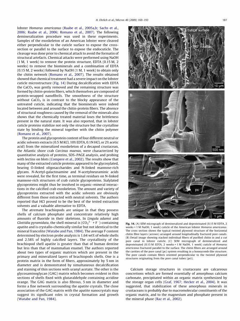
Fig. 14. (A) SEM micrograph of demineralized and deproteinated (0.15 M EDTA, 2
weeks + 1 M NaOH, 1 week) cuticle of the American lobster Homarus americanus.
The cross section shows the typical twisted plywood structure of the horizontal
chitin fibre layers (arrows) arranged around longitudinally fractured pore canals.
(B) Detail image showing stacked individual fibers of purified chitin in and a cut
pore canal in lobster cuticle. (C) SEM micrograph of demineralized and
deproteinated (0.15 M EDTA, 2 weeks + 1 M NaOH, 1 week) cuticle of Homarus
americanus fractured parallel to the surface. The chitin fibres are arranged around
the cavities of the pore canal (pc) system resulting in a honeycomb-like structure.
The pore canals contain fibres oriented perpendicular to the twisted plywood
structures originating from the pore canal tubes (pct).
H. Ehrlich et al. / Micron 40 (2009) 169–193 187
lobster Homarus americanus (Raabe et al., 2005a,b; Sachs et al.,2006; Raabe et al., 2006; Romano et al., 2007). The followingdemineralization procedure was used in these experiments.Samples of the exoskeleton of an American lobster were cleavedeither perpendicular to the cuticle surface to expose the cross-section or parallel to the surface to expose the endocuticle. Thecleavage was done prior to chemical attack to avoid the formatin ofstructural artefacts. Chemical attacks were performed using NaOH(1 M, 1 week) to remove the protein structure, EDTA (0.15 M, 2weeks) to remove the biominerals and a combination of EDTA(0.15 M, 2 weeks) followed by NaOH (1 M, 1 week) to obtain onlythe chitin network (Romano et al., 2007). The results obtainedshowed that chemical treatment had a severe impact on the lobstercuticle microstructure (Fig. 14) During decalcification with EDTAthe CaCO3 was gently removed and the remaining structure wasformed by chitin-protein fibers, which themselves are composed ofprotein-wrapped nanofibrils. The smoothness of the structurewithout CaCO3 is in contrast to the blocky appearance of theuntreated cuticle, indicating that the biominerals were indeedlocated between and around the chitin-protein fibers. The absenceof structural roughness caused by the removal of the minerals alsoshows that the chemically treated material loses the brittlenesspresent in the natural state. It was also reported, that in lobstercuticle proteins stabilize not only the structure but the crystallinestate by binding the mineral together with the chitin polymer(Romano et al., 2007).
The protein and glycoprotein content of four different neutral oracidic solvents extracts (0.5 M KCl, 10% EDTA, 0.1N HCl, or 2% aceticacid) from the mineralized exoskeleton of a decapod crustacean,the Atlantic shore crab Carcinus maenas, were characterized byquantitative analysis of proteins, SDS–PAGE analysis, and probingwith lectins on blots (Compere et al., 2002). The results show thatmany of the extracted cuticle proteins appeared to be glycosylated,bearing O-linked oligosaccharides and N-linked mannose-richglycans. N-Acetyl-galactosamine and N-acetylneuraminic acidswere revealed, for the first time, as terminal residues on N-linkedmannose-rich structures of crab cuticle glycoproteins. Sialylatedglycoproteins might thus be involved in organic–mineral interac-tions in the calcified crab exoskeleton. The amount and variety ofglycoproteins extracted with the acidic solvents are obviouslydifferent from those extracted with neutral solvents. The authorsreported that HCl proved to be the best of the tested extractionsolvents and a valuable alternative to EDTA.
The atremate brachiopods are unique in that they possessshells of calcium phosphate and concentrate relatively highamounts of fluoride in their skeletons. In Lingula adamsi andGlottidia pyramidata, the shell mineral is (CO3
2� + F�)-containingapatite and is crystallo-chemically similar but not identical to themineral francolite (Watabe and Pan, 1984). The average F contentdetermined by electron probe analysis is 1.64 wt% of whole shellsand 2.58% of highly calcified layers. The crystallinity of thebrachiopod shell apatite is greater than that of human dentinebut less than that of mammalian enamel. The authors reportedabout two types of organic matrices which are present in theprimary and mineralized layers of brachiopods shells. One is aprotein matrix in the form of fibers, approximately by 5 nm indiameter and is demonstrated by simultaneous decalcificationand staining of thin sections with uranyl acetate. The other is theglycosaminoglycan (GAG) matrix which becomes evident in thinsections of shells fixed with glutaraldehyde containing acridineorange. The GAG matrix is also fibrous, 5 nm in diameter andforms a fine network surrounding the apatite crystals. The closeassociation of the GAG matrix with the apatite nanocrystals maysuggest its significant roles in crystal formation and growth(Watabe and Pan, 1984).
Calcium storage structures in crustaceans are calcareousconcretions which are formed essentially of amorphous calciumcarbonate, precipitated within an organic matrix synthesized bythe storage organ cells (Graf, 1967; Hecker et al., 2004). It wassuggested, that stabilization of these amorphous minerals incrustaceans is probably due to macromolecular constituents of theorganic matrix, and to the magnesium and phosphate present inthe mineral phase (Raz et al., 2002).

Fig. 15. (A) Untreated spherules in the proximal spherular layer in fully developed
sternal ACC deposits of Porcellio scaber in late premolt stage. Material consists of
ACC-nanoparticles associated with fibrillar matrix structures which display
different states of dissolution. (B) SEM micrograph of sagittally cut fully
developed sternal ACC deposits of the woodlouse P. scaber in late premolt stage.
The sample surface has been polished using an ultra-microtome followed by
decalcification in 0.1 M EDTA with 2.5% glutaraldehyde for 10 min. The image from
the transition zone between the proximal spherular layer (psl) and homogeneous
layer (hl) shows the results of demineralization. (C) High resolution SEM
micrograph of a single cross sectioned spherule consisting of several concentric
matrix reticules (cm) still associated with undissolved ACC-nanoparticles which are
connected by radial matrix filaments (rm).
H. Ehrlich et al. / Micron 40 (2009) 169–193188
Orchestia cavimana is a terrestrial amphipod that provides adramatic example of calcium storage process with respect toformation of amorphous calcium carbonate phase which islocalized within an organic matrix synthesized by a caecalepithelial cells in the lumen of posterior caeca. After extraction
of the organic matrix in a buffer containing EDTA, electrophoreticanalysis of the proteinaceous components led to reveal about 11polypeptides in the EDTA-soluble fraction. Among them, an acidicpolypeptide migrating at 23 kDa in SDS–PAGE appeared specific tothe EDTA-soluble fraction organic matrix. Sequencing andcharacterization of this protein, called Orchestrin, revealed thatit is the only organic matrix component able to bind calcium and itis phosphorylated (Hecker et al., 2003). Recently, Marin and Luquet(2007) reported about occurrence of unusually acidic proteins(with pI below 4/4.5) isolated from crustaceans skeletal forma-tions. Thus, for crustaceans, 28 complete cuticular proteinsequences (plus nine partial sequences) have been retrieved. Inaddition, two complete protein sequences of calcium storagestructures have been determined. From these 30 sequences, 21 canbe considered as unusually acidic.
Recently, Fabritius et al. (2005) presented results of a detailedultrastructural investigation of the organic matrix of the sternalCaCO3 deposits of terrestrial crustacean P. scaber using high-resolution field-emission scanning electron microscopy. In P.
scaber 10–30 nm thick amorphous calcium carbonate nano-particles occur in mature spherules and on the surface of formingand degrading sperules. The authors showed that these nano-particles are often preserved in some of the concentric layers and atthe surface of radial strands of the organic matrix (Fig. 15) Theseparticles must be stabilized to a higher degree than the in EDTAdissolved material between radial strands and some concentriclayers. It is of interest that similar particles have been describedalong rod-like chitin-protein fibres in the exo- and endocuticle ofthe crab Carcinus maenas (Roer and Dillaman, 1984). Although themain mineral phase in the mature cuticle of decapods is calcite,ACC occurs in premolt cuticles suggesting that the calcitic nano-particles in the cuticle originate from amorphous precursors.
Gastroliths are calcified structures formed in the cardiacstomach wall in crustaceans (Luquet and Marin, 2004). Theyappear as paired discs in some decapods such as crayfishes,lobsters and as four more irregular concretions in gecarcinid landcrabs. Nagasawa and co-workers described solubilization techni-que of gastrolith matrix protein (GAMP) (Ishii et al., 1996, 1998;Takagi et al., 2000). Gastroliths were collected from the crayfish 7–10 days after bilateral eyestalk ablation. The gastroliths wereimmersed in 1 M acetic acid for two days. After the decalcifyingsolution was poured off, the residue was washed with distilledwater several times. Then the residue was extracted separatelywith mixtures of one or more of three solutions, 6 M urea, 1% SDS,and 10 mM dithiothreitol, in various combinations at 100 8C for10 min. Each extract was digested with trypsin to checkwhetherthe GAMP fragment was generated or not. Finally, an insolublematrix protein, referred to as gastrolith matrix protein, was madesoluble with 1% SDS containing 10 mMdithiothreitol, and waspurified by reverse-phase high-performance liquid chromatogra-phy. The protein had a molecular weight of about 50,500 and ablocked amino terminus. The GAMP fragment has a tandemlyrepeated sequence not reported before; each repeat consists of fiveamino acid residues. X-ray analysis showed that calcium carbonatein the gastrolith was amorphous, whereas that in the exoskeletonconsisted of calcite (Takagi et al., 2000).
6. Epilogue
It is apparent that decalcification is an intricately complexmechanism that serves several purposes, from organisms’ survivalto structural protection to calcium balance. Since centuries ago,man’s curiosity has been the driving force to delineate themechanisms involved in (either ‘‘natural’’ or ‘‘pathological’’)decalcification. Several scientific fields can benefit from knowing

H. Ehrlich et al. / Micron 40 (2009) 169–193 189
natural decalcification mechanisms. For example, calcium-con-taining deposits are known as significant problems in processwater systems (Demadis et al., 2005, 2006). These can bedecalcified/dissolved by the action of a variety of Ca2+ chelatingmolecules. It is apparent that decalcification and calcificationshould be envisioned as the two sides of the same coin. Natureoften uses these in admirable synergy to achieve a multitude ofpurposes. Continued advances in instrument technology will makeit possible to further untangle unresolved issues. There is aplethora of opportunities for innovative research and appliedscience. In particular, applications centered around human healthand well-being are the main drivers forward.
Acknowledgements
Authors would like to acknowledge Prof. S. Weiner, Prof. M. Ilan(Israel); Prof. M. Glimcher, Prof. H.C. Skinner, Prof. G. Nancollas,Prof. D. Morse, Prof. S. Golubic, Prof. Henry L. Ehrlich, Prof. A.George, Prof. G. Marshall, Prof. A. Luttge, Prof. C. Pueschel, and G.Callis (USA); Prof. C. Dawes (Canada); Prof. G. Gadd, Prof. J. Elliott,Dr. M. Barbour (UK); Prof. Y. Dauphin, Prof. J.P. Cuif, Prof. J. Vacelet,Prof. G. Luquet (France); Prof. D. Allemand (Monaco); Prof. K.D.Jandt, Prof. M. Epple, Prof. W.E.G. Muller, Prof. G. Worheide, Prof. E.Baeuerlein, Prof. B. Schone, Prof. H. Worch, Prof. D. Raabe, Prof. R.Kniep, Dr. P. Simon, Dr. A. Ziegler, Dr. C.H. Schonberg, Dr. H.Fabritius, Dr. M. Wisshak, Dr. S. Noe (Germany); Prof. E.Konigsberger (Australia); Dr. S. Dorozhkin (Russia) for discussionand comments. We wish to thank Vasily V. Bazhenov for excellenttechnical assistance.
This work was supported by German Research Foundation(DFG) grant WO 494/17-1, and the General Secrtariat of Sciene &Technology, Greece.
References
Ackerman, J.L., Raleigh, D.P., Glimcher, M.J., 1992. Phosphorus-31 magnetic reso-nance imaging of hydroxyapatite: a model for bone imaging. Magnetic reso-nance in medicine: official journal of the Society of Magnetic Resonance inMedicine. Soc. Magn. Reson. Med. 25 (1), 1–11.
Alexanderson, E.T., 1975. Marks of unknown carbonate-decomposing organelles incyanophyte borings. Nature 254, 237–238.
Allan, G.G., Fox, J.R., Kong, N., 1978. Marine polymers. Part 8: a critical evaluation ofthe potential sources of chitin and chitosan.In: Proc. Int. Conf. Chitin. Chitosan,vol. 1. pp. 64–78.
Amaechi, B.T., Higham, S.M., Edgar, W.M., 1999. Factors influencing the develop-ment of dental erosion in vitro: enamel type, temperature and exposure time. J.Oral. Rehabil. 26, 624–630.
Anderson, P., Bollet-Quivogne, F.R.G., Dowker, S.E.P., Elliott, J.C., 2004. Deminer-alization in enamel and hydroxyapatite aggregates at increasing ionic strengths.Arch. Oral Biol. 49, 199–207.
Ankel, W.E., 1937. Wie bohrt Natica? Biol. Zentr. 57, 75–82.Arends, J., Jongebloed, W.L., 1977. Mechanism of enamel dissolution and its pre-
vention. J. Biol. Buccale 5, 219–237.Barbour, M.E., Parker, D.M., Allen, G.C., Jandt, K.D., 2003. Enamel dissolution in citric
acid as a function of calcium and phosphate concentrations and degree ofsaturation with respect to hydroxyapatite. Eur. J. Oral Sci. 111, 428–4330.
Bayrak, B., Lacin, O., Bakan, F., Sarac, H., 2006. Investigation of dissolution kinetics ofnatural magnesite in gluconic acid solutions. Chem. Eng. J. 117, 109–115.
Belanger, L.F., Copp, D.H., Morton, M.A., 1965. Demineralization with EDTA byconstant replacement. Anat. Rec. 153, 41–48.
Birkedal-Hansen, H., 1974. Kinetics of acid demineralization in histologic technique.J. Histochem. Cytochem. 22, 434–441.
Blake, J.A., Evans, J.W., 1973. Polydora and related genera as borers in mollusc shellsand other calcareous substrates (Polychaeta: Spionidae). Veliger 15, 235–249.
Boquet, E., Boronat, A., Ramos-Cormenzana, A., 1973. Production of calcite (calciumcarbonate) crystals by soil bacteria is a general phenomenon. Nature 246, 527–529.
Boyde, A., Jones, S.J., 1996. Scanning electron microscopy of bone: instrument,specimen, and issues. Microsc. Res. Tech. 33, 92–120.
Brown, W.E., Chow, L.C., 1981. Thermodynamics of apatite crystal-growth anddissolution. J. Cryst. Growth 53, 31–41.
Brudevold, F., 1962. Chemical composition of teeth in relation to caries. In: Sognaes,R.f. (Ed.), Chemistry and Prevention of Dental Caries. Thomas-Springfield, pp.32–78.
Bryers, J.D., Ratner, B.D., 2006. Biomaterials approaches to combating oral biofilmsand dental disease. BMC Oral Health 6, S15–S22.
Budz, J.A., Lo Re, M., Nancollas, G.H., 1988a. The influence of high- and low-molecular-weight inhibitors on dissolution kinetics of hydroxyapatite andhuman enamel in lactate buffers: a constant composition study. J. Dent. Res.67, 1493–1498.
Budz, J.A., Nancollas, G.H., 1988b. The mechanism of dissolution of hydroxyapatiteand carbonated apatite in acidic solutions. J. Cryst. Growth 91, 490–496.
Calcinai, B., Cerrano, C., Sara, M., 2000. Borino sponges (Porifera. Demospongiae)from the Indian Ocean. Ital. J. Zool. 67, 203–219.
Callis, G., Sterchi, D., 1998. Decalcification of bone: literature review and practicalstudy of various decalcifying agents, methods, and their effects on bonehistology. J. Histotechnol. 21, 49–58.
Cameron, B., 1969. Paleozoic shell-boring annelids and their trace fossils. Am. Zool.9, 689–703.
Carano, A., Schlesinger, P.H., Athanasou, N.A., Teitelbaum, S.L., Blair, H.C., 1993. Acidand base effects on avian osteoclast activity. Am. J. Physiol. Cell Physiol. 264,C694–C701.
Cariker, M.R., Schaadt, J.G., Peters, V., 1974. Analysis by slow-motion picturephotography and scanning electron microscopy of radular function in Urosal-pinx cinerea follyensis (Muricidae, Gastropoda) during shell penetration. Mar.Biol. 25, 63–76.
Carriker, M.R., 1961. Comparative functional morphology of boring mechanisms ingastropods. Am. Zool. 1, 263–266.
Carriker, M.R., 1978. Ultrastructural analysis of dissolution of shell of the bivalveMytilus edulis by the accessory boring organ of the gastropod Urosalpinx cinerea.Mar. Biol. 48, 105–134.
Carriker, M.R., Smith, E.H., 1969. Comparative calcibiocavitology: summary andconclusions. Am. Zool. 9, 1011–1020.
Carriker, M.R., van Zandt, D., Charlton, G., 1967. Gastropod urosalpinx: pH ofaccessory boring organ while boring. Science 158, 920–922.
Carriker, M.R., Williams, L.G., 1978. Chemical mechanism of shell penetration byUrosalpinx: an hypothesis. Malacologia 17, 142–156.
Chave, K.E., 1984. Physics and chemistry of biomineralization. Ann. Rev. EarthPlanet. Sci. 12, 293–305.
Chen, W.C., Nancollas, G.H., 1986. The kinetics of dissolution of tooth enamel. Aconstant composition study. J. Dent. Res. 65, 663–668.
Chetal, M., Fournie, J., 1969. Shell-boring mechanism of the gastropod purpura(Thais) lapillus: a physiological demonstration of the role of carbonic anhydrasein the dissolution of CaCO3. Am. Zool. 9, 983–990.
Chockalingam, S., 1974. Nature and composition of the cephalic and thoraciccuticles of the hermit crab Clibanarius olivaceous. Mar. Biol. 26, 329–351.
Chow, L., Markovic, C., Takagi, M.S., 2003. A dual constant–composition titrationsystem as an in vitro resorption model for comparing dissolution rates ofcalcium phosphate biomaterials. J. Biomed. Mater. Res. B 65, 245–251.
Christoffersen, J., Christoffersen, M.R., 1992. A revised theory for the growth ofcrystals by surface nucleation. J. Cryst. Growth 121, 608–616.
Christoffersen, J., Christoffersen, M.R., 1979. Kinetics of dissolution of calciumhydroxy apatite. J. Cryst. Growth 47, 671–679.
Chughtai, I., Knight-Jones, E.W., 1988. Burrowing into limestone by sabellid poly-chaetes. Zool. Scr. 17 (3), 231–238.
Clelland, E.S., Saleuddin, A.S.M., 2000. Vacuolar-type ATPase in the accessory boringorgan of Nucella lamellosa (Gmelin) (Mollusca: Gastropoda): role in shellpenetration. Biol. Bull. 198, 272–283.
Cobb, W.R., 1969. Penetration of calcium carbonate substrates by the boring sponge,Cliona. Am. Zool. 9, 783–790.
Compere, P., Jaspar-Versali, M.-F., Goffinet, G., 2002. Glycoproteins from the Cuti-cole of the Atlantic Shore Crab Carcinus maenas. I. Electrophoresis and Westernblot analysis by use of lectins. Biol. Bull. 202, 61–73.
Compton, R.G., Brown, C.A., 1995. The inhibition of calcite dissolution/precipitation:1,2-dicarboxylic acids. J. Colloid Interf. Sci. 170, 586–590.
Compton, R.G., Sanders, G.H.W., 1993. The dissolution of calcite in aqueous acid: theinfluence of humic species. J. Colloid Interf. Sci. 158, 439–445.
Crane, N.J., Popescu, V., Morris, M.D., Steenhuis, P., Ignelzi Jr., M.A., 2006. Ramanspectroscopic evidence for calcium phosphate and other transient mineralspecies deposited during intramembranous mineralization. Bone 39, 343–442.
Cui, F.Z., Li, Y., Ge, J., 2007. Self-assembly of mineralized collagen composites. Mater.Sci. Eng. R 57, 1–27.
Cuif, J.-P., Dauphin, Y., Gautret, P., 1999. Compositional diversity of soluble miner-alizing matrices in some recent coral skeletons compared to fine-scale growthstructures of fibres: discussion of consequences for biomineralization anddiagenesis. Int. J. Earth Sci. 88, 582–592.
Cuif, J.-P., Dauphin, Y., 2004. The environment recording unit in coral skeletons:structural and chemical evidences of a biochemically driven stepping-growthprocess in coral fibres. Biogeosci. Discuss. 1, 625–658.
Cvejic, J., Tambuette, S., Lotto, S., Mikov, M., Slacanin, I., Allemand, D., 2007.Determination of canthaxantin in the red coral (Corallium rubrum) from Mar-seille by HPLC combined with UV and MS detection. Mar. Biol. 152, 855–862.
Daculsi, G., LeGeros, R., Mitre, Z.D., 1989. Crystal dissolution of biological andceramic apatites. Calcif. Tissue Int. 45, 95–103.
Dalas, E., Ioannou, P.V., Koutsoukos, P.G., 1990. In vitro calcification: effect ofmolecular variables of the phospholipid molecule. Langmuir 6, 535–538.
Dalas, E., Kallitsis, J.K., Koutsoukos, P.G., 1991. Crystallization of hydroxyapatite onpolymers. Langmuir 7, 1822–1826.

H. Ehrlich et al. / Micron 40 (2009) 169–193190
Dawes, C., 2003. What is the critical pH and why does a tooth dissolve in acid? J.Can. Dent. Assoc. 69, 722–724.
Dawes, C., 2006. Why does supragingival calculus form preferentially on the lingualsurface of the 6 lower anterior teeth? J. Can. Dent. Assoc. 70, 923–926.
Dawes, C., Jenkins, G.N., 1962. Some inorganic costituents of dental enamel andtheir relationship to early calculus formation and caries. Arch. Oral Biol. 7, 161–172.
de Rooij, J.F., Nancollas, G.H., 1984. The formation and remineralization of artificialwhite spot lesions: a constant composition approach. J. Dent. Res. 63, 864–867.
Dehbi, H., Thomas, G., 1987. Thermodynamic and kinetic experimental study of thecalcium phosphate dissolution in water. Ann. Chim. (Paris, France) 12, 47–70.
Demadis, K.D., Neofotistou, E., Mavredaki, E., Tsiknakis, M., Sarigiannidou, E.-E.,Katarachia, S.D., 2005. Inorganic foulants in membrane systems: chemicalcontrol strategies and the contribution of ‘‘green chemistry’’. Desalination179, 281–295.
Demadis, K.D., Lykoudis, P., Raptis, R.G., Mezei, G., 2006. Phosphonopolycarbox-ylates as chemical additives for calcite scale dissolution and metallic corrosioninhibition based on a calcium-phosphonotricarboxylate organic–inorganichybrid. Cryst. Growth Des. 6, 1064–1067.
Demir, F., Donmez, B., Colak, S., 2003. Leaching kinetics of magnesite in citric acidsolutions. J. Chem. Eng. Jpn. 36, 683–688.
Dittrich, M., Obst, M., 2004. Are picoplankton responsible for calcite precipitation inlakes? Ambio 33, 559–563.
Dorozhkin, S.V., 2002. A review on the dissolution models of calcium apatites. Prog.Cryst. Growth Charact. Mater. 44, 45–61.
Dorozhkin, S.V., 2007. Calcium orthophosphate. J. Mater. Sci. 42, 1061–1095.Ehrlich, H.L., 1996. Geomicrobiology. Marcel Dekker, New York.Ehrlich, H., Etnoyer, P., Litvinov, S.D., Olennikova, M.M., Domaschke, H., Hanke, T.,
Born, R., Meissner, H., Worch, H., 2006. Biomaterial structure in deep-seabamboo coral (Anthozoa: Gorgonaceae: Isididae): perspectives for the debe-lopment of bone implants and templates for tissue engineering. Mater. -Wiss. u.Werkstofftech. 37 (6), 552–557.
Ehrlich, H., Koutsoukos, P.G., Demadis, K.D., Pokrovsky, O.S., 2008. Principles ofdemineralization: modern strategies for the isolation of organic frameworks. PartI. Common definitions and history. Micron (doi:10.1016/j.micron.2008.02.004).
Eggert, F.M., Germain, J.P., 1979. Rapid demineralization in acidic buffers. Histo-chemistry 59, 215–224.
Fabritius, H., Walther, P., Ziegler, A., 2005. Architecture of the organic matrix in thesternal CaCO3 deposits of Porcellio scaber (Crustacea. Isopoda). J. Struct. Biol.150, 190–199.
Fabritius, H., Ziegler, A., 2003. Analysis of CaCO3 deposit formation and degradationduring the molt cycle of the terrestrial isopod Porcello scaber (Crustacea.Isopoda). J. Struct. Biol. 142, 281–291.
Ferris, F.G., Wiese, R.G., Fyfe, W.S., 1994. Precipitation of carbonate minerals bymicroorganisms: implications for silicate weathering and the global carbondioxide budget. Geomicrobiol. J. 12, 1–13.
Ferris, F.G., Phoenix, V., Fujita, Y., Smith, R.W., 2003. Kinetics of calcite precipitationinduced by ureolitic bacteria at 10 and 20 8C in artificial groundwater. Geochim.Cosmochim. Acta 67, 1701–1722.
Fine, M., Tchernov, D., 2007. Scleractinian coral species survive and recover fromdecalcification. Science 315, 1811.
Finke, M., Jandt, K.D., Parker, D.M., 2000. The early stages of native enameldissolution studied with atomic force microscopy. J. Colloid Interf. Sci. 232,156–164.
Fosdick, L.S., Hutchinson, A.C.W., 1965. The mechanism of caries of dental enamel.Ann. NY Acad. Sci. 131, 758–770.
Franc, S., Ledger, P.W., Garrone, R., 1985. Structural variability of collagen fibers inthe calcareous axial rod of a sea pen. J. Morphol. 184, 75–84.
Frye, G., Thomas, M., 1993. Adsorption of organic compounds on carbonate miner-als. 2. Extraction of carboxylic acids from recent and ancient carbonates. Chem.Geol. 109, 215–226.
Fredd, C.N., Fogler, H.S., 1998a. The influence of chelating agents on the kinetics ofcalcite dissolution. J. Colloid Interf. Sci. 204, 187–197.
Fredd, C.N., Fogler, H.S., 1998b. The kinetics of calcite dissolution in acetic acidsolutions. Chem. Eng. Sci. 53, 3863–3874.
Fukuda, I., Ooki, S., Fujita, T., Murayama, E., Nagasawa, H., Isa, Y., Watanabe, T., 2003.Molecular cloning of a cDNA encoding a soluble protein in the coral exoske-leton. Biochem. Biophys. Res. Commun. 304, 11–17.
Fujita, Y., Ferris, F.G., Lawson, R.D., Colwell, F.S., Smith, R.W., 2000. Calciumcarbonate precipitation by ureolitic subsurface bacteria. Geomicrobiol. J. 17,305–318.
Garcia-Pichel, F., 2006. Plausible mechanisms for the boring on carbonates bymicrobial phototrophs. Sedim. Geol. 185, 205–213.
Gerstenfeld, L.C., Feng, M., Gotoh, Y., Glimcher, M.J., 1994. Selective extractability ofnoncollagenous proteins from chicken bone. Calcif. Tissue Int. 55, 230–235.
Glimcher, M.J., Katz, E.P., Travis, D.F., 1965. The solubilization and reconstruction ofbone collagen. J. Ultrastruct. Res. 13, 163–171.
Glimcher, M.J., 2006. Bone: nature of the calcium phosphate crystals and cellular,structural, and physical chemical mechanisms in their formation. Rev. Mineral.Geochem. 64, 223–282.
Golubev, S.V., Bauer, A., Pokrovsky, O.S., 2006. Effect of pH and organic ligands onthe kinetics of smectite dissolution at 25 8C. Geochim. Cosmochim. Acta 70,4436–4451.
Golubev, S.V., Pokrovsky, O.S., 2006. Experimental study of the effect of organicligands on diopside dissolution kinetics. Chem. Geol. 235, 377–389.
Golubev, S.V., Pokrovsky, O.S., Oelkers, E.H., Benezeth, P., submitted for publication.Role of organic ligands in calcite dissolution at 25 8C. Geochim. Comsochim.Acta.
Golubic, S., Campbell, S.E., Drobne, K., Cameron, B., Blasman, W.L., Cimmermann, F.,Dubois, L., 1984. Microbial endoliths: a bentic overprint in the sedimentaryrecord, and a paleobathymetric cross-reference with foraminiferea. J. Paleontol.58, 351–361.
Gotoh, Y., Gerstenfeld, L.G., Glimcher, M.J., 1990. Identification and characterizationof the major chicken bone phosphoprotein. Analysis of its synthesis by culturedembryonic chick osteoblasts. Eur. J. Biochem. 187, 49–58.
Gotoh, Y., Salih, E., Glimcher, M.J., Gerstenfeld, L.C., 1995. Characterization of themajor non-collagenous proteins of chicken bone: identification of a Novel60 kDa non-collagenous phosphoprotein. Biochem. Biophys. Res. Commun.208, 863–870.
Graf, F., 1967. Dinamique du calcium dans l’epithelium des caecums posterieursd’Orchestia cavimana Heller (Crustace. Amphipode): role de l’espace intercel-lulare. C. R. Acad. Sci. Paris 273, 1828–1831.
Gramain, P., Thomann, J.M., Gumpper, M., Voegel, J.C., 1989. Dissolution kinetics ofhuman enamel powder. I. Stirring effects and surface calcium accumulation. J.Colloid Interf. Sci. 128, 370–381.
Grynpas, M.D., Omelon, S., 2007. Transient precursor strategy or very small biolo-gical apatite crystals? Bone 41 (2), 162–164.
Gustafson, G.T., Lerner, U., 1984. Bradykinin stimulates bone resorption and lyso-somal-enzyme release in cultured mouse calvaria. Biochem. J. 219, 329–332.
Habelitz, S., Balooch, M., Marshall, S.J., Balooch, G., Marshall Jr., G.W., 2002. In situatomic force microscopy of partially demineralized human dentin collagenfibrils. J. Struct. Biol. 138, 227–236.
Haigler, S.A., 1969. Boring mechanism of Polydora websteri inhabiting Crassostreavirginica. Am. Zool. 9, 821–828.
Hamdona, S.K., Hamza, S.M., Mangood, A.H., 1995. Effect of some amino acids on therate of dissolution of calcite crystals in aqueous systems. Desalination 101, 263–267.
Hannig, C., Hamkens, A., Becker, K., Attin, R., Attin, T., 2005. Erosive effects ofdifferent acids on bovine enamel: release of calcium and phosphate in vitro.Arch. Oral Biol. 50, 541–552.
Harper, E.M., 2005. Evidence of predation damage in Pliocene Apletosia maxima(Brachiopoda). Palaeontology 48, 1–12.
Hassenkam, T., Fanther, G.E., Cutroni, J.A., Weaver, J.C., Morse, D.E., Hansma, P.K.,2004. High-resolution AFM imaging of intact and fractured trabecular bone.Bone 35, 4–10.
Hatch, W.I., 1980. The implication of carbonic anhydrase in the physiologicalmechanism of penetration of carbonate substrata by the marine burrowingsponge Cliona celata (Demospongiae). Biol. Bull. 159, 135–147.
Hayashi, Y., Yanagiguchi, K., Virolia, I., Yukizaki, H., 1997. High-resolution electronmicroscopy of crystal contact in the demineralised dentine. J. Electron. Microsc.46, 189–192.
Hayward, D.C., Samuel, P.C., Pontynen, Catmull, J., Saint, R., Miller, D.J., Ball, E.E.,2002. Proc. Natl. Acad. Sci. U.S.A. 99, 8111–8116.
Hecker, A., Quennedey, B., Testaniere, O., Quennedey, A., Graf, F., Luquet, G., 2004.Orchestrin, a calcum-binding phosphoprotein, is a matrix component of twosuccessive transitori calcified biomineralizations cyclically elaborated by aterrestrial crustacean. J. Struct. Biol. 146, 310–324.
Hecker, A., Testeniere, O., Marin, F., Luquet, G., 2003. Phosphorylation of serineresidues is fundamental fort he calcium-binding ability of Orchestrin, a solublematrix protein from crustacean calcium storage structures. FEBS Lett. 535, 49–54.
Hoch, A.R., Reddy, M.M., Aiken, G.R., 2000. Calcite crystal growth inhibition byhumic substances with emphasis on hydrophobic acids from the Florida Ever-glades. Geochim. Cosmochim. Acta 64, 61–72.
Inoue, M., Yoshida, H., Akisaka, T., 1999. Visualization of acidic compartments incultured osteoclasts by use of an acidotrophic amine as a marker for low pH. CellTissue Res. 298, 527–537.
Ishii, K., Tsutsui, N., Watanabe, T., Yanagisawa, T., Nagasawa, H., 1998. Solubilizationand chemical characterization of an insoluble matrix protein in the gastrolithsof a crayfish, Procambarus clarkii. Biosci. Biotechnol. Biochem. 62, 291–296.
Ishii, K., Yanagisawa, T., Nagasawa, H., 1996. Characterization of a matrix protein inthe gastroliths of the crayfish Procambarus clarkii. Biosci. Biotechnol. Biochem.60, 1479–1482.
Isik, G., Ince, S., Saglam, F., Onan, U., 1997. Comparative SEM studyon the effect ofdifferent demineralization methods with tetracycline HCl on healty root sur-faces. J. Clin. Periodontol. 24, 589–594.
Jackson, D.J., Macis, L., Reitner, J., Degnan, B.M., Worheide, G., 2007. Spongepaleogenomics reveals an ancient role for carbonic anhydrase in skeletogenesis.Science 316, 1893–1895.
Jenkins, J.N., 1978. The Physiology and Biochemistry of the Mouth, 4th ed. BlackwellScience, 599 pp.
Johnston, I.S., 1980. The ultrastructure of skeletogenesis in. hermatypic corals. Int.Rev. Cytol. 67, 171–214.
Jordan, G., Pokrovsky, O.S., Guichet, X., Schmall, W., 2007. Organic and inorganicligand effects on magnesite dissolution at 100 8C and pH 5–10. Chem. Geol. 242,484–496.
Jung, W.J., Jo, G.H., Kuk, J.H., Kim, Y.J., Oh, K.T., Park, R.D., 2007. Production of chitinfrom red crab shell waste by successive fermentation with Lactobacillus para-casei KCTC-3074 and Serratia marcescens FS-3. Carbohydr. Polym. 68, 746–750.
Jung, W.J., Kuk, J.H., Kim, K.Y., Park, R.D., 2005. Demineralization of red crab shellwaste by lactic acid fermentation. Appl. Microbiol. Biotechnol. 67, 851–854.

H. Ehrlich et al. / Micron 40 (2009) 169–193 191
Kalinowski, B.E., Liermann, L.J., Brantley, S.L., Barnes, A., Pantano, C.G., 2000. X-rayphotoelectron evidence for bacteria-enhanced dissolution of hornblende. Geo-chim. Cosmochim. Acta 64, 1331–1343.
Kibby, C.L., Hall, W.K., 1972. Surface properties of calcium phosphates. In: Hair, M.L.(Ed.), The Chemistry of Biosurface II. Marcel Dekker, NY, pp. 663–729.
Kingsley, R.J., Tsuzaki, M., Watabe, N., Mechanic, G.L, 1995. Collagen in the spiculeorganic matrix of the gorgonian leptogorgia virgulata. Biol. Bull. 79, 203–213.
Kitano, Y., 1962. Polymorphic formation of calcium carbonate in thermal springswith an emphasis on the effect of temperature. Bull. Chem. Soc. Jpn. 35, 1980–1985.
Kossel, W., 1934. Zur energetik fur obervlachenvorga ngen. Ann. Phys. 21, 457–480.Koutsoukos, P.G., 1998. Influence of metal ions on the crystal growth of calcium
phosphates. In: Amjad, Z. (Ed.), Calcium Phosphates in Biological and IndustrialSystems. Kluwer Academic Publishers, pp. 145–171.
Koutsoukos, P.G., Valsami-Jones, E., 2003. Phosphorus removal technologies fromwater and wastewater. In: Valsami-Jones, E. (Ed.), Phosphorus in Environ-mental Technology: Principles and Applications. IWA Publishing, London, pp.193–248.
Kreitzman, S.N., Fritz, M.E., 1970. Demineralization of bone by phosphoproteinphosphatase. J. Dent. Res. 49, 1509–1512.
Kreitzman, S.N., Fritz, M.E., Saffir, A.J., 1970. Enzymatic destruction of bone in vitro.Nature 228, 575–576.
Kreitzman, S.N., Irving, S., Navia, J.M., Harris, R.S., 1969. Enzymatic release ofphosphate from rat molar enamel by phosphoprotein phosphatase. Nature223, 520–521.
Lacin, O., Donmez, B., Demir, F., 2005. Dissolution kinetics of natural magnesite inacetic acid solutions. Int. J. Miner. Process. 75, 91–99.
Lafon, M., 1948. Nouvelles recherches biochimiques et physiologiques sur lesquelette tegumentaire des crustaces. Bull. Inst. Oceanogr. 45, 1–28.
Lazar, B., 1991. Bioerosion of coral reefs—a chemical approach. Limnol. Oceanogr.36, 377–383.
Lewandrowski, K.-U., Tomford, W.W., Michaud, N.A., Schomaker, K.T., Deutsch, T.F.,1997. An electron microscopic study on the process of acid demineralization ofcortical bone. Calcif. Tissue Int. 61, 294–297.
Lewandrowski, K.U., Venugopalan, V., Tomford, W.W., Schomacker, K.T., Mankin,H.J., Deutsch, T.F., 1996. Kinetics of cortical bone demineralisation. A newmethod for modifying cortical bone allografts. J. Biomat. Res. 31, 365–372.
Lian, B., Hu, Q., Chen, J., Ji, J., Teng, H.H., 2006. Carbonate biomineralization inducedby soil bacterium Bacillus megaterium. Geochim. Cosmochim. Acta 70, 5522–5535.
Lijun, T.R., Bonstein, T., Orme, C.A., Bush, P.J., Nancollas, G.H., 2005. A new model fornanoscale enamel dissolution. J. Phys. Chem. B 109, 999–1005.
Lindskog, S., 1997. Pharmacol. Ther. 74, 1–20.Lippert, F., Parker, D.M., Jandt, K.D., 2004. In vitro demineralization/remineralization
cycles at human tooth enamel surfaces investigated by AFM and nanoindenta-tion. J. Colloid Interf. Sci. 280, 442–448.
Liu, P.-J., Hsieh, H.-L., 2000. Burrow architecture of the spionid polychaete Polydoravillosa in the corals Montipora and Porites. Zool. Stud. 39 (1), 47–54.
Lowenstam, H.A., 1971. Phosphatic hard tissues of marine invertebrates: theirnature and mechanical function and some fossil implications. Chem. Geol. 9,153–166.
Lower, S.K., Maurice, P.A., Traina, S.J., 1998. Simultaneous dissolution of hydro-xylapatite and precipitation of hydroxypyromorphite: direct evidence of homo-geneous nucleation. Geochim. Cosmochim. Acta 62, 1773–1780.
Lucas, J.M., Knapp, L.W., 1996. Biochemical characterizaton of purified carbonicanhydrase from the octocoral Leptogorgia virgulata. Mar. Biol. 126, 471–477.
Ludwig, C., Casey, W.H., Rock, P.A., 1995. Prediction of ligand-promoted dissolutionrates from the reactivities of aqueous complexes. Nature 375, 44–47.
Lunz, G.R., 1940. The annelid worm, Polydora, as an oyster pest. Science 92, 310.Luquet, G., Marin, F., 2004. Biomineralisations in crustaceans: storage strategies. C.
R. Palevol. 3, 515–534.Margolis, H.C., Moreno, E.C., 1992. Kinetics of hydroxyapatite dissolution in acetic,
lactic and phosphoric acid solutions. Calcif. Tissue Int. 50, 137–143.Marin, F., Luquet, G., 2007. Unusually acidic proteins in biomineralization. In:
Bauerlein, E. (Ed.), Handbook of Biomineralization. Wiley–VCH, Weinheim,pp. 273–290.
Marshall Jr., G.W., Balooch, M., Tench, R.J., Kinney, J.H., Marshall, S.J., 1993. Atomicforce microscopy of acid effects on dentin. Dent. Mater. 9, 265–268.
Martin, D., Britayev, T.A., 1998. Symbiotic polychaetes: review of known species.Oceanogr. Mar. Biol. Ann. Rev. 36, 217–340.
Mayer, I., Voegel, J.C., Bres, E.F., Frank, R.M., 1988. The release of carbonate duringthe dissolution of synthetic apatites and dental enamel. J. Cryst. Growth 87,129–136.
McDowell, H., Gregory, T.M., Brown, W.E., 1977. Solubility of hydroxyapatite(Ca5(PO4)3OH) in the system calcium hydroxide-phosphoric acid-water at 5,15, 25, and 37 8C. J. Res. Natl. Bureau Stand. A: Phys. Chem. 81A, 273–281.
Melikhov, I.V., Doroshkin, S.V., Nikolaev, A.L., Kozlovskaya, E.D., Rudin, V.N., 1990.Dislocations and dissolution rates of solids. Z. Fiz. Khim. 64, 3242–3248.
Meyran, J.C., Graf, F., Fournie, J., 1987. Carbonic anhydrase activity in a calcium-mobilizing epithelium of the crustacean Orchestia cavimana during molting.Histochemistry 87, 419–429.
Miles, A.E.W., 1967. Structural and Chemical Organization of the Teeth. AcademicPress, London, 525 pp.
Mitchell, A.C., Ferris, F.G., 2006. The influence of Bacillus pasteurii on the nucleationand growth of calcium carbonate. Geomicrobiol. J. 23, 213–226.
Moreno, E.C., Zahradnik, R.T., 1974. Chemistry of enamel subsurface demineraliza-tion in vitro. J. Dent. Res. 53, 226–235.
Monger, H.C., Daugherty, L.A., Lindemann, W.C., Liddell, C.M., 1991. Microbialprecipitation of pedogenic calcite. Geology 19, 997–1000.
Morita, R.Y., 1980. Calcite precipitation by marine bacteria. Geomicrobiol. J. 2, 63–82.Morton, B., Scott, P.J.B., 1988. Evidence for chemical boring in Petricola lapicida
(Gmelin 1791) (Bivalvia: Petricolidae). J. Moll. Stud. 54, 231–237.Mullin, J.W., 1993a. Crystallization, 3rd ed. Butterworth-Heinemann, London, 570
pp.Mullin, J.W., 1993b. Crystallization, 3rd ed. Butterworth-Heinemann, Oxford.Nancollas, G.H., 1966. Intreractions in Electrolyte Solutions. Elsevier, Amsterdam/
London/New York, 214 pp.Nancollas, G.H., 1989. In vitro studies of calcium phosphate crystallization. In:
Mann, S., Webb, J., Williams, R.J.P. (Eds.), Biomineralization. VCH, Basel, pp.156–187.
Nancollas, G.H., Wu, W., 2000. Biomineralization mechanisms: a kinetics andinterfacial energy approach. J. Cryst. Growth 211 (1–4), 137–142.
Nancollas, G.H., 1992. The involvement of calcium phosphates in biological miner-alization and demineralization processes. Pure Appl. Chem. 64 (11), 1673–1678.
Nancollas, W.F., Bareham, B.J., 1975. Evidence for the presence of secondary calciumphosphate in bone and its stabilization by acid production. Calcif. Tissue Res. 18,161–172.
Noe, S.U., Dullo, W.-C., 2006. Skeletal morphogenesis and growth mode of modernand fossil deep-water isidid gorgonians (Octocorallia) in the West Pacific (NewZealand and Sea of Okhotsk). Coral Reefs 25, 303–320.
Nordstrom, T., Shrode, L.D., Rotstein, O.D., Romanek, R., Goto, T., Heersche, J.N.M.,Manolson, M.F., Brisseau, G.F., Grinstein, S., 1997. Chronic extracellular acidosisinduces plasmalemmal vacuolar type H+ATPase activity in osteoclasts. J. Biol.Chem. 272, 6354–6360.
Oliveira, S.S.A., Marshall, S.J., Hilton, J.F., Marshall, G.W., 2002. Etching kinetics of aself-etching primer. Biomaterials 23, 4105–4112.
Olszta, M.J., Cheng, X., Jee, S.S., Kumar, R., Kim, Y.Y., Kaufman, M.J., Douglas, E.P.,Gower, L.B., 2007. Bone structure and formation: a new perspective. Mater. Sci.Eng. R 58, 77–116.
Parolo, C.C.F., Maltz, M., 2006. Microbial contamination of noncavitated carieslesions: a scanning electron microscopic study. Caries Res. 40, 536–541.
Paschalis, E.P., Tan, J., Nancollas, G.H., 1996. Constant composition dissolutionkinetics studies of human dentin. J. Dent. Res. 75, 1019–1026.
Patel, P.R., Brown, W.E., 1975. Thermodynamic solubility product of human toothenamel. J. Dent. Res. 54, 728–736.
Patel, M.V., Fox, J.L., Higuchi, W.I., 1987. Effect of acid type on kinetics andmechanism of dental enamel demineralization. J. Dent. Res. 66, 1425–1430.
Pawlowski, J., Holzmann, M., Berney, C., Fahrni, J., Gooday, A.J., Cedhagen, T., Habura,A., Bowser, S., 2003. The evolution of early Foraminifera. Proc. Natl. Acad. Sci.U.S.A. 100, 11494–11498.
Peterson, L.G., 1975. On topical Application of Fluorides and its Inhibiting Effect onCaries. Thesis. University of Lund. Odont. Rev. 26 suppl. 34.
Perrin, C., 2003. Compositional heterogeneity and microstructural diversity of coralskeletons: implifications for taxonomy and control on early diagenesis. CoralReefs 22, 109–120.
Perry, T.D., Duckworth, I.V., McNamara, O.W., Martin, C.J., Mitchell, S.T.R., 2004.Effects of the biologically produced polymer alginic acid on microscopic calcitedissolution rates. Environ. Sci. Technol. 38, 3040–3046.
Perry, T.D., Duckworth, O.W., Kendall, T.A., Martin, S.T., Mitchell, R., 2005. Chelatingligand alters the microscopic mechanism of mineral dissolution. J. Am. Chem.Soc. 127, 5744–5745.
Pokrovsky, O.S., Savenko, V.S., 1994. Influence of dissolved organic matter on thekinetics of homogeneous precipitation of aragonite in seawater. Oceanology 34,760–767.
Pokrovsky, O.S., Savenko, V.S., 1995. Experimental modeling of CaCO3 precipitationat the conditions of photosynthesis in seawater. Oceanology 35, 805–810.
Pokrovsky, O.S., Schott, J., 2001. Kinetics and mechanism of dolomite dissolution inneutral to alkaline solutions revisited. Am. J. Sci. 301, 597–626.
Pokrovsky, O.S., Schott, J., Castillo, A., 2005. Kinetics of brucite dissolution at 25 8C inthe presence of organic and inorganic ligands and divalent metals. Geochim.Cosmochim. Acta 69, 905–918.
Pokrovsky, O.S., Golubev, S.V., Jordan, G., 2008. Effect of organic and inorganicligands on calcite and magnesite dissolution rates at 60 8C and 30 atm pCO2.Chem. Geol. (in review).
Pomponi, S.A., 1980. Cytological mechanisms of calcium carbonate excavation byboring sponges. Int. Rev. Cytol. 65, 301–319.
Plummer, L.N., Busenberg, E., 1982. The solubilities of calcite, aragonite and vateritein CO2–H2O solutions between 0 and 90 8C. Geochim. Cosmochim. Acta 46,1011–1040.
Puverel, S., Tambuette, E., Zoccola, D., Domart-Coulon, I., Bouchot, A., Lotto, S.,Allemand, D., Tambuette, S., 2005a. Antibodies against the organic matrix inscleractinians: a new tool to study coral biomineralization. Coral Reefs 24, 149–156.
Puverel, S., Tambuette, E., Pereira-Mouries, L., Zoccola, D., Allemand, D., Tambuette,S., 2005b. Soluble organic matrix of two Scleractinian corals: partial andcomparative analysis. Comp. Biochem. Phisiol. Part B 141, 480–487.
Raabe, D., Romano, P., Sachs, C., Al-Sawalmih, A., Brokmeier, H.-G., Yi, S.-B., Servos,G., Hartwig, H.G., 2005a. Discovery of a honeycomb structure in the twistedplywood patterns of fibrous biological nanocomposite tissue. J. Cryst. Growth283, 1–7.

H. Ehrlich et al. / Micron 40 (2009) 169–193192
Raabe, D., Romano, P., Sachs, C., Fabritius, H., Al-Sawalmih, A., Yi, S.-B., Servos, G.,Hartwig, H.G., 2006. Microstructure and crystallographic texture of the chitin-protein network in the biological composite material of the exoskeleton of thelobster Homarus americanus. Mater. Sci. Eng. A 421, 143–153.
Raabe, D., Sachs, C., Romano, P., 2005b. The crustacean exoskeleton as an example ofa structurally and mechanically graded biological nanocomposite material. ActaMater. 53, 4281–4292.
Rahman, M.A., Isa, Y., 2005. Characterization of proteins from the matrix ofspicules from the alcyonarian, Lobophytum crassum. J. Exp. Mar. Biol. Ecol.321, 71–82.
Raz, S., Testeniere, O., Hecker, A., Weiner, S., Luquet, G., 2002. Stable amorphouscalcium carbonate is the main component of the calcium storage structures ofthe crustaceans Orchestria cavimana. Biol. Bull. 203, 269–274.
Risk, J.M., Samarco, W.P., Dinger, N.E., 1995. Bioerosion in Acropora across thecontinental shelf of the Great Barrier Reef. Coral Reefs 14, 79–86.
Robinson, C., Shore, R.C., Brookes, S.J., Strafford, S., Wood, S.R., Kirkham, J., 2000. Thechemistry of enamel caries. Crit. Rev. Oral Biol. Med. 11 (4), 481–495.
Rodriguez-Navarro, C., Jimenez-Lopez, C., Rodriguez-Navarro, A., Gonzalez-Munoz,M.T., Rodriguez-Gallego, M., 2007. Bacterially mediated mineralization ofvaterite. Geochim. Cosmochim. Acta 71, 1197–1213.
Roer, R., Dillaman, R., 1984. The structure and calcification of the crustacean cuticle.Am. Zool. 24, 893–909.
Romano, P., Fabritius, H., Raabe, D., 2007. The exoskeleton of the lobster Homarusamericanus a san example of a smart anisotropic biological material. ActaBiomater. 3, 301–309.
Rutzler, K., Rieger, G., 1973. Mar. Biol. 21, 144–162; Rutzler, K., 1975. Oecologia(Berlin) 19, 203–216.
Sachs, C., Fabritius, H., Raabe, D., 2006. Hardness and elastic properties of dehy-drated cuticle from the lobster Homarus americanus obtained by nanoidenta-tion. J. Mater. Res. 21, 1987–1995.
Schiemenz, P., 1891. Wie bohrt Natica die Muscheln an? Mitt. Zool. Sta. Neapel. 10,153–169.
Schmidt-Nielsen, B., 1946. The solubility of tooth substance in relation to thecomposition of saliva. Acta Odontol. Scand. 7 (Suppl. 2), 1–88.
Schonberg, C.H.L., 2002a. Pione lampa, a bioeroding sponge in a worm reef. Hydro-biologia 482, 49–68.
Schonberg, C.H.L., 2002b. Substrate effects on the bioeroding demosponge Clionaorientalis. 1. Bioerosion rates. Mar. Ecol. 23 (4), 313–326.
Schonberg, C.H.L., 2006. Growth and erosion of the zooxanthellate Australianbioeroding sponge Cliona orientalis are enchanced in light. In: Proceedingsof the 10th International Coral Reef Symposium. pp. 168–174.
Schupbach, P., Guggenheim, B., Lutz, F., 1989. Human root caries: histopathology ofinitial lesions in cementum and dentin. J. Oral Pathol. Med. 18, 146–156.
Senn, N., 1889. On the healing of aseptic bone cavities by implantation of antisepticdecalcified bone. Am. J. Med. Sci. 218–243.
Schindler, P.W., Stumm, W., 1987. The surface chemistry of oxides, hydroxides, andoxide minerals. In: Stumm, W. (Ed.), Aquatic Surface Chemistry. Wiley, pp.337–365.
Scott, J.E., Kyffin, T.W., 1978. Demineralization in organic solvents by alkylammo-nium salts of Ethylenediamine tetra-acetic acid. Biochem. J. 169, 697–701.
Sherwood, O.A., Scott, D.B., Risk, M.J., 2006. Late Holocene radiocarbon and asparticacid racemization dating of deep-sea octocorals. Geochim. Cosmochim. Acta 70,2806–2814.
Shyu, L.J., Perez, L., Zawacki, S., Heughebaert, J.C., Nancollas, G.H., 1983. Thesolubility of octacalcium phosphate at 37 8C in the system calcium hydro-xide–phosphoric acid–potassium nitrate–water. J. Dent. Res. 62, 398–400.
Simon, P., Lichte, H., Formanek, P., Lehmann, M., Huhle, R., Carrillo-Cabrera, W.,Harscher, A., Ehrlich, H., 2008. Electron holography of biological samples.Micron 39, 229–256.
Sjoberg, E.L., Rickard, D.T., 1983. The influence of experimental design on the rate ofcalcite dissolution. Geochim. Cosmochim. Acta 47, 2281–2285.
Smith, E.H., 1969. Functional morphology of Penitella conradi relative to shell-penetration. Am. Zool. 9, 869–880.
Spanos, N., Misirlis, D.Y., Kanellopoulou, D.G., Koutsoukos, P.G., 2006a. Seededgrowth of hydroxyapatite in simulated body fluid. J. Mater. Sci. 41 (6),1805–1812.
Spanos, N., Kanellopoulou, D.G., Koutsouskos, P.G., 2006b. The interaction of dipho-sphonates with calcitic surfaces: understanding the inhibition activity inmarble dissolution. Langmuir 22, 2074–2081.
Stanley Jr., G.D., 2003. The evolution of modern corals and their early history. Earth-Sci. Rev. 60, 195–225.
Stephan, R.M., 1940. Changes in hydrogen-ion concentration on tooth surfaces andin carious lesions. J. Am. Dent. Assoc. 27, 718–723.
Stevenson, J.R., 1985. Dynamics of the integument. In: Bliss, D.R., Mantel, L.H.(Eds.), The Biology of Crustacea. Academic Press, New York.
Stumm, W., 1992. Chemistry of the Solid–Water Interface. Wiley.Stumm, W., Morgan, J.J., 1996. Aquatic Chemistry. John Wiley and Sons, New York,
792 pp.Takagi, Y., Ishii, K., Ozaki, N., 2000. Immunolocalization of gastrolith matrix protein
(GAMP) in the gastroliths and exoskeleton of Procambarus clarkii. Zool. Sci. 17,179–184.
Tambutte, S., Tambutte, E., Zoccola, D., Allemand, D., 2007. Organic matrix andbiomineralization of scleractinian corals. In: Baeuerlein, E. (Ed.), Handbook ofBiomineralization, vol. 1. Wiley–VCH Verlag GmbH & Co., Weinheim, pp. 243–259.
Tang, R., Nancollas, G.H., Orme, C.A., 2001. Mechanism of dissolution of sparinglysoluble electrolytes. J. Am. Chem. Soc. 123, 5437–5443.
Tang, R., Hass, M., Wu, W., Gulde, S., Nancollas, G.H., 2003. Constant compositiondissolution of mixed phases. II. Selective dissolution of calcium phosphates. J.Colloid Interf. Sci. 26, 379–384.
Tang, R., Nancollas, G.H., 2002. New mechanism for the dissolution of sparinglysoluble minerals. Pure Appl. Chem. 74, 1851–1857.
Tang, R., Wang, L., Nancollas, G., 2004a. Size-effects in the dissolution of hydro-xyapatite: an understanding of biological demineralization. J. Mater. Chem. 14,2341–2346.
Tang, R., Wang, L., Orme, C.A., Bonstein, T., Bush, P.J., Nancollas, G.H., 2004b.Dissolution at the nanoscale: self-preservation of biominerals. Angew. Chem.Int. Ed. 43, 2697–2701.
Tauchmanova, L., Pivonello, R., Di Somma, C., Rossi, R., De Martino, M.C., Camera, L.,Klain, M., Salvatore, M., Lombardi, G., Colao, A., 2006. Bone demineralization andvertebral fractures in endogenous cortisol excess: role of disease etiology andgonadal status. J. Clin. Endocrinol. Metab. 91, 1779–1784.
Ten Cate, R., 1979. Remineralization of Enamel Lesions. Ph.D. Thesis. University ofGronigen, 127 pp.
Thomas, M., Clouse, J., Longo, J., 1993. Adsorption of organic compounds oncarbonate minerals. 3. Influence on dissolution rates. Chem. Geol. 109, 227–237.
Thylstrup, A., Bruun, C., Holmen, L., 1994. In vivo caries models- mechanisms forcaries initiation and arrestment. Adv. Dent. Res. 8, 144–157.
Tomazic, B.B., Brown, W.E., Schoen, F.J., 1994. Physicochemical properties of calcificdeposits isolated from porcine bioprosthetic heart valves removed frompatients following 2–13 years function. J. Biom. Mater. Res. 28, 35–47.
Torrecilla, I., Leganes, F., Bonilla, I., Fernandez-Pinas, F., 2001. Calcium transients inresponse to salinita and osmotic stress in the nitrogen-fixing cyanobacteriumAnabaena sp. PCC7120, expressing cytosolic aquaporin. Plant Cell Environ. 24,641–648.
Trautz, O.R., 1967. Crystalline organization of dental mineral. In: Miles, A.E.W.(Ed.), Structural and Chemical Organization of Teeth. Acad. Press, Washing-ton, DC, pp. 165–197.
Troschel, F.H., 1854. Uber die Speichel von Dolium galea. J. Prakt. Chem. 63, 170–179.Vacelet, J., 1981. Algal-sponge symbiosis in the coral reefs of New Caledonia: a
morphological study. In: Gomez, E.D., et al. (Eds.), The Reef and Man. Proceed-ings of the 4th International Coral Reef Symp, Manila 2, University of thePhilippines, Quezon City, Philippines, pp. 713–719.
Valsami-Jones, E., Ragnardsottir, K.V., Putnis, A., Bosbach, D., Kemp, A.J., Cressey, G.,1998. The dissolution of apatite in the presence of aqueous metal cations at pH2–7. Chem. Geol. 151, 215–233.
Veis, A., Sabsay, B., 1983. Bone and tooth formation. Insights into mineralizationstrategies. In: Westbroek, P., de Jong, E.W. (Eds.), Biomineralization and Bio-logical Metal Accumulation. Reidel Publishing Corporation, pp. 273–284.
Venec-Peyre, M.-T., 1987. Boring Foraminifera in French Polynesian coral reefs.Coral Reefs 5, 205–212.
Venec-Peyre, M.-T., 1996. Bioeroding foraminifera: a review. Mar. Micropaleontol.28, 19–30.
Wainwright, S.A., 1963. Skeletal organization in the coral. Pocillopora damicornis.Quart. J. Micr. Sci. 104, 169–183.
Wang, J., Zhou, H.-Y., Salih, E., Xu, L., Wunderlich, L., Gu, X., Hofstaetter, J.G., Torres,M., Glimcher, M.J., 2006a. Site-specific In vivo calcification and osteogenesissimulated by bone sialoprotein. Calcif. Tissue Int. 79, 179–189.
Wang, L.J., Tang, R., Bonstein, T., Bush, P., Nancollas, G.H., 2006b. Enamel deminer-alization in primary and permanent teeth. J. Dent. Res. 85, 359–363.
Wang, L., Tang, R., Bonstein, T., Orme, C.A., Bush, P.J., Nancollas, G.H., 2005. A newmodel for nanoscale enamel dissolution. J. Phys. Chem. 109, 999–1005.
Wang, L.J., Tang, R., Bonstein, T., Bush, P., Nancollas, G.H., 2006c. Enamel deminer-alization in primary and permanent teeth. J. Dent. Res. 85 (4), 359–363.
Warren, L.A., Maurice, P.A., Parmar, N., Ferris, F.G., 2001. Microbially mediatedcalcium carbonate precipitation: implications for interpreting calcite precipita-tion and for solid phase capture of inorganic contaminants. Geomicrobiol. J. 18,93–115.
Watabe, N., Pan, C.-M., 1984. Phosphatic shell formation in atremate brachiopods.Am. Zool. 24, 977–985.
Weatherell, J., Robinson, C., 1973. The inorganic composition of teeth. In: Zipkin, I.(Ed.), Biological Mineralization. Wiley, New York, pp. 43–74.
Weiner, S., 1984. Organization of organic matrix components in mineralized tissues.Am. Zool. 24, 945–951.
Weiner, S., 2006. Transient precursor strategy in mineral formation of bone. Bone39, 431–433.
Welsh, S., Vandevivere, A.P., 1994. Effect of microbial and other naturally occurringpolymers on mineral dissolution. Geomicrobiol. J. 12, 227–238.
Wheatly, M.G., 1999. Calcium homeostasis in crustacean: the evolving role ofbranchial, renal, digestive and hypodermal epithelia. J. Exp. Zool. 283, 620–640.
Wheatly, M.G., Ayers, J., 1995. Scaling of calcium, inorganic contents, and organiccontents to body mass during the moulting cycle of the fresh-water crayfishProcambarus clarkii (Girard). J. Crustacean Biol. 15, 409–417.
Williams, J.A., Margolis, S.V., 1974. Sipunculid burrows in coral reef: evidence forchemical and mechanical excavation. Pacif. Sci. 28, 357–359.
Wisshak, M., Ruggeberg, A., 2006. Colonisation and bioerosion of experimentalsubstrates by benthic foraminiferans from euphotic to aphtotic depths (Kos-terfjord. SW Sweden). Facies 52, 1–17.
Wu, Y.-T., Grant, C., 2002. Effect of chelation chemistry of sodium polyaspartate onthe dissolution of calcite. Langmuir 18, 6813–6820.

H. Ehrlich et al. / Micron 40 (2009) 169–193 193
Yamada, T., 2004. Bone-demineralized bone–bone graft for ligament reconstructionin rats. J. Med. Dent. Sci. 51, 45–52.
Yanasigawa, T., Miake, Y., 2003. High-resolution electron microscopy of enamel-crystal demineralization and remineralization in carious lesions. J. ElectronMicrosc. 52, 605–613.
Yoshida, Y., Van Meerbeek, B., Nakayama, Y., Yoshioka, M., Snauwaert, J., Abe, Y.,Lambrechts, P., Vanherle, G., Okazaki, M., 2001. Adhesion to and decalci-fication of hydroxyapatite by carboxylic acids. J. Dental Res. 80, 1565–1569.
Yoshioka, M., Yoshida, Y., Inoue, S., Lambrechts, P., Vanherle, G., Nomura, Y.,Okazaki, M., Shintani, H., Van Meerbeek, B., 2002. Adhesion/decalcificationmechanisms of acid interaction with human hard tissues. J. Biomed. Mater.Res. 59, 56–62.
Young, R.A., 1975. Biological apatite vs. hydroxyapatite at the atomic level. Clin.Orthop. 113, 249–262.
Zahradnik, R.T., Moreno, E.C., Burke, E.J., 1976. Effect of salivary pellicle on enamelsubsurface demineralization in vitro. J. Dent. Res. 55, 664–670.
Zhang, J., Nancollas, G.H., 1991. Dissolution kinetics of calcium phosphates involvedin biomineralization. In: Garside, J., Davey, R.J., Jones, A.G. (Eds.), Advances inIndustrial Crystallization. Butterworth-Heinemann, Oxford, pp. 47–62.
Ziegler, A., Hagedorn, M., Ahearn, G.A., Carefoot, T.H., 2007. Calcium translocationsduring the moulting cycle of the semiterrestrial isopod Ligia hawaiiensis(Oniscidea, Crustacea). J. Comp. Physiol. B 177, 99–108.
Ziegler, A., Miller, B., 1997. Ultrastructure of CaCO3 deposits of terrestrial isopods(Crustacea. Oniscidea). Zoomorphology 117, 181–187.
Ziegler, A., Weihrauch, D., Hagedorn, M., Towle, D., Blecher, R., 2004a. Expression andpolarity reversal of V-type H+-ATPase during the mineralization-demineraliza-tion cycle in Porcellio scaber sternal epithelial cells. J. Exp. Biol. 207, 1749–1756.
Ziegler, A., Weihrauch, D., Hagedorn, M., Towle, D.W., Blecher, R., 2004b. Expressionand polyrity reversal of V-type H+ATPase during the mineralization-deminer-alization cycle in Porcellio scaber sternal epithelial cells. J. Exp. Biol. 207, 1749–1756.
Zundelevich,A.,Lazar,B., Ilan,M.,2007.Chemicalversusmechanicalbioerosionofcoralreefs by boring sponges–lessons from Pione cf. vastifica. J. Exp. Biol. 210, 91–96.
Related Documents