Micromolar concentrations of rimonabant directly inhibits delta opioid receptor specific ligand binding and agonist-induced G-protein activity Ferenc Zádor ⇑ , Dóra Kocsis, Anna Borsodi, Sándor Benyhe Institute of Biochemistry, Biological Research Centre, Hungarian Academy of Sciences, Temesvári krt. 62, H-6726 Szeged, Hungary article info Article history: Received 24 June 2013 Received in revised form 26 November 2013 Accepted 17 December 2013 Available online 4 February 2014 Keywords: Rimonabant Delta opioid receptor Cannabinoid receptor G-protein [ 35 S]GTPcS binding Radioligand binding abstract What is known: There is a growing number of evidence showing, that the cannabinoid receptor 1 (CB 1 ) antagonist rimonabant has many non-cannabimimetic actions, such as affecting the opioid system. The direct effect of rimonabant on opioid receptors has been studied so far mainly on l-opioid receptors. However recently the d-opioid receptor (DOR) receives much more attention as before, due to its poten- tial therapeutic applications, such as nociception or treatment for psychiatric disorders. Objectives: To investigate the direct effect of rimonabant on DOR specific ligand binding and on the DOR mediated G-protein activation. Results: Micromolar concentrations of rimonabant directly inhibited the DOR specific agonist binding in radioligand competition binding experiments using Chinese hamster ovary cells stably transfected with mouse DOR (CHO-mDOR). However the inhibition occurred also in the subnanomolar range during DOR specific antagonist binding in similar experimental conditions. In functional [ 35 S]GTPcS binding assays rimonabant significantly decreased the basal receptor activity in CHO-mDOR but also in parental CHO cell membranes. During DOR agonist stimulation, micromolar concentration of rimonabant attenuated the DOR G-protein activation and the potency of the activator ligand in [ 35 S]GTPcS binding assays performed in CHO-mDOR, in wild type and also in CB 1 /CB 2 double knock-out mouse forebrain membranes. Yet again this inhibitory action was DOR specific, since it did not occur during other specific GPCR agonist mediated G-protein activation. Conclusion: Rimonabant directly inhibited DOR function in the micromolar concentrations. The inhibi- tory actions indicate an antagonistic behavior towards DOR which was established by the followings: (i) rimonabant inhibited DOR antagonist binding more effectively than agonist binding, (ii) the inverse agonistic, agonistic effect of the compound can be excluded, and (iii) additionally according to previous findings the allosteric mechanism can also be foreclosed. Ó 2013 Elsevier Ltd. All rights reserved. 1. Introduction Among the opioid receptors, l, d and j (MOR, DOR and KOR, respectively), the DOR is relatively studied in a less extent com- pared to its two other companions, especially to MOR (Pradhan et al., 2012). However recently there is an increasing number of studies showing DOR as a potential therapeutic target (for review see Pradhan et al., 2011), more interestingly the activation of DOR represents less of the known adverse effects of MOR stimula- tion such as addiction, respiratory depression, or constipation (Codd et al., 2009). Additionally certain DOR agonists have anxio- lytic and anti-depressant effects too (Broom et al., 2002; Perrine et al., 2006; Saitoh et al., 2004). Both cannabinoid receptor types – type 1 (CB 1 ) and type 2 (CB 2 ) – share many features with the opioid receptors: they all belong to the G-protein coupled receptor (GPCR) superfamily, and couple http://dx.doi.org/10.1016/j.neuint.2013.12.005 0197-0186/Ó 2013 Elsevier Ltd. All rights reserved. Abbreviations: [ 35 S]GTPcS, guanosine-5 0 -O-(3-[ 35 S]thio)triphosphate; BBB, blood–brain-barrier; CB 1 , type 1 cannabinoid receptor; CB 1 /CB 2 K.O., CB 1 /CB 2 double knock-out; CB 2 , type 2 cannabinoid receptor; CHO, Chinese hamster ovary cell line; CHO-mDOR, Chinese hamster ovary cell line overexpressed with mouse d-opioid receptor; CHO-MOR, Chinese hamster ovary cell line overexpressed with l-opioid receptor; DMSO, dimethyl sulphoxide; DOR, d-opioid receptor; DPDPE, [D-Pen 2,5 ]-enkephalin hydrate; EGTA, ethyleneglycol-tetraacetate; GDP, guanosine 5 0 -diphosphate; GPCR, G-protein coupled receptor; GTP, guanosin 5 0 -triphosphate; GTPcS, guanosine-5 0 -O-[c-thio] triphosphate; KOR, j-opioid receptor; MOR, l-opioid receptor; NTI, naltrindole; pCHO, parental CHO cell line; rim., rimonabant; S.E.M., standard error of means; TEM, Tris–HCl, EGTA, MgCl 2 ; Tris–HCl, tris- (hydroxymethyl)-aminomethane hydrochloride; w.t., wild type. ⇑ Corresponding author. Address: Institute of Biochemistry, Biological Research Center, Hungarian Academy of Sciences, PO Box 521, H-6701 Szeged, Hungary. Tel.: +36 62 599 636; fax: +36 62 433 506. E-mail address: [email protected] (F. Zádor). Neurochemistry International 67 (2014) 14–22 Contents lists available at ScienceDirect Neurochemistry International journal homepage: www.elsevier.com/locate/nci

Welcome message from author
This document is posted to help you gain knowledge. Please leave a comment to let me know what you think about it! Share it to your friends and learn new things together.
Transcript

Neurochemistry International 67 (2014) 14–22
Contents lists available at ScienceDirect
Neurochemistry International
journal homepage: www.elsevier .com/locate /nc i
Micromolar concentrations of rimonabant directly inhibits delta opioidreceptor specific ligand binding and agonist-induced G-protein activity
http://dx.doi.org/10.1016/j.neuint.2013.12.0050197-0186/� 2013 Elsevier Ltd. All rights reserved.
Abbreviations: [35S]GTPcS, guanosine-50-O-(3-[35S]thio)triphosphate; BBB,blood–brain-barrier; CB1, type 1 cannabinoid receptor; CB1/CB2 K.O., CB1/CB2
double knock-out; CB2, type 2 cannabinoid receptor; CHO, Chinese hamster ovarycell line; CHO-mDOR, Chinese hamster ovary cell line overexpressed with moused-opioid receptor; CHO-MOR, Chinese hamster ovary cell line overexpressed withl-opioid receptor; DMSO, dimethyl sulphoxide; DOR, d-opioid receptor; DPDPE,[D-Pen2,5]-enkephalin hydrate; EGTA, ethyleneglycol-tetraacetate; GDP, guanosine50-diphosphate; GPCR, G-protein coupled receptor; GTP, guanosin 50-triphosphate;GTPcS, guanosine-50-O-[c-thio] triphosphate; KOR, j-opioid receptor; MOR,l-opioid receptor; NTI, naltrindole; pCHO, parental CHO cell line; rim., rimonabant;S.E.M., standard error of means; TEM, Tris–HCl, EGTA, MgCl2; Tris–HCl, tris-(hydroxymethyl)-aminomethane hydrochloride; w.t., wild type.⇑ Corresponding author. Address: Institute of Biochemistry, Biological Research
Center, Hungarian Academy of Sciences, PO Box 521, H-6701 Szeged, Hungary. Tel.:+36 62 599 636; fax: +36 62 433 506.
E-mail address: [email protected] (F. Zádor).
Ferenc Zádor ⇑, Dóra Kocsis, Anna Borsodi, Sándor BenyheInstitute of Biochemistry, Biological Research Centre, Hungarian Academy of Sciences, Temesvári krt. 62, H-6726 Szeged, Hungary
a r t i c l e i n f o
Article history:Received 24 June 2013Received in revised form 26 November 2013Accepted 17 December 2013Available online 4 February 2014
Keywords:RimonabantDelta opioid receptorCannabinoid receptorG-protein[35S]GTPcS bindingRadioligand binding
a b s t r a c t
What is known: There is a growing number of evidence showing, that the cannabinoid receptor 1 (CB1)antagonist rimonabant has many non-cannabimimetic actions, such as affecting the opioid system. Thedirect effect of rimonabant on opioid receptors has been studied so far mainly on l-opioid receptors.However recently the d-opioid receptor (DOR) receives much more attention as before, due to its poten-tial therapeutic applications, such as nociception or treatment for psychiatric disorders.Objectives: To investigate the direct effect of rimonabant on DOR specific ligand binding and on the DORmediated G-protein activation.Results: Micromolar concentrations of rimonabant directly inhibited the DOR specific agonist binding inradioligand competition binding experiments using Chinese hamster ovary cells stably transfected withmouse DOR (CHO-mDOR). However the inhibition occurred also in the subnanomolar range during DORspecific antagonist binding in similar experimental conditions. In functional [35S]GTPcS binding assaysrimonabant significantly decreased the basal receptor activity in CHO-mDOR but also in parental CHO cellmembranes. During DOR agonist stimulation, micromolar concentration of rimonabant attenuated theDOR G-protein activation and the potency of the activator ligand in [35S]GTPcS binding assays performedin CHO-mDOR, in wild type and also in CB1/CB2 double knock-out mouse forebrain membranes. Yet againthis inhibitory action was DOR specific, since it did not occur during other specific GPCR agonist mediatedG-protein activation.Conclusion: Rimonabant directly inhibited DOR function in the micromolar concentrations. The inhibi-tory actions indicate an antagonistic behavior towards DOR which was established by the followings:(i) rimonabant inhibited DOR antagonist binding more effectively than agonist binding, (ii) the inverseagonistic, agonistic effect of the compound can be excluded, and (iii) additionally according to previousfindings the allosteric mechanism can also be foreclosed.
� 2013 Elsevier Ltd. All rights reserved.
1. Introduction
Among the opioid receptors, l, d and j (MOR, DOR and KOR,respectively), the DOR is relatively studied in a less extent com-pared to its two other companions, especially to MOR (Pradhanet al., 2012). However recently there is an increasing number ofstudies showing DOR as a potential therapeutic target (for reviewsee Pradhan et al., 2011), more interestingly the activation ofDOR represents less of the known adverse effects of MOR stimula-tion such as addiction, respiratory depression, or constipation(Codd et al., 2009). Additionally certain DOR agonists have anxio-lytic and anti-depressant effects too (Broom et al., 2002; Perrineet al., 2006; Saitoh et al., 2004).
Both cannabinoid receptor types – type 1 (CB1) and type 2 (CB2)– share many features with the opioid receptors: they all belong tothe G-protein coupled receptor (GPCR) superfamily, and couple

F. Zádor et al. / Neurochemistry International 67 (2014) 14–22 15
mainly to the Gi/o type G-proteins (Burford et al., 2000; Demuthand Molleman, 2006), therefore they inhibit the release of differenttypes of neurotransmitters such as GABA, glutamate, noradrena-line, acetylcholine or dopamine (Howlett et al., 2002; Katonaet al., 1999; Mansour et al., 1995; Shen et al., 1996). Furthermoretheir distribution overlaps in certain brain regions such as cerebralcortex or amygdala (Gong et al., 2006; Howlett et al., 2002; Man-sour et al., 1995; Sim and Childers, 1997). The CB1 receptor andDOR have not only similar functions, like nociception (Bie andPan, 2007; Bushlin et al., 2012; Maldonado and Valverde, 2003;Pertwee, 2001) and mood regulation (Bambico et al., 2007; Filliolet al., 2000), but they can allosterically alter each other’s activity(Berrendero et al., 2003; Bushlin et al., 2012; Rozenfeld et al.,2012; Urigüen et al., 2005) and form heteromers as well (Bushlinet al., 2012).
The CB1 receptor is known for having a well-established role inappetite control (for review see Pagotto et al., 2006), thus both CB1
agonists and antagonist are developed for therapeutic control offood intake (for review see Lee et al., 2009; Pagotto et al., 2006).The chief among the CB1 receptor antagonists, rimonabant wasfirstly developed (Rinaldi-Carmona et al., 1994) and marketed asan appetite suppressant under the trade name Acomplia� (Padwaland Majumdar, 2007). However 2 years after its introduction it waswithdrawn from the market because serious psychiatric side ef-fects such as severe depression, anxiety and suicidal thoughts oc-curred during chronic administration of the drug (see webreference). Before and also after the withdrawal, preclinical rodentstudies showed both anxiolytic/antidepressant and anxiogenic ef-fects and depression-like behavior during acute rimonabantadministration (Arévalo et al., 2001; Lockie et al., 2011; McGregoret al., 1996; Rodgers et al., 2003; Tzavara et al., 2003), whilechronic treatment of rimonabant clearly induced depression-likebehavior in the animals (Beyer et al., 2010). Although rimonabantis believed to behave as an antagonist on CB1 receptor, there areincreasing numbers of studies reporting that it can inhibit CB1
receptor mediated G-protein basal activity (Breivogel et al., 2001;MacLennan et al., 1998; Sim-Selley et al., 2001), thus it can behaveas an inverse agonist. This character of rimonabant results in oppo-site cannabimimetic effects to that of a CB1 agonist, such as en-hanced neurotransmitter release, increased memory activity oreven reduced food consumption (for review see Pertwee, 2005).However later on the inverse agonistic actions of rimonabant hasbeen proven to be CB1 receptor independent, probably occurringvia non-receptor mediated manner or allosterically on CB1 receptoror through other GPCRs different from CB1 receptor (Breivogelet al., 2001; Cinar and Szücs, 2009; Seely et al., 2012; Sim-Selleyet al., 2001).
Rimonabant can interact with other members of the GPCR fam-ily, such as opioid receptors (for review see Pertwee, 2010). It hasbeen shown that it can affect the function of MOR through the CB1
receptor (for review see Foll and Goldberg, 2005 and Robledo et al.,2008), moreover there is an increasing number of studies reportinga direct effect of rimonabant on opioid receptors, mainly focusingon MORs (Cinar and Szücs, 2009; Fong et al., 2009; Kathmannet al., 2006; Seely et al., 2012; Zádor et al., 2012). Recently ourgroup also demonstrated an antagonistic character of rimonabanton MOR (Zádor et al., 2012) which was later affirmed by Seelyand coworkers (Seely et al., 2012).
The aim of this study was to examine the direct effect of rimo-nabant on DOR function, since this opioid receptor type is not wellstudied in this aspect yet. Herein we investigated the effect ofrimonabant on DOR specific ligand binding and for the first timeon DOR G-protein basal activity and on DOR G-protein activity dur-ing agonist-stimulation. Furthermore we clarified the role of bothcannabinoid receptors in the forebrain region regarding to the ef-fect of rimonabant on DOR functionality.
2. Materials and methods
2.1. Chemicals
Tris–HCl, EGTA, NaCl, MgCl2 � 6H2O, GDP, the GTP analogGTPcS, the adrenerg receptor agonist L-epinephrine, the DOR spe-cific agonist peptide DPDPE, the DOR antagonist naltrindole andthe KOR specific agonist U50488 were purchased from Sigma–Al-drich (St. Louis, MO, USA). The MOR agonist morphine was ob-tained from the Alkaloid Chemic Factory (Tiszavasvár, Hungary).The radiolabelled GTP analog, [35S]GTPcS (specific activity:>1000 Ci/mmol) was purchased from the Isotope Institute Ltd.(Budapest, Hungary). SR141716 (rimonabant) was acquired fromSanta Cruz (Dallas, Texas, USA). The modified DOR specific deltor-phin II derivative, Ile5,6deltorphin II was synthesized and tritiated([3H]Ile5,6deltorphin II; specific activity: 28 Ci/mmol; Nevin et al.,1994) in the Isotope Laboratory of Biological Research Center (Sze-ged, Hungary). The opioid antagonist naloxone was kindly providedby the company Endo Laboratories DuPont de Nemours (Wilming-ton, DE, USA). [3H]naltrindole ([3H]NTI; specific activity: 24 Ci/mmol) was purchased from PerkinElmer (Boston, USA). Rimonabantwas dissolved in DMSO and similar to the other applied receptorligands, was stored in 1 mM stock solution at �20 �C.
2.2. Animals
The CB1/CB2 receptor double knockout mice were provided byDr. Andreas Zimmer’s laboratory (University of Bonn, Germany),the lack of both cannabinoid receptors was confirmed in previousstudies (Járai et al., 1999). The wild type mice were bought fromthe local animal house of the Biological Research Center (Szeged,Hungary). Both mice genotypes were derived from the C57BL/6Jstrain and 8 wild type and 8 double knock-out animals were usedfor membrane preparations. All the animals were housed at 21–24 �C under a 12:12 light: dark cycle and were provided with waterand food ad libitum. All housing and experiences were conducted inaccordance with the European Communities Council Directives(86/609/ECC) and the Hungarian Act for the Protection of Animalsin Research (XXVIII.tv. 32.§).
2.3. Cell culture and cell membrane preparations
CHO-mDOR cells overexpressing mouse DORs were provided byDr. Zvi Vogel (Rehovot, Israel). The maximal DOR binding capacityin the cell lines was 2730 ± 90 fmol/mg according to our group’sprevious reports (Ioja et al., 2007, 2005).
The growing of the cells was performed as we previously de-scribed (Zádor et al., 2012). Briefly the cells were grown in Dul-becco’s modified Eagle’s medium and in a-minimum essentialmedium, respectively. Both media were supplemented with 10%fetal calf serum, 2 mM glutamine, 100 IU/ml penicillin, 100 mg/ml streptomycin, 25 mg/ml fungizone and 0.5 mg/ml geneticin.Parental CHO cells (pCHO) were cultured in F-12 medium with L-glutamine which contained 10% fetal bovine serum. Both CHO celllines were kept in culture at 37 �C in a humidified atmosphere con-sisting of 5% CO2 and 95% air.
Cell membranes were prepared for competition binding exper-iments as we previously described (Zádor et al., 2012). For the[35S]GTPcS binding assays the membrane homogenate was pre-pared similarly except the cells were diluted in TEM buffer(50 mM Tris–HCl, 1 mM EGTA, and 5 mM MgCl2; pH 7.4).
2.4. Forebrain membrane preparations
Forebrain membrane fractions from CB1/CB2 K.O. mice and theirwild type controls were prepared according to the method

16 F. Zádor et al. / Neurochemistry International 67 (2014) 14–22
previously described (Benyhe et al., 1997) until the point wherethe membrane homogenate was prepared for the [35S]GTPcSbinding assay. In this procedure the sucrose was removed bycentrifugation (40,000g, 20 min, 4 �C) and the pellet wassuspended with ice-cold TEM buffer to obtain the appropriateprotein content for the assay (�10 lg/ml).
2.5. Radioligand competition binding assays
The direct binding affinity of rimonabant towards DOR wasinvestigated in radioligand competition binding experiments,where we measure the inhibition of fix concentrations of specificradioligand binding in the presence of increasing concentrationsof unlabeled competitor ligands.
Aliquots of frozen pCHO and CHO-mDOR membranes were firstcentrifuged (40,000g, 20 min, 4 �C) to remove sucrose and the pel-lets were suspended in 50 mM Tris–HCl buffer (pH 7.4). Mem-branes were incubated with gentle shaking at 35 �C for 35 min([3H]Ile5,6deltorphin II) or 25 �C for 60 min ([3H]NTI) in a final vol-ume of 1 ml with 10�10 to 10�5 M concentration interval of unla-beled rimonabant together with �1 nM of [3H]Ile5,6deltorphin IIor [3H]NTI. Total binding was measured in the presence of radioli-gand, in the absence of the competitor ligands. The non-specificbinding was determined in the presence of 10 lM unlabeled nalox-one. The reaction was terminated by rapid filtration under vacuum(Brandel M24R Cell Harvester), and washed three times with 5 mlice-cold 50 mM Tris–HCl (pH 7.4) buffer through Whatman GF/C([3H]Ile5,6deltorphin II) or GF/B ([3H]NTI; washed in 3% polyethy-lenimine for 60 min) glass fiber filters. The radioactivity of the fil-ters was detected in UltimaGold™ MV aqueous scintillationcocktail with Packard Tricarb 2300TR liquid scintillation counter.The competition binding assays were performed in duplicate andrepeated at least three times.
2.6. Functional [35S]GTPcS binding assays
The G-protein activation of DOR was measured in functional[35S]GTPcS binding experiments, which monitors the nucleotideexchange process of the Ga-protein using a non-hydrolysable radi-olabeled GTP analog, [35S]GTPcS in the presence of increasing con-centrations of the observed ligand.
The assays were performed according to previous studies (Simet al., 1995; Traynor et al., 1995), with slight modifications. Mem-brane fractions of pCHO, CHO-mDOR and CB1/CB2 K.O. and wildtype mouse forebrains were incubated in a final volume of 1 mlat 30 �C for 60 min in Tris-EGTA buffer (pH 7.4) composed of50 mM Tris–HCl, 1 mM EGTA, 3 mM MgCl2, 100 mM NaCl, contain-ing 0.05 nM [35S]GTPcS (20 MBq/0.05 cm3) and 30 lM GDP to-gether with the indicated concentrations of DPDPE, rimonabant,NTI, U50488, morphine or L-epinephrine. Total binding (T) wasmeasured in the absence of the ligands, non-specific binding (NS)was determined in the presence of 10 lM unlabeled GTPcS andsubtracted from total binding. The difference (TNS) represents ba-sal activity. Bound and free [35S]GTPcS were separated by vacuumfiltration through Whatman GF/B filters with Brandel M24R Cellharvester. Filters were washed three times with 5 ml ice-cold buf-fer (pH 7.4), and the radioactivity of the dried filters was detectedin UltimaGold™ MV scintillation cocktail with Packard Tricarb2300TR liquid scintillation counter. The [35S]GTPcS binding exper-iments were performed in triplicates and repeated at least threetimes.
2.7. Data analysis
Experimental data were presented as means ± S.E.M. and werefitted with the professional curve fitting program, GraphPad Prism
5.0 (GraphPad Prism Software Inc., San Diego, CA), using non-linearregression. During the competition binding assays the ‘One sitecompetition’ or in the case of [3H]NTI displacement studies the‘Two sites-Fit logIC50’ fitting equation was applied to determinethe concentration of the competitor ligands that displaced 50% ofthe radioligand (IC50). The inhibition of specifically bound[3H]Ile5,6deltorphin II and [3H]NTI was given in percentage, the to-tal specific binding and the minimum level of non-specific bindingwas defined as 100% and 0% respectively. Additionally competitionbinding experiments applied in pCHO cell membranes the bound[3H]Ile5,6deltorphin II was represented in cpm (counts per minute)since there was no specific binding observed in this experiments(see Fig. 1A). In the [35S]GTPcS binding assays the ‘Sigmoid dose-response’ fitting was used to establish the maximal stimulationor efficacy (Emax) of the receptors G-protein, and the potency(EC50) of the stimulator ligand. Stimulation was given as percentof the specific [35S]GTPcS binding observed over or under the basalactivity, which was settled as 100%. In case of two data sets un-paired t-test with two-tailed P value statistical analysis was used,while in case of three or more data sets One-way ANOVA with Bon-ferroni’s Multiple Comparison post hoc test was performed todetermine the significance level, using GraphPad Prism 5.0. Sinceboth the competitor and stimulator ligands were presented inthe logarithm form, the curve fitting program could only calculateS.E.M. for the logarithm form of IC50 (logIC50) and EC50 (logEC50)values. At the same time their antilogarithm form has also beenindicated on the figures for better understanding. Significancewas accepted at the P < 0.05 level.
3. Results
3.1. Direct binding affinity measurements of rimonabant towards DORin competition binding experiments in CHO-mDOR membranefractions
Our first objective was to measure the binding affinity of rimo-nabant directly towards DOR. Equilibrium competition bindingexperiments were carried out with the DOR specific tritiated ago-nist Ile5,6deltorphin II ([3H]Ile5,6deltorphin II) using membranefractions of CHO cells overexpressing mouse DOR (CHO-mDOR)to insure a better receptor-ligand interaction. Additionally we alsoapplied the DOR specific tritiated antagonist naltrindole ([3H]NTI)to see whether rimonabant can alter agonist or antagonist specificbinding differently on DOR. Initially we determined the presence ofDOR binding in parental CHO (pCHO) cell lines, which were notoverexpressed with DORs. According to our results no significantdisplacement was observed between [3H]Ile5,6deltorphin II andunlabeled Ile5,6deltorphin II (Fig. 1A), therefore the points couldnot be fitted with non-linear regression. Additionally similar[3H]Ile5,6deltorphin II binding values were detected in the presenceof unlabeled Ile5,6deltorphin II and 10 lM naloxone (Fig. 1A), whichis applied for the calculation of non-specific binding (see Sec-tion 2.5). Thus the detected bound [3H]Ile5,6deltorphin II was dueto non-specific binding. Accordingly we can conclude that in pCHOcell lines DOR are not expressed physiologically.
Rimonabant reduced the total specific binding (=100%) of theagonist [3H]Ile5,6deltorphin II only in micromolar concentrations(Fig 1B). In 10 lM concentrations the specific binding of the radio-ligand was inhibited approximately 50% (Fig. 1B) resulting a2.8 lM IC50 value for rimonabant (Fig 1C). During the displacementof the antagonist [3H]NTI – similar to [3H]Ile5,6deltorphin II – rimo-nabant at the highest applied concentrations reduced the total spe-cific binding of the radioligand approximately 50% (Fig. 1B).However interestingly rimonabant also inhibited the total specific[3H]NTI binding in the nanomolar range (10�10 to 10�9 M), whichwas followed by a short plateau phase (10�9 to 10�7 M) and in
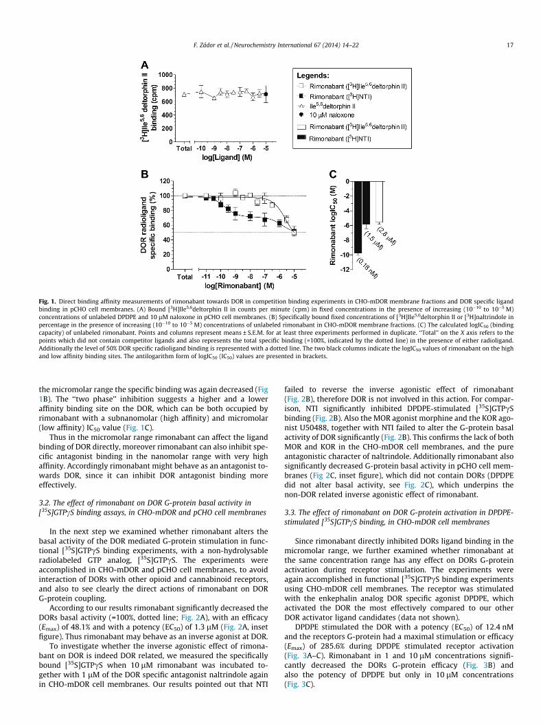
Fig. 1. Direct binding affinity measurements of rimonabant towards DOR in competition binding experiments in CHO-mDOR membrane fractions and DOR specific ligandbinding in pCHO cell membranes. (A) Bound [3H]Ile5,6deltorphin II in counts per minute (cpm) in fixed concentrations in the presence of increasing (10�10 to 10�5 M)concentrations of unlabeled DPDPE and 10 lM naloxone in pCHO cell membranes. (B) Specifically bound fixed concentrations of [3H]Ile5,6deltorphin II or [3H]naltrindole inpercentage in the presence of increasing (10�10 to 10�5 M) concentrations of unlabeled rimonabant in CHO-mDOR membrane fractions. (C) The calculated logIC50 (bindingcapacity) of unlabeled rimonabant. Points and columns represent means ± S.E.M. for at least three experiments performed in duplicate. ‘‘Total’’ on the X axis refers to thepoints which did not contain competitor ligands and also represents the total specific binding (=100%, indicated by the dotted line) in the presence of either radioligand.Additionally the level of 50% DOR specific radioligand binding is represented with a dotted line. The two black columns indicate the logIC50 values of rimonabant on the highand low affinity binding sites. The antilogarithm form of logIC50 (IC50) values are presented in brackets.
F. Zádor et al. / Neurochemistry International 67 (2014) 14–22 17
the micromolar range the specific binding was again decreased (Fig1B). The ‘‘two phase’’ inhibition suggests a higher and a loweraffinity binding site on the DOR, which can be both occupied byrimonabant with a subnanomolar (high affinity) and micromolar(low affinity) IC50 value (Fig. 1C).
Thus in the micromolar range rimonabant can affect the ligandbinding of DOR directly, moreover rimonabant can also inhibit spe-cific antagonist binding in the nanomolar range with very highaffinity. Accordingly rimonabant might behave as an antagonist to-wards DOR, since it can inhibit DOR antagonist binding moreeffectively.
3.2. The effect of rimonabant on DOR G-protein basal activity in[35S]GTPcS binding assays, in CHO-mDOR and pCHO cell membranes
In the next step we examined whether rimonabant alters thebasal activity of the DOR mediated G-protein stimulation in func-tional [35S]GTPcS binding experiments, with a non-hydrolysableradiolabeled GTP analog, [35S]GTPcS. The experiments wereaccomplished in CHO-mDOR and pCHO cell membranes, to avoidinteraction of DORs with other opioid and cannabinoid receptors,and also to see clearly the direct actions of rimonabant on DORG-protein coupling.
According to our results rimonabant significantly decreased theDORs basal activity (=100%, dotted line; Fig. 2A), with an efficacy(Emax) of 48.1% and with a potency (EC50) of 1.3 lM (Fig. 2A, insetfigure). Thus rimonabant may behave as an inverse agonist at DOR.
To investigate whether the inverse agonistic effect of rimona-bant on DOR is indeed DOR related, we measured the specificallybound [35S]GTPcS when 10 lM rimonabant was incubated to-gether with 1 lM of the DOR specific antagonist naltrindole againin CHO-mDOR cell membranes. Our results pointed out that NTI
failed to reverse the inverse agonistic effect of rimonabant(Fig. 2B), therefore DOR is not involved in this action. For compar-ison, NTI significantly inhibited DPDPE-stimulated [35S]GTPcSbinding (Fig. 2B). Also the MOR agonist morphine and the KOR ago-nist U50488, together with NTI failed to alter the G-protein basalactivity of DOR significantly (Fig. 2B). This confirms the lack of bothMOR and KOR in the CHO-mDOR cell membranes, and the pureantagonistic character of naltrindole. Additionally rimonabant alsosignificantly decreased G-protein basal activity in pCHO cell mem-branes (Fig 2C, inset figure), which did not contain DORs (DPDPEdid not alter basal activity, see Fig. 2C), which underpins thenon-DOR related inverse agonistic effect of rimonabant.
3.3. The effect of rimonabant on DOR G-protein activation in DPDPE-stimulated [35S]GTPcS binding, in CHO-mDOR cell membranes
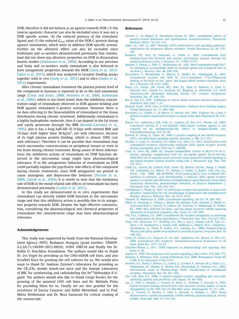
Since rimonabant directly inhibited DORs ligand binding in themicromolar range, we further examined whether rimonabant atthe same concentration range has any effect on DORs G-proteinactivation during receptor stimulation. The experiments wereagain accomplished in functional [35S]GTPcS binding experimentsusing CHO-mDOR cell membranes. The receptor was stimulatedwith the enkephalin analog DOR specific agonist DPDPE, whichactivated the DOR the most effectively compared to our otherDOR activator ligand candidates (data not shown).
DPDPE stimulated the DOR with a potency (EC50) of 12.4 nMand the receptors G-protein had a maximal stimulation or efficacy(Emax) of 285.6% during DPDPE stimulated receptor activation(Fig. 3A–C). Rimonabant in 1 and 10 lM concentrations signifi-cantly decreased the DORs G-protein efficacy (Fig. 3B) andalso the potency of DPDPE but only in 10 lM concentrations(Fig. 3C).

Fig. 2. The effect of rimonabant on DOR G-protein basal activity in ligand-modulated [35S]GTPcS binding assays, in CHO-mDOR and in pCHO cell membranes. (A) Specificallybound [35S]GTPcS in percentage in the presence of increasing concentrations (10�10 to 10�5 M) of rimonabant in CHO-mDOR cell membranes. Emax and logEC50 values arepresented in the inset figure. ⁄Denotes the significant alterations of DOR G-protein Emax value (unpaired t-test, two-tailed P value) compared to basal activity (=100%) in thepresence of rimonabant. (B) Specifically bound [35S]GTPcS in percentage in the presence of 10 lM of the indicated ligands alone or together with 1 lM naltrindole in CHO-mDOR cell membranes. �Indicates the significant alterations in specifically bound [35S]GTPcS (One-way ANOVA, Bonferroni’s Multiple Comparison post hoc test), comparedto basal activity (=100%) in the presence of rimonabant or DPDPE. ⁄Indicates the significant reduction in the specifically bound [35S]GTPcS during DPDPE stimulation in thepresence of NTI compared to DPDPE alone. (C) Specifically bound [35S]GTPcS in percentage in the presence of increasing concentrations (10�10 to 10�5 M) of rimonabant orDPDPE in pCHO cell membranes. In case of rimonabant error bars are shorter than the points itself. The logEC50 value of rimonabant and the G-protein Emax value afterrimonabant activation are presented in the inset figure. ⁄denotes the significant alterations of G-protein Emax value (unpaired t-test, two-tailed P value) compared to basalactivity (=100%) in the presence of rimonabant. Points and columns represent means ± S.E.M. for at least three experiments performed in triplicate. ‘‘Basal’’ on the X axis refersto the points which did not contain ligands. The dotted line represents the receptors basal activity level, which was defined as 100%. ���, ⁄⁄⁄: P < 0.001; �: P < 0.05.
18 F. Zádor et al. / Neurochemistry International 67 (2014) 14–22
Thus rimonabant inhibited the DPDPE-induced maximal G-pro-tein stimulation of the DOR and also the potency of the stimulatorligand in the micromolar range when no cannabinoid or other opi-oid receptors were present in the experimental system.
3.4. The effect of rimonabant on DOR G-protein activation in DPDPE-stimulated [35S]GTPcS binding, in wild type and CB1/CB2 receptordouble knock-out mouse forebrain membranes
Our next objective was to examine whether the inhibitory ac-tion of rimonabant upon DOR G-protein activation and stimulatoryligand potency occurs when the receptor is expressed in physiolog-ical conditions, when cannabinoid and other opioid receptors arepresent, thus interactions may occur between them. Membranefractions of mice forebrain were used, since both opioid and can-nabinoid receptors are expressed in eligible amount in this brainregion (Lever, 2007; Sim and Childers, 1997; Svízenská et al.,2008). Since the inhibitory actions of rimonabant occurred from1 lM in CHO-mDOR cell membranes, we carried out our furtherexperiments with this concentration.
In the wild type (w.t.) mouse forebrain DPDPE stimulated theDOR to a maximal stimulation level of 138.1% with an EC50 valueof 13.8 nM (Fig. 4A–C). The presence of rimonabant produced a sig-nificant attenuation in the maximal efficacy of DORs G-proteinactivity (Fig. 4A and B) and in the potency of the stimulatory ligandas well (Fig. 4C).
Previously our group demonstrated an interaction between theCB2 receptor and MOR in mice forebrain (Páldyová et al., 2008)therefore we studied the possible role of the CB1 receptor in the
inhibitory actions of rimonabant, and also the role of the CB2 recep-tor. To achieve this, we carried out our DPDPE-induced [35S]GTPcSbinding assays in CB1/CB2 K.O. mouse forebrain membranes.Accordingly the reduced maximal efficacy of the receptor and thepotency of the activating ligand (Fig. 4A–C, respectively) were alsoestablished in the CB1/CB2 K.O. mouse forebrain when rimonabantwas added. Interestingly in the absence of rimonabant, the lack ofboth cannabinoid receptors did not affect significantly the maximalstimulation of the G-protein activity or the stimulatory potency ofDPDPE (Fig. 4A–C).
Further on we wanted to verify that the inhibitory actions ofrimonabant are not due to the unspecific inverse agonistic effectdiscussed previously under Section 3.2. To exclude this possibilitywe applied L-epinephrine, which is a non-selective endogenousadrenergic receptor agonist, in [35S]GTPcS binding assays inCB1/CB2 K.O. mouse forebrain membranes. Adrenergic receptorsare highly expressed in the forebrain structures and can coupledto Gi/o type G-proteins such as the a2 type receptors (Cottinghamand Wang, 2012), therefore they can be monitored by L-epineph-rine-stimulated [35S]GTPcS binding assays. L-epinephrine activatedits specific receptor with a 31.6 nM potency (Fig. 5C) and the cou-pled G-protein had a 166.8% maximal efficacy during the activation(Fig. 5A and B). In the presence of 10 lM rimonabant neither of theparameters changed significantly (Emax: 162%; EC50: 26.7 nM;Fig. 5A–C), thus the inhibitory actions of rimonabant are indeedDOR specific.
To summarize the DPDPE-activated [35S]GTPcS binding experi-ments accomplished in the forebrain, we can conclude thatrimonabant inhibits agonist-stimulated DOR G-protein efficacy

Fig. 3. The effect of rimonabant on DOR G-protein activation in DPDPE-stimulated[35S]GTPcS binding, in CHO-mDOR cell membranes. (A) Specifically bound[35S]GTPcS in percentage in the presence of increasing concentrations (10�10 to10�5 M) of DPDPE in the absence or presence of the indicated rimonabantconcentrations. (B) The calculated Emax (maximal efficacy) value of DOR G-protein.(C) The calculated logEC50 (ligand potency) value of DPDPE. Points and columnsrepresent means ± S.E.M. for at least three experiments performed in triplicate.⁄Indicates the significant reduction in Emax and logEC50 values in the presence ofrimonabant compared to DPDPE alone (One-way ANOVA, Bonferroni’s MultipleComparison post hoc test). ‘‘Basal’’ on the X axis refers to the points which did notcontain DPDPE. The antilogarithm form of logEC50 (EC50) values are presented inbrackets. ⁄⁄⁄: P < 0.001; ⁄⁄: P < 0.01.
Fig. 4. The effect of rimonabant on DOR G-protein activation in DPDPE-stimulated[35S]GTPcS binding in wild type and CB1/CB2 receptor double knock-out mouseforebrain membranes. (A) Specifically bound [35S]GTPcS in percentage in thepresence of increasing concentrations (10�10 to 10�5 M) of DPDPE in the absence orpresence of rimonabant. (B) The calculated Emax (maximal efficacy) value of DOR G-protein. (C) The calculated logEC50 (ligand potency) value of DPDPE. Points andcolumns represent means ± S.E.M. for at least three experiments performed intriplicate. ⁄Indicates the significant reduction in Emax and logEC50 values in thepresence of rimonabant compared to DPDPE alone in wild type or in CB1/CB2
receptor double knock-out mouse (unpaired t-test, two-tailed P value). ‘‘Basal’’ onthe X axis refers to the points which did not contain activator ligands. Theantilogarithm form of logEC50 (EC50) values are presented in brackets. ⁄⁄⁄: P < 0.001;⁄⁄: P < 0.01; ⁄: P < 0.05.
F. Zádor et al. / Neurochemistry International 67 (2014) 14–22 19
and activator ligand potency in the micromolar range, when thereceptor is expressed in physiological conditions. Further on thisinhibitory action is independent from both cannabinoid receptorsand it is DOR specific.
4. Discussion
Rimonabant was the first CB1 receptor antagonist/inverse ago-nist to be developed, and it is still a perfect tool for antagonizingCB1 related effects (for review see Pertwee, 2005). Later it turnedout that rimonabant had several non-cannabinoid related effectssuch as decreasing alcohol intake, opiate self-administration andincreasing smoking cessation (for review see Foll and Goldberg,2005). There is also evidence that rimonabant can interact withseveral GPCRs and ion channels (for review see Pertwee, 2010).
This study focuses on the interaction between rimonabant andDOR. The interaction was studied at ligand-receptor level in com-petition binding experiments, and at the receptor-G-protein levelin functional [35S]GTPcS binding assays. These assays gather infor-mation about the receptors ligand binding capacity (Schütz, 1991)and functionality (Strange, 2010).
To our knowledge there has been only one paper so far studyingthe direct effect of rimonabant on DOR (Kathmann et al., 2006) incompetition binding experiments and in saturation and kineticbinding assays in cerebral cortical membranes with the DOR spe-cific tritiated antagonist [3H]NTI. In competition binding experi-ments they found that rimonabant inhibited [3H]NTI binding by
only 20%, while in our binding studies rimonabant decreased 50%of [3H]Ile5,6deltorphin II and [3H]NTI total specific binding(Fig. 1B). The diverse results in case of [3H]NTI can be partly ex-plained by the different experimental conditions applied duringthe assay (incubation time and temperature, final volume etc.)and by the applied CHO cell lines, which contained a homogeneouspopulation of overexpressed mouse DORs. CHO-mDOR cell mem-branes insure near optimal conditions for a rimonabant–DOR inter-action since no other opioid or even cannabinoid receptors werepresent in the membrane preparation. Additionally in case of bothradioligands rimonabant reached an IC50 value in the micromolarrange (Fig. 1C), similar to our previously reported data on CHO-MOR cell membranes (Zádor et al., 2012) and to other studies (Cin-ar and Szücs, 2009; Fong et al., 2009; Kathmann et al., 2006). Inter-estingly rimonabant also bound with an IC50 in the micromolarrange to other members of the GPCR family, such as adrenergicor 5-HT6 receptors or to ion channels, like calcium, sodium andpotassium channels (for review see Pertwee, 2010). However rimo-nabant also displaced the antagonist [3H]NTI in the subnanomolarrange (Fig. 1B) with a very high affinity (Fig. 1C), which did not oc-cur during the displacement of the agonist [3H]Ile5,6deltorphin II(Fig. 1B and C). It is known that antagonists stabilize the receptorsin the inactive state, thus they prefer the inactive receptor form,while agonists prefer the active receptor state, since they stabilizethe active form (Rosenbaum et al., 2009; Strange, 2002). Accord-ingly in our competition binding experiments rimonabant may be-have as an antagonist since it displaced [3H]NTI (which probablystabilized the DOR to the inactive state) from the higher affinity

Fig. 5. The effect of rimonabant on AR G-protein activation in L-epinephrine-stimulated [35S]GTPcS binding in CB1/CB2 receptor double knock-out mouseforebrain membranes. (A) Specifically bound [35S]GTPcS in percentage in thepresence of increasing concentrations (10�10 to 10�5 M) of L-epinephrine in theabsence or presence of rimonabant. (B) The calculated Emax (maximal efficacy) valueof AR G-protein. (C) The calculated logEC50 (ligand potency) value of L-epinephrine.Points and columns represent means ± S.E.M. for at least three experimentsperformed in triplicate. ‘‘Basal’’ on the X axis refers to the points which did notcontain L-epinephrine. The antilogarithm form of logEC50 (EC50) values arepresented in brackets. AR: adrenergic receptor.
20 F. Zádor et al. / Neurochemistry International 67 (2014) 14–22
binding site, which did not occur during the displacement of theagonist [3H]Ile5,6deltorphin II (which probably stabilized the DORin the active state). In our previous paper similar results were ob-served when rimonabant displaced the opioid antagonist [3H]nal-oxone from the high affinity binding site in CHO-MOR cellmembranes (Zádor et al., 2012). The high and low affinity bindingsites were previously described in the GPCR superfamily amongthe MORs as well (Baker and Hill, 2007; Krizsan et al., 1991).
It is well known that G-proteins have a constitutive activity (forreview see Seifert and Wenzel-Seifert, 2002) which can be furthermodified upon ligand binding. In turn the altered basal activity ini-tiates different types of signaling cascades (Hamm, 1998). For fur-ther investigations we examined the impact of rimonabant on thebasal activity of the DORs G-protein, which can give us valuableinformation about the binding character of rimonabant on DOR.In our [35S]GTPcS binding assays rimonabant significantly inhib-ited receptor basal activity in CHO-mDOR membranes (Fig. 2A),however we proved that this was not DOR related, since the effectwas not reversed by the DOR specific antagonist NTI (Fig. 2B) and italso occurred in pCHO cell membranes (Fig. 2C) which did not ex-press DORs (Figs. 1A and 2C). These findings also confirms previousreports in similar experimental conditions (Cinar and Szücs, 2009;Seely et al., 2012). Therefore the agonistic (since rimonabant didnot enhance DOR G-protein basal activity) and inverse agonisticcharacter of rimonabant at DOR can be excluded. As regard tothe non-CB1 related inverse agonistic effect of rimonabant it hasbeen proven to be pertussis toxin sensitive and it has beendemonstrated in different types of brain tissues in several other
publications (Breivogel et al., 2001; Cinar and Szücs, 2009;MacLennan et al., 1998; Sim-Selley et al., 2001).
Since rimonabant affected the DORs ligand binding, it raises thepossibility that it can also interfere with the receptors G-proteinactivation during DOR specific agonist stimulation. Indeed, whenG-protein signaling was initiated with the cyclic enkephalin analogDPDPE, rimonabant inhibited the maximal stimulation of the DORsG-protein and also the potency of the stimulator ligand signifi-cantly (Fig. 3A–C). Moreover the inhibition occurred in themicromolar concentration range, similarly as in direct affinitymeasurements in CHO-mDOR cell membranes. For comparison,in CHO-MOR cell membrane preparations rimonabant (10 lM)did not alter the DAMGO-stimulated (10 lM) G-protein activationsignificantly (Cinar and Szücs, 2009), in contrast in morphine-induced [35S]GTPcS assays accomplished in CHO-MOR cell mem-branes rimonabant reduced the potency of morphine (Seely et al.,2012). Thus so far the inhibitory action of rimonabant on themaximal stimulation of the DOR G-protein and on the potencyof the stimulator ligand in agonist-induced [35S]GTPcSexperiments indicates that rimonabant may behave as an antago-nist on DOR.
The CHO cell line with homogeneous population of overexpres-sed DOR is a useful tool to investigate the direct interaction be-tween rimonabant and DOR, since there is no cannabinoid orother opioid receptors in the system which can interact withDOR, thus the direct effect of rimonabant can be measured moreaccurately. However, it is well known that in physiological condi-tions GPCR receptors often communicate with each other via over-lapping signaling pathways or more even via heterodimerization(González-Maeso, 2011; Hur and Kim, 2002; Jordan and Devi,1999; Rozenfeld and Devi, 2011), which in addition alters therefunctionality (Birdsall, 2010; Jordan and Devi, 1999). Taking thisinto consideration we investigated the impact of rimonabant onDOR G-protein activation in mouse forebrain, where the cannabi-noid and opioid receptors are expressed in adequate quantity(Gong et al., 2006; Howlett et al., 2002; Mansour et al., 1995;Sim and Childers, 1997). According to our results, in micromolarconcentrations the inhibitory action of rimonabant also occurredwhen the DOR and its G-protein were expressed in physiologicalconditions (Fig. 4A–C). More importantly the inhibition was alsoobserved when neither cannabinoid receptors were expressed inthe forebrain (Fig. 4A–C); therefore it was a cannabinoid receptorindependent action, at least in the forebrain region. Previouslyour group demonstrated a cannabinoid independent G-proteininhibitory action of rimonabant on MOR, in the forebrain regionas well (Zádor et al., 2012). In another study, micromolar concen-trations of rimonabant decreased the potency of the MOR stimula-tor ligand CB1 receptor independently (Cinar and Szücs, 2009).However the inhibitory action of rimonabant on DPDPE-stimulatedDOR G-protein activation might be due to the non-CB1 related in-verse agonistic effect. To investigate this possibility we examinedL-epinephrine-induced G-protein activity in the presence of rimo-nabant in CB1/CB2 receptor double knock-out mouse forebrainmembranes. According to our results micromolar concentrationsof rimonabant did not have any effect on adrenergic receptor G-protein maximal activity or L-epinephrine potency during L-epi-nephrine stimulation (Fig. 5A–C). Therefore we can conclude thatthe decreased agonist mediated DOR G-protein activation by rimo-nabant are truly DOR specific.
Thus rimonabant can directly inhibit DOR specific binding andagonist-stimulated DOR G-protein efficacy and potency. The mech-anism behind these actions can be due to the antagonistic charac-ter of rimonabant towards the DOR, which possibility is confirmedby our results in many levels: (1) the reduced specific binding ofthe antagonist [3H]NTI in the subnanomolar concentrationrange, (2) rimonabant did not enhanced the basal activity of
F. Zádor et al. / Neurochemistry International 67 (2014) 14–22 21
DOR, therefore it did not behave as an agonist towards DOR, (3) theinverse agonistic character can also be excluded since it was not aDOR specific action, (4) the reduced potency of the stimulatorligand and (5) the reduced Emax value of the DOR G-protein duringagonist stimulation, which were in addition DOR specific actions.Further on the allosteric effect can also be excluded sinceKathmann and co-workers demonstrated previously that rimona-bant did not show any allosteric properties on DOR in dissociationkinetic studies (Kathmann et al., 2006). According to our previousand Seely and co-workers study rimonabant is also believed tohave antagonistic properties towards the MOR (Seely et al., 2012;Zádor et al., 2012), which was analyzed in receptor binding assaystogether with in vivo (Seely et al., 2012) and in silico (Zádor et al.,2012) experiments.
After chronic rimonabant treatment the plasma protein level ofthe compound in humans is reported to be in the mid nanomolarrange (Cinar and Szücs, 2009; Henness et al., 2006; Kathmannet al., 2006), which is ten times lower than the inhibitory concen-tration range of rimonabant observed in DOR agonist binding andDOR agonist stimulated G-protein activation. However there isno data referring to the bioavailability of rimonabant or the tissuedistribution during chronic treatment. Additionally rimonabant isa highly hydrophobic molecule, thus it can deposit in the fat tissueand easily penetrate through the BBB (Rinaldi-Carmona et al.,1995), also it has a long half-life (6–9 days with normal BMI and16 days with higher than 30 kg/m2, see web reference), becauseof its high plasma protein binding, which is almost 100% (seeweb reference). Therefore it can be possible that rimonabant mayreach micromolar concentrations in peripheral tissues or even inthe brain during chronic treatment. Being aware of these informa-tions the inhibitory actions of rimonabant on DOR function ob-served in the micromolar range might have pharmacologicalrelevance. If so the antagonistic behavior of rimonabant on DORcould partially explain the psychiatric side effects of the compoundduring chronic treatments, since DOR antagonists are proved tocause anxiogenic and depressive-like behavior (Perrine et al.,2006; Saitoh et al., 2004). It is worth to note that the mediatoryrole of KOR in the mood related side effects of rimonabant has beendemonstrated previously (Lockie et al., 2011).
In this study we demonstrated in in vitro experiments thatrimonabant can directly inhibit DOR function in the micromolarrange and that this inhibitory action is possibly due to its antago-nist property towards DOR. Despite the high effective concentra-tion, considering the pharmacological and chemical properties ofrimonabant this concentration range may have pharmacologicalrelevance.
Acknowledgements
This study was supported by funds from the National Develop-ment Agency (NFÜ), Budapest, Hungary (grant number: TÁMOP-4.2.2A-11/1KONV-2012-0024); OTKA 108518 and finally the Dr.Rollin D. Hotchkiss Foundation. The authors would like to thankDr. Zvi Vogel for providing us the CHO-mDOR cell lines, and alsoErzsébet Kusz for growing the cell cultures for us. We would alsowant to thank Dr. Andreas Zimmer’s laboratory for providing usthe CB1/CB2 double knock-out mice and the Isotope Laboratoryof BRC for synthetizing and radiolabeling the Ile5,6deltorphin II li-gand. The authors would also like to thank Gerg}o Kovács for thegrowing of the parental CHO cell lines and Dr. Melinda Pirityfor providing them for us. Finally we are also grateful for theassistance of Zsuzsa Canjavec and Ildikó Némethné, and to Prof.Mária Wollemann and Dr. Reza Samavati for critical reading ofthe manuscript.
References
Arévalo, C., de Miguel, R., Hernández-Tristán, R., 2001. Cannabinoid effects onanxiety-related behaviours and hypothalamic neurotransmitters. Pharmacol.Biochem. Behav. 70, 123–131.
Baker, J.G., Hill, S.J., 2007. Multiple GPCR conformations and signalling pathways:implications for antagonist affinity estimates. Trends Pharmacol. Sci. 28, 374–381.
Bambico, F.R., Katz, N., Debonnel, G., Gobbi, G., 2007. Cannabinoids elicitantidepressant-like behavior and activate serotonergic neurons through themedial prefrontal cortex. J. Neurosci. 27, 11700–11711.
Benyhe, S., Farkas, J., Tóth, G., Wollemann, M., 1997. Met5-enkephalin-Arg6-Phe7,an endogenous neuropeptide, binds to multiple opioid and nonopioid sites inrat brain. J. Neurosci. Res. 48, 249–258.
Berrendero, F., Mendizabal, V., Murtra, P., Kieffer, B.L., Maldonado, R., 2003.Cannabinoid receptor and WIN 55 212-2-stimulated [35S]-GTPgammaSbinding in the brain of mu-, delta- and kappa-opioid receptor knockout mice.Eur. J. Neurosci. 18, 2197–2202.
Beyer, C.E., Dwyer, J.M., Piesla, M.J., Platt, B.J., Shen, R., Rahman, Z., Chan, K.,Manners, M.T., Samad, T.a., Kennedy, J.D., Bingham, B., Whiteside, G.T., 2010.Depression-like phenotype following chronic CB1 receptor antagonism.Neurobiol. Dis. 39, 148–155.
Bie, B., Pan, Z.Z., 2007. Trafficking of central opioid receptors and descending paininhibition. Mol. Pain. 3, 37.
Birdsall, N.J.M., 2010. Class A GPCR heterodimers: evidence from binding studies.Trends Pharmacol. Sci. 31, 499–508.
Breivogel, C.S., Griffin, G., Di Marzo, V., Martin, B.R., 2001. Evidence for a new Gprotein-coupled cannabinoid receptor in mouse brain. Mol. Pharmacol. 60, 155–163.
Broom, D.C., Jutkiewicz, E.M., Folk, J.E., Traynor, J.R., Rice, K.C., Woods, J.H., 2002.Convulsant activity of a non-peptidic delta-opioid receptor agonist is notrequired for its antidepressant-like effects in Sprague-Dawley rats.Psychopharmacology 164, 42–48.
Burford, N., Wang, D., Sadee, W., 2000. G-protein coupling of mu-opioid receptors(OP3): elevated basal signalling activity. Biochem. J. 348, 531–537.
Bushlin, I., Gupta, A., Stockton, S.D., Miller, L.K., Devi, L.A., 2012. Dimerization withcannabinoid receptors allosterically modulates delta opioid receptor activityduring neuropathic pain. PLoS One 7, e49789.
Cinar, R., Szücs, M., 2009. CB1 receptor-independent actions of SR141716 on G-protein signaling: coapplication with the mu-opioid agonist Tyr-D-Ala-Gly-(NMe)Phe-Gly-ol unmasks novel, pertussis toxin-insensitive opioid signaling inmu-opioid receptor-Chinese hamster ovary cells. J. Pharmacol. Exp. Ther. 330,567–574.
Codd, E.E., Carson, J.R., Colburn, R.W., Stone, D.J., Besien, C.R.Van., Zhang, S., Wade,P.R., Gallantine, E.L., Meert, T.F., Molino, L., Pullan, S., Razler, C.M., Dax, S.L.,Flores, C.M., 2009. JNJ-20788560 [9-(8-azabicyclo[3.2.1]oct-3-ylidene)-9H-xanthene-3-carboxylic acid diethylamide], a selective delta opioid receptoragonist, is a potent and efficacious antihyperalgesic agent that does not producerespiratory depression, pharmacologic tolerance, or physical dependence. J.Pharmacol. Exp. Ther. 329, 241–251.
Cottingham, C., Wang, Q., 2012. a2 adrenergic receptor dysregulation in depressivedisorders: implications for the neurobiology of depression and antidepressanttherapy. Neurosci. Biobehav. Rev. 36, 2214–2225.
Demuth, D., Molleman, A., 2006. Cannabinoid signalling. Life Sci. 78, 549–563.Filliol, D., Ghozland, S., Chluba, J., Martin, M., Matthes, H.W., Simonin, F., Befort, K.,
Gavériaux-Ruff, C., Dierich, A., LeMeur, M., Valverde, O., Maldonado, R., Kieffer,B.L., 2000. Mice deficient for delta- and mu-opioid receptors exhibit opposingalterations of emotional responses. Nat. Genet. 25, 195–200.
Foll, B.Le., Goldberg, S.R., 2005. Cannabinoid CB1 receptor antagonists as promisingnew medications for drug dependence. J. Pharmacol. Exp. Ther. 312, 875–883.
Fong, T.M., Shearman, L.P., Stribling, D.S., Shu, J., Lao, J., Huang, C.R.-R., Xiao, J.C.,Shen, C.-P., Tyszkiewicz, J., Strack, A.M., DeMaula, C., Hubert, M.-F., Galijatovic-Idrizbegovic, A., Owen, R., Huber, A.C., Lanning, C.L., 2009. Pharmacologicalefficacy and safety profile of taranabant in preclinical species. Drug Dev. Res. 70,349–362.
Gong, J.P., Onaivi, E.S., Ishiguro, H., Liu, Q.R., Tagliaferro, P.A., Brusco, A., Uhl, G.R.,2006. Cannabinoid CB2 receptors: immunohistochemical localization in ratbrain. Brain Res. 1071, 10–23.
González-Maeso, J., 2011. GPCR oligomers in pharmacology and signaling. Mol.Brain. 4, 20.
Hamm, H., 1998. The many faces of G protein signaling. J. Biol. Chem. 273, 669–772.Henness, S., Robinson, D.M., Lyseng-Williamson, K.A., 2006. Rimonabant. Drugs 66,
2109–2119 (discussion 2120–2121).Howlett, A.C., Barth, F., Bonner, T.I., Cabral, G., Casellas, P., Devane, W.a., Felder, C.C.,
Herkenham, M., Mackie, K., Martin, B.R., Mechoulam, R., Pertwee, R.G., 2002.International Union of Pharmacology. XXVII. Classification of cannabinoidreceptors. Pharmacol. Rev. 54, 161–202.
Hur, E.M., Kim, K.T., 2002. G protein-coupled receptor signalling and cross-talk:achieving rapidity and specificity. Cell. Signal. 14, 397–405.
Ioja, E., Tóth, G., Benyhe, S., Tourwe, D., Péter, A., Tömböly, C., Borsodi, A., 2005.Opioid receptor binding characteristics and structure-activity studies of noveltetrapeptides in the TIPP (Tyr-Tic-Phe-Phe) series. Neurosignals 14, 317–328.
Ioja, E., Tourwé, D., Kertész, I., Tóth, G., Borsodi, A., Benyhe, S., 2007. Noveldiastereomeric opioid tetrapeptides exhibit differing pharmacological activityprofiles. Brain Res. Bull. 74, 119–129.

22 F. Zádor et al. / Neurochemistry International 67 (2014) 14–22
Járai, Z., Wagner, J.A., Varga, K., Lake, K.D., Compton, D.R., Martin, B.R., Zimmer, A.M.,Bonner, T.I., Buckley, N.E., Mezey, E., Razdan, R.K., Zimmer, A., Kunos, G., 1999.Cannabinoid-induced mesenteric vasodilation through an endothelial sitedistinct from CB1 or CB2 receptors. Proc. Natl. Acad. Sci. USA 96, 14136–14141.
Jordan, B., Devi, L., 1999. G-protein-coupled receptor heterodimerization modulatesreceptor function. Nature 399, 697–700.
Kathmann, M., Flau, K., Redmer, A., Tränkle, C., Schlicker, E., 2006. Cannabidiol is anallosteric modulator at mu- and delta-opioid receptors. Naunyn.Schmiedebergs. Arch. Pharmacol. 372, 354–361.
Katona, I., Sperlágh, B., Sík, A., Käfalvi, A., Vizi, E.S., Mackie, K., Freund, T.F., 1999.Presynaptically located CB1 cannabinoid receptors regulate GABA release fromaxon terminals of specific hippocampal interneurons. J. Neurosci. 19, 4544–4558.
Krizsan, D., Varga, E., Hosztafi, S., Benyhe, S., Szucs, M., Borsodi, A., 1991. Irreversibleblockade of the high and low affinity (3H) naloxone binding sites by C-6derivatives of morphinane-6-ones. Life Sci. 48, 439–451.
Lee, H.K., Choi, E.B., Pak, C.S., 2009. The current status and future perspectives ofstudies of cannabinoid receptor 1 antagonists as anti-obesity agents. Curr. Top.Med. Chem. 9, 482–503.
Lever, J.R., 2007. PET and SPECT imaging of the opioid system : receptors,radioligands and avenues for drug discovery and development. Curr. Pharm.Des. 13, 33–49.
Lockie, S.H., Czyzyk, T.A., Chaudhary, N., Perez-Tilve, D., Woods, S.C., Oldfield, B.J.,Statnick, M.A., Tschöp, M.H., 2011. CNS opioid signaling separates cannabinoidreceptor 1-mediated effects on body weight and mood-related behavior inmice. Endocrinology 152, 3661–3667.
MacLennan, S.J., Reynen, P.H., Kwan, J., Bonhaus, D.W., 1998. Evidence for inverseagonism of SR141716A at human recombinant cannabinoid CB1 and CB2receptors. Br. J. Pharmacol. 124, 619–622.
Maldonado, R., Valverde, O., 2003. Participation of the opioid system incannabinoid-induced antinociception and emotional-like responses. Eur.Neuropsychopharmacol. 13, 401–410.
Mansour, A., Fox, C.A., Akil, H., Watson, S.J., 1995. Opioid-receptor mRNA expressionin the rat CNS: anatomical and functional implications. Trends Neurosci. 18, 22–29.
McGregor, I.S., Dastur, F.N., McLellan, R.A., Brown, R.E., 1996. Cannabinoidmodulation of rat pup ultrasonic vocalizations. Eur. J. Pharmacol. 313, 43–49.
Nevin, S.T., Kabasakal, L., Otvös, F., Töth, G., Borsodi, A., 1994. Binding characteristicsof the novel highly selective delta agonist, [3H]IIe5,6deltorphin II.Neuropeptides 26, 261–265.
Padwal, R.S., Majumdar, S.R., 2007. Drug treatments for obesity: orlistat,sibutramine, and rimonabant. Lancet 369, 71–77.
Pagotto, U., Marsicano, G., Cota, D., Lutz, B., Pasquali, R., 2006. The emerging role ofthe endocannabinoid system in endocrine regulation and energy balance.Endocr. Rev. 27, 73–100.
Páldyová, E., Bereczki, E., Sántha, M., Wenger, T., Borsodi, A., Benyhe, S., 2008.Noladin ether, a putative endocannabinoid, inhibits mu-opioid receptoractivation via CB2 cannabinoid receptors. Neurochem. Int. 52, 321–328.
Perrine, S.A., Hoshaw, B.A., Unterwald, E.M., 2006. Delta opioid receptor ligandsmodulate anxiety-like behaviors in the rat. Br. J. Pharmacol. 147, 864–872.
Pertwee, R.G., 2001. Cannabinoid receptors and pain. Prog. Neurobiol. 63, 569–611.Pertwee, R.G., 2005. Inverse agonism and neutral antagonism at cannabinoid CB1
receptors. Life Sci. 76, 1307–1324.Pertwee, R.G., 2010. Receptors and channels targeted by synthetic cannabinoid
receptor agonists and antagonists. Curr. Med. Chem. 17, 1360–1381.Pradhan, A., Befort, K., Nozaki, C., Gavériaux-Ruff, C., Kieffer, B.L., 2011. The delta
opioid receptor: an evolving target for the treatment of brain disorders. TrendsPharmacol. Sci. 32, 581–590.
Pradhan, A.A., Smith, M.M.L., Kieffer, B.L., Evans, C.J., 2012. Ligand-directedsignalling within the opioid receptor family. Br. J. Pharmacol. 167, 960–969.
Rinaldi-Carmona, M., Barth, F., Héaulme, M., Shire, D., Calandra, B., Congy, C.,Martinez, S., Maruani, J., Néliat, G., Caput, D., 1994. SR141716A, a potent andselective antagonist of the brain cannabinoid receptor. FEBS Lett. 350, 240–244.
Rinaldi-Carmona, M., Barth, F., Héaulme, M., 1995. Biochemical andpharmacological characterisation of SR141716A, the first potent and selectivebrain cannabinoid receptor antagonist. Life Sci. 56, 1941–1947.
Robledo, P., Berrendero, F., Ozaita, A., Maldonado, R., 2008. Advances in the field ofcannabinoid–opioid cross-talk. Addict. Biol. 13, 213–224.
Rodgers, R.J., Haller, J., Halasz, J., Mikics, E., 2003. ‘‘One-trial sensitization’’ to theanxiolytic-like effects of cannabinoid receptor antagonist SR141716A in themouse elevated plus-maze. Eur. J. Neurosci. 17, 1279–1286.
Rosenbaum, D.M., Rasmussen, S.G.F., Kobilka, B.K., 2009. The structure and functionof G-protein-coupled receptors. Nature 459, 356–363.
Rozenfeld, R., Devi, L.A., 2011. Exploring a role for heteromerization in GPCRsignalling specificity. Biochem. J. 433, 11–18.
Rozenfeld, R., Bushlin, I., Gomes, I., Tzavaras, N., Gupta, A., Neves, S., Battini, L.,Gusella, G.L., Lachmann, A., Ma’ayan, A., Blitzer, R.D., Devi, L.A., 2012. Receptorheteromerization expands the repertoire of cannabinoid signaling in rodentneurons. PLoS One 7, e29239.
Saitoh, A., Kimura, Y., Suzuki, T., Kawai, K., Nagase, H., Kamei, J., 2004. Potentialanxiolytic and antidepressant-like activities of SNC80, a selective delta-opioidagonist, in behavioral models in rodents. J. Pharmacol. Sci. 95, 374–380.
Schütz, W., 1991. The pharmacological basis of receptor binding. Wien. Klin.Wochenschr. 103, 438–442.
Seely, K.A., Brents, L.K., Franks, L.N., Rajasekaran, M., Zimmerman, S.M., Fantegrossi,W.E., Prather, P.L., 2012. AM-251 and rimonabant act as direct antagonists atmu-opioid receptors: implications for opioid/cannabinoid interaction studies.Neuropharmacology 63, 905–915.
Seifert, R., Wenzel-Seifert, K., 2002. Constitutive activity of G-protein-coupledreceptors: cause of disease and common property of wild-type receptors.Naunyn. Schmiedebergs. Arch. Pharmacol. 366, 381–416.
Shen, M., Piser, T.M., Seybold, V.S., Thayer, S.A., 1996. Cannabinoid receptor agonistsinhibit glutamatergic synaptic transmission in rat hippocampal cultures. J.Neurosci. 16, 4322–4334.
Sim, L.J., Childers, S.R., 1997. Anatomical distribution of mu, delta, and kappaopioid- and nociceptin/orphanin FQ-stimulated [35S]guanylyl-50-O-(gamma-thio)-triphosphate binding in guinea pig brain. J. Comp. Neurol. 386, 562–572.
Sim, L.J., Selley, D.E., Childers, S.R., 1995. In vitro autoradiography of receptor-activated G proteins in rat brain by agonist-stimulated guanylyl 50-[gamma-[35S]thio]-triphosphate binding. Proc. Natl. Acad. Sci. U. S. A. 92, 7242–7246.
Sim-Selley, L.J., Brunk, L.K., Selley, D.E., 2001. Inhibitory effects of SR141716A on G-protein activation in rat brain. Eur. J. Pharmacol. 414, 135–143.
Strange, P.G., 2002. Mechanisms of inverse agonism at G-protein-coupled receptors.Trends Pharmacol. Sci. 23, 89–95.
Strange, P.G., 2010. Use of the GTPcS ([35S]GTPcS and Eu-GTPcS) binding assay foranalysis of ligand potency and efficacy at G protein-coupled receptors. Br. J.Pharmacol. 161, 1238–1249.
Svízenská, I., Dubovy, P., Sulcová, A., 2008. Cannabinoid receptors 1 and 2 (CB1 andCB2), their distribution, ligands and functional involvement in nervous systemstructures – a short review. Pharmacol. Biochem. Behav. 90, 501–511.
Traynor, R., Nahorski, R., Traynor, J.R., Nahorski, S.R., 1995. Modulation by mu-opioidagonists of guanosine-50-O-(3-[35S]thio)triphosphate binding to membranes fromhuman neuroblastoma SH-SY5Y cells. Mol. Pharmacol. 47, 848–854.
Tzavara, E.T., Davis, R.J., Perry, K.W., Li, X., Salhoff, C., Bymaster, F.P., Witkin, J.M.,Nomikos, G.G., 2003. The CB1 receptor antagonist SR141716A selectivelyincreases monoaminergic neurotransmission in the medial prefrontal cortex:implications for therapeutic actions. Br. J. Pharmacol. 138, 544–553.
Urigüen, L., Berrendero, F., Ledent, C., Maldonado, R., Manzanares, J., 2005. Kappa-and delta-opioid receptor functional activities are increased in the caudateputamen of cannabinoid CB1 receptor knockout mice. Eur. J. Neurosci. 22,2106–2110.
Zádor, F., Otvös, F., Benyhe, S., Zimmer, A., Páldy, E., 2012. Inhibition of forebrain l-opioid receptor signaling by low concentrations of rimonabant does not requirecannabinoid receptors and directly involves l-opioid receptors. Neurochem.Int. 61, 378–388.
Further reading
http://en.sanofi.com/press/press_releases/2008/news_list_2008.aspx (last accessed:2013).
http://www.drugbank.ca/drugs/DB06155 (last accessed: 2013).
Related Documents