The structural basis for agonist and partial agonist action on a β 1 -adrenergic receptor Tony Warne, Rouslan Moukhametzianov, Jillian G. Baker 1 , Rony Nehmé, Patricia C. Edwards, Andrew G.W. Leslie, Gebhard F.X. Schertler 2,* , and Christopher G. Tate * MRC Laboratory of Molecular Biology, Hills Road, Cambridge CB2 0QH, UK 1 Institute of Cell Signalling, C Floor Medical School, Queen’s Medical Centre, University of Nottingham, Nottingham NG7 2UH, UK β-Adrenergic receptors (βARs) are G protein-coupled receptors (GPCRs) that activate intracellular G proteins upon binding catecholamine agonist ligands such as adrenaline and noradrenaline 1 , 2 . Synthetic ligands have been developed that either activate or inhibit βARs for the treatment of asthma, hypertension or cardiac dysfunction. These ligands are classified as either full agonists, partial agonists or antagonists, depending on whether the cellular response is similar to that of the native ligand, reduced or inhibited, respectively. However, the structural basis for these different ligand efficacies is unknown. Here we present four crystal structures of the thermostabilised turkey (Meleagris gallopavo) β 1 - adrenergic receptor (β 1 AR-m23) bound to the full agonists carmoterol and isoprenaline and the partial agonists salbutamol and dobutamine. In each case, agonist binding induces a 1 Å contraction of the catecholamine binding pocket relative to the antagonist bound receptor. Full agonists can form hydrogen bonds with two conserved serine residues in transmembrane helix 5 (Ser 5.42 and Ser 5.46 ), but partial agonists only interact with Ser 5.42 (superscripts refer to Ballesteros-Weinstein numbering 3 ). The structures provide an understanding of the pharmacological differences between different ligand classes, illuminating how GPCRs function and providing a solid foundation for the structure-based design of novel ligands with predictable efficacies. Determining how agonists and antagonists bind to the β receptors has been the goal of research for more than 20 years 4 - 11 . Although the structures of the homologous β 1 and β 2 receptors 12 - 15 show how some antagonists bind to receptors in the inactive state 16 , structures with agonists bound are required to understand subsequent structural transitions involved in activation. GPCRs exist in an equilibrium between an inactive state (R) and an activated state (R*) that can couple and activate G proteins 17 . The binding of a full agonist, such as adrenaline or noradrenaline, is thought to increase the probability of the receptor converting to R*, with a conformation similar to that of opsin 18 , 19 . In the absence of any ligand, the βARs exhibit a low level of constitutive activity, indicating that there is always a small * Joint corresponding authors: MRC Laboratory of Molecular Biology, Hills Road, Cambridge CB2 0QH, UK, cgt@mrc- lmb.cam.ac.uk, [email protected], Telephone +44-(0)1223-402266, Fax +44-(0)1223-213556. 2 Current Address: Paul Scherrer Institut, Laboratory of Biomolecular Research, BMR, OFLC 109, CH-5232 Villigen PSI, Switzerland, Telephone +41 (0)56 310 4265 Author contributions: T.W. devised and performed receptor expression, purification, crystallization, cryo-cooling of the crystals, data collection and initial data processing. P.C.E. helped with crystal cryo-cooling and data collection. J.G.B. performed the pharmacological analyses on receptor mutants in whole cells and R.N. performed the ligand binding studies on baculovirus-expressed receptors. R.M. and A.G.W.L. were involved in data processing and structure refinement. Manuscript preparation was performed by T.W., C.G.T., A.G.W.L. and G.F.X.S. The overall project management was by G.F.X.S. and C.G.T. Author Information: Co-ordinates and structure factors have been submitted to the PDB database under accession codes 2y00, 2y01, 2y02, 2y03 and 2y04 for β44-m23 bound either to dobutamine (dob92 and dob102), carmoterol, isoprenaline or salbutamol, respectively. Europe PMC Funders Group Author Manuscript Nature. Author manuscript; available in PMC 2011 July 13. Published in final edited form as: Nature. 2011 January 13; 469(7329): 241–244. doi:10.1038/nature09746. Europe PMC Funders Author Manuscripts Europe PMC Funders Author Manuscripts

Welcome message from author
This document is posted to help you gain knowledge. Please leave a comment to let me know what you think about it! Share it to your friends and learn new things together.
Transcript
The structural basis for agonist and partial agonist action on aβ1-adrenergic receptor
Tony Warne, Rouslan Moukhametzianov, Jillian G. Baker1, Rony Nehmé, Patricia C.Edwards, Andrew G.W. Leslie, Gebhard F.X. Schertler2,*, and Christopher G. Tate*
MRC Laboratory of Molecular Biology, Hills Road, Cambridge CB2 0QH, UK1Institute of Cell Signalling, C Floor Medical School, Queen’s Medical Centre, University ofNottingham, Nottingham NG7 2UH, UK
β-Adrenergic receptors (βARs) are G protein-coupled receptors (GPCRs) that activateintracellular G proteins upon binding catecholamine agonist ligands such as adrenaline andnoradrenaline1,2. Synthetic ligands have been developed that either activate or inhibit βARsfor the treatment of asthma, hypertension or cardiac dysfunction. These ligands areclassified as either full agonists, partial agonists or antagonists, depending on whether thecellular response is similar to that of the native ligand, reduced or inhibited, respectively.However, the structural basis for these different ligand efficacies is unknown. Here wepresent four crystal structures of the thermostabilised turkey (Meleagris gallopavo) β1-adrenergic receptor (β1AR-m23) bound to the full agonists carmoterol and isoprenaline andthe partial agonists salbutamol and dobutamine. In each case, agonist binding induces a 1 Åcontraction of the catecholamine binding pocket relative to the antagonist bound receptor.Full agonists can form hydrogen bonds with two conserved serine residues intransmembrane helix 5 (Ser5.42 and Ser5.46), but partial agonists only interact with Ser5.42
(superscripts refer to Ballesteros-Weinstein numbering3). The structures provide anunderstanding of the pharmacological differences between different ligand classes,illuminating how GPCRs function and providing a solid foundation for the structure-baseddesign of novel ligands with predictable efficacies.
Determining how agonists and antagonists bind to the β receptors has been the goal ofresearch for more than 20 years4-11. Although the structures of the homologous β1 and β2receptors12-15 show how some antagonists bind to receptors in the inactive state16, structureswith agonists bound are required to understand subsequent structural transitions involved inactivation. GPCRs exist in an equilibrium between an inactive state (R) and an activatedstate (R*) that can couple and activate G proteins17. The binding of a full agonist, such asadrenaline or noradrenaline, is thought to increase the probability of the receptor convertingto R*, with a conformation similar to that of opsin18,19. In the absence of any ligand, theβARs exhibit a low level of constitutive activity, indicating that there is always a small
*Joint corresponding authors: MRC Laboratory of Molecular Biology, Hills Road, Cambridge CB2 0QH, UK, [email protected], [email protected], Telephone +44-(0)1223-402266, Fax +44-(0)1223-213556.2Current Address: Paul Scherrer Institut, Laboratory of Biomolecular Research, BMR, OFLC 109, CH-5232 Villigen PSI,Switzerland, Telephone +41 (0)56 310 4265Author contributions: T.W. devised and performed receptor expression, purification, crystallization, cryo-cooling of the crystals,data collection and initial data processing. P.C.E. helped with crystal cryo-cooling and data collection. J.G.B. performed thepharmacological analyses on receptor mutants in whole cells and R.N. performed the ligand binding studies on baculovirus-expressedreceptors. R.M. and A.G.W.L. were involved in data processing and structure refinement. Manuscript preparation was performed byT.W., C.G.T., A.G.W.L. and G.F.X.S. The overall project management was by G.F.X.S. and C.G.T.Author Information: Co-ordinates and structure factors have been submitted to the PDB database under accession codes 2y00, 2y01,2y02, 2y03 and 2y04 for β44-m23 bound either to dobutamine (dob92 and dob102), carmoterol, isoprenaline or salbutamol,respectively.
Europe PMC Funders GroupAuthor ManuscriptNature. Author manuscript; available in PMC 2011 July 13.
Published in final edited form as:Nature. 2011 January 13; 469(7329): 241–244. doi:10.1038/nature09746.
Europe PM
C Funders A
uthor Manuscripts
Europe PM
C Funders A
uthor Manuscripts
proportion of the receptor in the activated state, with the β2AR showing a 5-fold higher levelof basal activity than the β1AR20. Basal activity of β2AR is important physiologically, asshown by the T164I4.56 human polymorphism that reduces the basal activity of β2AR tolevels similar to β1AR21 and whose expression has been associated with heart disease22.
As a first step towards understanding how agonists activate receptors, we have determinedthe structures of β1AR bound to 4 different agonists. Native turkey β1AR is unstable indetergent23, so crystallization and structure determination relied on using a thermostabilisedconstruct (β1AR-m23) that contained six point mutations, which dramatically improved itsthermostability24. In addition, the thermostabilising mutations altered the equilibriumbetween R and R*, so that the receptor was preferentially in the R state24. However, it couldstill couple to G proteins after activation by agonists13 (Supplementary Fig. 1,Supplementary Tables 1-3), although the activation energy barrier is predicted to beconsiderably higher than for the wild-type receptor25. Here we report structures of β1AR-m23 (see Methods) bound to R-isoprenaline (2.85 Å resolution), R,R-carmoterol (2.6 Åresolution), R-salbutamol (3.05 Å resolution) and R-dobutamine (two independent structuresat 2.6 Å and 2.5 Å resolution) (Supplementary Table 5). The overall structures of β1AR-m23 bound to the agonists are very similar to the structure with the bound antagonistcyanopindolol13, as expected for a receptor mutant stabilised preferentially in the R state.None of the structures show the outward movement of the cytoplasmic end oftransmembrane helix H6 by 5-6 Å that is observed during light activation ofrhodopsin18,19,26. This suggests that the structures represent an inactive, non-signaling stateof the receptor formed on initial agonist binding.
All four agonists bind in the catecholamine pocket in a virtually identical fashion (Fig. 1).The secondary amine and β-hydroxyl groups shared by all the agonists (except fordobutamine, which lacks the β-hydroxyl; see Supplementary Figure 4) form potentialhydrogen bonds with Asp1213.32 and Asn3297.39, while the hydrogen bond donor/acceptorgroup equivalent to the catecholamine meta-hydroxyl (m-OH) generally forms a hydrogenbond with Asn3106.55. In addition, all the agonists can form a hydrogen bond withSer2115.42, as seen for cyanopindolol13, and they also induce the rotamer conformationchange of Ser2125.43 so that it makes a hydrogen bond with Asn3106.55. The majordifference between the binding of full agonists compared to the partial agonists is that onlyfull agonists make a hydrogen bond to the side chain of Ser2155.46 as a result of a change inside chain rotamer. All of these amino acid residues involved in the binding of thecatecholamine headgroups to β1AR are fully conserved in both β1 and β2 receptors (Fig. 2).Furthermore, the role of many of these amino acid residues in ligand binding is supported byextensive mutagenesis studies on β2AR that were performed before the first β2AR structurewas determined27. Thus it was predicted that Asp1133.32, Ser2035.42, Ser2075.46, Asn2936.55
and Asn3127.39 in β2AR were all involved in agonist binding4,5,7-9 (Fig. 3). Inspection ofthe region outside the catecholamine binding pocket in the structures with bounddobutamine and carmoterol allows the identification of non-conserved residues that interactwith these ligands (Fig. 2 and Supplementary Figure 7), which may contribute to the subtypespecificity of these ligands10,28.
There are three significant differences in the β1AR catecholamine binding pocket when fullagonists are bound compared to when an antagonist is bound, namely the rotamerconformation changes of side chains Ser2125.43 and Ser2155.46 (Fig.3) and the contractionof the catecholamine binding pocket by ~1 Å, as measured between the Cα atoms ofAsn3297.39 and Ser2115.42 (Fig. 4). So why should these small changes increase theprobability of R* formation? Agonist binding has not changed the conformation oftransmembrane helix H5 below Ser2155.46, although significant changes in this region arepredicted once the receptor has reached the fully activated state18,19. The only effect that the
Warne et al. Page 2
Nature. Author manuscript; available in PMC 2011 July 13.
Europe PM
C Funders A
uthor Manuscripts
Europe PM
C Funders A
uthor Manuscripts

agonist-induced rotamer conformation change of Ser2155.46 appears to have is to break thevan der Waals interaction between Val1724.56 and Ser2155.46, thus reducing the number ofinteractions between H4 and H5. As there is only a minimal interface betweentransmembrane helices H4 and H5 in this region (Supplementary Table 8 andSupplementary Fig. 8), this loss of interaction may be significant in the activation process.In this regard, it is noteworthy that the naturally occurring polymorphism in β2AR at the H4-H5 interface, T164I4.56, converts a polar residue to a hydrophobic residue as seen in β1AR(Val1724.56), which results in both reduced basal activity and reduced agonist stimulation21.This supports the hypothesis that the extent of interaction between H4 and H5 could affectthe probability of a receptor transition into the activated state.
In contrast to the apparent weakening of helix-helix interactions by the agonist-inducedrotamer conformation change of Ser2155.46, the agonist-induced rotamer conformationchange of Ser2125.43 probably results in the strengthening of interactions between H5 andH6. Upon agonist binding, Ser2125.43 forms a hydrogen bond with Asn3106.55 (Fig. 3) and,in addition, hydrogen bond interactions to Ser2115.43 and Asn3106.55mediated by the ligandserve to bridge H5 and H6. The combined effects of strengthening the H5-H6 interface andweakening the H4-H5 interface could facilitate the subsequent movements of H5 and H6, asobserved in the activation of rhodopsin.
Stabilisation of the contracted catecholamine binding pocket is probably the most importantrole of bound agonists in the activation process (Fig. 4). This probably requires stronghydrogen bonding interactions between the catechol (or equivalent) moiety and both H5 andH6, and strong interactions between the secondary amine and β-hydroxyl groups in theagonist and the amino acid side chains in helices H3 and H7. Reduction in the strength ofthese interactions is likely to reduce the efficacy of a ligand29. Both salbutamol anddobutamine are partial agonists of β1AR-m23 (Supplementary Table 3) and human β1AR. Inthe case of salbutamol, there are only two predicted hydrogen bonds between the headgroupand H5/H6, compared to 3-4 potential hydrogen bonds for isoprenaline and carmoterol.Dobutamine lacks the β-hydroxyl group, which similarly reduces the number of potentialhydrogen bonds to H3/H7 from 3-4 seen in the other agonists to only 2. We propose that thisweakening of agonist interactions with H5/H6 for salbutamol and H3/H7 for dobutamine is amajor contributing factor in making these ligands partial agonists rather than full agonists.
The agonist-bound structures of β1AR suggest there are three major determinants thatdictate the efficacy of any ligand; ligand-induced rotamer conformational changes of (i)Ser2125.43 and (ii) Ser2155.46 and (iii) stabilization of the contracted ligand binding pocket.The full agonists studied here achieve all three. The partial agonists studied here do not alterthe conformation of Ser2155.46 and may be less successful than isoprenaline or carmoterol atstabilizing the contracted catecholamine binding pocket due to reduced numbers ofhydrogen bonds between the ligand and the receptor. The antagonist cyanopindolol acts as avery weak partial agonist and none of the three agonist-induced changes are observed. Incontrast to partial agonists, neutral antagonists or very weak partial agonists such ascyanopindolol may also have a reduced ability to contract the binding pocket due to thegreater distance between the secondary amine and the catechol moiety (or equivalent). Forexample, the number of atoms in the linker between the secondary amine and the headgroupof cyanopindolol is 4 whereas the agonists in this study only contain 2 (Fig. 1 andSupplementary Fig. 4). A ligand with a sufficiently bulky headgroup that binds with high-affinity and which actively prevents any spontaneous contraction of the binding pocket and/or Ser5.46 rotamer change, would be predicted to act as a full inverse agonist. This is indeedwhat is observed in the recently determined structure15 of β2AR bound to the inverse agonistICI 118,551.
Warne et al. Page 3
Nature. Author manuscript; available in PMC 2011 July 13.
Europe PM
C Funders A
uthor Manuscripts
Europe PM
C Funders A
uthor Manuscripts

The significant structural similarities amongst GPCRs suggests that similar agonist-inducedconformational changes to those we have observed here may also be applicable to manyother members of the GPCR superfamily, though undoubtedly there will be many subtlevariations on this theme.
METHODS SUMMARYExpression, purification and crystallization
The β44-m23 construct was expressed in insect cells, purified in the detergent Hega-10 andcrystallized in the presence of cholesterol hemisuccinate (CHS), following previouslyestablished protocols30. Crystals were grown by vapour diffusion, with the conditions shownin Supplementary Table 4.
Data collection, structure solution and refinementDiffraction data were collected from a single cryo-cooled crystal (100 K) of each complex inmultiple wedges at beamline ID23-2 at ESRF, Grenoble. The structures were solved bymolecular replacement using the β1AR structure13 (PDB code 2VT4) as a model (see OnlineMethods). Data collection and refinement statistics are presented in Supplementary Table 5.
Supplementary MaterialRefer to Web version on PubMed Central for supplementary material.
AcknowledgmentsThis work was supported by core funding from the MRC and the BBSRC grant (BB/G003653/1). Financial supportfor G.F.X.S was also from a Human Frontier Science Project (HFSP) programme grant (RG/0052), a EuropeanCommission FP6 specific targeted research project (LSH-2003-1.1.0-1) and an ESRF long-term proposal. J.G.B.was funded by a Wellcome Trust Clinician Scientist Fellowship. We are grateful to P. Coli and A. Rizzi of ChiesiFarmaceutici S.P.A. (Parma, Italy) for the supply of (R,R)-carmoterol. F. Gorrec is thanked for his help withcrystallisation robotics. We would also like to thank beamline staff at the European Synchrotron Radiation Facility,particularly D. Flot and A. Popov at ID23-2 and F. Marshall, M. Weir, M. Congreve and R. Henderson for helpfulcomments on the manuscript.
METHODS ONLINE
Expression, purification and crystallizationThe turkey (M. gallopavo) β1AR construct, β36-m23, contains six thermostabilising pointmutations and truncations at the N-terminus, inner loop 3 and C-terminus30. Here we usedthe β44-m23 construct, which differs from the previously published β36-m23 construct onlyby the presence of two previously deleted amino acid residues at the cytoplasmic end ofhelix 6 (H6), Thr277 and Ser278. Baculovirus expression and purification were allperformed as described previously30, but with the detergent exchanged to Hega-10 (0.35%)on the alprenolol affinity column. Purified receptor was competitively eluted from thealprenolol sepharose column with 0.2 mM agonist ((R)-isoprenaline, (R,S)-salbutamol, (R,S)-dobutamine or (R,R)-carmoterol). The buffer was exchanged to 10 mM Tris-HCl, pH 7.7, 100mM NaCl, 0.1 mM EDTA, 0.35% Hega-10 and 1.0 mM agonist during concentration to 15–20 mg ml−1. Before crystallization, CHS and Hega-10 were added to 0.45-1.8 mg ml−1 and0.5-0.65 % respectively. Crystals were grown at 4°C in 200 nl sitting drops and cryo-protected by soaking in either PEG 400 or PEG 600 for ~5 minutes (Supplementary Table 4)prior to mounting on Hampton CrystalCap HT loops and cryo-cooling in liquid nitrogen.
Warne et al. Page 4
Nature. Author manuscript; available in PMC 2011 July 13.
Europe PM
C Funders A
uthor Manuscripts
Europe PM
C Funders A
uthor Manuscripts

Data collection, structure solution and refinementDiffraction data were collected at the European Synchrotron Radiation Facility, Grenoblewith a Mar 225 CCD detector on the microfocus beamline ID23-2 (wavelength, 0.8726 Å)using a 10 μm focused beam. The microfocus beam was essential for the location of the bestdiffracting parts of single crystals, as well as allowing several wedges to be collected fromdifferent positions. Images were processed with MOSFLM31 and SCALA32. Theisoprenaline complex was solved by molecular replacement with PHASER33 using theβ1AR structure (PDB code 2VT4) as a model. This structure was then used as a startingmodel for the structure solution of the carmoterol complex. Finally, the carmoterol complexwas used as a starting model for both the dobutamine complexes and for the salbutamolcomplex. Refinement and rebuilding were carried out with REFMAC534 and COOT35
respectively. The dob92 dobutamine crystal diffracted to a higher resolution (2.5 Å) than thedob102 crystal (2.6 Å), but the dob102 dataset was more complete and less anisotropic thandob92 and gave a lower Wilson B factor (Supplementary Table 5). Dictionary entries for theagonists were created using Jligand and PRODRG36. During refinement with REFMAC5tight non-crystallographic restraints (σ = 0.05 Å) were applied to the majority (172) of theresidues in the two molecules in the asymmetric unit, with their selection based onimprovements in Rfree values. For the salbutamol complex, where the resolution was lower(3.05 Å), all three standard rotamers were modelled for Ser211 and Ser215 side chains, andthe final choice was made based on the local stereochemistry and features in the differencemaps. Hydrogen bond assignments for the ligands were determined using hbplus37 butallowing a maximum hydrogen-acceptor distance of 2.7 Å and a minimum angle of 89degrees. Superposition of the different complexes was achieved by determining an initialtransformation based on the 12 C-terminal residues of helix 2 (90-101) and then using thelsq_imp option of the program O38 to find the largest number of residues that could besuperposed without a significant increase in the rmsd. Cutoff values of between 0.2-0.5 Åfor residues to be included in the superposition were found to produce the largest number ofresidues while maintaining a small rmsd (< 0.15-0.3 Å), depending on the structures beingcompared. This was repeated using the uppermost residues of helices 3, 6 and 7 to determinethe initial transformation, and all cases converged to give the same solution, with 147residues superposed and a final rmsd of 0.28 Å for the superposition of the carmoterol andcyanopindolol structures, and lower rmsd values for superposing different agonist structureson one another. The convergence to a common solution validates this procedure fordetermining the optimal transformation. Validation of the final refined models was carriedout using Molprobity39. Omit densities for the ligands and the surrounding side chains areshown in Supplementary Figure 3.
The two dobutamine crystals (dob92 and dob102) differed in the crystallisation buffer andpH (Supplementary Table 4) and this resulted in slightly different unit cell parameters(Supplementary Table 5) and packing arrangements. The differences between these twostructures (overall r.m.s.d 0.21 Å for monomer A, 0.21 Å for monomer B) provides ameasure of the influence of crystal packing forces on the detailed conformation of thereceptors. The observed differences in the ligand-binding pocket for monomer B, wherethere are no direct lattice contacts, emphasises the conformational flexibility of this region(Supplementary Figure 6).
Pharmacological analysis of agonist binding to the thermostabilised β1ARmutants in whole cells
Stable CHO-K1 cell lines expressing either the wild type turkey truncated receptor (βtrunc),or the β36, or the β6-m23 or the β36-m23 receptors and a CRE-SPAP reporter were used40.See Supplementary Table 1 for a description of the constructs. Cells were grown in
Warne et al. Page 5
Nature. Author manuscript; available in PMC 2011 July 13.
Europe PM
C Funders A
uthor Manuscripts
Europe PM
C Funders A
uthor Manuscripts

Dulbecco’s modified Eagle’s medium nutrient mix F12 (DMEM/F12) containing 10% foetalcalf serum and 2mM L-glutamine in a 37°C humidified 5% CO2 : 95% air atmosphere.
To analyse the affinities of agonist binding to β1AR mutants 3H-CGP 12177 saturationbinding and competition binding experiments were performed on whole cells(Supplementary Table 1). Cell lines were grown to confluence in white-sided tissue culture96-well view plates. 3H-CGP12177 whole cell competition binding was performed aspreviously described41 using 3H-CGP 12177 in the range of 0.82 – 1.80 nM. The KD valuesfor 3H-CGP 12177 were 0.32 nM (βtrunc), 0.85 nM (β6-m23), 0.34 nM (β36) and 0.88 nM(β36-m23)40. For the competition assays, all data points on each binding curve wereperformed in triplicate and each 96-well plate also contained 6 determinations of total andnon-specific binding. In all cases, the competing ligand completely inhibited the specificbinding of 3H-CGP 12177. A one-site sigmoidal response curve was then fitted to the datausing Graphpad Prism 2.01 and the IC50 was then determined as the concentration requiredto inhibit 50% of the specific binding as previously described41.
The ability of the receptors to couple to G proteins and induce an increase in cAMPconcentrations was determined by measuring the increase in secreted alkaline phosphatase(SPAP) under the transcriptional control of a cAMP response element (CRE). Cells weregrown to confluence in clear plastic tissue culture treated 96-well plates and CRE-SPAPsecretion into the media measured between 5 and 6 hours after the addition of agonist aspreviously described (Supplementary Figure 1 and Supplementary Table 3)41.
Binding of agonists to β1AR mutants expressed in insect cells for structuralstudies
Receptors β36 and β36-m23 were expressed using the baculovirus expression system ininsect cells (High Five™) as previously described30. Cells were disrupted by freeze-thawand membranes prepared by centrifugation. Saturation binding and competition bindingexperiments were performed using 3H-dihydroalprenolol as previously described42. Non-specific binding of radioligand to the receptor was determined by including 100 μMunlabelled alprenolol. The assay mixtures were incubated for 2 hours at 30°C and thenfiltered on a 96-well glass-fibre filter plates (Millipore) pre-treated with polyethyleneimine.The filters were washed three times with ice-cold buffer (Tris 20 mM pH 8, NaCl 150 mM),dried, and counted in a Beckmann LS 6000 scintillation counter. The apparent IC50 valueswere determined by nonlinear regression analysis using a one-site competition model inPrism software and Ki values were determined using the Cheng-Prusoff equation43.
References1. Evans BA, et al. Ligand-directed signalling at beta-adrenoceptors. Br J Pharmacol. 2010; 159:1022–
1038. [PubMed: 20132209]
2. Rosenbaum DM, Rasmussen SG, Kobilka BK. The structure and function of G-protein-coupledreceptors. Nature. 2009; 459:356–363. [PubMed: 19458711]
3. Ballesteros JA, Weinstein H. Integrated methods for the construction of three dimensional modelsand computational probing of structure function relations in G protein-coupled receptors. MethodsNeurosci. 1995; 25:366–428.
4. Strader CD, et al. Conserved aspartic acid residues 79 and 113 of the beta-adrenergic receptor havedifferent roles in receptor function. J Biol Chem. 1988; 263:10267–10271. [PubMed: 2899076]
5. Sato T, Kobayashi H, Nagao T, Kurose H. Ser203 as well as Ser204 and Ser207 in fifthtransmembrane domain of the human beta2-adrenoceptor contributes to agonist binding andreceptor activation. Br J Pharmacol. 1999; 128:272–274. [PubMed: 10510435]
Warne et al. Page 6
Nature. Author manuscript; available in PMC 2011 July 13.
Europe PM
C Funders A
uthor Manuscripts
Europe PM
C Funders A
uthor Manuscripts

6. Liapakis G, et al. The forgotten serine. A critical role for Ser-2035.42 in ligand binding to andactivation of the beta 2-adrenergic receptor. J Biol Chem. 2000; 275:37779–37788. [PubMed:10964911]
7. Strader CD, et al. Identification of two serine residues involved in agonist activation of the beta-adrenergic receptor. J Biol Chem. 1989; 264:13572–13578. [PubMed: 2547766]
8. Wieland K, et al. Involvement of Asn-293 in stereospecific agonist recognition and in activation ofthe beta 2-adrenergic receptor. Proc Natl Acad Sci U S A. 1996; 93:9276–9281. [PubMed:8799191]
9. Suryanarayana S, Kobilka BK. Amino acid substitutions at position 312 in the seventh hydrophobicsegment of the beta 2-adrenergic receptor modify ligand-binding specificity. Mol Pharmacol. 1993;44:111–114. [PubMed: 8101966]
10. Kikkawa H, Isogaya M, Nagao T, Kurose H. The role of the seventh transmembrane region in highaffinity binding of a beta 2-selective agonist TA-2005. Mol Pharmacol. 1998; 53:128–134.[PubMed: 9443940]
11. Isogaya M, et al. Identification of a key amino acid of the beta2-adrenergic receptor for highaffinity binding of salmeterol. Mol Pharmacol. 1998; 54:616–622. [PubMed: 9765503]
12. Cherezov V, et al. High-resolution crystal structure of an engineered human beta2-adrenergic Gprotein-coupled receptor. Science. 2007; 318:1258–1265. [PubMed: 17962520]
13. Warne T, et al. Structure of a beta1-adrenergic G-protein-coupled receptor. Nature. 2008; 454:486–491. [PubMed: 18594507]
14. Hanson MA, et al. A specific cholesterol binding site is established by the 2.8 A structure of thehuman beta2-adrenergic receptor. Structure. 2008; 16:897–905. [PubMed: 18547522]
15. Wacker D, et al. Conserved binding mode of human beta2 adrenergic receptor inverse agonists andantagonist revealed by X-ray crystallography. J Am Chem Soc. 2010; 132:11443–11445.[PubMed: 20669948]
16. Tate CG, Schertler GF. Engineering G protein-coupled receptors to facilitate their structuredetermination. Curr Opin Struct Biol. 2009; 19:386–395. [PubMed: 19682887]
17. Kobilka BK, Deupi X. Conformational complexity of G-protein-coupled receptors. TrendsPharmacol Sci. 2007; 28:397–406. [PubMed: 17629961]
18. Park JH, et al. Crystal structure of the ligand-free G-protein-coupled receptor opsin. Nature. 2008;454:183–187. [PubMed: 18563085]
19. Scheerer P, et al. Crystal structure of opsin in its G-protein-interacting conformation. Nature. 2008;455:497–502. [PubMed: 18818650]
20. Engelhardt S, Grimmer Y, Fan GH, Lohse MJ. Constitutive activity of the human beta(1)-adrenergic receptor in beta(1)-receptor transgenic mice. Mol Pharmacol. 2001; 60:712–717.[PubMed: 11562432]
21. Green SA, Rathz DA, Schuster AJ, Liggett SB. The Ile164 beta(2)-adrenoceptor polymorphismalters salmeterol exosite binding and conventional agonist coupling to G(s). Eur J Pharmacol.2001; 421:141–147. [PubMed: 11516429]
22. Piscione F, et al. Effects of Ile164 polymorphism of beta2-adrenergic receptor gene on coronaryartery disease. J Am Coll Cardiol. 2008; 52:1381–1388. [PubMed: 18940527]
23. Serrano-Vega MJ, Tate CG. Transferability of thermostabilizing mutations between beta-adrenergic receptors. Mol Membr Biol. 2009; 26:385–396. [PubMed: 19883298]
24. Serrano-Vega MJ, Magnani F, Shibata Y, Tate CG. Conformational thermostabilization of thebeta1-adrenergic receptor in a detergent-resistant form. Proc Natl Acad Sci U S A. 2008; 105:877–882. [PubMed: 18192400]
25. Balaraman G, Bhattacharya S, Nagarajan V. Structural Insights into Conformational Stability ofWild-Type and Mutant b1-Adrenergic Receptor. Biophys. J. 2010; 99:568–577. [PubMed:20643076]
26. Altenbach C, et al. High-resolution distance mapping in rhodopsin reveals the pattern of helixmovement due to activation. Proc Natl Acad Sci U S A. 2008; 105:7439–7444. [PubMed:18490656]
27. Rasmussen SG, et al. Crystal structure of the human beta2 adrenergic G-protein-coupled receptor.Nature. 2007; 450:383–387. [PubMed: 17952055]
Warne et al. Page 7
Nature. Author manuscript; available in PMC 2011 July 13.
Europe PM
C Funders A
uthor Manuscripts
Europe PM
C Funders A
uthor Manuscripts
28. Williams RS, Bishop T. Selectivity of dobutamine for adrenergic receptor subtypes: in vitroanalysis by radioligand binding. J Clin Invest. 1981; 67:1703–1711. [PubMed: 6263950]
29. Katritch V, et al. Analysis of full and partial agonists binding to beta2-adrenergic receptor suggestsa role of transmembrane helix V in agonist-specific conformational changes. J Mol Recognit.2009; 22:307–318. [PubMed: 19353579]
30. Warne T, Serrano-Vega MJ, Tate CG, Schertler GF. Development and crystallization of a minimalthermostabilised G protein-coupled receptor. Protein Expr Purif. 2009; 65:204–213. [PubMed:19297694]
References31. Leslie AG. The integration of macromolecular diffraction data. Acta Crystallogr D Biol
Crystallogr. 2006; 62:48–57. [PubMed: 16369093]
32. Evans P. Scaling and assessment of data quality. Acta Crystallogr D Biol Crystallogr. 2006; 62:72–82. [PubMed: 16369096]
33. McCoy AJ, et al. Phaser crystallographic software. J Appl Crystallogr. 2007; 40:658–674.[PubMed: 19461840]
34. Murshudov GN, Vagin AA, Dodson EJ. Refinement of macromolecular structures by themaximum-likelihood method. Acta Crystallogr D Biol Crystallogr. 1997; 53:240–255. [PubMed:15299926]
35. Emsley P, Lohkamp B, Scott WG, Cowtan K. Features and development of Coot. Acta CrystallogrD Biol Crystallogr. 66:486–501. [PubMed: 20383002]
36. Schuttelkopf AW, van Aalten DM. PRODRG: a tool for high-throughput crystallography ofprotein-ligand complexes. Acta Crystallogr D Biol Crystallogr. 2004; 60:1355–1363. [PubMed:15272157]
37. McDonald IK, Thornton JM. Satisfying hydrogen bonding potential in proteins. J Mol Biol. 1994;238:777–793. [PubMed: 8182748]
38. Jones TA, Zou JY, Cowan SW, Kjeldgaard M. Improved Methods for Building Protein Models inElectron-Density Maps and the Location of Errors in These Models. Acta Crystallogr A. 1991;47:110–119. [PubMed: 2025413]
39. Davis IW, et al. MolProbity: all-atom contacts and structure validation for proteins and nucleicacids. Nucleic Acids Res. 2007; 35:W375–383. [PubMed: 17452350]
40. Baker JG, Proudman RGW, Tate CG. An analysis of the pharmacological effects of thethermostabilising mutations and intracellular loop deletions in the turkey β-adrenoceptor that wereessential for its crystallisation. 2010 In preparation.
41. Baker JG, Hall IP, Hill SJ. Agonist actions of “beta-blockers” provide evidence for two agonistactivation sites or conformations of the human beta1-adrenoceptor. Mol Pharmacol. 2003;63:1312–1321. [PubMed: 12761341]
42. Warne T, Chirnside J, Schertler GF. Expression and purification of truncated, non-glycosylatedturkey beta-adrenergic receptors for crystallization. Biochim Biophys Acta. 2003; 1610:133–140.[PubMed: 12586387]
43. Cheng Y, Prusoff WH. Relationship between the inhibition constant (K1) and the concentration ofinhibitor which causes 50 per cent inhibition (I50) of an enzymatic reaction. Biochem Pharmacol.1973; 22:3099–3108. [PubMed: 4202581]
Warne et al. Page 8
Nature. Author manuscript; available in PMC 2011 July 13.
Europe PM
C Funders A
uthor Manuscripts
Europe PM
C Funders A
uthor Manuscripts

Figure 1.Structure of the β1-adrenergic receptor bound to agonists. (a) Structure of β1AR shown incartoon representation with the intracellular side at the bottom of the figure. The ligandcarmoterol is shown as a space filling model (C, yellow; O, red; N, blue). The N-terminus(N), C-terminus (C), extracellular loop 2 (EL2), and transmembrane helices 1-4 (H1-4) arelabeled. The same orientation of receptor is shown in panels (b-f); (b) the antagonistcyanopindolol; (c-d) the partial agonists dobutamine and salbutamol; (e-f) the full agonistsisoprenaline and carmoterol. The colour scheme of the ligand and labeling of the receptor isidentical in all panes, with amino acid sidechains that make hydrogen bonds to the ligandsdepicted (C, green; O, red; N, blue). For clarity, residues 171-196 and 94-119 have beenremoved in B-F, which correspond to the C-terminal region of H4 and EL2, and EL1 withthe C-terminal region of H2 and N-terminal region of H3, respectively. All structures shownare of monomer B (Supplementary Figure 2) and were generated using Pymol (DeLanoScientific Ltd). For a comparison of the positions of the ligands when bound to the receptor,see Supplementary Figure 5.
Warne et al. Page 9
Nature. Author manuscript; available in PMC 2011 July 13.
Europe PM
C Funders A
uthor Manuscripts
Europe PM
C Funders A
uthor Manuscripts
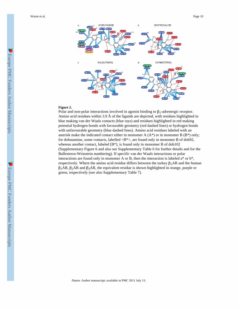
Figure 2.Polar and non-polar interactions involved in agonist binding to β1-adrenergic receptor.Amino acid residues within 3.9 Å of the ligands are depicted, with residues highlighted inblue making van der Waals contacts (blue rays) and residues highlighted in red makingpotential hydrogen bonds with favourable geometry (red dashed lines) or hydrogen bondswith unfavourable geometry (blue dashed lines). Amino acid residues labeled with anasterisk make the indicated contact either in monomer A (A*) or in monomer B (B*) only;for dobutamine, some contacts, labelled <B*>, are found only in monomer B of dob92,whereas another contact, labeled [B*], is found only in monomer B of dob102(Supplementary Figure 6 and also see Supplementary Table 6 for further details and for theBallesteros-Weinstein numbering). If specific van der Waals interactions or polarinteractions are found only in monomer A or B, then the interaction is labeled a* or b*,respectively. Where the amino acid residue differs between the turkey β1AR and the humanβ1AR, β2AR and β3AR, the equivalent residue is shown highlighted in orange, purple orgreen, respectively (see also Supplementary Table 7).
Warne et al. Page 10
Nature. Author manuscript; available in PMC 2011 July 13.
Europe PM
C Funders A
uthor Manuscripts
Europe PM
C Funders A
uthor Manuscripts

Figure 3.Comparison of the ligand binding pockets of the β1 and β2 adrenergic receptors. The ligandbinding pockets are shown as viewed from the extracellular surface with EL2 removed forclarity (same colour scheme as in Fig. 1). (a) β2AR with the antagonist carazolol bound(PDB code 2RH1); (b) β1AR with the antagonist cyanopindolol bound (PDB code 2VT4);(c) β1AR with the agonist isoprenaline bound.
Warne et al. Page 11
Nature. Author manuscript; available in PMC 2011 July 13.
Europe PM
C Funders A
uthor Manuscripts
Europe PM
C Funders A
uthor Manuscripts
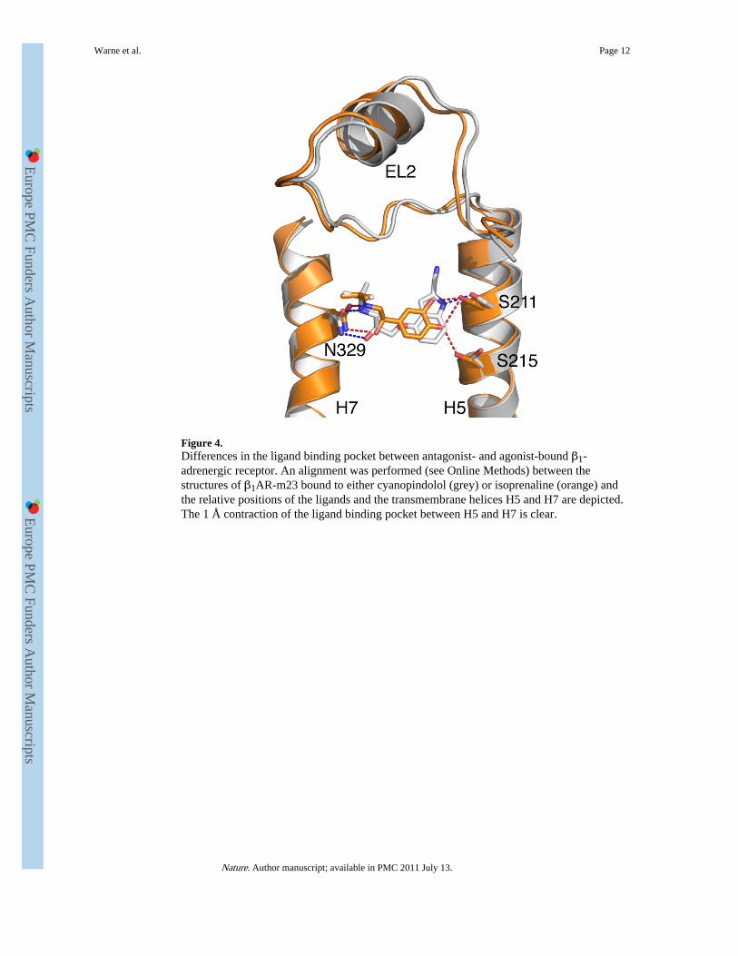
Figure 4.Differences in the ligand binding pocket between antagonist- and agonist-bound β1-adrenergic receptor. An alignment was performed (see Online Methods) between thestructures of β1AR-m23 bound to either cyanopindolol (grey) or isoprenaline (orange) andthe relative positions of the ligands and the transmembrane helices H5 and H7 are depicted.The 1 Å contraction of the ligand binding pocket between H5 and H7 is clear.
Warne et al. Page 12
Nature. Author manuscript; available in PMC 2011 July 13.
Europe PM
C Funders A
uthor Manuscripts
Europe PM
C Funders A
uthor Manuscripts
Related Documents


![Newer Medications for Lower Urinary Tract Symptoms ... · Detrol LA] Tolterodine. b [Detrol] Solifenacin. b [Vesicare] Trospium [Sanctura] Beta-3 adrenergic agonist - Increases bladder](https://static.cupdf.com/doc/110x72/5e853b72b6e81334644b600e/newer-medications-for-lower-urinary-tract-symptoms-detrol-la-tolterodine-b.jpg)

