TISSUE-SPECIFIC STEM CELLS Microgel Iron Oxide Nanoparticles for Tracking Human Fetal Mesenchymal Stem Cells Through Magnetic Resonance Imaging EDDY S.M. LEE, a JERRY CHAN, b BORYS SHUTER, a LAY GEOK TAN, b MARK S.K. CHONG, b DURRGAH L. RAMACHANDRA, b GAVIN S. DAWE, c JUN DING, d SWEE HIN TEOH, e OLIVIER BEUF, f ANDRE BRIGUET, f KAM CHIU TAM, g MAHESH CHOOLANI, b SHIH-CHANG WANG a a Department of Diagnostic Radiology, Yong Loo Lin School of Medicine, National University of Singapore, Republic of Singapore; b Experimental Fetal Medicine Group, Department of Obstetrics and Gynaecology, Yong Loo Lin School of Medicine, National University of Singapore, Republic of Singapore; c Department of Pharmacology, Yong Loo Lin School of Medicine, National University of Singapore, Republic of Singapore; d Department of Materials Science and Engineering, Faculty of Engineering, National University of Singapore, Republic of Singapore; e Department of Mechanical Engineering, Faculty of Engineering, National University of Singapore, Republic of Singapore; f Laboratoire de RMN, Universite ´ de Lyon, CREATIS-LRMN, CNRS UMR 5220; Inserm U630; INSA-Lyon; Universite ´ Lyon 1, Villeurbanne, France; g Chemical Engineering Department, University of Waterloo, Waterloo, Canada Key Words. Clinical translation • Tissue-specific stem cells • In vivo tracking • Mesenchymal stem cells • Microarray • Xenogeneic stem cell transplantation ABSTRACT Stem cell transplantation for regenerative medicine has made significant progress in various injury models, with the development of modalities to track stem cell fate and migration post-transplantation being currently pursued rigorously. Magnetic resonance imaging (MRI) allows se- rial high-resolution in vivo detection of transplanted stem cells labeled with iron oxide particles, but has been ham- pered by low labeling efficiencies. Here, we describe the use of microgel iron oxide (MGIO) particles of diameters spanning 100-750 nm for labeling human fetal mesenchy- mal stem cells (hfMSCs) for MRI tracking. We found that MGIO particle uptake by hfMSCs was size dependent, with 600-nm MGIO (M600) particles demonstrating three- to sixfold higher iron loading than the clinical particle fer- ucarbotran (33-263 versus 9.6-42.0 pg iron/hfMSC; p < .001). Cell labeling with either M600 particles or ferucar- botran did not affect either cellular proliferation or trili- neage differentiation into osteoblasts, adipocytes, and chondrocytes, despite differences in gene expression on a genome-wide microarray analysis. Cell tracking in a rat photothrombotic stroke model using a clinical 1.5-T MRI scanner demonstrated the migration of labeled hfMSCs from the contralateral cortex to the stroke injury, with M600 particles achieving a five- to sevenfold higher sensi- tivity for MRI detection than ferucarbotran (p < .05). However, model-related cellular necrosis and acute inflam- mation limited the survival of hfMSCs beyond 5-12 days. The use of M600 particles allowed high detection sensitiv- ity with low cellular toxicity to be achieved through a sim- ple incubation protocol, and may thus be useful for cellular tracking using standard clinical MRI scanners. STEM CELLS 2009;27:1921–1931 Disclosure of potential conflicts of interest is found at the end of this article. Author contributions: E.S.M.L.: conception and design, collection and assembly of data, data analysis and interpretation, manuscript writing; J.C.: conception and design, financial support, administrative support, assembly of data, provision of study material or patients, data analysis and interpretation, manuscript writing; B.S.: data analysis and interpretation, manuscript writing; L.G.T.: administrative support; M.S.K.C.: data analysis and interpretation; D.L.R.: collection of data; G.S.D.: data analysis and interpretation; J.D.: data analysis and interpretation; S.H.T.: administrative support; O.B.: manuscript writing; A.B.: manuscript writing; K.C.T.: provision of study material; M.C.: conception and design, data analysis and interpretation, financial support, administrative support, provision of study material or patients; S.-C.W.: conception and design, financial support, administrative support, data analysis and interpretation, manuscript writing, final approval of manuscript. Correspondence: Eddy S.M. Lee, B.Eng, Experimental Fetal Medicine Group, Department of Obstetrics and Gynaecology, National University of Singapore, 5 Lower Kent Ridge Road, Singapore 119074. Telephone: 65-67722672; Fax: 65-67794753; e-mail: [email protected]; Jerry Chan, M.R.C.O.G., PhD., Experimental Fetal Medicine Group, Department of Obstetrics and Gynaecology, National University of Singapore, 5 Lower Kent Ridge Road, Singapore 119074. Telephone: 65-67722672; Fax: 65-67794753; e-mail: [email protected] Received February 4, 2009; accepted for publication April 19, 2009; first published online in STEM CELLS EXPRESS April 30, 2009. V C AlphaMed Press 1066-5099/2009/$30.00/0 doi: 10.1002/stem.112 STEM CELLS 2009;27:1921–1931 www.StemCells.com

Welcome message from author
This document is posted to help you gain knowledge. Please leave a comment to let me know what you think about it! Share it to your friends and learn new things together.
Transcript

TISSUE-SPECIFIC STEM CELLS
Microgel Iron Oxide Nanoparticles for Tracking Human Fetal
Mesenchymal Stem Cells Through Magnetic Resonance Imaging
EDDY S.M. LEE,aJERRY CHAN,
bBORYS SHUTER,
aLAY GEOK TAN,
bMARK S.K. CHONG,
b
DURRGAH L. RAMACHANDRA,b GAVIN S. DAWE,c JUN DING,d SWEE HIN TEOH,e OLIVIER BEUF,f
ANDRE BRIGUET,f KAM CHIU TAM,g MAHESH CHOOLANI,b SHIH-CHANG WANGa
aDepartment of Diagnostic Radiology, Yong Loo Lin School of Medicine, National University of Singapore,
Republic of Singapore; bExperimental Fetal Medicine Group, Department of Obstetrics and Gynaecology, Yong
Loo Lin School of Medicine, National University of Singapore, Republic of Singapore; cDepartment of
Pharmacology, Yong Loo Lin School of Medicine, National University of Singapore, Republic of Singapore;dDepartment of Materials Science and Engineering, Faculty of Engineering, National University of Singapore,
Republic of Singapore; eDepartment of Mechanical Engineering, Faculty of Engineering, National University of
Singapore, Republic of Singapore; fLaboratoire de RMN, Universite de Lyon, CREATIS-LRMN, CNRS UMR
5220; Inserm U630; INSA-Lyon; Universite Lyon 1, Villeurbanne, France; gChemical Engineering Department,
University of Waterloo, Waterloo, Canada
Key Words. Clinical translation • Tissue-specific stem cells • In vivo tracking • Mesenchymal stem cells • Microarray • Xenogeneic stem
cell transplantation
ABSTRACT
Stem cell transplantation for regenerative medicine hasmade significant progress in various injury models, with
the development of modalities to track stem cell fate andmigration post-transplantation being currently pursued
rigorously. Magnetic resonance imaging (MRI) allows se-rial high-resolution in vivo detection of transplanted stemcells labeled with iron oxide particles, but has been ham-
pered by low labeling efficiencies. Here, we describe theuse of microgel iron oxide (MGIO) particles of diameters
spanning 100-750 nm for labeling human fetal mesenchy-mal stem cells (hfMSCs) for MRI tracking. We found thatMGIO particle uptake by hfMSCs was size dependent,
with 600-nm MGIO (M600) particles demonstrating three-to sixfold higher iron loading than the clinical particle fer-ucarbotran (33-263 versus 9.6-42.0 pg iron/hfMSC; p <.001). Cell labeling with either M600 particles or ferucar-
botran did not affect either cellular proliferation or trili-neage differentiation into osteoblasts, adipocytes, and
chondrocytes, despite differences in gene expression on agenome-wide microarray analysis. Cell tracking in a rat
photothrombotic stroke model using a clinical 1.5-T MRIscanner demonstrated the migration of labeled hfMSCsfrom the contralateral cortex to the stroke injury, with
M600 particles achieving a five- to sevenfold higher sensi-tivity for MRI detection than ferucarbotran (p < .05).
However, model-related cellular necrosis and acute inflam-mation limited the survival of hfMSCs beyond 5-12 days.The use of M600 particles allowed high detection sensitiv-
ity with low cellular toxicity to be achieved through a sim-ple incubation protocol, and may thus be useful for
cellular tracking using standard clinical MRI scanners.STEM CELLS 2009;27:1921–1931
Disclosure of potential conflicts of interest is found at the end of this article.
Author contributions: E.S.M.L.: conception and design, collection and assembly of data, data analysis and interpretation, manuscriptwriting; J.C.: conception and design, financial support, administrative support, assembly of data, provision of study material or patients,data analysis and interpretation, manuscript writing; B.S.: data analysis and interpretation, manuscript writing; L.G.T.: administrativesupport; M.S.K.C.: data analysis and interpretation; D.L.R.: collection of data; G.S.D.: data analysis and interpretation; J.D.: dataanalysis and interpretation; S.H.T.: administrative support; O.B.: manuscript writing; A.B.: manuscript writing; K.C.T.: provision ofstudy material; M.C.: conception and design, data analysis and interpretation, financial support, administrative support, provision ofstudy material or patients; S.-C.W.: conception and design, financial support, administrative support, data analysis and interpretation,manuscript writing, final approval of manuscript.
Correspondence: Eddy S.M. Lee, B.Eng, Experimental Fetal Medicine Group, Department of Obstetrics and Gynaecology, NationalUniversity of Singapore, 5 Lower Kent Ridge Road, Singapore 119074. Telephone: 65-67722672; Fax: 65-67794753; e-mail:[email protected]; Jerry Chan, M.R.C.O.G., PhD., Experimental Fetal Medicine Group, Department of Obstetrics andGynaecology, National University of Singapore, 5 Lower Kent Ridge Road, Singapore 119074. Telephone: 65-67722672; Fax:65-67794753; e-mail: [email protected] Received February 4, 2009; accepted for publication April 19, 2009; first published onlinein STEM CELLS EXPRESS April 30, 2009. VC AlphaMed Press 1066-5099/2009/$30.00/0 doi: 10.1002/stem.112
STEM CELLS 2009;27:1921–1931 www.StemCells.com

INTRODUCTION
Stem cell transplantation is a rapidly emerging field of regen-erative medicine undergoing intensive investigation. Severalclinical trials are already in progress for the treatment of vari-ous diseases, such as ischemic stroke [1], skeletal dysplasia[2], spinal cord injury [3], and myocardial infarction [4].Transplanted stem cells have been shown to home and engraft[5, 6] into areas of tissue injury, where they replace the defec-tive cell types and/or produce benefit through paracrine mech-anisms [7].
Development of this field requires identification and track-ing of transplanted cells to monitor their survival and local-ization. This has traditionally been achieved through longitu-dinal histological analyses, using techniques such as gendermismatches, fluorescent proteins, enzymes (LacZ), and thymi-dine analogs [8] (e.g., 5-bromo-20-deoxyuridine). Thesemodalities, however, require serial sacrifice of animals ormultiple biopsies, and are beset by problems of interanimalvariability. Moreover, these approaches would not be appro-priate in clinical studies, and hence there is an urgent needfor the development of noninvasive in vivo imaging modal-ities [9].
Magnetic resonance imaging (MRI) is a sensitive three-dimensional imaging method that provides high-resolutionimages at depth in opaque living animals and patients, with-out ionizing radiation. Serial tracking of transplanted stemcells is feasible if the cells are labeled with MRI-visible par-ticles. Currently available particles span a wide range ofdiameters (20 nm to 5.8 lm). Dextran-coated superparamag-netic iron oxide (SPIO) nanoparticles in clinical use, such asferucarbotran (ResovistVR ; Bayer Schering Pharma, Berlin,Germany, http://www.bayerhealthcare.com) [10] and ferumox-ide (Feridex I.V., AMAG pharmaceuticals, Lexington, MA,http://www.amagpharma.com) [11], as well as larger (0.9-5.8lm) polystyrene-based particles [12, 13], have been investi-gated as cell labels for MRI tracking. Typically, cells areincubated with the particles for up to a few days, allowinguptake through endocytosis prior to transplantation. Althoughthese particles have been successfully used to image labeledcells clinically and in animal models, the low uptake of par-ticles in nonphagocytotic cell types has resulted in limitedMRI sensitivity and hampered imaging. The development ofMR contrast particles that can be taken up efficiently by non-phagocytotic cell types is therefore a high priority in thisfield.
A variety of techniques to increase the cellular uptake ofiron oxide particles have been described, including reversibleelectroporation [14] and the addition of transfection agents(reviewed elsewhere by Bulte and Kraitchman [15]) such aspoly-L-lysine [16, 17], activated-dendrimer [17], and prota-mine sulfate [18]. All these approaches require careful optimi-zation to avoid possible cytotoxicity [17]. Particle conjugationto the HIV tat peptide increases uptake but results in its local-ization within the nucleus rather than in endosomes, and maythus interfere with nuclear function [19]. The size of the par-ticles appears to influence iron loading of cells, with bothultrasmall SPIO particles (20 nm) [20] and the larger experi-mental polystyrene particles (0.9 lm) [12] resulting in lowercellular loading than obtainable with clinical SPIO particles(62 nm) [21]. However, particles of similar composition inthe size range of 100-750 nm in diameter have not been pre-viously investigated for this application.
Among various stem cell types, mesenchymal stem cells(MSCs) have shown the ability to home and migrate towardinjury sites [22], such as cerebral infarcts, where they can
induce angiogenesis or functional recovery [23, 24]. Humanfetal MSCs (hfMSCs) are primitive MSCs, with greater prolif-erative and differentiation capacities than their adult counter-parts [25–28], and have been under investigation for variouscellular therapy applications [25, 28–30]
In this study, we investigated the use of microgel iron ox-ide (MGIO) nanoparticles with a range in diameter of 100-750 nm as MRI contrast particles for cellular labeling ofhfMSCs. We showed that MGIO particles of 600 nm diameter(M600) were taken up more avidly by hfMSCs than ferucar-botran, without affecting the stem cell functions of self-renewal and differentiation. M600 labeling allowed betterdetection in a rat photothrombotic stroke model on MRI thanwith the use of ferucarbotran. In the growing field of cellulartransplantation applications, the availability of highly effi-cient, low toxicity MRI contrast particles should greatly facili-tate cellular tracking and monitoring of transplanted cells.
METHODS AND MATERIALS
Ethics and Samples
All human tissue collection was approved by the domain spe-cific review board of National University Hospital and was incompliance with international guidelines regarding the use offetal tissue for research [31]. In all cases, patients undergoingclinically indicated termination of pregnancies gave separatewritten consent for the use of the collected tissue. FemaleWistar rats (200-250 g) were acquired from the Centre forAnimal Resources (Singapore) and all procedures wereapproved by the Institutional Animal Care and Use Commit-tee at National University of Singapore.
Synthesis of MGIO Particles
The development of MGIO particles started with the conden-sation polymerization of ethyl acrylate, methacrylic acid, anddi-allyl phthalate to form a nonmagnetic precursor microgel(PMG), as previously described elsewhere [32]. PMG wasmagnetized by the coprecipitation of iron salt with ammoniato form primary iron oxide cores within PMG in situ. Afterpurification by density and magnetic field strength, MGIOparticles were obtained. The polymerization and coprecipita-tion conditions were altered to produce MGIO particles withdiffering diameters of 100-750 nm. MGIO was air-dried on a200-mesh copper grid and imaged by transmission electronmicroscopy (TEM) (JEOL JEM-100CX microscope; JEOL,Tokyo, Japan, http://www.jeol.com). The hydrated diameterof MGIO particles in suspension was determined by dynamiclight scattering (n ¼ 3–6, Brookhaven BI-200SM system;Brookhaven Instruments Corporation, Holtsville, NY, http://www.bic.com), using a power-adjustable 488-nm argon lasersource.
hfMSC Isolation and Differentiation
hfMSCs were isolated from human fetal bone marrow as pre-viously described (8-12 weeks of gestation, n ¼ 2) [26].Briefly, bone marrow cells were flushed from the femursusing a 22-gauge needle into a culture medium (CM10) con-sisting of 10% fetal bovine serum (FBS) (Sigma-Aldrich, St.Louis, http://www.sigmaaldrich.com) in Dulbecco’s modifiedEagle’s medium (DMEM) (Sigma-Aldrich, Singapore) supple-mented with 2 mM L-glutamine, 50 IU/ml penicillin, and 50mg/ml streptomycin (Invitrogen, Carlsbad, CA, http://www.invitrogen.com). Single-cell suspensions were plated in 100-mm dishes at 105 nucleated cells per ml and cultured inCM10 at 37�C in 5% CO2. After 3 days, nonadherent
1922 MRI Tracking of hfMSCs

cells were removed and the medium was replaced. Adherentcell colonies were detached with 0.25% trypsin EDTA (StemCell Technologies, Vancouver, BC, Canada, http://www.stemcell.com), expanded, cultured to subconfluence, trypsi-nized, and stored in liquid nitrogen.
hfMSCs were characterized by immunocytochemistry forCD14, CD34, CD45, CD31, von Willebrand factor (vWF),CD105 (SH2), CD73 (SH3, SH4) (Abcam, Cambridge, MA,http://www.abcam.com), vimentin, laminin, CD29 (Chemicon,Temecula, CA, http://www.chemicon.com), CD44 (BD Bio-sciences, San Diego, http://www.bdbiosciences.com), CD106,CD90 (Chemicon), human leukocyte antigen (HLA) I, HLA II(Dako, Carpinteria, CA, http://www.dakousa.com), Oct-4, andNanog (Abcam) and flow cytometry was used to screen forStro-1 (Chemicon) as previously described elsewhere [33].Cells at passages 5-6 were used in all experiments. Osteo-genic, adipogenic, and chondrogenic differentiation and theirrespective assays were performed as previously describedelsewhere [28, 33].
Cellular Labeling Protocol
Prior to labeling, 5 � 105 hfMSCs were cultured for 24 hoursat 2 � 103 cells/cm2 in CM10. The cells were labeled withMGIO or ferucarbotran by incubation at 0.025-0.2 mg iron/mlwithin a labeling culture medium (CM2: 2% FBS in DMEMsupplemented with 2 mM L-glutamine, 50 IU/ml penicillin,and 50 mg/ml streptomycin) at 37�C in 5% CO2. After 24hours, adherent cells were repeatedly washed with freshchanges of phosphate-buffered saline (PBS) until the PBSappeared free of particles under light microscopy to removeunattached particles. The cells were then trypsinized, resus-pended in CM10, and layered on Ficoll-paque PLUS (Amer-sham Biosciences, Piscataway, NJ, http://www.gelifesciences.com) for density centrifugation at 100g for 30 minutes toremove loosely attached, extracellular particles. LabeledhfMSCs were recovered at the interface between CM10 andFicoll-paque PLUS and washed with PBS by centrifugation toremove the remaining Ficoll-paque. Mock-labeled cells wereused as controls where hfMSCs were subjected to the aboveprocedures but without incubation with any particles.
Cellular TEM and Iron Quantification
To quantify intracellular iron mass, cells were counted, lysedin 0.2 ml of aqua regia, reconstituted to 5 ml with distilledwater, and analyzed for iron using inductively coupled plasmaoptical emission spectroscopy (ICP-OES) (Optima 5300V,PerkinElmer, Waltham, MA, http://www.perkinelmer.com.The iron mass of the mock-labeled control was below the reli-able ICP-OES detectability of 0.01 ppm (<0.1 pg/cell). TEM(Leica, Heerbrugg, Switzerland, http://www.leica.com) for la-beled cells was performed after the cells were fixed, dehy-drated, resin embedded, cut in 100-nm sections, and stainedwith lead citrate on a copper grid.
Genome-Wide Microarray Expression Analysis
Total RNA was extracted from M600-, ferucarbotran-, andmock-labeled hfMSCs in biological triplicate, using theRNeasy kit (Qiagen, Valencia, CA, http://www1.qiagen.com)in accordance with the manufacturer’s protocol. Ten micro-grams of total RNA was used to generate labeled cRNA andhybridized to Human Genome U133 Plus 2.0 arrays (Affyme-trix, Santa Clara, CA, http://www.affymetrix.com). Differ-entially expressed genes were identified with GeneSpringGX 7.3.1 (Agilent Technologies, Palo Alto, CA, http://www.agilent.com). The associated gene ontology terms wereenriched with the web-based functional annotation software
FatiGO [35]. Further details of this process are described insupporting information data.
In Vivo Imaging
Focal Photothrombotic Stroke Induction. Female Wistarrats were anesthetized with 7.5 mg/100 g body weight (BW)ketamine (Parnell Laboratories, Alexandria, Australia, http://www.parnell.biz) and 1 mg/100 g BW xylazine (Troy Labora-tories, Smithfield, Australia, http://www.troylab.com.au) i.p.and mounted in a stereotactic frame. A 7.5-mg/ml solution ofRose Bengal in saline was filtered (0.22 lm) and injected viaa tail vein cannula at 1 mg/100 g BW at a rate of 0.2 ml/mi-nute. Simultaneously, the skull, �2 mm anteroposterior (AP)and �3 mm mediolateral (ML) from the bregma, was exposedto 60 W of blue-green passband-filtered (BG39; Schott, Dur-yea, PA, http://www.us.schott.com) white light from a halo-gen lamp via a fiber optic waveguide for 10 minutes to gener-ate a photochemical cortical stroke. The spot size wasadjusted to a diameter of approximately 3 mm with an opticalaperture.
Transplantation of hfMSCs. Two days after induction ofphotothrombotic injury to the cerebral cortex (day 0), we xen-otransplanted (a) 2 � 104 M600-labeled hfMSCs (M600-hfMSCs, n ¼ 9) or (b) 2 � 104 ferucarbotran-labeled hfMSCs(ferucarbotran-hfMSCs, n ¼ 4) into the contralateral cerebralcortex. A third group (c) had 2 � 106 M600-labeled hfMSCsinfused i.v. through the tail vein (n ¼ 2) and a control group(d) of animals without cortical injury was transplanted with 2� 104 M600-hfMSCs (n ¼ 3). A further two control groupsconsisted of animals with contralateral stroke injury eitherwithout cellular transplantation (n ¼ 3) or transplanted withmock-labeled hfMSCs (n ¼ 1).
A 33-gauge needle was used for the intraparenchymalcortical injection of hfMSCs. Passage of hfMSCs through a33-gauge needle did not affect cellular viability, as demon-strated through trypan blue exclusion tests, nor did it affectcolony-forming unit-erythroid ability (data not shown), sug-gesting its suitability for transplantation purposes.
For cortical injection contralateral to the stroke site, aburr hole (1 mm) was made on the right side of the skull toexpose the dura overlying the cortex. hfMSCs were resus-pended by repeated pipetting before loading into Hamiltonsyringes just before cellular injection to avoid aggregation ofthe cells. hfMSCs (2 � 104) were injected slowly in 5 ll ofPBS over a 10-minute period using a 33-gauge Hamilton sy-ringe into the contralateral hemisphere �2 mm AP, 3 mmML, and 3.5 mm dorsoventral from the bregma. For i.v. deliv-ery, 2 � 106 cells in 0.5 ml PBS were injected into the lateraltail vein. Immunosuppression with i.p. cyclosporin (20 mg/kgBW on alternate days, Sandimmune Injection; Novartis Inter-national, Basel, Switzerland, http://www.novartis.com) wasinitiated at the time of cellular transplantation and maintainedthroughout the experimental duration.
MRI. In vivo MRI was performed on a 1.5-T whole-bodyclinical MR scanner with a clinical wrist radiofrequency coil(General Electric, Waukesha, WI, http://www.gehealthcare.-com). Anesthesia was induced with 4% isoflurane and main-tained with 1.5%–2.5% isoflurane in 100% oxygen deliveredthrough a cone mask. In vivo transverse images were obtainedusing turbo spin echo (TSE)—field of view (FOV), 5 cm; ma-trix, 192 � 192 and zero-filled to 512 � 512; voxel dimen-sions, 260 lm � 260 lm � 1.5 mm; repetition time (TR)/echo time (TE)/echo train length (ETL)/flip angle (FA)/num-ber of excitations (NEX), 2 seconds/81 ms/16/90�/12;
Lee, Chan, Shuter et al. 1923
www.StemCells.com

acquisition time, approximately 9 minutes—and gradient echo(GRE)—FOV, 5 cm; matrix: 160 � 160 and zero-filled to512 � 512; voxel dimensions, 313 lm � 313 lm � 1.5 mm;TR/TE/FA/NEX, 280 ms/20 ms/20�/15; acquisition time,approximately 9 minutes—pulse sequences as 10 two-dimen-sional slices.
Analysis of Hypointense Voxels on MR Images. A voxelwas considered hypointense if its signal intensity was belowthe signal intensity threshold (SH). Using Rose’s criterion, theSH of a day 5 or day 12 image was determined with respectto a reference signal (SR) and the image standard deviation(SDR), as:
SH ¼ SR � k � SDR
The SR was determined from the corresponding day �1GRE image as the mean signal intensity of an 8 mm2 regionof interest positioned at the cortical region contralateral to thestroke. The SDR was calculated from the standard deviationof MR signal from the air, reduced by a factor of 0.655 toaccount for the non-Gaussian noise of magnitude images. Thecontrast to noise ratio, k, was assumed to be 5.
Histology
Animals (n ¼ 3) were euthanized, and intracardiac perfusionwith 250 ml of 2% 2,3,5-triphenyltetrazolium (TTC) (Sigma-Aldrich) 24 hours after the stroke induction procedure wasdone for confirmation of thrombotic stroke. Following recov-ery of the brain, 1-mm sections were fixed, laid onto slides,and visualized under light microscopy.
For histological analysis of transplanted cells at varioustime points, the rats were euthanized and perfused with 4%paraformaldehyde, and the brains were paraffin embedded and10-lm coronal sections were laid onto polylysine-coatedslides for staining.
Prussian blue (PB) iron staining was performed by incu-bating with freshly prepared 5% potassium ferrocyanide in5% HCl 1:1 for 30 minutes and washing with deionizedwater. For 3,30-diaminobenzidine (DAB) enhancement of PBstaining [36], sections were incubated in 3% H2O2 for 3minutes pre-PB staining, PB stained, and incubated in 0.05%DAB in PBS for 5 minutes followed by another incubation in0.05% DAB in PBS and 0.03% H2O2 for 3 minutes.
Immunohistochemical staining was done after deparaffini-zation, rehydration, and an antigen retrieval step performed at95�C for 30 minutes (H-3300; Vector Laboratories, Peterbor-ough, U.K., http://www.vectorlabs.com). Sections wereblocked with 5% goat and fetal calf serum, and the nuclearenvelope was permeabilized with 0.2% Triton X-100 for 1hour before being incubated overnight at 4�C with primaryantibodies of mouse anti-rat ED1 1:100 (MCA341R; AbDSerotec, Oxford, U.K., http://www.abdserotec.com) and rabbitanti-human vimentin 1:100 (ab16700; Abcam, Cambridge,U.K., http://www.abcam.com). After washing of the slideswith PBS, incubation with secondary antibodies, either goatanti-rabbit Alexa Fluor 488 or goat anti-mouse Alexa Fluor594 at 1:100, for 30 minutes was performed. Sections werethen mounted with 40,6-diamidino-2-phenylindole or propi-dium iodide for nuclear visualization (both from VectorLaboratories).
Statistics
Parametric data are shown as mean � standard error of themean. Iron loading at various labeling concentrations was an-alyzed using two-way analysis of variance with post hoc Bon-
ferroni correction, or with a t-test. A p-value < .05 was con-sidered indicative of a statistically significant result.
RESULTS
Generation and Characterization of MGIO
Nonmagnetic PMG was synthesized and followed by magnet-ization by alkaline coprecipitation of iron oxide primary par-ticles within PMG. Resulting synthesized MGIO particlesspanned a ninefold range of hydrated diameters (87-766 nm)(Fig. 1A) and consisted of multiple iron oxide primary nano-particles (� 5 nm) held within soft, water-swellable polymericcages as seen on TEM(Fig. 1B).
Isolation and Characterization of hfMSCs
hfMSCs appeared as plastic-adherent spindle-shaped cells inculture. They had an immunophenotype that was negative forthe hemopoietic and endothelial markers CD14, CD34, CD45,CD31, vWF, and HLA II and positive for the mesenchymalmarkers CD105 and CD73, the intracellular markers vimentinand laminin, the cell adhesion molecules CD29, CD44,CD106, and CD90, and HLA I, as previously reported (datanot shown) [28, 29]. Under permissive induction media, theyunderwent osteogenic, adipogenic, and chondrogenic differen-tiation (data not shown) confirming their bona fide identity asMSCs [36].
Uptake of MGIO Particles by hfMSCs
In order to test the utility of these novel particles, we labeledprimary hfMSCs with MGIO particles of varying sizes andferucarbotran. By simple incubation of hfMSCs with MGIOparticles or ferucarbotran (0.05 mg iron/ml) over 24 hours,we observed the incorporation of nanoparticles into the cyto-plasm of hfMSCs (Fig. 2A) and localization to endosomalstructures (Fig. 2B). We found 97.3% � 0.9% M600-labeledcells staining positive for PB (range, 21-38 per low poweredfield [LPF]; total cells counted, 212) and 98.2% � 1.1%(range, 20-35 per LPF; total cells counted, 216) ferucarbo-tran-labeled cells staining positive for PB. Interestingly, weobserved a distinct size-dependent uptake of MGIO particles.Using an initial iron concentration of 0.05 mg/ml, we consis-tently observed threefold greater cellular iron loading withM600 particles (33.3 � 4.0 pg/cell, n ¼ 9) than with ferucar-botran (9.6 � 1.3 pg/cell, n ¼ 9; p ¼ .0003), and significantlyhigher iron loading for M600 particles than for MGIO par-ticles of other sizes (p < .001) (Fig. 2C). This difference iniron uptake between M600 particles and ferucarbotran waseven more marked on incubation with increasing iron concen-trations. Up to sixfold greater cellular iron loading wasachieved at an incubation concentration of 0.2 mg iron/ml(Fig. 2D) (263 � 27 pg/cell, n ¼ 3, versus 41 � 6 pg/cell;p < .001).
Upon passaging the labeled cells in culture, we found thatthe half-life of the cellular iron content was one populationdoubling, as can be expected when the iron labels have beenfully passed onto the two daughter cells. We also investigatedthe effects of labeling hfMSCs using MGIO particles of 400-750 nm in diameter and ferucarbotran on their stem cell prop-erties. Compared with mock-labeled cells, labeling of hfMSCswith either MGIO particles or ferucarbotran did not affect cellmorphology, doubling time, viability (>95% throughout), ortrilineage differentiation into osteoblasts, adipocytes, andchondrocytes (Fig. 3).
In order to broaden our understanding of the effects ofiron loading on hfMSCs, we performed a global gene
1924 MRI Tracking of hfMSCs

expression analysis using a genome-wide microarray (Affy-metrix HG U-133 Plus 2.0) approach. We found 114 differen-tially regulated genes upregulated at least twofold and 102genes downregulated at least twofold after labeling withM600 particles (Fig. 2E). These genes are largely associatedwith upregulation of metal and cation binding and downregu-lation of the cell cycle (supporting information data, FatiGOanalysis [35]). Labeling of hfMSCs with ferucarbotranresulted in a smaller set of differentially regulated genes, with32 genes upregulated and 29 genes downregulated. The up-
regulated genes after ferucarbotran labeling are similarly asso-ciated with ion binding, and the downregulated genes areprincipally involved in prostanoid metabolic processes (sup-porting information data).
MR Tracking of Labeled hfMSCs in aFocal Stroke Model
Next, we carried out a cellular transplantation experiment totest the sensitivity and efficacy of MGIO particles as a cellu-lar label for MRI tracking in a well-established rat photo-thrombotic stroke injury model [37]. One day after strokeinduction, the photothrombotic injury was visualized on MRIthrough a TSE (Fig. 4, yellow arrowheads) sequence as awedge-shaped focal hyperintense region, involving the cortexpredominantly, and was confirmed by the absence of meta-bolic activity through TTC staining (supporting informationFig. 1A). In the M600-hfMSC group, intracerebrally trans-planted cells (Fig. 4A, green arrowhead) appeared as hypoin-tense regions on GRE images of the transplanted side of thebrain. By day 5, a small area of hypointensity could be seenaround the peripheral region of the stroke (Fig. 4A, redarrowhead), suggesting the migration of M600-hfMSCs to thestroke site. On day 12, the area of hypointensity around theinjury could be clearly seen encompassing the periphery ofthe stroke (Fig. 4A, red arrowheads). Animals injected withferucarbotran-hfMSCs showed a smaller area of hypointensityat the stroke site (Fig. 4B). i.v. delivery of M600-hfMSCswas well tolerated and resulted in the appearance of hypoin-tensity by day 5 at the site of the stroke, which increasedfrom day 5 to day 12 on GRE images (Fig. 4C). MRI of
Figure 1. Properties of ferucarbotran and microgel iron oxide(MGIO) particles. (A): Measurements of hydrated diameter dH. (B):Transmission electron microscopy of air-dried 600-nm MGIO par-ticles shows iron oxide primary particles of approximately 5 nm in di-ameter (electron dense) held together by a polymeric matrix (lessdense).
Figure 2. Properties of labelled hfMSC. (A): 600-nm microgel iron oxide (M600) particles localized to human fetal mesenchymal stem cell(hfMSC) cytoplasm (Prussian blue stain) where they were found within endosomes (B). (C, D) M600 particles demonstrated threefold higher cel-lular uptake in hfMSCs than other microgel iron oxide particles and ferucarbotran (0.05 mg iron per ml), with up to sixfold higher efficiency athigher labeling concentrations (0.2 mg iron per ml). (E): Microarray analysis of hfMSCs revealed 114 and 102 differentially regulated genes thatwere upregulated and downregulated at least twofold, respectively, after M600 particle labeling (lanes 4-6) compared with mock-labeled hfMSCs(lanes 1-3). Corresponding changes in ferucarbotran-labeled cells (Fc) are shown in lanes 7-9.
Lee, Chan, Shuter et al. 1925
www.StemCells.com

control animals transplanted with M600-hfMSCs but withouta contralateral stroke demonstrated no development of hypo-intensity in the contralateral cerebral cortex (Fig. 4D).
Histological Analysis of Transplanted Animals
We harvested animals in the M600-hfMSC group to correlatethe MRI findings with histologic and immunostaining evi-
dence of cell and label fate. On day 1, PBþ cells (staining foriron) could be seen only at the injection site (Fig. 5A, bluestain) and not at the stroke site (Fig. 5B, 5C). Double immu-nostaining of the injection site revealed hfMSCs as humanvimentin-positive cells among an infiltration of host ED1þ
macrophages (Fig. 5D–5F), with a discernible shift of cellsfrom the injected site toward the stroke area at day 1. Therewere only ED1þ host macrophages, with no vimentin-positivecells at the stroke site at day 1 (Fig. 5G, 5H).
Figure 3. Labeling of human fetal mesenchymal stem cells with 600-nm microgel iron oxide particles or ferucarbotran did not alter their spin-dle-shaped morphology (CM10) or their capacity to differentiate into osteoblasts (black extracellular crystals, von Kossa staining), adipocytes (oilred O staining), or chondrocytes (Safranin O [red] and Alcian Blue [blue] staining in micromass pellet cultures).
Figure 4. In vivo imaging with TSE (day �1) and GRE (day �1 through day 12) sequences. A focal cortical stroke (yellow arrows) wasinduced at day �2 and cellular transplantation took place on day 0 by contralateral intracerebral (green arrows) or systemic injection (i.v.). (A):An area of hypointensity appeared in the area of the stroke (red arrows) noticeable at day 5, and increased over time to day 12 in M600-hfMSC-injected animals. (B): A similar observation was made in ferucarbotran-hfMSC-injected animals, albeit with a smaller area of hypointensity seen.(C): Animals injected with M600-hfMSCs i.v. showed the appearance of hypointensity in the stroke region by day 5, which increased over timeto day 12. (D): In comparison, there was no hypointensity at the contralateral cerebral cortex where no stroke injury had been induced. Abbrevia-tions: GRE, gradient echo; hfMSC, human fetal mesenchymal stem cell; M600, 600-nm microgel iron oxide; TSE, turbo spin echo.
1926 MRI Tracking of hfMSCs

By day 5, in keeping with the MRI findings, iron-labeledDAB-enhanced PBþ cells appeared at the periphery of thestroke injury (Fig. 6A–6C) along with the appearance vimen-tin-positive human cells together with a heavy infiltration ofhost ED1þ cells (Fig. 6D–6F). Histological sections at day 12showed an abundance of globular heavily PBþ cells (Fig. 7A–7C), correlating well with the increase in hypointensity onMRI. However, immunostaining revealed only ED1þ cells atthe stroke area and no human vimentin-positive cells (Fig.7D, 7E). Inspection of the injection site revealed only fewvimentin-positive cells (data not shown) amid a large infiltrateof ED1þ cells.
In animals transplanted with ferucarbotran-hfMSCs,immunohistological staining at day 12 similarly showed PBþ
and ED1þ cells at the stroke site (supporting information Fig.
2A–2E) and a few vimentin-positive human cells among amajority of ED1þ macrophages at the injection site (data notshown). In animals that had been transplanted with M600-hfMSCs through tail vein injection, analysis at day 19 demon-strated similar findings of iron-laden macrophages at thestroke site, again with no visible human cells (supporting in-formation Fig. 3A–3E).
Transplantation of mock-labeled hfMSCs into the contra-lateral cerebral cortex resulted in no MRI-hypointense regionsat either the injection or the stroke site, with infiltration ofPB� ED1þ cells into both the injection and stroke sites, andonly a few vimentin-positive human cells at the injection siteby day 12 (supporting information Fig. 4A–4E). Examinationof animals with a stroke injury but without cellular transplan-tation also demonstrated no MRI hypointensity at either site,
Figure 5. Immunohistological analysisof animals transplanted with 2 � 104
M600-hfMSCs on day 1. (A–C): Prus-sian blue/hematoxylin & eosin stainingdemonstrated iron-laden cells at theinjection site, but not the stroke site.(D–F): Immunohistochemical stainingof adjacent sections showed these to bemainly human vimentin-positivehfMSCs (green), infiltrated by ED1þ ratmacrophages (red). (G, H): Examina-tion of the stroke area demonstrates thepresence of ED1þ cells and no vimen-tin-positive hfMSCs. Nuclei werestained with DAPI (blue). Abbrevia-tions: DAPI, 40,6-diamidino-2-phenylin-dole; hfMSC, human fetal mesenchymalstem cell; M600, 600-nm microgel ironoxide; Vim, vimentin.
Lee, Chan, Shuter et al. 1927
www.StemCells.com

with infiltration of only ED1þ cells and no vimentin-positivecells at the stroke site by day 12 (data not shown).
Immunostaining for host CD8þ cells revealed an increas-ing infiltrate at the stroke region between day 5 and day 12,but these were not found at injection sites.
DISCUSSION
MRI is an attractive tool for the detection of transplanted cellsin living organisms, with high spatiotemporal localization in anoninvasive manner. This requires the development of highlyefficient iron oxide particles for cellular labeling to improveon detection sensitivities, lower toxicity, and reduce therequirements for more powerful MRI technologies. Severalresearch particles have demonstrated similar efficacies to clin-ical SPIO particles but have poor efficiencies in labeling pri-mary cell types. The use of M600 particles for labeling ofhfMSCs has demonstrated high efficiencies with correspond-ing low toxicities, suggesting their utility as a cellular labelfor MRI tracking.
Incubation of hfMSCs with MGIO particles resulted ininternalization into endosomes, suggesting an endocytoticmechanism for cellular uptake, as previously described forferucarbotran [38]. The labeling of hfMSCs with ferucarbo-tran resulted in similar cellular loading as previouslyreported by Mailander et al. [21] using adult bone marrow-derived MSCs (9 pg/MSC), which is higher than thatachieved with ultrasmall SPIO particles (3.8 pg/MSC) [20]and the larger polystyrene particles (7.5 pg/MSC) [12]. Theunexpected finding of a three- to sixfold greater uptake withM600 particles seems to reflect a size-dependent effect thathas been previously reported in the uptake of other nanopar-ticles by HeLa cells [39] and T cells [40]. The mechanismsfor this preferential uptake at 600 nm are presentlyunknown, but may in part be explained by differences in thepathways involved in endocytotic uptake with different parti-cle sizes. For example, 500-nm polystyrene particles undergouptake via a caveolae-mediated pathway, whereas thosemeasuring 50-200 nm are taken up by cells through a cla-thrin-mediated pathway [41]. The lower uptake of the largerM750 particles may be explained in part by a size limitationof the endocytotic machinery of nonphagocytotic cell types[41].
Figure 6. Immunohistological analysisof animals transplanted with 2 � 104
M600-hfMSCs on day 5. (A–C): Byday 5, the presence of iron-laden cellscan be seen at the stroke site throughDAB enhancement of Prussian bluestaining ((B, C), brown). (D–F): Immu-nohistological staining of adjacent sec-tions showed the presence of hfMSCs(green vimentin positive cells; (F), z-stacked confocal) surrounded by ED1þ
macrophages at the stroke site. Abbrevi-ations: DAB, 3,30-diaminobenzidine;DAPI, 40,6-diamidino-2-phenylindole;hfMSC, human fetal mesenchymal stemcell; M600, 600-nm microgel iron ox-ide; Vim, vimentin.
1928 MRI Tracking of hfMSCs

Labeling of hfMSCs with either MGIO particles or feru-carbotran did not affect either cellular proliferation or triline-age differentiation. This finding is in contrast to an earlierreport by Kostura et al. [42] of the lower chondrogenic differ-entiation potential of ferumoxide-labeled adult MSCs,although it remains uncertain whether the cause lies with theparticle type used or the choice of transfection agent (poly-L-lysine) used to increase particle uptake [43].
The greater differences in differentially regulated genes inthe genome-wide microarray analysis in the M600-labeledhfMSCs may be a result of greater iron loading than with fer-ucarbotran-labeled cells or differences in the polymeric con-tent between the two particle types. However, we did notobserve any phenotypic differences in the labeled cells interms of proliferation, viability, and differentiation capacity.The longer term effects of such alterations in gene regulationwhen labeling cells with iron oxide particles, however, arecurrently unknown. There have been limited studies on theimpact of iron oxide labeling on cellular gene expression.Schafer et al. [20] demonstrated, through flow cytometry, thatthe transferrin receptor was upregulated after labeling of ratMSCs with SPIO, whereas Berry et al. [44] demonstrated thatcytoskeleton and signaling genes were upregulated in humandermal fibroblasts after iron oxide labeling. To our knowl-edge, there is no genome-wide array studies performed inhuman MSC types after labeling with iron oxide particles.
In keeping with the higher iron loading of hfMSCs withthe use of M600 particles over ferucarbotran, the hypointen-
sities developing in the stroke region appeared more strikingin animals transplanted with M600- than ferucarbotran-labeledhfMSCs. Using the Rose criterion [45] (as described in sup-porting information data) to obtain an image-based quantita-tive assessment for cellular detection, animals transplantedwith M600-hfMSCs had greater numbers of hypointense vox-els at day 5 (106.2 � 15.2, n ¼ 5, versus 14.0 � 5.5, n ¼ 3;p ¼ .002) and at day 12 (235.3 � 64.7, n ¼ 4, versus 44.0 �22.1, n ¼ 3; p ¼ .03) at the stroke region than animals trans-planted with ferucarbotran-hfMSCs. Thus, M600 labeling pro-vided a six- to sevenfold higher sensitivity for cellular detec-tion at both time points, which should provide more reliabledetection of transplanted cells.
In order to quantify the superior cell detection with M600labeling over ferucarbotran labeling, we measured the signal-to-noise ratio (SNR) in our GRE images to be 15.2 � 0.2[46]. By applying the detection threshold reported by Heynet al. [47], our detection limit was calculated to be 777 � 126pg iron per voxel. In terms of cell numbers, the lower limit atwhich M600-hfMSCs (33.3 pg/cell) can, therefore, bedetected is � 23 cells, compared with � 81 cells for ferucar-botran-hfMSCs (9 pg/cell). Under the microimaging condi-tions possible with research scanners or custom-built hardware(100-lm isotropic voxel dimensions and an SNR of 60) [47],the detection limit would be lowered from 777 to 1.34 � 0.22pg iron per voxel, which should allow a single M600-hfMSCto be detected even after four cellular divisions, assuming thatthe intracellular iron halves with each cellular division.
Figure 7. Immunohistological analysisof animals transplanted with 2 � 104
M600-hfMSCs on day 12. By day 12,Prussian blue staining demonstratedincreased iron-laden cells at the strokesite (A–C), which were exclusivelyED1þ when stained for both ED1 andhuman vimentin on adjacent sections(D, E). Abbreviations: DAPI, 40,6-dia-midino-2-phenylindole; hfMSC, humanfetal mesenchymal stem cell; M600,600-nm microgel iron oxide; Vim,vimentin.
Lee, Chan, Shuter et al. 1929
www.StemCells.com
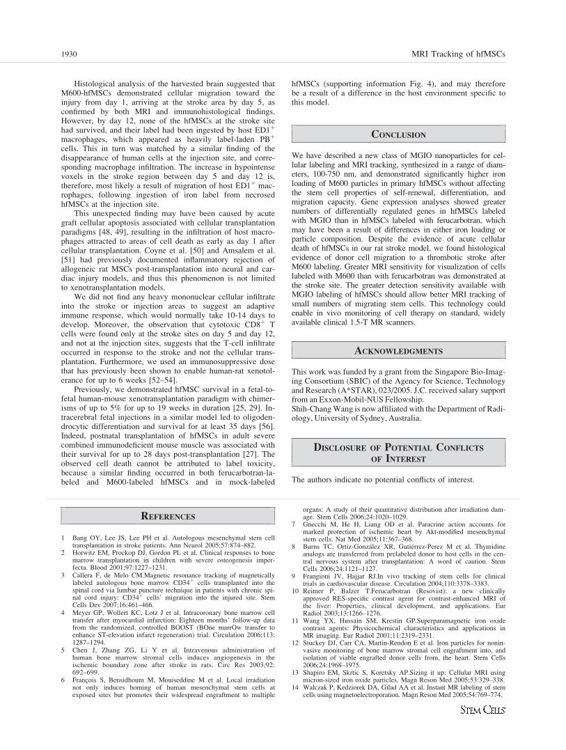
Histological analysis of the harvested brain suggested thatM600-hfMSCs demonstrated cellular migration toward theinjury from day 1, arriving at the stroke area by day 5, asconfirmed by both MRI and immunohistological findings.However, by day 12, none of the hfMSCs at the stroke sitehad survived, and their label had been ingested by host ED1þ
macrophages, which appeared as heavily label-laden PBþ
cells. This in turn was matched by a similar finding of thedisappearance of human cells at the injection site, and corre-sponding macrophage infiltration. The increase in hypointensevoxels in the stroke region between day 5 and day 12 is,therefore, most likely a result of migration of host ED1þ mac-rophages, following ingestion of iron label from necrosedhfMSCs at the injection site.
This unexpected finding may have been caused by acutegraft cellular apoptosis associated with cellular transplantationparadigms [48, 49], resulting in the infiltration of host macro-phages attracted to areas of cell death as early as day 1 aftercellular transplantation. Coyne et al. [50] and Amsalem et al.[51] had previously documented inflammatory rejection ofallogeneic rat MSCs post-transplantation into neural and car-diac injury models, and thus this phenomenon is not limitedto xenotransplantation models.
We did not find any heavy mononuclear cellular infiltrateinto the stroke or injection areas to suggest an adaptiveimmune response, which would normally take 10-14 days todevelop. Moreover, the observation that cytotoxic CD8þ Tcells were found only at the stroke sites on day 5 and day 12,and not at the injection sites, suggests that the T-cell infiltrateoccurred in response to the stroke and not the cellular trans-plantation. Furthermore, we used an immunosuppressive dosethat has previously been shown to enable human-rat xenotol-erance for up to 6 weeks [52–54].
Previously, we demonstrated hfMSC survival in a fetal-to-fetal human-mouse xenotransplantation paradigm with chimer-isms of up to 5% for up to 19 weeks in duration [25, 29]. In-tracerebral fetal injections in a similar model led to oligoden-drocytic differentiation and survival for at least 35 days [56].Indeed, postnatal transplantation of hfMSCs in adult severecombined immunodeficient mouse muscle was associated withtheir survival for up to 28 days post-transplantation [27]. Theobserved cell death cannot be attributed to label toxicity,because a similar finding occurred in both ferucarbotran-la-beled and M600-labeled hfMSCs and in mock-labeled
hfMSCs (supporting information Fig. 4), and may thereforebe a result of a difference in the host environment specific tothis model.
CONCLUSION
We have described a new class of MGIO nanoparticles for cel-lular labeling and MRI tracking, synthesized in a range of diam-eters, 100-750 nm, and demonstrated significantly higher ironloading of M600 particles in primary hfMSCs without affectingthe stem cell properties of self-renewal, differentiation, andmigration capacity. Gene expression analyses showed greaternumbers of differentially regulated genes in hfMSCs labeledwith MGIO than in hfMSCs labeled with ferucarbotran, whichmay have been a result of differences in either iron loading orparticle composition. Despite the evidence of acute cellulardeath of hfMSCs in our rat stroke model, we found histologicalevidence of donor cell migration to a thrombotic stroke afterM600 labeling. Greater MRI sensitivity for visualization of cellslabeled with M600 than with ferucarbotran was demonstrated atthe stroke site. The greater detection sensitivity available withMGIO labeling of hfMSCs should allow better MRI tracking ofsmall numbers of migrating stem cells. This technology couldenable in vivo monitoring of cell therapy on standard, widelyavailable clinical 1.5-T MR scanners.
ACKNOWLEDGMENTS
This work was funded by a grant from the Singapore Bio-Imag-ing Consortium (SBIC) of the Agency for Science, Technologyand Research (A*STAR), 023/2005. J.C. received salary supportfrom an Exxon-Mobil-NUS Fellowship.Shih-ChangWang is now affiliated with the Department of Radi-ology, University of Sydney, Australia.
DISCLOSURE OF POTENTIAL CONFLICTS
OF INTEREST
The authors indicate no potential conflicts of interest.
REFERENCES
1 Bang OY, Lee JS, Lee PH et al. Autologous mesenchymal stem celltransplantation in stroke patients. Ann Neurol 2005;57:874–882.
2 Horwitz EM, Prockop DJ, Gordon PL et al. Clinical responses to bonemarrow transplantation in children with severe osteogenesis imper-fecta. Blood 2001;97:1227–1231.
3 Callera F, de Melo CM.Magnetic resonance tracking of magneticallylabeled autologous bone marrow CD34þ cells transplanted into thespinal cord via lumbar puncture technique in patients with chronic spi-nal cord injury: CD34þ cells’ migration into the injured site. StemCells Dev 2007;16:461–466.
4 Meyer GP, Wollert KC, Lotz J et al. Intracoronary bone marrow celltransfer after myocardial infarction: Eighteen months’ follow-up datafrom the randomized, controlled BOOST (BOne marrOw transfer toenhance ST-elevation infarct regeneration) trial. Circulation 2006;113:1287–1294.
5 Chen J, Zhang ZG, Li Y et al. Intravenous administration ofhuman bone marrow stromal cells induces angiogenesis in theischemic boundary zone after stroke in rats. Circ Res 2003;92:692–699.
6 Francois S, Bensidhoum M, Mouiseddine M et al. Local irradiationnot only induces homing of human mesenchymal stem cells atexposed sites but promotes their widespread engraftment to multiple
organs: A study of their quantitative distribution after irradiation dam-age. Stem Cells 2006;24:1020–1029.
7 Gnecchi M, He H, Liang OD et al. Paracrine action accounts formarked protection of ischemic heart by Akt-modified mesenchymalstem cells. Nat Med 2005;11:367–368.
8 Burns TC, Ortiz-Gonzalez XR, Gutierrez-Perez M et al. Thymidineanalogs are transferred from prelabeled donor to host cells in the cen-tral nervous system after transplantation: A word of caution. StemCells 2006;24:1121–1127.
9 Frangioni JV, Hajjar RJ.In vivo tracking of stem cells for clinicaltrials in cardiovascular disease. Circulation 2004;110:3378–3383.
10 Reimer P, Balzer T.Ferucarbotran (Resovist): a new clinicallyapproved RES-specific contrast agent for contrast-enhanced MRI ofthe liver: Properties, clinical development, and applications. EurRadiol 2003;13:1266–1276.
11 Wang YX, Hussain SM, Krestin GP.Superparamagnetic iron oxidecontrast agents: Physicochemical characteristics and applications inMR imaging. Eur Radiol 2001;11:2319–2331.
12 Stuckey DJ, Carr CA, Martin-Rendon E et al. Iron particles for nonin-vasive monitoring of bone marrow stromal cell engraftment into, andisolation of viable engrafted donor cells from, the heart. Stem Cells2006;24:1968–1975.
13 Shapiro EM, Skrtic S, Koretsky AP.Sizing it up: Cellular MRI usingmicron-sized iron oxide particles. Magn Reson Med 2005;53:329–338.
14 Walczak P, Kedziorek DA, Gilad AA et al. Instant MR labeling of stemcells using magnetoelectroporation. Magn Reson Med 2005;54:769–774.
1930 MRI Tracking of hfMSCs

15 Bulte JW, Kraitchman DL.Iron oxide MR contrast agents for molecu-lar and cellular imaging. NMR Biomed 2004;17:484–499.
16 Arbab AS, Bashaw LA, Miller BR et al. Characterization of biophysi-cal and metabolic properties of cells labeled with superparamagneticiron oxide nanoparticles and transfection agent for cellular MR imag-ing. Radiology 2003;229:838–846.
17 Frank JA, Miller BR, Arbab AS et al. Clinically applicable labeling ofmammalian and stem cells by combining superparamagnetic ironoxides and transfection agents. Radiology 2003;228:480–487.
18 Arbab AS, Yocum GT, Kalish H et al. Efficient magnetic cell labelingwith protamine sulfate complexed to ferumoxides for cellular MRI.Blood 2004;104:1217–1223.
19 Lewin M, Carlesso N, Tung CH et al. Tat peptide-derivatized mag-netic nanoparticles allow in vivo tracking and recovery of progenitorcells. Nat Biotechnol 2000;18:410–414.
20 Schafer R, Kehlbach R, Wiskirchen J et al. Transferrin receptor upreg-ulation: In vitro labeling of rat mesenchymal stem cells with superpar-amagnetic iron oxide. Radiology 2007;244:514–523.
21 Mailander V, Lorenz MR, Holzapfel V et al. Carboxylated superpara-magnetic iron oxide particles label cells intracellularly without trans-fection agents. Mol Imaging Biol 2008;10:138–146.
22 Hordijk PL.Endothelial signalling events during leukocyte transmigra-tion. FEBS J 2006;273:4408–4415.
23 Chen J, Li Y, Zhang R et al. Combination therapy of stroke in ratswith a nitric oxide donor and human bone marrow stromal cellsenhances angiogenesis and neurogenesis. Brain Res 2004;1005:21–28.
24 Borlongan CV, Lind JG, Dillon-Carter O et al. Bone marrow graftsrestore cerebral blood flow and blood brain barrier in stroke rats.Brain Res 2004;1010:108–116.
25 Guillot PV, Abass O, Bassett JH et al. Intrauterine transplantation ofhuman fetal mesenchymal stem cells from first-trimester blood repairsbone and reduces fractures in osteogenesis imperfecta mice. Blood2008;111:1717–1725.
26 Chan J, O’Donoghue K, de la Fuente J et al. Human fetal mesenchy-mal stem cells as vehicles for gene delivery. Stem Cells 2005;23:93–102.
27 Chan J, O’Donoghue K, Gavina M et al. Galectin-1 induces skeletalmuscle differentiation in human fetal mesenchymal stem cells andincreases muscle regeneration. Stem Cells 2006;24:1879–1891.
28 Zhang ZY, Teoh SH, Chong MS et al. Superior osteogenic capacityfor bone tissue engineering of fetal compared to perinatal and adultmesenchymal stem cells. Stem Cells 2009;27:126–137.
29 Chan J, Waddington SN, O’Donoghue K et al. Widespread distribu-tion and muscle differentiation of human fetal mesenchymal stem cellsafter intrauterine transplantation in dystrophic mdx mouse. Stem Cells2007;25:875–884.
30 Le Blanc K, Gotherstrom C, Ringden O et al. Fetal mesenchymalstem-cell engraftment in bone after in utero transplantation in a patientwith severe osteogenesis imperfecta. Transplantation 2005;79:1607–1614.
31 Polkinghorne JC. Review of the Guidance on the Research Use ofFetuses and Fetal Material. London: Her Majesty’s Stationery Office(HMSO), 1989:1–762.
32 Tan BH, Tam KC, Lam YC et al. A semi-empirical approach formodeling charged soft microgel particles. J Rheol 2004;48:915–926.
33 Campagnoli C, Roberts IA, Kumar S et al. Identification of mesenchy-mal stem/progenitor cells in human first-trimester fetal blood, liver,and bone marrow. Blood 2001;98:2396–2402.
34 Al-Shahrour F, Minguez P, Tarraga J et al. BABELOMICS: A sys-tems biology perspective in the functional annotation of genome-scaleexperiments. Nucleic Acids Res 2006;34:W472–W476.
35 Schroeter M, Saleh A, Wiedermann D et al. Histochemical detectionof ultrasmall superparamagnetic iron oxide (USPIO) contrast medium
uptake in experimental brain ischemia. Magn Reson Med 2004;52:403–406.
36 Dominici M, Le Blanc K, Mueller I et al. Minimal criteria for defin-ing multipotent mesenchymal stromal cells. The International Societyfor Cellular Therapy position statement. Cytotherapy 2006;8:315–317.
37 Watson BD, Dietrich WD, Busto R et al. Induction of reproduciblebrain infarction by photochemically initiated thrombosis. Ann Neurol1985;17:497–504.
38 Matuszewski L, Persigehl T, Wall A et al. Cell tagging with clinicallyapproved iron oxides: Feasibility and effect of lipofection, particle size,and surface coating on labeling efficiency. Radiology 2005;235:155–161.
39 Chithrani BD, Ghazani AA, Chan WC.Determining the size and shapedependence of gold nanoparticle uptake into mammalian cells. NanoLett 2006;6:662–668.
40 Thorek DL & Tsourkas A.Size, charge and concentration dependentuptake of iron oxide particles by non-phagocytic cells. Biomaterials2008;29:3583–3590.
41 Rejman J, Oberle V, Zuhorn IS et al. Size-dependent internalizationof particles via the pathways of clathrin- and caveolae-mediated endo-cytosis. Biochem J 2004;377:159–169.
42 Kostura L, Kraitchman DL, Mackay AM et al. Feridex labeling ofmesenchymal stem cells inhibits chondrogenesis but not adipogenesisor osteogenesis. NMR Biomed 2004;17:513–517.
43 Arbab AS, Yocum GT, Rad AM et al. Labeling of cells with ferumox-ides-protamine sulfate complexes does not inhibit function or differen-tiation capacity of hematopoietic or mesenchymal stem cells. NMRBiomed 2005;18:553–559.
44 Berry CC, Charles S, Wells S et al. The influence of transferrin stabi-lised magnetic nanoparticles on human dermal fibroblasts in culture.Int J Pharm 2004;269:211–225.
45 Rose A.The sensitivity performance of the human eye on an absolutescale. J Opt Soc Am 1948;38:196–208.
46 Firbank MJ, Coulthard A, Harrison RM et al. A comparison of twomethods for measuring the signal to noise ratio on MR images. PhysMed Biol 1999;44:N261–N264.
47 Heyn C, Bowen CV, Rutt BK et al. Detection threshold of singleSPIO-labeled cells with FIESTA. Magn Reson Med 2005;53:312–320.
48 Beauchamp JR, Morgan JE, Pagel CN et al. Dynamics of myoblasttransplantation reveal a discrete minority of precursors with stem cell-like properties as the myogenic source. J Cell Biol 1999;144:1113–1122.
49 Skuk D, Caron NJ, Goulet M et al. Resetting the problem of celldeath following muscle-derived cell transplantation: Detection, dynam-ics and mechanisms. J Neuropathol Exp Neurol 2003;62:951–967.
50 Coyne TM, Marcus AJ, Woodbury D et al. Marrow stromal cellstransplanted to the adult brain are rejected by an inflammatoryresponse and transfer donor labels to host neurons and glia. StemCells 2006;24:2483–2492.
51 Amsalem Y, Mardor Y, Feinberg MS et al. Iron-oxide labeling andoutcome of transplanted mesenchymal stem cells in the infarcted myo-cardium. Circulation 2007;116(11 suppl):I38–I45.
52 Wennersten A, Meier X, Holmin S et al. Proliferation, migration, anddifferentiation of human neural stem/progenitor cells after transplanta-tion into a rat model of traumatic brain injury. J Neurosurg 2004;100:88–96.
53 Zhao LR, Duan WM, Reyes M et al. Human bone marrow stem cellsexhibit neural phenotypes and ameliorate neurological deficits aftergrafting into the ischemic brain of rats. Exp Neurol 2002;174:11–20.
54 Guo C, Haider HK, Shim WS et al. Myoblast-based cardiac repair:Xenomyoblast versus allomyoblast transplantation. J Thorac Cardio-vasc Surg 2007;134:1332–1339.
55 Kennea NL, Waddington SN, Chan J et al. Differentiation of humanfetal mesenchymal stem cells into cells with an oligodendrocyte phe-notype. Cell Cycle 2009;8:1069–1079.
See www.StemCells.com for supporting information available online.
Lee, Chan, Shuter et al. 1931
Related Documents