RESEARCH ARTICLE Open Access Intracellular trafficking and endocytosis of CXCR4 in fetal mesenchymal stem/stromal cells Rebecca A Pelekanos 1*† , Michael J Ting 1† , Varda S Sardesai 1 , Jennifer M Ryan 1 , Yaw-Chyn Lim 2 , Jerry KY Chan 1,3,4 and Nicholas M Fisk 1,4,5 Abstract Background: Fetal mesenchymal stem/stromal cells (MSC) represent a developmentally-advantageous cell type with translational potential. To enhance adult MSC migration, studies have focussed on the role of the chemokine receptor CXCR4 and its ligand SDF-1 (CXCL12), but more recent work implicates an intricate system of CXCR4 receptor dimerization, intracellular localization, multiple ligands, splice variants and nuclear accumulation. We investigated the intracellular localization of CXCR4 in fetal bone marrow-derived MSC and role of intracellular trafficking in CXCR4 surface expression and function. Results: We found that up to 4% of human fetal MSC have detectable surface-localized CXCR4. In the majority of cells, CXCR4 is located not at the cell surface, as would be required for ‘sensing’ migratory cues, but intracellularly. CXCR4 was identified in early endosomes, recycling endosomes, and lysosomes, indicating only a small percentage of CXCR4 travelling to the plasma membrane. Notably CXCR4 was also found in and around the nucleus, as detected with an anti-CXCR4 antibody directed specifically against CXCR4 isoform 2 differing only in N-terminal sequence. After demonstrating that endocytosis of CXCR4 is largely independent of endogenously-produced SDF-1, we next applied the cytoskeletal inhibitors blebbistatin and dynasore to inhibit endocytotic recycling. These increased the number of cells expressing surface CXCR4 by 10 and 5 fold respectively, and enhanced the number of cells migrating to SDF1 in vitro (up to 2.6 fold). These molecules had a transient effect on cell morphology and adhesion, which abated after the removal of the inhibitors, and did not alter functional stem cell properties. Conclusions: We conclude that constitutive endocytosis is implicated in the regulation of CXCR4 membrane expression, and suggest a novel pharmacological strategy to enhance migration of systemically-transplanted cells. Keywords: Fetal mesenchymal stromal cells, Bone marrow, MSC, CXCR4, Chemokine receptor, Migration, Small molecule, Endocytosis Background Fetal mesenchymal stem/stromal cells (fMSC) have proper- ties intermediate between adult and embryonic stem cells, and thus considerable therapeutic potential. Advantageous characteristics of fetal over adult MSC include their higher proliferative rate, greater differentiation capacity and longer telomeres with reduced senescence [1]. Indeed, a primary fetal bone marrow MSC line from our team had the fastest migratory capacity of 70 cell lines assessed at a recent American Society of Cell Biology (5.2 μm/minute or 0.000000312 kilometers per hour) [2]. Successful transla- tion of cell therapies requires an in-depth understanding not only of the basic stem cell properties of differentiation and self-renewal, but also of the cell type’ s adhesive and migratory properties. MSC can be mobilized from their sedentary niches by a range of external stimuli and triggered to migrate to and occupy distant tissue sites [3]. Preferential homing to sites of tissue injury has been demonstrated with both adult [4,5] and fetal MSC [6-8]. However poor understand- ing of the underlying processes in adult and particularly fetal MSC limits our ability to exploit them for targeting MSC to regions of tissue damage. Improved homing would facilitate therapeutic development and reduce inappropriate uptake of MSC in healthy tissues. * Correspondence: [email protected] † Equal contributors 1 UQ Centre for Clinical Research, The University of Queensland, Herston QLD 4029, Australia Full list of author information is available at the end of the article © 2014 Pelekanos et al.; licensee BioMed Central Ltd. This is an Open Access article distributed under the terms of the Creative Commons Attribution License (http://creativecommons.org/licenses/by/2.0), which permits unrestricted use, distribution, and reproduction in any medium, provided the original work is properly credited. The Creative Commons Public Domain Dedication waiver (http://creativecommons.org/publicdomain/zero/1.0/) applies to the data made available in this article, unless otherwise stated. Pelekanos et al. BMC Cell Biology 2014, 15:15 http://www.biomedcentral.com/1471-2121/15/15

Welcome message from author
This document is posted to help you gain knowledge. Please leave a comment to let me know what you think about it! Share it to your friends and learn new things together.
Transcript

Pelekanos et al. BMC Cell Biology 2014, 15:15http://www.biomedcentral.com/1471-2121/15/15
RESEARCH ARTICLE Open Access
Intracellular trafficking and endocytosis of CXCR4in fetal mesenchymal stem/stromal cellsRebecca A Pelekanos1*†, Michael J Ting1†, Varda S Sardesai1, Jennifer M Ryan1, Yaw-Chyn Lim2, Jerry KY Chan1,3,4
and Nicholas M Fisk1,4,5
Abstract
Background: Fetal mesenchymal stem/stromal cells (MSC) represent a developmentally-advantageous cell typewith translational potential.To enhance adult MSC migration, studies have focussed on the role of the chemokine receptor CXCR4 and its ligandSDF-1 (CXCL12), but more recent work implicates an intricate system of CXCR4 receptor dimerization, intracellularlocalization, multiple ligands, splice variants and nuclear accumulation. We investigated the intracellular localization ofCXCR4 in fetal bone marrow-derived MSC and role of intracellular trafficking in CXCR4 surface expression and function.
Results: We found that up to 4% of human fetal MSC have detectable surface-localized CXCR4. In the majority of cells,CXCR4 is located not at the cell surface, as would be required for ‘sensing’ migratory cues, but intracellularly. CXCR4was identified in early endosomes, recycling endosomes, and lysosomes, indicating only a small percentage of CXCR4travelling to the plasma membrane. Notably CXCR4 was also found in and around the nucleus, as detected with ananti-CXCR4 antibody directed specifically against CXCR4 isoform 2 differing only in N-terminal sequence. Afterdemonstrating that endocytosis of CXCR4 is largely independent of endogenously-produced SDF-1, we nextapplied the cytoskeletal inhibitors blebbistatin and dynasore to inhibit endocytotic recycling. These increased thenumber of cells expressing surface CXCR4 by 10 and 5 fold respectively, and enhanced the number of cells migratingto SDF1 in vitro (up to 2.6 fold). These molecules had a transient effect on cell morphology and adhesion, which abatedafter the removal of the inhibitors, and did not alter functional stem cell properties.
Conclusions: We conclude that constitutive endocytosis is implicated in the regulation of CXCR4 membraneexpression, and suggest a novel pharmacological strategy to enhance migration of systemically-transplanted cells.
Keywords: Fetal mesenchymal stromal cells, Bone marrow, MSC, CXCR4, Chemokine receptor, Migration,Small molecule, Endocytosis
BackgroundFetal mesenchymal stem/stromal cells (fMSC) have proper-ties intermediate between adult and embryonic stem cells,and thus considerable therapeutic potential. Advantageouscharacteristics of fetal over adult MSC include their higherproliferative rate, greater differentiation capacity and longertelomeres with reduced senescence [1]. Indeed, a primaryfetal bone marrow MSC line from our team had the fastestmigratory capacity of 70 cell lines assessed at a recentAmerican Society of Cell Biology (5.2 μm/minute or
* Correspondence: [email protected]†Equal contributors1UQ Centre for Clinical Research, The University of Queensland, Herston QLD4029, AustraliaFull list of author information is available at the end of the article
© 2014 Pelekanos et al.; licensee BioMed CentCommons Attribution License (http://creativecreproduction in any medium, provided the orDedication waiver (http://creativecommons.orunless otherwise stated.
0.000000312 kilometers per hour) [2]. Successful transla-tion of cell therapies requires an in-depth understandingnot only of the basic stem cell properties of differentiationand self-renewal, but also of the cell type’s adhesive andmigratory properties.MSC can be mobilized from their sedentary niches by
a range of external stimuli and triggered to migrate toand occupy distant tissue sites [3]. Preferential homingto sites of tissue injury has been demonstrated with bothadult [4,5] and fetal MSC [6-8]. However poor understand-ing of the underlying processes in adult and particularlyfetal MSC limits our ability to exploit them for targetingMSC to regions of tissue damage. Improved homing wouldfacilitate therapeutic development and reduce inappropriateuptake of MSC in healthy tissues.
ral Ltd. This is an Open Access article distributed under the terms of the Creativeommons.org/licenses/by/2.0), which permits unrestricted use, distribution, andiginal work is properly credited. The Creative Commons Public Domaing/publicdomain/zero/1.0/) applies to the data made available in this article,
Pelekanos et al. BMC Cell Biology 2014, 15:15 Page 2 of 15http://www.biomedcentral.com/1471-2121/15/15
CXCR4 is a G-protein coupled receptor belonging tothe CXC family of chemokine receptors. Interaction ofCXCR4 with its ligand, stromal derived factor (SDF-1α,CXCL12) directs the movement of cells in hematopoieticstem cell homing [9], leukocyte trafficking [10,11] andtumour metastasis [12,13]. The CXCR4-SDF-1α axis isactive in adult MSC, which migrate toward SDF-1αin vitro [14]. CXCR4 is expressed on the surface of onlya subset of adult MSC, but up-regulation by viral trans-duction increases MSC homing [15], rendering CXCR4 atarget for modulating migration. However the viralapproach is unsuitable for clinical translation due tothe risk of insertional mutagenesis. CXCR4 expressionis dynamically regulated by external cues like hypoxia[16], and can be up-regulated in adult MSC followingin vitro priming with a mixture of cytokines, as shownto enhance migration in vitro toward an SDF-1α gradientas well as homing in vivo to bone marrow [17]. Recently,SDF-1 exposure was shown to up regulate low basalCXCR4 surface expression in fetal blood derived-MSC,which increased chemotaxis [18].Like other G-protein coupled receptors, CXCR4
undergoes internalization after interaction with ligand.Ligand-induced endocytosis of CXCR4 and its internalsequestration has been extensively studied in leukocytes[19,20] and to a lesser degree in hematopoietic stem cells[21,22] and tumour cells [23]. Although these studies con-firm the existence of a general regulatory mechanism, theextent of intracellular expression and endocytosis/recyc-ling kinetics differs between cell types, implicating cellularcontext in the regulation of CXCR4 trafficking and itsfunctional consequences [24,25]. The predominant intra-cellular localization of CXCR4 suggests that dynamicequilibrium between the cytoplasm and plasma mem-brane may modulate CXCR4 availability at the cell surface,and thus fMSC responsiveness to SDF-1α gradients.We investigated the intracellular localization and traffick-
ing of CXCR4 in fetal bone marrow MSC, and treatedfMSC with blebbistatin and dynasore, specific inhibitors ofmyosin IIA and dynamin subunits of the actin cytoskeletonresponsible for cytoskeletal movement and chemotaxis, andcommonly associated with G-protein endocytosis. Ourfindings demonstrate that surface expression of CXCR4on fMSC and their SDF-1α induced-chemotaxis in vitrocan be increased through inhibition of receptor endo-cytosis. These data support further development ofsmall molecule agents to up-regulate the functional expres-sion of a key receptor involved in homing and engraftmentof MSC.
MethodsMSC cultureFetal tissue was collected from consenting women under-going clinically indicated termination of pregnancy in
accordance with national guidelines and as approvedby the Human Research Ethics Committee of the RoyalBrisbane and Women’s Hospital. Early trimester bonemarrow MSC (passage 1–7) derived from different donors(n = 9, gestation 10–13 weeks) and adult bone marrowMSC (aMSC) from a bone marrow donor were culturedin Dulbecco’s modified Eagle’s medium (DMEM) high glu-cose (Invitrogen) supplemented with 10% fetal bovineserum (FBS), 100 IU/mL penicillin, and 100 μg/mLstreptomycin (Invitrogen), expanded at 5000 cells/cm2
at 37°C with 5% CO2. Isolated fMSC and aMSC werecharacterised by typical cell surface phenotype and dif-ferentiation capacity as previously reported [26-28].Antibodies used to characterize MSC are listed in [28].Mesodermal differentiation methods are described in theAdditional file 1.
Priming fMSC with endocytosis inhibitorsFor flow cytometry, cells were primed with (−)-blebbista-tin (1-Phenyl-1,2,3,4-tetrahydro-4-hydroxypyrrolo [2.3-b]-7-methylquinolin-4-one, cat. # B0560, Sigma Aldrich) anddynasore hydrate (3-Hydroxy-naphthalene-2-carboxylicacid (3,4-dihydroxy-benzylidene)-hydrazide hydrate, cat. #D7693, Sigma Aldrich) as follows: cells were detachedwith TrypLE Select and washed twice with serum-freeDMEM. Cells were resuspended in 600 μl of DMEM notcontaining FCS but with 25 mM HEPES (4 × 10⁴ cells/ml)with blebbistatin or dynasore added at a concentration of80 μM. Cells were incubated at 37°C for 15, 30 or 60 min.For morphology and immunofluorescence studies, cellswere seeded into 24 well plates with or without glasscoverslips. When confluent, cells were washed oncewith DMEM and treated as indicated. Treatment mediaconsisted of 250 μl of DMEM + 25 mM HEPES withoutFCS and 80 μM blebbistatin or dynasore as necessary.Vehicle treatment was 0.45% DMSO in the same media,equivalent to the amount of DMSO in 80 μM blebbistatinsolution. Cells were imaged immediately or washed withPBS, fixed with 1% PFA in PBS pH 7.4 for 10 min, washedand stored in PBS at 4°C.
Flow cytometryFor flow cytometry, cells were detached using TrypLEreagent (Invitrogen). The antibodies used are detailed inAdditional file 1: Table S1. Because of expression differ-ences with different commercially available antibodies,we detected intracellular and extracellular CXCR4 usinga monoclonal anti-human CXCR4 PE-Cy5 conjugatedantibody (clone 12G5) following the manufacturer’s in-structions (1:20 dilution, eBioscience), a non-conjugatedpolyclonal antibody (clone ab2074, 1:50 dilution, Abcam)and/or a biotin-conjugated monoclonal antibody (clone4417, 1:20 dilution, R and D Systems). For indirect stain-ing with the polyclonal antibody, cells were incubated

Pelekanos et al. BMC Cell Biology 2014, 15:15 Page 3 of 15http://www.biomedcentral.com/1471-2121/15/15
with primary antibody at 4°C for 30 min then washedonce with PBS + 2% FCS. After washing cells were resus-pended and incubated with FITC- or Alexa 488 conjugatedsecondary anti-rabbit antibody or PE- or FITC labelledstreptavidin at 4°C for 30 min. After incubation cells werewashed three times with PBS. For intracellular staining fordetection of total cellular CXCR4 (surface + cytoplasmic),cells were fixed with 4% paraformaldehyde (Sigma) for10 min, permeabilised with 0.5% Triton X-100 (Sigma) for10 min, and stained with anti-human CXCR4-PE Cy5 for30 min. Cells were analysed by FACS Calibur (BectonDickinson - fetal MSC, adult MSC and THP-1 cells) orGallios (Beckman Coulter - fetal MSC and HeLa cells).For flow cytometry experiments the isotype controls,analysis gates were optimized on each flow cytometer,for permeablized or intact cells, and for each antibody,fluorophore and cell line used according to standardpractice. For cytoplasmic staining, cells were fixed andpermeabilised as above. Cells were then stained withCXCR4-PE-Cy5, and incubated for 5 min in 0.25 Macetic acid (pH 4) to remove surface bound antibody.Surface CXCR4 was expressed as a percentage of totalCXCR4 as follows: [MFI (surface CXCR4)/MFI (totalCXCR4)] × 100. The CXCR4 antibodies were validatedusing THP-1 cells (leukemic monocytes, obtained fromATCC), a control cell haematopoietic cell with consti-tutively high surface expression. Antibodies were alsoassessed in human cervical carcinoma HeLa cells(ATCC) cultured in MSC medium.
Immunofluorescence microscopyfMSC (P3-7) were cultured on glass coverslips and thenfixed with 4% PFA in PBS (pH 7.4) for 10 min at roomtemperature [28]. Cells were washed with PBS, and thenincubated with blocking and permeabilisation buffer (2%bovine serum albumin, 0.1% Tween) for 1 hr at roomtemperature. Blocking buffer was discarded and antibodieswere diluted in diluent buffer (blocking buffer diluted 1:10with PBS) and incubated overnight with primary antibodiesat 4°C. Rabbit anti-CXCR4 (ab2074) and either mouseanti-Rab5, anti-Rab11A, or anti-Lamp1 (all 1:50 dilution,detailed in Additional file 1: Table S1) was added. Afterincubation with primary antibodies, cells were washed3 × 5 min and secondary anti-mouse-Alexa 568 andanti-rabbit Alexa 488 (1:1000 dilution, from Invitrogen)were added. Cells were incubated with secondary anti-bodies at room temperature for 1 hr and then washed 3 ×5 min. Coverslips were placed on microscope slides withProlong Gold with DAPI mounting medium (Invitrogen).For blebbistatin and dynasore treatment experiments,
coverslips were treated with 0.1% gelatin for 10 min priorto seeding or seeded onto fibronectin coated chamberslides. After inhibitor treatment, cells were fixed with pre-warmed 1% PFA for 10 min at RT, blocked in 1% BSA
with 0.05% Tween in PBS for permeabilisation for 1 hrprior to proceeding with antibody staining. Tween wasomitted for surface CXCR4 staining. Antibodies were di-luted in PBS. Four x 30 sec washes were carried out aftereach antibody incubation. Slides were examined on a ZeissAxio epi-fluorescence or LSM 510 confocal microscope.
In situ Proximity Ligation Assay (PLA)Cells were cultured on coverslips, fixed with PFA as perImmunofluorescence Microscopy (above). Cells were per-meabilised with 0.1% Tween in PBS for 10 min at roomtemperature. The Duolink II proximity ligation assay(PLA, mouse-rabbit starter kit in orange, Olink Biosci-ences) was carried out as per manufacturer’s instructionsand using supplied reagents for a 1 cm2 reaction volume.Primary antibodies were incubated overnight at 4°C -rabbit anti-CXCR4 (1:50, ab2074) and either mouse anti-Rab5 (1:200, 1 μg/ml), anti-Rab11A (1:50, 1 μg/ml) oranti-Lamp1 (1:100, 1 μg/ml) were used. The positive con-trol was the PLA reaction between rabbit anti-GrowthHormone Receptor (GHR) clone H300 (1:200, Santa Cruz)and the mouse anti-GHR clone 3A12 (1:200, Sigma Al-drich), as these 2 different antibodies detect differentepitopes of the GHR protein.
Transwell assaysFetal MSC were detached using TrypLE reagent (Invitro-gen), washed twice in PBS, and resuspended in DMEM.Cells were stained for 8 min at room temperature withCFSE (0.1 mg/ml) and the reaction was then inactivatedby adding PBS with 2% FCS. Cells were washed twiceand resuspended in DMEM with no FCS. Cells weretreated with either endocytosis inhibitors or vehicle only(DMSO) for 1 hr. 300 μl of fMSC suspension (1.6 × 10⁴
cells/well) were placed in the upper compartments of aFluoroblok transwell migration chamber (BD Biosciences).The transwell membrane (8 μm pore size) was coated withfibronectin 2.5 μg/ml for 30 min at room temperature,then washed with water and allowed to air-dry. SDF-1αwas placed in the lower compartment (30, 100 or 200 ng/ml). Cells were allowed to undergo chemotaxis for 4 hoursand then migration was determined by fluorescence inten-sity in the bottom well using the Paradigm fluorescenceplate reader (Beckman Coulter). Each experiment was per-formed in triplicate. The migration index was calculatedas the ratio of the fluorescence intensity of cells migratingtowards the chemo-attractant to the fluorescence intensityof cells migrating towards media alone.
Adhesion assaysFor adhesion assays, CFSE stained cells (35,000/well) wereincubated in fibronectin or collagen I coated wells of96-well plates in DMEM containing no FBS at 37°C and5% CO2 for 1 hr, and then washed with DMEM without

Pelekanos et al. BMC Cell Biology 2014, 15:15 Page 4 of 15http://www.biomedcentral.com/1471-2121/15/15
FBS. The number of adherent cells/well was estimatedby reading the fluorescence intensity with the Paradigmmicroplate reader.
Quantitative wound healing assayMigration was assessed by performing scratch wound as-says in a real-time cell imaging system (IncuCyte Live-cell,ESSEN BioScience Inc.). Briefly, 5 × 10⁴ cells per well wereplated into an Essen-Costar 96 well plate coated with 0.1%gelatin (n = 3 independent donors, performed in replicatesof 5). Twenty four hours later, the confluent monolayer ofcells was washed 1x PBS, scratched with the Essen Bio-science 96 well plate scratcher, incubated with blebbistatinor dynasore as above (80 μM, for 1 hr 37°C). The mediawas removed and normal growth media added, plateswere transferred to the Incucyte live cell imaging systemand the Essen 96 well wound assay protocol was run onthe software. Cells were imaged at 4 hr intervals for 24 hrto monitor cell migration. Wound confluence was calcu-lated automatically by the Incucyte software (v1.5) for12 hr, i.e. until wounds were greater than 90% confluent.The data were then analysed using an integrated metric:Relative Wound density.
Statistical analysisNormally-distributed data were expressed as mean ±standard deviation and analysed by paired t-test andANOVA. p < 0.05 was considered significant. All experi-ments were conducted in triplicate or with a minimum of 3independent donor-derived fMSC, unless otherwise stated.For scratch wound, a 2-way ANOVA with Bonferroni cor-rection was used (Prism, GraphPad).
ResultsFetal bone marrow derived-MSC exhibit typical MSCcharacteristicsIsolated fMSC and aMSC were characterised by typicalcell surface phenotype and differentiation capacity as pre-viously reported [26-28]. Cultured MSC met the criteriaset by the International Society for Cellular Therapy [29],being plastic adherent, fibroblastic in morphology andable to differentiate osteogenic, adipogenic and chondro-genic lineages in vitro (Additional file 2: Figure S1A-D).Fetal MSC were positive for the various MSC markers(including CD73, CD105, CD29, CD90) and were negativefor hematopoietic and endothelial markers (includingCD45, CD11b, CD34, CD31) (Additional file 2: Figure S1Eand F).
CXCR4 expression is predominantly intracellular andlocalized to the endosomal, lysosomal and nuclearcompartment of fMSCOnly 3.8 ± 0.3% of fetal bone marrow-derived mesenchymalstem/stromal cells (fMSC, n = 4) displayed positive surface
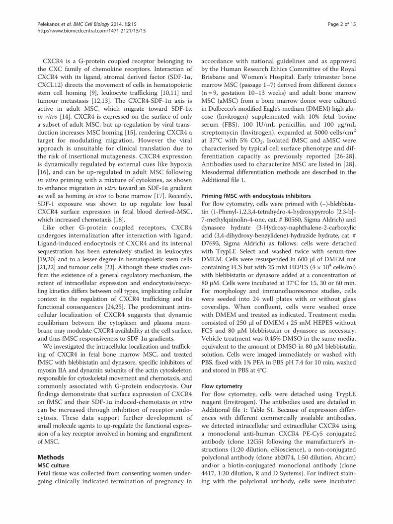
immune staining for CXCR4 (Figure 1, Additional file 3:Figure S2A). However, when cells were fixed and permeabi-lised, ~50-90% of fMSC stained positive for intracellularlylocalised CXCR4 (Figure 1B, Additional file 3: Figure S2B).This low surface and high intracellular CXCR4 staining pat-tern was observed regardless of the anti-CXCR4 antibodyclone used (clone 12G5, 4417 or ab2074) or the methodof antibody detection (direct or indirect fluorophore-conjugated primary antibody). Furthermore, detachingfMSC with enzymatic (TrypLE) or chemical methods(5 mM EDTA) produced only a modest change inCXCR4 (12G5) antibody surface staining (3.8% vs 5.3%,Additional file 3: Figure S2H). Therefore, enzymaticdegradation of the CXCR4 is not a major reason forlow surface CXCR4 expression observed in this study.A similar low surface/high intracellular CXCR4 immuno-positivity was seen for adult bone marrow MSC (passage5, n = 1, Figure 1C and D), as previously reported [30].This is in contrast to THP-1 monocytic leukemic andHeLa cell lines, that both displayed a high percentage ofcells with surface CXCR4 immuno-positivity (Figure 1E-F,Additional file 3: Figure S2E) and previously reported mi-gration capacity to SDF-1, the CXCR4 ligand [31,32].Although cytoplasmic sequestration of CXCR4 has
been described in adult bone marrow and decidual MSC[30], the internal distribution of CXCR4 has not to datebeen characterised in any MSC. Two CXCR4 antibodyclones (4417 and 12G5) detected an even surface distribu-tion of CXCR4 on a small number of fMSC by immuno-fluorescence microscopy (Figure 2A, and not shown).This supported the flow cytometry data that deter-mined only 3.8 ± 0.3% fMSC had positive CXCR4 sur-face immuno-reactivity (Figure 1). Permeabilization ofcells with Triton-X showed CXCR4 to be localized in punc-tate endosomal/lysosomal-like compartments (Figure 2B),some of which aligned to the cytoskeleton or nuclear mem-brane (Figure 2B, upper and lower insets respectively).However, the third anti-CXCR4 (ab2074) antibody tested,produced a different pattern of diffuse punctate cytoplasmicstaining with a large accumulation of CXCR4 in the nucleus(Figure 2D). Antibody staining conditions were optimizedas described in the additional information, but this did notchange the nuclear localization of CXCR4 detected bythe ab2074 antibody (Additional file 4: Figure S3A-C).We thus examined the localization of CXCR4 usingwell-characterised markers of the endocytotic pathway,Rab5 and Rab11 for early/recycling endosomes, andLamp-1 for lysosomes. Immuno-fluorescence stainingof fMSC revealed a similar diffuse punctate distributionof anti-CXCR4 (ab2074) antibody with both Rab5 andRab11 labelled endosomes (Figure 2D and E). Subcellu-lar localization patterns of Rab5 and Rab11 were con-firmed by transient transfection of fMSC and HeLa cells(Additional file 4: Figure S3D and E). CXCR4 staining
Figure 1 A low number of human fetal bone marrow MSC (fMSC) display surface CXCR4. Three different anti-CXCR4 antibody clones wereused 12G5, ab2074 and 4417 as detailed in the methods, with fluorophore labelled secondary antibodies used where necessary. A) CXCR4 surfaceexpression (fluorescence intensity vs. forward scatter) by flow cytometry shown as dot plots. B) After permeabilisation, the majority of cells showintracellular stores of CXCR4. The horizontal gates delineate the position of relevant isotype control antibody. Human adult bone marrow MSCalso show low expression of CXCR4 on the cell surface (C) with large intracellular stores of this receptor (D). E) Each of the anti-CXCR4 antibodiesare able to detect >80% cells with surface expression of CXCR4 on HeLa cells. F) Both the anti-CXCR4 antibodies 12G5 and ab2074 are able todetect >80% cells with surface expression of CXCR4 on THP-1 human monocytic leukemia cells.
Pelekanos et al. BMC Cell Biology 2014, 15:15 Page 5 of 15http://www.biomedcentral.com/1471-2121/15/15
on immuno-fluorescence microscopy also overlapped withLamp1, although Lamp1 displayed a different peri-nuclearlocation (Figure 2F).
To confirm co-localization of CXCR4 in endosomalcompartments, an in cell co-immunoprecipation (prox-imity ligation assay, PLA) was conducted (Figure 2G).

Figure 2 Subcellular localization of CXCR4 expression in MSC. A) Incubation of non-permeabilised fMSC with the anti-CXCR4 antibody (clone4417) shows a distinct plasma membrane labelling of a small percentage of cells (a representative image of positive cell in the centre is shown).B) When incubated with permeabilised fMSC, the anti-CXCR4 (4417) antibody labels endosomal-like structures in a majority of cells. These CXCR4positive vesicles have an arrangement along the cytoskeleton (upper inset) and also perinuclear accumulation (lower inset) with light nuclearstaining. C) Negative control for ab 4417, using the same imaging settings. D-F) Immunofluorescence staining of fMSC with anti-CXCR4 cloneab2074 (red) strong nuclear localization of CXCR4, with diffuse, punctate cytoplasmic staining. CXCR4 colocalises with the endocytotic markersRab5 (D) and Rab11 (E) and lysosomal marker Lamp1 (F, all green). Lamp1 displays a distinct peri-nuclear location, with larger sized vesicles.Nuclei, counterstained with DAPI (x40 magnification). G) The Duolink II proximity ligation assay (PLA) shows colocalisation of CXCR4 (ab2074)with all three Rab5, Rab11 and Lamp1 positive compartments. Each red spot corresponds to a molecular interaction (x20 magnification). H) Thepositive control experiment is two different antibodies to the Growth Hormone Receptor, where the bound antibodies are in close proximity toeach other. Negative controls have one (#1) or both (#2) primary antibodies omitted from the PLA procedure.
Pelekanos et al. BMC Cell Biology 2014, 15:15 Page 6 of 15http://www.biomedcentral.com/1471-2121/15/15
Anti-CXCR4 antibody successfully co-localized with Rab5,Rab11 and Lamp1, with each red spot indicating a pointof co-localization. Positive and negative controls for thePLA assay shown in Figure 2H support the specificity of
these CXCR4 interactions. These PLA results are con-sistent with the immunofluorescence microscopy, andindicate that cytoplasmic CXCR4 in fMSC is distributedwithin all three endosomal compartments.

Pelekanos et al. BMC Cell Biology 2014, 15:15 Page 7 of 15http://www.biomedcentral.com/1471-2121/15/15
To investigate what drives internalization of CXCR4,we investigated whether endogenously-produced SDF-1α,the CXCR4 ligand highly expressed in MSC [33], couldtrigger this endocytosis of CXCR4 through a ligand-dependent process. Fetal MSC expressed very low levelsof CXCR4 mRNA, consistent with recycling of receptorsrather than synthesis (Additional file 5: Figure S4A). Al-though fMSC expressed high levels of SDF-1α mRNA(Additional file 5: Figure S4A), treatment of fMSC cultureswith a neutralizing antibody against SDF-1α producedonly a moderate increase in cell surface expression ofCXCR4, as measured by mean fluorescent intensity (MFI)on flow cytometry (Figure 3A). This suggests althoughligand-dependent endocytosis occurs, it cannot accountfor all and particularly basal receptor internalization, andthus an overriding constitutive mechanism is likely to beresponsible for the CXCR4 trafficking pattern observed.
Treatment with endocytosis inhibitors increases CXCR4surface expressionGiven the large proportion of CXCR4 located within theendosomal compartment, we explored the regulatorymechanism responsible by using two inhibitors of endo-cytosis, blebbistatin and dynasore, to probe the dynamicsof CXCR4 trafficking. Blebbistatin is an inhibitor of non-muscle myosin II and inhibits endocytosis, while dynasoreis a small molecule inhibitor of dynamin I and dynamin IIproteins with important roles in clathrin-dependent endo-cytosis. fMSC were initially treated with different concen-trations (ranging from 20–100 μM) of either blebbistatinor dynasore. After 30 min the highest levels of surfaceCXCR4 expression determined by flow cytometry (bleb-bistatin: 22.6% ± 7.9 of cells; dynasore: 17.2% ± 3.8 of cells,n = 3) were achieved at a concentration of 80 μM for bothagents (p < 0.01, Figure 3D). Quantitatively similar resultswere obtained using a different anti-CXCR4 antibody(ab2074) and an indirect staining method on adultMSC (blebbistatin: 20.5% ± 3.0 of cells and dynasore:16.4% ± 2.4 of cells, Figure 3E). To determine the kineticsof CXCR4 receptor exocytosis, fMSC were treated with80 μM blebbistatin or dynasore (n = 3 each) at 37°C andthe cells then collected at select time points. The cellswere stained with CXCR4 antibody (12G5), fixed and thelevel of surface and cytoplasmic CXCR4 expression exam-ined by flow cytometry. Blebbistatin resulted in a return ofCXCR4 to the cell surface with maximal surface expres-sion (33.4% ± 11.3 of cells) reaching steady state after1 hour (Figure 3C). Dynasore treatment revealed a similarreturn of surface expression of CXCR4, but with a lowermaximal level (25.6% ± 5.4 of cells) compared with bleb-bistatin. After two hours, CXCR4 surface expressionwas maintained stably at a level around 5 fold higherthan untreated control MSC, but returned to baseline overthe subsequent 6 hours (not shown). These endocytosis
inhibitors also reduced ligand-induced internalization ofCXCR4 in THP-1 cells. This hematopoietic leukemic cellline served as a control cell type with >90% endogenoussurface expression when untreated, whereas SDF-1 treat-ment induced rapid internalization of CXCR4 (Figure 3Fand G). However, co-incubation of THP-1 cells with SDF-1 and blebbistatin or dynasore kept the surface levels ofCXCR4 similar to untreated (Figure 3H and I).
Translocation kinetics and modelling indicates CXCR4endocytosis rate regulates surface expression of CXCR4At steady state, the relative distribution of a membraneprotein between the surface and interior of cells is deter-mined by the ratio of the recycling to endocytosis rate con-stants. Inhibition of endocytosis in fMSC with blebbistatinand dynasore permits an approximation of a recycling rate(kr) and thus derivation of the endocytosis rate (ke). Thehigh rate of basal endocytosis is consistent with highcytoplasmic sequestration of CXCR4 and the low surfaceexpression we document in fMSC.Additional file 6: Figure S5B shows the fit of the above
model to the experimental data obtained before and aftertreatment with blebbistatin or dynasore. Simulations usingrate constants derived from the experimental data showlevels of CXCR4 surface expression of 4.8% for restingfMSC, 35% for blebbistatin and 26% for dynasore.Additional file 6: Figure S5C shows a simulation in whichthe total cellular CXCR4 is initially present in the intracel-lular pool. As the system approaches equilibrium, CXCR4is transferred from the cytoplasmic compartment to thecell surface. Simulations show that the level of membraneexpression of CXCR4 in fMSC is sensitive to the rate ofendocytosis (ke). The series of theoretical curves generateddemonstrate how ke and kr inter-determine the level ofCXCR4 receptor surface expression and internalization infMSC. Additional file 6: Figure S5C shows the effect onreceptor cell surface expression level of varying ke whilemaintaining kr at its basal rate (0.04 min−1). When ke issmall, little internalization is seen and a large proportionof CXCR4 in present on the cell surface (~70%), but as kerises, a greater proportion of receptors are found insidethe cell at steady state. Thus, fMSC that exhibit small krand large ke have substantial levels of internalization. Themodel also demonstrates that altering the rate of endo-cytosis independent of the recycling rate is sufficient tocause substantial receptor re-surfacing. The model sup-ports the rate of endocytosis of CXCR4 being a criticalregulatory point for surface expression of CXCR4 infMSC, and one amenable to manipulation.
Transient effect of endocytosis inhibitors on fMSCmorphology and cytoskeleton arrangementThe treatment of blebbistatin and dynasore had a markedeffect on the morphology and fragility of cells (Figure 4
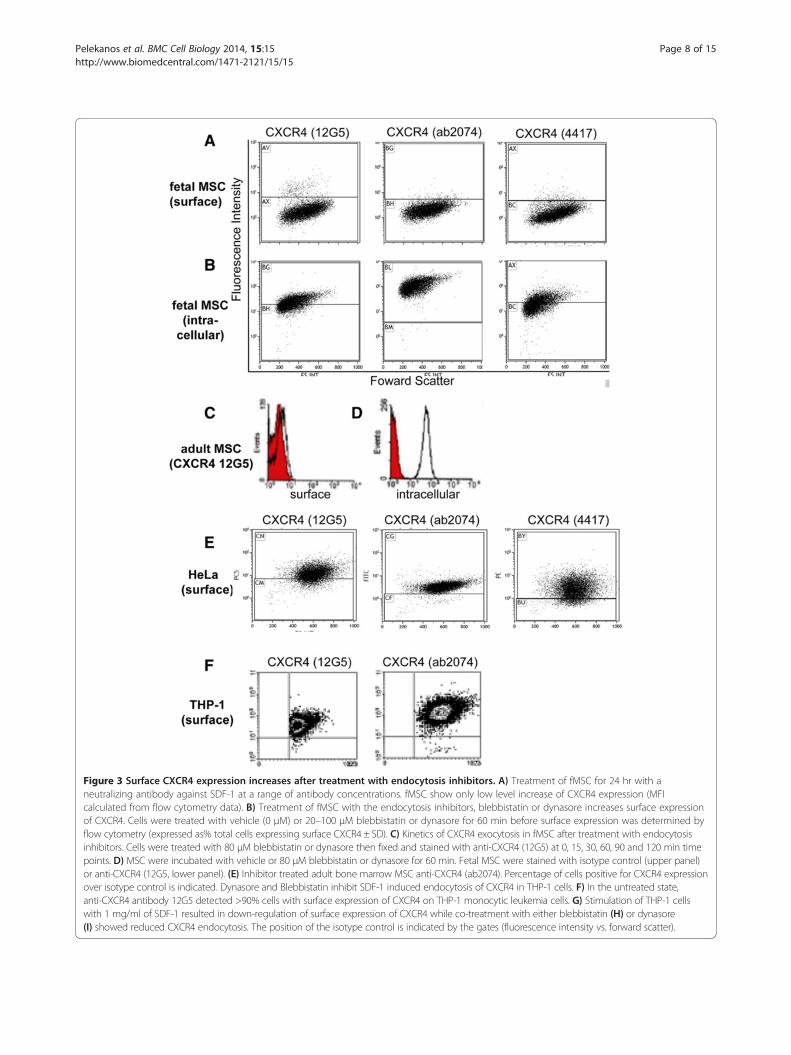
Figure 3 Surface CXCR4 expression increases after treatment with endocytosis inhibitors. A) Treatment of fMSC for 24 hr with aneutralizing antibody against SDF-1 at a range of antibody concentrations. fMSC show only low level increase of CXCR4 expression (MFIcalculated from flow cytometry data). B) Treatment of fMSC with the endocytosis inhibitors, blebbistatin or dynasore increases surface expressionof CXCR4. Cells were treated with vehicle (0 μM) or 20–100 μM blebbistatin or dynasore for 60 min before surface expression was determined byflow cytometry (expressed as% total cells expressing surface CXCR4 ± SD). C) Kinetics of CXCR4 exocytosis in fMSC after treatment with endocytosisinhibitors. Cells were treated with 80 μM blebbistatin or dynasore then fixed and stained with anti-CXCR4 (12G5) at 0, 15, 30, 60, 90 and 120 min timepoints. D) MSC were incubated with vehicle or 80 μM blebbistatin or dynasore for 60 min. Fetal MSC were stained with isotype control (upper panel)or anti-CXCR4 (12G5, lower panel). (E) Inhibitor treated adult bone marrow MSC anti-CXCR4 (ab2074). Percentage of cells positive for CXCR4 expressionover isotype control is indicated. Dynasore and Blebbistatin inhibit SDF-1 induced endocytosis of CXCR4 in THP-1 cells. F) In the untreated state,anti-CXCR4 antibody 12G5 detected >90% cells with surface expression of CXCR4 on THP-1 monocytic leukemia cells. G) Stimulation of THP-1 cellswith 1 mg/ml of SDF-1 resulted in down-regulation of surface expression of CXCR4 while co-treatment with either blebbistatin (H) or dynasore(I) showed reduced CXCR4 endocytosis. The position of the isotype control is indicated by the gates (fluorescence intensity vs. forward scatter).
Pelekanos et al. BMC Cell Biology 2014, 15:15 Page 8 of 15http://www.biomedcentral.com/1471-2121/15/15

Figure 4 Endocytosis inhibitors disrupt the actin cytoskeleton of fMSC. The morphology of the actin cytoskeleton was visualized with Alexa568-conjugated phalloidin after incubation for 1 hr in serum-free (treatment) media, or treatment media + vehicle, blebbistatin or dynasore. A)Cells at high confluence are less prone to cytoskeletal change, whereas cells at lower confluence are impacted more by inhibitors (B and C showsenlargement of key regions of B). D) A schematic illustration of C, showing actin filaments as white lines, the outline of each cell as a red line,and membrane ruffles as blue lines or spots. A normal, filamentous actin cytoskeleton is seen in fMSC with treatment media. Treatment withvehicle (DMSO) has some impact on cytoskeletal integrity, with the partial loss of long actin filaments in most cells. Blebbistatin and dynasoretreatment dramatically disrupts the actin cytoskeleton in most cells. The cells develop membrane ruffles, neural-like projections and begindetaching from the culture dish (x40 magnification).
Pelekanos et al. BMC Cell Biology 2014, 15:15 Page 9 of 15http://www.biomedcentral.com/1471-2121/15/15
and Additional file 6: Figure S5). Inhibitor treatmentaltered the morphology of the cells’ cytoskeletonwithin 15 min to a more rounded cell type with pro-jections (Additional file 6: Figure S5), which continuedto increase over the 60 min treatment time. However,24 hr after treatment was stopped, the cells regainedtheir usual shape. This was enhanced by the factthere were no serum or attachment factors in themedia and further the vehicle, DMSO, elicited a par-tial loss of actin cytoskeleton polymerization (Figure 4).The change in structural features in the fMSC with differ-ent treatments is illustrated in the schematic shown inFigure 4D. Where a monolayer of cells was less confluent,the inhibitor treatment had a more dramatic effect onmorphology than areas with higher cell density (Figure 4Avs. B).When the cellular distribution of CXCR4 and Rab5
following blebbistatin treatment was assessed by micros-copy, there was little change in the size or distributionof the endocytotic vesicles, or in the number of CXCR4+endosomes (Figure 5).
Partial inhibition of endocytosis does not adversely affectdirectional migration and can augment CXCR4 functionWe next investigated whether CXCR4 retained functional-ity after treatment with blebbistatin and dynasore. It hadbeen reported that blebbistatin can disrupt cell migration[34], although other reports suggested it may have littleadverse impact on, or even enhance, cellular migration[35,36]. Accordingly we performed a scratch wound assayto determine if pre-treatment of fMSC with either inhibi-tor alters directional migration. Blebbistatin or dynasoredid not affect migration of cells into the in vitro woundzone (Figure 6A and B, n = 3 donors).Adhesion to extracellular matrix (ECM) proteins is im-
portant not only for tissue cohesion but also cell migration[37]. Therefore, we assessed adhesion of treated and un-treated fMSC to two ECM components, fibronectin andtype I collagen. Interestingly, treatment with blebbistatinand dynasore reduced adhesion to both fibronectin andcollagen I by up to a third (p < 0.05, n = 3) compared tountreated fMSC (Figure 6C). This suggests a role for theseinhibitors in cell mobilisation, likely via disruption of the

Figure 5 Blebbistatin does not dramatically alter the morphology of CXCR4 positive endosomes. The cellular distribution CXCR4 without(A) and with blebbistatin treatment (B). C) Rab5 distribution with blebbistatin (80 μM, 1 hr). There was little change in the size or distribution ofthe CXCR4+ or endocytotic vesicles, despite a change in actin cytoskeleton stained with phalloidin (red, A vs. B and C). However, there was aqualitative increase in the amount of staining endosomes trapped throughout the cytoplasm in cells treated with blebbistatin (A vs. B).
Pelekanos et al. BMC Cell Biology 2014, 15:15 Page 10 of 15http://www.biomedcentral.com/1471-2121/15/15
cytoskeleton (Figure 4). Chemotaxis of fMSC was then ex-amined in a transwell assay. fMSC were either treated with80 μM blebbistatin or 80 μM dynasore, or control-treatedwith vehicle (0.45% DMSO). Cells were exposed to the in-hibitors for 1 hour to maximize cell surface expression ofCXCR4 prior to being placed in the top chamber ofTranswell plates. Migration to three different concentra-tions of SDF-1 (30, 100, and 200 ng/ml) was determinedafter 4 hours (Figure 6D). Both blebbistatin and dynasoreenhanced chemotaxis toward SDF-1α compared to con-trol cells, by over two fold at all concentrations examined(2.6 fold higher than vehicle-treated cells, p < 0.01).
Effect of endocytosis inhibitors on fMSC viability anddifferentiationTo exclude any adverse impact of treating fMSC with in-hibitors on cell viability and osteogenic/adipogenic differ-entiation, fMSC were exposed to differing concentrationsof blebbistatin for 1 hour, then counted and live/deaddiscrimination determined. Measures of cell survival in-cluding Picogreen assay for DNA content and 7-AADflow cytometry staining showed no effect of 1 hour ofblebbistatin treatment (Additional file 7: Figure S6). Onlyblebbistatin was tested as this increased the level of sur-face CXCR4 expression the greatest.
To ensure transient treatment with blebbistatin anddynasore do not have long-term effects on mesenchymaldifferentiation, fMSC exposed to either blebbistatin ordynasore for 2 hours, and the cells then cultured understandard inductive conditions for 3 weeks. All culturesshowed evidence of the relevant osteogenic or adipogenicdifferentiation after staining with Alizarin Red or Oil RedO (Figure 6E and F). Thus, short-term blebbistatin anddynasore exposure had no lasting detrimental effect onfMSC that would hinder in vivo use after inhibitortreatment.
DiscussionWe report a role for CXCR4 receptor trafficking in themigration of first trimester fetal MSC toward SDF-1.fMSC showed a large intracellular pool of CXCR4 protein(Figure 1), but only marginal expression of CXCR4 on thecell surface. In this basal state, internal sequestrationof CXCR4 was associated with only modest migrationof naïve fMSC toward SDF-1. Immunofluorescence mi-croscopy and in situ PLA experiments examining therelationship between CXCR4 and components of theendosomal compartment showed that CXCR4 receptorswere present within Rab5+ and Rab11+ endosomes, withanother fraction residing within Lamp1+ late endosomes/

Figure 6 Effect of endocytosis inhibitors on fMSC migration, attachment, chemotaxis and differentiation. A and B) Scratch wound assaywas carried out in a 96 well plate using the Incucyte live cell imaging system. A confluent monolayer of fMSC was wounded and treated for 1 hrwith serum-free treatment media, or treatment media with DMSO vehicle, 80 μM blebbistatin or dynasore. Images were taken 4 hourly, withrepresentative images of media and blebbistatin treated cells at 0, 6 and 12 hr intervals shown in A. The grey overlay is the automatically generatedwound outline at 0 hr. B) Quantitative analysis of percentage wound confluence (N = 3 donors, replicates of 5) show no statistically significantdifference in ability to migrate into the wound zone for any cell treatment. C) fMSC were prestained with CFSE, then treated with either blebbistatin ordynasore before being placed into fibronectin or collagen I coated wells of a 96 well plate. Cell adhesion was determined by fluorescence intensityafter a 1 hr incubation and removal of non-adherent cells. D) Untreated or inhibitor-treated cells were placed in the upper well of a Transwell plate.Cells were incubated for 4 hr at 37°C and migration determined at a range of concentrations of SDF-1 by fluorescence intensity in the bottom wellwas higher (p < 0.01** for both) in inhibitor-treated cells. fMSC were treated with either vehicle, 80 μM of blebbistatin or dynasore for 2 hours and themedia then replaced with either (E) osteogenic- or (F) adipogenic-induction media. After 3 weeks culture, differentiation was determined by AlizarinRed (osteogenesis) or Oil Red O (adipogenesis) staining. Undifferentiated fMSC cultured in normal growth media are shown on the right.
Pelekanos et al. BMC Cell Biology 2014, 15:15 Page 11 of 15http://www.biomedcentral.com/1471-2121/15/15
lysosomes (Figure 2A-C) [38]. The kinetic profile ofCXCR4 trafficking in fMSC as supported by translocationmodelling indicates that CXCR4 endocytosis rate regulatessurface expression of CXCR4. Together this indicates astate of accelerated internalization of CXCR4 in restingfMSC (Figure 3), which when relieved by inhibition ofendocytosis led to an increase in CXCR4 surface expres-sion and potentiated fMSC migration toward an SDF-1αgradient in Transwell assays (Figure 6D).Flow cytometry data indicate that there was up to 10-
fold increased CXCR4 expression at the cell surface afterblebbistatin treatment (Figure 3B and D). However thiswas difficult to confirm conclusively with microscopydue to the differences in sensitivity between the two tech-niques and the low total expression of CXCR4 in fMSC,or alternatively perhaps because inhibiting endocytosis is
not the main mechanism of the increase in surfaceCXCR4. This 5-10-fold increase in surface CXCR4expression, with a 2.6 fold increase in migration index,is superior to what has been reported for genetic over-expression of CXCR4. Recently Marquez-Curtis et al.reported that non-transfected human fetal umbilicalcord blood-derived (UCB)-MSC had <2% CXCR4+ popu-lation [9]. However, after transiently transfecting with aCXCR4 expression construct, 40% of cells were CXCR4+,resulting in a ~3-fold increased migration to SDF-1. Fur-thermore, Marquez-Curtis et al. claim their transienttransfection method in fetal UCB-MSC produced superiorresults to other expression methods in adult rat MSC,which although increased CXCR4 expression to 54-95% ofcells, but showed a mere 2–3 fold increased migration toSDF-1 [9,15,39].

Pelekanos et al. BMC Cell Biology 2014, 15:15 Page 12 of 15http://www.biomedcentral.com/1471-2121/15/15
The marked effect of blebbistatin and dynasore oncytoskeletal morphology is not unexpected as mechanis-tically they transiently disrupt cytoskeletal componentsmyosin IIA and dynamin [40]. Therefore, a small mol-ecule with reversible effects, such as blebbistatin, is likelyto be more effective at enhancing migration rather thanover expression or knock down approaches to a singlechemokine receptor/ligand. However, this cytoskeletaldisruption also makes the cells less adherent and moresensitive to handling.Although we only used one adult MSC donor in this
study, the small number of cells expressing CXCR4 atthe plasma membrane was similar to what we foundwith nine fetal MSC donors and as reported by othersfor adult MSC [30]. Surface expression of CXCR4reported in adult MSC has been variable, in the range of2-25% of cells [41-43]. In keeping with this, there was adisparity between the 4% of cells we found with surfaceCXCR4 expression in first trimester bone marrow fMSCcompared to the Jones et al. figure of 23% in blood-derived fMSC [18]. Indeed discrepancies on levels ofother fMSC markers in particular Oct4, between ourcollaborating laboratories was the subject of a recentreview [44]. This difference in CXCR4 expression mightreflect inherent biological variation in MSC samples (donoror organ sourced, age, sampling method) coupled withdiffering methods of culture (serum batch) or differentantibodies used in analysis.Our finding of nuclear localized CXCR4 in bone marrow
fMSC is consistent with recent findings in blood fMSC[18]. The human CXCR4 contains a nuclear localizationmotif [45] and nuclear CXCR4 translocation has been re-ported to be a negative prognostic marker in several highlyproliferative cancers [46-49]. Furthermore, a number ofstudies have found, similar to our study, that differentantibody clones against CXCR4 can show disparate sub-cellular localization patterns, including some with nuclearlocalization and others with cytoplasmic localization ofCXCR4 [50]. This could be due to the epitope recognisedby the antibody; for example the epitope of the CXCR4ab2074 antibody, which we found to detect the nuclearCXCR4, is the N-terminal 20 amino acids, 9 residues ofwhich differ between the CXCR4 mRNA splice variants 1and 2 [51,52]. A number of molecular weight forms ofCXCR4 protein have been described by western blottingof various cells and tissues, which have been determinedto be due to splice variants, dimeric receptors, and post-translational modifications [32,53].Fetal MSC express both CXCR4 and its ligand SDF-1 as
confirmed here. Reports of an inverse correlation betweenCXCR4 and SDF-1 expression by MSC [54], is one possibleexplanation for our finding. That is, endogenously pro-duced SDF-1 binds surface CXCR4, induces internalizationof CXCR4, and potentially forms a suppressive autocrine
loop down regulating CXCR4 expression. To investigatethis we showed that treatment with a neutralizing antibodyagainst SDF-1, resulted in only a small increase in surfaceexpression of CXCR4 (p < 0.01). Furthermore, prolongedtreatment or increased antibody concentration, whichwould be expected to lessen autocrine suppression, did notrestore CXCR4 surface expression to a substantial level.Thus while autocrine SDF-1 may trigger some ligand-dependent CXCR4 internalization, this is not the principalmechanism responsible for intracellular localization ofCXCR4 in fMSC. These data also support previous find-ings in non-MSC cell lines [53].The chemokine receptor and other migratory mecha-
nisms in MSC are not well understood, leading toseemingly contradictory findings in the literature [55-61].This may be due to different migratory assays detectingdifferent type of cellular movements. Similarly, we foundthat blebbistatin and dynasore did not have any effect ontwo dimensional migration in the scratch wound assay,but did have a significant increase in the number of cellsthat migrated in transwell assay in response to SDF-1. Inthe transwell assay, cells migrate through a membrane inresponse to a chemotactic ligand (e.g. SDF-1), mimickingan in vivo injury paradigm. Different migratory effectsobserved may also depend on the suite of chemokinereceptors and ligand isoforms expressed by MSC, and thein vitro or in vivo environment [58]. Furthermore, a num-ber of studies ignore the capacity of CXCR4 to cross talkwith other receptors directly or indirectly, especiallythe heterodimerizing CXCR4-CXCR7 pair [41,62] orhave alternative ligands [57,63]. Nor do they take intoaccount that the ligands of CXCR4 and CXCR7 homo-and heterodimeric complexes, SDF-1 and MIF, arehighly expressed by MSC [64]. Park et al. demonstratedCXCR4-overexpressing MSC displayed enhanced migra-tion to SDF-1, but more so to glioma-conditioned media,which contains a multitude of migratory factors [65].
ConclusionsIn conclusion, this study is the first to propose a reversible,small molecule method for enhancing MSC migration.Understanding the novel role of the cytoplasmic and nu-clear localised CXCR4 described here may further augmentfetal MSC migration for translation therapeutic uses.
Additional files
Additional file 1: Pelekanos et al. Supplementary Text R1.
Additional file 2: Figure S1. Characterization of fetal MesenchymalStem/Stromal Cells. A) Light microscopy (x10 magnification) of fetal MSCshowing an adherent, fibroblast like morphology. Fetal MSC displayosteogenic (B), adipogenic (C) and chondrogenic (D) differentiation capacityafter incubation with specific induction media and staining with Alizarin Red,Oil red-O or Alcian Blue respectively (x4, x10 and x2 magnification respectively).E) Flow cytometry for MSC positive markers: CD73, CD105, CD90, CD44,
Pelekanos et al. BMC Cell Biology 2014, 15:15 Page 13 of 15http://www.biomedcentral.com/1471-2121/15/15
HLA-ABC, CD29, CD49b, CD49d. F) Flow cytometry for MSC negativemarkers: CD11b, CD34, CD45, CD117, CD31, HLA-DR, CD14. Fluorophoreconjugates are indicated.
Additional file 3: Figure S2. Flow cytometry of CXCR4 expression by MSCcontrol cell lines. Three different anti-CXCR4 antibody clones were used 12G5,ab2074 and 4417 as detailed in the methods, with fluorophore labelledsecondary antibodies used where necessary. A) Fetal MSC CXCR4 surfaceexpression (cell count vs fluorescence intensity) by flow cytometry shown ashistograms plots (same data as for Figure 3). B) After permeabilization, themajority of cells show intracellular stores of CXCR4. Red histogram indicatesisotype control. Human adult bone marrow MSC also show low expression ofCXCR4 on the cell surface (C) but large intracellular store of this receptor (D).E and F) All the anti-CXCR4 antibodies are able to detect >80% cells withsurface expression of CXCR4 on HeLa human cervical cancer cells. G) Both theanti-CXCR4 antibodies 12G5 and ab2074 are able to detect >80% cells withsurface expression of CXCR4 on THP-1 human monocytic leukaemia cells. H)Fetal MSC were detached with TrypLE trypsin replacement or 5 mM EDTAand stained with CXCR4 (12G5) antibody to assess the effect of enzymaticdissociation on the number of cells staining positive for surface CXCR4.
Additional file 4: Figure S3. Optimization of the fixation andpermeabilisation conditions for the anti-CXCR4 (ab2074) antibody in fMSCor HeLa cells. (A and B) fMSC or HeLa cells were fixed and permeabilised asfollows (from left to right): methanol, 4% paraformaldehyde (PFA) + 0.3%Triton and PFA + 0.1% Tween, and then stained with CXCR4 (ab2074) asdetailed in the methods section. The negative control was PFA + Tritontreated cells but the primary CXCR4 antibody omitted. C) HeLa cells withthe anti CXCR4 antibody clone ab2074 or 12G5, where the cells were fixed inPFA to show surface staining or treated with PFA + Triton to show intracellularstaining. D and E) Fetal MSC and HeLa cells were transiently transfected withRab5-GFP or Rab11-GFP constructs to ensure the accuracy of the Rab antibodystaining. Note that Rab5-GFP shows classic punctate endosomal localization inboth fMSC and HeLa, whereas the Rab11 shows diffuse cytoplasmiclocalization in fMSC and classical endosomal punctae in the HeLa cells,similar to antibody staining in Figure 2.
Additional file 5: Figure S4. Two compartment modelling of CXCR4trafficking. (A) Real time PCR for SDF-1: Expression of SDF-1 in fMSC (n = 4)shows SDF-1 and CXCR4 transcripts relative to the housekeeping geneGAPDH. B) Kinetics of CXCR4 exocytosis in fMSC after treatment withendocytosis inhibitors: Cells were treated with 80 μM blebbistatin or80 μM dynasore then fixed and stained with anti-CXCR4. Surface expressionwas determined by flow cytometry and data fitted to a two-compartmentmodel of endocytosis (same data as Figure 1C). C) Fit of CXCR4 surfaceexpression data for naïve (kr = 0.04 min−1, ke = 0.79 min−1), blebbistatin-(kr = 0.04 min−1, ke = 0.078 min−1, r2 = 0.89) and dynasore-treated cells(kr = 0.03 min−1, ke = 0.09 min−1, r2 = 0.91) to the two compartment model.Simulation was initiated with cytoplasm (endosomes) containing the entirecellular CXCR4. Response of surface expression to changes in the endocytosisrate (ke): kr was maintained at 0.04 min−1 while ke was varied from 0.79 min−1
to 0.02 min−1. D) Data that was used in the mathematical modelling of CXCR4trafficking.
Additional file 6: Figure S5. Endocytosis inhibitors have a rapid buttransient effect on fMSC morphology. Light microscopy images showmorphology of fMSC rapidly changes from wide flattened fibroblast-likestate (far left panels, 0 min) to cells that become rounded in the centrewith long projections after vehicle (DMSO), blebbistatin and dynasoretreatment. Cell morphology returns to normal by 24 hr post treatmentand removal of the reagents (x10 magnification).
Additional file 7: Figure S6. Comparison of fMSC survival afterinhibitor treatment. A) The Picogreen assay showed that there was nosignificant difference in the number of cells in control (serum freemedia), vehicle (0.45% DMSO) conditions or with increasing dose ofblebbistatin for 1 hr. B) 7-AAD dye exclusion analysed by flow cytometryshowed no significant difference between control media (vehicle) andblebbistatin treated fMSC after 1 hr incubation.
AbbreviationsCXCR4: chemokine receptor C-X-C motif 4; F: fetal; MSC: mesenchymal stemor stromal cell; SDF-1: stromal derived factor-1; PLA: Proximity Ligation Assay.
Competing interestsThe authors report no potential conflicts of interests.
Authors’ contributionsMT was responsible for the original concept, carried out experimentaldesign, data acquisition and analysis, especially in relation to the blebbistatinand dynasore treatment experiments and mathematical modelling. RPcarried out experimental design, data acquisition, analysis, and interpretationfor morphology, immunofluorescence, PLA, wound assay, viability andmesodermal characterization. RP and VS carried out flow cytometry, viabilityand morphology experiments. VS and JR collected samples, and helpedcharacterize MSC. LYC and JC undertook parallel investigations in Singaporeand assisted with data analysis and interpretation. NF assisted MT in studydesign and data analysis and coordinated the research. MT, RP, JC and NFwrote the paper. All authors have read and approve the final manuscript.
AcknowledgementsWe acknowledge equipment support from the Clive & Vera RamaciottiFoundation. JC received salary support from the National Medical ResearchCouncil, Singapore (NMRC/CSA/012/2009 and CSA/043/2012). RP issupported by a National Health and Medical Research Council PostdoctoralTraining Fellowship. VS is supported by a University of QueenslandInternational Postgraduate Student scholarship. We thank R Ellis and SJesuadian for technical assistance. We thank Prof. Jennifer Stow, Dr AdamWall and Darren Brown for helpful discussions and provision of studymaterials.
Author details1UQ Centre for Clinical Research, The University of Queensland, Herston QLD4029, Australia. 2Department of Physiology, National University of Singapore,Singapore, Singapore. 3Department of Reproductive Medicine, KK Women’sand Children’s Hospital, Singapore, Singapore. 4Experimental Fetal MedicineGroup, Department of Obstetrics and Gynaecology, National University ofSingapore, Singapore, Singapore. 5Centre for Advanced Prenatal Care, RoyalBrisbane & Women’s Hospital, Brisbane 4029, Australia.
Received: 17 January 2014 Accepted: 2 May 2014Published: 16 May 2014
References1. Guillot PV, Gotherstrom C, Chan J, Kurata H, Fisk NM: Human first-trimester
fetal MSC express pluripotency markers and grow faster and have longertelomeres than adult MSC. Stem Cells 2007, 25(3):646–654.
2. World’s first cell race no small affair. Updated [http://blogs.nature.com/news/2011/12/worlds_first_cell_race_no_smal.html].
3. Ramirez M, Lucia A, Gomez-Gallego F, Esteve-Lanao J, Perez-Martinez A, FosterC, Andreu AL, Martin MA, Madero L, Arenas J, García-Castro J: Mobilisation ofmesenchymal cells into blood in response to skeletal muscle injury.Br J Sports Med 2006, 40(8):719–722.
4. Horita Y, Honmou O, Harada K, Houkin K, Hamada H, Kocsis JD: Intravenousadministration of glial cell line-derived neurotrophic factor gene-modifiedhuman mesenchymal stem cells protects against injury in a cerebralischemia model in the adult rat. J Neurosci Res 2006, 84(7):1495–1504.
5. Hauger O, Frost EE, van Heeswijk R, Deminiere C, Xue R, Delmas Y, CombeC, Moonen CT, Grenier N, Bulte JW: MR evaluation of the glomerularhoming of magnetically labeled mesenchymal stem cells in a rat modelof nephropathy. Radiology 2006, 238(1):200–210.
6. Guillot PV, Abass O, Bassett JH, Shefelbine SJ, Bou-Gharios G, Chan J, KurataH, Williams GR, Polak J, Fisk NM: Intrauterine transplantation of humanfetal mesenchymal stem cells from first-trimester blood repairs boneand reduces fractures in osteogenesis imperfecta mice. Blood 2008,111(3):1717–1725.
7. Guillot PV, Cook HT, Pusey CD, Fisk NM, Harten S, Moss J, Shore I, Bou-GhariosG: Transplantation of human fetal mesenchymal stem cells improvesglomerulopathy in a collagen type I alpha 2-deficient mouse. J Pathol 2008,214(5):627–636.
8. O’Donoghue K, Sultan HA, Al-Allaf FA, Anderson JR, Wyatt-Ashmead J,Fisk NM: Microchimeric fetal cells cluster at sites of tissue injury inlung decades after pregnancy. Reprod Biomed Online 2008,16(3):382–390.
Pelekanos et al. BMC Cell Biology 2014, 15:15 Page 14 of 15http://www.biomedcentral.com/1471-2121/15/15
9. Marquez-Curtis LA, Gul-Uludag H, Xu P, Chen J, Janowska-Wieczorek A:CXCR4 transfection of cord blood mesenchymal stromal cells with theuse of cationic liposome enhances their migration toward stromalcell-derived factor-1. Cytotherapy 2013, 15(7):840–849.
10. Fernandis AZ, Cherla RP, Ganju RK: Differential regulation of CXCR4-mediatedT-cell chemotaxis and mitogen-activated protein kinase activation by themembrane tyrosine phosphatase, CD45. J Biol Chem 2003, 278(11):9536–9543.
11. Ehlin-Henriksson B, Liang W, Cagigi A, Mowafi F, Klein G, Nilsson A: Changesin chemokines and chemokine receptor expression on tonsillar B cellsupon Epstein-Barr virus infection. Immunology 2009, 127(4):549–557.
12. O’Hayre M, Salanga CL, Kipps TJ, Messmer D, Dorrestein PC, Handel TM:Elucidating the CXCL12/CXCR4 signaling network in chronic lymphocyticleukemia through phosphoproteomics analysis. PLoS One 2010,5(7):e11716.
13. Saur D, Seidler B, Schneider G, Algul H, Beck R, Senekowitsch-Schmidtke R,Schwaiger M, Schmid RM: CXCR4 expression increases liver and lungmetastasis in a mouse model of pancreatic cancer. Gastroenterology 2005,129(4):1237–1250.
14. Wynn RF, Hart CA, Corradi-Perini C, O’Neill L, Evans CA, Wraith JE, FairbairnLJ, Bellantuono I: A small proportion of mesenchymal stem cells stronglyexpresses functionally active CXCR4 receptor capable of promotingmigration to bone marrow. Blood 2004, 104(9):2643–2645.
15. Zhang D, Fan GC, Zhou X, Zhao T, Pasha Z, Xu M, Zhu Y, Ashraf M, Wang Y:Over-expression of CXCR4 on mesenchymal stem cells augmentsmyoangiogenesis in the infarcted myocardium. J Mol Cell Cardiol 2008,44(2):281–292.
16. Schioppa T, Uranchimeg B, Saccani A, Biswas S, Doni A, Rapisarda A,Bernasconi S, Saccani S, Nebuloni M, Vago L, Mantovani A, Melillo G, Sica A:Regulation of the chemokine receptor CXCR4 by hypoxia. J Exp Med2003, 198(9):1391–1402.
17. Shi M, Li J, Liao L, Chen B, Li B, Chen L, Jia H, Zhao RC: Regulation ofCXCR4 expression in human mesenchymal stem cells by cytokinetreatment: role in homing efficiency in NOD/SCID mice. Haematologica2007, 92(7):897–904.
18. Jones GN, Moschidou D, Lay K, Abdulrazzak H, Vanleene M, Shefelbine SJ,Polak J, de Coppi P, Fisk NM, Guillot PV: Upregulating CXCR4 in humanfetal mesenchymal stem cells enhances engraftment and bonemechanics in a mouse model of osteogenesis imperfecta. Stem CellsTransl Med 2012, 1(1):70–78.
19. Rose JJ, Foley JF, Murphy PM, Venkatesan S: On the mechanism andsignificance of ligand-induced internalization of human neutrophil chemokinereceptors CXCR1 and CXCR2. J Biol Chem 2004, 279(23):24372–24386.
20. Su Y, Raghuwanshi SK, Yu Y, Nanney LB, Richardson RM, Richmond A:Altered CXCR2 signaling in beta-arrestin-2-deficient mouse models.J Immunol 2005, 175(8):5396–5402.
21. Zhang Y, Foudi A, Geay J, Berthebaud M, Buet D, Jarrier P, Jalil A,Vainchenker W, Louachea F: Intracellular Localization and ConstitutiveEndocytosis of CXCR4 in Human CD34+ Hematopoietic Progenitor Cells.Stem Cells 2004, 22:1015–1029.
22. Kollet O, Petit I, Kahn J, Samira S, Dar A, Peled A, Deutsch V, Gunetti M,Piacibello W, Nagler A, Lapidot T: Human CD34 (+) CXCR4 (−) sorted cellsharbor intracellular CXCR4, which can be functionally expressed andprovide NOD/SCID repopulation. Blood 2002, 100(8):2778–2786.
23. Kim SW, Kim HY, Song IC, Jin SA, Lee HJ, Yun HJ, Kim S, Jo DY: Cytoplasmictrapping of CXCR4 in hepatocellular carcinoma cell lines. Cancer Res Treat2008, 40(2):53–61.
24. Ding Z, Issekutz TB, Downey GP, Waddell TK: L-selectin stimulationenhances functional expression of surface CXCR4 in lymphocytes:implications for cellular activation during adhesion and migration. Blood2003, 101(11):4245–4252.
25. Goichberg P, Kalinkovich A, Borodovsky N, Tesio M, Petit I, Nagler A, HardanI, Lapidot T: cAMP-induced PKCzeta activation increases functional CXCR4expression on human CD34+ hematopoietic progenitors. Blood 2006,107(3):870–879.
26. Barlow S, Brooke G, Chatterjee K, Price G, Pelekanos R, Rossetti T, Doody M,Venter D, Pain S, Gilshenan K, Atkinson K: Comparison of human placenta- andbone marrow-derived multipotent mesenchymal stem cells. Stem Cells Dev2008, 17(6):1095–1107.
27. Campagnoli C, Roberts IA, Kumar S, Bennett PR, Bellantuono I, Fisk NM:Identification of mesenchymal stem/progenitor cells in human first-trimesterfetal blood, liver, and bone marrow. Blood 2001, 98(8):2396–2402.
28. Chen Y-S, Pelekanos RA, Ellis RL, Horne R, Wolvetang EJ, Fisk NM: Smallmolecule mesengenic nduction of human induced pluripotent stem cellsto generate mesenchymal stem/stromal cells. Stem Cells Transl Med 2012,1(2):83–95.
29. Dominici M, Le Blanc K, Mueller I, Slaper-Cortenbach I, Marini F, Krause D,Deans R, Keating A, Prockop D, Horwitz E: Minimal criteria for definingmultipotent mesenchymal stromal cells: the International Society forCellular Therapy position statement. Cytotherapy 2006, 8(4):315–317.
30. Brooke G, Tong H, Levesque JP, Atkinson K: Molecular traffickingmechanisms of multipotent mesenchymal stem cells derived fromhuman bone marrow and placenta. Stem Cells Dev 2008, 17(5):929–940.
31. Gupta SK, Pillarisetti K, Lysko PG: Modulation of CXCR4 expression andSDF-1alpha functional activity during differentiation of human mono-cytes and macrophages. J Leukoc Biol 1999, 66(1):135–143.
32. Sloane AJ, Raso V, Dimitrov DS, Xiao X, Deo S, Muljadi N, Restuccia D, Turville S,Kearney C, Broder CC, Zoellner H, Cunningham AL, Bendall L, Lynch GW:Marked structural and functional heterogeneity in CXCR4: separation ofHIV-1 and SDF-1alpha responses. Immunol Cell Biol 2005, 83(2):129–143.
33. Van Overstraeten-Schlögel NBY, Gothot A: Role of stromal-derived factor-1in the hematopoietic-supporting activity of human mesenchymal stemcells. Eur J Haematol 2006, 76(6):488–493.
34. Wang HH, Tanaka H, Qin X, Zhao T, Ye LH, Okagaki T, Katayama T, Nakamura A,Ishikawa R, Thatcher SE, Wright GL, Kohama K: Blebbistatin inhibits thechemotaxis of vascular smooth muscle cells by disrupting the myosin II-actininteraction. Am J Physiol Heart Circ Physiol 2008, 294(5):H2060–H2068.
35. Liu Z, van Grunsven LA, Van Rossen E, Schroyen B, Timmermans JP, GeertsA, Reynaert H: Blebbistatin inhibits contraction and accelerates migrationin mouse hepatic stellate cells. Br J Pharmacol 2010, 159(2):304–315.
36. Niggli V, Schmid M, Nievergelt A: Differential roles of Rho-kinase andmyosin light chain kinase in regulating shape, adhesion, and migrationof HT1080 fibrosarcoma cells. Biochem Biophys Res Commun 2006,343(2):602–608.
37. Munevar S, Wang YL, Dembo M: Distinct roles of frontal and rearcell-substrate adhesions in fibroblast migration. Mol Biol Cell 2001,12(12):3947–3954.
38. Zhu B, Xu D, Deng X, Chen Q, Huang Y, Peng H, Li Y, Jia B, Thoreson WB,Ding W, Ding J, Zhao L, Wang Y, Wavrin KL, Duan S, Zheng J: CXCL12enhances human neural progenitor cell survival through a CXCR7- andCXCR4-mediated endocytotic signaling pathway. Stem Cells 2012,30(11):2571–2583.
39. Cheng Z, Ou L, Zhou X, Li F, Jia X, Zhang Y, Liu X, Li Y, Ward CA, Melo LG,Kong D: Targeted migration of mesenchymal stem cells modified withCXCR4 gene to infarcted myocardium improves cardiac performance.Mol Ther 2008, 16(3):571–579.
40. Zhang M, Rao PV: Blebbistatin, a novel inhibitor of myosin II ATPaseactivity, increases aqueous humor outflow facility in perfusedenucleated porcine eyes. Invest Ophthalmol Vis Sci 2005, 46(11):4130–4138.
41. Li Y, Yu X, Lin S, Li X, Zhang S, Song YH: Insulin-like growth factor 1enhances the migratory capacity of mesenchymal stem cells. BiochemBiophys Res Commun 2007, 356(3):780–784.
42. Hung SC, Pochampally RR, Hsu SC, Sanchez C, Chen SC, Spees J, ProckopDJ: Short-term exposure of multipotent stromal cells to low oxygenincreases their expression of CX3CR1 and CXCR4 and their engraftmentin vivo. PLoS One 2007, 2(5):e416.
43. Sordi V, Malosio ML, Marchesi F, Mercalli A, Melzi R, Giordano T, Belmonte N,Ferrari G, Leone BE, Bertuzzi F, Zerbini G, Allavena P, Bonifacio E, Piemonti L:Bone marrow mesenchymal stem cells express a restricted set offunctionally active chemokine receptors capable of promoting migrationto pancreatic islets. Blood 2005, 106(2):419–427.
44. Ryan JM, Pettit AR, Guillot PV, Chan JK, Fisk NM: Unravelling thePluripotency Paradox in fetal and placental mesenchymal stem cells:Oct-4 expression and the case of the Emperor’s new clothes. Stem CellRev 2013, 9(4):408–421.
45. Wang LH, Liu Q, Xu B, Chen W, Yang Q, Wang ZX, Sun YH: Identification ofnuclear localization sequence of CXCR4 in renal cell carcinoma byconstructing expression plasmids of different deletants. Plasmid 2010,63(1):68–72.
46. Speetjens FM, Liefers GJ, Korbee CJ, Mesker WE, van de Velde CJ, vanVlierberghe RL, Morreau H, Tollenaar RA, Kuppen PJ: Nuclear localization ofCXCR4 determines prognosis for colorectal cancer patients. CancerMicroenviron 2009, 2(1):1–7.

Pelekanos et al. BMC Cell Biology 2014, 15:15 Page 15 of 15http://www.biomedcentral.com/1471-2121/15/15
47. Wang SC, Lin JK, Wang HS, Yang SH, Li AF, Chang SC: Nuclear expressionof CXCR4 is associated with advanced colorectal cancer. Int J ColorectalDis 2010, 25(10):1185–1191.
48. Wang L, Wang Z, Yang B, Yang Q, Sun Y: CXCR4 nuclear localizationfollows binding of its ligand SDF-1 and occurs in metastatic but notprimary renal cell carcinoma. Oncol Rep 2009, 22(6):1333–1339.
49. Oda Y, Ohishi Y, Basaki Y, Kobayashi H, Hirakawa T, Wake N, Ono M, NishioK, Kuwano M, Tsuneyoshi M: Prognostic implications of the nuclearlocalization of Y-box-binding protein-1 and CXCR4 expression in ovariancancer: their correlation with activated Akt, LRP/MVP and P-glycoproteinexpression. Cancer Sci 2007, 98(7):1020–1026.
50. Fischer T, Nagel F, Jacobs S, Stumm R, Schulz S: Reassessment of CXCR4chemokine receptor expression in human normal and neoplastic tissuesusing the novel rabbit monoclonal antibody UMB-2. PLoS One 2008,3(12):e4069.
51. Wegner SA, Ehrenberg PK, Chang G, Dayhoff DE, Sleeker AL, Michael NL:Genomic organization and functional characterization of the chemokinereceptor CXCR4, a major entry co-receptor for human immunodeficiencyvirus type 1. J Biol Chem 1998, 273(8):4754–4760.
52. Gupta SK, Pillarisetti K: Cutting edge: CXCR4-Lo: molecular cloning andfunctional expression of a novel human CXCR4 splice variant. J Immunol1999, 163(5):2368–2372.
53. Carlisle AJ, Lyttle CA, Carlisle RY, Maris JM: CXCR4 expression heterogeneity inneuroblastoma cells due to ligand-independent regulation. Mol Cancer2009, 8:126.
54. Lisignoli G, Cristino S, Piacentini A, Cavallo C, Caplan AI, Facchini A:Hyaluronan-based polymer scaffold modulates the expressionof inflammatory and degradative factors in mesenchymal stemcells: Involvement of Cd44 and Cd54. J Cell Physiol 2006,207(2):364–373.
55. Wu Y, Zhao RC: The role of chemokines in mesenchymal stem cellhoming to myocardium. Stem Cell Rev 2012, 8(1):243–250.
56. Kollar K, Cook MM, Atkinson K, Brooke G: Molecular mechanisms involvedin mesenchymal stem cell migration to the site of acute myocardialinfarction. Int J Cell Biol 2009, 2009:904682.
57. Ziarek JJ, Veldkamp CT, Zhang F, Murray NJ, Kartz GA, Liang X, Su J, BakerJE, Linhardt RJ, Volkman BF: Heparin oligosaccharides inhibit chemokine(C-X-C motif) ligand 12 (CXCL12) cardioprotection by binding orthogonalto the dimerization interface, promoting oligomerization, andcompeting with the chemokine (C-X-C motif) receptor 4 (CXCR4) Nterminus. J Biol Chem 2013, 288(1):737–746.
58. Munoz LM, Holgado BL, Martinez AC, Rodriguez-Frade JM, Mellado M:Chemokine receptor oligomerization: a further step toward chemokinefunction. Immunol Lett 2012, 145(1–2):23–29.
59. Munoz LM, Lucas P, Holgado BL, Barroso R, Vega B, Rodriguez-Frade JM,Mellado M: Receptor oligomerization: a pivotal mechanism for regulatingchemokine function. Pharmacol Ther 2011, 131(3):351–358.
60. Kramp BK, Sarabi A, Koenen RR, Weber C: Heterophilic chemokine receptorinteractions in chemokine signaling and biology. Exp Cell Res 2011,317(5):655–663.
61. Wang J, Norcross M: Dimerization of chemokine receptors in living cells:key to receptor function and novel targets for therapy. Drug Discov Today2008, 13(13–14):625–632.
62. Pello OM, Moreno-Ortiz Mdel C, Rodriguez-Frade JM, Martinez-Munoz L, LucasD, Gomez L, Lucas P, Samper E, Aracil M, Martinez C, Bernad A, Mellado M:SOCS up-regulation mobilizes autologous stem cells through CXCR4blockade. Blood 2006, 108(12):3928–3937.
63. Schwartz V, Kruttgen A, Weis J, Weber C, Ostendorf T, Lue H, Bernhagen J:Role for CD74 and CXCR4 in clathrin-dependent endocytosis of thecytokine MIF. Eur J Cell Biol 2012, 91(6–7):435–449.
64. Pelekanos RA, Li J, Gongora M, Chandrakanthan V, Scown J, Suhaimi N,Brooke G, Christensen ME, Doan T, Rice AM, Osborne GW, Grimmond SM,Harvey RP, Atkinson K, Little MH: Comprehensive transcriptome andimmunophenotype analysis of renal and cardiac MSC-like populationssupports strong congruence with bone marrow MSC despite maintenanceof distinct identities. Stem Cell Res 2012, 8(1):58–73.
65. Park SA, Ryu CH, Kim SM, Lim JY, Park SI, Jeong CH, Jun JA, Oh JH, Park SH,Oh W, Jeun SS: CXCR4-transfected human umbilical cord blood-derivedmesenchymal stem cells exhibit enhanced migratory capacity towardgliomas. Int J Oncol 2011, 38(1):97–103.
doi:10.1186/1471-2121-15-15Cite this article as: Pelekanos et al.: Intracellular trafficking andendocytosis of CXCR4 in fetal mesenchymal stem/stromal cells. BMC CellBiology 2014 15:15.
Submit your next manuscript to BioMed Centraland take full advantage of:
• Convenient online submission
• Thorough peer review
• No space constraints or color figure charges
• Immediate publication on acceptance
• Inclusion in PubMed, CAS, Scopus and Google Scholar
• Research which is freely available for redistribution
Submit your manuscript at www.biomedcentral.com/submit
Related Documents

![Intracellular Trafficking Network of Protein Nanocapsules: Endocytosis… · 2016-09-13 · endocytosis, recycling endocytosis and exocytosis pathways [22]. Rab5 and Rab7 have been](https://static.cupdf.com/doc/110x72/5f34351cd6125f288673d8b5/intracellular-trafficking-network-of-protein-nanocapsules-endocytosis-2016-09-13.jpg)


