ARTICLE IN PRESS +Model J Pediatr (Rio J). 2014;xxx(xx):xxx---xxx www.jped.com.br ORIGINAL ARTICLE Microarray-based comparative genomic hybridization analysis in neonates with congenital anomalies: detection of chromosomal imbalances Luiza Emy Dorfman a , Júlio César L. Leite b , Roberto Giugliani a,b , Mariluce Riegel a,b,∗ a Programa de Pós-graduac ¸ão em Genética e Biologia Molecular, Universidade Federal do Rio Grande do Sul (UFRGS), Porto Alegre, RS, Brazil b Servic ¸o de Genética Médica, Hospital de Clínicas, Porto Alegre, RS, Brazil Received 27 February 2014; accepted 28 April 2014 KEYWORDS Birth defects; Congenital anomalies; Newborn selective screening; Chromosomal abnormalities; Molecular cytogenetics; Array-CGH Abstract Objective: to identify chromosomal imbalances by whole-genome microarray-based comparative genomic hybridization (array-CGH) in DNA samples of neonates with con- genital anomalies of unknown cause from a birth defects monitoring program at a public maternity hospital. Methods: a blind genomic analysis was performed retrospectively in 35 stored DNA samples of neonates born between July of 2011 and December of 2012. All potential DNA copy number variations detected (CNVs) were matched with those reported in public genomic databases, and their clinical significance was evaluated. Results: out of a total of 35 samples tested, 13 genomic imbalances were detected in 12/35 cases (34.3%). In 4/35 cases (11.4%), chromosomal imbalances could be defined as pathogenic; in 5/35 (14.3%) cases, DNA CNVs of uncertain clinical significance were identified; and in 4/35 cases (11.4%), normal variants were detected. Among the four cases with results considered causally related to the clinical findings, two of the four (50%) showed causative alterations already associated with well-defined microdeletion syndromes. In two of the four samples (50%), the chromosomal imbalances found, although predicted as pathogenic, had not been previously associated with recognized clinical entities. Conclusions: array-CGH analysis allowed for a higher rate of detection of chromosomal anoma- lies, and this determination is especially valuable in neonates with congenital anomalies of unknown etiology, or in cases in which karyotype results cannot be obtained. Moreover, although the interpretation of the results must be refined, this method is a robust and precise tool that Please cite this article as: Dorfman LE, Leite JC, Giugliani R, Riegel M. Microarray-based comparative genomic hybridiza- tion analysis in neonates with congenital anomalies: detection of chromosomal imbalances. J Pediatr (Rio J). 2014. http://dx.doi.org/10.1016/j.jped.2014.05.007 ∗ Corresponding author. E-mail: [email protected] (M. Riegel). http://dx.doi.org/10.1016/j.jped.2014.05.007 0021-7557/© 2014 Sociedade Brasileira de Pediatria. Published by Elsevier Editora Ltda. All rights reserved. JPED-202; No. of Pages 8

Welcome message from author
This document is posted to help you gain knowledge. Please leave a comment to let me know what you think about it! Share it to your friends and learn new things together.
Transcript

ARTICLE IN PRESS+Model
J Pediatr (Rio J). 2014;xxx(xx):xxx---xxx
www.jped.com.br
ORIGINAL ARTICLE
Microarray-based comparative genomic hybridizationanalysis in neonates with congenital anomalies:detection of chromosomal imbalances�
Luiza Emy Dorfmana, Júlio César L. Leiteb, Roberto Giugliania,b, Mariluce Riegela,b,∗
a Programa de Pós-graduacão em Genética e Biologia Molecular, Universidade Federal do Rio Grande do Sul (UFRGS),Porto Alegre, RS, Brazilb Servico de Genética Médica, Hospital de Clínicas, Porto Alegre, RS, Brazil
Received 27 February 2014; accepted 28 April 2014
KEYWORDSBirth defects;Congenitalanomalies;Newborn selectivescreening;Chromosomalabnormalities;Molecularcytogenetics;Array-CGH
AbstractObjective: to identify chromosomal imbalances by whole-genome microarray-basedcomparative genomic hybridization (array-CGH) in DNA samples of neonates with con-genital anomalies of unknown cause from a birth defects monitoring program at a publicmaternity hospital.Methods: a blind genomic analysis was performed retrospectively in 35 stored DNA samples ofneonates born between July of 2011 and December of 2012. All potential DNA copy numbervariations detected (CNVs) were matched with those reported in public genomic databases,and their clinical significance was evaluated.Results: out of a total of 35 samples tested, 13 genomic imbalances were detected in 12/35cases (34.3%). In 4/35 cases (11.4%), chromosomal imbalances could be defined as pathogenic;in 5/35 (14.3%) cases, DNA CNVs of uncertain clinical significance were identified; and in 4/35cases (11.4%), normal variants were detected. Among the four cases with results consideredcausally related to the clinical findings, two of the four (50%) showed causative alterationsalready associated with well-defined microdeletion syndromes. In two of the four samples (50%),the chromosomal imbalances found, although predicted as pathogenic, had not been previouslyassociated with recognized clinical entities.
Conclusions: array-CGH analysis allowed for a higher rate of detection of chromosomal anoma-lies, and this determination is especially valuable in neonates with congenital anomalies ofunknown etiology, or in cases in which karyotype results cannot be obtained. Moreover, althoughthe interpretation of the results must be refined, this method is a robust and precise tool that� Please cite this article as: Dorfman LE, Leite JC, Giugliani R, Riegel M. Microarray-based comparative genomic hybridiza-tion analysis in neonates with congenital anomalies: detection of chromosomal imbalances. J Pediatr (Rio J). 2014.http://dx.doi.org/10.1016/j.jped.2014.05.007
∗ Corresponding author.E-mail: [email protected] (M. Riegel).
http://dx.doi.org/10.1016/j.jped.2014.05.0070021-7557/© 2014 Sociedade Brasileira de Pediatria. Published by Elsevier Editora Ltda. All rights reserved.
JPED-202; No. of Pages 8

ARTICLE IN PRESS+Model
2 Emy Dorfman L et al.
can be used in the first-line investigation of congenital anomalies, and should be considered forprospective/retrospective analyses of DNA samples by birth defect monitoring programs.© 2014 Sociedade Brasileira de Pediatria. Published by Elsevier Editora Ltda. All rights reserved.
PALAVRAS-CHAVEDefeitos congênitos;Anomaliascongênitas;Triagem seletiva derecém-nascidos;Anomaliascromossômicas;Citogenéticamolecular;CGH-array
Hibridizacão genômica comparativa baseada em microarranjos em neonatos comanomalias congênitas: deteccão de desequilíbrios cromossômicos
ResumoObjetivo: identificar desequilíbrios cromossômicos por meio da hibridizacão genômica compa-rativa baseada em microarranjos (CGH-array) em amostras de DNA de neonatos com anomaliascongênitas de causa desconhecida de um programa de monitoramento de defeitos congênitosem uma maternidade pública.Métodos: uma análise genômica cega foi realizada retrospectivamente em 35 amostrasarmazenadas de DNA de neonatos nascidos entre julho de 2011 e dezembro de 2012. Todasas possíveis variacões no número de cópias (CNVs) de DNA foram comparadas com as relatadasem bases de dados genômicos públicas, e sua relevância clínica foi avaliada.Resultados: de um total de 35 amostras testadas, foram detectados 13 desequilíbrios genômicosem 12/35 casos (34,3%). Em 4/35 casos (11,4%), os desequilíbrios cromossômicos poderiam serdefinidos como patogênicos; em 5/35 (14,3%) deles foram identificadas CNVs de DNA de relevân-cia clínica incerta; e, em 4/35 (11,4%), foram detectadas variacões normais. Dentre os quatrocasos com resultados considerados relacionados causalmente aos achados clínicos, 2/4 (50%)apresentaram alteracões causais já relacionadas a síndromes de microdelecão bem definidas.Em 2/4 amostras (50%), os desequilíbrios cromossômicos encontrados, embora preditivos comopatogênicos, não estavam relacionados anteriormente a entidades clínicas reconhecidas.Conclusões: a análise de CGH-array permitiu maior taxa de deteccão de anomalias cromossômi-cas, e essa determinacão é valiosa principalmente em neonatos com anomalias congênitas deetiologia desconhecida ou em casos em que os resultados do cariótipo não podem ser obtidos.Além disso, embora a interpretacão dos resultados deva ser refinada, esse método é uma fer-ramenta robusta e precisa que pode ser usada na investigacão de primeira linha de anomaliascongênitas e deve ser considerada em análises futuras/retrospectivas de amostras de DNA porprogramas de monitoramento de defeitos congênitos.© 2014 Sociedade Brasileira de Pediatria. Publicado por Elsevier Editora Ltda. Todos os direitosreservados.
I
AclsibHdatdtcdtwbstf
aaaaiaflctdm3aigsow
ntroduction
lthough Mendelian, chromosomal, and environmentalauses have been established for many congenital anoma-ies and dysmorphic syndromes, the precise etiology ofeveral such conditions has not yet been identified. Etiologicnvestigations of congenital anomalies suggest that 6% ofirth defects are related to chromosomal abnormalities.1
owever, the proportion of chromosomal anomalies in birthefects may be higher. Some individuals with congenitalnomalies may have genomic imbalances below the resolu-ion (> 5 Mb) of standard chromosome analysis. In the lastecade, significant developments in the molecular detec-ion of chromosomal imbalances have occurred, and theirausal relationship to congenital anomalies and mentalisabilities has increased. The considerable gap betweenhe resolution for detection of chromosome abnormalitiesith light microscopy and molecular gene analysis was
ridged with the introduction of molecular approaches,uch as microarray-based comparative genomic hybridiza-ion (array-CGH). Array-CGH is currently a powerful methodor the simultaneous detection of chromosomal imbalancesraip
nd the most prevalent chromosome abnormalities. Itllows for the detection of trisomies and large chromosomalnomalies (already recognized by standard karyotypenalysis) as well as smaller submicroscopic chromosomalmbalances (deletions, duplications, or triplications ofny chromosomal region, few of which are recognized byuorescence in situ hybridization [FISH]) that result inopy-number variations (CNVs). Several studies have shownhat the use of array-based technologies increases theetection rate of chromosomal abnormalities to approxi-ately 14% to 18%, compared with a rate of approximately
% (excluding trisomy 21) using standard cytogeneticpproaches in individuals with developmental delays,ntellectual disabilities, learning difficulties, multiple con-enital abnormalities (MCAs), autistic spectrum disorders,chizophrenia, and other neuropsychiatric disorders.2 Theverall frequency of unbalanced chromosome abnormalitiesas reported in neonates as 0.43%, according to recent
3,4
eports. Therefore, the introduction of genome-widerray-CGH analysis at the neonatal period, when few clin-cal findings related to recognized causes may be present,otentially increases the possibility of early detection of
IN+Model
amtccrztri
D
Wbinrtic(dCn(mrbpriowwo
R
Tu2ar1FwcTdvoacrw
ARTICLEArray CGH in neonates with congenital anomalies
chromosomal abnormalities consistent with a genetic/genomic disorder.
Therefore, the aim of this study was to identify chro-mosomal imbalances using a retrospective whole-genomearray-CGH analysis in stored DNA samples of neonates withcongenital anomalies of unknown cause. In addition, thisstudy evaluated the contribution of array-CGH as a first-line diagnostic tool in neonates with congenital anomaliesevaluated by a birth defects monitoring program at a publicmaternity hospital in Southern Brazil.
Methods
Sample selection
This retrospective study was performed using de-identifiedDNA samples extracted from the blood of neonates, whichwere obtained from the biorepository of the Programa deMonitoramento de Defeitos Congênitos (PMDC) of Hospi-tal de Clínicas de Porto Alegre (HCPA), Brazil. Subjectswere less than 30 days of age, presenting a wide rangeof congenital anomalies of unknown cause and in whom achromosomal abnormality was suspected. The clinical indi-cations for cytogenetic analysis at the time of referral weretaken from the clinical and laboratory data collected atbirth and available in hospital records, and did not includefollow-up investigations and information about disease out-comes. Cases without enough clinical data were excluded,as were cases where mothers had clinical or laboratorysuspicion of infectious/parasitic diseases or a history ofuse/abuse of illicit drugs/alcohol during pregnancy. Accord-ing to these criteria, a total of 45 samples were selected,but ten were excluded because they did not achieve theoptimal DNA quality needed for the array-CGH analysis, andthus, the study was conducted with 35 samples. The resultsof previous chromosome analyses were obtained in 32 cases.Conventional cytogenetic testing at the 500-550 band levelresolution was initially normal for all cases, but in one casea report of an abnormal karyotype was provided later. Thisstudy was approved by the Institutional Review Board of theHCPA and was conducted in accordance with current institu-tional ethics rules regarding the use of biological materialsfrom biorepositories.5
Whole-genome Array-CGH
Oligonucleotide array-based CGH was performed using an8 × 60 k whole-genome platform (design 021924, AgilentTechnologies, Santa Clara, USA), which has an average spac-ing of 40 kb between probes. Genomic DNA was isolatedfrom the peripheral blood (provided by the PMDC-HCPA)of 35 neonatal individuals and subsequently analyzed. Foreach experiment, a gender-mismatched normal reference(Promega Corp. Madison, WI, USA) was used. The exper-iments were performed according to the manufacturer’sprotocol. Images of the arrays were taken using a microar-ray scanner (design G2600D, Agilent, California, USA) and
processed using Feature Extraction software (design v9.5.1,Agilent, California, USA), both from Agilent. The raw datawere analyzed by Agilent Cytogenomics (design v2.7.8.0,Agilent, California, USA) software with the statisticalsiem
PRESS3
lgorithm ADM-2, using a threshold of 6.0 and a four-probeinimum aberration call. Subsequent software normaliza-
ion of the data was performed for the verification of DNAopy number changes. The p-values for each probe werealculated, providing additional objective statistical crite-ia to determine whether the deviation of each probe fromero was statistically significant.6 All experiments includedwo array hybridizations per sample, and the results wereecorded and compared. Only genomic imbalances detectedn both dye-swap experiments were reported.
ata Analysis
hole-genome array-CGH data analyses were performed in alinded fashion; samples were received, de-identified, andnvestigators who performed the array-CGH analyses wereot aware of the previous clinical and laboratory informationelated to each sample. The DNA copy number varia-ions (CNVs) detected were compared with CNVs reportedn publicly available online resources and databases ofhromosomal abnormalities and variants.7---13 The CNVsgains/duplications or losses/deletions) were classified intoifferent categories: benign CNV (normal genomic variant);NV of uncertain clinical relevance (variant of uncertain sig-ificance [VOUS]); and CNV of possible clinical relevancepathogenic variant). In this study, the pathogenic abnor-alities included the detection of CNVs in known pathogenic
egions, deletion/duplication > 3 Mb in size, or visible by G-anded karyotype that have not been reported in the normalopulation, and deletions or duplications < 3 Mb previouslyeported as pathogenic. Benign deletions or duplicationsncluded variants well documented in the normal populationr previously reported as benign. Deletions or duplicationsere classified as being VOUS when insufficient evidenceas available to conclude if the CNV was either pathogenicr benign.
esults
he data of the 35 neonates with congenital anomalies ofnknown cause, born between July of 2011 and December of012, whose DNA samples were analyzed by whole-genomerray-CGH, are presented in Table 1. The maternal ageanged from 16 to 41 years of age. This study identified2 (34.3%) cases with DNA copy number variations (CNVs).rom those cases, 7/12 (58.3%) were male and 5/12 (41.7%)ere female. The details of the array-CGH results from theases with genomic imbalances are summarized in Table 2.hirteen CNVs were identified in 12 individuals. Overall,uplications were verified in 6/35 (17%) and deletions wereerified in 7/35 (20%) of the cases. In 6/35 (17%) of the cases,nly a deletion was identified; 5/35 cases (14.3%) only had
duplication, and 1/35 (2.8%) had a deletion and a dupli-ation. Additionally, a FISH test confirmed the array-CGHesults in one deletion case (case 14) from which stored cellsere available (data not shown).
Among the five individuals with syndromic or non-
yndromic orofacial clefts (cases 14, 25, 30, 34, and 37)n whom genomic imbalances were detected, one casexhibited a clinically significant 7.2 Mb deletion at chro-osome 17p13.3-p13.1 (case 14) that coincides with the
ARTICLE IN PRESS+Model
4 Emy Dorfman L et al.
A B
chr1 chr4 chr6 chr10 chr17
C D E
210–1–2210–1–2210–1–2210–1–2
210–1–2
Figure 1 Array-CGH ratio profiles of the chromosomes in four neonates with pathogenic chromosomal imbalances using genomicDNA from the neonates as test (in red) and DNA from normal subjects as reference (in blue). The test/reference ratio data for eachchromosome are shown. Each dot represents a single probe (oligo) spotted on the array. The log ratio of the chromosome probesis plotted as a function of chromosomal position. Copy number loss shifts the ratio to the left (value of approximately -1x). Copynumber gain shifts the ratio to the right (value of approximately +1x). The ideogram of each chromosome (left margin) shows thelocation of each probe. The probe log2 ratios were plotted according to genomic coordinates (based on the UCSC Genome Browser,February 2009, NCBI Build 37 reference sequence). A: A ∼1.5 Mb terminal deletion at chromosome 1q44 (blue line) in case 31.B 3 (bc termA ue b
kc(stalastociwc
pjbpcT(tg
: A ∼12.9 Mb terminal deletion at chromosome 4p16.3-p15.3hromosome 6q22.31-q37 (blue box) in case 31. D: A ∼ 2.37 Mb
∼ 7.2 Mb terminal deletion at chromosome 17p13.3-p13.1 (bl
nown Miller-Dieker syndrome (MDS) region. FISH analysisonfirmed the deletion of the chromosome 17p13.3 regiondata not shown). The other four cases with oral facial cleftshowed CNVs that were classified as benign or as VOUS. Ofhe two cases with arthrogryposis multiplex congenital (17nd 31), in one individual an interstitial duplication of theong arm of chromosome 6 at band q22.31-q27 was found,s well as a terminal deletion of the long arm of chromo-ome 1 at band q44. The previous karyotype analysis showedhe identification of a chromosomal abnormality of unknownrigin involving the long arm of chromosome 6, but not the
hromosomal imbalance that involved chromosome 1. Thisnfant died at the age of 35 days. Of five additional casesith MCAs (1, 16, 22, 38, and 48), this study identifiedlinically significant chromosomal imbalances or potentialD
Ti
lue box) in case 1. C: A ∼ 49.7 Mb interstitial duplication atinal deletion at chromosome 10q26.3 (blue box) in case 48. E:
ox) in case 14.
athogenic CNVs in three cases (1, 31, and 48). The sub-ects died at the age of 2 days, 5 hours, and 3 days afterirth, respectively. Overall, the deletions were classified asathogenic in three cases (1, 14, and 48), as benign in twoases (16 and 30), and as VOUS in two cases (17 and 31).he duplications were classified as pathogenic in one case31), as benign in two cases (34 and 38), and as VOUS inhree cases (cases 22, 25, and 37). Examples of array-CGHraphical overviews are shown in Fig. 1.
iscussion
he aim of this study was to retrospectively identify genomicmbalances using whole-genome array-CGH in samples

ARTICLE IN+Model
Array CGH in neonates with congenital anomalies
Table 1 Summary of the clinical indications from the 35samples at the time of referral for chromosomal analysis.
Case Main clinical associated features
1a Female, CDH, microtia, hypertelorism, CHD(dextroposition of the heart), anal atresia
2 Male, CHD (tetralogy of Fallot)4 Male, omphalocele, microcephaly5 Male, omphalocele, limb agenesis, ambiguous
genitalia, anal atresia, bladder dysfunction10 Female, gastroschisis12a Male, CDH13 Male, unilateral phocomelia, hip dysplasia14 Male, cranial asymmetry, cleft palate (soft), ocular
hypertelorism, esophageal atresia type IIIB,camptodactyly of the 3rd, 4th, and 5th fingers,clinodactyly of the 5th finger, clubfeet
15 Male, anal atresia, club feet16 Female, anal atresia, hypoplastic genitalia, CHD17 Female, arthrogryposis multiplex congenital
(amyoplasia)19 Female, non-syndromic unilateral cleft lip (left) and
cleft palate22a Female, microcephaly, cerebellar hypoplasia,
olygohydramnios, pulmonary hypoplasia, renaldysplasia, genital hypoplasia, frontal microgyria,occipital encephalocoele, cerebellar hypoplasia
23 Female, omphalocele, microcephaly24 Male, micrognathia, single upper median incisor25 Female, non-syndromic cleft lip and palate
(bilateral)29 Male, CHD (tetralogy of Fallot)30 Male, non-syndromic cleft lip and palate (bilateral)31a Male, arthrogryposis multiplex congenita
(amyoplasia), CHD (tetralogy of Fallot), hip dysplasia32a Female, bilateral multicystic dysplastic kidney,
oligohydramnios33 Male, cleft palate, clubfeet34 Male, cleft lip (left) and cleft palate, widow’s peak,
widely spaced nipples, genital hypoplasia,hypospadias
35 Female, HPE, oligohydramnios, microcephaly,unilateral choanal atresia
37 Male, non-syndromic cleft lip and palate (bilateral)38 Male, esophageal atresia type IIIB40 Male, intrauterine growth retardation41 Female, gastrosquisis42 Female, CHD (tetralogy of Fallot)43 Male, non-syndromic cleft palate44 Male, microgyria, incomplete lissencephaly,
micrognathia46 Male, meningocele, club foot (left)47 Female, gastroschisis48a Male, bilateral multicystic dysplastic kidney,
oligohydramnios, bilateral pulmonary hypoplasia49 Female, gastroschisis50 Male, arthrogryposis multiplex congenita,
micrognathia
CDH, congenital diaphragmatic hernia; CHD, congenital heartdefect; HPE, holoprosencephaly.
a patient died.
aucimS
lrct
sy7(iw
adpascrpfii
sfgbidusOna
woTdweCterst
apthtMa
PRESS5
vailable from neonates with congenital anomalies ofnknown etiology. In addition, this study evaluated theontribution of array-CGH as a first-line diagnostic tooln neonates with congenital anomalies in a birth defectsonitoring program at a public maternity hospital in
outhern Brazil.To date, the largest newborn screening (in 20,126 unse-
ected cases) using array-CGH analysis as a first-line testevealed that 87/20,126 (0.43%) of the neonatal cases hadhromosomal imbalances (53 cases of aneuploidies, 23 dele-ions, and 11 duplications).4
Reddy et al.14 reported the results of a population-basedtudy of 532 stillbirths. In this sample, array-CGH analysisielded more results than did karyotype analysis (87.4% vs.0.5%), provided better detection of genetic abnormalitiesaneuploidy or pathogenic CNVs, 8.3% vs. 5.8%), and alsodentified more genomic imbalances among 67 stillbirthsith congenital anomalies (29.9% vs. 19.4%).
When selective screening is performed, the use ofrray-based technologies demonstrates the ability toetect pathogenic imbalances in approximately 14%-18% ofostnatal cases with developmental delays, intellectual dis-bilities, and MCAs referred for analysis.2,15---18 The presenttudy verified genomic imbalances in 4/35 (14.3%) of theases that could be defined as pathogenic and causallyelated to the abnormal phenotypes. Although this study waserformed in a relatively small cohort, the rate of positivendings detected through array-CGH is in the range reported
n several postnatal series.Although a clear association exists between CNVs in both
yndromic and non-syndromic congenital anomalies, onlyew large cohort studies have specifically performed whole-enome array-CGH analysis in samples of neonates withirth defects. Lu et al.19 reported the frequency of genomicmbalances identified in 638 neonates with various birthefects referred for chromosomal microarray analysis. Theysed three different array platforms with increasingly exten-ive genomic coverage and compared the results obtained.verall, 17.1% of patients were identified with clinically sig-ificant abnormalities, with detection rates of 13.7%, 16.6%,nd 19.9%, depending on the array platform used.
In the present study, a previous karyotype analysisas available in 32 cases and showed that the frequencyf chromosomal imbalances detected was 1/32 (3.1%).he detection yield of genomic imbalances not previouslyetected by karyotype analysis increased to 9/32 cases (28%)ith the use of array-CGH, which was in agreement with thexpected increased detection yield. In 4/35 cases (11.4%),NVs could be defined as pathogenic and causally related tohe abnormal phenotypes. Rate differences between differ-nt studies may be due to the cohort size, differences in theesolution of the array platform used, the criteria for patientelection, and the interpretation of the clinical relevance ofhe CNVs.
Among the 4/35 pathogenic cases, in two cases (31nd 48), the identified abnormalities found had not beenreviously associated with well-recognized syndromes. Inhe two other cases (1 and 14), causative alterations
ad already been associated with well-defined microdele-ion syndromes20 (Wolf-Hirschhorn Syndrome [WHS] andDS, respectively). In these two cases with CNVs associ-ted with well-defined genetic disorders, the chromosomal
ARTICLE IN PRESS+Model
6 Emy Dorfman L et al.
Table 2 Details of the array-CGH from 12 samples with chromosomal imbalances.
Case Del/Dup Chromosomeband location
Size (Mb) Genomiccoordinates(hg 19)
Classification Main clinical associatedfeatures
1a del 4p16.3-p15.33 12.90 71,552-12,976,346 Pathogenic Female, CDH, microtia,hypertelorism, CHD(dextroposition of the heart),anal atresia
14 del 17p13.3-p13.1 7.22 87,309-7,306,339 Pathogenic Male, cranial asymmetry, cleftpalate (soft), ocularhypertelorism, esophagealatresia type IIIB,camptodactyly, clinodactyly ofthe fifth finger, clubfeet
16 del 8p11.2 0.13 39,258,894-39,386,158 Benign Female, anal atresia, club feet17 del 11p14.2-p14.1 0.22 27,006,061-27,225,374 VOUS Female, distal arthrogryposis,
club feet22a dup 9q31.3-q32 1.53 113,919,284-115,449,137 VOUS Female, microcephaly,
cerebellar hypoplasia,olygohydramnios, pulmonaryhypoplasia, renal dysplasia,genital hypoplasia, frontalmicrogyria, occipitalencephalocele, cerebellarhypoplasia
25 dup 9p13.3-p13.2 1.89 36,163,040-38,050,778 VOUS Female, non-syndromic cleftlip and palate (bilateral)
30 del 15q11.1-.q11.2 1.91 20,575,646-22,486,999 Benign Male, non-syndromic cleft lipand palate (bilateral)
31a del 1q44 1.52 247,695,693-249,212,668 VOUS Male, arthrogryposis multiplexcongenita (amyoplasia), CHD(Tetralogy of Fallot), hipdysplasia
dup 6q22.31-q27 49.75 118,718,417-168,473,515 Pathogenic34 dup 22q11.23 0.23 25,664,618-25,892,253 Benign Male, cleft lip (left) and cleft
palate, widow’s peak, widelyspaced nipples, genitalhypoplasia, hypospadias
37 dup 3q29 0.19 197,574,293-197,766,791 VOUS Male, non-syndromic cleft lipand palate (bilateral)
38 dup 2p22.3 0.64 32,654,837-33,294,782 Benign Male, esophageal atresia typeIIIB
48a del 10q26.3 2.37 132,720,766-135,089,504 Pathogenic Male, bilateral multicysticdysplastic kidney,oligohydramnios, bilateralpulmonary hypoplasia
CDH, congenital diaphragmatic hernia; CHD, congenital heart defect; VOUS, variant of uncertain significance; array-CGH, microarray-licat
itpfitwek
gsbd
based comparative genomic hybridization; del, deletion; dup, dupa patient died.
mbalances could have been previously diagnosed by karyo-ype analysis or by FISH analysis alone (using locus-specificrobes for the critical chromosome region) if the clinicalndings at the time of referral were indicative of a par-
icular microdeletion syndrome that could inform exactlyhich region(s) and/or chromosome(s) to investigate. How-ver, both samples were from subjects in whom neitheraryotype nor FISH analysis results were available. Certainbdbt
ion.
enetic disorders, such as WHS and MDS, are microdeletionyndromes with CNV of variable size known to be causedy dosage-sensitive genes, and atypical recognized syn-romes associated with non-recurrent microdeletions might
e clinically missed at birth. Furthermore, even in a well-efined syndrome, non-recurrent chromosome deletions cane of different sizes, leading to a broad phenotypic spec-rum.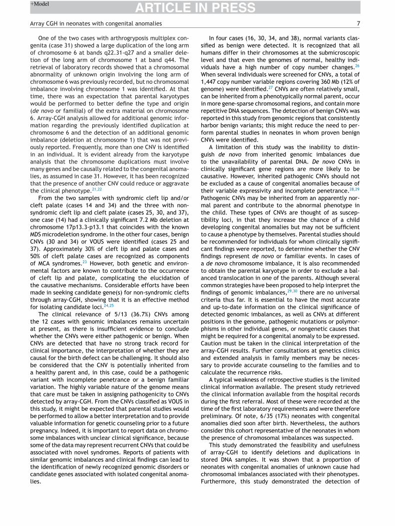
IN+Model
shlvW1gcirrhfC
gtccbtPmttdtbcfiatacficadppmCaasc
ctdtpact
o
ARTICLEArray CGH in neonates with congenital anomalies
One of the two cases with arthrogryposis multiplex con-genita (case 31) showed a large duplication of the long armof chromosome 6 at bands q22.31-q27 and a smaller dele-tion of the long arm of chromosome 1 at band q44. Theretrieval of laboratory records showed that a chromosomalabnormality of unknown origin involving the long arm ofchromosome 6 was previously recorded, but no chromosomalimbalance involving chromosome 1 was identified. At thattime, there was an expectation that parental karyotypeswould be performed to better define the type and origin(de novo or familial) of the extra material on chromosome6. Array-CGH analysis allowed for additional genomic infor-mation regarding the previously identified duplication atchromosome 6 and the detection of an additional genomicimbalance (deletion at chromosome 1) that was not previ-ously reported. Frequently, more than one CNV is identifiedin an individual. It is evident already from the karyotypeanalysis that the chromosome duplications must involvemany genes and be causally related to the congenital anoma-lies, as assumed in case 31. However, it has been recognizedthat the presence of another CNV could reduce or aggravatethe clinical phenotype.21,22
From the two samples with syndromic cleft lip and/orcleft palate (cases 14 and 34) and the three with non-syndromic cleft lip and cleft palate (cases 25, 30, and 37),one case (14) had a clinically significant 7.2 Mb deletion atchromosome 17p13.3-p13.1 that coincides with the knownMDS microdeletion syndrome. In the other four cases, benignCNVs (30 and 34) or VOUS were identified (cases 25 and37). Approximately 30% of cleft lip and palate cases and50% of cleft palate cases are recognized as componentsof MCA syndromes.23 However, both genetic and environ-mental factors are known to contribute to the occurrenceof cleft lip and palate, complicating the elucidation ofthe causative mechanisms. Considerable efforts have beenmade in seeking candidate gene(s) for non-syndromic cleftsthrough array-CGH, showing that it is an effective methodfor isolating candidate loci.24,25
The clinical relevance of 5/13 (36.7%) CNVs amongthe 12 cases with genomic imbalances remains uncertainat present, as there is insufficient evidence to concludewhether the CNVs were either pathogenic or benign. WhenCNVs are detected that have no strong track record forclinical importance, the interpretation of whether they arecausal for the birth defect can be challenging. It should alsobe considered that the CNV is potentially inherited froma healthy parent and, in this case, could be a pathogenicvariant with incomplete penetrance or a benign familiarvariation. The highly variable nature of the genome meansthat care must be taken in assigning pathogenicity to CNVsdetected by array-CGH. From the CNVs classified as VOUS inthis study, it might be expected that parental studies wouldbe performed to allow a better interpretation and to providevaluable information for genetic counseling prior to a futurepregnancy. Indeed, it is important to report data on chromo-some imbalances with unclear clinical significance, becausesome of the data may represent recurrent CNVs that could beassociated with novel syndromes. Reports of patients with
similar genomic imbalances and clinical findings can lead tothe identification of newly recognized genomic disorders orcandidate genes associated with isolated congenital anoma-lies.sncF
PRESS7
In four cases (16, 30, 34, and 38), normal variants clas-ified as benign were detected. It is recognized that allumans differ in their chromosomes at the submicroscopicevel and that even the genomes of normal, healthy indi-iduals have a high number of copy number changes.26
hen several individuals were screened for CNVs, a total of,447 copy number variable regions covering 360 Mb (12% ofenome) were identified.27 CNVs are often relatively small,an be inherited from a phenotypically normal parent, occurn more gene-sparse chromosomal regions, and contain moreepetitive DNA sequences. The detection of benign CNVs waseported in this study from genomic regions that consistentlyarbor benign variants; this might reduce the need to per-orm parental studies in neonates in whom proven benignNVs were identified.
A limitation of this study was the inability to distin-uish de novo from inherited genomic imbalances dueo the unavailability of parental DNA. De novo CNVs inlinically significant gene regions are more likely to beausative. However, inherited pathogenic CNVs should note excluded as a cause of congenital anomalies because ofheir variable expressivity and incomplete penetrance.28,29
athogenic CNVs may be inherited from an apparently nor-al parent and contribute to the abnormal phenotype in
he child. These types of CNVs are thought of as suscep-ibility loci, in that they increase the chance of a childeveloping congenital anomalies but may not be sufficiento cause a phenotype by themselves. Parental studies shoulde recommended for individuals for whom clinically signifi-ant findings were reported, to determine whether the CNVndings represent de novo or familiar events. In cases of
de novo chromosome imbalance, it is also recommendedo obtain the parental karyotype in order to exclude a bal-nced translocation in one of the parents. Although severalommon strategies have been proposed to help interpret thendings of genomic imbalances,29,30 there are no universalriteria thus far. It is essential to have the most accuratend up-to-date information on the clinical significance ofetected genomic imbalances, as well as CNVs at differentositions in the genome, pathogenic mutations or polymor-hisms in other individual genes, or nongenetic causes thatight be required for a congenital anomaly to be expressed.aution must be taken in the clinical interpretation of therray-CGH results. Further consultations at genetics clinicsnd extended analysis in family members may be neces-ary to provide accurate counseling to the families and toalculate the recurrence risks.
A typical weakness of retrospective studies is the limitedlinical information available. The present study retrievedhe clinical information available from the hospital recordsuring the first referral. Most of these were recorded at theime of the first laboratory requirements and were thereforereliminary. Of note, 6/35 (17%) neonates with congenitalnomalies died soon after birth. Nevertheless, the authorsonsider this cohort representative of the neonates in whomhe presence of chromosomal imbalances was suspected.
This study demonstrated the feasibility and usefulnessf array-CGH to identify deletions and duplications in
tored DNA samples. It was shown that a proportion ofeonates with congenital anomalies of unknown cause hadhromosomal imbalances associated with their phenotypes.urthermore, this study demonstrated the detection of
IN+Model
8
cdfi
avmd
F
C
C
T
A
TDc
R
1
1
1
1
1
1
1
1
1
1
2
2
2
2
2
2
2
2
2
2
ARTICLE
hromosomal abnormalities consistent with genetic syn-romes at an early age, when often, only a few clinicalndings are clear.
In conclusion, retrospective or prospective array-CGH as first-line diagnostic tool would benefit families by pro-iding a more accurate diagnosis and impact the overallanagement in a significant number of cases from birthefects monitoring programs.
unding
NPq/Brazil, grant 402012/2010-0.
onflicts of interest
he authors declare no conflicts of interest.
cknowledgments
he authors would like to thank the Conselho Nacional deesenvolvimento Científico e Tecnológico (CNPq) for finan-ial support (grant 402012/2010-0).
eferences
1. Schinzel A. Catalogue of unbalanced chromosome aberrationsin man. 2nd ed. Berlin: Walter de Gruyter; 2001.
2. Hochstenbach R, Buizer-Voskamp JE, Vorstman JA, Ophoff RA.Genome arrays for the detection of copy number variations inidiopathic mental retardation, idiopathic generalized epilepsyand neuropsychiatric disorders: lessons for diagnostic workflowand research. Cytogenet Genome Res. 2011;135:174---202.
3. Wellesley D, Dolk H, Boyd PA, Greenlees R, Haeusler M, Nelen V,et al. Rare chromosome abnormalities, prevalence and prena-tal diagnosis rates from population-based congenital anomalyregisters in Europe. Eur J Hum Genet. 2012;20:521---6.
4. Park SJ, Jung EH, Ryu RS, Kang HW, Chung HD, Kang HY. Theclinical application of array CGH for the detection of chromo-somal defects in 20,126 unselected newborns. Mol Cytogenet.2013;6:21.
5. Fernandes MS, Ashton-Prolla P, Matte U, Meurer L, Osvaldt A,Bittelbrunn AC, et al. The Hospital de Clinicas de Porto Ale-gre normative for the storage and use of human biologicalmaterials and their associated information in research: an inter-disciplinary approach. Revista HCPA. 2010;30:169---79.
6. Vermeesch JR, Melotte C, Froyen G, Van Vooren S, Dutta B,Maas N, et al. Molecular karyotyping: array CGH quality crite-ria for constitutional genetic diagnosis. J Histochem Cytochem.2005;53:413---22.
7. Database of Chromosomal Imbalance and Phenotype in HumansUsing Ensemble Resources (Decipher). [cited 26 Jan 2014].Available from: http://decipher.sanger.ac.uk/
8. MacDonald JR, Ziman R, Yuen RK, Feuk L, Scherer SW. TheDatabase of Genomic Variants: a curated collection of struc-tural variation in the human genome. Nucleic Acids Res.2014;42:D986---92.
9. European Cytogeneticists Association Register of Unbal-anced Chromosome Aberrations (ECARUCA). [cited 23 Feb
2014]. Available from: http://umcecaruca01.extern.umcn.nl:8080/ecaruca/ecaruca.jsp0. Ensembl Genome Browser. [cited 23 Feb 2014]. Available from:http://www.ensembl.org/index.html
3
PRESSEmy Dorfman L et al.
1. The International Standards for Cytogenomic Arrays(ISCA). [cited 23 Feb 2014]. Available from: https://www.iscaconsortium.org/index.php
2. National Center for Biotechnology Information (NCBI). [cited 23Feb 2014]. Available from: http://www.ncbi.nlm.nih.gov/
3. University California Santa Cruz (UCSC) Genome Browser. [cited23 Feb 2014]. Available from: http://genome.ucsc.edu/
4. Reddy UM, Page GP, Saade GR, Silver RM, Thorsten VR,Parker CB, et al. Karyotype versus microarray testing forgenetic abnormalities after stillbirth. N Engl J Med. 2012;367:2185---93.
5. Iourov IY, Vorsanova SG, Kurinnaia OS, Zelenova MA, SilvanovichAP, Yurov YB. Molecular karyotyping by array CGH in a Russiancohort of children with intellectual disability, autism,epilepsy and congenital anomalies. Mol Cytogenet. 2012;5:46.
6. Wapner RJ, Martin CL, Levy B, Ballif BC, Eng CM, Zachary JM,et al. Chromosomal microarray versus karyotyping for prenataldiagnosis. N Engl J Med. 2012;367:2175---84.
7. Ahn JW, Bint S, Bergbaum A, Mann K, Hall RP, Ogilvie CM. ArrayCGH as a first line diagnostic test in place of karyotyping forpostnatal referrals - results from four years’ clinical applicationfor over 8,700 patients. Mol Cytogenet. 2013;6:16.
8. Hillman SC, McMullan DJ, Hall G, Togneri FS, James N, MaherEJ, et al. Use of prenatal chromosomal microarray: prospectivecohort study and systematic review and meta-analysis. Ultra-sound Obstet Gynecol. 2013;41:610---20.
9. Lu XY, Phung MT, Shaw CA, Pham K, Neil SE, Patel A, et al.Genomic imbalances in neonates with birth defects: high detec-tion rates by using chromosomal microarray analysis. Pediatrics.2008;122:1310---8.
0. Vissers LE, Stankiewicz P. Microdeletion and microduplicationsyndromes. Methods Mol Biol. 2012;838:29---75.
1. Girirajan S, Rosenfeld JA, Coe BP, Parikh S, Friedman N,Goldstein A, et al. Phenotypic heterogeneity of genomicdisorders and rare copy-number variants. N Engl J Med.2012;367:1321---31.
2. Girirajan S. Genomic disorders: complexity at multiple levels.Genome Med. 2013;5:43.
3. Stanier P, Moore GE. Genetics of cleft lip and palate: syndromicgenes contribute to the incidence of non-syndromic clefts. HumMol Genet. 2004;13:R73---81.
4. Osoegawa K, Vessere GM, Utami KH, Mansilla MA, Johnson MK,Riley BM, et al. Identification of novel candidate genes associ-ated with cleft lip and palate using array comparative genomichybridisation. J Med Genet. 2008;45:81---6.
5. Shi M, Mostowska A, Jugessur A, Johnson MK, Mansilla MA, Chris-tensen K, et al. Identification of microdeletions in candidategenes for cleft lip and/or palate. Birth Defects Res A Clin MolTeratol. 2009;85:42---51.
6. Iafrate AJ, Feuk L, Rivera MN, Listewnik ML, Donahoe PK, Qi Y,et al. Detection of large-scale variation in the human genome.Nat Genet. 2004;36:949---51.
7. Redon R, Ishikawa S, Fitch KR, Feuk L, Perry GH, Andrews TD,et al. Global variation in copy number in the human genome.Nature. 2006;444:444---54.
8. Kearney HM, South ST, Wolff DJ, Lamb A, Hamosh A, RaoKW, et al. American College of Medical Genetics recommenda-tions for the design and performance expectations for clinicalgenomic copy number microarrays intended for use in thepostnatal setting for detection of constitutional abnormalities.Genet Med. 2011;13:676---9.
9. Vermeesch JR, Brady PD, Sanlaville D, Kok K, Hastings RJ.Genome-wide arrays: quality criteria and platforms to be used
in routine diagnostics. Hum Mutat. 2012;33:906---15.0. de Leeuw N, Dijkhuizen T, Hehir-Kwa JY, Carter NP, Feuk L, FirthHV, et al. Diagnostic interpretation of array data using publicdatabases and internet sources. Hum Mutat. 2012;33:930---40.
Related Documents