Chapter 6 Calcium Imbalances Because many factors affect calcium regulation, there are a multitude of causes of disturbed calcium balance. For this reason, both hypocalcemia and hypercalcemia are relatively common imbalances. To facilitate understanding of cal- cium disturbances, it is helpful to review factors that affect calcium balance. CALCIUM BALANCE Distribution and Function Calcium is the body’s most abundant divalent cation. 1 More than 99% of the body’s calcium is concentrated in the skele- tal system, and approximately 1% is rapidly exchangeable with blood calcium (the remainder is more stable and exchanged only slowly). The small amount of calcium located outside the bone circulates in the serum, partly bound to protein and partly ionized. Calcium has a major role in transmission of nerve impulses. It helps regulate muscle contraction and relaxation, including normal heart- beat. Calcium has a vital role in the cardiac action potential and is essential for cardiac pacemaker automaticity. This ion is also involved in blood clotting and hormone secre- tion. Recommended adequate intake for calcium for both men and women aged 19 to 50 years is 1000 mg/day; some experts recommend increasing this amount to 1200 mg/day for those persons older than 50 years. 2 Evidence suggests that calcium and vitamin D play important roles in the primary prevention of colorectal neo- plasia. 3 It appears that calcium binds bile acids in the bowel lumen, inhibiting bile-induced mucosal damage. 4 Calcium is closely tied to magnesium and phosphorus regulation. 5 Measurement of Calcium in Blood The test most frequently performed in clinical settings to mea- sure serum calcium is total calcium, with results normally ranging from 8.9 to 10.3 mg/dL (roughly equivalent to 2.23 to 2.57 mmol/L). The total calcium in serum is the sum of the ionized (47%) and non-ionized (53%) calcium components. In the non-ionized portion, calcium is primarily bound to albumin (and, to a lesser extent, to other anions such as citrate and phosphate). When serum albumin levels and pH are within normal ranges, readings from total calcium are gener- ally useful. In contrast, when the serum albumin level is abnor- mal, corrections must be made in the reported total serum calcium levels. In noncritically ill patients, it is estimated that a 1.0 g/dL decrease in the serum albumin is accompanied by a 0.8 mg/dL decrease in the total calcium. The following is a convenient formula sometimes used to calculate the “cor- rected” calcium level when hypoalbuminemia is present: Corrected calcium (mg/dL) measured serum calcium 0.8 (4.0 – measured serum albumin g/dL) For example, if the patient’s serum albumin level is below normal by 1 g/dL (e.g., 3.0 g/dL rather than 4.0 g/dL), a measured total serum calcium concentration of 8.0 mg/dL should be adjusted upward to 8.8 mg/dL. In this situation, the ionized calcium level would be estimated at approxi- mately half of the adjusted value. The direct relationship between albumin and total calcium often leads clinicians to ignore a low total serum calcium level in the presence of a similarly low serum albumin level. The ionized calcium level in the bloodstream is affected by plasma pH. For example, when the arterial pH increases 91 © Jones & Bartlett Learning, LLC. NOT FOR SALE OR DISTRIBUTION.

Welcome message from author
This document is posted to help you gain knowledge. Please leave a comment to let me know what you think about it! Share it to your friends and learn new things together.
Transcript
Chapter 6
Calcium Imbalances
Because many factors affect calcium regulation, there are amultitude of causes of disturbed calcium balance. For thisreason, both hypocalcemia and hypercalcemia are relativelycommon imbalances. To facilitate understanding of cal-cium disturbances, it is helpful to review factors that affectcalcium balance.
CALCIUM BALANCE
Distribution and Function
Calcium is the body’s most abundant divalent cation.1 Morethan 99% of the body’s calcium is concentrated in the skele-tal system, and approximately 1% is rapidly exchangeablewith blood calcium (the remainder is more stable andexchanged only slowly). The small amount of calciumlocated outside the bone circulates in the serum, partlybound to protein and partly ionized. Calcium has a majorrole in transmission of nerve impulses. It helps regulatemuscle contraction and relaxation, including normal heart-beat. Calcium has a vital role in the cardiac action potentialand is essential for cardiac pacemaker automaticity. Thision is also involved in blood clotting and hormone secre-tion. Recommended adequate intake for calcium for bothmen and women aged 19 to 50 years is 1000 mg/day; someexperts recommend increasing this amount to 1200 mg/dayfor those persons older than 50 years.2
Evidence suggests that calcium and vitamin D playimportant roles in the primary prevention of colorectal neo-plasia.3 It appears that calcium binds bile acids in the bowellumen, inhibiting bile-induced mucosal damage.4 Calcium isclosely tied to magnesium and phosphorus regulation.5
Measurement of Calcium in Blood
The test most frequently performed in clinical settings to mea-sure serum calcium is total calcium, with results normallyranging from 8.9 to 10.3 mg/dL (roughly equivalent to 2.23 to2.57 mmol/L). The total calcium in serum is the sum of theionized (47%) and non-ionized (53%) calcium components.In the non-ionized portion, calcium is primarily bound toalbumin (and, to a lesser extent, to other anions such as citrateand phosphate). When serum albumin levels and pH arewithin normal ranges, readings from total calcium are gener-ally useful. In contrast, when the serum albumin level is abnor-mal, corrections must be made in the reported total serumcalcium levels. In noncritically ill patients, it is estimated that a1.0 g/dL decrease in the serum albumin is accompanied by a0.8 mg/dL decrease in the total calcium. The following is aconvenient formula sometimes used to calculate the “cor-rected” calcium level when hypoalbuminemia is present:
Corrected calcium (mg/dL) � measured serum calcium �
0.8 � (4.0 – measured serum albumin g/dL)
For example, if the patient’s serum albumin level is belownormal by 1 g/dL (e.g., 3.0 g/dL rather than 4.0 g/dL), ameasured total serum calcium concentration of 8.0 mg/dLshould be adjusted upward to 8.8 mg/dL. In this situation,the ionized calcium level would be estimated at approxi-mately half of the adjusted value. The direct relationshipbetween albumin and total calcium often leads clinicians toignore a low total serum calcium level in the presence of asimilarly low serum albumin level.
The ionized calcium level in the bloodstream is affectedby plasma pH. For example, when the arterial pH increases
91
81644_CH06_091_110_QXP6.qxd 9/14/10 11:07 AM Page 91
© Jones & Bartlett Learning, LLC. NOT FOR SALE OR DISTRIBUTION.
© Jones & Bartlett Learning, LLCNOT FOR SALE OR DISTRIBUTION
© Jones & Bartlett Learning, LLCNOT FOR SALE OR DISTRIBUTION
© Jones & Bartlett Learning, LLCNOT FOR SALE OR DISTRIBUTION
© Jones & Bartlett Learning, LLCNOT FOR SALE OR DISTRIBUTION
© Jones & Bartlett Learning, LLCNOT FOR SALE OR DISTRIBUTION
© Jones & Bartlett Learning, LLCNOT FOR SALE OR DISTRIBUTION
© Jones & Bartlett Learning, LLCNOT FOR SALE OR DISTRIBUTION
© Jones & Bartlett Learning, LLCNOT FOR SALE OR DISTRIBUTION
© Jones & Bartlett Learning, LLCNOT FOR SALE OR DISTRIBUTION
© Jones & Bartlett Learning, LLCNOT FOR SALE OR DISTRIBUTION
© Jones & Bartlett Learning, LLCNOT FOR SALE OR DISTRIBUTION
© Jones & Bartlett Learning, LLCNOT FOR SALE OR DISTRIBUTION
© Jones & Bartlett Learning, LLCNOT FOR SALE OR DISTRIBUTION
© Jones & Bartlett Learning, LLCNOT FOR SALE OR DISTRIBUTION
© Jones & Bartlett Learning, LLCNOT FOR SALE OR DISTRIBUTION
© Jones & Bartlett Learning, LLCNOT FOR SALE OR DISTRIBUTION
© Jones & Bartlett Learning, LLCNOT FOR SALE OR DISTRIBUTION
© Jones & Bartlett Learning, LLCNOT FOR SALE OR DISTRIBUTION
© Jones & Bartlett Learning, LLCNOT FOR SALE OR DISTRIBUTION
© Jones & Bartlett Learning, LLCNOT FOR SALE OR DISTRIBUTION
(alkalosis), more calcium becomes bound to protein.Although the total serum calcium remains unchanged, theionized portion decreases. Therefore, symptoms ofhypocalcemia often occur when alkalosis is present (despitea normal total calcium level). Acidosis (low pH) has theopposite effect; that is, less calcium is bound to proteinand, therefore, more exists in the ionized form. Signs ofhypocalcemia will develop only rarely in the presence ofacidosis, even when the total serum calcium level is lowerthan normal.
Direct measurement of ionized calcium by the labora-tory is highly desirable, especially in critically ill patients.Recall that the ionized calcium concentration is the physi-ologically active and clinically important component.6
Whole blood, heparinized plasma, or serum may be usedfor the measurement of calcium ionization.7 Instructionsfrom the laboratoy that is performing the analysis need tobe carefully followed to assure accurate results. The nor-mal value for urinary calcium is dependent on dietary cal-cium intake. Urine specimen collected for calcium analysisneed to be appropriately acidified to prevent calcium saltprecipitation.8
Regulation
Many biochemical and hormonal factors act to maintain anormal calcium balance. Among the most important areparathyroid hormone (PTH), calcitonin, and calcitriol (anactive metabolite of vitamin D). PTH promotes transfer ofcalcium from the bone to the plasma, thereby raising theplasma calcium level. The bones and teeth are ready sourcesfor replenishment of low plasma calcium levels. PTH alsoaugments the intestinal absorption of calcium and enhancesthe net renal calcium reabsorption. Calcium is absorbedprimarily in the duodenum and jejunum.9
Calcitonin (which is produced in the thyroid as well asseveral other tissues) is a physiological antagonist of PTH.Calcitonin secretion is directly stimulated by a high serumcalcium concentration. At high levels, calcitonin inhibitsbone resorption; the resultant reduced flux of calcium frombone causes a reduction in the serum calcium level.
Calcitriol (1,25-dihydroxyvitamin D) is a hormone thatincreases the extracellular calcium concentration by threemain actions: promotion of calcium absorption from theintestine, enhancement of bone resorption of calcium, andstimulation of renal tubular reabsorption of calcium. Cal-citriol has a synergistic effect with parathyroid hormone onbone resorption.
Osteoporosis
Osteoporosis is associated with prolonged low intake of cal-cium. It is characterized by loss of bone mass, which in turncauses bones to become porous, brittle, and susceptible tofracture. In the United States, osteoporosis is estimated tocause 1.5 million fractures annually, primarily of the hipand spine.10 Although serum calcium levels are usually nor-mal in individuals with osteoporosis, total body calciumstores are greatly diminished. Bone loss begins at an earlierage in women than in men and is accelerated by meno-pause. However, men also develop a negative calcium bal-ance in later years, at which point they may becomevulnerable to osteoporosis. A dual process is involved inosteoporosis: increased bone resorption and inadequatebone formation. Menopause leads to rapid bone loss inwomen because estrogen deficiency reduces calciumabsorption and increases excretion; as a result, bone loss faroutpaces bone deposition.
Risk of developing serious bone problems is greater inpostmenopausal, physically inactive women who areelderly, thin and small-framed, and smokers, and in thosewho have a diet deficient in calcium.11 Inactivity predis-poses to bone loss by reducing the efficiency of calciumuse. Conversely, regular physical exercise (such as running,walking, or bicycling) slows the rate of bone loss andimproves calcium balance. Considering the magnitude ofthe problems associated with osteoporosis, prevention isthe only cost-effective approach. Also important in the pre-vention of osteoporosis is elimination of bone toxins (suchas cigarettes and heavy alcohol ingestion). Hypogonadalwomen who take estrogen have a reduced risk of develop-ing osteoporosis; thus it is one factor to consider whendeciding whether to take estrogen (hormone replacementtherapy [HRT]).12 It appears that low doses of estrogen areadequate to prevent postmenopausal osteoporosis; how-ever, once osteoporosis has developed, it is not an effectivetreatment.13
Bisphosphonates are the most commonly used drugsfor the treatment of osteoporosis. Since the first suchagent was introduced, more than 190 million prescrip-tions have been written for their use.14 All patients receiv-ing bisphosphonates should have adequate calcium andvitamin D intake before and during therapy.15 Somereports have described development of bisphosphonate-induced hypocalcemia in patients with unrecognizedhypoparathyroidism, vitamin D deficiency, or impairedrenal function.16
92 CHAPTER 6 CALCIUM IMBALANCES
81644_CH06_091_110_QXP6.qxd 9/14/10 11:07 AM Page 92
© Jones & Bartlett Learning, LLC. NOT FOR SALE OR DISTRIBUTION.
© Jones & Bartlett Learning, LLCNOT FOR SALE OR DISTRIBUTION
© Jones & Bartlett Learning, LLCNOT FOR SALE OR DISTRIBUTION
© Jones & Bartlett Learning, LLCNOT FOR SALE OR DISTRIBUTION
© Jones & Bartlett Learning, LLCNOT FOR SALE OR DISTRIBUTION
© Jones & Bartlett Learning, LLCNOT FOR SALE OR DISTRIBUTION
© Jones & Bartlett Learning, LLCNOT FOR SALE OR DISTRIBUTION
© Jones & Bartlett Learning, LLCNOT FOR SALE OR DISTRIBUTION
© Jones & Bartlett Learning, LLCNOT FOR SALE OR DISTRIBUTION
© Jones & Bartlett Learning, LLCNOT FOR SALE OR DISTRIBUTION
© Jones & Bartlett Learning, LLCNOT FOR SALE OR DISTRIBUTION
© Jones & Bartlett Learning, LLCNOT FOR SALE OR DISTRIBUTION
© Jones & Bartlett Learning, LLCNOT FOR SALE OR DISTRIBUTION
© Jones & Bartlett Learning, LLCNOT FOR SALE OR DISTRIBUTION
© Jones & Bartlett Learning, LLCNOT FOR SALE OR DISTRIBUTION
© Jones & Bartlett Learning, LLCNOT FOR SALE OR DISTRIBUTION
© Jones & Bartlett Learning, LLCNOT FOR SALE OR DISTRIBUTION
© Jones & Bartlett Learning, LLCNOT FOR SALE OR DISTRIBUTION
© Jones & Bartlett Learning, LLCNOT FOR SALE OR DISTRIBUTION
© Jones & Bartlett Learning, LLCNOT FOR SALE OR DISTRIBUTION
© Jones & Bartlett Learning, LLCNOT FOR SALE OR DISTRIBUTION
HYPOCALCEMIA
Hypocalcemia may be defined as a total serum calcium levelof less than 8.9 mg/dL and an ionized calcium concentra-tion of less than 4.6 mg/dL. However, it is important to rec-ognize that reporting laboratories often have slightlydiffering values for normal calcium levels. Hypocalcemia isa common imbalance in critically ill patients. For example,the prevalence of ionized hypocalcemia is reported to rangefrom 60% to 85% in medical, surgical, and trauma pa-tients.17 Hypocalcemia is a serious imbalance, in that it canpotentiate cardiac arrhythmias and seizures.18 Althoughmortality is greater in patients with hypocalcemia, this out-come does not appear to be independently associated withthe imbalance.19
As discussed earlier, hypoalbuminemia can produce afalsely low total serum calcium test result (referred to aspseudohypocalcemia). In this situation, the ionized calciumlevel remains normal and the patient is asymptomatic andrequires no treatment.
Causes
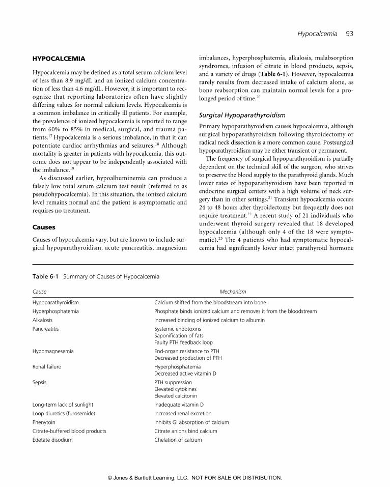
Causes of hypocalcemia vary, but are known to include sur-gical hypoparathyroidism, acute pancreatitis, magnesium
imbalances, hyperphosphatemia, alkalosis, malabsorptionsyndromes, infusion of citrate in blood products, sepsis,and a variety of drugs (Table 6-1). However, hypocalcemiararely results from decreased intake of calcium alone, asbone reabsorption can maintain normal levels for a pro-longed period of time.20
Surgical Hypoparathyroidism
Primary hypoparathyroidism causes hypocalcemia, althoughsurgical hypoparathyroidism following thyroidectomy orradical neck dissection is a more common cause. Postsurgicalhypoparathyroidism may be either transient or permanent.
The frequency of surgical hypoparathyroidism is partiallydependent on the technical skill of the surgeon, who strivesto preserve the blood supply to the parathyroid glands. Muchlower rates of hypoparathyroidism have been reported inendocrine surgical centers with a high volume of neck sur-gery than in other settings.21 Transient hypocalcemia occurs24 to 48 hours after thyroidectomy but frequently does notrequire treatment.22 A recent study of 21 individuals whounderwent thyroid surgery revealed that 18 developedhypocalcemia (although only 4 of the 18 were sympto-matic).23 The 4 patients who had symptomatic hypocal-cemia had significantly lower intact parathyroid hormone
Hypocalcemia 93
Table 6-1 Summary of Causes of Hypocalcemia
Cause Mechanism
Hypoparathyroidism Calcium shifted from the bloodstream into bone
Hyperphosphatemia Phosphate binds ionized calcium and removes it from the bloodstream
Alkalosis Increased binding of ionized calcium to albumin
Pancreatitis Systemic endotoxinsSaponification of fatsFaulty PTH feedback loop
Hypomagnesemia End-organ resistance to PTHDecreased production of PTH
Renal failure HyperphosphatemiaDecreased active vitamin D
Sepsis PTH suppressionElevated cytokinesElevated calcitonin
Long-term lack of sunlight Inadequate vitamin D
Loop diuretics (furosemide) Increased renal excretion
Phenytoin Inhibits GI absorption of calcium
Citrate-buffered blood products Citrate anions bind calcium
Edetate disodium Chelation of calcium
81644_CH06_091_110_QXP6.qxd 9/14/10 11:07 AM Page 93
© Jones & Bartlett Learning, LLC. NOT FOR SALE OR DISTRIBUTION.
© Jones & Bartlett Learning, LLCNOT FOR SALE OR DISTRIBUTION
© Jones & Bartlett Learning, LLCNOT FOR SALE OR DISTRIBUTION
© Jones & Bartlett Learning, LLCNOT FOR SALE OR DISTRIBUTION
© Jones & Bartlett Learning, LLCNOT FOR SALE OR DISTRIBUTION
© Jones & Bartlett Learning, LLCNOT FOR SALE OR DISTRIBUTION
© Jones & Bartlett Learning, LLCNOT FOR SALE OR DISTRIBUTION
© Jones & Bartlett Learning, LLCNOT FOR SALE OR DISTRIBUTION
© Jones & Bartlett Learning, LLCNOT FOR SALE OR DISTRIBUTION
© Jones & Bartlett Learning, LLCNOT FOR SALE OR DISTRIBUTION
© Jones & Bartlett Learning, LLCNOT FOR SALE OR DISTRIBUTION
© Jones & Bartlett Learning, LLCNOT FOR SALE OR DISTRIBUTION
© Jones & Bartlett Learning, LLCNOT FOR SALE OR DISTRIBUTION
© Jones & Bartlett Learning, LLCNOT FOR SALE OR DISTRIBUTION
© Jones & Bartlett Learning, LLCNOT FOR SALE OR DISTRIBUTION
© Jones & Bartlett Learning, LLCNOT FOR SALE OR DISTRIBUTION
© Jones & Bartlett Learning, LLCNOT FOR SALE OR DISTRIBUTION
© Jones & Bartlett Learning, LLCNOT FOR SALE OR DISTRIBUTION
© Jones & Bartlett Learning, LLCNOT FOR SALE OR DISTRIBUTION
© Jones & Bartlett Learning, LLCNOT FOR SALE OR DISTRIBUTION
© Jones & Bartlett Learning, LLCNOT FOR SALE OR DISTRIBUTION
(iPTH) levels than did the 14 patients without symptomsand the remaining 3 without hypocalcemia. The researchersconcluded that a 1-hour postoperative iPTH level of 2.5pmol/L or less can identify individuals at risk for developingsymptomatic hypocalcemia.
The most likely mechanism in terms of causing hypocal-cemia after radical neck dissection is ischemia to theparathyroid tissue following dissection and hemostaticmaneuvers. It is also possible that trauma to the parathyroidglands precludes PTH from increasing to the level needed toelevate the low serum calcium concentration, thus con-tributing to the development of hypocalcemia. If perma-nent parathyroid damage has not occurred, parathyroidinsufficiency resolves as edema at the surgical site lessensand revascularization occurs, allowing reestablishment ofparathyroid gland integrity. Extensive neck surgery (as inradical neck dissection for cancer) is more likely to be asso-ciated with permanent hypoparathyroidism than are lessinvolved surgical maneuvers.
Most patients who develop hypocalcemia after neck sur-gery are asymptomatic; however, some may develop pares-thesias, laryngeal spasm, or tetany. It is common practice tocheck serum ionized calcium levels at regular intervals inthe early postoperative period in patients who have under-gone neck surgery.
With the emphasis on cost containment in the currenthealthcare environment, concerns have been raised thatpatients who have undergone thyroid or parathyroid glandsurgery may be discharged before postoperative hypocalcemiabecomes manifest. A retrospective study of 197 patients whohad undergone such operations indicated that early post-operative calcium levels give a good indication of whetherhypocalcemia is likely to occur.24 In the study, postoperativecalcium levels were plotted as a function of time, and theslope between the first two postoperative calcium levels wasexamined. The results indicated that an initial upsloping post-operative calcium curve based on these two early postopera-tive calcium measurements is a strong predictor of a stablepostoperative calcium level; conversely, a steeply downslopinginitial calcium curve is worrisome for eventual hypocalcemia.
Acute Pancreatitis
Hypocalcemia is not uncommon during acute pancreatitisand is associated with a poor outcome. While it is unclearprecisely which mechanisms cause hypocalcemia in this set-ting, several possibilities have been identified. Inflammationof the pancreas causes release of proteolytic and lipolyticenzymes; it is believed that calcium ions combine with the
fatty acids, forming soaps and thereby decreasing the serumcalcium concentration.25 In contrast, other researchers haveconcluded that systemic endotoxin exposure may play a sig-nificant role in the development of hypocalcemia in patientswith acute pancreatitis.26 Other investigators have foundthat there is an inadequate PTH response to the hypocal-cemia caused by acute pancreatitis.27 In any event, ionizedhypocalcemia is a common problem, occurring in as manyas 85% of patients with acute severe pancreatitis.28 SeeChapter 20 for a more extensive discussion of this topic.
Magnesium Abnormalities
The serum magnesium level influences both PTH secretionand action and, therefore, the serum calcium level. Severehypomagnesemia (less than 1 mg/dL) inhibits PTH secre-tion. One study reported that 22% of the patients withhypocalcemia also had hypomagnesemia.29 Hypomagne-semic hypocalcemia responds poorly to calcium therapyalone but can be resolved through concurrent calcium andmagnesium replacement.
Hyperphosphatemia
Hyperphosphatemia that develops rapidly is associated withhypocalcemia. This condition might be seen in patients whoreceive excessive hypertonic sodium phosphate enemas. Forexample, as described in a recent report, a 13-year-old boywith chronic constipation developed severe hyperphos-phatemia and hypocalcemia after receiving four hypertonicsodium phosphate pediatric enemas for severe constipa-tion.30 It is important for clinicians to recognize that theseenemas are absorbable and can lead to potentially lethalcomplications if given improperly. See Chapters 8 and 13for a more detailed discussion of this topic.
Alkalosis
Blood pH alters Ca�� binding to serum proteins. In alkalo-sis, a greater amount of calcium is bound to plasma pro-teins, resulting in a smaller percentage of ionized calcium.Thus patients with alkalosis are more susceptible tohypocalcemic tetany.
Inadequate Vitamin D
Inadequate consumption of vitamin D or insufficient expo-sure to the sun (ultraviolet radiation) can cause reducedcalcium absorption, leading to hypocalcemia. Deficiency ofvitamin D occurs in malabsorptive states, as described inthe next subsection. It is not uncommon for elderly personsto have low vitamin D levels. Breastfed infants born to
94 CHAPTER 6 CALCIUM IMBALANCES
81644_CH06_091_110_QXP6.qxd 9/14/10 11:07 AM Page 94
© Jones & Bartlett Learning, LLC. NOT FOR SALE OR DISTRIBUTION.
© Jones & Bartlett Learning, LLCNOT FOR SALE OR DISTRIBUTION
© Jones & Bartlett Learning, LLCNOT FOR SALE OR DISTRIBUTION
© Jones & Bartlett Learning, LLCNOT FOR SALE OR DISTRIBUTION
© Jones & Bartlett Learning, LLCNOT FOR SALE OR DISTRIBUTION
© Jones & Bartlett Learning, LLCNOT FOR SALE OR DISTRIBUTION
© Jones & Bartlett Learning, LLCNOT FOR SALE OR DISTRIBUTION
© Jones & Bartlett Learning, LLCNOT FOR SALE OR DISTRIBUTION
© Jones & Bartlett Learning, LLCNOT FOR SALE OR DISTRIBUTION
© Jones & Bartlett Learning, LLCNOT FOR SALE OR DISTRIBUTION
© Jones & Bartlett Learning, LLCNOT FOR SALE OR DISTRIBUTION
© Jones & Bartlett Learning, LLCNOT FOR SALE OR DISTRIBUTION
© Jones & Bartlett Learning, LLCNOT FOR SALE OR DISTRIBUTION
© Jones & Bartlett Learning, LLCNOT FOR SALE OR DISTRIBUTION
© Jones & Bartlett Learning, LLCNOT FOR SALE OR DISTRIBUTION
© Jones & Bartlett Learning, LLCNOT FOR SALE OR DISTRIBUTION
© Jones & Bartlett Learning, LLCNOT FOR SALE OR DISTRIBUTION
© Jones & Bartlett Learning, LLCNOT FOR SALE OR DISTRIBUTION
© Jones & Bartlett Learning, LLCNOT FOR SALE OR DISTRIBUTION
© Jones & Bartlett Learning, LLCNOT FOR SALE OR DISTRIBUTION
© Jones & Bartlett Learning, LLCNOT FOR SALE OR DISTRIBUTION
mothers who are vitamin D deficient are at risk for develop-ing vitamin D deficiency and hypocalcemia; unfortunately,maternal vitamin D deficiency is not uncommon.31
Malabsorption Syndromes
Intestinal malabsorptive disorders are likely to lead tohypocalcemia by decreasing the absorption of vitamin D,bile salts, and calcium. In a study involving 82 patients whounderwent biliopancreatic bypass from 1988 to 2001, 26%were found to have hypocalcemia and 50% were found tohave low vitamin D levels (despite the fact that most tookmultivitamins).32
Infusion of Citrate in Blood Products
Decreases in ionized Ca�� during blood transfusion corre-late with speed of the transfusion and circulating citrate lev-els. Hypocalcemia is seen more commonly during thetransfusion of plasma and platelets, which have high citrateconcentrations.33 Citrate is added to banked blood to act asan anticoagulant and to preserve the life of the blood. Usu-ally the citrate in blood is rapidly metabolized by the liver asit is transfused and presents no problem for calcium bal-ance. However, when blood is transfused faster than metab-olism of the excess citrate can occur, hypocalcemia results.Recall that citrate is a negatively charged ion and that cal-cium is a positively charged ion; thus the two ions areattracted to each other. Therefore, transient hypocalcemiacan occur with massive administration of citrated blood (asin exchange transfusions in neonates), as calcium ions com-bine with the citrate and are temporarily removed from thecirculation (a process referred to as chelation). Citratemetabolism is hindered in patients with liver disease, shock,and hypothermia. Small children and osteoporotic adultsare also at increased risk for citrate/calcium imbalancesbecause they tend to have inadequate stores of bone calciumand, therefore, are less able to compensate for decliningionized calcium levels. When citrate intoxication occurs, itmay be manifested as circumoral paresthesias, muscletremors, or tetany.
The infusion of packed red blood cells (instead of wholeblood) lowers the amount of citrate infused, therebydecreasing the already low risk for hypocalcemia after trans-fusions. However, there is sufficient citrate even in packedred blood cells to affect calcium balance. Two cases werereported in which the transfusion of small volumes ofpacked red blood cells proved sufficient to precipitatesymptomatic hypocalcemia.34 Subsequent investigationrevealed that both of the patients had preexisting,
untreated, and asymptomatic hypocalcemia (one followingpartial thyroidectomy years earlier and the other with docu-mented hypocalcemia but without a definitive cause).
As indicated previously, hypocalcemia is more com-monly observed during plasmapheresis than during bloodtransfusions. Ordinarily the citrate anticoagulant used dur-ing apheresis procedures is considered a safe medicationbecause it is rapidly metabolized by the donor; however,life-threatening hypocalcemia can occur if the infusion rateof the citrate is too fast. A case was reported in which citratewas inadvertently administered too rapidly to a 54-year-oldwoman due to malfunction of the anticoagulant line of anapheresis instrument.35 Seven minutes into the procedurethe patient developed muscle spasm, chest pain, andhypotension; her serum ionized calcium level was 0.64mmol/L (the normal level in the reporting laboratory was inthe range of 1.18–1.38 mmol/L).
Drugs
A variety of medications can predispose to hypocalcemia. Forexample, loop diuretics increase renal excretion of calciumand phenytoin inhibits intestinal absorption of calcium.Phosphate-containing agents bind calcium in the intestinaltract and, therefore, interfere with its absorption. Edetatedisodium (EDTA) is a chelating agent used in the treatmentof toxic metal poisoning; three deaths were recentlyreported from hypocalcemia following the administrationof this agent.36
Alcoholism
Alcoholics are at risk for hypocalcemia for many reasons.Among these are intestinal malabsorption, low levels of 25-hydroxyvitamin D, hypomagnesemia, hypoalbuminemia,respiratory and metabolic alkalosis, and pancreatitis. Themost significant of these conditions is probably hypomag-nesemia (caused by the toxic effects of alcohol). Magnesiumreplacement in alcoholics helps to correct hypocalcemia byincreasing responsiveness to PTH.
Neonatal Hypocalcemia
Two types of hypocalcemia can occur in newborn infants.The first develops early, during the first 3 days of life. Thistype is attributed to parathyroid immaturity or maternalhyperparathyroidism (or both), resulting in neonatal para-thyroid gland suppression; it most often resolves within thefirst week of life. Among the predisposing factors for thiscondition are prematurity, maternal insulin-dependent dia-betes mellitus, and asphyxia at birth. A recent study of 381
Hypocalcemia 95
81644_CH06_091_110_QXP6.qxd 9/14/10 11:07 AM Page 95
© Jones & Bartlett Learning, LLC. NOT FOR SALE OR DISTRIBUTION.
© Jones & Bartlett Learning, LLCNOT FOR SALE OR DISTRIBUTION
© Jones & Bartlett Learning, LLCNOT FOR SALE OR DISTRIBUTION
© Jones & Bartlett Learning, LLCNOT FOR SALE OR DISTRIBUTION
© Jones & Bartlett Learning, LLCNOT FOR SALE OR DISTRIBUTION
© Jones & Bartlett Learning, LLCNOT FOR SALE OR DISTRIBUTION
© Jones & Bartlett Learning, LLCNOT FOR SALE OR DISTRIBUTION
© Jones & Bartlett Learning, LLCNOT FOR SALE OR DISTRIBUTION
© Jones & Bartlett Learning, LLCNOT FOR SALE OR DISTRIBUTION
© Jones & Bartlett Learning, LLCNOT FOR SALE OR DISTRIBUTION
© Jones & Bartlett Learning, LLCNOT FOR SALE OR DISTRIBUTION
© Jones & Bartlett Learning, LLCNOT FOR SALE OR DISTRIBUTION
© Jones & Bartlett Learning, LLCNOT FOR SALE OR DISTRIBUTION
© Jones & Bartlett Learning, LLCNOT FOR SALE OR DISTRIBUTION
© Jones & Bartlett Learning, LLCNOT FOR SALE OR DISTRIBUTION
© Jones & Bartlett Learning, LLCNOT FOR SALE OR DISTRIBUTION
© Jones & Bartlett Learning, LLCNOT FOR SALE OR DISTRIBUTION
© Jones & Bartlett Learning, LLCNOT FOR SALE OR DISTRIBUTION
© Jones & Bartlett Learning, LLCNOT FOR SALE OR DISTRIBUTION
© Jones & Bartlett Learning, LLCNOT FOR SALE OR DISTRIBUTION
© Jones & Bartlett Learning, LLCNOT FOR SALE OR DISTRIBUTION
calcium levels from 111 extremely low birthweight (ELBW)infants during the first 48 hours of life found that themajority (59.9%) had at least one hypocalcemic value.37 Theinvestigators concluded that serum calcium values are lowerin ELBW infants and that the values may be inconsequen-tial; they recommended that hypocalcemia be redefined forELBW infants.
A second type of neonatal hypocalcemia occurs approxi-mately 1 week after birth and is associated with hyperphos-phatemia and hypomagnesemia. Hypocalcemia in infantswith this “late-onset” condition can be caused by feedingthem milk with a high phosphorus level, leading to hyper-phosphatemia and then to hypocalcemia. Low serum cal-cium levels can persist until the child’s parathyroid glandsfunction well enough to respond.
Sepsis
Although hypocalcemia in critically ill, septic patients is com-mon, the underlying basis for this condition is unclear.Researchers have postulated that calcium shifts from theextracellular compartment into the cells and that the hor-monal response to the resultant hypocalcemia is inadequate.Possible causes for hypocalcemia in a group of patients withgram-negative sepsis described by Zaloga and Chernowincluded acquired parathyroid gland insufficiency, dietaryvitamin D deficiency, and renal hydroxylase insufficiency.38
Other sources have reported that hypocalcemia in septic criti-cally ill patients may be related to an inflammatory response.39
In experimental settings, calcium administration in sepsishas been shown to increase or have no effect on mortality.40 Itis not known if sepsis-induced hypocalcemia is protective orharmful to the patient; there is no evidence that routine cal-cium replacement is needed but treatment is generally advo-cated for symptomatic patients.41 A review of the literature byForsythe et al. found no clear evidence that parenteral cal-cium supplementation affects the outcome of critically illpatients.42
Other Factors
Conditions commonly associated with low serum albuminlevels (such as cirrhosis of the liver and the nephrotic syn-drome) are frequently associated with a low total serum cal-cium concentration. Often, the ionized calciumconcentration is normal and no symptoms of hypocalcemiaappear. Medullary thyroid carcinoma may produce hypo-calcemia if calcitonin (a calcium-lowering hormone) issecreted by the tumor.
Hypocalcemia was reported in almost 18% of 66 patientswith acquired immune deficiency syndrome (AIDS).43 Theresearchers postulated that intestinal malabsorption of vita-min D is the most likely cause of hypocalcemia in thispatient population.
Clinical Signs
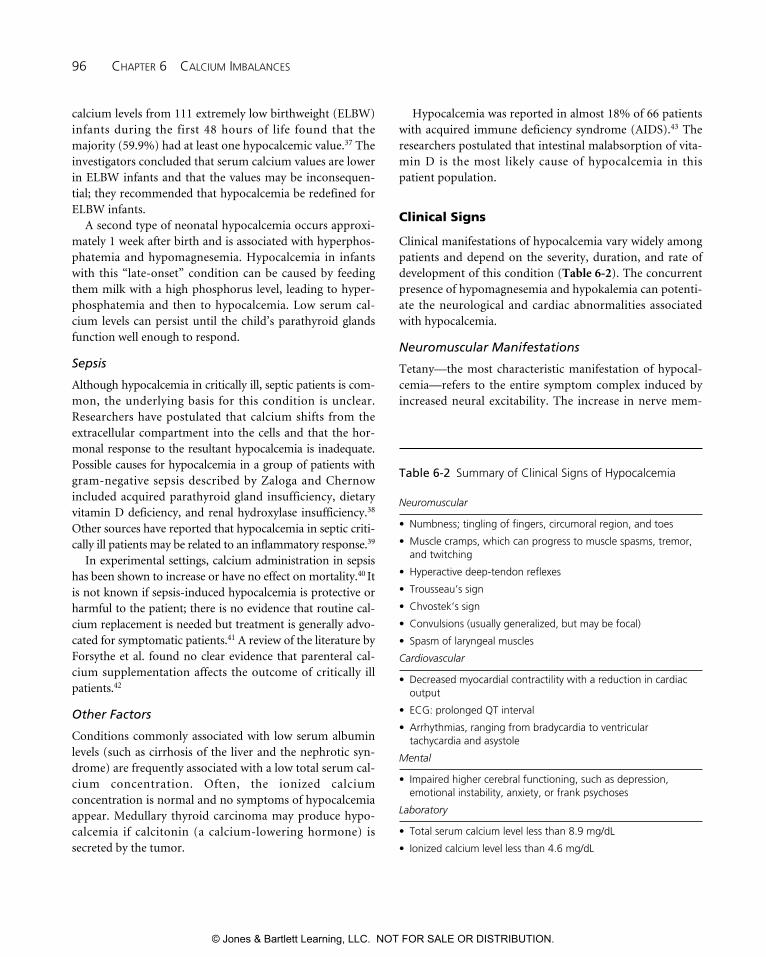
Clinical manifestations of hypocalcemia vary widely amongpatients and depend on the severity, duration, and rate ofdevelopment of this condition (Table 6-2). The concurrentpresence of hypomagnesemia and hypokalemia can potenti-ate the neurological and cardiac abnormalities associatedwith hypocalcemia.
Neuromuscular Manifestations
Tetany—the most characteristic manifestation of hypocal-cemia—refers to the entire symptom complex induced byincreased neural excitability. The increase in nerve mem-
96 CHAPTER 6 CALCIUM IMBALANCES
Table 6-2 Summary of Clinical Signs of Hypocalcemia
Neuromuscular
• Numbness; tingling of fingers, circumoral region, and toes
• Muscle cramps, which can progress to muscle spasms, tremor,and twitching
• Hyperactive deep-tendon reflexes
• Trousseau’s sign
• Chvostek’s sign
• Convulsions (usually generalized, but may be focal)
• Spasm of laryngeal muscles
Cardiovascular
• Decreased myocardial contractility with a reduction in cardiacoutput
• ECG: prolonged QT interval
• Arrhythmias, ranging from bradycardia to ventriculartachycardia and asystole
Mental
• Impaired higher cerebral functioning, such as depression,emotional instability, anxiety, or frank psychoses
Laboratory
• Total serum calcium level less than 8.9 mg/dL
• Ionized calcium level less than 4.6 mg/dL
81644_CH06_091_110_QXP6.qxd 9/14/10 11:07 AM Page 96
© Jones & Bartlett Learning, LLC. NOT FOR SALE OR DISTRIBUTION.
© Jones & Bartlett Learning, LLCNOT FOR SALE OR DISTRIBUTION
© Jones & Bartlett Learning, LLCNOT FOR SALE OR DISTRIBUTION
© Jones & Bartlett Learning, LLCNOT FOR SALE OR DISTRIBUTION
© Jones & Bartlett Learning, LLCNOT FOR SALE OR DISTRIBUTION
© Jones & Bartlett Learning, LLCNOT FOR SALE OR DISTRIBUTION
© Jones & Bartlett Learning, LLCNOT FOR SALE OR DISTRIBUTION
© Jones & Bartlett Learning, LLCNOT FOR SALE OR DISTRIBUTION
© Jones & Bartlett Learning, LLCNOT FOR SALE OR DISTRIBUTION
© Jones & Bartlett Learning, LLCNOT FOR SALE OR DISTRIBUTION
© Jones & Bartlett Learning, LLCNOT FOR SALE OR DISTRIBUTION
© Jones & Bartlett Learning, LLCNOT FOR SALE OR DISTRIBUTION
© Jones & Bartlett Learning, LLCNOT FOR SALE OR DISTRIBUTION
© Jones & Bartlett Learning, LLCNOT FOR SALE OR DISTRIBUTION
© Jones & Bartlett Learning, LLCNOT FOR SALE OR DISTRIBUTION
© Jones & Bartlett Learning, LLCNOT FOR SALE OR DISTRIBUTION
© Jones & Bartlett Learning, LLCNOT FOR SALE OR DISTRIBUTION
© Jones & Bartlett Learning, LLCNOT FOR SALE OR DISTRIBUTION
© Jones & Bartlett Learning, LLCNOT FOR SALE OR DISTRIBUTION
© Jones & Bartlett Learning, LLCNOT FOR SALE OR DISTRIBUTION
© Jones & Bartlett Learning, LLCNOT FOR SALE OR DISTRIBUTION
brane excitability causes fibers to discharge spontaneously,eliciting tetanic contractions. Findings may include sensa-tions of tingling around the mouth (circumoral paresthesia)and in the hands and feet, as well as spasms of the musclesof the extremities and face. Although laryngeal spasms mayoccur, they rarely result in asphyxia.44 Ordinarily, tetanyoccurs when the blood concentration of calcium falls fromits normal value to about 6 mg/dL (approximately 35%below the normal calcium concentration).45
When hypocalcemic patients fail to show overt signs oftetany, latent tetany can be elicited in two ways. Oneinvolves placing a blood pressure cuff on the upper arm andinflating it to above systolic pressure for about 3 minutesand observing for carpal spasm (Trousseau’s sign; see Fig-ure 2-3). Trousseau’s sign is not specific for hypocalcemiabecause it is negative in approximately 30% of individualswith latent tetany and positive for a small percentage ofhealthy individuals. Another test involves tapping over thefacial nerve just anterior to the ear and observing for ipsilat-eral facial muscle contraction (Chvostek’s sign). This sign isalso not specific for hypocalcemia because it may occur insome healthy adults.
Cardiovascular Manifestations
In some patients, altered cardiovascular hemodynamicsmay be the most significant effect of hypocalcemia. Thisoutcome is understandable given the important role thatcalcium ions play in the contraction of cardiac muscle. Thecardiovascular effects of hypocalcemia include decreasedmyocardial contractility leading to reduced cardiac output,hypotension that is refractory to fluid replacement andvasoconstrictive agents, and decreased responsiveness todigitalis.46 Dysrhythmias associated with hypocalcemia canrange from bradycardia to ventricular tachycardia and asys-tole. Hypocalcemia prolongs the QT interval, predisposingthe patient to life-threatening ventricular dysrhythmia.Often cardiac patients are already predisposed to bothhypocalcemia and hypomagnesemia because they are takingpotent loop diuretics.
Long-standing hypocalcemia can be complicated byreversible cardiomyopathy. In one case, a 46-year-oldwoman with chronic severe hypocalcemia (associated withuntreated hypoparathyroidism) developed severe heart dys-function.47 After the hypocalcemia was corrected, near-normal cardiac function returned within a few months.Similar cases have been reported by other authors.48
Central Nervous System Manifestations
Convulsions may be the initial symptom of severe hypocal-cemia.49 Hypocalcemia may also cause impaired highercerebral function, such as anxiety, depression, confusion,and frank psychoses.
Treatment
Treatment of hypocalcemia depends on the underlyingcause, the magnitude of the serum calcium deficiency, andthe severity of symptoms. Numerous etiological factors areassociated with hypocalcemia. Ideally, treatment is directedat alleviating the cause.
Intravenous Calcium Replacement for AcuteHypocalcemia
Acute symptomatic hypocalcemia is a medical emergency,requiring prompt administration of intravenous (IV) cal-cium. Parenteral calcium salts include calcium gluconate,calcium chloride, and calcium gluceptate. Although calciumchloride produces a significantly higher ionized calciumlevel than does an equimolar amount of calcium gluconate,it is not used as often because it is more irritating to the veinand can cause tissue sloughing if allowed to infiltrate.Because calcium is very irritating, it should be administeredthrough a central line whenever possible. For symptoms ofsevere hypocalcemia in an average-sized adult, the physicianmay prescribe 10 mL of 10% calcium gluconate (90 mg ele-mental calcium/10 mL), to be administered over a 10-minute period.50 This dose may be followed by the infusionof additional calcium gluconate diluted in 500 or 1000 mLof 5% aqueous dextrose (D5W) or 0.9% NaCl. (Calciumshould not be mixed with any solution containing bicar-bonate because of the possibility of precipitation.) Patientsreceiving digitalis should be monitored with an electrocar-diogram (ECG) during the infusion because calciumadministration may produce fatal arrhythmias if the infu-sion is given too rapidly. The serum calcium level should bemonitored every 4 to 6 hours and the infusion rate adjustedto avoid recurrent symptomatic hypocalcemia.
Oral Calcium Replacement for ChronicHypocalcemia
When oral calcium supplements are tolerated, the oral routeof administration is preferred over the IV route because it issafer. Oral calcium can be provided as carbonate, gluconate,
Hypocalcemia 97
81644_CH06_091_110_QXP6.qxd 9/14/10 11:07 AM Page 97
© Jones & Bartlett Learning, LLC. NOT FOR SALE OR DISTRIBUTION.
© Jones & Bartlett Learning, LLCNOT FOR SALE OR DISTRIBUTION
© Jones & Bartlett Learning, LLCNOT FOR SALE OR DISTRIBUTION
© Jones & Bartlett Learning, LLCNOT FOR SALE OR DISTRIBUTION
© Jones & Bartlett Learning, LLCNOT FOR SALE OR DISTRIBUTION
© Jones & Bartlett Learning, LLCNOT FOR SALE OR DISTRIBUTION
© Jones & Bartlett Learning, LLCNOT FOR SALE OR DISTRIBUTION
© Jones & Bartlett Learning, LLCNOT FOR SALE OR DISTRIBUTION
© Jones & Bartlett Learning, LLCNOT FOR SALE OR DISTRIBUTION
© Jones & Bartlett Learning, LLCNOT FOR SALE OR DISTRIBUTION
© Jones & Bartlett Learning, LLCNOT FOR SALE OR DISTRIBUTION
© Jones & Bartlett Learning, LLCNOT FOR SALE OR DISTRIBUTION
© Jones & Bartlett Learning, LLCNOT FOR SALE OR DISTRIBUTION
© Jones & Bartlett Learning, LLCNOT FOR SALE OR DISTRIBUTION
© Jones & Bartlett Learning, LLCNOT FOR SALE OR DISTRIBUTION
© Jones & Bartlett Learning, LLCNOT FOR SALE OR DISTRIBUTION
© Jones & Bartlett Learning, LLCNOT FOR SALE OR DISTRIBUTION
© Jones & Bartlett Learning, LLCNOT FOR SALE OR DISTRIBUTION
© Jones & Bartlett Learning, LLCNOT FOR SALE OR DISTRIBUTION
© Jones & Bartlett Learning, LLCNOT FOR SALE OR DISTRIBUTION
© Jones & Bartlett Learning, LLCNOT FOR SALE OR DISTRIBUTION
lactate, or citrate salts. In some cases, long-term managementmay require the use of vitamin D preparations. These med-ications should be used with caution if severe hyperphos-phatemia is present because of the danger of calciumphosphate precipitation in the soft tissues. If hyperphos-phatemia is present, oral phosphate-binding medications(such as aluminum hydroxide) may be indicated.
Calcium carbonate is the least expensive and most fre-quently supplied oral calcium salt.51 However, its rate ofabsorption is greatly reduced in patients with achlorhydria(unless taken with meals). Patients with achlorhydria, orhypochlorhydria, should be encouraged to take calcium car-bonate with meals or to consider taking calcium citrateinstead (at any time of the day).52 Some experts recommendthat patients taking proton pump inhibitors take calcium cit-rate instead of calcium carbonate. Unfortunately, calcium cit-rate costs approximately 50% more than calcium carbonate.53
Clinical Considerations
1. Be aware of patients at risk for hypocalcemia andmonitor for its occurrence (see Table 6-2).
2. Be prepared to implement seizure precautions whenhypocalcemia is severe.
3. Monitor the patient’s airway closely because laryngealstridor can occur.
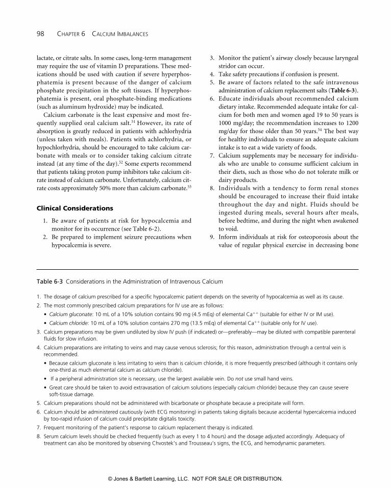
4. Take safety precautions if confusion is present.5. Be aware of factors related to the safe intravenous
administration of calcium replacement salts (Table 6-3).6. Educate individuals about recommended calcium
dietary intake. Recommended adequate intake for cal-cium for both men and women aged 19 to 50 years is1000 mg/day; the recommendation increases to 1200mg/day for those older than 50 years.54 The best wayfor healthy individuals to ensure an adequate calciumintake is to eat a wide variety of foods.
7. Calcium supplements may be necessary for individu-als who are unable to consume sufficient calcium intheir diets, such as those who do not tolerate milk ordairy products.
8. Individuals with a tendency to form renal stonesshould be encouraged to increase their fluid intakethroughout the day and night. Fluids should beingested during meals, several hours after meals,before bedtime, and during the night when awakenedto void.
9. Inform individuals at risk for osteoporosis about thevalue of regular physical exercise in decreasing bone
98 CHAPTER 6 CALCIUM IMBALANCES
Table 6-3 Considerations in the Administration of Intravenous Calcium
1. The dosage of calcium prescribed for a specific hypocalcemic patient depends on the severity of hypocalcemia as well as its cause.
2. The most commonly prescribed calcium preparations for IV use are as follows:
• Calcium gluconate: 10 mL of a 10% solution contains 90 mg (4.5 mEq) of elemental Ca�� (suitable for either IV or IM use).
• Calcium chloride: 10 mL of a 10% solution contains 270 mg (13.5 mEq) of elemental Ca�� (suitable only for IV use).
3. Calcium preparations may be given undiluted by slow IV push (if indicated) or—preferably—may be diluted with compatible parenteralfluids for slow infusion.
4. Calcium preparations are irritating to veins and may cause venous sclerosis; for this reason, administration through a central vein isrecommended.
• Because calcium gluconate is less irritating to veins than is calcium chloride, it is more frequently prescribed (although it contains onlyone-third as much elemental calcium as calcium chloride).
• If a peripheral administration site is necessary, use the largest available vein. Do not use small hand veins.
• Great care should be taken to avoid extravasation of calcium solutions (especially calcium chloride) because they can cause severesoft-tissue damage.
5. Calcium preparations should not be administered with bicarbonate or phosphate because a precipitate will form.
6. Calcium should be administered cautiously (with ECG monitoring) in patients taking digitalis because accidental hypercalcemia inducedby too-rapid infusion of calcium could precipitate digitalis toxicity.
7. Frequent monitoring of the patient’s response to calcium replacement therapy is indicated.
8. Serum calcium levels should be checked frequently (such as every 1 to 4 hours) and the dosage adjusted accordingly. Adequacy oftreatment can also be monitored by observing Chvostek’s and Trousseau’s signs, the ECG, and hemodynamic parameters.
81644_CH06_091_110_QXP6.qxd 9/14/10 11:07 AM Page 98
© Jones & Bartlett Learning, LLC. NOT FOR SALE OR DISTRIBUTION.
© Jones & Bartlett Learning, LLCNOT FOR SALE OR DISTRIBUTION
© Jones & Bartlett Learning, LLCNOT FOR SALE OR DISTRIBUTION
© Jones & Bartlett Learning, LLCNOT FOR SALE OR DISTRIBUTION
© Jones & Bartlett Learning, LLCNOT FOR SALE OR DISTRIBUTION
© Jones & Bartlett Learning, LLCNOT FOR SALE OR DISTRIBUTION
© Jones & Bartlett Learning, LLCNOT FOR SALE OR DISTRIBUTION
© Jones & Bartlett Learning, LLCNOT FOR SALE OR DISTRIBUTION
© Jones & Bartlett Learning, LLCNOT FOR SALE OR DISTRIBUTION
© Jones & Bartlett Learning, LLCNOT FOR SALE OR DISTRIBUTION
© Jones & Bartlett Learning, LLCNOT FOR SALE OR DISTRIBUTION
© Jones & Bartlett Learning, LLCNOT FOR SALE OR DISTRIBUTION
© Jones & Bartlett Learning, LLCNOT FOR SALE OR DISTRIBUTION
© Jones & Bartlett Learning, LLCNOT FOR SALE OR DISTRIBUTION
© Jones & Bartlett Learning, LLCNOT FOR SALE OR DISTRIBUTION
© Jones & Bartlett Learning, LLCNOT FOR SALE OR DISTRIBUTION
© Jones & Bartlett Learning, LLCNOT FOR SALE OR DISTRIBUTION
© Jones & Bartlett Learning, LLCNOT FOR SALE OR DISTRIBUTION
© Jones & Bartlett Learning, LLCNOT FOR SALE OR DISTRIBUTION
© Jones & Bartlett Learning, LLCNOT FOR SALE OR DISTRIBUTION
© Jones & Bartlett Learning, LLCNOT FOR SALE OR DISTRIBUTION
loss. Walking is well tolerated by all age groups and isan excellent form of exercise, as is bicycling.
10. Discuss the calcium loss associated with the use ofalcohol and nicotine. Smoking lowers estrogen levelsand interferes with the body’s absorption of calcium;as a consequence, women who smoke are at greaterrisk of developing osteoporosis.
HYPOCALCEMIA CASE STUDIES
Case Study 6-1
A 46-year-old woman with end-stage renal disease wasadmitted with a secondary diagnosis of seizure activity andmulti-infarct dementia. She required dialysis twice a week.On admission, laboratory data from a venous blood samplerevealed the following: Na 138 mEq/L, BUN 41 mg/dL, K5.8 mEq/L, serum creatinine 8.2 mg/dL, total Ca 7.0 mg/dL,albumin 3.0 g/dL, phosphorus 7.1 mg/dL, and HCO3 13.5mEq/L.
Commentary. Note the low total calcium level and the pres-ence of hypoalbuminemia. With correction for the lowserum albumin level, the serum calcium would be nearer tonormal. (Recall that for every gram the serum albumin isbelow the normal level, 0.8 mg must be added to thereported calcium level.) Using the following equation:
Corrected calcium (mg/dL) � measured serum calcium �
0.8 � (4.0 – measured serum albumin g/dL)
The corrected total calcium is 7.8 mg/dL. Although thecorrected total calcium level is still below normal, the ion-ized fraction of the calcium was normal; thus symptoms ofhypocalcemia were not present. Note that this patient hasmetabolic acidosis (evidenced by the low serum bicarbonatelevel); both hypoalbuminemia and acidosis favor increasedcalcium ionization. Rapid correction, or over-correction, ofacidosis in a patient with renal disease predisposes thepatient to precipitation of hypocalcemic symptoms. Hyper-phosphatemia was present, a major factor in explaining thehypocalcemia in this case.
Case Study 6-2
A hysterical young woman was admitted to the emergencydepartment after an automobile accident in which she frac-tured her arm. She complained of circumoral paresthesiaand then fainted. Arterial blood gas findings included a pHof 7.55 (alkalosis) and an arterial carbon dioxide pressure
(PaCO2) of 20 mm Hg (normal, 40 mm Hg), indicating res-piratory alkalosis.
Commentary. Hyperventilation secondary to hysteria is acommon cause of tetany in the hospital emergency depart-ment. In this situation, the tetany resulted from a reductionin the plasma ionized calcium level consequent to respira-tory alkalosis. Fainting was due to cerebral ischemia causedby the low PaCO2 (recall that a low PaCO2 causes cerebralvasoconstriction). The total serum calcium level was proba-bly normal, although the ionized fraction decreased. Cor-rection of the hyperventilation (and thus of respiratoryalkalosis) will restore the ionized calcium level to normaland alleviate symptoms.
Case Study 6-3
A case was reported in which a 40-year-old female was afirst-time apheresis platelet donor.55 Her history includedhypertension, hyperlipidemia, and depression. Medicationsincluded bumetanide (a loop diuretic), pravastatin (acholesterol-lowering drug), and paroxetine (a selective sero-tonin reuptake inhibitor [SSRI] antidepressant). Thirtyminutes after the procedure started, the patient complainedof tingling around her mouth, hands, and feet. Shortlythereafter, she developed acute-onset severe facial andextremity tetany. Treatment with intravenous calcium glu-conate was started and the muscle contractions subsidedover 10 to 15 minutes.
Commentary. These events are consistent with a severe reac-tion to calcium chelation by the sodium citrate anticoagulantused in the apheresis donation procedure. It is possible thatthe loop diuretic (bumetanide) contributed to the hypocal-cemia. Authors of the reported case study concluded thatcareful screening is needed to help prevent severe reactions tocitrate toxicity; it may be wise to measure pre-procedure cal-cium levels in selected donors to identify cases requiring extravigilance. The authors also pointed out the need of maintain-ing preparedness for managing rare but serious reactions involunteer apheresis blood donors.56
Case Study 6-4
A 32-year-old woman was admitted to an acute care facilitywith severe hypocalcemia and convulsions. She had under-gone a subtotal thyroidectomy two weeks earlier. At the timeof admission, her total serum calcium level was 3.2 mg/dL(normal for the reporting laboratory was 8.8 to 10.6 mg/dL).
Hypocalcemia Case Studies 99
81644_CH06_091_110_QXP6.qxd 9/14/10 11:07 AM Page 99
© Jones & Bartlett Learning, LLC. NOT FOR SALE OR DISTRIBUTION.
© Jones & Bartlett Learning, LLCNOT FOR SALE OR DISTRIBUTION
© Jones & Bartlett Learning, LLCNOT FOR SALE OR DISTRIBUTION
© Jones & Bartlett Learning, LLCNOT FOR SALE OR DISTRIBUTION
© Jones & Bartlett Learning, LLCNOT FOR SALE OR DISTRIBUTION
© Jones & Bartlett Learning, LLCNOT FOR SALE OR DISTRIBUTION
© Jones & Bartlett Learning, LLCNOT FOR SALE OR DISTRIBUTION
© Jones & Bartlett Learning, LLCNOT FOR SALE OR DISTRIBUTION
© Jones & Bartlett Learning, LLCNOT FOR SALE OR DISTRIBUTION
© Jones & Bartlett Learning, LLCNOT FOR SALE OR DISTRIBUTION
© Jones & Bartlett Learning, LLCNOT FOR SALE OR DISTRIBUTION
© Jones & Bartlett Learning, LLCNOT FOR SALE OR DISTRIBUTION
© Jones & Bartlett Learning, LLCNOT FOR SALE OR DISTRIBUTION
© Jones & Bartlett Learning, LLCNOT FOR SALE OR DISTRIBUTION
© Jones & Bartlett Learning, LLCNOT FOR SALE OR DISTRIBUTION
© Jones & Bartlett Learning, LLCNOT FOR SALE OR DISTRIBUTION
© Jones & Bartlett Learning, LLCNOT FOR SALE OR DISTRIBUTION
© Jones & Bartlett Learning, LLCNOT FOR SALE OR DISTRIBUTION
© Jones & Bartlett Learning, LLCNOT FOR SALE OR DISTRIBUTION
© Jones & Bartlett Learning, LLCNOT FOR SALE OR DISTRIBUTION
© Jones & Bartlett Learning, LLCNOT FOR SALE OR DISTRIBUTION
Commentary. The hypocalcemia associated with thyroidec-tomy may occur weeks to months after surgery (as evi-denced in this case). For this reason, patients should betaught about the symptoms of hypocalcemia and told toreport signs early to their healthcare providers.
Case Study 6-5
A case was reported in which a 32-year-old mentallyimpaired man presented with a year-long history of loss ofseizure control (after being seizure-free for 5 years on a reg-imen of phenytoin and phenobarbital).57 Physical examina-tion revealed a positive Chvostek’s sign and a serumcalcium level of 5.9 mg/dL (normal for the reporting labo-ratory was 8.8 to 10.4 mg/dL). An intravenous infusion ofcalcium was administered until the patient’s serum calciumlevel reached 8.0 mg/dL. The method of calcium adminis-tration was then changed to the oral route. The patient’sseizure activity diminished following calcium replacement.
Commentary. Phenytoin can interfere with vitamin Dmetabolism and impair calcium absorption for the intes-tinal and mobilization from the bone. The authors of thecase report stated that vitamin D and calcium treatmentshould probably be maintained during the use ofantiepileptic drugs.
Case Study 6-6
A case was reported in which a 43-year-old woman with a20-year history of Crohn’s disease presented to the emer-gency department with fatigue and weight loss.58 For thepast month, she had experienced tetany and muscle crampsas well as peripheral and perioral paresthesia. In addition,she experienced colicky abdominal pain. Upon examina-tion, positive Chvostek’s and Trousseau’s signs were found.A serum calcium level of 1.3 mmol/L (corrected, 1.7mmol/L) was found (normal, 2.23–2.57 mmol/L). Alsopresent were hypomagnesemia and hypokalemia. Treat-ment consisted of electrolyte replacement with intravenouscalcium, potassium, and magnesium. Vitamin D wasreplaced and a semi-elemental tube feeding was started.One month after discharge from the hospital, the patientcontinued to receive tube feedings and electrolyte replace-ment therapy. Plasma electrolyte levels normalized and thepatient was gaining weight.
Commentary. Short bowel associated with severe Crohn’sdisease seriously impairs the ability of the bowel to absorb
adequate amounts of carbohydrates, proteins, fats, vitamins,minerals, and electrolytes. Note that this patient requiredreplacement of magnesium along with replacement of cal-cium to allow the serum calcium level to normalize.
HYPERCALCEMIA
Hypercalcemia occurs when calcium enters the extracellularfluid more rapidly than it can be excreted by the kidneys.59
The incidence of this imbalance depends on the setting inwhich it occurs.60 If allowed to become severe, hypercal-cemia is associated with significant morbidity and mortal-ity; therefore, it is important to detect this imbalance early.
Causes
Primary hyperparathyroidism and malignancy account formore than 90% of the cases of hypercalcemia in ambulatoryand noncritically ill patients.61 Only a small percentage ofhypercalcemia cases are due to immobilization, vitamin Aand D intoxication, lithium use, and thiazide diuretics.
Primary Hyperparathyroidism
Primary hyperparathyroidism accounts for more than halfof the cases of hypercalcemia in ambulatory patients.62 Thisdisorder is far more common in women than in men.63 Dueto increased PTH production, hyperparathyroidism causesincreased release of calcium from bone, augmented intes-tinal calcium absorption, and renal reabsorption of calcium.Mild hypercalcemia is found in approximately 10% ofpatients with thyrotoxicosis.64
Malignancies
Approximately 40% of the cases of hypercalcemia in hospi-talized patients are associated with cancer.65 Pathogenesis ofthe hypercalcemia of malignancy is complex and varies withthe type of tumor. The malignancies most often associatedwith hypercalcemia include breast and lung cancers andhematologic malignancies such as multiple myeloma orlymphoma. Hypercalcemia is usually present only inpatients with advanced cancer.66 In a recent retrospectivestudy, hypercalcemia was found to be a reliable indicator ofimpending death in cancer patients cared for in a hospicecare setting.67 Chapter 22 provides a more thorough discus-sion of tumors associated with hypercalcemia.
Immobilization
Bone mineral is lost during immobilization, sometimescausing an elevated total calcium concentration in the
100 CHAPTER 6 CALCIUM IMBALANCES
81644_CH06_091_110_QXP6.qxd 9/14/10 11:07 AM Page 100
© Jones & Bartlett Learning, LLC. NOT FOR SALE OR DISTRIBUTION.
© Jones & Bartlett Learning, LLCNOT FOR SALE OR DISTRIBUTION
© Jones & Bartlett Learning, LLCNOT FOR SALE OR DISTRIBUTION
© Jones & Bartlett Learning, LLCNOT FOR SALE OR DISTRIBUTION
© Jones & Bartlett Learning, LLCNOT FOR SALE OR DISTRIBUTION
© Jones & Bartlett Learning, LLCNOT FOR SALE OR DISTRIBUTION
© Jones & Bartlett Learning, LLCNOT FOR SALE OR DISTRIBUTION
© Jones & Bartlett Learning, LLCNOT FOR SALE OR DISTRIBUTION
© Jones & Bartlett Learning, LLCNOT FOR SALE OR DISTRIBUTION
© Jones & Bartlett Learning, LLCNOT FOR SALE OR DISTRIBUTION
© Jones & Bartlett Learning, LLCNOT FOR SALE OR DISTRIBUTION
© Jones & Bartlett Learning, LLCNOT FOR SALE OR DISTRIBUTION
© Jones & Bartlett Learning, LLCNOT FOR SALE OR DISTRIBUTION
© Jones & Bartlett Learning, LLCNOT FOR SALE OR DISTRIBUTION
© Jones & Bartlett Learning, LLCNOT FOR SALE OR DISTRIBUTION
© Jones & Bartlett Learning, LLCNOT FOR SALE OR DISTRIBUTION
© Jones & Bartlett Learning, LLCNOT FOR SALE OR DISTRIBUTION
© Jones & Bartlett Learning, LLCNOT FOR SALE OR DISTRIBUTION
© Jones & Bartlett Learning, LLCNOT FOR SALE OR DISTRIBUTION
© Jones & Bartlett Learning, LLCNOT FOR SALE OR DISTRIBUTION
© Jones & Bartlett Learning, LLCNOT FOR SALE OR DISTRIBUTION
bloodstream and resultant calciuria with the possibility offormation of renal stones. Hypercalcemia will result if therate of bone resorption exceeds the kidneys’ ability to excretethe excess calcium. In particular, patients with spinal cordinjuries are at risk for immobilization-related hypercalcemia.Notable risk factors in this population include an age lessthan 21 years, male gender, and extensive cord injury.68
Immobilization-related hypercalcemia has also beenreported in individuals without spinal cord injury, such asthose with prolonged illnesses in geriatric and critical caresettings.69, 70 The hypercalcemia of immobilization remitswhen activity is restored; if treatment is required, bisphos-phonates may be the treatment of choice.71
Drugs
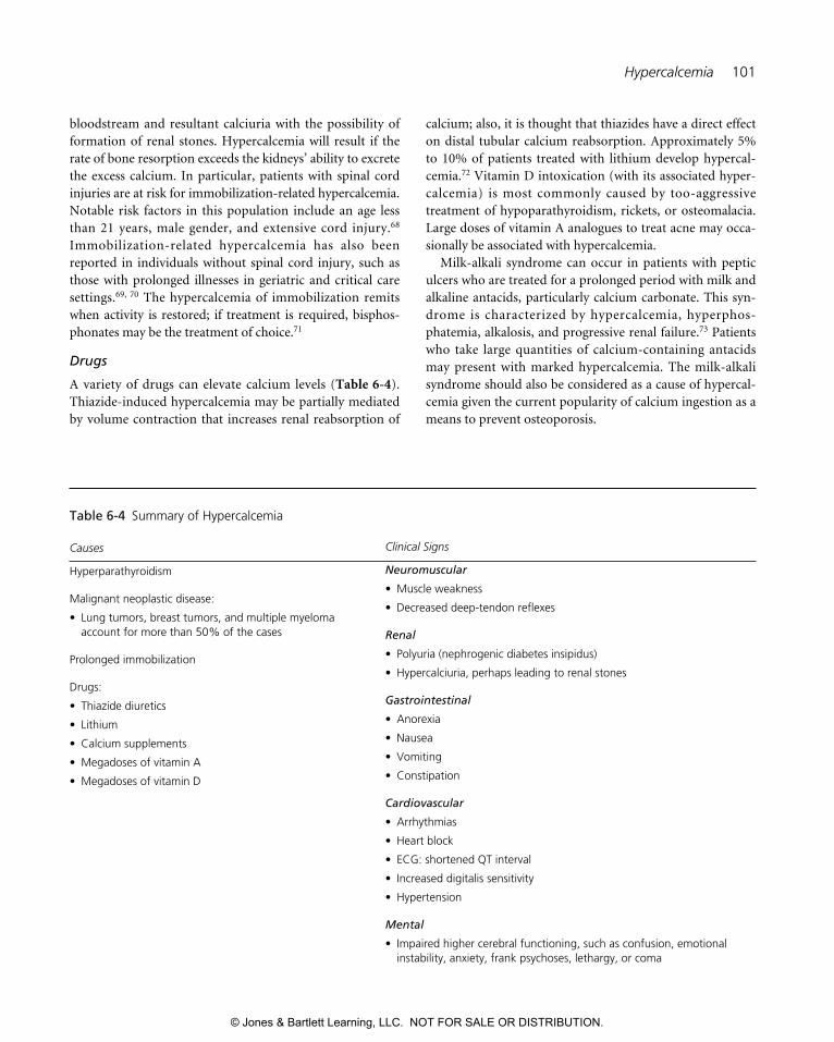
A variety of drugs can elevate calcium levels (Table 6-4).Thiazide-induced hypercalcemia may be partially mediatedby volume contraction that increases renal reabsorption of
calcium; also, it is thought that thiazides have a direct effecton distal tubular calcium reabsorption. Approximately 5%to 10% of patients treated with lithium develop hypercal-cemia.72 Vitamin D intoxication (with its associated hyper-calcemia) is most commonly caused by too-aggressivetreatment of hypoparathyroidism, rickets, or osteomalacia.Large doses of vitamin A analogues to treat acne may occa-sionally be associated with hypercalcemia.
Milk-alkali syndrome can occur in patients with pepticulcers who are treated for a prolonged period with milk andalkaline antacids, particularly calcium carbonate. This syn-drome is characterized by hypercalcemia, hyperphos-phatemia, alkalosis, and progressive renal failure.73 Patientswho take large quantities of calcium-containing antacidsmay present with marked hypercalcemia. The milk-alkalisyndrome should also be considered as a cause of hypercal-cemia given the current popularity of calcium ingestion as ameans to prevent osteoporosis.
Hypercalcemia 101
Table 6-4 Summary of Hypercalcemia
Causes
Hyperparathyroidism
Malignant neoplastic disease:
• Lung tumors, breast tumors, and multiple myelomaaccount for more than 50% of the cases
Prolonged immobilization
Drugs:
• Thiazide diuretics
• Lithium
• Calcium supplements
• Megadoses of vitamin A
• Megadoses of vitamin D
Clinical Signs
Neuromuscular
• Muscle weakness
• Decreased deep-tendon reflexes
Renal
• Polyuria (nephrogenic diabetes insipidus)
• Hypercalciuria, perhaps leading to renal stones
Gastrointestinal
• Anorexia
• Nausea
• Vomiting
• Constipation
Cardiovascular
• Arrhythmias
• Heart block
• ECG: shortened QT interval
• Increased digitalis sensitivity
• Hypertension
Mental
• Impaired higher cerebral functioning, such as confusion, emotionalinstability, anxiety, frank psychoses, lethargy, or coma
81644_CH06_091_110_QXP6.qxd 9/14/10 11:07 AM Page 101
© Jones & Bartlett Learning, LLC. NOT FOR SALE OR DISTRIBUTION.
© Jones & Bartlett Learning, LLCNOT FOR SALE OR DISTRIBUTION
© Jones & Bartlett Learning, LLCNOT FOR SALE OR DISTRIBUTION
© Jones & Bartlett Learning, LLCNOT FOR SALE OR DISTRIBUTION
© Jones & Bartlett Learning, LLCNOT FOR SALE OR DISTRIBUTION
© Jones & Bartlett Learning, LLCNOT FOR SALE OR DISTRIBUTION
© Jones & Bartlett Learning, LLCNOT FOR SALE OR DISTRIBUTION
© Jones & Bartlett Learning, LLCNOT FOR SALE OR DISTRIBUTION
© Jones & Bartlett Learning, LLCNOT FOR SALE OR DISTRIBUTION
© Jones & Bartlett Learning, LLCNOT FOR SALE OR DISTRIBUTION
© Jones & Bartlett Learning, LLCNOT FOR SALE OR DISTRIBUTION
© Jones & Bartlett Learning, LLCNOT FOR SALE OR DISTRIBUTION
© Jones & Bartlett Learning, LLCNOT FOR SALE OR DISTRIBUTION
© Jones & Bartlett Learning, LLCNOT FOR SALE OR DISTRIBUTION
© Jones & Bartlett Learning, LLCNOT FOR SALE OR DISTRIBUTION
© Jones & Bartlett Learning, LLCNOT FOR SALE OR DISTRIBUTION
© Jones & Bartlett Learning, LLCNOT FOR SALE OR DISTRIBUTION
© Jones & Bartlett Learning, LLCNOT FOR SALE OR DISTRIBUTION
© Jones & Bartlett Learning, LLCNOT FOR SALE OR DISTRIBUTION
© Jones & Bartlett Learning, LLCNOT FOR SALE OR DISTRIBUTION
© Jones & Bartlett Learning, LLCNOT FOR SALE OR DISTRIBUTION
Clinical Signs
The magnitude of the serum calcium elevation and the timeit takes to develop have major effects on clinical findings, asdoes the underlying cause of hypercalcemia. For example,acute hypercalcemia produces more symptoms than doeschronic hypercalcemia. Also, malignancies can present withsevere hypercalcemia (serum calcium level � 14 mg/dL)more commonly than with other conditions.74
Clinical signs of hypercalcemia are summarized in Table6-4. In some patients, mild hypercalcemia is found on rou-tine examinations; other patients may present in hypercal-cemic crisis. Although there are no firm diagnostic criteriafor hypercalcemic crisis, it is generally thought to representthe presence of volume depletion, neurological manifesta-tions, and cardiac arrhythmias in a patient with a serum cal-cium level greater than 14 mg/dL. As a rule, symptoms ofhypercalcemia are proportional to the serum calcium level,although this is not always the case.
Serum calcium levels less than 11.5 mg/dL rarely producesymptoms. By comparison, levels between 11.5 and 13mg/dL may be associated with lethargy, anorexia, nausea,and polyuria.75 Further, calcium levels greater than 13mg/dL constitute severe hypocalcemia and are associatedwith more severe symptoms (such as muscle weakness,impaired memory, emotional lability, stupor, and coma). Atotal calcium concentration greater than 14 mg/dL repre-sents hypercalcemic crisis and is a medical emergency.76
Neuromuscular Changes
Hypercalcemia reduces neuromuscular excitability becauseit acts as a sedative at the myoneural junction. Symptomssuch as muscular weakness and depressed deep-tendonreflexes may occur.
Gastrointestinal Symptoms
Constipation, anorexia, nausea, vomiting, and adynamicileus are common symptoms of hypercalcemia. Constipa-tion results from decreased GI motility caused by calcium’saction on smooth muscle and nerve conduction, as well asfrom dehydration.77 Delayed gastric emptying, nausea, andvomiting are also related to altered motility. Patients withhypercalcemia are predisposed to duodenal ulcer diseasebecause of the increased gastric acid secretion, which is pro-moted by calcium’s action on the parietal cells of the stom-ach. Pancreatitis is another potential complication of severehypercalcemia and is probably related to calcium depositsin the pancreatic ducts.
Behavior Changes
Behavior changes associated with hypercalcemia may rangefrom subtle alterations in personality to acute psychosis andmay include confusion, impairment of memory, andbizarre behavior. Patients may become inattentive and losetheir ability to concentrate; recent memory is affected moredramatically than is distant memory. Other mental statuschanges sometimes seen in patients with hypercalcemiainclude lethargy and drowsiness, as well as psychiatric dis-turbances such as irritability and depression; severe casesare associated with stupor or coma. Although the cause ofthese symptoms is not known, it has been suggested thatincreased calcium in the cerebrospinal fluid is involved. Themore severe symptoms tend to occur when the serum cal-cium level approaches or exceeds 15 mg/dL. In a study ofeight hypercalcemic inpatients with cancer during a periodof 66 patient-days, Mahon found that the most evidentchanges were those affecting mental status.78 For example,many subjects could not remember their home telephonenumbers or perform simple mathematical computations.Some displayed inappropriate behaviors, such as pullingout a Foley catheter while the balloon was inflated. Asserum calcium levels decreased toward normal, the mentalsymptoms gradually subsided.
Renal Changes
Disturbed renal tubular function produced by hypercal-cemia can cause polyuria and polydipsia. More specifi-cally, this disturbed function is a form of nephrogenicdiabetes insipidus (NDI) that is usually reversible within 1 to 12 weeks after correction of the imbalance. The con-centrating defect may become clinically apparent when theplasma calcium concentration exceeds 11 mg/dL. Renalcolic may occur as a result of kidney stones, which mayform from the excess calcium presented to the kidneys forexcretion. Calcium salts deposited in the kidney can causerenal failure.
Cardiovascular Changes
Calcium is important in cardiac function; it exerts a positiveinotropic effect on the heart and reduces heart rate in a waysimilar to the effect of cardiac glycosides. Calcium adminis-tration to patients receiving digitalis must be done withextreme care because it can precipitate severe arrhythmias.
Cardiac effects of hypercalcemia include QT-intervalshortening and arrhythmias.79 Bradycardia; first-, second-,and third-degree heart block; and bundle branch block may
102 CHAPTER 6 CALCIUM IMBALANCES
81644_CH06_091_110_QXP6.qxd 9/14/10 11:07 AM Page 102
© Jones & Bartlett Learning, LLC. NOT FOR SALE OR DISTRIBUTION.
© Jones & Bartlett Learning, LLCNOT FOR SALE OR DISTRIBUTION
© Jones & Bartlett Learning, LLCNOT FOR SALE OR DISTRIBUTION
© Jones & Bartlett Learning, LLCNOT FOR SALE OR DISTRIBUTION
© Jones & Bartlett Learning, LLCNOT FOR SALE OR DISTRIBUTION
© Jones & Bartlett Learning, LLCNOT FOR SALE OR DISTRIBUTION
© Jones & Bartlett Learning, LLCNOT FOR SALE OR DISTRIBUTION
© Jones & Bartlett Learning, LLCNOT FOR SALE OR DISTRIBUTION
© Jones & Bartlett Learning, LLCNOT FOR SALE OR DISTRIBUTION
© Jones & Bartlett Learning, LLCNOT FOR SALE OR DISTRIBUTION
© Jones & Bartlett Learning, LLCNOT FOR SALE OR DISTRIBUTION
© Jones & Bartlett Learning, LLCNOT FOR SALE OR DISTRIBUTION
© Jones & Bartlett Learning, LLCNOT FOR SALE OR DISTRIBUTION
© Jones & Bartlett Learning, LLCNOT FOR SALE OR DISTRIBUTION
© Jones & Bartlett Learning, LLCNOT FOR SALE OR DISTRIBUTION
© Jones & Bartlett Learning, LLCNOT FOR SALE OR DISTRIBUTION
© Jones & Bartlett Learning, LLCNOT FOR SALE OR DISTRIBUTION
© Jones & Bartlett Learning, LLCNOT FOR SALE OR DISTRIBUTION
© Jones & Bartlett Learning, LLCNOT FOR SALE OR DISTRIBUTION
© Jones & Bartlett Learning, LLCNOT FOR SALE OR DISTRIBUTION
© Jones & Bartlett Learning, LLCNOT FOR SALE OR DISTRIBUTION
occur. Hypercalcemia can also affect the systemic vascula-ture, perhaps leading to hypertension. The mechanism forthe increase in blood pressure may be multifactorial. Forexample, serum levels of epinephrine and norepinephrineare higher in patients with hypercalcemia than in those withnormocalcemia.
Treatment
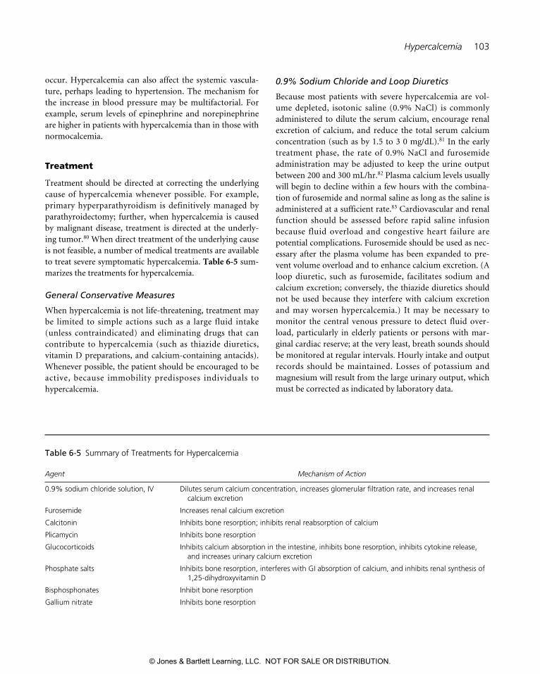
Treatment should be directed at correcting the underlyingcause of hypercalcemia whenever possible. For example,primary hyperparathyroidism is definitively managed byparathyroidectomy; further, when hypercalcemia is causedby malignant disease, treatment is directed at the underly-ing tumor.80 When direct treatment of the underlying causeis not feasible, a number of medical treatments are availableto treat severe symptomatic hypercalcemia. Table 6-5 sum-marizes the treatments for hypercalcemia.
General Conservative Measures
When hypercalcemia is not life-threatening, treatment maybe limited to simple actions such as a large fluid intake(unless contraindicated) and eliminating drugs that cancontribute to hypercalcemia (such as thiazide diuretics,vitamin D preparations, and calcium-containing antacids).Whenever possible, the patient should be encouraged to beactive, because immobility predisposes individuals tohypercalcemia.
0.9% Sodium Chloride and Loop Diuretics
Because most patients with severe hypercalcemia are vol-ume depleted, isotonic saline (0.9% NaCl) is commonlyadministered to dilute the serum calcium, encourage renalexcretion of calcium, and reduce the total serum calciumconcentration (such as by 1.5 to 3 0 mg/dL).81 In the earlytreatment phase, the rate of 0.9% NaCl and furosemideadministration may be adjusted to keep the urine outputbetween 200 and 300 mL/hr.82 Plasma calcium levels usuallywill begin to decline within a few hours with the combina-tion of furosemide and normal saline as long as the saline isadministered at a sufficient rate.83 Cardiovascular and renalfunction should be assessed before rapid saline infusionbecause fluid overload and congestive heart failure arepotential complications. Furosemide should be used as nec-essary after the plasma volume has been expanded to pre-vent volume overload and to enhance calcium excretion. (Aloop diuretic, such as furosemide, facilitates sodium andcalcium excretion; conversely, the thiazide diuretics shouldnot be used because they interfere with calcium excretionand may worsen hypercalcemia.) It may be necessary tomonitor the central venous pressure to detect fluid over-load, particularly in elderly patients or persons with mar-ginal cardiac reserve; at the very least, breath sounds shouldbe monitored at regular intervals. Hourly intake and outputrecords should be maintained. Losses of potassium andmagnesium will result from the large urinary output, whichmust be corrected as indicated by laboratory data.
Hypercalcemia 103
Table 6-5 Summary of Treatments for Hypercalcemia
Agent Mechanism of Action
0.9% sodium chloride solution, IV Dilutes serum calcium concentration, increases glomerular filtration rate, and increases renalcalcium excretion
Furosemide Increases renal calcium excretion
Calcitonin Inhibits bone resorption; inhibits renal reabsorption of calcium
Plicamycin Inhibits bone resorption
Glucocorticoids Inhibits calcium absorption in the intestine, inhibits bone resorption, inhibits cytokine release,and increases urinary calcium excretion
Phosphate salts Inhibits bone resorption, interferes with GI absorption of calcium, and inhibits renal synthesis of1,25-dihydroxyvitamin D
Bisphosphonates Inhibit bone resorption
Gallium nitrate Inhibits bone resorption
81644_CH06_091_110_QXP6.qxd 9/14/10 11:07 AM Page 103
© Jones & Bartlett Learning, LLC. NOT FOR SALE OR DISTRIBUTION.
© Jones & Bartlett Learning, LLCNOT FOR SALE OR DISTRIBUTION
© Jones & Bartlett Learning, LLCNOT FOR SALE OR DISTRIBUTION
© Jones & Bartlett Learning, LLCNOT FOR SALE OR DISTRIBUTION
© Jones & Bartlett Learning, LLCNOT FOR SALE OR DISTRIBUTION
© Jones & Bartlett Learning, LLCNOT FOR SALE OR DISTRIBUTION
© Jones & Bartlett Learning, LLCNOT FOR SALE OR DISTRIBUTION
© Jones & Bartlett Learning, LLCNOT FOR SALE OR DISTRIBUTION
© Jones & Bartlett Learning, LLCNOT FOR SALE OR DISTRIBUTION
© Jones & Bartlett Learning, LLCNOT FOR SALE OR DISTRIBUTION
© Jones & Bartlett Learning, LLCNOT FOR SALE OR DISTRIBUTION
© Jones & Bartlett Learning, LLCNOT FOR SALE OR DISTRIBUTION
© Jones & Bartlett Learning, LLCNOT FOR SALE OR DISTRIBUTION
© Jones & Bartlett Learning, LLCNOT FOR SALE OR DISTRIBUTION
© Jones & Bartlett Learning, LLCNOT FOR SALE OR DISTRIBUTION
© Jones & Bartlett Learning, LLCNOT FOR SALE OR DISTRIBUTION
© Jones & Bartlett Learning, LLCNOT FOR SALE OR DISTRIBUTION
© Jones & Bartlett Learning, LLCNOT FOR SALE OR DISTRIBUTION
© Jones & Bartlett Learning, LLCNOT FOR SALE OR DISTRIBUTION
© Jones & Bartlett Learning, LLCNOT FOR SALE OR DISTRIBUTION
© Jones & Bartlett Learning, LLCNOT FOR SALE OR DISTRIBUTION
Bisphosphonates
Bisphosphonates, such as pamidronate and etidronate,inhibit bone resorption and, therefore, can treat hypercal-cemia. The bisphosphonate dosage is determined by theseverity of the hypercalcemia. Bisphosphonates are themainstay of treatment for hypercalcemia associated withmalignancy. They normalize calcium in more than 70% ofpatients, although it may take as long as 48 to 72 hoursbefore the full therapeutic effect becomes evident.84 Serumcalcium levels may remain in the normal range for weeks tomonths after bisphosphonate therapy.
Plicamycin
Plicamycin (an antineoplastic agent) lowers serum calciumby blocking bone resorption. Because of the potential fornephrotoxicity and hepatotoxicity, long-term use of pli-camycin is limited; use of this agent should be avoided inpatients with underlying renal or liver dysfunction. Sincethe advent of the bisphosphonates, plicamycin has beenused much less often than in the past.85
Calcitonin
By inhibiting bone resorption, salmon calcitonin has aslight and short-term effect on plasma calcium levels. Theefficacy of calcitonin is largely limited to the first 48 hoursafter its administration (which limits its use in the controlof long-term hypercalcemia).86 Calcitonin is used largely asadjunctive therapy in controlling hypercalcemia in acutecare settings until more powerful (but slower-acting) drugstake effect.87
Glucocorticoids
Glucocorticoids can reduce the serum calcium level byinhibiting cytokine release, inhibiting absorption of calciumin the intestine, and increasing urinary calcium excretion.88
They are effective in reducing serum calcium in hypercal-cemia due to sarcoidosis, vitamin D intoxication, multiplemyeloma, or other hematologic malignancies. A drawbackof glucocorticoids is that clinically significant reductions inserum calcium may not occur until at least 5 to 10 days aftertherapy is initiated.89 Possible complications associated withglucocorticoids include hyperglycemia and sodium andwater retention.
Gallium Nitrate
Gallium nitrate lowers the serum calcium level by inhibitingbone resorption. It has a delayed action onset of 2 days.This agent carries a significant risk of nephrotoxicity and is
contraindicated when the serum creatinine is greater than2.5 mg/dL.90
Phosphate Salts
When ingested orally, phosphate produces a small reduc-tion in the serum calcium level by inhibiting bone resorp-tion, interfering with the GI absorption of calcium, andinhibiting renal synthesis of 1,25-dihydroxy vitamin D.Because of its modest effect on the serum calcium level, oralphosphate use is limited to long-term treatment of mildhypercalcemia. It is important to remember that increasingthe serum phosphate concentration alters the extracellularcalcium–phosphate equilibrium to promote the formationof calcium-phosphate precipitates, which are then de-posited in various body tissues including bone, soft tissues,blood vessels, lung, myocardium, and kidneys. Because ofthe risk of soft-tissue calcification, phosphate therapy islimited primarily to patients with low serum phosphate lev-els (less than 3.0 mg/dL) and adequate renal function.
Clinical Considerations
1. Be aware of patients at risk for hypercalcemia andmonitor for its presence. (See Table 6-4.)
2. Increase patient mobilization when feasible; recall thatimmobilization favors hypercalcemia. Hospitalizedpatients at risk for hypercalcemia should be ambu-lated as soon as possible; outpatients should be toldthe importance of frequently moving about.
3. Encourage the oral intake of sufficient fluids to keepthe patient well hydrated. Sodium-containing fluidsshould be given, unless contraindicated by other con-ditions, because sodium favors calcium excretion.Always consider the patient’s preferences when en-couraging oral fluids.
4. Encourage adequate bulk in the diet to offset the ten-dency toward constipation.
5. Take safety precautions if confusion or other mentalsymptoms of hypercalcemia are present. Explain tothe patient and family that the mental changes associ-ated with hypercalcemia are reversible with treatment.
6. Be aware that cardiac arrest can occur in patients withsevere hypercalcemia; be prepared to deal with thisemergency situation.
7. Be aware that bones may fracture more easily inpatients with chronic hypercalcemia because boneresorption has been excessive, weakening the bonystructure. Transfer patients cautiously.
104 CHAPTER 6 CALCIUM IMBALANCES
81644_CH06_091_110_QXP6.qxd 9/14/10 11:07 AM Page 104
© Jones & Bartlett Learning, LLC. NOT FOR SALE OR DISTRIBUTION.
© Jones & Bartlett Learning, LLCNOT FOR SALE OR DISTRIBUTION
© Jones & Bartlett Learning, LLCNOT FOR SALE OR DISTRIBUTION
© Jones & Bartlett Learning, LLCNOT FOR SALE OR DISTRIBUTION
© Jones & Bartlett Learning, LLCNOT FOR SALE OR DISTRIBUTION
© Jones & Bartlett Learning, LLCNOT FOR SALE OR DISTRIBUTION
© Jones & Bartlett Learning, LLCNOT FOR SALE OR DISTRIBUTION
© Jones & Bartlett Learning, LLCNOT FOR SALE OR DISTRIBUTION
© Jones & Bartlett Learning, LLCNOT FOR SALE OR DISTRIBUTION
© Jones & Bartlett Learning, LLCNOT FOR SALE OR DISTRIBUTION
© Jones & Bartlett Learning, LLCNOT FOR SALE OR DISTRIBUTION
© Jones & Bartlett Learning, LLCNOT FOR SALE OR DISTRIBUTION
© Jones & Bartlett Learning, LLCNOT FOR SALE OR DISTRIBUTION
© Jones & Bartlett Learning, LLCNOT FOR SALE OR DISTRIBUTION
© Jones & Bartlett Learning, LLCNOT FOR SALE OR DISTRIBUTION
© Jones & Bartlett Learning, LLCNOT FOR SALE OR DISTRIBUTION
© Jones & Bartlett Learning, LLCNOT FOR SALE OR DISTRIBUTION
© Jones & Bartlett Learning, LLCNOT FOR SALE OR DISTRIBUTION
© Jones & Bartlett Learning, LLCNOT FOR SALE OR DISTRIBUTION
© Jones & Bartlett Learning, LLCNOT FOR SALE OR DISTRIBUTION
© Jones & Bartlett Learning, LLCNOT FOR SALE OR DISTRIBUTION
8. Educate home-bound oncology patients with a pre-disposition for hypercalcemia, as well as their families,regarding symptoms that occur with this condition.Instruct them to report symptoms to healthcareproviders before they become severe. In a study re-ported by Mahon, constipation, confusion, anorexia,increasing bone pain, weight loss, and weakness werethe symptoms that most frequently caused readmis-sion of patients with cancer. In a study of 22 hospital-ized and 18 ambulatory cancer patients,91 Cowardreported that 90% were unaware that hypercalcemiamight be a complication of their cancer.92 Further-more, only one of the patients knew the symptoms ofcancer-induced hypercalcemia. Almost 70% of thepatients did not recall being told of measures thatmight prevent hypercalcemia.
9. Be alert for signs of digitalis toxicity when hypercal-cemia occurs in digitalized patients.
10. Be familiar with the treatment modalities for hyper-calcemia and associated nursing functions (see thetreatment section in this chapter and in Chapter 22).
HYPERCALCEMIA CASE STUDIES
Case Study 6-7
A 47-year-old woman who appeared thin and dehydratedwas admitted to the hospital with abdominal pain, weightloss, and a history of vomiting for 3 months. Her bodyweight of 38 kg was only 84% of her ideal body weight; hermid-arm circumference was 60% of standard. On ex-amination, a nodule was felt near the right lobe of the thy-roid gland and was later found to be an adenoma. A ser-um calcium level was obtained and was found to be high(13.8 mg/dL).
The patient was rehydrated with 0.9% NaCl solution;however, saline diuresis did not represent a long-term solu-tion to this woman’s problem of severe hypercalcemia.Thus the right upper parathyroid gland was surgicallyremoved. At the time of discharge from the hospital, thepatient’s calcium concentration had normalized to 8.7mg/dL.
Commentary. This patient had hypercalcemia due to aparathyroid adenoma, which is one of the most commoncauses of hypercalcemia. She received the usual treatmentfor this disorder—rehydration with isotonic saline and sur-gical removal of the tumor.
Case Study 6-8
A 49-year-old woman with a history of breast cancer andmetastases to the bone and liver was admitted to the hospi-tal with polyuria and polydipsia; her total serum calciumlevel was 3.08 mmol/L (12.3 mg/dL). A single IV infusion ofpamidronate was administered. The serum calcium levelreturned to normal and did not change until 6 weeks later.Several subsequent recurrences of hypercalcemia were alsotreated with single infusions of pamidronate given in theoutpatient department. The patient was able to continuereceiving palliative therapy at home, but died 6 monthslater from other complications.
Commentary. The polyuria associated with hypercalcemiaoccurs as a result of a renal concentrating defect; althoughthe precise cause of this abnormality is not certain, a defectin the action of antidiuretic hormone in the collecting ductsand damage to the concentrating segments of the nephronsplay a role. Provided the patient is not too nauseated todrink fluids, fluid intake will increase as the patient be-comes more thirsty. Treatment of this woman’s intermit-tent bouts of hypercalcemia allowed her to be morecomfortable during her remaining months of life.
Case Study 6-9
A 59-year-old woman with carcinoma of the left breast under-went a radical mastectomy and adjuvant chemotherapy. Shewas later admitted to the hospital for management of hippain, confusion, and hypercalcemia (corrected serum cal-cium, 15.2 mg/dL). Isotonic saline was administered IV, andshe was given calcitonin subcutaneously every 8 hours. Shealso received pamidronate IV. The patient remained confusedafter the serum calcium level normalized. No further treat-ment was administered when the hypercalcemia recurred, andshe died 1 month later from a pulmonary embolus.
Commentary. Because hypercalcemia can impair mentalfunction, it is not unusual to observe confusion in patientswith hypercalcemia. However, the behavior changes usuallygradually subside once the hypercalcemia is resolved.Therefore, it was concluded that the confusion was due tosomething other than hypercalcemia.
Case Study 6-10
A 50-year-old man with dysphagia due to an adenocarcinomain the gastroesophageal junction became acutely confused and
Hypercalcemia Case Studies 105
81644_CH06_091_110_QXP6.qxd 9/14/10 11:07 AM Page 105
© Jones & Bartlett Learning, LLC. NOT FOR SALE OR DISTRIBUTION.
© Jones & Bartlett Learning, LLCNOT FOR SALE OR DISTRIBUTION
© Jones & Bartlett Learning, LLCNOT FOR SALE OR DISTRIBUTION
© Jones & Bartlett Learning, LLCNOT FOR SALE OR DISTRIBUTION
© Jones & Bartlett Learning, LLCNOT FOR SALE OR DISTRIBUTION
© Jones & Bartlett Learning, LLCNOT FOR SALE OR DISTRIBUTION
© Jones & Bartlett Learning, LLCNOT FOR SALE OR DISTRIBUTION
© Jones & Bartlett Learning, LLCNOT FOR SALE OR DISTRIBUTION
© Jones & Bartlett Learning, LLCNOT FOR SALE OR DISTRIBUTION
© Jones & Bartlett Learning, LLCNOT FOR SALE OR DISTRIBUTION
© Jones & Bartlett Learning, LLCNOT FOR SALE OR DISTRIBUTION
© Jones & Bartlett Learning, LLCNOT FOR SALE OR DISTRIBUTION
© Jones & Bartlett Learning, LLCNOT FOR SALE OR DISTRIBUTION
© Jones & Bartlett Learning, LLCNOT FOR SALE OR DISTRIBUTION
© Jones & Bartlett Learning, LLCNOT FOR SALE OR DISTRIBUTION
© Jones & Bartlett Learning, LLCNOT FOR SALE OR DISTRIBUTION
© Jones & Bartlett Learning, LLCNOT FOR SALE OR DISTRIBUTION
© Jones & Bartlett Learning, LLCNOT FOR SALE OR DISTRIBUTION
© Jones & Bartlett Learning, LLCNOT FOR SALE OR DISTRIBUTION
© Jones & Bartlett Learning, LLCNOT FOR SALE OR DISTRIBUTION
© Jones & Bartlett Learning, LLCNOT FOR SALE OR DISTRIBUTION

reported recent nausea, anorexia, and polyuria. Because ofhis nausea, he was unable to consume fluids; his skin turgorwas poor and his mucous membranes were dry. The ionizedserum calcium level was elevated (8.7 mg/dL). Isotonic salineand a single dose of pamidronate were administered IV. Theserum calcium level normalized within 3 days and thepatient’s confusion improved. Later, the confusion returnedand he was found to have multiple metastases to the lung,bone, and brain. After consultation with his family, treatmentof hypercalcemia was withdrawn, and he was treated pallia-tively at home until he died 10 days later.
Commentary. Nausea and vomiting are classic symptoms ofhypercalcemic states, as is constipation. High serum cal-cium levels cause slowed GI motility and delayed gastricemptying; these conditions contribute to anorexia, nausea,and vomiting. The polyuria associated with hypercalcemiais related to a renal concentrating defect; inability to replacethe large urinary losses with increased oral fluid intakeresults in fluid volume depletion. Although this patient’sconfusion was partially alleviated by correction of theserum calcium level, other causes were present that causedit to recur (namely, metastases to the brain).
Case Study 6-11
A case was reported in which a 40-year-old woman wasadmitted to the emergency department with eclampsia.93
During her examination, she was found to have profoundhypercalcemia and metabolic alkalosis (pH 7.57) secondaryto milk-alkai syndrome. The corrected calcium level was4.71 mmol/L (normal range is 2.23 to 2.57 mmol/L). Thepatient revealed that she had self-medicated with multipleantacid tablets for dyspepsia. Treatment consisted of aggres-sive rehydration, bisphosphonates, and discontinuation ofantacid tablets. The patient made a full recovery and deliv-ered a normal infant.
Commentary. It is possible to consume enough calciumand alkali with over-the-counter antacids to induce milk-alkali syndrome. Upon questioning, it was learned that thepatient had taken approximately 24 antacid tablets per day,in addition to an unknown multivitamin preparation.
Case Study 6-12
A case was reported in which a 32-year-old man had a 3-day history of nausea, vomiting, and constipation.94 In
addition, he complained of muscular weakness andpolyuria/polydipsia. Laboratory analysis revealed a serumcalcium level of 6.9 mmol/L (equivalent to about 27.6mg/dL), a hematocrit of 52%, and an extremely high para-thyroid hormone concentration (70.2 pmol/L (referencerange, 1.6 to 6.8 pmol/L). A parathyroid adenoma wasidentified and removed.
Commentary. Parathyroid adenoma accounts for 96% ofthe cases of primary hyperparathyroidism.
Case Study 6-13
A 50-year-old woman with breast cancer and metastases tothe bone and liver was admitted with polyuria and polydip-sia. Laboratory results showed that her total calcium levelwas 12.3 mg/dL. Following treatment with a bisphospho-nate, her serum calcium level returned to normal for almost2 months. Several recurrences of hypercalcemia were man-aged the same way. The patient was able to continue pallia-tive therapy at home before ultimately dying fromcomplications other than hypercalcemia.
Commentary. The polyuria and polydipsia noted in thiscase were attributable to nephrogenic diabetes insipidussecondary to hypercalcemia. Fortunately, this condition wascorrected when the hypercalcemia was treated. The value ofbisphosphonates in treating hypercalcemia is immense inthat they provide relief from the multiple symptoms ofsevere hypercalcemia.
Case Study 6-14
A case was reported in which a 69-year-old woman withsquamous cancer of the anus was admitted to the hospitalin an unresponsive state.95 She had a 3-day history oflethargy, weakness, obstipation, and emesis of feculentmaterial. A fecal impaction of the transverse and ascend-ing colon was identified. Laboratory results showed atotal calcium level of 14.7 mg/dL (corrected for a lowalbumin level) and a BUN level of 33 mg/dL. The patientwas treated with hydration, calcitonin, and a bisphospho-nate. The lethargy and bowel dysfunction were ultimatelycorrected.
Commentary. This case demonstrates the profound effectthat hypercalcemia can have on the bowel (slowed bowelfunction due to smooth muscle depression).
106 CHAPTER 6 CALCIUM IMBALANCES
81644_CH06_091_110_QXP6.qxd 9/14/10 11:07 AM Page 106
© Jones & Bartlett Learning, LLC. NOT FOR SALE OR DISTRIBUTION.
© Jones & Bartlett Learning, LLCNOT FOR SALE OR DISTRIBUTION
© Jones & Bartlett Learning, LLCNOT FOR SALE OR DISTRIBUTION
© Jones & Bartlett Learning, LLCNOT FOR SALE OR DISTRIBUTION
© Jones & Bartlett Learning, LLCNOT FOR SALE OR DISTRIBUTION
© Jones & Bartlett Learning, LLCNOT FOR SALE OR DISTRIBUTION
© Jones & Bartlett Learning, LLCNOT FOR SALE OR DISTRIBUTION
© Jones & Bartlett Learning, LLCNOT FOR SALE OR DISTRIBUTION
© Jones & Bartlett Learning, LLCNOT FOR SALE OR DISTRIBUTION
© Jones & Bartlett Learning, LLCNOT FOR SALE OR DISTRIBUTION
© Jones & Bartlett Learning, LLCNOT FOR SALE OR DISTRIBUTION
© Jones & Bartlett Learning, LLCNOT FOR SALE OR DISTRIBUTION
© Jones & Bartlett Learning, LLCNOT FOR SALE OR DISTRIBUTION
© Jones & Bartlett Learning, LLCNOT FOR SALE OR DISTRIBUTION
© Jones & Bartlett Learning, LLCNOT FOR SALE OR DISTRIBUTION
© Jones & Bartlett Learning, LLCNOT FOR SALE OR DISTRIBUTION
© Jones & Bartlett Learning, LLCNOT FOR SALE OR DISTRIBUTION
© Jones & Bartlett Learning, LLCNOT FOR SALE OR DISTRIBUTION
© Jones & Bartlett Learning, LLCNOT FOR SALE OR DISTRIBUTION
© Jones & Bartlett Learning, LLCNOT FOR SALE OR DISTRIBUTION
© Jones & Bartlett Learning, LLCNOT FOR SALE OR DISTRIBUTION
Case Study 6-15
A 70-year-old man presented with progressive weaknessover a period of 3 weeks. He had experienced nausea, vom-iting, and constipation. The patient had a history of heart-burn and self-medicated with 200 antacid (calciumcarbonate) tablets per week; in addition, he drank threequarts of milk per day. Laboratory results showed a serumcalcium level of 15 mg/dL. Also evident were signs of renaldamage (serum creatinine 5.9 mg/dL and BUN 60 mg/dL).
Commentary. This patient presented with evidence of milk-alkali syndrome. A typical calcium carbonate antacid tablemay contain 200 mg of elemental calcium (as calcium car-bonate). Multiplying this number by 200 tablets and addingthe amount of calcium consumed in three quarts of milkper day shows the extremely high calcium intake consumedby this patient. (One quart of milk contains approximately1200 mg of calcium.)
Also see Case Studies 14-2, 14-5, and 22-3 for a discussionof other patients with calcium problems.
NOTES
1. Bongard, F. S., Sue, D. Y., & Vintch, J. R. (2008). Current diagno-sis and treatment, critical care (3rd ed.). New York: McGraw-Hill/Lange, p. 51.
2. Nix, S. (2009). Williams’ basic nutrition and diet therapy (13thed.). St. Louis: Mosby Elsevier, p. 126.
3. Holt, P. R. (2008). New insights into calcium, dairy and coloncancer. World Journal of Gastroenterology, 14(28), 4429–4433.
4. McPhee, S. J., Papadakis, M. S., & Tierney, L. M. (2008). 2008 cur-rent medical diagnosis and treatment. New York: McGraw-Hill, p. 1394.
5. Bongard et al., note 1, p. 51.
6. McPhee et al., note 4, p. 768.
7. McPherson, R. A., & Pincus, M. R. (2007). Henry’s clinical diagno-sis and management by laboratory methods (21st ed.). Philadel-phia: Saunders, p. 172.
8. McPherson & Pincus, note 7, p. 172.
9. Bongard et al., note 1, p. 51.
10. McPhee et al., note 4, p. 994.
11. Orwoll, E. S., Bauer, D. C., Vogt, T. M., & Fox, K. M. (1996).Axial bone mass in older women: Study of osteoporotic fracturesresearch group. Annals of Internal Medicine, 124(2), 187–196.
12. McPhee et al., note 4, p. 996.
13. McPhee et al., note 4, p. 996.
14. Recker, R. R., Lewiecki, E. M., Miller, P. D., & Reiffel, J. (2009).Safety of bisphosphonates in the treatment of osteoporosis. Amer-ican Journal of Medicine, 122(2 suppl), S22–S32.
15. Recker et al., note 14.
16. Maalouf, N. M., Heller, H. J., Odvina, C. V., Kim, P. J., & Sakhae,A. (2006). Bisphosphonate-induced hypocalcemia: Report of 3cases and review of literature. Endocrine Practice, 12(1), 48–53.
17. Parrillo, J. E., & Dellinger, R. P. (2008). Critical care: Principles ofdiagnosis and management in the adult (3rd ed.). Philadelphia:Mosby Elsevier, p. 1227.
18. Bongard et al., note 1, p. 52.
19. Parrillo & Dellinger, note 17, p. 1129.
20. Brunicardi, F. C., Andersen, D., Billiar, T. R., Dunn, D. L.,Hunter, J. G., Pollock, R. E. et al. (2005). Schwartz’s principles ofsurgery (8th ed.). New York: McGraw-Hill, p. 50.
21. Thomusch, O. (2003). The impact of surgical technique on post-operative hypoparathyroidism in bilateral thyroid surgery: A mul-tivariate analysis of 5846 consecutive patients. Surgery, 133,185–188.
22. Klingensmith, M. E., Chen, L. E., Glasgow, S. C., Goers, T. A., &Melby, S. J. (2007). The Washington manual of surgery (5th ed.).Philadelphia: Lippincott Williams & Wilkins, p. 347.
23. Lim, J. P., Irvine, R., Bugis, S., Holmes, D., & Wiseman, S. M.(2009). Intact parathyroid hormone measurement 1 hour afterthyroid surgery identifies individuals at high risk for developmentof symptomatic hypocalcemia. American Journal of Surgery,197(5), 648–653.
24. Adams, J., Andersen, P., Everts, E., & Cohen, J. (1998). Early post-operative calcium levels as predictors of hypocalcemia. Laryngo-scope, 108, 1829–1831.
25. Bongard et al., note 1, p. 53.
26. Ammori, B. J., Barclay, G. R., Larvin, M., & McMahon, M. J.(2003). Hypocalcemia in patients with acute pancreatitis: A putativerole for systematic endotoxin exposure. Pancreas, 26(3), 213–217.
27. Robertson, G. M., Moore, E. W., Switz, D. M., Sizemore, G. W., &Estep, H. L. (1976). Inadequate parathyroid response to acutepancreatitis. New England Journal of Medicine, 294(10), 512–516.
28. Ammori et al., note 26.
29. Whang, R, Oei, T. O., Aikawa, J. K., Vannatta, A., Fryer, J., &Markanich, M. (1984). Predictors of clinical hypomagnesemia.Archives of Internal Medicine, 144, 1794–1796.
30. Biebl, A., Grillenberger, A., & Schmitt, K. (2009). Enema-inducedsevere hyperphosphatemia in children. European Journal of Pedi-atrics, 168(1), 111–112.
31. Camadoo, L., Tibbott, R., & Isaza, F. (2007). Maternal vitamin Ddeficiency associated with neonatal hypocalcemia and convul-sions. Nutrition Journal, 6, 23.
32. Newbury, L., Dolan, K., Hatzifortis, M., Low, N., & Fielding, G.(2003). Calcium and vitamin D depletion and elevated parathy-roid hormone following biliopancreatic diversion. ObesitySurgery, 13(6), 893–895.
33. Perkins, J. G., Cap, A. P., Weiss, B. M., Reid, T. J., & Bolan, C. D.(2008). Massive transfusion and nonsurgical hemostatic agents.Critical Care Medicine, 36(suppl), S325–S339.
34. Niven, M., Zohar, M., Shimoni, Z., & Glick, J. (1998). Sympto-matic hypocalcemia precipitated by small-volume transfusion.Annals of Emergency Medicine, 32(4), 498–501.
Notes 107
81644_CH06_091_110_QXP6.qxd 9/14/10 11:07 AM Page 107
© Jones & Bartlett Learning, LLC. NOT FOR SALE OR DISTRIBUTION.
© Jones & Bartlett Learning, LLCNOT FOR SALE OR DISTRIBUTION
© Jones & Bartlett Learning, LLCNOT FOR SALE OR DISTRIBUTION
© Jones & Bartlett Learning, LLCNOT FOR SALE OR DISTRIBUTION
© Jones & Bartlett Learning, LLCNOT FOR SALE OR DISTRIBUTION
© Jones & Bartlett Learning, LLCNOT FOR SALE OR DISTRIBUTION
© Jones & Bartlett Learning, LLCNOT FOR SALE OR DISTRIBUTION
© Jones & Bartlett Learning, LLCNOT FOR SALE OR DISTRIBUTION
© Jones & Bartlett Learning, LLCNOT FOR SALE OR DISTRIBUTION
© Jones & Bartlett Learning, LLCNOT FOR SALE OR DISTRIBUTION
© Jones & Bartlett Learning, LLCNOT FOR SALE OR DISTRIBUTION
© Jones & Bartlett Learning, LLCNOT FOR SALE OR DISTRIBUTION
© Jones & Bartlett Learning, LLCNOT FOR SALE OR DISTRIBUTION
© Jones & Bartlett Learning, LLCNOT FOR SALE OR DISTRIBUTION
© Jones & Bartlett Learning, LLCNOT FOR SALE OR DISTRIBUTION
© Jones & Bartlett Learning, LLCNOT FOR SALE OR DISTRIBUTION
© Jones & Bartlett Learning, LLCNOT FOR SALE OR DISTRIBUTION
© Jones & Bartlett Learning, LLCNOT FOR SALE OR DISTRIBUTION
© Jones & Bartlett Learning, LLCNOT FOR SALE OR DISTRIBUTION
© Jones & Bartlett Learning, LLCNOT FOR SALE OR DISTRIBUTION
© Jones & Bartlett Learning, LLCNOT FOR SALE OR DISTRIBUTION
35. Uhl, L., Maillet, S., King, S., & Kruskall, M. S. (1997). Unexpectedcitrate toxicity and severe hypocalcemia during apheresis. Trans-fusion, 37, 1063–1065.
36. Brown, M. J., Wills, T., Omalu, B., & Leiker, R. (2006). Deathsresulting from hypocalcemia after administration of edetate dis-odium: 2003–2005. Pediatrics, 118(2), e534–e536.
37. Altirkawi, K., & Rozycki, H. J. (2008). Hypocalcemia is commonin the first 48 hours of life in ELBW infants. Journal of PerinatalMedicine, 36(4), 348–353.
38. Zaloga, G., & Chernow, B. Mechanisms for hypocalcemia duringgram negative sepsis [abstract]. Critical Care Medicine, 14, 405.
39. Lind, L., Carlstedt, F., Rastad, J., Stiernstrom, H., Ljunggren, O.,Wide, L., et al. (2000). Hypocalcemia and parathyroid hormonesecretion in critically ill patients. Critical Care Medicine, 28(1),93–99.
40. Fink, M. P., Abraham, E., Vincent, J. L., & Kochanek, P. M.(2005). Textbook of critical care (5th ed.). Philadelphia: ElsevierSaunders, p. 80.
41. Fink et al., note 40, p. 80.
42. Forsythe, R. M., Wessel, C. B., Billiar, T. R., Angus, D. C., &Rosengart, M. R. (2008). Parenteral calcium for intensive careunit patients. Cochrane Database of Systematic Reviews, 4,CD006163.
43. Peter, S. A. (1992). Disorders of serum calcium in acquiredimmunodeficiency syndrome. Journal of the National MedicalAssociation, 84, 626–628.
44. Olinger, M. L (1989). Disorders of calcium and magnesium.Emergency Medical Clinics of North America, 7, 795–822
45. Guyton, A. C., & Hall, J. E. (2006). Textbook of medical physiology(11th ed.). Philadelphia: W. B. Saunders, p. 979.
46. Yucha, C. B., & Toto, K. H. (1994) Calcium and phosphorusderangements. Critical Care Nursing Clinics of North America,6(4), 747–766.
47. Rallidis, L. S., Gregoropoulos, P., & Papasteriadis, E. (1997). Acase of severe hypocalcemia mimicking myocardial infarction.International Journal of Cardiology, 61, 89–91.
48. Suzuki, T., Ikeda, U., Fukikawa, H., Saito, K., & Shimada, K.(1997). Hypocalcemic heart failure: A reversible form of heartmuscle disease. Clinical Cardiology, 21, 227–228.
49. Olinger, note 44.
50. Murray, M. J. Coursin, D. B, Pearl, R. G., & Prough, D.S. (2002).Critical care medicine: Perioperative management (2nd ed.).Philadelphia: Lippincott Williams & Wilkins, p. 218.
51. Steichen, O. (2008). Use of oral calcium to treat hypocalcemia.British Medical Journal, 336(7658), 1298–1392.
52. Steichen, note 51.
53. Steichen, note 51.
54. Nix, note 2, p. 126.
55. Bell, A. M., Nolen, J. D., Knudson, C. M., & Raife, T. J. (2007).Severe citrate toxicity complication volunteer donor apheresisplatelet donation. Journal of Clinical Apheresis, 22(1), 15–16.
56. Bell et al., note 55.
57. Ali F. E, Al-Bustan, M. A, Al-Busairi, W. A, & Al-Mulla, F. A.(2004). Loss of seizure control due to anticonvulsant-inducedhypocalcemia. Annals of Pharmacotherapy, 38, 1002–1005.
58. Kelly, A. P., Robb, B. J., & Gearry, R. B. (2008). Hypocalcaemiaand hypomagnesaemia: A complication of Crohn’s disease. NewZealand Medical Journal, 121, 77–79.
59. Murray et al., note 55, p. 219.
60. Parrillo & Dellinger, note 17, p. 1229.
61. Parrillo & Dellinger, note 17, p. 1229.
62. Parrillo & Dellinger, note 17, p. 1230.
63. Gardner, D. G., & Shoback, D. (2007). Greenspan’s basic and clini-cal endocrinology (8th ed.). New York: McGraw-Hill, p. 300.
64. Gardner & Shoback, note 63, p. 308.
65. Parrillo & Dellinger, note 17, p. 1230.
66. Parrillo & Dellinger, note 17, p. 1230.
67. Alsirafy, S. A., Sroor, M. Y., & Al-Shahri, M. Z. (2009). Predictiveimpact of electrolyte abnormalities on the admission outcomeand survival of palliative care cancer referrals. Journal of PalliativeMedicine, 12(2), 177–180.
68. Maynard, F. M. (1986). Immobilization hypercalcemia followingspinal cord injury. Archives of Physician Medicine & Rehabilita-tion, 67(1), 41–44.
69. Wick, J. Y. (2007). Immobilization hypercalcemia in the elderly.Consultant Pharmacist, 22(11), 892–905.
70. Alborzi, F., & Leibowitz, A. B. (2002) Immobilization hypercal-cemia in critical illness following bariatric surgery. ObesityResearch. 12(6), 871–873.
71. Gardner & Shoback, note 63, p. 309.
72. Parrillo & Dellinger, note 17, p. 1230.
73. Fink et al., note 40, p. 1124.
74. Edelson, G. W., & Kleerekoper, M. (1995). Hypercalcemic crisis.Medical Clinics of North America, 79, 79–92.
75. Murray et al., note 55, p. 219.
76. Edelson & Kleerekoper, note 74.
77. Edelson & Kleerekoper, note 74.
78. Mahon, S. M. (1987). Symptoms as clues to calcium levels. Ameri-can Journal of Nursing, 87, 354.
79. Klingensmith et al., note 22, p. 80.
80. Kaye, T. B. (1995). Hypercalcemia: How to pinpoint the causeand customize treatment. Postgraduate Medicine, 97, 153.
81. Murray et al., note 55, p. 219.
82. Klingensmith et al., note 22, p. 80.
83. Bongard et al., note 1, p. 56.
84. McPhee & Papadakis, note 4, p. 771.
85. Bongard et al., note 1, p. 56.
86. Govindan, R. (2008). The Washington manual of oncology (2nded.). Philadelphia: Lippincott Williams & Wilkins, p. 405.
87. Gardner & Shoback, note 63, p. 883.
108 CHAPTER 6 CALCIUM IMBALANCES
81644_CH06_091_110_QXP6.qxd 9/14/10 11:07 AM Page 108
© Jones & Bartlett Learning, LLC. NOT FOR SALE OR DISTRIBUTION.
© Jones & Bartlett Learning, LLCNOT FOR SALE OR DISTRIBUTION
© Jones & Bartlett Learning, LLCNOT FOR SALE OR DISTRIBUTION
© Jones & Bartlett Learning, LLCNOT FOR SALE OR DISTRIBUTION
© Jones & Bartlett Learning, LLCNOT FOR SALE OR DISTRIBUTION
© Jones & Bartlett Learning, LLCNOT FOR SALE OR DISTRIBUTION
© Jones & Bartlett Learning, LLCNOT FOR SALE OR DISTRIBUTION
© Jones & Bartlett Learning, LLCNOT FOR SALE OR DISTRIBUTION
© Jones & Bartlett Learning, LLCNOT FOR SALE OR DISTRIBUTION
© Jones & Bartlett Learning, LLCNOT FOR SALE OR DISTRIBUTION
© Jones & Bartlett Learning, LLCNOT FOR SALE OR DISTRIBUTION
© Jones & Bartlett Learning, LLCNOT FOR SALE OR DISTRIBUTION
© Jones & Bartlett Learning, LLCNOT FOR SALE OR DISTRIBUTION
© Jones & Bartlett Learning, LLCNOT FOR SALE OR DISTRIBUTION
© Jones & Bartlett Learning, LLCNOT FOR SALE OR DISTRIBUTION
© Jones & Bartlett Learning, LLCNOT FOR SALE OR DISTRIBUTION
© Jones & Bartlett Learning, LLCNOT FOR SALE OR DISTRIBUTION
© Jones & Bartlett Learning, LLCNOT FOR SALE OR DISTRIBUTION
© Jones & Bartlett Learning, LLCNOT FOR SALE OR DISTRIBUTION
© Jones & Bartlett Learning, LLCNOT FOR SALE OR DISTRIBUTION
© Jones & Bartlett Learning, LLCNOT FOR SALE OR DISTRIBUTION
88. Cooper, D. H., Krainik, A. J., Lubner, S. J., & Reno, H. E. (2007).The Washington manual of medical therapeutics (32nd ed.).Philadelphia: Lippincott Williams & Wilkins, p. 81.
89. Cooper et al., note 88, p. 81.
90. Cooper et al., note 88, p. 81.
91. Mahon, note 78.
92. Coward, D. D. (1988). Hypercalcemia knowledge assessment inpatients at risk for developing cancer-induced hypercalcemia.Oncology Nursing Forum, 15, 471–476.
93. Bailey, C. S., Weiner, J. J., Gibby, O. M., & Penney, M. D. (2008).Excessive calcium ingestion leading to milk-alkai syndrome.Annals of Clinical Biochemistry, 45, 527–529.
94. Marienhagen, K., Due, J., Hanssen, T., & Svartberg, J. (2005). Sur-viving extreme hypercalcemia: A case report and review of the lit-erature. Journal of Internal Medicine, 258(1), 86–89.
95. Bush, E., & Friedman, I. (2004). Case report: An unusual case ofconstipation. American Journal of Hospital & Palliative Medicine,21, 455–456.
Notes 109
81644_CH06_091_110_QXP6.qxd 9/14/10 11:07 AM Page 109
© Jones & Bartlett Learning, LLC. NOT FOR SALE OR DISTRIBUTION.
© Jones & Bartlett Learning, LLCNOT FOR SALE OR DISTRIBUTION
© Jones & Bartlett Learning, LLCNOT FOR SALE OR DISTRIBUTION
© Jones & Bartlett Learning, LLCNOT FOR SALE OR DISTRIBUTION
© Jones & Bartlett Learning, LLCNOT FOR SALE OR DISTRIBUTION
© Jones & Bartlett Learning, LLCNOT FOR SALE OR DISTRIBUTION
© Jones & Bartlett Learning, LLCNOT FOR SALE OR DISTRIBUTION
© Jones & Bartlett Learning, LLCNOT FOR SALE OR DISTRIBUTION
© Jones & Bartlett Learning, LLCNOT FOR SALE OR DISTRIBUTION
© Jones & Bartlett Learning, LLCNOT FOR SALE OR DISTRIBUTION
© Jones & Bartlett Learning, LLCNOT FOR SALE OR DISTRIBUTION
© Jones & Bartlett Learning, LLCNOT FOR SALE OR DISTRIBUTION
© Jones & Bartlett Learning, LLCNOT FOR SALE OR DISTRIBUTION
© Jones & Bartlett Learning, LLCNOT FOR SALE OR DISTRIBUTION
© Jones & Bartlett Learning, LLCNOT FOR SALE OR DISTRIBUTION
© Jones & Bartlett Learning, LLCNOT FOR SALE OR DISTRIBUTION
© Jones & Bartlett Learning, LLCNOT FOR SALE OR DISTRIBUTION
© Jones & Bartlett Learning, LLCNOT FOR SALE OR DISTRIBUTION
© Jones & Bartlett Learning, LLCNOT FOR SALE OR DISTRIBUTION
© Jones & Bartlett Learning, LLCNOT FOR SALE OR DISTRIBUTION
© Jones & Bartlett Learning, LLCNOT FOR SALE OR DISTRIBUTION

81644_CH06_091_110_QXP6.qxd 9/14/10 11:07 AM Page 110
© Jones & Bartlett Learning, LLC. NOT FOR SALE OR DISTRIBUTION.
© Jones & Bartlett Learning, LLCNOT FOR SALE OR DISTRIBUTION
© Jones & Bartlett Learning, LLCNOT FOR SALE OR DISTRIBUTION
© Jones & Bartlett Learning, LLCNOT FOR SALE OR DISTRIBUTION
© Jones & Bartlett Learning, LLCNOT FOR SALE OR DISTRIBUTION
© Jones & Bartlett Learning, LLCNOT FOR SALE OR DISTRIBUTION
© Jones & Bartlett Learning, LLCNOT FOR SALE OR DISTRIBUTION
© Jones & Bartlett Learning, LLCNOT FOR SALE OR DISTRIBUTION
© Jones & Bartlett Learning, LLCNOT FOR SALE OR DISTRIBUTION
© Jones & Bartlett Learning, LLCNOT FOR SALE OR DISTRIBUTION
© Jones & Bartlett Learning, LLCNOT FOR SALE OR DISTRIBUTION
© Jones & Bartlett Learning, LLCNOT FOR SALE OR DISTRIBUTION
© Jones & Bartlett Learning, LLCNOT FOR SALE OR DISTRIBUTION
© Jones & Bartlett Learning, LLCNOT FOR SALE OR DISTRIBUTION
© Jones & Bartlett Learning, LLCNOT FOR SALE OR DISTRIBUTION
© Jones & Bartlett Learning, LLCNOT FOR SALE OR DISTRIBUTION
© Jones & Bartlett Learning, LLCNOT FOR SALE OR DISTRIBUTION
© Jones & Bartlett Learning, LLCNOT FOR SALE OR DISTRIBUTION
© Jones & Bartlett Learning, LLCNOT FOR SALE OR DISTRIBUTION
© Jones & Bartlett Learning, LLCNOT FOR SALE OR DISTRIBUTION
© Jones & Bartlett Learning, LLCNOT FOR SALE OR DISTRIBUTION
Related Documents