Micro-PIXE study of Ag in digestive glands of a nano-Ag fed arthropod (Porcellio scaber, Isopoda, Crustacea) Z ˇ iva Pipan Tkalec a , Damjana Drobne a,c , Katarina Vogel-Mikuš a , Paula Pongrac a,b , Marjana Regvar a , Jasna Štrus a , Primoz ˇ Pelicon b,⇑ , Primoz ˇ Vavpetic ˇ b , Nataša Grlj b , Maja Remškar b,c a Department of Biology, University of Ljubljana, Vec ˇna pot 111, SI- 1000 Ljubljana, Slovenia b Joz ˇef Stefan Institute, Jamova 39, SI-1000 Ljubljana, Slovenia c Centre of Excellence, Advanced Materials and Technologies for the Future (CO NAMASTE), Joz ˇef Stefan Institute, Jamova 39, SI-1000 Ljubljana, Slovenia article info Article history: Available online xxxx Keywords: Nanobiology Nanotoxicology Nano-Ag particles Micro-PIXE Porcellio scaber abstract Micro-proton induced X-ray emission (micro-PIXE) method was applied to study the micro-localization of silver (Ag) in digestive glands of a terrestrial arthropod (Porcellio scaber) after feeding on silver nano- particles (nano-Ag) dosed food. The aim of our work was to assess whether feeding on nano-Ag results in the assimilation of silver (Ag) in digestive gland cells. To study micro-localization and elemental distri- bution of Ag, the animals were fed on food dosed with nanoparticles for 14 days under controlled labo- ratory conditions. At the end of the feeding exposure, the animals were dissected and digestive glands prepared for micro-PIXE analyses and TEM investigation. The results obtained by micro-PIXE docu- mented high amounts of Ag inside S-cells of the digestive gland epithelium; however, TEM investigation did not show particle aggregates inside digestive gland cells. Also no adverse effect on feeding behavior was recorded what is a measure of toxic effects. We explain the presence of Ag inside the cells as a result of the assimilation of dissoluted Ag ions from ingested nano-Ag particles. Assimilation of excessive amounts of ingested metal ions in S-cells is a well known metal detoxification mechanism in isopods. We discuss the advantages of using micro-PIXE for the micro-localization of elements in biological tissue in studies of interactions between nanoparticles and biological systems. Ó 2011 Elsevier B.V. All rights reserved. 1. Introduction Among the 580 consumer products containing known nanoma- terials, Ag-based nanoparticles are the most commonly mentioned in product descriptions [1]. Known for their anti-microbial activity these were developed in order to improve human health [2] and they are found in disinfectants, deodorants, anti-microbial sprays and powders, bedding, machine washers, humidifiers, water puri- fication and air filters, toothpastes, shampoos and rinses, re-usable bottle nursing nipples, and in multiple fabrics, kitchen utensils and toys [3]. With increased application of nano-Ag products inevitably comes the possibility of adverse effects on humans and on the environment [4]. There are already several reports on toxic effects of nano-Ag in vitro [5,3,6–8] and in vivo [9–12] systems. Fewer data are avail- able on the absorption of nano-Ag by organisms themselves. Entry of nano-Ag into organisms may occur by inhalation, or oral or der- mal routes, however, there is little information on the subsequent distribution of the nano-Ag and its possible accumulation in spe- cific tissues [13,14,1]. Bioaccumulation of metals can be studied by different tech- niques which provide data on the distribution and chemical state of elements in biological systems at the cellular level [15–17]. Imaging techniques based on X-ray fluorescence currently rank among the most sensitive modalities for detection of elements in biological samples with submicrometer resolution [17]. These are microprobe methods which use electron beams, proton beams or X-ray beams and rely on the excitation of the atom’s core-shell electrons, which subsequently relax, emitting photons. Among these techniques, the energy dispersive X-ray analysis (EDX) in a transmission electron microscope provides the highest lateral res- olution, but only a moderate chemical sensitivity [16]. It requires specimen analysis performed in vacuo, preparation of thin sections of a specimen, and a conductive sample surface. An alternative technique, particle induced X-ray emission (PIXE) is gaining impor- tance in the analysis of elemental distribution and concentration in biological samples at the tissue level and delivers lateral resolution in the micron range [18]. In the present paper, we report on the bioaccumulation of Ag in the digestive gland epithelium (hepatopancreas) of a terrestrial isopod Porcellio scaber fed on nano-Ag dosed food. Digestive glands 0168-583X/$ - see front matter Ó 2011 Elsevier B.V. All rights reserved. doi:10.1016/j.nimb.2011.02.068 ⇑ Corresponding author. Address: Jamova 39, SI-1000 Ljubljana, Slovenia. Tel.: +386 1 588 5294; fax: +386 1 477 3151. E-mail address: [email protected] (P. Pelicon). Nuclear Instruments and Methods in Physics Research B xxx (2011) xxx–xxx Contents lists available at ScienceDirect Nuclear Instruments and Methods in Physics Research B journal homepage: www.elsevier.com/locate/nimb Please cite this article in press as: Z ˇ .P. Tkalec et al., Micro-PIXE study of Ag in digestive glands of a nano-Ag fed arthropod (Porcellio scaber, Isopoda, Crus- tacea), Nucl. Instr. and Meth. B (2011), doi:10.1016/j.nimb.2011.02.068

Welcome message from author
This document is posted to help you gain knowledge. Please leave a comment to let me know what you think about it! Share it to your friends and learn new things together.
Transcript

Nuclear Instruments and Methods in Physics Research B xxx (2011) xxx–xxx
Contents lists available at ScienceDirect
Nuclear Instruments and Methods in Physics Research B
journal homepage: www.elsevier .com/locate /n imb
Micro-PIXE study of Ag in digestive glands of a nano-Ag fed arthropod(Porcellio scaber, Isopoda, Crustacea)
Ziva Pipan Tkalec a, Damjana Drobne a,c, Katarina Vogel-Mikuš a, Paula Pongrac a,b, Marjana Regvar a,Jasna Štrus a, Primoz Pelicon b,⇑, Primoz Vavpetic b, Nataša Grlj b, Maja Remškar b,c
a Department of Biology, University of Ljubljana, Vecna pot 111, SI- 1000 Ljubljana, Sloveniab Jozef Stefan Institute, Jamova 39, SI-1000 Ljubljana, Sloveniac Centre of Excellence, Advanced Materials and Technologies for the Future (CO NAMASTE), Jozef Stefan Institute, Jamova 39, SI-1000 Ljubljana, Slovenia
a r t i c l e i n f o
Article history:Available online xxxx
Keywords:NanobiologyNanotoxicologyNano-Ag particlesMicro-PIXEPorcellio scaber
0168-583X/$ - see front matter � 2011 Elsevier B.V.doi:10.1016/j.nimb.2011.02.068
⇑ Corresponding author. Address: Jamova 39, SI-1+386 1 588 5294; fax: +386 1 477 3151.
E-mail address: [email protected] (P. Pelicon).
Please cite this article in press as: Z.P. Tkalec ettacea), Nucl. Instr. and Meth. B (2011), doi:10.1
a b s t r a c t
Micro-proton induced X-ray emission (micro-PIXE) method was applied to study the micro-localizationof silver (Ag) in digestive glands of a terrestrial arthropod (Porcellio scaber) after feeding on silver nano-particles (nano-Ag) dosed food. The aim of our work was to assess whether feeding on nano-Ag results inthe assimilation of silver (Ag) in digestive gland cells. To study micro-localization and elemental distri-bution of Ag, the animals were fed on food dosed with nanoparticles for 14 days under controlled labo-ratory conditions. At the end of the feeding exposure, the animals were dissected and digestive glandsprepared for micro-PIXE analyses and TEM investigation. The results obtained by micro-PIXE docu-mented high amounts of Ag inside S-cells of the digestive gland epithelium; however, TEM investigationdid not show particle aggregates inside digestive gland cells. Also no adverse effect on feeding behaviorwas recorded what is a measure of toxic effects. We explain the presence of Ag inside the cells as a resultof the assimilation of dissoluted Ag ions from ingested nano-Ag particles. Assimilation of excessiveamounts of ingested metal ions in S-cells is a well known metal detoxification mechanism in isopods.We discuss the advantages of using micro-PIXE for the micro-localization of elements in biological tissuein studies of interactions between nanoparticles and biological systems.
� 2011 Elsevier B.V. All rights reserved.
1. Introduction
Among the 580 consumer products containing known nanoma-terials, Ag-based nanoparticles are the most commonly mentionedin product descriptions [1]. Known for their anti-microbial activitythese were developed in order to improve human health [2] andthey are found in disinfectants, deodorants, anti-microbial spraysand powders, bedding, machine washers, humidifiers, water puri-fication and air filters, toothpastes, shampoos and rinses, re-usablebottle nursing nipples, and in multiple fabrics, kitchen utensils andtoys [3]. With increased application of nano-Ag products inevitablycomes the possibility of adverse effects on humans and on theenvironment [4].
There are already several reports on toxic effects of nano-Agin vitro [5,3,6–8] and in vivo [9–12] systems. Fewer data are avail-able on the absorption of nano-Ag by organisms themselves. Entryof nano-Ag into organisms may occur by inhalation, or oral or der-mal routes, however, there is little information on the subsequent
All rights reserved.
000 Ljubljana, Slovenia. Tel.:
al., Micro-PIXE study of Ag in d016/j.nimb.2011.02.068
distribution of the nano-Ag and its possible accumulation in spe-cific tissues [13,14,1].
Bioaccumulation of metals can be studied by different tech-niques which provide data on the distribution and chemical stateof elements in biological systems at the cellular level [15–17].Imaging techniques based on X-ray fluorescence currently rankamong the most sensitive modalities for detection of elements inbiological samples with submicrometer resolution [17]. These aremicroprobe methods which use electron beams, proton beams orX-ray beams and rely on the excitation of the atom’s core-shellelectrons, which subsequently relax, emitting photons. Amongthese techniques, the energy dispersive X-ray analysis (EDX) in atransmission electron microscope provides the highest lateral res-olution, but only a moderate chemical sensitivity [16]. It requiresspecimen analysis performed in vacuo, preparation of thin sectionsof a specimen, and a conductive sample surface. An alternativetechnique, particle induced X-ray emission (PIXE) is gaining impor-tance in the analysis of elemental distribution and concentration inbiological samples at the tissue level and delivers lateral resolutionin the micron range [18].
In the present paper, we report on the bioaccumulation of Ag inthe digestive gland epithelium (hepatopancreas) of a terrestrialisopod Porcellio scaber fed on nano-Ag dosed food. Digestive glands
igestive glands of a nano-Ag fed arthropod (Porcellio scaber, Isopoda, Crus-
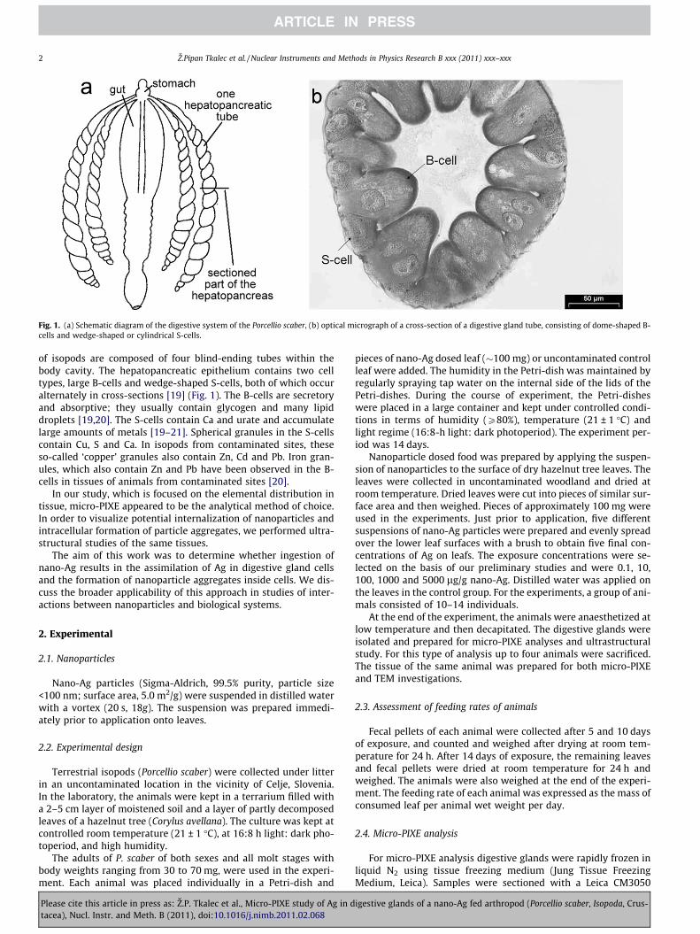
Fig. 1. (a) Schematic diagram of the digestive system of the Porcellio scaber, (b) optical micrograph of a cross-section of a digestive gland tube, consisting of dome-shaped B-cells and wedge-shaped or cylindrical S-cells.
2 Z.Pipan Tkalec et al. / Nuclear Instruments and Methods in Physics Research B xxx (2011) xxx–xxx
of isopods are composed of four blind-ending tubes within thebody cavity. The hepatopancreatic epithelium contains two celltypes, large B-cells and wedge-shaped S-cells, both of which occuralternately in cross-sections [19] (Fig. 1). The B-cells are secretoryand absorptive; they usually contain glycogen and many lipiddroplets [19,20]. The S-cells contain Ca and urate and accumulatelarge amounts of metals [19–21]. Spherical granules in the S-cellscontain Cu, S and Ca. In isopods from contaminated sites, theseso-called ‘copper’ granules also contain Zn, Cd and Pb. Iron gran-ules, which also contain Zn and Pb have been observed in the B-cells in tissues of animals from contaminated sites [20].
In our study, which is focused on the elemental distribution intissue, micro-PIXE appeared to be the analytical method of choice.In order to visualize potential internalization of nanoparticles andintracellular formation of particle aggregates, we performed ultra-structural studies of the same tissues.
The aim of this work was to determine whether ingestion ofnano-Ag results in the assimilation of Ag in digestive gland cellsand the formation of nanoparticle aggregates inside cells. We dis-cuss the broader applicability of this approach in studies of inter-actions between nanoparticles and biological systems.
2. Experimental
2.1. Nanoparticles
Nano-Ag particles (Sigma-Aldrich, 99.5% purity, particle size<100 nm; surface area, 5.0 m2/g) were suspended in distilled waterwith a vortex (20 s, 18g). The suspension was prepared immedi-ately prior to application onto leaves.
2.2. Experimental design
Terrestrial isopods (Porcellio scaber) were collected under litterin an uncontaminated location in the vicinity of Celje, Slovenia.In the laboratory, the animals were kept in a terrarium filled witha 2–5 cm layer of moistened soil and a layer of partly decomposedleaves of a hazelnut tree (Corylus avellana). The culture was kept atcontrolled room temperature (21 ± 1 �C), at 16:8 h light: dark pho-toperiod, and high humidity.
The adults of P. scaber of both sexes and all molt stages withbody weights ranging from 30 to 70 mg, were used in the experi-ment. Each animal was placed individually in a Petri-dish and
Please cite this article in press as: Z.P. Tkalec et al., Micro-PIXE study of Ag in dtacea), Nucl. Instr. and Meth. B (2011), doi:10.1016/j.nimb.2011.02.068
pieces of nano-Ag dosed leaf (�100 mg) or uncontaminated controlleaf were added. The humidity in the Petri-dish was maintained byregularly spraying tap water on the internal side of the lids of thePetri-dishes. During the course of experiment, the Petri-disheswere placed in a large container and kept under controlled condi-tions in terms of humidity (P80%), temperature (21 ± 1 �C) andlight regime (16:8-h light: dark photoperiod). The experiment per-iod was 14 days.
Nanoparticle dosed food was prepared by applying the suspen-sion of nanoparticles to the surface of dry hazelnut tree leaves. Theleaves were collected in uncontaminated woodland and dried atroom temperature. Dried leaves were cut into pieces of similar sur-face area and then weighed. Pieces of approximately 100 mg wereused in the experiments. Just prior to application, five differentsuspensions of nano-Ag particles were prepared and evenly spreadover the lower leaf surfaces with a brush to obtain five final con-centrations of Ag on leafs. The exposure concentrations were se-lected on the basis of our preliminary studies and were 0.1, 10,100, 1000 and 5000 lg/g nano-Ag. Distilled water was applied onthe leaves in the control group. For the experiments, a group of ani-mals consisted of 10–14 individuals.
At the end of the experiment, the animals were anaesthetized atlow temperature and then decapitated. The digestive glands wereisolated and prepared for micro-PIXE analyses and ultrastructuralstudy. For this type of analysis up to four animals were sacrificed.The tissue of the same animal was prepared for both micro-PIXEand TEM investigations.
2.3. Assessment of feeding rates of animals
Fecal pellets of each animal were collected after 5 and 10 daysof exposure, and counted and weighed after drying at room tem-perature for 24 h. After 14 days of exposure, the remaining leavesand fecal pellets were dried at room temperature for 24 h andweighed. The animals were also weighed at the end of the experi-ment. The feeding rate of each animal was expressed as the mass ofconsumed leaf per animal wet weight per day.
2.4. Micro-PIXE analysis
For micro-PIXE analysis digestive glands were rapidly frozen inliquid N2 using tissue freezing medium (Jung Tissue FreezingMedium, Leica). Samples were sectioned with a Leica CM3050
igestive glands of a nano-Ag fed arthropod (Porcellio scaber, Isopoda, Crus-
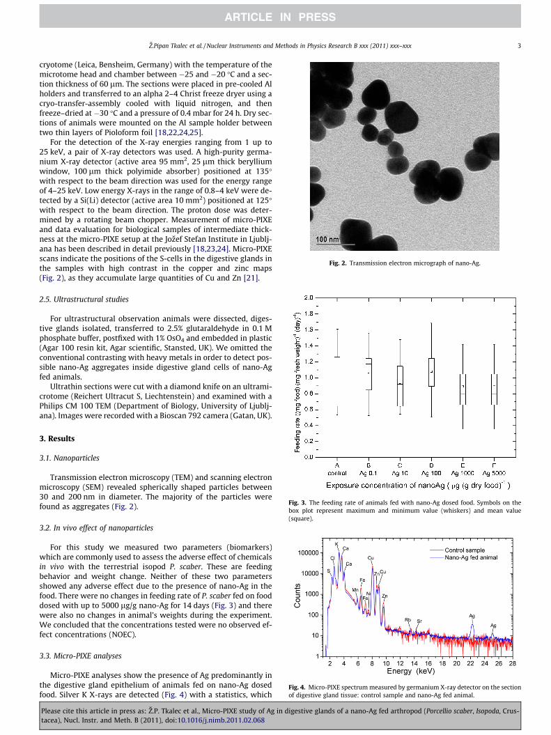
Fig. 2. Transmission electron micrograph of nano-Ag.
Z.Pipan Tkalec et al. / Nuclear Instruments and Methods in Physics Research B xxx (2011) xxx–xxx 3
cryotome (Leica, Bensheim, Germany) with the temperature of themicrotome head and chamber between �25 and �20 �C and a sec-tion thickness of 60 lm. The sections were placed in pre-cooled Alholders and transferred to an alpha 2–4 Christ freeze dryer using acryo-transfer-assembly cooled with liquid nitrogen, and thenfreeze–dried at �30 �C and a pressure of 0.4 mbar for 24 h. Dry sec-tions of animals were mounted on the Al sample holder betweentwo thin layers of Pioloform foil [18,22,24,25].
For the detection of the X-ray energies ranging from 1 up to25 keV, a pair of X-ray detectors was used. A high-purity germa-nium X-ray detector (active area 95 mm2, 25 lm thick berylliumwindow, 100 lm thick polyimide absorber) positioned at 135�with respect to the beam direction was used for the energy rangeof 4–25 keV. Low energy X-rays in the range of 0.8–4 keV were de-tected by a Si(Li) detector (active area 10 mm2) positioned at 125�with respect to the beam direction. The proton dose was deter-mined by a rotating beam chopper. Measurement of micro-PIXEand data evaluation for biological samples of intermediate thick-ness at the micro-PIXE setup at the Jozef Stefan Institute in Ljublj-ana has been described in detail previously [18,23,24]. Micro-PIXEscans indicate the positions of the S-cells in the digestive glands inthe samples with high contrast in the copper and zinc maps(Fig. 2), as they accumulate large quantities of Cu and Zn [21].
2.5. Ultrastructural studies
For ultrastructural observation animals were dissected, diges-tive glands isolated, transferred to 2.5% glutaraldehyde in 0.1 Mphosphate buffer, postfixed with 1% OsO4 and embedded in plastic(Agar 100 resin kit, Agar scientific, Stansted, UK). We omitted theconventional contrasting with heavy metals in order to detect pos-sible nano-Ag aggregates inside digestive gland cells of nano-Agfed animals.
Ultrathin sections were cut with a diamond knife on an ultrami-crotome (Reichert Ultracut S, Liechtenstein) and examined with aPhilips CM 100 TEM (Department of Biology, University of Ljublj-ana). Images were recorded with a Bioscan 792 camera (Gatan, UK).
Fig. 3. The feeding rate of animals fed with nano-Ag dosed food. Symbols on thebox plot represent maximum and minimum value (whiskers) and mean value(square).
Fig. 4. Micro-PIXE spectrum measured by germanium X-ray detector on the sectionof digestive gland tissue: control sample and nano-Ag fed animal.
3. Results
3.1. Nanoparticles
Transmission electron microscopy (TEM) and scanning electronmicroscopy (SEM) revealed spherically shaped particles between30 and 200 nm in diameter. The majority of the particles werefound as aggregates (Fig. 2).
3.2. In vivo effect of nanoparticles
For this study we measured two parameters (biomarkers)which are commonly used to assess the adverse effect of chemicalsin vivo with the terrestrial isopod P. scaber. These are feedingbehavior and weight change. Neither of these two parametersshowed any adverse effect due to the presence of nano-Ag in thefood. There were no changes in feeding rate of P. scaber fed on fooddosed with up to 5000 lg/g nano-Ag for 14 days (Fig. 3) and therewere also no changes in animal’s weights during the experiment.We concluded that the concentrations tested were no observed ef-fect concentrations (NOEC).
3.3. Micro-PIXE analyses
Micro-PIXE analyses show the presence of Ag predominantly inthe digestive gland epithelium of animals fed on nano-Ag dosedfood. Silver K X-rays are detected (Fig. 4) with a statistics, which
Please cite this article in press as: Z.P. Tkalec et al., Micro-PIXE study of Ag in digestive glands of a nano-Ag fed arthropod (Porcellio scaber, Isopoda, Crus-tacea), Nucl. Instr. and Meth. B (2011), doi:10.1016/j.nimb.2011.02.068

Fig. 5. Scanning Transmission Ion Microscopy (STIM) images and micro-PIXE qualitative elemental maps of S, Cu, Zn and Ag of Porcellio scaber: (a) section along the body axisof control animal; (b) section perpendiculary to the body axis of a control animal; (c) cross-section of a digestive gland of a control animal; (d) cross-section of a digestiveglands of a nano-Ag fed animal from a laboratory experiment.
4 Z.Pipan Tkalec et al. / Nuclear Instruments and Methods in Physics Research B xxx (2011) xxx–xxx
allows a reasonable two-dimensional mapping (Fig. 5d). In controlanimals, Ag was not found inside digestive gland cells (Fig. 5a–c).Since the spatial distribution of Ag in digestive gland epitheliumcorresponds to that of Cu and Zn and co-localization of Ag andCu is evident, we assume that Ag is also stored in S cells.
3.4. Ultrastructural studies
There were no differences between controls and nanoparticlefed animals in the presence of electron dense aggregates in diges-tive gland cells (Fig. 6a and b) investigated by TEM. In control ani-mals and also in nano-Ag fed animals we observed electron densematerial. We did not found any aggregates, which would indicatenano-Ag. We examined the same region of the epithelium whereAg was observed by the micro-PIXE method. As expected, metalstorage granules were detected in S-cells. These corresponded tothe Cu-granules described by many authors. No significant differ-ences in the appearances of these Cu-granules were observedwhen control animals and those fed on nano-Ag dosed food werecompared (Fig. 6c).
4. Discussion
Evidence is provided that feeding on nano-Ag dosed food resultsin assimilation of Ag in the digestive gland epithelium cells of amodel invertebrate animal. Ultrastructurally we could not detectthe formation of electron dense aggregates corresponding to inter-nalized nanoparticles and no effects on feeding behavior andweight of animals fed for 14 days on up to 5000 lg/g nano-Ag inthe food were observed.
Please cite this article in press as: Z.P. Tkalec et al., Micro-PIXE study of Ag in dtacea), Nucl. Instr. and Meth. B (2011), doi:10.1016/j.nimb.2011.02.068
Elemental distribution in digestive gland cells determined bythe micro-PIXE method showed that Ag, Cu and also Zn maps over-lap. It appears that majority of the Ag is in small cells of the diges-tive glands which are known to accumulate metals [26]. Reports inthe literature indicate that Ag accumulation results from free Agions that are bioavailable [1]. Ion release rates from nano-Ag in-crease with temperature in the range 0�37 �C, and decrease withincreasing pH or the addition of humic or fulvic acids. Silver nano-particle surfaces can adsorb Ag+, so even simple colloids containthree forms of Ag: Ag0 solids, free Ag+ or its complexes, and sur-face-adsorbed Ag+. Both thermodynamic analysis and kinetic mea-surements indicate that Ag0 nanoparticles will not persist inrealistic environmental compartments containing dissolved oxy-gen [27]. In isopods, there are two types of granules which accu-mulate metal ions. These are Type B and Type C granules. InType B Cd, Cu, Hg and Ag were reported to accumulate. In Type Cgranules Fe is usually found. We did not find a co-existence of Agand Fe, but a parallel existence of Cu and Ag. This is in accordanceto literature data [20]. The observation that Ag, but not Fe is foundin the same micro-locations as Cu and Zn suggests that ionic formsof Ag, but not entire particles of Ag were accumulated. The appear-ance of Cu-granules in S-cells of digestive gland epithelium indi-cates that Ag ions and not Ag nanoparticles are stored in thecells. Cu-granules are composed of homogeneous electron densematerial and are up to 1 lm in size. When nanoparticles are inter-nalized, they preserved the nanoparticle’s size and original shape.Within cells they are commonly in membrane bound vesicles[28,29].
Many authors also report cellular internalization of nanoparti-cles, which are detected as membrane bound micron-sized clusterseverywhere in the cytoplasm [28,29]. We observed membrane
igestive glands of a nano-Ag fed arthropod (Porcellio scaber, Isopoda, Crus-

Fig. 6. Transmission electron micrographs of digestive gland epithelium: (a) control group (on the left side B-cell and on the right side S-cell with electron dense material);(b) nano-Ag fed animals Porcellio scaber from a laboratory experiment (B-cell and S-cell with electron dense material); (c) metal granules in S-cell (on the left is control groupand on the right is nano-Ag fed animals Porcellio scaber from a laboratory experiment). Arrows indicate metal granules.
Z.Pipan Tkalec et al. / Nuclear Instruments and Methods in Physics Research B xxx (2011) xxx–xxx 5
bound electron dense clusters in the cytoplasm of both cell types,but there were no differences between control and nano-Ag fedanimals in the presence of these clusters. Therefore, it seems likelythat the clusters are not related to the consumption of nano-Ag.
The inability to detect electron dense aggregates in digestivegland cells of nano-Ag fed animals does not exclude the presenceof nanoparticles inside the cells, but other imaging modalities arenecessary to gain this information. Synchrotron X-ray fluorescencemicroscopy (SXRF) is a technique which offers simultaneous anal-ysis of trace element sensitivity and submicron spatial resolutioncombined with the ability to provide information regarding the
Please cite this article in press as: Z.P. Tkalec et al., Micro-PIXE study of Ag in dtacea), Nucl. Instr. and Meth. B (2011), doi:10.1016/j.nimb.2011.02.068
oxidation state and degree of coordination of metals [30]. Researchcurrently in progress is aimed at the determination of the oxida-tion state of elements detected inside cells and whether dissolvedions or nanoparticles, or both penetrated the cells.
In the study presented here, we used a terrestrial isopod to as-sess the toxic potential of substances added to food [31]. The re-sults show that exposure concentrations up to 5000 lg/g nano-Ag in the food did not affect the feeding behavior and weight ofthe animals and are therefore considered to be non-toxic. The pres-ence of Ag in digestive gland cells may be a result of a successfulsequestration mechanism in which excessive amounts of ingested
igestive glands of a nano-Ag fed arthropod (Porcellio scaber, Isopoda, Crus-
6 Z.Pipan Tkalec et al. / Nuclear Instruments and Methods in Physics Research B xxx (2011) xxx–xxx
metals or metal ions are stored in metal granules predominantly inS-cell. This mechanism is viewed as a detoxification mechanismwhich helps the organism to cope with high amounts of metalsin the environment. On the other hand, organisms which are ableto accumulate bioavailable metals could serve as a biological sys-tem with which it would be possible to assess bioavailable metalfractions in the substances added to food [32] and the dissolutionrate of ions from nanoparticles.
5. Conclusions
Our results show that the micro-PIXE method with its high sen-sitivity and good lateral resolution is a method of choice in studieswhere elemental distribution at organism/tissue levels is studied.Among the outstanding advantages of this method is chemical-freesample preparation. The micro-PIXE analysis, in combination withother methods could be of great benefit for studies of interactionsbetween nanoparticles and biological systems.
Acknowledgements
Support of the Research Programmes P1-0184, P1-0112, P1-0212, P1-0099, the Research Projects J7-0352, J1-9475, the acceler-ator operation within the framework of the Infrastructure ResearchCentres and the Ph.D. students within the framework of youngresearchers (Z.P. and N.G.) by the Slovenian Research Agency isacknowledged. Microprobe instrumentation is partially upgradedwithin the 7th FP EU Project No. 227012 ‘‘SPIRIT’’. We thankG.W.A. Milne for editorial assistance.
References
[1] S.W.P. Wijnhovenen, W.J.G.M. Peijnenburg, C.A. Herberts, W.I. Hagens, A.G.Oomen, W.H.W. Heugens, B. Roszek, J. Bisschops, I. Gosens, D. Van De Meent, S.Dekkers, W.H. De Jong, M. Van Zijverden, A.J.A.M. Sips, R.E. Geertsma,Nanotoxicology 3 (2009) 109.
[2] S.N. Luoma, Woodrow Wilson International Center for Scholars, Project onEmerging Nanotechnologies, 2008, p. 66.
[3] S.M. Hussain, K.L. Hess, J.M. Gearhart, K.T. Geiss, J.J. Schlager, Toxicol. In Vitro19 (2005) 975.
[4] N. Lubick, Environ. Sci. Technol. 42 (2008) 8617.[5] N. Miura, Y. Shinohara, Biochem. Biophys. Res. Commun. 390 (2009) 733.
Please cite this article in press as: Z.P. Tkalec et al., Micro-PIXE study of Ag in dtacea), Nucl. Instr. and Meth. B (2011), doi:10.1016/j.nimb.2011.02.068
[6] J. Farkas, P. Christian, J.A. Urrea, N. Roos, M. Hassellöv, K.E. Tollefsen, K.V.Thomas, Aquat. Toxicol. 96 (2010) 44.
[7] M.E. Samberg, S.J. Oldenburg, N.A. Monteiro-Riviere, Environ. Health Perspect.118 (2010) 407.
[8] L. Braydich-Stolle, S. Hussain, J.J. Schlager, M.C. Hofmann, Toxicol. Sci. 88(2005) 412.
[9] G. Laban, L.F. Nies, R.F. Turco, J.W. Bickham, M.S. Sepúlveda, Ecotoxicology 19(2010) 185.
[10] Y.J. Chae, C.H. Pham, J. Lee, E. Bae, J. Yi, M.B. Gu, Aquat. Toxicol. 94 (2009) 320.[11] E. Navarro, F. Piccapietra, B. Wagner, F. Marconi, R. Kaegi, N. Odzak, L. Sigg, R.
Behra, Environ. Sci. Technol. 42 (2008) 8959.[12] Y.S. Kim, J.S. Kim, H.S. Cho, D.S. Rha, J.M. Kim, J.D. Park, B.S. Choi, R. Lim, H.K.
Chang, Y.H. Chung, I.H. Kwong, J. Jeong, B.S. Han, J.I. Yu, Inhal. Toxicol. 20(2008) 575.
[13] S. Takenaka, E. Karg, C. Roth, H. Schulz, A. Ziesenis, U. Heinzmann, P. Schramel,J. Heyder, Environ. Health Perspect 109 (2001) 547.
[14] J.H. Ji, J.H. Jung, S.S. Kim, J.U. Joon, J.D. Park, B.S. Choi, Y.H. Chung, I.H. Kwon, J.Jeong, B.S. Han, J.H. Shin, J.H. Sung, K.S. Song, I.J. Yu. Inhal. Toxicol. 19 (2007)857.
[15] P.M. Bertsch, D.B. Hunter, Chem. Rev. 101 (2001) 1809.[16] B. Kaulich, A. Gianoncelli, A. Beran, D. Eichert1, I. Kreft, P. Pongrac, M. Regvar,
K. Vogel-Mikuš, M. Kiskinova, J. R. Soc. Interface 5 (2009) 641.[17] R. McRae, P. Bagchi, S. Sumalekshmy, C.J. Fahrni, Chem. Rev. 109 (2009) 4780.[18] K. Vogel-Mikuš, P. Pongrac, P. Pelicon, P. Vavpetic, B. Povh, H. Bothe, M. Regvar,
Micro-PIXE analysis for localisation and quantification of elements in roots ofmycorrhizal metal-tolerant plants, in: A. Varma, A.C. Kharkwal (Eds.),Symbiotic Fungi: Principles and Practice, (Soil biology, 18), Springer,Heidelberg, 2009, p. 227.
[19] J.W. Wägele, Isopoda, in: F.W. Harrison, A.G. Humes (Eds.), MicroscopicAnatomy of Invertebrates, Crustacea, vol. 9, Wiley-Liss, New York, p. 529.
[20] S.P. Hopkin, M.H. Martin, Tissue Cell 4 (1982) 703.[21] M. Tarnawska, P. Migula, W. Przybyłowicz, J. Mesjasz-Przybyłowicz, M.
Augustyniak, Nucl. Instrum. Meth. B 260 (2007) 213.[22] T. Schneider, S. Sheloske, B. Povh, Inter. J. PIXE 12 (2002) 101.[23] K. Vogel-Mikuš, P. Pongrac, P. Kump, M. Necemer, J. Simcic, P. Pelicon, M.
Budnar, B. Povh, M. Regvar, Environ. Pollut. 147 (2007) 50.[24] K. Vogel-Mikuš, M. Regvar, J. Mesjasz-Przybyłowicz, W.J. Przybyłowicz, J.
Simcic, P. Pelicon, M. Budnar, New Phytol. 179 (2008) 712.[25] K. Vogel-Mikuš, J. Simcic, P. Pelicon, M. Budnar, P. Kump, M. Necemer, J.
Mesjasz-Przybyłowicz, W.J. Przybyłowicz, M. Regvar, Plant Cell Environ. 31(2008) 1484.
[26] S.P. Hopkin, Ecophysiology of metals in Terrestrial Invertebrates, Elsevier,London, New York, 1989. p. 366.
[27] S. Elzey, V.H. Grassian, J. Nanopart. Res. 12 (2010) 1945.[28] C. Carlson, S.M. Hussain, A.M. Schrand, L.K. Braydich-Stolle, K.L. Hess, R.L.
Jones, J.J. Schlager, J. Phys. Chem. B 112 (2008) 13608.[29] A.M. Schrand, L.K. Braydich-Stolle, J.J. Schlager, L. Dai, S.M. Hussain,
Nanotechnology 19 (2008) 1.[30] C.J. Fahrni, Curr. Opin. Chem. Biol. 11 (2007) 121.[31] D. Drobne, Environ. Toxicol. 16 (1997) 1159.[32] M. Udovic, D. Drobne, D. Leštan, Environ. Pollut. 157 (2009) 2822.
igestive glands of a nano-Ag fed arthropod (Porcellio scaber, Isopoda, Crus-
Related Documents