Cellular Internalization of Dissolved Cobalt Ions from Ingested CoFe 2 O 4 Nanoparticles: In Vivo Experimental Evidence Sara Novak, † Damjana Drobne, †,‡,§, * Miha Golobic ̌ , † Jernej Zupanc, † Tea Romih, † Alessandra Gianoncelli, ∥ Maya Kiskinova, ∥ Burkhard Kaulich, ∥ Primoz ̌ Pelicon, ⊥ Primoz ̌ Vavpetic ̌ , ⊥ Luka Jeromel, ⊥ Nina Ogrinc, ⊥,∇ and Darko Makovec # † Department of Biology, Biotechnical Faculty, University of Ljubljana, Več na pot 111, 1000 Ljubljana, Slovenia ‡ Centre of Excellence, Advanced Materials and Technologies for the Future (CO NAMASTE), Jamova cesta 39, 1000 Ljubljana, Slovenia § Center of Exelence, Nanoscience and Nanotechnology (Nanocentre), Jamova cesta 39, 1000 Ljubljana, Slovenia ∥ Elettra-Sinchrotrone Trieste S.C.p.A., Strada Statale 14-km 163.5 in AREA Science Park, 34149 Basovizza, Trieste, Italy ⊥ Jož ef Stefan Institute, Microanalytical Center, Department for Low and Medium Energy Physics, Jamova cesta 39, 1000 Ljubljana, Slovenia # Institute Jozef Stefan, Jamova cesta 39, 1000 Ljubljana, Slovenia ∇ LOTRIC Metrology, Selca 163, 4227 Selca, Slovenia * S Supporting Information ABSTRACT: With a model invertebrate animal, we have assessed the fate of magnetic nanoparticles in biologically relevant media, i.e., digestive juices. The toxic potential and the internalization of such nanoparticles by nontarget cells were also examined. The aim of this study was to provide experimental evidence on the formation of Co 2+ , Fe 2+ , and Fe 3+ ions from CoFe 2 O 4 nanoparticles in the digestive juices of a model organism. Standard toxicological parameters were assessed. Cell membrane stability was tested with a modified method for measurement of its quality. Proton-induced X-ray emission and low energy synchrotron radiation X-ray fluorescence were used to study internalization and distribu- tion of Co and Fe. Co 2+ ions were found to be more toxic than nanoparticles. We confirmed that Co 2+ ions accumulate in the hepatopancreas, but Fe n+ ions or CoFe 2 O 4 nanoparticles are not retained in vivo. A model biological system with a terrestrial isopod is suited to studies of the potential dissolution of ions and other products from metal-containing nanoparticles in biologically complex media. ■ INTRODUCTION In the past decade, magnetic nanoparticles (NPs) have attracted much attention because of their potential use in different fields such as medicine, electronics, and energetics. 1−8 Because of increased use in many fields, high concentrations of CoFe 2 O 4 NPs in industrial wastewaters and surrounding soils can occur and negatively affect humans and the environment. One of the important issues which must be resolved before CoFe 2 O 4 nanoparticles can be widely used and safely controlled refers to dissolution of cobalt (Co) from CoFe 2 O 4 nanoparticles. Cobalt ions (Co 2+ ) may induce the formation of Reactive Oxygen Species (ROS), 9 oxidize proteins, 10 and cause oxidative DNA damage. 11 Consequently, the possible dissolution of the cobalt ions from CoFe 2 O 4 particles must be controlled. It is now widely recognized that such dissolution plays an important role in nanoparticle toxicity, but the reach of this phenomenon remains unclear. It has been found to be influenced mainly by pH but also by the specific surface area of the nanoparticles. 12 Natural organic compounds in the cellular media may either enhance or reduce the release of ions from nanoparticles, depending on their chemical composition and concentration. 12,13 Methods used for studying dissolution include different chemical analytical methods, such as atomic absorption spectroscopy (AAS), 14 inductively coupled plasma mass spectrometry (ICP-MS), 15 and localized surface plasmon resonance (LSPR). 16 These methods may be used in conjunction with ultracentrifugation of a suspension that Received: December 24, 2012 Revised: March 29, 2013 Accepted: April 11, 2013 Published: April 11, 2013 Article pubs.acs.org/est © 2013 American Chemical Society 5400 dx.doi.org/10.1021/es305132g | Environ. Sci. Technol. 2013, 47, 5400−5408

Welcome message from author
This document is posted to help you gain knowledge. Please leave a comment to let me know what you think about it! Share it to your friends and learn new things together.
Transcript
Cellular Internalization of Dissolved Cobalt Ions from IngestedCoFe2O4 Nanoparticles: In Vivo Experimental EvidenceSara Novak,† Damjana Drobne,†,‡,§,* Miha Golobic,† Jernej Zupanc,† Tea Romih,†
Alessandra Gianoncelli,∥ Maya Kiskinova,∥ Burkhard Kaulich,∥ Primoz Pelicon,⊥ Primoz Vavpetic,⊥Luka Jeromel,⊥ Nina Ogrinc,⊥,∇ and Darko Makovec#
†Department of Biology, Biotechnical Faculty, University of Ljubljana, Vecna pot 111, 1000 Ljubljana, Slovenia‡Centre of Excellence, Advanced Materials and Technologies for the Future (CO NAMASTE), Jamova cesta 39, 1000 Ljubljana,Slovenia§Center of Exelence, Nanoscience and Nanotechnology (Nanocentre), Jamova cesta 39, 1000 Ljubljana, Slovenia∥Elettra-Sinchrotrone Trieste S.C.p.A., Strada Statale 14-km 163.5 in AREA Science Park, 34149 Basovizza, Trieste, Italy⊥Jozef Stefan Institute, Microanalytical Center, Department for Low and Medium Energy Physics, Jamova cesta 39, 1000 Ljubljana,Slovenia#Institute Jozef Stefan, Jamova cesta 39, 1000 Ljubljana, Slovenia∇LOTRIC Metrology, Selca 163, 4227 Selca, Slovenia
*S Supporting Information
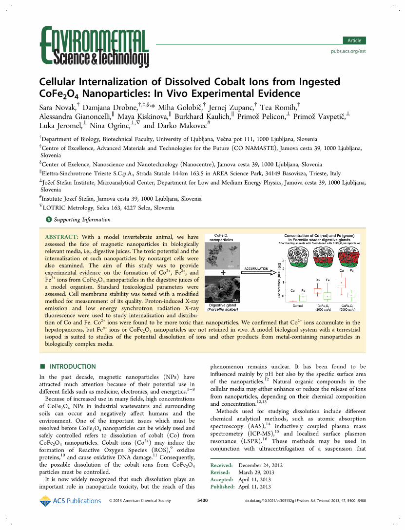
ABSTRACT: With a model invertebrate animal, we haveassessed the fate of magnetic nanoparticles in biologicallyrelevant media, i.e., digestive juices. The toxic potential and theinternalization of such nanoparticles by nontarget cells werealso examined. The aim of this study was to provideexperimental evidence on the formation of Co2+, Fe2+, andFe3+ ions from CoFe2O4 nanoparticles in the digestive juices ofa model organism. Standard toxicological parameters wereassessed. Cell membrane stability was tested with a modifiedmethod for measurement of its quality. Proton-induced X-rayemission and low energy synchrotron radiation X-rayfluorescence were used to study internalization and distribu-tion of Co and Fe. Co2+ ions were found to be more toxic than nanoparticles. We confirmed that Co2+ ions accumulate in thehepatopancreas, but Fen+ ions or CoFe2O4 nanoparticles are not retained in vivo. A model biological system with a terrestrialisopod is suited to studies of the potential dissolution of ions and other products from metal-containing nanoparticles inbiologically complex media.
■ INTRODUCTION
In the past decade, magnetic nanoparticles (NPs) haveattracted much attention because of their potential use indifferent fields such as medicine, electronics, and energetics.1−8
Because of increased use in many fields, high concentrationsof CoFe2O4 NPs in industrial wastewaters and surroundingsoils can occur and negatively affect humans and theenvironment. One of the important issues which must beresolved before CoFe2O4 nanoparticles can be widely used andsafely controlled refers to dissolution of cobalt (Co) fromCoFe2O4 nanoparticles. Cobalt ions (Co2+) may induce theformation of Reactive Oxygen Species (ROS),9 oxidizeproteins,10 and cause oxidative DNA damage.11 Consequently,the possible dissolution of the cobalt ions from CoFe2O4particles must be controlled.It is now widely recognized that such dissolution plays an
important role in nanoparticle toxicity, but the reach of this
phenomenon remains unclear. It has been found to beinfluenced mainly by pH but also by the specific surface areaof the nanoparticles.12 Natural organic compounds in thecellular media may either enhance or reduce the release of ionsfrom nanoparticles, depending on their chemical compositionand concentration.12,13
Methods used for studying dissolution include differentchemical analytical methods, such as atomic absorptionspectroscopy (AAS),14 inductively coupled plasma massspectrometry (ICP-MS),15 and localized surface plasmonresonance (LSPR).16 These methods may be used inconjunction with ultracentrifugation of a suspension that
Received: December 24, 2012Revised: March 29, 2013Accepted: April 11, 2013Published: April 11, 2013
Article
pubs.acs.org/est
© 2013 American Chemical Society 5400 dx.doi.org/10.1021/es305132g | Environ. Sci. Technol. 2013, 47, 5400−5408
separates the insoluble nanoparticles from ions that remain insolution.Measurements of dissolved ions from particles in biological
studies are useful only if they are conducted in biologicallyrelevant media. As a rule, it is not possible to mimic biologicalmedia; therefore, it is necessary to conduct experiments in invivo systems. Data on dissolution are very important to providean insight into the potentially compromised efficiency ofapplied nanomaterials or their toxicity when coming in contactwith biological fluids.In the work presented here, we have selected a model
biological system, in which CoFe2O4 nanoparticles areintroduced via food. The digestive system of the terrestrialisopod Porcellio scaber is composed of a stomach, four blind-ending digestive gland tubes (hepatopancreas), and a gut. Foodenters the digestive glands directly via the short stomach orafter the reflux from the gut. In the hepatopancreas, theingested material is mixed with digestive fluids. Measurementsof pH in the gut of terrestrial isopods (Porcellio scaber) with aLIX-type pH microelectrode showed pH 5.5−6.0 in theanterior hindgut, and pH 6.0−6.5 in the posterior hindgut.17
The pH value of the P. scaber digestive glands (hepatopancreas)is 6.1 ± 0.3 and at its distal region it is slightly more acidic, witha pH of 5.8−6.1.18 This biological system is a complexbiological environment which acts as an assembly of bio-logically relevant conditions that may affect dissolution ofcobalt or iron ions from nanoparticulate CoFe2O4. Whenconsumed particles entered the gut and digestive system, theyare retained in the digestive system for 2−4 h. Reflux of partlydigested food also reaches the digestive glands, but with somedelay. Apart from digestion and absorption of food, one of themajor roles of the digestive system is to accumulate metals inproportion to that in the gland lumen. In bioaccumulationstudies with isopods, it is expected that accumulation of a metalis related to the bioavailable metal ion fraction of the glandfluid.19,20
The aim of this study was to provide experimental evidenceon the formation of Co2+ and/or Fe2+/Fe3+ ions from CoFe2O4
nanoparticles in the digestive juices of a model organism,accumulation of dissolved ions by digestive gland epithelium,cellular internalization of particles, as well as on their toxicpotential. We hypothesize that metal ions are generated fromingested CoFe2O4 nanoparticles in a digestive system and thations are accumulated by digestive gland cells proportional toexposure doses. We also hypothesize that if particles enter cellsthey may reach a location distinct from that occupied by ionsthat have been accumulated via other pathways.
■ MATERIALS AND METHODS
Chemicals. Acridine orange (AO), ethidium bromide (EB),sodium chloride (NaCl), potassium chloride (KCl), magnesiumchloride (MgCl2), glucose, and 2-amino-2-hydroxymethyl-propane-1,3-diol (TRIS), were purchased from Merck. Cobalt-(II) chloride hexahydrate (CoCl2·6H2O), 99.9% (metal basis)was purchased from Alfa Aesar Johnson Mathey Company.CoFe2O4 nanoparticles were synthesized at the Department forMaterials Synthesis, Jozef Stefan Institute in Ljubljana.
Model Organisms. Terrestrial isopods, Porcellio scaber(Isopoda, Crustacea), were collected in July, 2011 at anuncontaminated location near Ljubljana, Slovenia. The animalswere kept in a terrarium filled with a layer of moistened soil anda thick layer of partly decomposed hazelnut tree leaves (Corylusavellana), at a temperature of 20 ± 2 °C and a 16:8 h light:darkphotoperiod. Only adult animals of both sexes and weighingmore than 30 mg were used. If moulting or the presence ofmarsupia were observed, then the animals were excluded fromthe experiment in order to keep the investigated population asphysiologically homogeneous as possible.
Characterization of Nanoparticles. The nanoparticleswere synthesized by coprecipitation using NaOH from aqueoussolutions of Co(II) and Fe (III) ions at elevated temper-atures.21 The nanoparticle samples were thoroughly washedwith water and then suspended in an aqueous solution ofglucose. The suspended CoFe2O4 nanoparticles show strongagglomeration in distilled H2O.Nanoparticles were inspected with transmission electron
microscopy (TEM) coupled with energy dispersive X-rayspectroscopy (EDXS) (JEOL 2010F) and the Zeta potentialwas also measured (Brookhaven Instruments Corp., Zeta-PALS). The aim of these analyses was to provide data on thesuspension of particles and allow comparisons among differentstudies and within our experiments.After exposure in feeding experiments, remnants of selected
leaves were dried and attached to mounts with silver paint(SPI), gold−palladium sputtered (Sputter coater SCD 050,BAL-TEC), and investigated by field emission scanningelectron microscopy (SEM) (Jeol JSM-6500F) at the Instituteof Metals and Technology in Ljubljana. Energy dispersive X-rayanalysis (EDX) was used to establish their chemicalcomposition (EDS/WDS Oxford Instruments INCA, JeolJSM-6500F) at the Institute of Metals and Technology.
Food Preparation. Hazelnut leaves were collected in anuncontaminated area and dried at room temperature and theanimals consumed particles applied to a leaf surface. Driedleaves were cut into pieces of approximately 100 mg. TheCoCl2 or CoFe2O4 nanoparticles was suspended in distilled
Table 1. Parameters of Experimentsa
experiment A experiment B experiment C experiment D
CoFe2O4 NPs concentration 1000 2000 1000 2000on leaves (μg/g leaf) 2000 / 2000 5000CoCl2 concentration on 1000 2000 / 2000leaves (μg/g leaf) 2000 / / 5000number of animals in each exposure group 20 15 10 15digestive gland membrane stability analyses (AO/EB) + + + −tissue distribution of Co and Fe (PIXE) + − − −cell distribution, colocalization of Co and Fe in gland cells (LE-XRF) + + + −concentration of Co and Fe indigestive glands (AAS) + − − +
aConcentrations of tested substances, number of exposed animals per group and measured parameters in different experiments (+, parametersanalyzed; −, parameters not analyzed).
Environmental Science & Technology Article
dx.doi.org/10.1021/es305132g | Environ. Sci. Technol. 2013, 47, 5400−54085401
water before each experiment to obtain final concentrations ofcobalt of 1000, 2000, and 5000 μg/mL. To diminishagglomeration, a suspension of nanoparticles in H2O wassonicated in an ultrasonic bath for 1 h and then immediatelyapplied to the leaves.In the control group, the leaves were treated with distilled
water and in the test group, a suspension of particles was spreadwith a paintbrush onto the abaxial surface of leaves to give finalnominal concentrations of 1000, 2000, and 5000 μg CoCl2 orCoFe2O4 nanoparticles per gram (dry mass) of leaf. The leaveswere allowed to stand until dry.Experimental Procedure. Each individual animal was
placed in a 9 cm Petri dish. One hazelnut leaf was treated withdistilled water, or a suspension of CoCl2 or nano-CoFe2O4 andplaced in the dish as the animal’s only food source. Humidity inthe Petri dish was maintained by spraying tap water on theinternal side of the lid every day. All Petri dishes were kept in alarge glass container under controlled conditions in terms of airhumidity (≥80%), temperature (21 ± 1 °C) and light regime(16:8 h light:dark photoperiod).Different numbers of animals in each individual experiment
were exposed to varying concentrations of nanoparticles for 14days (Table 1). Four experiments, A, B, C, and D, wereperformed one at a time, and the exposure concentrations ofsuspensions and initial number of tested animals were selectedon the basis of the type of analyses conducted after exposure.The concentrations were chosen on the basis of our
preliminary experiments. After exposure, the animals wereanaesthetized at low temperature and then decapitated andtheir digestive glands isolated. In experiments, digestive glandtubes were used for different analyses (Table 1).Feeding Parameters, Mass Change and Survival. After
14 days of exposure of the animals to treated leaves, faecalpellets and leaves were removed from the Petri dishes, dried atroom temperature for 24 h, and weighed. The feeding rate ofisopods was calculated as the mass of consumed leaves per wetmass of the animal per day. The food assimilation efficiency wascalculated as the difference between the mass of consumedleaves and mass of faecal pellets divided by the mass ofconsumed leaf. The mass change of animals was determined asthe difference in its mass from the beginning to the end of theexperimentAO/EB Analysis: Digestive Gland Cell Membrane
Stability. Cell membrane stability was tested with a modifiedmethod for assessment of cell membrane stability, previouslydescribed by Valant et al.22 In short, different permeability bythe two dyes results in differentially stained nuclei as follows:Acridine orange is taken up by cells with membranes that areintact or destabilized, and the cell emits green fluorescence.Ethidium bromide on the other hand, is taken up only by cellswith destabilized cell membranes, and such cells emit orangefluorescence, after intercalation into DNA.23
A single isolated hepatopancreatic tube was incubated for 5min in a mixture of the fluorescent dyes acridine orange andethidium bromide and then put on a microscope slide. Freshsamples were photographed and examined with an AxioimagerZ1 fluorescent microscope (Zeiss) with two different sets offilters. The excitation filter 450 to 490 nm and the emissionfilter 515 nm (filter set 09) were used to visualize AO and EBstained nuclei, while the excitation filter 365 nm and theemission filter 397 nm (filter set 01) were used to visualizenuclei stained with EB only. Cell membrane integrity wasassessed by examination of micrographs. Photographs of intact
digestive glands were examined by the same observer twice atintervals of at least 24 h and cell membrane integrity was ratedon the scale from 0 to 9 by visual inspection. On the basis ofpreliminary experiments, it was concluded that nontreated(control) animals showed <5% of nuclei stained by EB, whileseverely stressed animals have up to 100% of EB-stained nuclei.The <5% of hepatopancreatic tubes nuclei stained with EB wereclassified as 0, and those with the highest proportion (>95%) ofEB stained nuclei as 9. 22
Micro-PIXE Analysis: Tissue Distribution of Co and Fe.For microparticle induced X-ray emission (micro-PIXE)analysis, digestive glands were shock-frozen in liquid propaneor N2, using tissue-freezing medium (Jung Tissue FreezingMedium, Leica). Samples were sectioned with a sectionthickness of 60 μm using a Leica CM3050 cryotome (Leica)with the temperature of the microtome head and chambermaintained between −25 °C and −20 °C. The sections wereplaced in precooled Al holders, transferred to an alpha 2−4Christ freeze-dryer using a cryo-transfer assembly cooled withliquid N2, and then freeze-dried at −30 °C and 0.4 mbar for 24h. Dry sections were mounted between two thin layers ofPioloform foil on the Al sample holder.24,25
For detection of X-rays between 1 and 25 keV, two X-raydetectors were used simultaneously. A high-purity germaniumX-ray detector (active area 95 mm2; beryllium window, 25-μmthick; polyimide absorber, 100 μm thick) positioned at 135° tothe beam direction was used for the energy range of 4 keV-25keV. Low energy X-rays in the range of 0.8−6 keV weredetected by a Si(Li) detector (active area 10 mm2) positionedat 125° to the beam direction. The proton dose was determinedby a rotating in-beam chopper. Measurement of micro-PIXEemission and data evaluation for the biological samples ofintermediate thickness at the micro-PIXE laboratory, previouslydescribed in detail,24,26,27 was performed at the Jozef StefanInstitute in Ljubljana.We analyzed cross sections of isolated digestive gland tubes
from six animals. Two analyzed animals were control ones, twowere fed on food dosed with 2000 μg/g with CoCl2, and twoanimals on food dosed with 2000 μg/g nano-CoFe2O4. Two orthree digestive gland tubes were isolated from each animal andanalyzed.
LE-XRF Analysis: Cell Distribution and Colocalizationof Co and Fe in Digestive Gland Cells. For Low EnergySynchrotron Radiation X-ray Fluorescence (LE-XRF) analysis,isolated digestive glands were shock-frozen in liquid N2, usingtissue-freezing medium (Jung Tissue Freezing Medium, Leica).Samples were sectioned with a section thickness of 14 μm usinga Leica CM3050 cryotome (Leica) with the temperature of themicrotome head and chamber maintained between −25 °C and−20 °C. The sections were placed in precooled Al holders,transferred to an alpha 2−4 Christ freeze-dryer using a cryo-transfer assembly cooled with liquid N2, and then freeze-driedat −30 °C and 0.4 mbar for 24 h. Dry sections were mountedbetween two thin layers of Pioloform foil on the sample holder.The LE-XRF experiments were carried out with the x-ray
microscope TwinMic at the Elettra synchrotron radiationfacility in Trieste28,29 operating in the 400−2200 eV photonenergy range. During the experiments, TwinMic was operatedin STXM mode,29 in which a microprobe is formed by a zoneplate lens and the specimen is raster scanned across it. TheTwinMic microscope can provide submicrometer spatialresolution, but we used a spot size and a step size of 1 μm,
Environmental Science & Technology Article
dx.doi.org/10.1021/es305132g | Environ. Sci. Technol. 2013, 47, 5400−54085402
which is a useful compromise between lateral resolutionadequate for the features of interest and good XRF signal.The STXM mode allows simultaneous acquisition of X-ray
transmission (absorption and phase contrast images) andphoton emission (XRF) signals.30 The low X-ray energy rangeis particularly suited to biological investigations, and allows thesimultaneous acquisition of the elemental distributions ofelements of low atomic number (B to P) from the K emissionlines and elements of higher atomic number (Ca to Nb) fromthe L emission lines. The absorption and phase contrast imagesare collected by a configurable detector arrangement consistingof a fast read-out electron multiplying CCD camera coupled toan X-ray-to-visible light conversion system. The LEXRF setupused for this experiment consists of an annular arrangement of8 Si drift detectors (SDDs) (PNSensor, Munich, Germany),only 5 of which, coupled to read-out electronics, were used.31,32
This arrangement currently allows only qualitative analyses.Full elemental quantification will be the subject of futurereports.The X-ray fluorescence spectra obtained for each pixel in the
raster scan were batch processed by fitting the peaks with aGaussian model and with a linear subtraction, using thePyMCA data analysis software.33 Elemental maps weregenerated by plotting the intensity of the fluorescence peaksas a function of their position in the sample plane.In experiment A (Table 1), a cross section of one isolated
gland from a control animal, two isolated glands from oneanimal fed on food dosed with 2000 μg/g CoCl2, and twosections of digestive gland tubes from one animal fed on fooddosed with 2000 μg/g CoFe2O4 nanoparticles were analyzed. Inexperiment B, samples from one control animal and twosections from two animals from each treated group (animals fedon food dosed with 2000 μg/g CoCl2 or CoFe2O4 nano-particles) were analyzed. In experiment C, two sections fromtwo different animals both fed on food dosed with 2000 μg/gnano-CoFe2O4 were analyzed. Altogether, 12 different cross-sections of digestive glands were analyzed.AAS Analysis: Concentration of Co and Fe in
Digestive Glands. Cobalt and iron were measured by atomicabsorption spectroscopy in one or two isolated digestive glandtubes from each animal in experiments A and D. Prior to theanalysis, samples were lyophilized, weighed, and completelydigested in a 7:1 nitric acid/perchloric acid mixture. Afterevaporation of the acid, the residue was taken up in 0.2%HNO3 and total Co and Fe concentrations in the digestiveglands were determined by flame atomic absorption spectrom-etry (Perkin-Elmer AAnalyst 100) in the Department ofBiology, University of Ljubljana. Within each measurement,certified reference material (TORT-2, National ResearchCouncil of Canada) was used to check the accuracy andprecision of the analytical procedure. Along with the samples,10 replicates of a known amount of certified reference materialwere also acid digested and each sample was measured intriplicate. Calculations followed the approach of Jorhem34 andPhillips et al.35 The certified concentration of Co in thereference material was 0.51 ± 0.09 mg/kg; our measurementwas 0.64 ± 0.14 mg/kg (mean ± SD, n = 30); recovery was125.5%, Z′ = 2.62. For Fe, the certified concentration in thereference material was 105 ± 13 mg/kg; our measurement was101 ± 14 mg/kg (mean ± SD, n = 30); recovery was 96.2%, Z′= −0.626.Concentration of Co and Fe in the nano-CoFe2O4
Suspension Supernatant. For this analysis, suspensions of
CoFe2O4 NPs and CoCl2 in deionized water were prepared inthe same way as that for the in vivo tests. The finalconcentrations were 2000 mg Co and 5000 mg Co/mL; eachconcentration was prepared in triplicate. The suspensions wereultracentrifuged twice at 100 000 g for 30 min at 20 °C. Thesupernatant was separated from the pellet formed by thenanoparticles and divided into two aliquots for further dilution.One aliquot was diluted with an equal volume of deionizedwater, the other one with an equal volume of 1 M HCl.36 Then,a 3-mL portion from each aliquot was used for analysis by flameatomic absorption spectrometry (AAS, Perkin-Elmer Analyst100). The difference in Co and Fe ion content between theacidified and nonacidified suspension indicates that nano-particles remaining in the supernatant after centrifugation aredissolved. Measurement of CoCl2 solution served as a checkthat centrifugation did not remove ions from the solution.Detection of Co and Fe by AAS is possible, provided that theconcentrations of Co and Fe ions are above the detection limit(9 μg/L and 5 μg/L, respectively).The original suspensions of CoFe2O4 NPs (2000 and 5000
mg/L of CoFe2O4) were also analyzed by AAS. Prior to theanalysis the suspensions were diluted (1:1000 and 1:2500,respectively) with 1 M HCl and incubated in acid for 3 days toachieve complete dissolution.
Data Analysis. Data were analyzed by standard statisticalmethods. The difference in the median measured parameters inexposed and unexposed groups was tested with the non-parametric Mann−Whitney U test. All calculations wereperformed with Statgraphics Plus 4.0. Statistically significantdifferences between exposed and control animals were dividedinto three categories with different numbers of stars assigned (*p < 0.05, ** p < 0.01, ***p < 0.001).
■ RESULTSCharacterization of Nanoparticles. TEM analysis
showed the nanoparticles to have a relatively broad sizedistribution between 5 and 15 nm (Figure S1A of theSupporting Information, SI). The smaller nanoparticles areglobularly shaped; whereas the larger ones have an octahedralshape. Energy dispersive X-ray spectroscopy (EDS) analysisshowed that their composition matched to stoichiometry ofCoFe2O4. The size of the agglomerates of the nanoparticles onthe surfaces of leaves in the experiments was between 1 and 10μm (inspected with SEM, Figure S2 of the SI).Zeta potential measurements showed the isoelectric point for
CoFe2O4 nanoparticles at neutral pH. At pH 7.4, the zetapotential is slightly negative. SEM revealed that particlesremained spread over the entire leaf surface (Figure S1B of theSI) and energy dispersive X-ray analysis (EDX) revealed theirchemical composition (Table S1 of the SI).
Feeding Parameters, Mass Change and Mortality.Animals were exposed to leaves dosed with CoCl2 or asuspension of CoFe2O4 NPs, providing nominal concentrationsof 1000, 2000, or 5000 μg CoCl2 or CoFe2O4 nanoparticles perg of leaf. Mass change and survival were not affected at theseconcentrators of CoCl2 or nano-CoFe2O4 (Figures S3 and S4of the SI). In all four experiments, there was a statisticallysignificant difference in feeding rate between control groupsand groups in which animals were exposed to 2000 or 5000 μg/g of CoCl2. Exposure to CoFe2O4 nanoparticles had no effecton the feeding rate of animals in any experiment for the 14 daysof the experiment. Compared to the control animals, thereduction in the feeding rate of animals exposed to CoCl2 was
Environmental Science & Technology Article
dx.doi.org/10.1021/es305132g | Environ. Sci. Technol. 2013, 47, 5400−54085403
statistically significant in higher concentrations (2000, 5000 μg/g) of Co in food as shown in Figure S5 of the SI).AO/EB Analysis: Digestive Gland Cell Membrane
Stability. Our previously published data demonstrate thatthe digestive gland cell membrane stability value was higherthan 2 (on a scale from 0 to 9) in only 5% of animals from astock culture and in good physiological condition, and thisnumber was used as a benchmark.22 The cell membranes areconsidered destabilized when this value is higher than 2.We combine results on cell membrane stability from all three
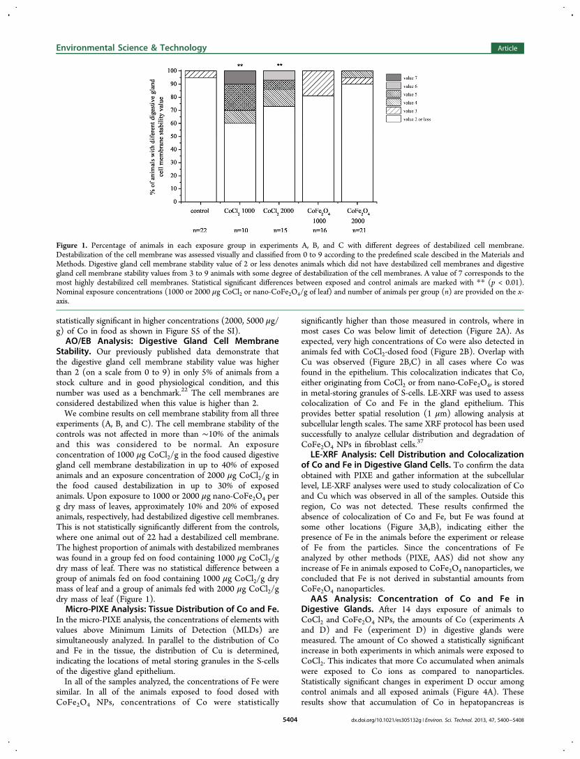
experiments (A, B, and C). The cell membrane stability of thecontrols was not affected in more than ∼10% of the animalsand this was considered to be normal. An exposureconcentration of 1000 μg CoCl2/g in the food caused digestivegland cell membrane destabilization in up to 40% of exposedanimals and an exposure concentration of 2000 μg CoCl2/g inthe food caused destabilization in up to 30% of exposedanimals. Upon exposure to 1000 or 2000 μg nano-CoFe2O4 perg dry mass of leaves, approximately 10% and 20% of exposedanimals, respectively, had destabilized digestive cell membranes.This is not statistically significantly different from the controls,where one animal out of 22 had a destabilized cell membrane.The highest proportion of animals with destabilized membraneswas found in a group fed on food containing 1000 μg CoCl2/gdry mass of leaf. There was no statistical difference between agroup of animals fed on food containing 1000 μg CoCl2/g drymass of leaf and a group of animals fed with 2000 μg CoCl2/gdry mass of leaf (Figure 1).Micro-PIXE Analysis: Tissue Distribution of Co and Fe.
In the micro-PIXE analysis, the concentrations of elements withvalues above Minimum Limits of Detection (MLDs) aresimultaneously analyzed. In parallel to the distribution of Coand Fe in the tissue, the distribution of Cu is determined,indicating the locations of metal storing granules in the S-cellsof the digestive gland epithelium.In all of the samples analyzed, the concentrations of Fe were
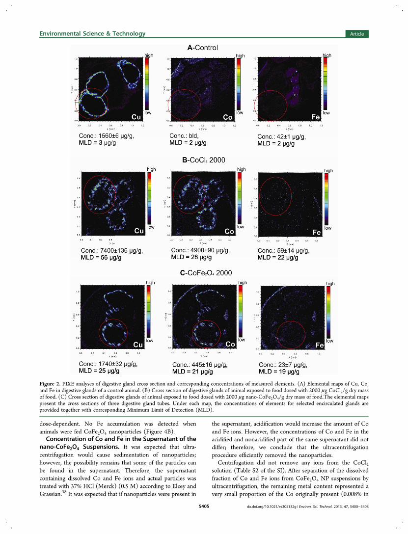
similar. In all of the animals exposed to food dosed withCoFe2O4 NPs, concentrations of Co were statistically
significantly higher than those measured in controls, where inmost cases Co was below limit of detection (Figure 2A). Asexpected, very high concentrations of Co were also detected inanimals fed with CoCl2-dosed food (Figure 2B). Overlap withCu was observed (Figure 2B,C) in all cases where Co wasfound in the epithelium. This colocalization indicates that Co,either originating from CoCl2 or from nano-CoFe2O4, is storedin metal-storing granules of S-cells. LE-XRF was used to assesscolocalization of Co and Fe in the gland epithelium. Thisprovides better spatial resolution (1 μm) allowing analysis atsubcellular length scales. The same XRF protocol has been usedsuccessfully to analyze cellular distribution and degradation ofCoFe2O4 NPs in fibroblast cells.37
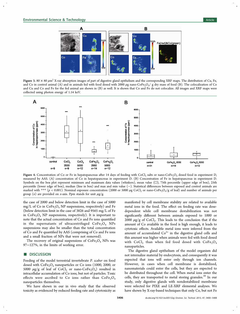
LE-XRF Analysis: Cell Distribution and Colocalizationof Co and Fe in Digestive Gland Cells. To confirm the dataobtained with PIXE and gather information at the subcellularlevel, LE-XRF analyses were used to study colocalization of Coand Cu which was observed in all of the samples. Outside thisregion, Co was not detected. These results confirmed theabsence of colocalization of Co and Fe, but Fe was found atsome other locations (Figure 3A,B), indicating either thepresence of Fe in the animals before the experiment or releaseof Fe from the particles. Since the concentrations of Feanalyzed by other methods (PIXE, AAS) did not show anyincrease of Fe in animals exposed to CoFe2O4 nanoparticles, weconcluded that Fe is not derived in substantial amounts fromCoFe2O4 nanoparticles.
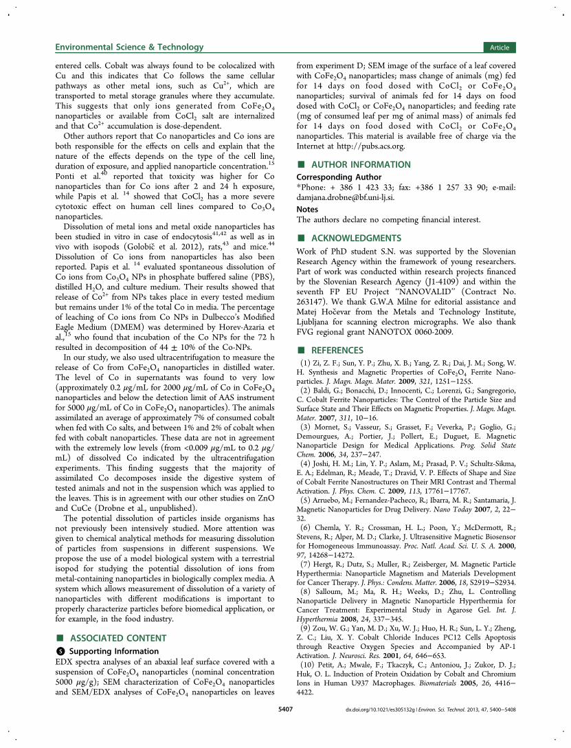
AAS Analysis: Concentration of Co and Fe inDigestive Glands. After 14 days exposure of animals toCoCl2 and CoFe2O4 NPs, the amounts of Co (experiments Aand D) and Fe (experiment D) in digestive glands weremeasured. The amount of Co showed a statistically significantincrease in both experiments in which animals were exposed toCoCl2. This indicates that more Co accumulated when animalswere exposed to Co ions as compared to nanoparticles.Statistically significant changes in experiment D occur amongcontrol animals and all exposed animals (Figure 4A). Theseresults show that accumulation of Co in hepatopancreas is
Figure 1. Percentage of animals in each exposure group in experiments A, B, and C with different degrees of destabilized cell membrane.Destabilization of the cell membrane was assessed visually and classified from 0 to 9 according to the predefined scale descibed in the Materials andMethods. Digestive gland cell membrane stability value of 2 or less denotes animals which did not have destabilized cell membranes and digestivegland cell membrane stability values from 3 to 9 animals with some degree of destabilization of the cell membranes. A value of 7 corresponds to themost highly destabilized cell membranes. Statistical significant differences between exposed and control animals are marked with ** (p < 0.01).Nominal exposure concentrations (1000 or 2000 μg CoCl2 or nano-CoFe2O4/g of leaf) and number of animals per group (n) are provided on the x-axis.
Environmental Science & Technology Article
dx.doi.org/10.1021/es305132g | Environ. Sci. Technol. 2013, 47, 5400−54085404
dose-dependent. No Fe accumulation was detected whenanimals were fed CoFe2O4 nanoparticles (Figure 4B).Concentration of Co and Fe in the Supernatant of the
nano-CoFe2O4 Suspensions. It was expected that ultra-centrifugation would cause sedimentation of nanoparticles;however, the possibility remains that some of the particles canbe found in the supernatant. Therefore, the supernatantcontaining dissolved Co and Fe ions and actual particles wastreated with 37% HCl (Merck) (0.5 M) according to Elzey andGrassian.38 It was expected that if nanoparticles were present in
the supernatant, acidification would increase the amount of Coand Fe ions. However, the concentrations of Co and Fe in theacidified and nonacidified part of the same supernatant did notdiffer; therefore, we conclude that the ultracentrifugationprocedure efficiently removed the nanoparticles.Centrifugation did not remove any ions from the CoCl2
solution (Table S2 of the SI). After separation of the dissolvedfraction of Co and Fe ions from CoFe2O4 NP suspensions byultracentrifugation, the remaining metal content represented avery small proportion of the Co originally present (0.008% in
Figure 2. PIXE analyses of digestive gland cross section and corresponding concentrations of measured elements. (A) Elemental maps of Cu, Co,and Fe in digestive glands of a control animal. (B) Cross section of digestive glands of animal exposed to food dosed with 2000 μg CoCl2/g dry massof food. (C) Cross section of digestive glands of animal exposed to food dosed with 2000 μg nano-CoFe2O4/g dry mass of food.The elemental mapspresent the cross sections of three digestive gland tubes. Under each map, the concentrations of elements for selected encirculated glands areprovided together with corresponding Minimum Limit of Detection (MLD).
Environmental Science & Technology Article
dx.doi.org/10.1021/es305132g | Environ. Sci. Technol. 2013, 47, 5400−54085405
the case of 2000 and below detection limit in the case of 5000mg/L of Co in CoFe2O4 NP suspensions, respectively) and Fe(below detection limit in the case of 3826 and 9565 mg/L of Fein CoFe2O4 NP suspensions, respectively). It is important tonote that the actual concentration of Co and Fe ions quantifiedin the supernatants of ultracentrifuged CoFe2O4 NPssuspensions may also be smaller than the total concentrationof Co and Fe quantified by AAS (comprising of Co and Fe ionsand a small fraction of NPs that were not removed).The recovery of original suspensions of CoFe2O4 NPs was
97−127%, in the limits of working error.
■ DISCUSSION
Feeding of the model terrestrial invertebrate P. scaber on fooddosed with CoFe2O4 nanoparticles or Co ions (1000, 2000, or5000 μg/g of leaf of CoCl2 or nano-CoFe2O4) resulted inintracellular accumulation of Co ions, but not of particles. Toxiceffects were ascribed to Co ions rather than CoFe2O4nanoparticles themselves.We have shown in our in vivo study that the observed
toxicity as evidenced by reduced feeding rate and cytotoxicity as
manifested by cell membrane stability are related to availablemetal ions in the food. The effect on feeding rate was dose-dependent while cell membrane destabilization was notsignificantly different between animals exposed to 1000 or2000 μg/g of CoCl2. This leads to the conclusion that if theamount of Co available in the food is high enough, it leads tocytotoxic effects. Available metal ions were inferred from theamount of accumulated Co2+ in the digestive gland cells andthis amount was higher when animals were fed with food dosedwith CoCl2 than when fed food dosed with CoFe2O4
nanoparticles.The digestive gland epithelium of the model organism did
not internalize material by endocytosis, and consequently it wasexpected that ions will enter only through ion channels.However, in cases when cell membrane is destabilized,nanomaterials could enter the cells, but they are expected tobe distributed throughout the cell. When metal ions enter thecells, they are transported to metal storing granules.39 In ourstudy, only digestive glands with nondestabilized membranewere selected for PIXE and LE-XRF elemental analyses. Wehave shown by X-ray-based techniques that only Co, but not Fe
Figure 3. 80 × 80 μm2 X-ray absorption images of part of digestive gland epithelium and the corresponding XRF maps. The distribution of Cu, Fe,and Co in control animal (A) and in animals fed with food dosed with 2000 μg nano-CoFe2O4/ g dry mass of food (B). The colocalization of Coand Cu and Co and Fe for the fed animal are shown in (B) as well. It is shown that Co and Fe do not colocalize. All images and XRF maps werecollected using photon energy of 1.14 keV.
Figure 4. Concentration of Co or Fe in hepatopancreas after 14 days of feeding with CoCl2 salts or nano-CoFe2O4 dosed food in experiment D,measured by AAS. (A) concentration of Co in hepatopancreas in experiment D. (B) Concentration of Fe in hepatopancreas in experiment D.Symbols on the box plot represent minimum and maximum data values (whiskers), mean value (□), 75th percentile (upper edge of box), 25thpercentile (lower edge of box), median (line in box) and max and min value (−). Statistical differences between exposed and control animals aremarked with *** (p < 0.001). Nominal exposure concentration (2000 or 5000 μg CoCl2 or nano-CoFe2O4/g of leaf) and number of animals pergroup (n) are provided on x-axis. Ppm stands for unit μg/g.
Environmental Science & Technology Article
dx.doi.org/10.1021/es305132g | Environ. Sci. Technol. 2013, 47, 5400−54085406
entered cells. Cobalt was always found to be colocalized withCu and this indicates that Co follows the same cellularpathways as other metal ions, such as Cu2+, which aretransported to metal storage granules where they accumulate.This suggests that only ions generated from CoFe2O4nanoparticles or available from CoCl2 salt are internalizedand that Co2+ accumulation is dose-dependent.Other authors report that Co nanoparticles and Co ions are
both responsible for the effects on cells and explain that thenature of the effects depends on the type of the cell line,duration of exposure, and applied nanoparticle concentration.15
Ponti et al.40 reported that toxicity was higher for Conanoparticles than for Co ions after 2 and 24 h exposure,while Papis et al. 14 showed that CoCl2 has a more severecytotoxic effect on human cell lines compared to Co3O4nanoparticles.Dissolution of metal ions and metal oxide nanoparticles has
been studied in vitro in case of endocytosis41,42 as well as invivo with isopods (Golobic et al. 2012), rats,43 and mice.44
Dissolution of Co ions from nanoparticles has also beenreported. Papis et al. 14 evaluated spontaneous dissolution ofCo ions from Co3O4 NPs in phosphate buffered saline (PBS),distilled H2O, and culture medium. Their results showed thatrelease of Co2+ from NPs takes place in every tested mediumbut remains under 1% of the total Co in media. The percentageof leaching of Co ions from Co NPs in Dulbecco’s ModifiedEagle Medium (DMEM) was determined by Horev-Azaria etal.,15 who found that incubation of the Co NPs for the 72 hresulted in decomposition of 44 ± 10% of the Co-NPs.In our study, we also used ultracentrifugation to measure the
release of Co from CoFe2O4 nanoparticles in distilled water.The level of Co in supernatants was found to very low(approximately 0.2 μg/mL for 2000 μg/mL of Co in CoFe2O4nanoparticles and below the detection limit of AAS instrumentfor 5000 μg/mL of Co in CoFe2O4 nanoparticles). The animalsassimilated an average of approximately 7% of consumed cobaltwhen fed with Co salts, and between 1% and 2% of cobalt whenfed with cobalt nanoparticles. These data are not in agreementwith the extremely low levels (from <0.009 μg/mL to 0.2 μg/mL) of dissolved Co indicated by the ultracentrifugationexperiments. This finding suggests that the majority ofassimilated Co decomposes inside the digestive system oftested animals and not in the suspension which was applied tothe leaves. This is in agreement with our other studies on ZnOand CuCe (Drobne et al., unpublished).The potential dissolution of particles inside organisms has
not previously been intensively studied. More attention wasgiven to chemical analytical methods for measuring dissolutionof particles from suspensions in different suspensions. Wepropose the use of a model biological system with a terrestrialisopod for studying the potential dissolution of ions frommetal-containing nanoparticles in biologically complex media. Asystem which allows measurement of dissolution of a variety ofnanoparticles with different modifications is important toproperly characterize particles before biomedical application, orfor example, in the food industry.
■ ASSOCIATED CONTENT*S Supporting InformationEDX spectra analyses of an abaxial leaf surface covered with asuspension of CoFe2O4 nanoparticles (nominal concentration5000 μg/g); SEM characterization of CoFe2O4 nanoparticlesand SEM/EDX analyses of CoFe2O4 nanoparticles on leaves
from experiment D; SEM image of the surface of a leaf coveredwith CoFe2O4 nanoparticles; mass change of animals (mg) fedfor 14 days on food dosed with CoCl2 or CoFe2O4nanoparticles; survival of animals fed for 14 days on fooddosed with CoCl2 or CoFe2O4 nanoparticles; and feeding rate(mg of consumed leaf per mg of animal mass) of animals fedfor 14 days on food dosed with CoCl2 or CoFe2O4nanoparticles. This material is available free of charge via theInternet at http://pubs.acs.org.
■ AUTHOR INFORMATIONCorresponding Author*Phone: + 386 1 423 33; fax: +386 1 257 33 90; e-mail:[email protected] authors declare no competing financial interest.
■ ACKNOWLEDGMENTSWork of PhD student S.N. was supported by the SlovenianResearch Agency within the framework of young researchers.Part of work was conducted within research projects financedby the Slovenian Research Agency (J1-4109) and within theseventh FP EU Project ‘‘NANOVALID’’ (Contract No.263147). We thank G.W.A Milne for editorial assistance andMatej Hocevar from the Metals and Technology Institute,Ljubljana for scanning electron micrographs. We also thankFVG regional grant NANOTOX 0060-2009.
■ REFERENCES(1) Zi, Z. F.; Sun, Y. P.; Zhu, X. B.; Yang, Z. R.; Dai, J. M.; Song, W.H. Synthesis and Magnetic Properties of CoFe2O4 Ferrite Nano-particles. J. Magn. Magn. Mater. 2009, 321, 1251−1255.(2) Baldi, G.; Bonacchi, D.; Innocenti, C.; Lorenzi, G.; Sangregorio,C. Cobalt Ferrite Nanoparticles: The Control of the Particle Size andSurface State and Their Effects on Magnetic Properties. J. Magn. Magn.Mater. 2007, 311, 10−16.(3) Mornet, S.; Vasseur, S.; Grasset, F.; Veverka, P.; Goglio, G.;Demourgues, A.; Portier, J.; Pollert, E.; Duguet, E. MagneticNanoparticle Design for Medical Applications. Prog. Solid StateChem. 2006, 34, 237−247.(4) Joshi, H. M.; Lin, Y. P.; Aslam, M.; Prasad, P. V.; Schultz-Sikma,E. A.; Edelman, R.; Meade, T.; Dravid, V. P. Effects of Shape and Sizeof Cobalt Ferrite Nanostructures on Their MRI Contrast and ThermalActivation. J. Phys. Chem. C. 2009, 113, 17761−17767.(5) Arruebo, M.; Fernandez-Pacheco, R.; Ibarra, M. R.; Santamaria, J.Magnetic Nanoparticles for Drug Delivery. Nano Today 2007, 2, 22−32.(6) Chemla, Y. R.; Crossman, H. L.; Poon, Y.; McDermott, R.;Stevens, R.; Alper, M. D.; Clarke, J. Ultrasensitive Magnetic Biosensorfor Homogeneous Immunoassay. Proc. Natl. Acad. Sci. U. S. A. 2000,97, 14268−14272.(7) Hergt, R.; Dutz, S.; Muller, R.; Zeisberger, M. Magnetic ParticleHyperthermia: Nanoparticle Magnetism and Materials Developmentfor Cancer Therapy. J. Phys.: Condens. Matter. 2006, 18, S2919−S2934.(8) Salloum, M.; Ma, R. H.; Weeks, D.; Zhu, L. ControllingNanoparticle Delivery in Magnetic Nanoparticle Hyperthermia forCancer Treatment: Experimental Study in Agarose Gel. Int. J.Hyperthermia 2008, 24, 337−345.(9) Zou, W. G.; Yan, M. D.; Xu, W. J.; Huo, H. R.; Sun, L. Y.; Zheng,Z. C.; Liu, X. Y. Cobalt Chloride Induces PC12 Cells Apoptosisthrough Reactive Oxygen Species and Accompanied by AP-1Activation. J. Neurosci. Res. 2001, 64, 646−653.(10) Petit, A.; Mwale, F.; Tkaczyk, C.; Antoniou, J.; Zukor, D. J.;Huk, O. L. Induction of Protein Oxidation by Cobalt and ChromiumIons in Human U937 Macrophages. Biomaterials 2005, 26, 4416−4422.
Environmental Science & Technology Article
dx.doi.org/10.1021/es305132g | Environ. Sci. Technol. 2013, 47, 5400−54085407
(11) De Boeck, M.; Lison, D.; Kirsch-Volders, M. Evaluation of the inVitro Direct and Indirect Genotoxic Effects of Cobalt CompoundsUsing the Alkaline Comet Assay. Influence of Interdonor andInterexperimental Variability. Carcinogenesis 1998, 19, 2021−2029.(12) Dokoumetzidis, A.; Macheras, P. a Century of DissolutionResearch: From Noyes and Whitney to the BiopharmaceuticsClassification System. Int. J. Pharm. 2006, 321, 1−11.(13) Miao, A. J.; Zhang, X. Y.; Luo, Z. P.; Chen, C. S.; Chin, W. C.;Santschi, P. H.; Quigg, A. Zinc Oxide Engineered NanoparticlesDissolution and Toxicity to Marine Phytoplankton. Environ. Toxicol.Chem. 2010, 29, 2814−2822.(14) Papis, E.; Rossi, F.; Raspanti, M.; Dalle-Donne, I.; Colombo, G.;Milzani, A.; Bernardini, G.; Gornati, R. Engineered Cobalt OxideNanoparticles Readily Enter Cells. Toxicol. Lett. 2009, 189, 253−259.(15) Horev-Azaria, L.; Kirkpatrick, C. J.; Korenstein, R.; Marche, P.N.; Maimon, O.; Ponti, J.; Romano, R.; Rossi, F.; Golla-Schindler, U.;Sommer, D.; Uboldi, C.; Unger, R. E.; Villiers, C. PredictiveToxicology of Cobalt Nanoparticles and Ions: Comparative In VitroStudy of Different Cellular Models Using Methods of KnowledgeDiscovery from Data. Toxicol. Sci. 2011, 122, 489−501.(16) Zook, J. M.; Long, S. E.; Cleveland, D.; Geronimo, C. L. A.;MacCuspie, R. I. Measuring Silver Nanoparticle Dissolution inComplex Biological and Environmental Matrices Using UV-VisibleAbsorbance. Anal. Bioanal. Chem. 2011, 401, 1993−2002.(17) Zimmer, M.; Topp, W. Homeostatic Responses in the Gut ofPorcellio scaber (Isopoda: Oniscidea) Optimize Litter Degradation. J.Comp. Physiol., B. 1997, 167, 582−585.(18) Zimmer, M.; Brune, A. Physiological Properties of the GutLumen of Terrestrial Isopods (Isopoda: Oniscidea): Adaptive toDigesting Lignocellulose? J. Comp. Physiol., B. 2005, 275−283.(19) Udovic, M.; Drobne, D.; Lestan, D. Bioaccumulation in Porcellioscaber (Crustacea, Isopoda) As a Measure of the EDTA RemediationEfficiency of Metal-Polluted Soil. Environ. Pollut. 2009, 157, 2822−2829.(20) Pipan-Tkalec, Z.; Drobne, D.; Jemec, A.; Romih, T.; Zidar, P.;Bele, M. Zinc Bioaccumulation in a Terrestrial Invertebrate Fed a DietTreated with Particulate ZnO or ZnCl2 Solution. Toxicology 2010, 269,198−203.(21) Gyergyek, S.; Drofenik, M.; Makovec, D. Oleic-Acid-CoatedCoFe2O4 Nanoparticles Synthesized by Co-Precipitation and Hydro-thermal Synthesis. Mater. Chem. Phys. 2012, 133, 515−522.(22) Valant, J.; Drobne, D.; Sepcic, K.; Jemec, A.; Kogej, K.;Kostanjsek, R. Hazardous Potential of Manufactured NanoparticlesIdentified by in Vivo Assay. J. Hazard. Mater. 2009, 171, 160−165.(23) McGahon, A. J.; Martin, S. J.; Bissonnette, R. P.; Mahboubi, A.;Shi, Y. F.; Mogil, R. J.; Nishioka, W. K.; Green, D. R. The End of the(Cell) LineMethods for the Study of Apoptosis in Vitro. MethodsCell Biol. 1995, 46, 153−185.(24) Vogel-Mikus, K.; Pelicon, P.; Vavpetic, P.; Krett, I.; Regvar, M.Elemental Analysis of Edible Grains by Micro-PIXE: CommonBuckwheat Case Study. Nucl. Instrum. Methods B 2009, 267, 2884−2889.(25) Schneider, T.; Strasser, O.; Gierth, M.; Scheloske, S.; Povh, B.Micro-PIXE Investigations of Apoplastic Iron in Freeze-Dried RootCross-Sections of Soil Grown Barley. Nucl. Instrum. Methods B 2002,189, 487−493.(26) Vogel-Mikus, K.; Pongrac, P.; Kump, P.; Necemer, M.; Simcic,J.; Pelicon, P.; Budnar, M.; Povh, B.; Regvar, M. Localisation andQuantification of Elements within Seeds of Cd/Zn HyperaccumulatorThlaspi praecox by Micro-PIXE. Environ. Pollut. 2007, 147, 50−59.(27) Vogel-Mikus, K.; Regvar, M.; Mesjasz-Przybylowicz, J.;Przybylowicz, W. J.; Simcic, J.; Pelicon, P.; Budnar, M. SpatialDistribution of Cadmium in Leaves of Metal HyperaccumulatingThlaspi praecox Using Micro-PIXE. New Phytol. 2008, 179, 712−721.(28) Kaulich, B.; S., J.; David, C.; Di Fabrizio, E.; Morrison, G.;Charalambous, P.; et al. A European Twin X-ray Microscopy StationCommissioned at ELETTRA. Proc. 8th Int. Conf. X-ray Microsc. Conf.Proc. Ser. IPAP. 2006, 7, 22−25.
(29) Kaulich, B.; Thibault, P.; Gianoncelli, A.; Kiskinova, M.Transmission and Emission X-ray Microscopy: Operation Modes,Contrast Mechanisms and Applications. J. Phys.: Condens. Matter.2011, 23, 8.(30) Morrison, G. R.; A., G.; B., K.; D., B.; Kovac, a. J. A Fast-Readout CCD System for Configured-Detector Imaging in STXM.Proc. 8th Int. Conf. X-ray Microsc. IPAP Conf. Ser. 2006, 377−379.(31) Alberti, R.; Klatka, T.; Longoni, A.; Bacescu, D.; Marcello, A.;De Marco, A.; Gianoncelli, A.; Kaulich, B. Development of a low-energy x-ray fluorescence system with sub-micrometer spatialresolution. X-Ray Spectrom. 2009, 205−209.(32) Gianoncelli, A.; Kaulich, B.; Alberti, R.; Klatka, T.; Longoni, A.;de Marco, A.; Marcello, A.; Kiskinova, M. Simultaneous Soft X-rayTransmission and Emission Microscopy. Nucl. Instrum. Methods Phys.Res., Sect. A 2009, 608, 195−198.(33) Sole, V. A.; Papillon, E.; Cotte, M.; Walter, P.; Susini, J. AMultiplatform Code for the Analysis of Energy-Dispersive X-rayFluorescence Spectra. Spectrochim. Acta, Part B. 2007, 62 (1), 63−68.(34) Jorhem, L. Certified Reference Materials As a Quality Tool inFood Control: Much UsedOften MisusedSometimes Abused.Accredit. Qual. Assur. 2004, 9, 305−310.(35) Phillips, K. M.; Wolf, W. R.; Patterson, K. Y.; Sharpless, K. E.;Holden, J. M. Reference Materials to Evaluate Measurement Systemsfor the Nutrient Composition of Foods: Results from USDA’sNational Food and Nutrient Analysis Program (NFNAP). Anal.Bioanal. Chem. 2007, 389, 219−229.(36) Golobic, M.; Jemec, A.; Drobne, D.; Romih, T.; Kasemets, K.;Kahru, A. Upon Exposure to Cu Nanoparticles, Accumulation ofCopper in the Isopod Porcellio scaber Is Due to the Dissolved Cu IonsInside the Digestive Tract. Environ. Sci. Technol. 2012, 46, 12112−12119.(37) Marmorato, P.; Ceccone, G.; Gianoncelli, A.; Pascolo, L.; Ponti,J.; Rossi, F.; Salome, M.; Kaulich, B.; Kiskinova, M. CellularDistribution and Degradation of Cobalt Ferrite Nanoparticles inBalb/3T3 Mouse Fibroblasts. Toxicol. Lett. 2011, 207, 128−136.(38) Elzey, S.; Grassian, V. H. Nanoparticle Dissolution from theParticle Perspective: Insights from Particle Sizing Measurements.Langmuir 2010, 26, 12505−12508.(39) Bibic, A.; Drobne, D.; Strus, J.; Byrne, A. R. Assimilation of zincby Porcellio scaber (Isopoda, Crustacea) Exposed to Zinc. Bull. Environ.Contam. Toxicol. 1997, 58, 814−821.(40) Ponti, J.; Colognato, R.; Franchini, F.; Gioria, S.; Simonelli, F.;Abbas, K.; Uboldi, C.; Kirkpatrick, C. J.; Holzwarth, U.; Rossi, F. AQuantitative in Vitro Approach to Study the Intracellular Fate of GoldNanoparticles: From Synthesis to Cytotoxicity. Nanotoxicology 2009,3, 296−306.(41) Studer, A. M.; Limbach, L. K.; Van Duc, L.; Krumeich, F.;Athanassiou, E. K.; Gerber, L. C.; Moch, H.; Stark, W. J. NanoparticleCytotoxicity Depends on Intracellular Solubility: Comparison ofStabilized Copper Metal and Degradable Copper Oxide Nanoparticles.Toxicol. Lett. 2010, 197, 169−174.(42) Brunner, T. J.; Wick, P.; Manser, P.; Spohn, P.; Grass, R. N.;Limbach, L. K.; Bruinink, A.; Stark, W. J. In Vitro Cytotoxicity ofOxide Nanoparticles: Comparison to Asbestos, Silica, And the Effectof Particle Solubility. Environ. Sci. Technol. 2006, 40, 4374−4381.(43) Karabanovas, V.; Zakarevicius, E.; Sukackaite, A.; Streckyte, G.;Rotomskis, R. Examination of the Stability of Hydrophobic (CdSe)-ZnS Quantum Dots in the Digestive Tract of Rats. Photochem.Photobiol. Sci. 2008, 7, 725−729.(44) Loginova, Y. F.; Kazachkina, N. I.; Zherdeva, V. V.; Rusanov, A.L.; Shirmanova, M. V.; Zagaynova, E. V.; Sergeeva, E. A.; Dezhurov, S.V.; Wakstein, M. S.; Savitsky, A. P. Biodistribution of IntactFluorescent CdSe/CdS/ZnS Quantum Dots Coated by Mercaptopro-pionic Acid after Intravenous Injection into Mice. J. Biophotonics 2012,5, 848−859.
Environmental Science & Technology Article
dx.doi.org/10.1021/es305132g | Environ. Sci. Technol. 2013, 47, 5400−54085408
Related Documents