Metabolism in iron Prof. Mamoun Ahram 2019

Welcome message from author
This document is posted to help you gain knowledge. Please leave a comment to let me know what you think about it! Share it to your friends and learn new things together.
Transcript

Metabolism in iron
Prof. Mamoun Ahram2019

Catabolism of heme
Fe2+
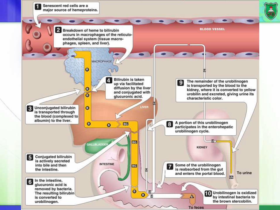

Jaundice
Accumulation of bilirubin in the plasma and tissues results in jaundice
Tissues such as skin, nails, and sclerae (whites of eyes) have a yellow color.
Hemolytic jaundice: Massive lysis of RBC causes higher levels of unconjugated bilirubin.
Hepatocellular jaundice: Liver damage, decreased conjugation efficiency, defective secretion of conjugated bilirubin into bile increase bilirubin in blood.
Levels of urobilinogen increase in urine (dark) and pale stool.
Jaundice in newborns: inefficient conjugating enzyme.
Biliary obstruction: subnormal amounts of conjugated bilirubin reach the intestine for conversion to urobilinogen and conjugated bilirubin is excreted in urine (dark).


Importance of iron
Within the body, iron exist in two oxidation states: ferrous (Fe2+) or ferric (Fe3+)
Iron is important for oxygen metabolism and transport.
It is also the prosthetic group of a number of enzymes such as redox cytochromes and the P450 class of detoxifying cytochromes.
Yet…
Iron can be potentially toxic due its ability to form free radicals.
Solution: iron is not free.
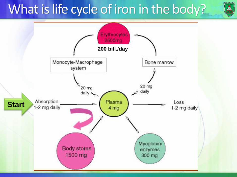
What is life cycle of iron in the body?
Start
200 bill./day
Normal levels of iron
Well-nourished people have 3-4 grams of iron in their bodies.
Iron is mainly used for hemoglobin synthesis (70% of all iron).
The iron for hemoglobin synthesis is recovered from dead erythrocytes.
The recycled iron binds to transferrin in blood circulation and is distributed to iron-consuming cells, mainly erythrocytes.
Additional iron (0.3–0.4 g) is channeled to other cellular proteins (myoglobin and cytochromes).
3-4 mg circulates through the plasma.
Small amounts are lost (about 1–2 mg/day) and are replaced by dietary absorption.
1 mg a day for men and 1.5–2 mg a day for women with regular menstrual periods
Sources of iron
Most of the iron in the body is recycled from destroyed red blood cells.
The released iron is scavenged by macrophages in the reticuloendothelial system.
A significant portion is stored in the liver, both in the hepatocytes, and in the Kupffer cells (reticuloendothelial cells).


State of iron
Under conditions of neutral or alkaline pH, iron is found in the Fe3+ state and in the Fe2+ state at acidic pH.
In the stomach, iron will be in the ferrous state.
In the duodenum iron is in the ferric state.
However, to be absorbed, dietary iron must be in its ferrous Fe2+ form.
Site of absorption
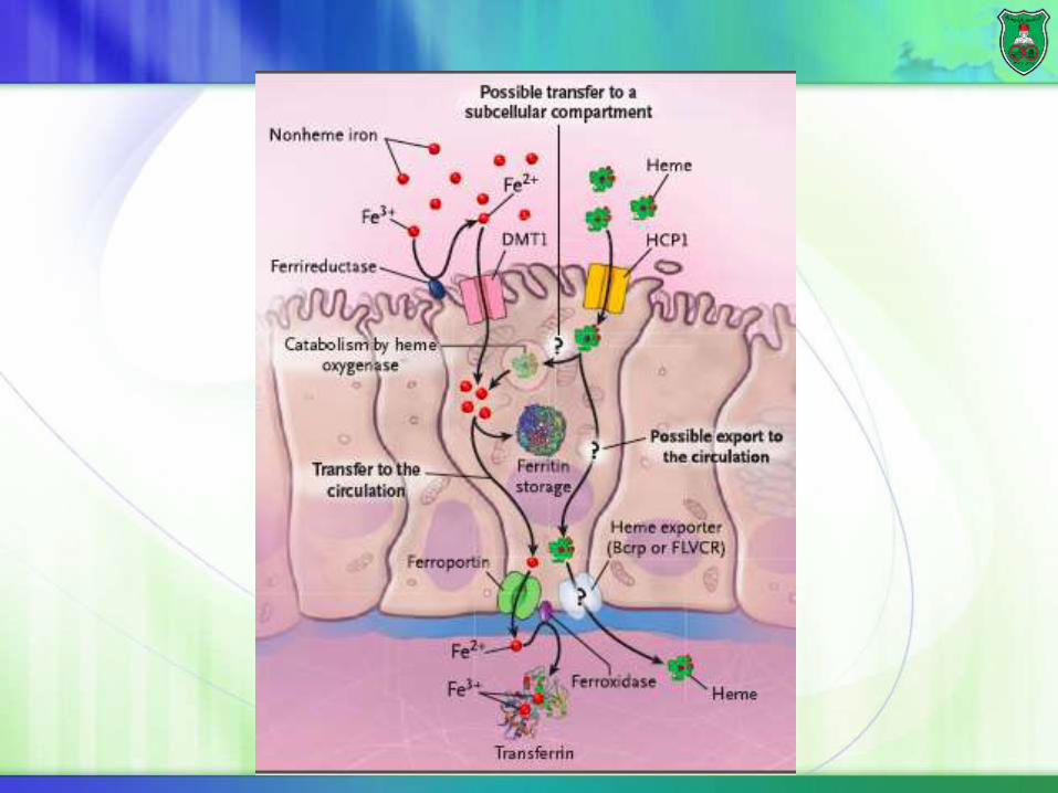
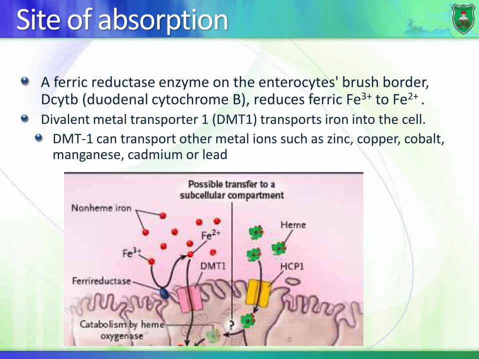
A ferric reductase enzyme on the enterocytes' brush border, Dcytb (duodenal cytochrome B), reduces ferric Fe3+ to Fe2+ .Divalent metal transporter 1 (DMT1) transports iron into the cell.
DMT-1 can transport other metal ions such as zinc, copper, cobalt, manganese, cadmium or lead

Heme oxygenase
Iron can also be obtained from ingested heme.
Heme is absorbed by a receptor called heme-carrier protein (HCP) and iron is released by heme oxygenase-1 (HO-1).
In other cells such as macrophages, heme oxidase extracts iron from heme.

Fate 1: storage into ferritin
Cells can then store iron as ferritin.Each Ferritin complex can store about 4500 iron (Fe3+) ions
But, if cells are sloughed off from the tip of the villus into feces, iron is eliminated from the body.

Fate 2: transport
Iron is transported out via a basolateral transporter known as ferroportin, which is distributed throughout the body on all cells.

Ferroxidase and transferrin
Once iron leaves intestinal cells, an iron oxidase, known as hephaestin or ferroxidase, converts iron from the ferrous state to the ferric state.
Nonintestinal cells use the plasma protein ceruloplasmin to oxidize iron.
Iron is rapidly bound to transferrin, an iron-binding protein of the blood that delivers iron to liver cells and from liver cells to other tissues via receptor-mediated endocytosis.
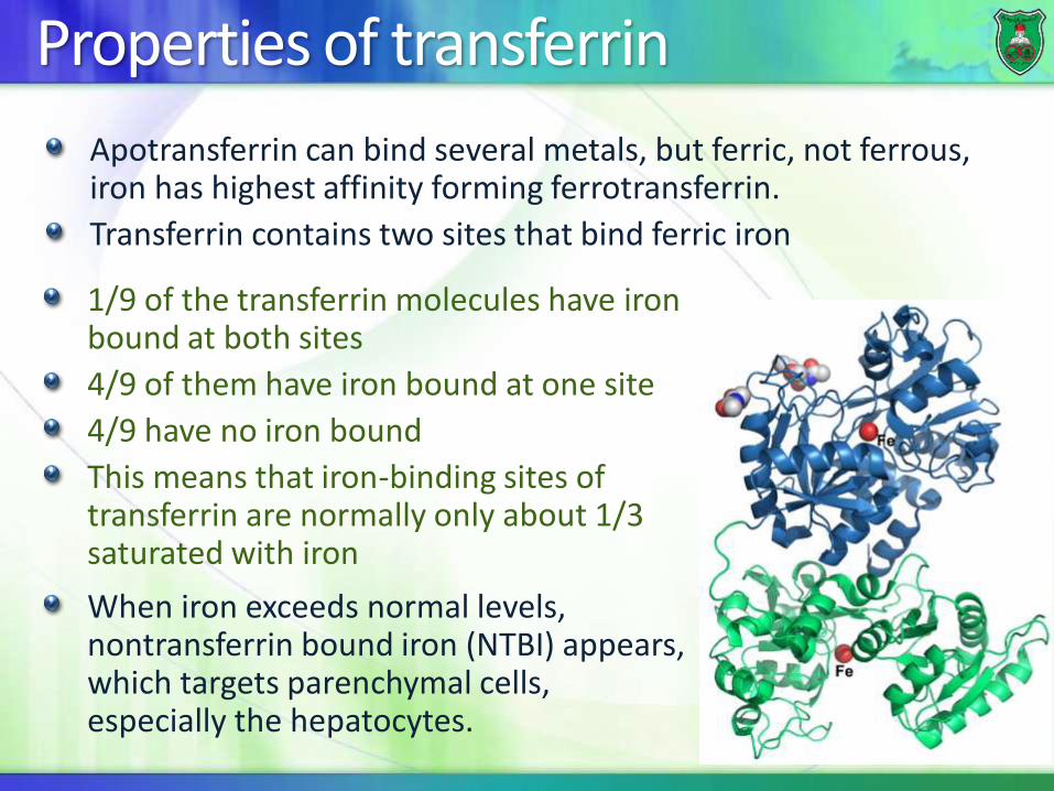
Properties of transferrin
Apotransferrin can bind several metals, but ferric, not ferrous, iron has highest affinity forming ferrotransferrin.
Transferrin contains two sites that bind ferric iron
1/9 of the transferrin molecules have iron bound at both sites
4/9 of them have iron bound at one site
4/9 have no iron bound
This means that iron-binding sites of transferrin are normally only about 1/3 saturated with iron
When iron exceeds normal levels, nontransferrin bound iron (NTBI) appears, which targets parenchymal cells, especially the hepatocytes.
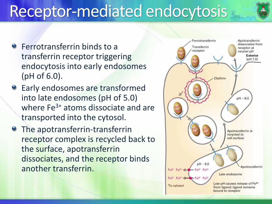
Receptor-mediated endocytosis
Ferrotransferrin binds to a transferrin receptor triggering endocytosis into early endosomes (pH of 6.0).
Early endosomes are transformed into late endosomes (pH of 5.0) where Fe3+ atoms dissociate and are transported into the cytosol.
The apotransferrin-transferrin receptor complex is recycled back to the surface, apotransferrindissociates, and the receptor binds another transferrin.

Hepcidin
Hepcidin is a peptide hormone secreted by the liver that induces internalization and lysosomal degradation of ferroportin.
Its production is increased by iron overload and inflammation and is suppressed by iron deficiency .

Mechanism 1
Hepcidin binds to the basolateral iron transporter ferroportin.
This causes ferroportin to be internalized and degraded.
Hepcidin may also decrease iron release by hepatocyte and macrophage resulting in an increase in stored iron in these cells

Mechanism 2
Hepcidin functions by inhibiting the presentation of one or more of the iron transporters (e.g. DMT1) in intestinal membranes decreasing iron absorption

Regulation of hepcidinThe expression of hepcidin is regulated positively by transferrin receptor 2, inflammatory cytokines, and negatively by anemia and hypoxia (through erythroferrone, produced by the erythroblasts in response to EPO (erythropoietin) synthesis by the kidney).
In addition, the release of bone morphogenetic protein 6 is induced by intracellular iron stimulating hepcidin synthesis.
Post-transcriptionl regulation of expression

Iron-response element and its binding protein
FerritinFerroportin
ALAS
TransferrinDMT1
Iron-related diseases

Diseases to be covered
Hemochromatosis (HC)Hepcidin deficiency-related HCQuantitative hepcidin deficiency
Hepcidin resistance
The ferroportin disease
Iron deficiency anemia
Hereditary hemochromatosis
It is an autosomal recessive disorder in iron metabolism that is characterized by excess iron absorption, saturation of iron-binding proteins and deposition of hemosiderin in the tissues.
more commonly in males than in females (why?)The primary cause of hemochromatosis is the inheritance of an autosomal recessive allele designated as HFE, but four other genes that regulate the hepcidin–ferroportin axis can also be involved.
Affected organs
Liver (hepatic fibrosis)
Pancreas (diabetes mellitus)
Joints (arthropathy)
Skin (pigmentation)
Heart (cardiomyopathy)
Gonadotrophin-secreting cells (hypogonadotrophic hypogonadism)

Groups/classes of hereditary hemochromatosis
Type 1 (hemochromatosis protein, HFE dependent)Most common
Type 2A [HFE2 (HJV) dependent]
Type 2B (hepcidin, HAMP dependent)
Type 3 (TfR2, TfR2 dependent)
Type 4 (ferroportin dependent).

Mechanism of action
Mutation or absence of
HFE or TFR2 prevents
formation of a functional
iron sensor and signal
transduction effector
complex leading to
dysregulation of systemic
iron homeostasis
TFR1 exists as a
complex at the
plasma membrane
during low or basal
serum iron conditions.
β2-microglobulin is
needed for this
interaction.
Serum Fe2+ -TF
competes with HFE
for binding to TFR1.
Increased serum
transferrin saturation
results in the
dissociation of HFE
from TFR1.
HFE binds TFR2
and induces a
intracellular
signaling that
stimulates hepcidin
production.
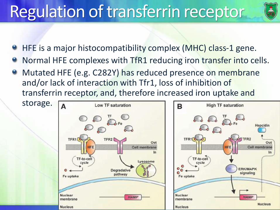
Regulation of transferrin receptor
HFE is a major histocompatibility complex (MHC) class-1 gene.
Normal HFE complexes with TfR1 reducing iron transfer into cells.
Mutated HFE (e.g. C282Y) has reduced presence on membrane and/or lack of interaction with Tfr1, loss of inhibition of transferrin receptor, and, therefore increased iron uptake and storage.

Juvenile hemochromatosis
Type 2A hereditary hemochromatosisAKA HFE2 (HJV)-dependent hereditary hemochromatosis
A very rare, severe juvenile form of hemochromatosis is due to a homozygous deletion of the gene for hepcidin
Mutations in HJV gene, which encodes the protein “hemojuvelin”, account for the majority of JH.
HJV upregulates expression of hepcidin.
Type 2B is also juvenile hemochromatosis, but is caused by mutations in hepcidin gene.

Hemosiderin
The normal total body iron stores may range from 2 to 6 gm, but persons with hemochromatosis have much greater stores.
The total iron stores of affected persons may exceed 50 gm
If the capacity for storage of iron in ferritin is exceeded, iron is stored as water-insoluble deposits known as hemosiderin.
Excess hemosiderin leads to cellular dysfunction and damage.

Iron deficiency anemia Anemias are characterized by a deficiency in the number of mature erythrocytes in the circulation, lowering the oxygen-carrying capacity of the blood, causing tissue hypoxia, and clinical symptoms such as fatigue, weakness, increased cardiac output, as well as increased morbidity and mortality.
Cells including cannot
synthesize DNA and,
hence, cannot divide.
Megaloblasts
accumulate. Folate is not regenerated
Anemia of chronic disease
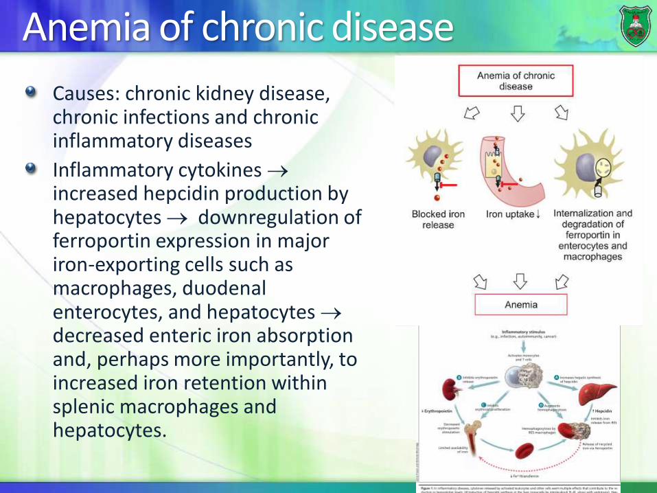
Causes: chronic kidney disease, chronic infections and chronic inflammatory diseases
Inflammatory cytokines increased hepcidin production by hepatocytes downregulation of ferroportin expression in major iron-exporting cells such as macrophages, duodenal enterocytes, and hepatocytes decreased enteric iron absorption and, perhaps more importantly, to increased iron retention within splenic macrophages and hepatocytes.
Related Documents