Lead Review Article October 1996: 295-317 Iron Metabolism: A Comprehensive Review John L. Beard, Ph.D., Harry Dawson, B.S., and Doming0 J. Pifiero, M.Sc. Despite its abundance in the earth’s crust, iron deficiency is a serious health issue in many parts of the world. Although fundamental obserwations about iron metabolism and the significance of iron nutriture were first noted some time ago, the mo- lecular mechanisms involved in iron metabolism are just now being defined. Historical Perspective Among all of the micronutrients, iron has the lon- gest and best described history. Iron is the fourth most abundant terrestrial element, comprising ap- proximately 4.7% of the earth’s crust in the form of the minerals hematite, magnetite, and siderite. Pri- mordial iron compounds were probably responsible for the catalytic generation of some of the atmo- spheric oxygen upon which most modem life forms depend.’ Iron is an essential nutrient for all living organisms with the exception of certain members of the bacterial genera Lactobacillus and Bacillus. In these organisms, the functions of iron are replaced by other transition metals, especially manganese and cobalt, which reside next to iron in the periodic table. In all other life forms, iron is an essential component of, or cofactor for, hundreds of proteins and enzymes. Based on extrapolations made from modem ab- original societies, prehistoric humans had an ade- quate intake of iron.2 The ancient Arabs, Chinese, Egyptians, Greeks, and Romans, although ignorant about the nutritional importance of iron, attributed Dr. Beard, Mr. Dawson, and Mr. Pinero are with the Department of Nutrition, Pennsylvania State Uni- versity, University Park, PA 16802, USA. This paper was originally presented in May 1996 at a Workshop on Iron Deficiency in Buenos Aires, Ar- gentina to commemorate the founding of the Centro de Estudios Sobre Nutricion lnfantil (CESNI). It will ap- pear in a slightly different format in O’Dell and Sunde, eds. Handbook of Nutritionally Essential Minerals; New York, NY, Marcel-Dekker, Inc., 1997. therapeutic properties to iron.3 The ancient Greeks administered iron to their injured soldiers to im- prove muscle weakness, which probably derived from hemorrhagic anemia.4 Alchemists and physi- cians of the 16th century prescribed iron for medic- inal US^.^.^ Iron salts were given to young women to treat what was then described as chlorosis, an arcane term for anemia usually due to iron or pro- tein defi~iency.~ Various physicians during this time also prescribed iron pills for anemia and were un- ceremoniously ridiculed by their successors in the medical Iron was identified as a constituent of animal liver and blood in the early 18th ~ e n t u r y . ~ . ~ . ’ ~ In 1825, hemoglobin (Hb) iron content was deter- mined to be 0.35%: a value extremely close to the Hb iron content of 0.347%” calculated by modem methods. Between 1832 and 1843 chlorosis was de- fined by low levels of iron in the blood and reduced number of red cell^.^.^.'^ Boussingault first described the nutrition essentiality of iron in 1872.12 In 1895, Bunge accurately described the anemia of chlorosis in terms of nutritional iron deficien~y.’~ Key observations about iron nutrition were made during the first half of this cent~ry.4.~.’~.’~ Moore discovered the enhancing effect of ascorbic acid on iron absorption.15Granick later proposed the “mucosal block” theory for control of body iron.16 Although these fundamental observations on the metabolism of iron and its nutritional significance were made some time ago, the molecular mecha- nisms involved in iron metabolism are just now be- ing described. Whole Animal Metabolism Food Sources Despite its abundance in the earth’s crust, iron de- ficiency is a serious health issue in many parts of the world. The iron nutrition status of an individual and of populations is largely a function of the amount of dietary iron, the bioavailability of that iron, and the extent of iron losses. Many foods that are potentially good sources of iron are limited by the bioavailability of that iron.I7 The bioavailability Nutrition Reviews, Vol. 54, No. 10 295 Downloaded from https://academic.oup.com/nutritionreviews/article/54/10/295/1836886 by guest on 31 March 2022

Welcome message from author
This document is posted to help you gain knowledge. Please leave a comment to let me know what you think about it! Share it to your friends and learn new things together.
Transcript
Lead Review Article October 1996: 295-317
Iron Metabolism: A Comprehensive Review John L. Beard, Ph.D., Harry Dawson, B.S., and Doming0 J. Pifiero, M.Sc.
Despite its abundance in the earth’s crust, iron deficiency is a serious health issue in many parts of the world. Although fundamental obserwations about iron metabolism and the significance of iron nutriture were first noted some time ago, the mo- lecular mechanisms involved in iron metabolism are just now being defined.
Historical Perspective
Among all of the micronutrients, iron has the lon- gest and best described history. Iron is the fourth most abundant terrestrial element, comprising ap- proximately 4.7% of the earth’s crust in the form of the minerals hematite, magnetite, and siderite. Pri- mordial iron compounds were probably responsible for the catalytic generation of some of the atmo- spheric oxygen upon which most modem life forms depend.’ Iron is an essential nutrient for all living organisms with the exception of certain members of the bacterial genera Lactobacillus and Bacillus. In these organisms, the functions of iron are replaced by other transition metals, especially manganese and cobalt, which reside next to iron in the periodic table. In all other life forms, iron is an essential component of, or cofactor for, hundreds of proteins and enzymes.
Based on extrapolations made from modem ab- original societies, prehistoric humans had an ade- quate intake of iron.2 The ancient Arabs, Chinese, Egyptians, Greeks, and Romans, although ignorant about the nutritional importance of iron, attributed
Dr. Beard, Mr. Dawson, and Mr. Pinero are with the Department of Nutrition, Pennsylvania State Uni- versity, University Park, PA 16802, USA.
This paper was originally presented in May 1996 at a Workshop on Iron Deficiency in Buenos Aires, Ar- gentina to commemorate the founding of the Centro de Estudios Sobre Nutricion lnfantil (CESNI). It will ap- pear in a slightly different format in O’Dell and Sunde, eds. Handbook of Nutritionally Essential Minerals; New York, NY, Marcel-Dekker, Inc., 1997.
therapeutic properties to iron.3 The ancient Greeks administered iron to their injured soldiers to im- prove muscle weakness, which probably derived from hemorrhagic anemia.4 Alchemists and physi- cians of the 16th century prescribed iron for medic- inal US^.^.^ Iron salts were given to young women to treat what was then described as chlorosis, an arcane term for anemia usually due to iron or pro- tein defi~iency.~ Various physicians during this time also prescribed iron pills for anemia and were un- ceremoniously ridiculed by their successors in the medical
Iron was identified as a constituent of animal liver and blood in the early 18th ~ e n t u r y . ~ . ~ . ’ ~ In 1825, hemoglobin (Hb) iron content was deter- mined to be 0.35%: a value extremely close to the Hb iron content of 0.347%” calculated by modem methods. Between 1832 and 1843 chlorosis was de- fined by low levels of iron in the blood and reduced number of red cell^.^.^.'^ Boussingault first described the nutrition essentiality of iron in 1872.12 In 1895, Bunge accurately described the anemia of chlorosis in terms of nutritional iron deficien~y.’~
Key observations about iron nutrition were made during the first half of this cent~ry.4.~.’~.’~ Moore discovered the enhancing effect of ascorbic acid on iron absorption.15 Granick later proposed the “mucosal block” theory for control of body iron.16 Although these fundamental observations on the metabolism of iron and its nutritional significance were made some time ago, the molecular mecha- nisms involved in iron metabolism are just now be- ing described.
Whole Animal Metabolism
Food Sources Despite its abundance in the earth’s crust, iron de- ficiency is a serious health issue in many parts of the world. The iron nutrition status of an individual and of populations is largely a function of the amount of dietary iron, the bioavailability of that iron, and the extent of iron losses. Many foods that are potentially good sources of iron are limited by the bioavailability of that iron.I7 The bioavailability
Nutrition Reviews, Vol. 54, No. 10 295
Dow
nloaded from https://academ
ic.oup.com/nutritionreview
s/article/54/10/295/1836886 by guest on 31 March 2022

of iron is a function of its chemical form and the presence of food items that promote or inhibit ab- ~ o r p t i o n . ~ ~ - ' ~ - ~ ~ Basal obligatory iron losses in hu- mans are approximately 1 mg/day and must be re- placed by an equivalent amount of iron derived from the diet. The typical Western diet provides an average of 6 mg of heme and nonheme iron per 1000 kcal of energy intake.26 Heme iron is an im- portant dietary source of iron because it is more effectively absorbed than nonheme iron. From 5% to 35% of heme iron is absorbed from a single meal, whereas nonheme iron absorption from a single meal can range from 2% to 20%, depending on the iron status of the individual and the ratio of en- hancers and inhibitors in the diet. Thus, although it constitutes about 10% of the iron found in the diet, heme iron may provide up to a third of total ab- sorbed dietary iron.27
Nonheme iron, which constitutes 90% of the remaining dietary iron, accounts for 60% of the iron from animal sources and about 100% of the iron found in vegetable material. Intentional iron fortification or iron contamination of food during preparation may account for as much as 10-15% of dietary nonheme iron.28 The primary overall ef- fect of food on nonheme iron absorption is inhib- itory. The rat, which has been the standard model of iron absorption for humans, appears to be less sensitive to these factors compared with humans. Therefore, the actual influence of these factors in humans may be undere~timated.~~ Conversely, the long-term contributions of these enhancers and promoters to body iron stores may be more limited than first
For the most part, attempts to alleviate iron de- ficiency through food fortification and diet supple- mentation have been successful in the United States. Many different forms of iron have been employed for this purpose. In the United States, fortification of food with iron started in the 1940s. The Food and Drug Administration has since upwardly re- vised standards for iron enrichment of cereal prod- ucts and has issued guidelines for fortification of infant formulas. These actions have prompted much debate about potential iron overloading in people with adequate iron
Supplementation, although more able to target specific populations at risk for iron deficiency, is limited by dosage requirements and potential side effects, both of which lead to noncompliance. Side effects of high-dose (>200 mg) iron supplementa- tion include gastrointestinal distress and constipa- tion. The additions of bovine Hb to foods38 and bo- vine lactoferrin to infant formulas39 appear to be effective methods of iron supplementation that have minimal side effects.
Iron Absorption
General The vast amount of work that has contributed to our knowledge of iron absorption will not be extensive- ly rereviewed here. We refer the readers to other recent and more comprehensive We would, however, like to direct our attention to some of the mechanistic aspects of iron absorption that have been suggested in recent years.
The process of iron absorption can be divided into three stages: (1) iron uptake, (2) intraenterocyte transport, and (3) storage and extraenterocyte trans- fer (Figure 1). During the intestinal phase of diges- tion, iron binds to specific mucosal membrane sites, is internalized, and then either is retained by the mucosal cell or is transported to the basolateral membrane where it is bound to transferrin (Tf) in the plasma pool. The process of iron absorption is controlled by intraluminal, mucosal, and somatic factors. A multitude of intraluminal factors affect the amount of iron available for absorption as either inhibitors or promoters. Mucosal factors include the amount of mucosal surface and intestinal motility. Somatic factors that influence iron absorption in- clude erythropoiesis and hypoxia.
Luminai Phase No absorption of iron occurs in the mouth, esoph- agus, or stomach. However, the stomach does con- tribute hydrochloric acid. This not only helps to re- move protein-bound iron by protein denaturation but also helps solubilize iron by reducing it from the ferric to the ferrous state. Reduction of ferric iron is necessary because the majority of iron in the diet is found in the relatively insoluble ferric form (Ksp = lO-I7 M) and is poorly a b ~ o r b e d . ~ ~ . ~ ~ De- creased stomach acidity due to over consumption of antacids, ingestion of alkaline clay, or pathologic conditions such as achlorhydria or partial gastrec- tomy may lead to impaired iron The combined actions of gastric acid and pepsin account for slightly less than half of the release of conju- gated dietary iron and the reduction of a third of total dietary ferric iron.
Other gastrointestinal components also have roles in iron absorption. Pancreatic ductal cells se- crete bicarbonate, which, by increasing the pH of the lumen, potentially decreases iron absorption. This effect is counterbalanced by pancreatic prote- ases that liberate nonheme iron from digestiva. Al- terations in this balance, as seen in patients with pancreatic insufficiency or cystic fibrosis, may have potential adverse consequences.& It has been sug- gested that consumption of pancreatic enzymes by these patients may predispose them to iron over- 10ad.47
296 Nutrition Reviews, Vol. 54, No. 10
Dow
nloaded from https://academ
ic.oup.com/nutritionreview
s/article/54/10/295/1836886 by guest on 31 March 2022
Parncellular 7 Transport
-
t I 2.
0 Fez+ Q k3+ x & 1 . 1 1 ) id&
trans feuin-Like Protein 7
I)- - ma \ 4. 5 .
f- 7.
Lysosom e 0 ' f6. //'+ Q k '
4
Bilir u b in + co Hem e
Luin en En terocyte
c o I I I I ? I I +
F2+
* 0 Apotransferrin
Ferrotransferrin
Basolateral Surface
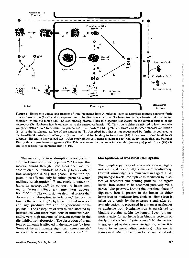
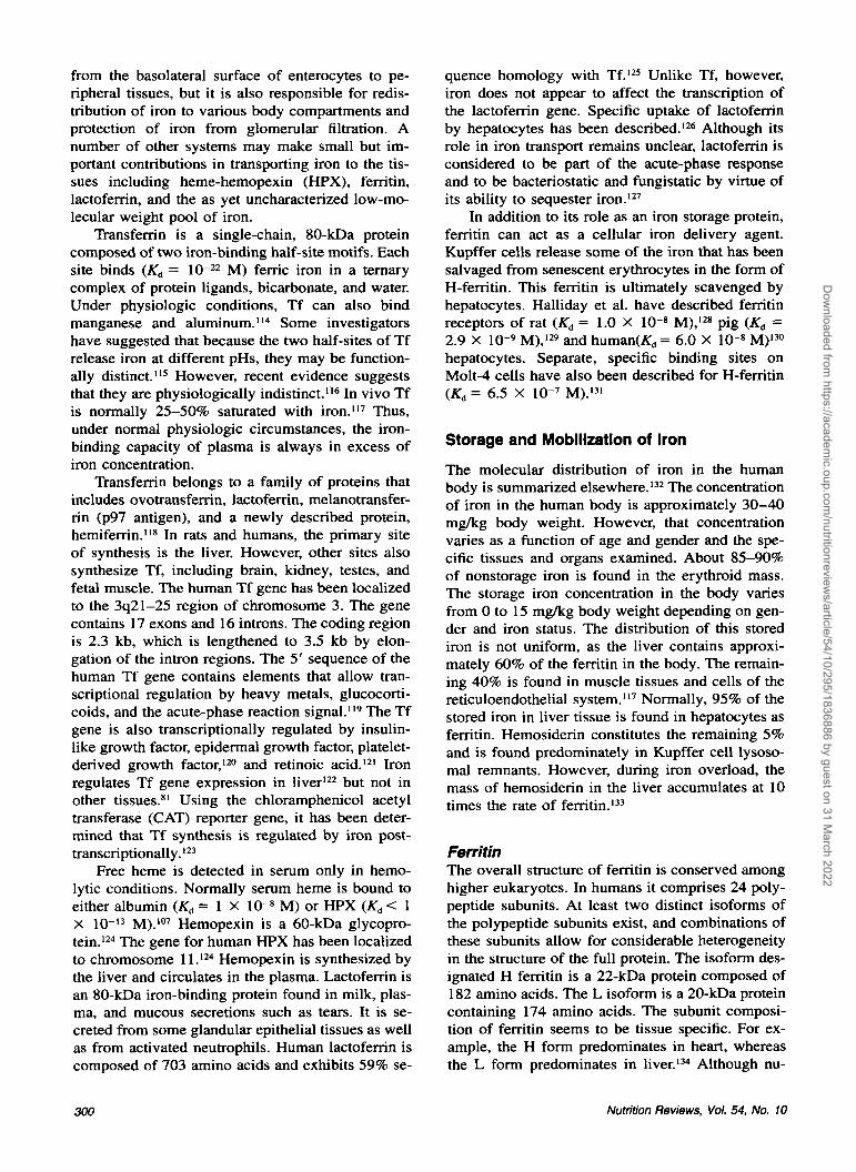
Figure 1. Enterocyte uptake and transfer of iron. Nonheme iron: A reductant such as ascorbate reduces nonheme femc iron to ferrous iron (1). Chelators sequester and solubilize nonheme iron. Nonheme iron is then transferred to a binding protein(s) within the lumen (2). The iron-binding protein binds to a specific transporter on the luminal surface of the enterocyte (3). Nonheme iron is transported to the enterocyte interior (4). This iron is either transferred to low molecular weight chelates or to a transfemn-like protein (5). The transfemn-like protein delivers iron to either mucosal cell fenitin (6) or to the basolateral surface of the enterocyte (8). Absorbed iron that is not sequestered by ferritin is delivered to the basolateral surface of enterocytes (9) and oxidized for binding to transferrin (10). Heme iron: Heme binds to its receptor (lh) and is internalized (2h). After entering the cell, heme is degraded to iron, carbon monoxide, and bilirubin IXa by the enzyme heme oxygenase (3h). This iron enters the common intracellular (enterocyte) pool of iron (4h) (5) and is processed like nonheme iron (6-10).
The majority of iron absorption takes place in the duodenum and upper j e j u n ~ m . 4 ~ . ~ ~ Factors that increase transit through these areas decrease iron ab~orpt ion .~~ A multitude of dietary factors affect iron absorption during this phase. Heme iron ap- pears to be affected only by animal proteins, which facilitate its abs0rption,5'.~~ and calcium, which in- hibits its ab~orpt ion .~~ In contrast to heme iron, many factors affect nonheme iron absorp- tion.20*24.41,54-56 The extrinsic intraluminal factors that decrease iron absorption include bran,57 hemicellu- lose, cellulose, pectin,58 phytic acid found in wheat and soy p r o d u ~ t s , ~ ~ . ~ ~ and polyphenolic com- pounds.21 The absorption of iron is also affected by interactions with other metal ions or minerals. Gen- erally, very high amounts of divalent cations in the diet inhibit iron absorption. The absorption of metal ions or minerals is affected in the same way by iron. Some of the nutritionally significant known metals/ minerals interactions are summarized el~ewhere.6~-'~
Mechanisms of Intestinal Cell Uptake
The complete pathway of iron absorption is largely unknown and is currently a matter of controversy. Current knowledge is summarized in Figure 1. At physiologic levels iron uptake is mediated by a se- ries of receptors and binding proteins. At higher levels, iron seems to be absorbed passively via a paracellular pathway. During the intestinal phase of digestion, iron is present in the lumen as either heme iron or nonheme iron chelates. Heme iron is taken up directly by the enterocyte and, after en- zymatic action, is processed in a manner analogous to nonheme iron. Nonheme iron is transferred to binding proteins within the lumen. Specific trans- porters exist for nonheme iron binding proteins on the luminal surface of enterocyte~.~~ Nonheme iron is transported to the enterocyte interior, where it is bound to an iron-binding protein(s). This iron is transferred either to ferritin or to the basolateral side
Nutrition Reviews, Vol. 54, No. 10 297
Dow
nloaded from https://academ
ic.oup.com/nutritionreview
s/article/54/10/295/1836886 by guest on 31 March 2022
of the enterocyte. Given the observation of in- creased iron absorption with low iron stores and de- creased absorption with high iron stores, it is tempt- ing to speculate that there is genetic regulation of both receptors and binding proteins. This regulation appears to be exerted across the basolateral mem- brane in a manner that is correlated to whole-body iron stores. Iron is then either lost when the cell is sloughed or bound to Tf in the circulation. The tools of molecular biology are now being used to deter- mine what receptors, binding proteins, and cells contribute to iron absorption. At this time, unfor- tunately, the description of the process of iron ab- sorption is incomplete.
Heme Iron Heme is soluble in an alkaline environment. Hence, no binding proteins are necessary for its luminal absorption. Although specific transporters exist for heme on the surface of rat e n t e r ~ c y t e s , ~ ~ . ~ ~ rats do not absorb heme iron as efficiently as do humans.75 A specific receptorkransporter for heme has not yet been described in humans. After binding to its re- ceptor, the heme molecule is then internalized. After entering the cell, heme is degraded to iron, carbon monoxide, and bilirubin IXa by the enzyme heme o x y g e n a ~ e . ~ ~ , ~ ~ This enzyme is not induced by oral administration of Hb (a source of heme) but is in- duced by iron def i~ iency .~~ Its distribution in the intestine is identical to the areas of maximal heme iron ab~orpt ion .~~ It is thought that the iron that is liberated from heme by heme oxygenase enters the common intracellular (enterocyte) pool of iron.
Luminal Nonheme Iron-Binding Proteins Ferrous iron that has been liberated by gastric and pancreatic proteases is readily oxidized to the ferric form in an alkaline environment. It would be ren- dered insoluble and biologically unavailable except for the presence of intraluminal iron-binding mol- ecules. Various attempts have been made to identify these molecules. The interpretation of these and oth- er studies seeking to identify physiologic iron-bind- ing molecules is difficult because of the large amount of nonspecific binding by iron. It was orig- inally proposed that the enterocyte or some other gastrointestinal entity such as the stomach or liver synthesized Tf and that Tf was extruded into the lumen to sequester iron.78 This Tf, replete with di- etary iron, was then absorbed by the enterocyte. Transferrin protein has been detected within duo- denal mucosal ~ e l l s ~ ~ . ~ ~ ; however, most investigators have failed to detect Tf mRNA in these cells.81.8z Although one study has claimed to detect Tf recep- tors (TfR) on the luminal surface of en tero~ytes ,~~ most others have failed to do s0.84.85 Furthermore,
even though Tf-bound iron is efficiently absorbed by the rat,78 when administered to patients with ach- lorhydria, it is not effectively absorbed.86 In addi- tion, humans and mice with hypotransferrinemia be- come iron overloaded rather than iron deficient.87 Although the intestinal absorption of Tf-bound iron is an attractive hypothesis, most of the available ev- idence argues against this scenario and suggests a very limited role for Tf in the direct absorption of iron.
Several investigators have reported the presence of luminal iron-binding proteins that are distinct from Tf. One of these proteins is mucin, which binds (Kd = 1.1 X M) and solubilizes ferric iron in an acidic milieu.88 Mucin also binds zinc but with lower affinity.88 Iron chelates of histidine, ascorbate, and fructose, which enhance iron absorp- tion in vivo, donate iron to mucin at neutral pH and may represent true in vivo complexes. More stable chelates of anions that inhibit iron absorption in vivo, such carbonate and oxalate, do not seem to donate iron to mucin in vitro.
At least two groups have described a 160-kDa glycoprotein from human microvillous membrane vesicles that may participate in facilitated transport of iron.72.89 This protein is composed of three iden- tical 54-kDa monomers.89 Other iron-binding pro- teins of 35, 95, and 120 kDa have been isolated from cultured rat intestinal epithelial cells.90*91
lntraenterocyte Transport and Storage
Transport of absorbed iron through the enterocyte may involve a Tf-like protein.92 One candidate for this Tf-like protein might be mobilferrin, a 56-kDa cytosolic protein isolated from rat and human duo- denal mucosa that can bind iron (Kd = 9 X M).92 It is a homologue of calreticulin and can also bind calcium, copper, and zinc.93 The multiple metal ion-binding properties of mobilferrin have been suggested as explanations of the interactions be- tween the absorption of these elements. Mobilfemn coprecipitates with the a and p subunits of integrin during its purification, prompting Conrad to suggest that mobilferrin is involved in cytosolic acceptance of iron from membrane-bound integ~in.~]
In the conceptual model of feedback regulation of iron absorption, an adequate iron status increases the amount of iron retained by the enterocyte. In this regard, femtin has been proposed to act as an “iron sink” for intestinal mucosal cells. Iron that is not transferred to the plasma is stored in mucosal cell femtin and is lost when the enterocyte dies and is subsequently It is unlikely that any mu- cosal cell ferritin reaches the circulation before the enterocyte is shed. The lower duodenal levels of ferritin mRNA found in iron-deficient subjects and
298 Nutrition Reviews, Vol. 54, NO. 10
Dow
nloaded from https://academ
ic.oup.com/nutritionreview
s/article/54/10/295/1836886 by guest on 31 March 2022

higher duodenal levels of ferritin mRNA found in secondary iron overload support the role of mucosal ferritin as a major regulator of iron absorption.82 If ferritin is the mucosal “iron sink,” dysregulation of mucosal ferritin mRNA expression could lead to iron deficiency or iron overload. Consistent with this hypothesis is the fact that the concentrations of mucosal cell ferritin mRNA and femtin protein in patients with familial hemochromatosis are lower than those of patients with secondary iron over- load.80.82.96
Extraenterocyte Transfer
Absorbed iron is delivered and bound to Tf at the basolateral surface of enterocytes. It has been pro- posed that ceruloplasmin is the protein responsible for oxidation of iron, which is necessary for its binding to Tf at the basolateral membrane.97~98 The evidence for involvement of ceruloplasmin in this process is largely circumstantial. Copper deficiency results in accumulation of iron in the mucosa and liver, reduced iron transport to peripheral tissues, and anemia.99 The classic explanation for this type of anemia has been attributed to lack of basolateral mucosal cell ferroxidase I (ceruloplasmin) action. This explanation is not fully adequate, as brindled mice and patients with Menkes’ or Wilson’s disease all have low serum ceruloplasmin levels but do not exhibit iron deficiency anemia.99
Regulation of Iron Absorption
Mucosal Factors The majority of iron absorption takes place in the duodenum and upper jejunum. These areas also adapt during iron deficiency to promote iron ab- sorption.49 In these areas the amount of functional absorptive mucosal surface is important for iron ab- sorption. Consequently, surgical removal of any part of the duodenum and upper jejunum or the presence of factors that increase enterocyte turnover decreases iron a b ~ o r p t i o n . ~ ~ Clinical disorders that affect iron absorption at this level include malab- sorption syndromes such as steatorrhea and tropical ~prue.~O
Somatic Factors The regulation of iron absorption involves somatic factors that notify the enterocyte of the need for iron absorption. It is clear that a person’s iron status cor- relates inversely with the amount of iron ab- sorbed.Im More recent investigations have shown that iron deficiency is the most potent somatic in- ducer of both heme and nonheme iron absorption. The mechanism(s) for this induction is largely un-
known. One possible contributing factor is intestinal heme oxygenase, which is activated by somatic iron defi~iency.’~
Hemoglobin and serum ferritin apparently have limited roles in signaling the enterocyte about the need for iron absorption.82.101.’02 It has been sug- gested that internalized plasma ferro-Tf may allow the enterocyte to monitor body iron status and reg- ulate iron absorption. Exposure to low intracellular amounts of plasma ferro-Tf would signal the enter- ocyte to up-regulate iron entry into the body. Trans- ferrin receptors are only found at the basolateral surface of e n t e r o c y t e ~ . ~ ~ ~ J ~ The amount of entero- cyte TfR mRNA and protein increases during iron deficiency and decreases during secondary iron loading.82.103.105 Acute hemolysis, which stimulates iron absorption, does not influence enterocyte ba- solateral TfR number.Io5 Of interest is that the levels of TfR and TfR mRNA in mucosal cells of people with hemochromatosis are higher than suitably matched controls.82.104
Active erythropoiesis, induced either by bleed- ingIm or by acute hernolysis,lo6 increases the ab- sorption of iron. It has therefore been proposed that erythropoietin is an endogenous signal for iron ab- sorption; there is limited evidence for this hypoth- esis. In fact, recombinant human erythropoietin does not increase intestinal iron absorption when given to iron-overloaded rats.Io7 Moreover, ex- change transfusion of reticulocytes with a large number of TfR into rats stimulates iron absorption in iron-replete animals independently of erythro- poietin production or active erythropoiesis. log
Hypoxia increases iron absorption1@’ indepen- dently of erythropoiesis.llO.lll Increased plasma iron turnover, which occurs not only in erythropoiesis but also in disorders of ineffective erythropoiesis such as thalassemia, hemolytic anemias, and side- roblastic anemias, is associated with increased iron absorption.112 Other clinical disorders such as he- mochromatosis, congenital ferrochelatase deficien- cy, and porphyria cutanea tardaS0 result in an in- creased iron absorption by mechanisms that are yet unexplained. Finally, inflammatory processes may decrease iron absorption,’I3 probably by eliciting the production of cytokines that have a direct effect on the mucosal cell.
Transport of Iron
In addition to being oxidized to the insoluble ferric state in an oxygen-rich environment such as that found under physiologic conditions, free iron is an extremely toxic substance capable of catalyzing many deleterious reactions. Quantitatively, the most significant iron transport molecule in vertebrates is Tf. Not only is it responsible for delivery of iron
Nutrition Reviews, Vol. 54, No. 10 299
Dow
nloaded from https://academ
ic.oup.com/nutritionreview
s/article/54/10/295/1836886 by guest on 31 March 2022
from the basolateral surface of enterocytes to pe- ripheral tissues, but it is also responsible for redis- tribution of iron to various body compartments and protection of iron from glomerular filtration. A number of other systems may make small but im- portant contributions in transporting iron to the tis- sues including heme-hemopexin (HPX), ferritin, lactoferrin, and the as yet uncharacterized low-mo- lecular weight pool of iron.
Transferrin is a single-chain, 80-kDa protein composed of two iron-binding half-site motifs. Each site binds (Kd = M) ferric iron in a ternary complex of protein ligands, bicarbonate, and water. Under physiologic conditions, Tf can also bind manganese and aluminum.II4 Some investigators have suggested that because the two half-sites of Tf release iron at different pHs, they may be function- ally distinct.115 However, recent evidence suggests that they are physiologically indistinct.'l6 In vivo Tf is normally 25-50% saturated with iron.II7 Thus, under normal physiologic circumstances, the iron- binding capacity of plasma is always in excess of iron concentration.
Transferrin belongs to a family of proteins that includes ovotransfemn, lactoferrin, melanotransfer- rin (p97 antigen), and a newly described protein, hemiferrin.II8 In rats and humans, the primary site of synthesis is the liver. However, other sites also synthesize Tf, including brain, kidney, testes, and fetal muscle. The human Tf gene has been localized to the 3q21-25 region of chromosome 3. The gene contains 17 exons and 16 introns. The coding region is 2.3 kb, which is lengthened to 3.5 kb by elon- gation of the intron regions. The 5' sequence of the human Tf gene contains elements that allow tran- scriptional regulation by heavy metals, glucocorti- coids, and the acute-phase reaction signal.Il9 The Tf gene is also transcriptionally regulated by insulin- like growth factor, epidermal growth factor, platelet- derived growth factor,Iz0 and retinoic acid.I2I Iron regulates Tf gene expression in liverIz2 but not in other tissues.81 Using the chloramphenicol acetyl transferase (CAT) reporter gene, it has been deter- mined that Tf synthesis is regulated by iron post- transcriptionally. l z 3
Free heme is detected in serum only in hemo- lytic conditions. Normally serum heme is bound to either albumin (Kd = 1 X M) or HPX (Kd < 1 X lO-I3 M).Io7 Hemopexin is a 60-kDa glycopro- tein.'24 The gene for human HPX has been localized to chromosome 1 1 . I z 4 Hemopexin is synthesized by the liver and circulates in the plasma. Lactofemn is an 80-kDa iron-binding protein found in milk, plas- ma, and mucous secretions such as tears. It is se- creted from some glandular epithelial tissues as well as from activated neutrophils. Human lactoferrin is composed of 703 amino acids and exhibits 59% se-
quence homology with Tf.Iz5 Unlike Tf, however, iron does not appear to affect the transcription of the lactoferrin gene. Specific uptake of lactoferrin by hepatocytes has been described.lZ6 Although its role in iron transport remains unclear, lactoferrin is considered to be part of the acute-phase response and to be bacteriostatic and fungistatic by virtue of its ability to sequester iron.Iz7
In addition to its role as an iron storage protein, ferritin can act as a cellular iron delivery agent. Kupffer cells release some of the iron that has been salvaged from senescent erythrocytes in the form of H-ferritin. This ferritin is ultimately scavenged by hepatocytes. Halliday et al. have described ferritin receptors of rat (& = 1.0 X M),'** pig (Kd = 2.9 X M),Iz9 and human(Kd = 6.0 X M)I3O hepatocytes. Separate, specific binding sites on Molt-4 cells have also been described for H-ferritin (Kd = 6.5 X M).13'
Storage and Mobilization of Iron
The molecular distribution of iron in the human body is summarized e 1 s e ~ h e r e . l ~ ~ The concentration of iron in the human body is approximately 30-40 mgkg body weight. However, that concentration varies as a function of age and gender and the spe- cific tissues and organs examined. About 85-90% of nonstorage iron is found in the erythroid mass. The storage iron concentration in the body varies from 0 to 15 mg/kg body weight depending on gen- der and iron status. The distribution of this stored iron is not uniform, as the liver contains approxi- mately 60% of the ferritin in the body, The remain- ing 40% is found in muscle tissues and cells of the reticuloendothelial system.II7 Normally, 95% of the stored iron in liver tissue is found in hepatocytes as ferritin. Hemosiderin constitutes the remaining 5% and is found predominately in Kupffer cell lysoso- ma1 remnants. However, during iron overload, the mass of hemosiderin in the liver accumulates at 10 times the rate of f e r ~ i t i n . ' ~ ~
Ferritin The overall structure of ferritin is conserved among higher eukaryotes. In humans it comprises 24 poly- peptide subunits. At least two distinct isoforms of the polypeptide subunits exist, and combinations of these subunits allow for considerable heterogeneity in the structure of the full protein. The isoform des- ignated H ferritin is a 22-kDa protein composed of 182 amino acids. The L isoform is a 20-kDa protein containing 174 amino acids. The subunit composi- tion of ferritin seems to be tissue specific. For ex- ample, the H form predominates in heart, whereas the L form predominates in liver.'" Although nu-
300 Nutrition Reviews, Vol. 54, No. 10
Dow
nloaded from https://academ
ic.oup.com/nutritionreview
s/article/54/10/295/1836886 by guest on 31 March 2022
merous pseudogenes exist for femtin on multiple chromosomes, the actively transcribed gene for the H subunit is found on chromosome 1 1135 and for the L form on chromosome 19.'36J37 The genes are 3 kb each, with four exons that are processed into 1-kb transcripts. The synthesis of both subunits of ferritin is stimulated by iron.138J39 The H subunit appears to be regulated only at the level of trans- lation.Ia The L subunit is apparently regulated at the level of t ran~cr ip t ion l~~. '~~ and tran~1ation.l~~ By coupling these two mechanisms a 25-50-fold change in the level of ferritin mRNA can be achieved.142 Theil'43 has proposed that differential ferritin gene expression plays a role in the "house- keeping" of cellular iron, and that the proportion of H-to L-chain ferritin is related to the developmental demands of the cells.
Theoretically, up to 4500 ferric iron atoms can be stored in ferritin.lU Even though ferritin with 1200-1400 molecules of iron appears to be the most efficient in the acquisition or release of iron, in vivo ferritin is normally 20% saturated (800 out of 4500 iron sites The structure and composi- tion of the mineralized core is analogous to a poly- mer of ferrhydrite (5Fe2O3.9H,O) with a variable amount of phosphate.l6
Ferritin iron core formation was recently re- viewed by Crichton and Ward.146 First, ferrous iron enters the protein through specific channels. Then iron is oxidized either at various sites within the protein or on the core surface. The H chain pos- sesses a distinct ferroxidase site(s), and the homo- geneous polymer of H-chain femtin is capable of self-10ading.l~~ The L chain of ferritin lacks this site, but the homogeneous polymer of L-chain ferritin is evidently also capable of some self iron loading at physiologic pH.148 The L chain is also more efficient than the H chain in the formation of a mineraliza- tion nuclei. Therefore it has been suggested that there is cooperativity between the H and L subunits in the process of iron 10ading.l~~ Alternatively, some investigators have proposed a model of femtin iron loading by which ceruloplasmin is responsible for iron oxidation and subsequent incorporation into ferritin. 150
Iron is rapidly released from ferritin by reduc- tion of the iron core. It has been suggested that as- corbic acid or reduced flavin mononucleotide is the endogenous reductant in this process in vivo. At the present moment however, the identity of the reduc- tant is unknown. In vitro studies of iron oxidation sometimes use ascorbic acid to mobilize iron from iron-loaded ferritin; some authors have suggested that excessive ascorbic acid intakes could lead to increased mobilization of storage iron, which could promote oxidative tissue damage.I5' There is little in vivo evidence to suggest that this occurs in peo-
ple with normal iron status or normal iron-handling capabilities. However, in iron-loaded people with thalassemia who are treated with deferoxamine, supplemental ascorbic acid may be
The rate of iron release from ferritin is influ- enced by several factors. For example, the last iron atoms entering the mineralized core of ferritin are more easily liberated than those loaded first.153 The H-chain femtin also releases iron more rapidly than does the L - ~ h a i n . ' ~ ~ In addition, heme binds to fer- iti in,'^^ which increases the rate of iron re1ea~e.I~~
It has been proposed that ceruloplasmin is nec- essary for the oxidation of ferritin-derived iron and for its subsequent attachment to Tf. Copper-defi- cient rats accumulate liver iron in the form of fer- iti in.'^^ Liver perfusion of these animals with blood containing ceruloplasmin causes immediate transfer of ferritin-bound iron to Tf.15'
Hemoside fin When the average tissue iron stores of ferritin ap- proach about 4000 atoms of iron per femtin mole- cule, ferritin is degraded by lysosomal proteases to form hemosiderin, an insoluble iron storage pro- tein.'58 In this process the protein shell of ferritin is partially degraded so that up to 40% of the mass of hemosiderin consists of iron. The description of the type of iron stored in hemosiderin depends on the sources and conditions under which it was obtained. These forms of iron include amorphous ferric oxide, ferrhydrite, and goethite.14 These forms of iron are less chemically reactive than those found in ferritin and may be less available for mobilization.
Iron Turnover and Redistribution
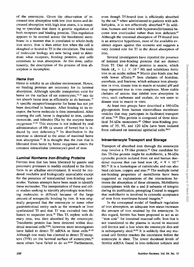
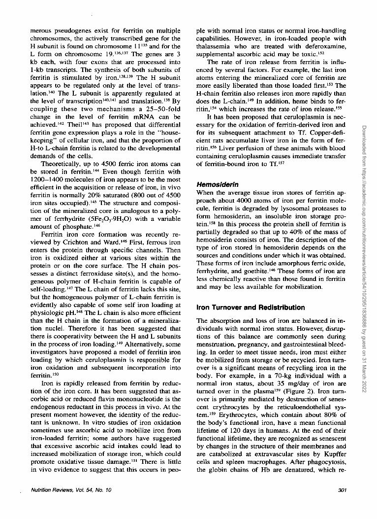
The absorption and loss of iron are balanced in in- dividuals with normal iron status. However, disrup- tions of this balance are commonly seen during menstruation, pregnancy, and gastrointestinal bleed- ing. In order to meet tissue needs, iron must either be mobilized from storage or be recycled. Iron turn- over is a significant means of recycling iron in the body. For example, in a 70-kg individual with a normal iron status, about 35 mg/day of iron are turned over in the plasmaIs9 (Figure 2). Iron turn- over is primarily mediated by destruction of senes- cent erythrocytes by the reticuloendothelial sys- tem.Is9 Erythrocytes, which contain about 80% of the body's functional iron, have a mean functional lifetime of 120 days in humans. At the end of their functional lifetime, they are recognized as senescent by changes in the structure of their membranes and are catabolized at extravascular sites by Kupffer cells and spleen macrophages. After phagocytosis, the globin chains of Hb are denatured, which re-
Nutrition Reviews, Vol. 54, No. 10 301
Dow
nloaded from https://academ
ic.oup.com/nutritionreview
s/article/54/10/295/1836886 by guest on 31 March 2022

. . . . . . . . . . . . . . . . . . . . . . . . . . . . . . . . . . . . . . . . . . . . . . . . . . . . . . . . . . . . . . . . . . . . . . . . . . . . . . . . . . . . . . . . . . . . . . . . . . . . . . . . . . . . . . . . . . . . . . . . . . . . . . . . . . . . . . . . . . . . . . . . . . . . . . . . . . . : : : : : : : : : :zs(x(T : :mk: . . . . . . . . . . . . . . . . . . . . . . . . . . . . . . . . . . . . . . . . . . . . . . . . . . . . . . . . . . . . . . . . . . . . . . . . . . . . . . . . . . . . . . . . . . . . . . . . . . . . . . . . . . . . . . . . . . . . . . . . . . . . . . . . . . . . . . . . . . . . . . . . . . . . . . . . . . . a\ 20 m g i - Hemog lob in
5 m g Plasma
Eryth r op oie sis
Myoglobi n, Heme-, and Nonheme- [lob]
Iron Containing Proteins Ab s o r I) e d
Losses 1-2 nig
. . . .
. . . Ferritin and Hemosid erin
I n g e s t e d 10-14 m g
Figure 2. Iron distribution and exchange pools.
Unabsorbed 8-13.5 m g
leases bound heme. Intracellular unbound heme is ultimately degraded by heme oxygenase, which lib- erates iron. About 85% of the iron derived from Hb degradation is rereleased to the body in the form of iron bound to Tf or ferritin.. Each day 0.66% of the body's total iron content is recycled in this man- n e ~ . ' ~ ~ Smaller contributions are made to plasma iron turnover by the degradation of myoglobin and iron-containing enzymes.
Iron Losses
The low solubility of iron precludes excretion as a major mechanism for maintaining iron homeostasis. In contrast to most other trace minerals whose ho- meostasis in maintained by excretion, the primary mechanism of maintaining whole-body iron homeo- stasis is to regulate the amount of iron absorbed so that it approximates iron losses. Iron losses can vary considerably with gender. In human males, total iron losses from the body have been calculated to be 1 mg/day. For premenopausal female humans, this loss is slightly higher. The predominant route
of loss is from the gastrointestinal tract, with losses amounting to 0.6 mg/day in adult males.Im Fecal iron losses derive from shed enterocytes, extrava- sated red blood cells, and biliary heme breakdown products that are poorly absorbed. Urogenital and integumental iron losses have been estimated at >0.1 mg/day and 0.3 mg/day, respectively, in adult males.I6' Menstrual iron loss, estimated from an av- erage blood loss of 33 mL/month, equals 1.5 mg/day, but may range as high as 2.1 mg/day.'* Oral contraceptives reduce this loss'60.162 and intra- uterine devices increase it.'60.163.161 Pregnancy is as- sociated with losses approximating 1 g over the pregnancy course, consisting of basal losses of 230 mg iron; increased maternal red cell mass of 450 mg iron; fetal needs of 270-300 mg iron; and pla- centa, decidua, and amniotic fluid iron content of 50-90 mg.
A number of clinical and pathological conditions are accompanied by variable amounts of blood loss. These include hemorrhage, hookworm infestation, peptic gastric or anastomotic ulceration, ulcerative colitis, colonic neoplasia, cow's milk feeding to in-
302 Nutrition Reviews, Vol. 54, No. 10
Dow
nloaded from https://academ
ic.oup.com/nutritionreview
s/article/54/10/295/1836886 by guest on 31 March 2022
fants, aspirin, nonsteroidal anti-inflammatory drugs or corticosteroid administration, and hereditary hem- orrhagic telangiectasia.'65-'70 In addition to these con- ditions, a significant amount of iron (210-240 mghnit) can be lost with regular blood d0nati0n.l~~
lntracellular Metabolism of Iron
Acquisition of Iron via the Transferrin Receptor Because most cellular iron acquisition occurs via Tf uptake, we will focus on the role of the TfR in maintaining intracellular iron homeostasis. The TfR is a 180-kDa glycoprotein composed of two iden- tical 95-kDa subunits that are linked by two disul- fide bridges (Cys-89 and C Y S - ~ ~ ) . ' ~ ~ The human TfR gene has been localized to chromosome 3, re- gion q26.2 qter.173.174 The promoter region of this gene contains several metal response elements and appears to be transcriptionally (two- to threefold de- crease)175 and t ran~lat ional lyl~~ regulated by iron. Gene transcription is also negatively regulated by retinoic and variably regulated by 1,25-di- hydroxyvitamin D,.
Each subunit of the TfR is composed of 760 amino Specific sequences in the intracel- lular domain composed of Tyr-Thr-Arg-Phe (YTRF) appear to be necessary for aggregation in clathrin- coated pits.17y Serine 24 on the cytoplasmic domain of the TfR is phosphorylated by protein kinase C.Igo The functional consequences of this phosphoryla- tion are unknown, but it is not required for inter- nalization. I g 1
The transmembrane region of the TfR consists of a single hydrophobic domain (24-28 amino ac- ids) above position 65. The transmembrane segment of the human TfR functions as a signal peptide and is necessary for translocation to the cell surface.Ig2 This hydrophobic domain is also acylated in posi- tions Cys-62 and possibly C ~ s - 6 7 . ' ~ ~ This acylation is apparently not required for transport to the cell surface. The receptor is (N-linked) glycosylated on Asn-25 1, Asn-3 17, and A ~ n - 7 2 7 . I ~ ~ Glycosylation facilitates Tf-TfR binding through its effects on the tertiary and quaternary structure of the TfR. Each TfR subunit binds one Tf molecule with high affin- ity.Ig3 The affinity of the TfR for Tf increases with Tf iron-binding site occupancy. The affinity is high- est for diferric Tf (Kd = 1.1 X M) and lowest for apo-Tf (& = 4.6 X M).Ig4 Because the con- centration of Tf in plasma is 30-40 X M, cell surface TfRs are usually saturated with Tf. There- fore, the regulation of cellular iron uptake is me- diated by altering the number of TfRs present on the cell surface. At any one time about a third of the cellular mass of TfRs is found on the cell sur- face. This number can be increased either by im-
mediate translocation of cytoplasmic receptors to the surface or by de novo synthesis. The number of receptors present on the cell surface is a function of intracellular iron status, cell proliferative status, and metabolic need such as production of hemoglobin and myoglobin. Consequently, erythroblasts (1 X lo5) and reticulocytes (8 X lo5) have the highest number of TfRs per cell, as their iron needs are very high. As these cells mature into erythrocytes, they lose functioning TfR on their cell surface.lg5
Ferro-transferrin is taken up by TfR-mediated endocytosis. Some researchers have demonstrated that internalization of the TfR can occur with apo-Tf attached,Ig6 although others have shown that only ferro-Tf is internalized.Ig7 After internalization, the endosomal compartment containing the Tf-TfR complex sheds its clathrin coat. An endosomal pro- ton pump (H+-ATPase) then lowers the pH in the endosome to pH 5-6. This acidic milieu lowers the affinity of Tf for iron. Binding of chloride to an anion-binding site of TfR-bound Tf facilitates the removal of iron from Tf.Ig8 Additionally, part of the TfR also participates in this process.Igy Some inves- tigators have described an endosomal enzyme that uses NADH to reduce Tf-derived ferric iron to the ferrous state.lyO.lyl Other investigators have suggest- ed that ascorbate may nonenzymatically assist in this action.lY2
After iron is removed from Tf, the iron-contain- ing portion of the endosome separates from the compartment containing the Tf-TfR complex. Iron in the endosomal compartment is transported across the membrane to the cytosol where it appears either to enter a pool of low molecular weight iron che- l a t e ~ ~ ~ ~ or attach to an intracellular iron-binding pro- tein. Recent evidence suggests that the endosomal proton pump (H+-ATPase) may participate in the transport of iron from the endosome to the cyto-
This iron is then channeled into one of three pathways: iron-regulatory protein(s), iron-utilizing proteins, or storage iron.
The endosomal portion containing the TfR- apo-Tf complex travels to the Golgi apparatus, where it is packaged, along with newly synthesized receptors, and is translocated to the cell surface. The affinity of the TfR for apo-Tf at pH 7.4 is much lower than that at pH 5.5. Consequently, apo-Tf is released when the TfR-apo-Tf complex returns to the cell surface. The complete cycling of TfR-Tf occurs in about 10 minutes and can occur repeatedly about 100 times before either TfR or Tf is degraded. In sheep and rat reticulocytes the TfR receptor can be actively shed from the cell ~ u r f a c e . ~ ~ ~ J ~ ~ A trun- cated form of the TfR that lacks cytoplasmic and transmembrane regions is found in human plasma bound to Tf.1y7.1y8 It is not known whether the hu- man TfR fragment arises from alternate splicing of
Nutrition Reviews, Vol. 54, No. 70 303
Dow
nloaded from https://academ
ic.oup.com/nutritionreview
s/article/54/10/295/1836886 by guest on 31 March 2022
the TfR gene or posttranslational cleavage. The fragment of TfR that circulates in plasma can be detected by enzyme-linked immunosorbent assay and is the basis of a new method for determining an individual's iron status.
lntracellular Low Molecular Weight Species
Several investigators have reported the presence of an intracellular pool of low molecular weight iron- containing species. The nature of this pool is largely speculative and suggestions of its composition range from citrate, nucleotide, pyrophosphate, ami- no acid, and/or protein chelates or complexes of iron.199-201 The intracellular concentration of this pool is constant throughout conditions ranging from iron deficiency to iron overload.200
lntracellular Iron Homeostasis
Iron Response Elements Intracellular iron homeostasis requires the coordi- nated regulation of the synthesis and action of pro- teins involved in iron acquisition, utilization, and storage. When intracellular iron is scarce the cell needs to increase its acquisition of iron either by mobilization of storage iron or acquisition of plasma iron. The cell also needs to prioritize its iron utili- zation so that life-sustaining, iron-containing pro- teins preferentially receive iron. In animals, much of this process is regulated at the genetic level by iron.123.175.202-205 In addition to the ill-defined tran- scriptional regulation by iron of Tf, TfRs, and fer- ritin, iron participates directly in its own homeosta- sis. It binds to a trans-acting element(s) known as the iron responsive element binding protein(s) (IRE- BP[s]). During intracellular iron deficiency, an IRE- BP binds to cis-acting iron regulatory or responsive elements (IREs) located in either the 3' or 5' un- translated region of some mRNAs. These IREs are a family of stem loop structures,2M which are highly conserved among species.207 Their secondary and tertiary structures are important for high-affinity binding of the IRE-BF? Five IREs have been iden- tified in the 3' untranslated region of TfR mRNA.176*
A single IRE has been located in the 5 ' untran- slated region of the mRNAs of femtin-erythroid D-aminolevulinate synthase (e-D-ALAS)202 and a mitochondria1 isoform of aconitase (m-aconitase),211 which repress translation of these genes when an IRE-BP is b o ~ n d . ~ ~ ~ - ~ ~ ~ An IRE has also been lo- cated in the amyloid precursor mRNA.215.216 In con- trast to e-D-ALAS, which contains a functional IRE, examination of the mRNA for housekeeping D-am- inolevulinate synthase (h-D-ALAS) did not reveal an IRE.2o2 Finally, although Tf synthesis may be regulated at the level of translation by iron,123 no
IRE has yet been identified in the mRNA for Tf. Thus, translational control of iron-containing pro- teins by the IRE-IRE-BP system is widespread in animals but may not be universal.
Iron Response Element-Binding Protein(s) The IRE-BPs, also known as the femtin repressor protein,217 the iron regulatory factor,218 or P-90,219 show broad tissue distribution.220 The gene for one IRE-BP has been localized to chromosome 9221 and has been cloned from a number of species.222 This IRE-BP has a molecular mass of 98 kDa, shows 95% homology between four different species, and shows considerable homology to m-aconitase (30%) and isopropylmalate i s o m e r a ~ e . ~ ~ ~ This IRE-BP has been putatively identified as the cytosolic form of aconitase (c-a~onitase).~" Iron-replete c-aconitase is an enzyme that converts citrate to isocitrate. The regulation of translation of mRNA by this IRE-BP does not involve changes in the level of IRE-BPZZ5 Disassembly of its iron-sulfur cluster (4Fe-4s) to (3Fe-4s) results in loss of aconitase activity and promotes high-affinity binding to mRNA.226-228 However, simple reduction and removal of the iron bound in the fourth coordination site of c-aconitase is insufficient to produce the high-affinity RNA binding properties found in the native IRE-BI? It appears that endogenously produced nitric oxide serves to promote disassembly of the iron-sulfur cluster of IRE-BP and enhances the high-affinity binding of IRE-BP to mRNA.229,230 The IRE-BP is also phosphorylated by protein kinase C, which en- hances the high affinity binding of IRE-BP to mRNA.231 Although the hypothesis of 3Fe/4Fe switching is attractive with regard to signaling IRE- BP binding to mRNA, other mechanisms also seem likely; further study of the binding region of IRE with IRE-BP is needed.
A second IRE-BP has recently been identified that differs from the above IRE-BP in size (105 kDa) and tissue distribution (primarily brain and in- te~ t ine) .~~* The location of the IREs and their bind- ing proteins in tissues and cellular organelles will help clarify the regulatory system within and be- tween organ systems.
Chemical Properties and Biochemical Functions of Iron
Introduction Iron is a d-block transition element, which can exist in oxidation states ranging from -2 to +6. In bio- logic systems, these oxidation states are primarily limited to the ferrous (+2), ferric (+3), and ferry1 (+4) states. The interconversion of iron oxidation states is not only a mechanism whereby iron partic-
304 Nutrition Reviews, Vol. 54, No. 10
Dow
nloaded from https://academ
ic.oup.com/nutritionreview
s/article/54/10/295/1836886 by guest on 31 March 2022
ipates in electron transfer but also a mechanism whereby iron can reversibly bind ligands. Iron can bind to many ligands by virtue of its unoccupied d orbitals. The preferred biologic ligands for iron are oxygen, nitrogen, or sulfur atoms. The electronic spin state and biologic redox potential (from + lo00 mV for some heme proteins to -550 mV for some bacterial ferredoxins) of iron can change according to the ligand to which it is bound. By exploiting the oxidation state, redox potential, and electron spin state of iron, nature can precisely adjust the chem- ical reactivity of iron. Thus, iron is particularly suit- ed to participate in a large number of useful bio- chemical reaction^.^^^^^^ The activity of many of the enzymes involved in these biochemical reactions decreases during tissue iron deficiency. Only rarely, however, have direct connections between biochem- ical events and clinical manifestations been firmly established.
Four major classes of iron-containing proteins carry out these reactions in the mammalian system: iron-containing proteins (Hb and myoglobin), iron- sulfur enzymes, heme proteins, and iron-containing enzymes (noniron sulfur and nonheme enzymes). In iron-sulfur enzymes, iron can be bound to sulfur in four possible arrangements (FeS, 2Fe-2S, 4Fe-4S, 3Fe-4s). In humans only three of these occur. In heme proteins, iron is bound to various forms of heme that dif€er not only in the composition of their side chains but also in the methods whereby they are attached to proteins. In humans, however, the predominant form of heme is protoporphyrin IX (PP-IX) .
Oxygen Transport and Storage
The movement of oxygen from the environment to terminal oxidases is one of the key functions of iron. Dioxygen is bound to porphyrin ring iron con- taining molecules either as part of the prosthetic group of Hb within red blood cells or as the facil- itator of oxygen diffusion in tissue-myoglobin. Hemoglobin is a tetrameric protein with two pairs of identical subunits (a2, p2, MW 64 kDa) with either 141 or 142 amino acids in the a chain and 146 in the b chain. Each subunit has one prosthetic group, Fe-PP-IX, whose ferrous iron reversibly binds dioxygen. The four subunits are not cova- lently attached to each other but do react coopera- tively with dioxygen with specific modulation by pH, pCO,, organic phosphates, and temperature. These modulators determine the efficiency of oxy- gen transport from the alveoli capillary interface in the lung to the red cell + capillary + tissue inter- face in peripheral tissues. The allosteric effect of decreasing pH-the Bohr effect4ecreases binding affinity of heme-Fe for dioxygen via protonation of
His-146 on p chains and Val-1 on a chains in the presence of C1- and CO,. CO, forms a Schiff base with the terminal amino acids of each chain and decreases dioxygen affinity. This favors the unload- ing of oxygen in tissues where the pH is lower and pC0, is higher than in arterial blood. 2,3-Diphos- phoglycerate is a product of a side pathway within erythrocytes and binds to a specific region of the p chain to decrease Hb-0,-binding affinity. This right shift of the dissociation curve is evident in times of greater need for oxygen delivery such as in anemia, when the blood content of Hb is significantly re- duced and increased cardiac output is only partially compensatory.
Myoglobin is the single chain hemoprotein of 17 kDa MW in cytoplasm. Its role is to increase the diffusion rate of dioxygen from capillary red blood cells to cytoplasm and mitochondria. The concen- tration of muscle myoglobin is drastically reduced in tissue iron deficiency, thus limiting the rate of diffusion of dioxygen from erythrocytes to mito- c hondri a. 235
Electron Transport
The cytochromes contain heme as the active site with the Fe-porphyrin ring functioning to reduce ferric iron to ferrous iron with the acceptance of electrons. The iron-sulfur proteins also act as elec- tron carriers via the action of iron bound to either two or four sulfur atoms and cysteine side chains. The 40 different proteins comprising the respiratory chain contain 6 different heme proteins, 6 iron-sul- fur centers, 2 copper centers, as well as ubiquinone to connect NADH to oxygen. Several hundred en- zyme activities have also been ascribed to the cy- tochrome P450 family of enzymes. Some demon- strate limited substrate specificity, but most exhibit broad substrate specificity. They have been termed the mixed function oxidases. At least 39 rat and 28 human cytochrome P450 genes have been identified so far and more are likely to be discovered.236 The microsomal P450 enzyme system participates in the biosynthesis of steroid hormones such as pregnen- olone, corticosterone, aldosterone, and 1,250H-vi- tamin D,. The microsomal P450 enzyme system participates in the metabolism of xenobiotics such as drugs and aromatic hydrocarbons. This enzyme system includes cholesterol 7a-monooxygenase, the rate-limiting step in bile acid synthesis. This en- zyme system is also responsible for the formation of prostacyclin, thromboxane, and leukotrienes.
With the exception of the glutathione peroxi- dases, all mammalian peroxidases contain iron. Cat- alase degrades hydrogen peroxide formed as a by- product of some oxidase reactions. Myeloperoxi- dase forms hypochlorite anion, which is an impor-
Nutrition Reviews, Vol. 54, No. 10 305
Dow
nloaded from https://academ
ic.oup.com/nutritionreview
s/article/54/10/295/1836886 by guest on 31 March 2022
tant cytotoxic molecule produced by neutrophils. Thyroperoxidase is responsible not only for the or- ganification of iodide but also for the conjugation of iodinated tyrosine residues on thyroglobulin. Ni- tric oxide (NO’) is a potent biologic effector mol- ecule. Its synthase is a cytochrome P450-like pro- tein that occurs in at least four isoforms. At least two of these forms, NO synthase I and 11, contain iron in the form of PP-IX.237.238 They may also con- tain a catalytically active nonheme iron.239
Physiologic Functions of Iron and Signs of Iron Deficiency
The overt physical manifestations of iron deficiency are glossitis, angular stomatitis, koilonychia (spoon nails), blue sclera, esophageal webbing (Plummer- Vinson syndrome), and anemia. Behavioral distur- bances such as pica (characterized by abnormal con- sumption of nonfood items such as dirt [geophagia] and ice [pagophagia]) are often present in iron de- ficiency. The physiologic manifestations of iron de- ficiency have also been noted in immune function, cognitive performance and behavior, thermoregula- tory performance, energy metabolism, and exercise or work p e r f ~ r m a n c e . ~ ~ ~ . ~ ~ ~ Many of these manifes- tations of iron deficiency are not mutually exclusive events and do not occur independently of each oth- er. Furthermore, many of these manifestations occur only during certain stages of iron deficiency.
The progression of iron deficiency occurs in two steps related to depletion of iron stores prior to de- pletion of functional iron: (1) bone marrow, spleen, and liver stores depletion; and (2) diminished eryth- ropoiesis due to a negative iron balance leading to anemia and decreased activity of iron-dependent en- zymes. Clinical iron deficiency is frequently diag- nosed by virtue of anemia secondary to long-term diminished erythropoiesis. With a few exceptions, depletion of the storage iron pool is generally with- out influence on physiologic f u n ~ t i o n . ~ ~ ’ , ~ ~ * In those studies, correlations were noted between electroen- cephalogram asymmetry (a central nervous system abnormality) and plasma femtin within the iron-ad- equate range. Nonetheless, nearly all functional consequences are more strongly related to “ane- mia” rather than to tissue iron deficits. The chal- lenge of separating 0, transport events from tissue iron deficits still looms large. Good examples are the decreases in muscle myoglobin content, cyto- chrome oxidase activity, and electron transport.
Several well-known consequences of iron defi- ciency occur after the depletion of iron s tores-de- cline in Hb concentration, decrease in mean cor- puscular Hb concentration, a decrease in the size and volume of the new red cells, reduced myoglo- bin, and reduced amounts of both Fe-S and heme
iron-containing cytochromes within cells. Diffusion of dioxygen from Hb into tissue becomes limited in this situation due to fewer erythrocytes packed close together in capillaries, increased membrane diffu- sivity, and a decreased tissue myoglobin concentra- tion. The heterogeneity of distribution of mitochon- dria around and adjacent to capillary walls is well known but not well studied in the iron-deficient hu- man or animal model. The delivery of red cells to tissue is under a complex regulation by both sys- temic and local regulatory features. The reader is urged to consider that the matching of oxygen de- livery to tissue needs for oxygen is the ultimate goal of these regulators. In severe anemia, oxygen trans- port is clearly limiting to tissue oxidative function at anything but the resting condition.235 This is de- spite a significant right shifting of the Hb-0, dis- sociation curve (decreased affinity) and an increase in cardiac output in an attempt to increase TaO,. Tissue extraction of oxygen is increased by this compensation and mixed venous PO, is significant- ly lower in anemic individuals. While Hb-0, affin- ity compensation is reasonable at sea level, the op- posite direction of compensation occurs in anemic people at high altitudes (4000 m). The Hb-0, dis- sociation curve is “left-shifted” in these hypobaric hypoxic conditions to increase 0, loading in the lung at the expense of tissue delivery.243 The very significant decrease in myoglobin and other iron- containing proteins in skeletal muscle seen in iron- deficiency anemia contributes significantly to the decline in muscle aerobic capacity.235
A recent study244 used 31P nuclear magnetic res- onance spectroscopy to examine the functional state of bioenergetics in iron-deficient and iron-replete rat gastrocnemius muscle at rest and during 10 seconds of contraction at 2 Hz. Iron-deficient animals had a clear increase in phosphocreatine (PCr) breakdown and a decrease in pH compared to controls. They also had a slower recovery of PCr and inorganic phosphate concentrations after exercise. Repletion for 2-7 days with iron dextran showed no substan- tial improvement in these indicators of muscle mi- tochondrial energetics. These authors concluded that “tissue factors” such as decreased mitochon- drial enzyme activity, decreased number of mito- chondria, and altered morphology of the mitochon- dria might be responsible for these observations. It is not uncommon for phosphate : oxygen ratios to be normal in iron deficiency despite very significant alterations in activity of iron-containing respiratory chain enzymes.
A more typical repair curve for muscle iron- containing and oxidative enzymes during iron re- pletion experiments has been described.245 Pyruvate and malate oxidase were decreased to 35% of nor- mal in iron-deficient muscle and improved to 85%
306 Nutrition Reviews, Vol. 54, No. 10
Dow
nloaded from https://academ
ic.oup.com/nutritionreview
s/article/54/10/295/1836886 by guest on 31 March 2022

Table 1. Diagnostic Criteria for Iron Deficiency (Adapted from Herbert246 and Ferguson et aLZ4’)
Iron-Deficient Iron Iron Er ythro- Iron-Deficiency Overload Normal Depletion poiesis Anemia
Erythrocyte morphology Normal Normal Normal Normal Microcytid hypochromic
Hemoglohin (gn- l ) < 120 (female)
Hernatocrit <0.35 (female)
Plasma iron (pg/dL-’) >I75 115 ? 50 115 <60 <40
<135 (male)
<0.40 (male)
RBC protoporphyrin (pg/dL-I) 30 30 30 100 200 RE marrow Fe 4 + 2-3 + 0-1 + 0 0 Sideroblasts (%) 40-60 40-60 40-60 < 10 < 10
Transfenin saturation (%) >60 3 5 ? 15 30 <I5 <I5 Plasma femtin (pg/dL-’) >300 100 5 60 20 10 < 10 TW relative amount 1 1.5 3 3-4
TIBC (pg/dL-’) <300 300 f 30 360 390 410
5.36 2 0.82 13.9 2 4.6 TfR (mg dL-’)”
“This will vary depending on the antibody used.
of normal in 10 days of treatment. 2-Oxoglutarate oxidase was decreased to 47% of normal and im- proved to 90%; in contrast, succinate oxidase was only 10% of normal in iron deficiency and im- proved to only 42% of normal after 10 days. Cy- toplasmic enzymes hexokinase and lactate dehydro- genase were unaffected by iron status. The 50-90% decrease in both the Fe-S enzymes and in the heme- containing mitochondria1 cytochromes is consistent with many other observations over the last two de- c a d e ~ . ~ ~ ~ What seems to determine the amount of decline in activity with iron deprivation is the rate of turnover of that particular iron-containing protein in the time of cellular deprivation of iron.
The last stage of iron deficiency occurs when stores are depleted and there is no longer sufficient iron to meet daily requirements. This stage of iron- deficient erythropoiesis leads to a significant com- promise in cellular function in many organs.235 The rate at which individual tissues and cellular organ- elles within those tissues develop a true iron “def- icit” is dependent on the rate of turnover of iron- containing proteins and rate of cell growth, as well as the intracellular mechanisms for recycling iron.235
The reader is encouraged to look at recent re- views that describe a number of functional conse- quences of iron d e f i ~ i e n c y . ’ ~ ~ . ~ ~ ~
Assessment of Iron Status
The iron status of humans can range from iron over- load to iron-deficiency anemia. Historically, many different methods have been used to assess the iron status of an individual including dietary intake, he- matocrit, Hb, mean cellular Hb, mean cell volume,
erythrocyte mean index, free erythrocyte protopor- phyrin, bone marrow iron stain, serum iron, total iron-binding capacity, serum Tf, Tf saturation, se- rum ferritin, and serum TfR. These methods vary considerably in their sensitivity and selectivity. The more commonly accepted diagnostic tests and their associated values are summarized in Table 1 .246,247
~ e ~ o g / o ~ ~ ~ / ~ e ~ ~ ~ o c ~ ~ ~ Several reviews of iron metabolism in the last de- ~ a d e ~ ~ ~ - ~ ~ ~ , ~ ~ ~ have noted that the term “iron defi- ciency” has no universal interpretation. Clinical se- quelae are most frequently recognized at the end stages of the iron depletion process when body iron stores have been depleted. For clinicians, the prev- alence of iron deficiency is equated with the prev- alence of iron-deficiency anemia. The use of anemia as a clinical indicator may be due either to the ease of assessment in measuring Hb concentration248 or to the assumption that iron deficiency exerts its del- eterious effects only if anemia is present.249 The use of Hb and hematocrit as indices of iron status must be used carefully, as significant false-positive indi- cations occur.25o Although a single replicate has been determined to accurately predict iron status, adjustments must be made for significant variations due to age, gender, and race in clinical assessment of iron status of individuals or populations. Hemo- globin concentrations are also altered in/by poly- cythemia, dehydration, cigarette smoking, chronic inflammation, chronic infection, hemorrhage, pro- tein-energy malnutrition, vitamin B ,z deficiency, folic acid deficiency, hemoglobinopathies, and preg- n a n ~ y . ~ ~ ’ Thus, considerable information about nu-
Nutrition Reviews, Vol. 54, No. 10 307
Dow
nloaded from https://academ
ic.oup.com/nutritionreview
s/article/54/10/295/1836886 by guest on 31 March 2022
trition and health status is needed in addition to Hb determination if one is to use Hb to assess iron sta- tus.
Ferritin A long-term negative iron balance eventually leads to the depletion of the storage iron pool, leading to dramatic declines in plasma ferritin concentrations. The most realistic tool to date in a nonclinical set- ting for assessment of the size of the storage pool is the measurement of serum or plasma ferritin con- centrations. The concentration of serum ferritin re- flects the size of the storage iron compartment if the subject is not also in an inflammatory state.252 Se- rum ferritin values usually fall in the range of 20- 300 mgL, with each mg/L representing 10 mg of storage iron.145 Plasma ferritin concentration can in- crease dramatically with both acute and chronic in- flammations, vitamin BIZ deficiency, folic acid de- ficiency, liver disease, leukemia, Hodgkin's Disease, alcohol intake, and hyperthyroidism.1'7.253-255 In ad- dition, it is now known that there is a large within- subject day-to-day coefficient of variation (25- 40%) in plasma ferritin concentration^.^^^
Transferrin Saturation Once the storage iron pool is depleted due to a pro- longed or acute negative iron balance, there is a decline in the Tf saturation. Consequently, less than adequate iron is available for essential body iron protein^.^^^.^^^ People in this stage of iron depletion have a Tf saturation below 15-16% and an inade- quate supply of iron for bone marrow to support normal e r y t h r o p o i e s i ~ . ~ ~ ~ - ~ ~ ~ The measure of eryth- ropoiesis is clearly an important aspect in this iron delivery scheme, as decreased erythropoiesis can lower iron transport requirements by 5040%.
Transferrin Receptor The measurement of TfR concentration in the plasma has diagnostic value for the assessment of iron defi- ciency anemia and ineffective erythropoiesis.260-262 The amount of TfR in circulation has been shown to vary with individual iron status. Plasma TfR con- centrations increase even in mild iron deficiency of recent onset.263,264 The plasma concentration of TfR is increased in P-thalassemia, autoimmune hemo- lytic anemia, sickle cell anemia, hereditary spher- osis, Hb H disease, polycythemia Vera, secondary polycythemia, myelofibrosis, and chronic lympho- cytic leukemia.260-262 The plasma TfR concentration is decreased in hemochromatosis, aplastic anemia- bone marrow ablation, posttransplantation anemia, and chronic renal failure.247.260-262 Unlike ferritin, levels of plasma TfR are not significantly affected
by i n f l a m m a t i ~ n ~ ~ . ~ ~ ~ or by liver disease.247 Unlike most other methods of assessment, TfR concentra- tion can be used to distinguish between iron-defi- ciency anemia and other anemias, including the ane- mia of chronic disease.247 Hence, the TfR is partic- ularly useful in assessing iron status.
Populations at Risk for Iron Deficiency
Despite the effectiveness of intervention therapies, iron deficiency is the primary nutrition deficiency in the United States.266 One to six percent of the U.S. population has impaired iron status, including 3.5-12.1% of males 11-14 years of age and 2.5- 14.2% of females 15-44 years of age.267 Approxi- mately 6-1 1% of reproductive age females, 14% of 15-19-year-old females, and 25% of pregnant wom- en were iron deficient in the United States and Can- ada in the 1980s.267.268 Fortification of the food sup- ply in combination with additional intakes of iron from supplements, as well as changes in dietary pat- terns have effectively eradicated iron deficiency from nearly all segments of the U.S. population. The exceptions are pregnant women and a small proportion of young children, adolescents, and re- productive age women. However, special clinical populations, some elderly, and perhaps athletes may still require targeted intervention.
International statistics demonstrate the immen- sity of the p r ~ b l e m , ~ ~ ~ . ~ ~ ~ with 15% of the world's population having iron-deficiency anemia. The World Health Organization estimates that 1.3 billion people are anemic, and that nearly half (500-600 million) have iron deficiency as the causal agent. For populations at risk the prevalence of iron defi- ciency can approach 50%. As noted in the various sections of this review, iron requirements are deter- mined by growth and maintenance requirements.271 There are additional requirements associated with clinical and pathological conditions leading to in- creased blood loss. Iron requirements in infancy, childhood, adolescence, and during pregnancy are covered in detail e l s e ~ h e r e . ~ ~ ~ . ~ ~ ~ The increased di- etary requirements during these periods of acceler- ated growth amount to 1 mg/kg/day in infants, 10 mg/day in children, up to 15 mg/day in female ad- olescents, and an additional 5-6 mg/day above the Recommended Dietary Allowance during the sec- ond and third trimester of pregnancy. The rapid ex- pansion of the maternal blood volume, placental growth, and fetal growth place tremendous demands on maternal stores of iron. It is not uncommon for >70% of women to emerge from pregnancy with iron-deficiency anemia. Menstrual blood losses dur- ing reproductive years elevate iron requirements in female on average 5 mg/day above that of males.
308 Nutrition Reviews, Vol. 54, No. 10
Dow
nloaded from https://academ
ic.oup.com/nutritionreview
s/article/54/10/295/1836886 by guest on 31 March 2022

The requirements for adult male and postmenopau- sal women is 10 mg/day.
Public health nutritionists are more interested in how much of the population is at risk of iron de- pletion and are thus more concerned with iron status adequacy and prevalence of depleted iron stores. Iron deficiency can be defined as that moment in time when body stores of iron-femtin and hemo- siderin-become depleted of iron and a restriction of supply of iron to various tissues becomes appar- ent.lI7 Conceptually, the process of depletion of iron stores can occur rapidly or very slowly and is de- pendent on the balance between iron intake and iron requirements.
Clearly, iron intake depends on the quantity of iron in the diet in addition to the presence of a num- ber of inhibitors and a smaller number of enhancers of iron absorption in Iron requirements de- pend on body needs for tissue growth and tissue maintenance, which vary with the life cycle and cer- tain environmental factors. The systematically greater prevalence of iron deficiency in women than men is likely due to a shift in the iron balance equa- tion and not due to “femaleness or maleness.” The gradual increase in iron status in females after men- opause regardless of estrogen replacement supports this concept. Requirements for iron in pregnancy are increased dramatically to approximately 4 mg/day above normal and perhaps even higher if the pregnancy occurs in adolescent^.^^^-^^^ The re- quirements for iron are of course, higher in the growing child and give rise to the concept of “crit- ical periods” during both prenatal and postnatal
That is, there exists a particular period of time during early development when the brain and other specific organs are especially susceptible to a nutrient deficiency state. The restoration of normal dietary iron intake at later times may not restore normal function or tissue content of these organs and “catch-up growth” may not occur.
A number of clinical and pathologic conditions associated with blood loss can cause iron deficiency. In addition, treatments that actively increase iron requirements, such as erythropoietin therapy, can lead to iron d e f i c i e n ~ y . ~ ~ ~ , ~ ~ ~
Conclusions and Future Research Perspectives
This review is not a comprehensive examination of all aspects of iron nutrition in health and disease. Rather, our intent is to review the current knowl- edge in key aspects of the field of iron nutrition and biology and to, hopefully, stimulate new intellectual efforts by scholars to better understand the role of iron in human biology. Scientific investigations spanning several decades have increased our under-
standing of the role of this mineral in many aspects of metabolism, but it is clear that many questions remain. For example, what are all of the compo- nents of the iron absorption pathway? What is the somatic regulator of iron absorption? What is the nature of the low molecular weight intra-and extra- cellular pools of iron? What are the full conse- quences of iron deficiency, particularly as related to cognitive function, and are they reversible? How does hemochromatosis arise? Finally, what role does iron, as found in the body under normal nu- trition states, play in oxidative stress? Hopefully, scientists in the next decade will unravel some of these mysteries.
1.
2.
3.
4.
5.
6.
7.
8.
9.
10.
11.
12.
13.
de Duve C. Prelude to a cell. The Sciences
Eaton SB, Konner M. Paleolithic nutrition. A consid- eration of its nature and current implications. N Engl J Med 1985;312:283-9 Vannotti A, Delachaux A. Iron Metabolism and Its Clinical Significance. New York: Grune and Strat- ton, 1949 Hughes ER. Human iron metabolism. In: Sigel H, ed. Metal Ions in Biological Systems. Iron in Model and Natural Compounds. New York: Marcel Dekker, 1977 MacKay C. Memoirs of Extraordinary Popular De- lusions. London: Richard Bently, 1841 Marks G, Beatty WK. The Precious Metals of Med- icine. New York: Charles Scribner & Sons, 1975 Cule J. The iron mixture of Dr. Griffith. Pharm J. 1967;CXCVlll:399-400 Fairbanks VF, Fahey JL, Beutler E. Clinical Disor- ders of Iron Metabolism. New York: Grune & Strat- ton, 1971 McCollum EV. A History of Nutrition. Boston: Houghton Mifflin, 1957 McCay CM. Notes on the History of Nutrition Re- search. Bern: Huber, 1973 Blood-inorganic substances. In: Lentner C, ed. Geigy Scientific Tables. Physical Chemistry, Com- position of Blood, Hematology, Somatometric Data, 8th ed. West Cadwell: Medical Education Division, Ciba-Geigy Corporation, 1984 Boussingault JB. Du fer contenu dans le sang et dans les aliments. CR Acad Sci Paris 1872;74: 1353-9 Schmidt JE, ed. Medical Discoveries. Springfield: Charles Thomas, 1959
1990;30:22-8
14. McCance RA, Widdowson EM. Absorption and ex- cretion of iron. Lancet 1937;2:680-4
15. Moore CV, Arrowsmith WR, Welch J, Minnich V. Studies in iron transportation and metabolism. IV. Observations on the absorption of iron from the gastrointestinal tract. J Clin Invest. 1939;18:553-80
16. Granick S. Protein apoferritin in iron feeding and ab- sorption. Science 1946;103:107-13
17. Iron in Human Nutrition. National Live Stock and Meat Board, 1990
Nutrition Reviews, Vol. 54, No. 10 309
Dow
nloaded from https://academ
ic.oup.com/nutritionreview
s/article/54/10/295/1836886 by guest on 31 March 2022

18.
19.
20.
21.
22.
23.
24.
25.
26.
27.
28.
29.
30.
31.
32.
33,
McCance RA, Widdowson EM. Mineral metabolism of healthy adults on white and brown bread dietar- ies. J Physiol 19423 01 :44-85 McCance RA, Edgecomb CN, Widdowson EM. Phy- tic acid and iron absorption. Lancet 1943;2:12&8 Van Campen D, Gross C. Effect of histidine and certain other amino acids on the absorption of iron-59 by rats. J Nutr 1969;99:68-74 Disler PB, Lynch SR, Charlton RW, Torrance JD, Bothwell TH. The effect of tea on iron absorbtion. Gut 1975; 1 6: 1 93-200 Monsen ER, Cook JD. Food iron absorption in hu- man subjects. IV. The effects of calcium and phos- phate salts on the absorption of nonheme iron. Am J Clin Nutr 1976;29:1142-8 Breddy M, Chidambaram MV, Fonseca J, Bates GW. Potential role of in vitro iron bioavailability stud- ies in combatting iron deficiency: a study of the ef- fects of phosvitin on iron mobilization from pinto beans. USAid Cooperative Agreement 1976;l: 1-45 Layrisse M, Martinez-Torres C, Leets I, Taylor P, Ramirez J. Effect of histidine, cysteine, glutathione or beef on iron absorption in humans. Br J Nutr 1984;52:37-46 Charlton RW, Bothwell TH. Iron absorption. Annu Rev Med 1983;34:55-68 Takkunen H, Seppanen R. Iron deficiency and di- etary factors in Finland. Am J Clin Nutr 1975;28:
Bjorn-Rasmussen E, Hallberg L, lsaksson B, Ar- vidsson B. Food iron absorption in man. Application of the two-pool extrinsic tag method to measure heme and non-heme iron absorption. J Clin Invest.
Cook JD, Reusser ME. Iron fortification: an update. J Food Sci 1983;48:1340-9 Reddy MB, Cook JD. Assessment of dietary deter- minants of nonheme-iron absorption in humans and rats. Am J Clin Nutr 1991;54:723-8 Forbes RM, Erdman JW. The bioavailability of trace mineral elements. Ann Rev Nutr 1983;3:213-31 Cook JD, Dassenko SA, Lynch SR. Asessment of the role of non-heme-iron availibility in iron balance. Am J Clin Nutr 1991;54:717-22 Cook JD, Watson SS, Simpson KM, Lipschitz DA, Skikne BS. The effect of high ascorbic acid supple- mentation on body iron stores. Blood 1984;64:721-6 Beard J. Iron fortification-rationale and effects. Nutr Todav 1986:21:17-20
1141-7
197433: 247-56
34. Crosby WH. Yin,'yang and iron. Nutr Today 1986; 21:14-6
35. Sullivan JL. Stored iron and ischemic heart dis- ease-empirical support for a new paradigm. Cir- culation 1992;86:1036-7
36. Lauffer R. Preventive measures for the mainte- nance of low but adequate iron stores. In: Lauffer R, ed. Iron and Human Disease. Boca Raton: CRC Press, 1992
37. Natow AB, Heslin J-A. The Iron Counter. New York: Pocket Books, 1993
38. Walter T, Hertrampf E, Pizarro F, et al. Effect of bovine-hemoglobin fortified cookies on iron status
of schoolchildren-a nationwide program in Chile. Am J Clin Nutr 1993;57:190-4
39. Chierici R, Sawatzki G, Tamisari L, Volpato S, Vigi V. Supplementation of an adapted formula with bo- vine lactoferrin. 2. Effects on serum iron, ferritin and zinc levels. Acta Paediatr 1992;81:475-9
40. Finch CA, Huebers HA. Iron absorption. Am J Clin Nutr 1988;47: 102-7
41. Carpenter CE, Mahoney AW. Contributions of heme and nonheme iron to human nutrition. Crit Rev Food Sci Nutr 1992;31:333-67
42. Wollenberg P, Rummel W. Dependence of intestinal iron absorption on the valency state of iron. Naunyn Schmiedebegs Arch Pharmacol 1987;336:578-82
43. Raja KB, Simpson RJ, Peters TJ. Comparison of 59Fe3+ uptake in vitro and in vivo by mouse duode- num. Biochim Biophys Acta 1987;901:52-60
44. Kelly KA, Turnbull EE, Cammock CT, Bombeck LM, Nyhus LM, Finch CA. Iron absorption after gastrec- tomy: an experimental study in the dog. Surgery 1967;62:35660
45. Conrad ME. Iron Absorption. In: Johnson LR, ed. Physiology of the Gastrointestinal Tract, 2nd ed. New York: Raven Press, 1987
46. Murry MJ, Stein N. Does the pancreas influence iron absorption? Gastroenterol 1966;51:694-700
47. Zempsky WT, Rosenstein BJ, Carroll JA, Oski FA. Effect of pancreatic enzyme supplements on iron absorption. Am J Disease Children 1989;143:969- 972.
48. Hastings-Wilson T. lntestinal Absorption. Philadel- phia: W. B. Saunders Co, 1962
49. Schumann K, Elsenhans B, Ehtechami C, Forth W. Rat intestinal iron transfer capacity and the longi- tudinal distribution of its adaptation to iron deficien- cy. Digestion. 1990;46:35-45
50. Conrad ME, Umbreit JN, Moore EG. Iron absorption and cellular uptake of iron. Adv Exp Med & Biology 1994;356:69-79
51. Layrisse M, Martinez-Torres C. Model for measur- ing dietary absorption of heme iron: test with a com- plete meal. Am J Clin Nutr 1972;25:401-11
52. Lynch SR, Dassenko SA, Morck TA, Beard JL, Cook JD. Soy protein products and heme iron ab- sorption in humans. Am J Clin Nutr 1985;41:13-20
53. Hallberg L, Rossanderhulthen L, Brune M, Gleerup A. Inhibition of haem-iron absorption in man by cal- cium. Br J Nutr 1993;69:533-40
54. Sayers MH, Lynch SR, Jacobs P, et al. The effects of ascorbic acid supplementation on the absorption of iron in maize, wheat and soy. Br J Haematol 1973;24:20%18
55. Cook JD, Monsen ER. Food iron absorption in hu- man subjects. Ill. comparison of the effect of animal proteins on non heme iron absorption. Am J Clin Nutr 1976;29:859-67
56. Taylor PG, Martinez-Torres C, Romano EL, Latrisse M. The effect of cysteine-containing peptides re- leased during meat digestion on iron absorption in humans. Am J Clin Nutr 1986;43:68-71
57. Simpson KM, Morris ER, Cook JD. The inhibitory
310 Nutrition Reviews, Vol. 54, No. 10
Dow
nloaded from https://academ
ic.oup.com/nutritionreview
s/article/54/10/295/1836886 by guest on 31 March 2022
58.
59.
60.
61.
62.
63.
64.
65.
66.
67.
68.
69.
70.
71.
72.
73.
74.
75.
effect of bran on iron absorption in humans. Am J Clin Nutr 1981;34:1469-78 Baig MM, Burgin CW, Cerda JJ. Effect of dietary pectin on iron absorption and turnover in the rat. J Nutr 1983;113:2615-22 Thompson DB, Erdman JEW. The effect of soy pro- tein isolate in the diet on retention by the rat of iron from radio-labeled test meals. J Nutr 1984;114:307- 11 Cook JD, Morck TA, Lynch SR. The inhibitory effect of soy products on nonheme iron absorption in man. Am J Clin Nutr 1981;34:2180-4 Crichton R. Ferritin-new molecular and medical perspectives. Noavelle Revue Francaise d’Hema- tologie 1982;24:49-53 Schafer SG, Forth W. The influence of tin, nickel, and cadmium on the intestinal absorption of iron. Ecotoxicol Environ Safety 1982;7:87-95 Lonnerdal B, Keen CL, Hurley LS. Manganese bind- ing proteins in human and cow’s milk. Am J Clin Nutr 1985;41:550-9 Solomons NW, Jacob RA. Studies on the bioavail- ibility of zinc in humans IV: effects of heme and non- heme iron on the absorption of zinc. Am J Clin Nutr 1981 ;34:475-82 Craig WJ, Balbach L, Harris S, Vyhmeister N. Plas- ma zinc and copper levels of infants fed different formulas. J Am Coll Nutr 1984;3:183-6 Yip R, Reeves JD, Lonnerdal B, Keen CL, Dallman PR. Does iron supplementation compromise zinc nu- trition in healthy infants. J Nutr 1983;113:2159-70 Haschke F, Zeigler EE, Edwards BB, Fomon SJ. Effect of iron fortification of infant formula on trace mineral absorption. J Pediatr Gastroenterol Nutr 19865768-73 Hambidge KM, Krebs NF, Sibley L, English J. Acute effects of iron therapy on zinc status during preg- nancy. Obstet Gynecol 1987;70:593-6 Davis CD, Malecki EA, Greger JL. Interactions among dietary manganese, heme iron, and nonheme iron in women. Am J Clin Nutr 1992;56:926-32 Sokoll W, Dawson-Hughes B. Calcium supplemen- tation and plasma ferritin concentrations in premen- opausal women. Am J Clin Nutr 1992;56:1045-8 Hallberg L, Rossander-Hulten L, Brune M, Gleerup A. Calcium and iron absorption: mechanism of ac- tion and nutritional importance. Eur J Clin Nutr
Stremmel W, Lotz G, Niederau C, Teschke R, Strohmeyer G. Iron uptake by rat duodenal micro- villous membrane vesicles: evidence for a carrier mediated transport system. Eur J Clin Invest 1987; 17: 136-45 Conrad M, Burton B, Williams H, Foy A. Human ab- sorption of hemoglobin-iron. Gastroenterology 1967;53:5-10 Grasbeck R, Majuri I, Kouvonen I, Tenhun R. Spectral and other studies on the intestinal haem receptor of the pig. Biochim Biophys Acta 1982;700:137-47 Weintraub LR, Conrad ME, Crosby WH. Absorption of hemoglobin iron by the rat. Proc SOC Exp Biol Med 19653 20:840-3
1992;46:317-27
76. Raffin SB, Woo CH, Roost KT, Price DC, Schmid R. Intestinal absorption of hemoglobin heme iron cleavage by mucosal heme oxygenase. J Clin In- vest 1974;54:1344
77. Rosenberg DW, Kappas A. Characterization of heme oxygenase in small intestinal epithelium. Arch Biochem Biophys 1989;274:471-80
78. Huebers HA, Huebers E, Csiba E, Rummel W, Finch CA. The significance of transferrin for intes- tinal iron absorption. Blood 1983;61:283-90
79. lsobe K, Sakurami T, Ysobe Y. Studies on iron transport in human intestine by immunoperoxidase technique. I. The localization of ferritin, lactoferritin and transferrin in human duodenal mucosa. Acta Haematol Jpn 1978;41:294-99
80. Fracanzani AL, Fargion S, Romano R, et al. Im- munohistochemical evidence for a lack of ferritin in duodenal absorptive epithelial cells in idiopathic he- mochromatosis. Gastroenterology 1989;96:1071-8
81. ldzerda KL, Huebers H, Finch CA, McKnight GS. Rat transferrin gene expression: tissue-specificity regulation by iron deficiency. Proc Natl Acad Sci USA 1986;83:3723-7
82. Pietrangelo A, Rocchi E, Casalgrandi G, et al. Reg- ulation of transferrin, transferrin receptor, and ferri- tin genes in human duodenum. Gastroenterology
83. Diponkar B, Flanagan P, Cluett J, Valberg L. Trans- ferrin receptors in the human gastrointestinal tract. Gastroenterology 1986;91:861-9
84. Levine JS, Seligman PA. The ultrastructural immu- nocytochemical localization of transferrin receptor (TFR) and transferrin (TF) in the gastrointestinal tract. [Abstract] Gastroenterology 1984;86:1161
85. Parmley RT, Barton JC, Conrad ME. Ultrastructural localization of transferrin, transferrin receptor, and iron-binding sites on human placental and duodenal microvilli. Br J Haematol 1985;60:81-9
86. Bezwoda WR, MacPhail AP, Bothwell TH, Baynes RD, Derman DP, Torrance JD. Failure of transferrin to enhance iron absorption in achlorohydric human subjects. Br J Haematol 1986;63:749-58
87. Buys SS, Martin CB, Eldridge M, Kushner JP, Kap- Ian J. Iron absorption in hypotransferrinemic mice. Blood 1991 ;78:328&90
88. Conrad ME, Umbreit JN, Moore EG. A role for mu- cin in the absorption of inorganic iron and other met- al cations. A study in rats. Gastroenterology 1991 ;100:129-36
89 Teichmann R, Stremmel W. Iron uptake by human upper small intestine microvillous membrane vesi- cles: indication for a facilitated transport mechanism mediated by a membrane iron-binding protein. J Clin Invest 1990;86:2145
90. Nichols GM, Pearce AR, Alverez X, et al. The mechanisms of nonheme iron uptake determined in IEC-6 rat intestinal cells. J Nutr 19923 22:945-52
91. Conrad ME, Umbreit JN, Peterson RDA, Moore EG, Harper KP. Function of integrin in duodenal muco- sal uptake of iron. Blood 1993;81:517-21
92. Pollack S, Lasky FD. A new iron-binding protein iso-
1992;102:802-9
Nutrition Reviews, Vol. 54, No. 10 31 1
Dow
nloaded from https://academ
ic.oup.com/nutritionreview
s/article/54/10/295/1836886 by guest on 31 March 2022
93.
94.
95.
96.
97.
98.
99.
100.
101.
102.
103.
104.
105.
106.
107.
lated from intestinal mucosa. J Lab Clin Med 1976;87:670-9 Conrad ME, Umbreit JN, Moore EG. Rat duodenal iron-binding protein mobilferrin is a homologue of calreticulin. Gastroenterology 1993;104:1700-4 Hahn PF, Bale WF, Ross JF, Balfour WM, Whipple GH. Radioactive iron absorption by gastro-intestinal tract: influence of anemia, anoxia and antecedent feeding distribution in growing dogs. J Exp Med 1 943;78: 1 69-88 Granick S. Ferritin. IX. Increase of the protein apo- ferritin in the gastrointestinal mucosa as a direct re- sponse to iron feeding. The function of ferritin in the regulation of iron absorption. J Biol Chem 19463 64: 737-46 Whittaker P, Skikne BS, Covell AM, Flowers C, Cooke A, Lynch SL. Duodenal iron proteins in id- iopathic hemochromatosis. J Clin Invest 1989;83:
Osaki S, Johnson DA, Freiden E. The possible sig- nificance of the ferrous oxidase activity of cerulo- plasmin in normal human serum. J Biol Chem 1966;241:2746 Wollenberg P, Malberg R, Rummel W. The valency state of absorbed iron appearing in the portal blood and ceruloplasmin substitution. Biometals 1990;336:1 ODell BL. Copper. In: Brown ML, ed. Present Knowledge in Nutrition, 6th ed. Washington, DC: In- ternational Life Sciences Institute Nutrition Foun- dation, 1990, p 261-267 Bothwell TH, Pirzio-Biroli G, Finch CA. Iron absorp- tion. I. Factors influencing absorption. J Lab Clin Med 1958;51:24-36 Scade SG, Bernier GM, Conrad ME. Normal iron absorption in hypertransferremic mice. Br J Hae- matol 1 969; 1 7: 1 87-90 Cook JD, Dassenko S, Skikne BS. Serum transfer- rin receptor as an index of iron absorbtion. Br J Haematol 1990;75:603-9 Banerjee D, Flanagan PR, Cluett J, Valberg LS. Transferrin receptors in the human gastrointestinal tract. Relationship to body iron stores. Gastroenter-
Lombard M, Bomford AB, Polson RJ, Bellingham AJ, Williams R. Differential expression of transferrin receptor in duodenal mucosa in iron overload. Evi- dence for a site-specific defect in genetic hemo- chromatosis. Gastroenterology 1990;98:976-84 Anderson GJ, Powell LW, Halliday JW. Transferrin receptor distribution and regulation in the small in- testine. Effect of iron stores and erythropoiesis. Gastroenterology 1990;98:576-84 Erlandson ME, Walden B, Stern G, Hilgartner MW, Wehman J, Smith CH. Studies on congenital he- molytic syndromes. IV. Gastrointestinal absorption of iron. Blood 1962;19:359 Adams PC, Chau LA, Lin E, Muirhead N. The effect of human recombinant erythropoietin on iron ab- sorption and hepatic iron in a rat model. Clin Invest Med 1991 ; 14:43243
261-7
ology 1986;91:861-9
transfused reticulocytes on iron exchange. Blood 1982;59:364-9
109. Vassar PS, Taylor DM. Effect of hypoxia on iron absorption in rats. Proc SOC Exp Biol Med 1956;93: 504-6
110. Raja KB, Simpson RJ, Pippard MJ, Peters TJ. In vivo studies on the relationship between intestinal iron (Fe3+) absorption, hypoxia, and erythropoiesis in the mouse. Br J Haematol 1988;68:373-84
11 1. Mendel GA. Studies on iron absorption. I. The re- lationship between the rate of erythropoiesis, hyp- oxia and iron absorption. Blood 1961;18:727
112. Weintraub LR, Conrad ME, Crosby WH. The signif- icance of iron turnover in the control of iron absorp- tion. Blood 1964;24: 19-24
113. Hershko C. Storage iron kinetics. VI. The effects of inflammation on iron exchange in the rat. Br J Hae- matol 1977;26:67-75
114. Aschner M, Aschner JL. Manganese transport across the blood brain barrier: Relationship to iron homeostasis. Brain Res Bull 1990;24:857-60
1 15. Princiotto JV, Zapolski FJ. Functional heterogeneity and pH dependent dissociation properties of human transferrin. Biochim Biophys Acta 1976;428:766-71
116. van Der Heul C, Roos MJK, van Noort WL, van Eijk HG. No functional difference of the two iron-binding sites of human transferrin in vitro. Br J Haematol
117. Bothwell TH, Charlton RW, Cook JD, Finch CA. Iron Metabolism in Man. Oxford: Blackwell Scientific Publications, 1979
118. Stallard BJ, Collard MW, Griswold MD. A transferrin (hemiferrin) mRNA is expressed in the germ cells of rat testis. Mol Cell Biol 1991 ;I 1 :1448-53
11 9. Adrian GS, Korinek BW, Bowman BH, Yang F. The human transferrin gene: 5' region contains con- served sequences which match the control ele- ments regulated by heavy metals, glucocorticoids and acute phase reaction. Gene 1986;49: 167-75
120. Davis RJ, Czech MP. Regulation of transferrin re- ceptor expression at the cell surface by insulin-like growth factors, epidermal growth factor and platelet- derived growth factor. EMBO J 1986;5:653-8
121. Hsu SL, Lin YF, Chou CK. Transcriptional regula- tion of transferrin and albumin genes by retinoic acid in human hepatoma cell line Hep3B. Biochem
122. McKnight GS, Lee DC, Hemmaplardh D, Finch CA, Palmiter RD. Transferrin gene expression. Effects of nutritional iron deficiency. J Biol Chem 1980;255: 144-7
123. Cox LA, Adrian GS. Posttranscriptional regulation of chimeric human transferrin genes by iron. Biochem- istry 1993;32:473&45
124. Muller-Eberhard U, Nikkila H. Transport of tetrapyr- roles by proteins. Semin Hematol 1989;26:86-104
125. Metz-Boutique M-H, Joll6s J, Marzurier J, et al. Hu- man lactoferrin: amino acid sequence and structural comparison with other transferrins. Eur J Biochem 1 984; 145:659-76
126. Ziere GJ, Van Dijk MC, Bijsterbosch MK, Van Ber-
1980;46:417-26
J 1992;283:611-5
108. Finch CA, Heuber H, Eng M, Miller L. Effect of kel TJ. Lactoferrin uptake by the rat liver. Charac-
312 Nutrition Reviews, Vol. 54, No. 10
Dow
nloaded from https://academ
ic.oup.com/nutritionreview
s/article/54/10/295/1836886 by guest on 31 March 2022
terization of the recognition site and effect of selec- tive modification of arginine residues. J Biol Chem 1992;267: 1 1229-35
127. Baynes R, Bezwoda W, Bothwell T, Khan Q, Man- soor N. The non-immune inflammatory response: serial changes in plasma iron, TIBC, lactoferrin, fer- ritin, and C-reactive protein. Scand J Clin Lab Invest 1986;46:695-704
128. Mack U, Powell LW, Halliday JW. Detection and iso- lation of a hepatic membrane receptor for ferritin. J Biol Chem 1983;258:4672-5
129. Adams PC, Mack U, Powell LW, Halliday JW. Iso- lation of a porcine hepatic ferritin receptor. Comp Biochem Physiol 1988;90:837-41
130. Adams PC, Powell LW, Halliday JW. Isolation of a human hepatic ferritin receptor. Hepatology 1988;8: 719-21
131. Moss D, Fargion S, Fracanzani AL, et al. Functional roles of the ferritin receptors of human liver, hepa- toma, lymphoid and erythroid cells. J lnorg Biochem 1992;47:219-27
132. Hunt SM, Groff JL. Advanced Nutrition and Human Metabolism. St. Paul: West Publishing, 1990
133. Selden C, Owen JMP, Hopkins JMP, Peters TJ. Studies on the concentration and intracellular local- ization of iron proteins in liver biopsy specimens from patients with iron overload with special refer- ence to their role in lysosomal disruption. Br J Hae- matol 1980;44:593
134. Thiel EC. Ferritin: structure, gene regulation, and cellular function in animals, plants and microorgan- isms. Ann Rev Biochem 1987;56:289-315
135. Cragg SJ, Drysdale J, Wowood M. Genes for the ‘H’ subunit of human ferritin are present on a num- ber of human chromosomes. Hum Genet 1985;71: 108-1 2
136. Lebo RV, Kan YW, Cheung MC, Jain SK, Drysdale J. Human ferritin light chain gene sequences mapped to several assorted chromosomes. Human Genet 1985;71:325-8
137. McGill, JR, Naylor SL, Sakaguchi AY, et al. Human ferritin H and L sequences lie on ten different chro- mosomes. Human Genet 1987;76:66-70
138. Zahringer J, Baliga BS, Munro HN. Novel mecha- nism for translational control in regulation of ferritin synthesis by iron. Proc Natl Acad Sci USA 1976;73: 857-61
139. Aziz N, Munro HN. Both subunits of rat liver ferritin are regulated at translational level by iron induction. Nucleic Acids Res 1986; 14:915-27
140. White K, Munro HN. Induction of ferritin subunit syn- thesis by iron is regulated at both the transcriptional and translational level. J Biol Chem 1988;263: 8938-42
141. Cairo G, Bardella L, Schiaffonati L, Arosio P, Levi S, Berneli-Zazzera A. Multiple mechanisms of iron- induced ferritin synthesis of HeLa cells. Biochem Biophys Res Comm 1985;133:314-21
142. Coulson RMR, Cleveland DW. Ferritin synthesis is controlled by iron-dependent translational derepres- sion and by changes in synthesisltransport of nu-
clear ferritin RNAs. Proc Natl Acad Sci USA 1993;90:7613-7
143. Thiel EC. Ferritin mRNA translation, structure, and gene transcription during development of animals and plants. Enzyme 1990;44:68-82
144. Fishbach FA, Andreregg JW. An x-ray scattering study of ferritin and apoferritin. J Mol Biol 1965;14: 458-73
145. Cook JD, Skikne BS. Serum ferritin: a possible model for the assessment of nutrient stores. Am J Clin Nutr 1982;35:1180-5
146. Crichton R, Ward RJ. Iron metabolism-new per- spectives in view. Biochemistry 1992;31 :I 1255-64
147. Levi S, Luzzago A, Cesareni G, et al. Mechanism of ferritin iron uptake: activity of the H-chain and deletion mapping of the ferro-oxidase site. J Biol Chem 1988;263:18086-92
148. Levi S, Franceschinelli F, Cozzi A, Doerner MH, Arosio P. Expression and structure and functional properties of human ferritin L-chain from fscherich- ia coli. Biochemistry 1989;28:5179-85
149. Levi S, Yewdall SJ, Harrison PM, et al. Evidence that H-and L-chains have cooperative roles in the iron-uptake mechanism of human ferritin. Biochem J 1992;288:591-6
150. De Silva D, Aust SD. Stoichiometry of Fe(ll) oxi- dation during ceruloplasmin-catalyzed loading of ferritin. Arch Biochem Biophys 1992;298:259-64
151. Herbert V, Shaw S, Jayatilleke E. Vitamin C driven free radical generation from iron. J Nutr 1996;126: 121 3s-1220s
152. Roeser HP. The role of ascorbic acid in the turnover of storage iron. Sem Hematol 1983;20:91-100
153. Treffry A, Harrison PM. Non-random distribution of iron entering rat liver ferritin in vivo. Biochem J
154. Kuhn LC, Hentze MW. Haem binding to ferritin and possible mechanisms of physiological iron uptake and release by ferritin. J lnorg Biochem 1992;47: 175-81
155. Kadir FH, al Massad F, Moore GR. Haem binding to horse spleen ferritin and its effect on the rate of iron release. Biochem J 1992;282:867-70
156. Osaki S, Johnson DA, Freiden E. Mobilization of liv- er iron by ferroxidase (ceruloplasmin). J Biol Chem 1969;244:5757-65
157. Roeser HP, Lee GR, Nacht S, Camright GE. The role of ceruloplasmin in iron metabolism. J Clin In- vest 1970;49:2408-17
158. Weir MP, Gibson JF, Peters TJ. Biochemical stud- ies on the isolation and characterisation of human spleen haemosiderin. Biochem J 1984;223:31-8
159. Finch CA, Deubelbliss K, Cook JD, et al. Ferroki- netics in man. Medicine 1970;49:17-53
160. Cole SK, Billewicz WZ, Thomson AM. Sources of variation in menstrual blood loss. J Obstet Gynaecol Br Commonw 1971 ;78:933-9
161. Green R, Charlton R, Seftel H, Bothwell TH, Mayet F. Body iron excretion in man. A collaborative study. Am J Med 1968;45:336-53
162. Frassinelli-Gunderson EP, Margen S, Brown JR.
1984;220:857-9
Nutrition Reviews, Vol. 54, No. 10 313
Dow
nloaded from https://academ
ic.oup.com/nutritionreview
s/article/54/10/295/1836886 by guest on 31 March 2022
Iron stores in users of oral contraceptive agents. Am J Clin Nutr 1985;41:703-12
163. Guillebaud J, Barnett MD, Gordon YB. Plasma fer- ritin levels as an index of iron deficiency in women using intrauterine devices. Br J Obstet Gynaecol
164. Kivijarvi A, Timonen H, Rajamaki A, Gronroos M. Iron deficiency in women using modern copper in- trauterine devices. Obstet Gynecol 1986;67:95-8
165. Pierson RNJ, Holt PR, Watson RM, Keating RP. As- pirin and gastrointestinal bleeding chromate blood loss studies. Am J Med 1961;31:259-65
166. Layrisse M, Roche M. The relationship between anemia and hookworm infection. Results of a sur- vey of a rural Venezuelan population. Am J Hyg 1964;79:279-301
167. Fomon SJ, Ziegler EE, Nelson SE, Edwards BB. Cow milk feeding in infancy: gastrointestinal blood loss and iron nutritional status. J Pediatr 1981 ;98: 540-5
168. Flower RJ, Moncada S, Vane JR. Analgesic-anti- pyretics and anti-inflammatory agents; drugs em- ployed in the treatment of gout. In: Gilman AG, Goodman LS, Rall TW, Murad F, eds. The Phar- macological Basis of Therapeutics, 7th ed. New York: Macmillan, 1985:682-728
169. Haynes RC Jr, Murad F. Adrenocorticotropic hor- mone; adrenocortical steroids and their synthetic analogs; inhibitors of adrenocortical steroid biosyn- thesis. In: Gilman AG, Goodman LS, Rall TW, Murad F, eds. The Pharmacological Basis of Therapeutics, 7th ed. New York: Macmillan, 1985:1466-96
170. Peery WH. Clinical spectrum of hereditary hemor- rhagic telangiectasia (Osler-Weber-Rendu disease). Am J Med 1987;82:98!3-97
171. Finch CA, Cook JD, Labbe RF, Culala M. Effect of blood donation of iron stores as evaluated by serum ferritin. Blood 1977;50:441-7
172. Jing S, Trowbridge IS. Identification of the inter- molecular disulfide bonds of the human transferrin receptor and its lipid attachment site. EMBO J
173. Enns CA, Suomalainen H, Gebhardt J, Schroder J, Sussman HH. Human transferrin receptor: expres- sion of the receptor is assigned to chromosome 3. Proc Natl Acad Sci USA 1982;79:3241-5
174. Rabin M, McClelland A, Kuhn L, Ruddle FH. Re- gional localization of human transferrin receptor gene to 3q26.2 ---qter. Am J Human Genet 1985;37: 1 1 12-6
175. Rao K, Harford JB, Rouault T, McClelland A, Rud- dle FH, Klausner RD. Transcriptional regulation by iron of the gene for the transferrin receptor. Mol Cell Biol 1986;6:236-40
176. Casey JL, Hentze MW, Koeller DM, et al. Iron-re- sponsive elements: regulatory RNA sequences that control mRNA levels and translation. Science
177. lturralde M, Vass JK, Oria R, Brock JH. Effect of iron and retinoic acid on the control of transferrin receptor and ferritin in the human promonocytic cell line U937. Biochim Biophys Acta 1992;1133:241-6
1979;86:51-5
1987;6:327-31
1988;240:924-28
178. McClelland A, Kuhn LC, Ruddle FH. The human transferrin receptor gene: genomic organisation and the complete primary structure of the receptor de- duced from a cDNA sequence. Cell 1984;39:267-74
179. Collawa JF, Stangel M, Kuhn LA, et al. Transferrin internalization sequence YXRF implicates a tight turn as the structural recognition motif for internali- zation. Cell 1990;63: 1061 -72
180. Davis RJ, Johnson GL, Kelleher DJ, Anderson JK, Mole JE, Czech MP. Identification of serine 24 as the unique site on the transferrin receptor phos- phorylated by protein kinase C. J Biol Chem 1986;261:9034-41
181. Zerial M, Suomalainen M, Zanetti-Schneider M, Schneider C, Garoff H. Phosphorylation of the hu- man transferrin receptor by protein kinase C is not required for endocytosis and recycling in mouse 3T3 cells. EMBO J 1987;6:2661-7
182. Zerial M, Melancon P, Schneider C, Garoff H. The transmembrane segment of the human transferrin receptor functions as a signal peptide. EMBO J 1986;5:1543-50
183. Wada HD, Hass PE, Sussman HH. Transferrin in human placental brush border membranes. J Biol Chem 1979;254:12629-35
184. Young SP, Bomford A, Williams R. The effect of iron saturation of transferrin on its binding and uptake by rabbit reticulocytes. Biochem J 1984;219:505-10
185. lacopetta BJ, Morgan EH, Yeoh GCT. Transferrin receptors and iron uptake during erythroid cell de- velopment. Biochim Biophys Acta 1982;687:204-10
186. Watts CA. Rapid endocytosis of the transferrin re- ceptor in the absence of bound transferrin. J Cell Biol 19853 00:633-7
187. Klausner RD, Hartford J, van Renswoude J. Rapid internalization of the transferrin receptor in K562 cells is triggered by ligand binding or treatment with a phorbol ester. Proc Natl Acad Sci USA 1984;81: 3005-9
188. Egan TJ, Zak 0, Aisen P. The anion requirement for iron release from transferrin is preserved in the receptor transferrin complex. Biochemistry 1993;32:
189. Bali PK, Zak 0, Aisen P. A new role for the trans- ferrin receptor in the release of iron from transferrin. Biochemistry 1991 ;30:324-9
190. Nutiez MT, Gaete V, Watkins JA, Glass J. Mobili- zation of iron from endocytotic vesicles. J Biol Chem 1990;265:6688-92
191. Scheiber B, Goldenberg H. NAD(P)H:ferric iron re- ductase in endosomal membranes from rat liver. Arch Biochem Biophys 1993;305:225-30
192. Escobar A, Gaete V, Nutiez MT. Effect of ascorbate in the reduction of transferrin-associated iron in en- docytic vesicles. J Bioenerg Biomembrane 1992;24:
193. Richardson DR, Baker E. Intermediate steps in cel- lular iron uptake from transferrin. Detection of a cy- toplasmic pool of iron, free of transferrin. J Biol Chem 1992;267:21384-9
194. Li CY, Watkins JA, Glass J. The H+-ATPase from reticulocyte endosomes reconstituted into lipo-
81 62-7
227-33
314 Nutrition Reviews, Vol. 54, No. 10
Dow
nloaded from https://academ
ic.oup.com/nutritionreview
s/article/54/10/295/1836886 by guest on 31 March 2022

195.
196.
197.
198.
199.
200.
201.
202.
203.
204.
205.
206.
207.
208.
209.
210.
somes acts as an iron transporter. J Biol Chem 1994;269: 10242-6 Pan BT, Johnstone R. Selective externalization of the transferrin receptor by sheep reticulocytes in vitro. Response to ligands and inhibitors of endo- cytosis. J Biol Chem 1984;259:9776-82 Chitambar CR, Loebel AL, Noble NA. Shedding of transferrin receptor from rat reticulocytes during maturation in vitro: soluble transferrin receptor is de- rived from receptor shed in vesicles. Blood 1991 ;78: 2444-2450 Kohgo Y, Nishisato T, Kondo H, et al. Circulating transferrin receptor in human serum. Br J Haematol
Shih YJ, Baynes RD, Hudson BG, Flowers CH, Ski- kne BS, Cook JD. Serum transferrin receptor is a truncated form of tissue receptor. J Biol Chem 1990;265: 19077-81 Mulligan M, Linder M. The size of small molecular weight iron pools in rat tissues. In: Saltman P, Hag- enaue J, eds. The Biochemistry and Physiology of Iron. New York: Elsevier, 1982 p 313-314 Mulligan M, Althaus 8, Linder MC. Non-ferritin, non- heme iron pools in rat tissues. Int J Biochern 19863 8:791-801 Weaver J, Pollack S. Low molecular weight isolated from guinea pig reticulocytes as AMP-iron and ATP- iron complexes. Biochem J 1989;261:787-93 Dandekar T, Stripecke R, Gray NK, et al. Identifi- cation of a novel iron-responsive element in murine and human erythroid d-aminolevulinic acid synthase mRNA. EMBO J 1991;10:1903-9 Rangarajan PN, Padmanaban G. Regulation of cy- tochrome P-450 b/e gene expression by a heme- and phenobarbitone-modulated transcription factor. Proc Natl Acad Sci USA 1989;86:3963-7 Alam J, Smith A. Receptor-mediated transport of heme by hemopexin regulates gene expression in mammalian cells. J Biol Chem 1989;264:17637-40 Alam J, Smith A. Heme-hemopexin-mediated induc- tion of metallothionein gene expression. J Biol Chem 1992;267:16379-84 Bettany A, Eisenstein RS, Munro HN. Mutagenesis of the iron-regulatory element further defines a role for rna secondary structure in the regulation offer- ritin and transferrin receptor expression. J Biol Chem 1992;267:16531-7 Munro HN, Kikinis Z, Eisenstein RS. Iron-dependent regulation of ferritin synthesis. In: Berdanier C, Har- grove JL, eds. Nutrition and Gene Expression. Boca Raton: CRC Press, 1993:525-45 Mullner EW, Kuhn LC. A stem-loop in the 3' un- translated region mediates iron-dependent regula- tion of transferrin receptor mRNA stabilitv in the cy- toplasm. Cell 1988;53:815-25 Owen D, Kuhn LC. Noncoding 3' sequences of the transferrin receptor gene are required for mRNA regulation by iron. EMBO J 1987;6:1287-95 Casey JL, Koeller DM, Ramin VC, Klausner RD, Harford JB. Iron regulation of transferrin receptor mRNA requires iron-responsive elements and a
1986;64:277-81
rapid turnover determinant in the 3' untranslated re- gion of the rnRNA. EMBO J 1989;8:3693-9
21 1. Zheng L, Kennedy MC, Blondin GA, Beinert H, Zalk- in H. Binding of cytosolic aconitase to the iron re- sponsive element of porcine mitochondria1 aconita- se mRNA. Arch Biochem Biophys 1992;299:356-60
212. Aziz N, Munro HN. Iron regulates ferritin mRNA translation through a segment of its 5' untranslated region. Proc Natl Acad Sci USA 1987;84:847&82
213. Leibold EA, Munro HN. Cytoplasmic protein binds in vitro to a highly conserved sequence in the 5' untranslated region of ferritin heavy-and light-sub- unit mRNAs. Proc Natl Acad Sci USA 1988;85:
214. Melefors 0, Goossen B, Johansson HE, Stripecke R, Gray NK, Hentze MW. Translational control of 5-aminolevulinate synthase mRNA by iron-respon- sive elements in erythroid cells. J Biol Chem 1993;268:5974-8
215. Panter SS, Braughler JM, Hall ED. Clinically-silent mutation in the putative iron-responsive element in exon-1 7 of the beta-amyloid precursor protein gene. J Neuropathol Exp Neurol 1992;51:459-63
216. Zubenko GS, Farr J, Stiffer JS, Hughes HB, Kaplan BB. Clinically silent mutation in the putative iron-re- sponse element in exon 17 of the p-amyloid pre- cursor protein gene. J Neuropathol Exp Neurol 1992;51:459-66
217. Walden WE, Daniels-McQueen S, Brown PH, et al. Translational repression in eukaryotes: partial puri- fication and characterization of a repressor of ferritin mRNA translation. Proc Natl Acad Sci USA 1988;85:9503-7
218. Mullner EW, Neupert B, Kuhn LC. A specific mRNA binding factor regulates the iron-dependent stability of cytoplasmic transferrin receptor mRNA. Cell 1989;58:373-82
219. Harrell CM, McKenzie AR, Patino MM, Walden WE, Thiel EC. Ferritin mRNA: interactions of iron regu- latory elements with translational regulator protein P-90 and the effect on base-paired flanking regions. Proc Natl Acad Sci USA 1991;88:4166-70
220. Mullner EW, Rothenberger S, Muller AM, Kuhn LC. In vivo and in vitro modulation of the mRNA-binding activity of iron-regulatory factor. Tissue distribution and effects of cell proliferation, iron levels and redox state. Eur J Biochem 1992;208:597-605
221. Hentz MW, Senanez HN, OBrien SJ, Hartford JB, Klausner RD. Chromosomal localization of nucleic acid-binding proteins by affinity mapping: assign- ment of the IRE-binding protein gene to human chromosome 9. Nucleic Acids Res 19893 7:6103-8
222. Yu Y, Radisky E, Leibold EA. The iron-responsive element binding protein-purification, cloning, and reg- ulation in rat liver. J Biol Chem 1992;267:1900!5-10
223. Hentz MW, Argos P. Homology between IRE-BP, a regulatory RNA-binding protein, aconitase, and is- opropylmalate isomerase. Nucleic Acids Res 1991;19:1739-40
224. Kennedy MC, Mende-Mueller L, Blondin GA, Be- inert H. Purification and characterization of cytosolic aconitase from beef liver and its relationship to the
2171-5
Nutrition Reviews, Vol. 54, No. 10 315
Dow
nloaded from https://academ
ic.oup.com/nutritionreview
s/article/54/10/295/1836886 by guest on 31 March 2022
225.
226.
227.
228.
229.
230.
231.
iron-responsive element binding protein. Proc Natl Acad Sci USA 1992;89: 1 1730-4 Tang CK, Chin J, Harford JB, Klausner RD, Rouault TA. Iron regulates the activity of the iron-responsive element binding protein without changing its rate of synthesis or degradation. J Biol Chem 1992;267: 2446670 Haile DJ, Rouault TA, Tang CK, Chin J, Hartford JB, Klausner RD. Reciprocal control of RNA-binding and aconitase activity in the regulation of the iron- responsive element binding protein-role of the iron-sulfur cluster. Proc Natl Acad Sci USA 1992;89: 7536-40 Haile DJ, Rouault TA, Hartford JB, Kennedy MC, Blondin GA, Beinert H, Klausner RD. Cellular reg- ulation of the iron-responsive element binding pro- tein: disassembly of the cubane iron-sulfur cluster results in high affinity RNA binding. Proc Natl Acad Sci USA 1992;89:11735-9 Emery-Goodman A, Hirling H, Scarpellino L, Hen- derson B, Kuhn LC. Iron regulatory factor ex- pressed from recombinant baculovirus-conversion between the RNA-binding apoprotein and Fe-S cluster containing aconitase. Nucleic Acids Res 1993;21:1457-61 Drapier JC, Hirling H, Wietzerbin J, Kaldy P, Kuhn LC. Biosynthesis of nitric oxide activates iron regu- latory factor in macrophages. EMBO J 1993;12: 3643-9 Weiss G, Goossen B, Doppler W, et al. Translation- al regulation via iron-responsive elements by the ni- tric oxide/NO-synthase pathway. EMBO J 1993;12: occ4 7 30a 1-1
, Eisenstein RS, Tuazon PT, Schalinske KL, Ander- son SA, Traugh JA. Iron-responsive element bind- ing protein. phosphorylation by protein kinase C. J Biol Chem 1993;268:27363-70
232. Henderson BR, Seiser C, Kuhn LC. Characteriza- tion of a second RNA-binding protein in rodents with specificity for iron-responsive elements. J Biol Chem 1993;268:27327-34
233. Webb EC. Enzyme Nomenclature. San Diego: Ac- ademic Press, 1992
234. Cammack R, Wriggleworth JM, Baum H. Iron-de- pendent enzymes in mammalian systems. In: Pon- ka P, ed. Iron Transport and Storage. Boca Raton: CRC Press, 1990:17-39
235. Dallman PR. Biochemical basis for the manifesta- tions of iron deficiency. Ann Rev Nutr 1986;6:13-40
236. Nebert DW, Nelson DR, Coon MJ, et al. The P450 superfamily: update on new sequences, gene map- ping, and recommended nomenclature. DNA Cell Biol 1 991 ; 1 0: 1-33
237. Stuehr DJ, lkeda SM. Spectral characterization of brain and macrophage nitric oxide synthases. Cy- tochrome P-450-like hemeproteins that contain a flavin semiquinone radical. J Biol Chem 1992;267:
238. White KA, Marletta MA. Nitric oxide synthase is a cytochrome P450 type hemoprotein. Biochemistry
239. Mayer B, John M, Heinzel B, Werner ER, et al.
20547-50
1992;31:6627-31
Brain nitric oxide synthase is a biopterin-and flavin- containing multi-functional oxido-reductase. Febs Lett 1991 ;288:187-91
240. Beard JL. Neuroendocrine alterations in iron defi- ciency. Prog Food Nutr Sci 1990;14:45-82
241. Tucker DM, Sandstead HH. Spectral electroen- cephalographic correlates of iron status: tired blood revisited. Physiol Behav 1981 ;26:439-49
242. Tucker DM, Sandstead HH, Swenson RA, Sawler BG, Penland JG. Longitudinal study of brain func- tion and depletion of iron stores in individual sub- jects. Physiol Behav 1982;29:737-40
243. Beard JL, Haas JD, Tufts H, Spielvogel E, Vargas E, Rodriguez C. Iron deficiency anemia and steady- state work performance at high altitude. J Appl Physiol 1988;64:1878-84
244. Thompson CH, Green YS, Ledingham JG, Radda GK, Rajagopalan B. The effect of iron deficiency on skeletal muscle metabolism of the rat. Acta Physiol Scand 1993; 147:8590
245. Azevedo JL, Willis WT, Turcotte LP, Rovner AS, Dallman PR, Brooks GA. Reciprocal changes of muscle oxidases and liver enzymes with recovery from iron deficiency. Am J Physid 1989;256:E4015
246. Herbert V. Recommended dietary intakes (RDI) of iron in humans. Am J Clin Nutr 1987;45:679-86
247. Ferguson BJ, Skikne BS, Simpson KM, Baynes RD, Cook JD. Serum transferrin receptor distinguishes the anemia of chronic disease from iron deficiency anemia. J Lab Clin Med 1992;119:385-90
248. Beard JL, Finch CA. Iron deficiency. In: Clydesdale F, Weimer KL, eds. lron Fortification of foods. New York: Academic Press, 1985 p 3-16
249. Finch CA, Heubers H. Perspectives in iron metab- olism. N Engl J Med 1982;306:1520-8
250. Bore1 MJ, Smith SM, Derr J, Beard JL. Day-to-day variation in iron-status indices in healthy men and women. Am J Clin Nutr 1991;54:729-35
251. Gibson RS. Principles of Nutritional Assessment. New York: Oxford University Press, 1990
252. Measurement of Iron Status. Washington, DC: In- ternational Life Science Institute, 1984
253. Lipschitz DA, Cook JD, Finch CA. A clinical evalu- ation of serum ferritin as an index of iron stores. Proc SOC Exp Biol Med 1975;148:358-64
254. Macaron CI, Macaron ZG. Increased serum ferritin levels in hyperthyroidism. J Clin Endocrinol Metab 1985;61:672-6
255. Leggett BA, Brown NN, Bryant SJ, Duplock L, Pow- ell LW, Halliday JW. Factors affecting the concen- tration of ferritin in serum in a healthy Australian population. Clin Chem 1990;36:1350-5
256. Dallman PR. Tissue effects of iron deficiency. In: Jacobs A, Worwood M, eds. Iron in Biochemistry and Medicine. London: Academic Press, 1974 p 437-476
257. Huebers HA, Finch CA. Transferrin: physiologic be- havior and clinical implications. Blood 1984;64:76%7
258. Dallman PR. Manifestations of iron deficiency. Sem- in Hematol 1982;19:19-30
259. Siimes MA, Refino C, Dallman PR. Manifestations
316 Nutrition Reviews, Vol. 54, No. 10
Dow
nloaded from https://academ
ic.oup.com/nutritionreview
s/article/54/10/295/1836886 by guest on 31 March 2022
of iron deficiency at various levels of dietary iron intake. Am J Clin Nutr 1980;33:570-4
260. Beguin Y. The soluble transferrin receptor: biologi- cal aspects and clinical usefulness as quantitative measure of erythropoiesis [editorial]. Haematologi- ca 1992;77:1-10
261. Cook JD, Skikne BS, Baynes RD. Serum transferrin receptor. Annu Rev Med 1993;44:63-74
262. Thorstensen K, Romslo I. The transferrin receptor: its diagnostic value and its potential as therapeutic target. Scan J Clin Lab Invest 1993;53(Suppl 21 5): 113-20
263. Carriaga MT, Skikne BS, Finley B, Cutler B, Cook JD. Serum transferrin receptor for the detection of iron deficiency in pregnancy. Am J Clin Nutr 1991 54: 1077-81
264. Skikne BS, Flowers CH, Cook JD. Serum transferrin receptor: a quantitative measure of tissue iron de- ficiency. Blood 1990;75:1870-6
265. Beguin Y, Huebers HA, Josephson B, Finch CA. Transferrin receptors in rat plasma. Proc Natl Acad Sci USA 1988;85:637-40
266. Nutrition monitoring in the United States. Washing- ton, DC: US. Government Printing Office, 1986
267. Group ESW. Summary of a report on assessment of the iron nutritional status of the United States population. Am J Clin Nutr 1985;42:1318-30
268. Seoane NA, Roberge AG, Page M, Allard C, Bou-
chard C. Selected indices of iron status in adoles- cents. J Can Diet Assoc 1985;46:298-303
269. DeMaeyer E, Adiels-Tegman M. The prevalence of anaemia in the world. World Health Stat Q 1985;38:
270. Hillman RS, Finch CA. Red Cell Manual, 5th ed. Philadelphia: FA Davis Company, 1985
271. National Research Council, Food and Nutrition Board. Recommended Dietary Allowances, 10th ed. Washington, DC: The National Academy of Sci- ences, 1989
272. Dallman PR. Changing iron needs from birth to ad- olescence. In: Fomon SJ, Zlotkin s, eds. Nutritional Anemias. New York: VerveyIRaven Press, 1992 p
273. Hallberg L. Iron balance in pregnancy and lactation. In: Fomon SJ, Zlotkin S, eds. Nutritional Anemias. New York: VerveyIRaven Press, 1992 p 13-28
274. Romslo I, Haram K, Sagen N, Augensen K. Iron requirements in normal pregnancy as assessed by serum ferritin, serum transferrin saturation and erythrocyte protoporphyrin determinations. Br J Ob- stet Gynaecol 1983;90:101-7
275. Filer W. Dietary Iron: Birth to Two Years. New York: Raven Press, 1989
276. Macdougall IC, Cavil1 I, Hulme B, et al. Detection of functional iron deficiency during erythropoietin treat- ment-a new approach. Br Med J 1992;304:225-6
277. Humphries JE. Anemia of renal failure. Use of eryth- ropoietin. Med Clin North Am 1992;76:711-25
302-1 6
29-38
Nutrition Reviews, Vol. 54, No. 10 317
Dow
nloaded from https://academ
ic.oup.com/nutritionreview
s/article/54/10/295/1836886 by guest on 31 March 2022
Related Documents