Essays in Biochemistry (2020) 64 607–647 https://doi.org/10.1042/EBC20190041 Received: 08 June 2020 Revised: 23 July 2020 Accepted: 29 July 2020 Version of Record published: 24 August 2020 Review Article Metabolism Ayesha Judge and Michael S. Dodd Centre for Sport, Exercise and Life Sciences, Faculty of Health and Life Sciences, Coventry University, Whitefriars Road, Coventry, U.K. Correspondence: Michael S. Dodd ([email protected]) Metabolism consists of a series of reactions that occur within cells of living organisms to sustain life. The process of metabolism involves many interconnected cellular pathways to ultimately provide cells with the energy required to carry out their function. The importance and the evolutionary advantage of these pathways can be seen as many remain unchanged by animals, plants, fungi, and bacteria. In eukaryotes, the metabolic pathways occur within the cytosol and mitochondria of cells with the utilisation of glucose or fatty acids providing the majority of cellular energy in animals. Metabolism is organised into distinct metabolic pathways to either maximise the capture of energy or minimise its use. Metabolism can be split into a series of chemical reactions that comprise both the synthesis and degradation of complex macromolecules known as anabolism or catabolism, respectively. The basic principles of energy consumption and production are discussed, alongside the biochemical pathways that make up fundamental metabolic processes for life. Introduction The basics of metabolism When many people think about metabolism, they think of food and drink or the huge metabolic pathway diagram with thousands of connections. However, understanding metabolism is key to understanding life and this has been a subject of fascination with biochemists for more than 150 years. The great Nobel Prize-winning scientist Hans Krebs was inspired to study metabolism by his university professor Prof France Knoop (who discovered β-oxidation of fatty acids). He unpicked and described both the citric acid cycle and the urea cycle which lie as fundamental processes of metabolism. Prof Franz Knoop said: “e final goal of physiological chemistry/(metabolism)” is to “present ascheme that puts together an unbroken series of equations of all of the reactions from the food stuffs which continuously supply to the organism its energy needs, all the way to the slag that again leaves the organism as energyless final oxidation products.” Prof Franz Knoop 1931 - Hans Krebs: e formation of a scientific life 1900–1933 by F.L. Holmes. Whilst it can be daunting to think about every metabolic pathway that is occurring, we can break it down and understand its smaller aspects. Knoop’s words underpin the true meaning of metabolism and one of its central roles in biochemistry and physiological chemistry. Metabolism is derived from the Greek word, metabol ¯ e meaning ‘to change’ and comprises the total of all chemical reactions that take place in the cell that are essential for life. These chemical reactions comprise both the synthesis and degradation of complex macromolecules and can be divided into either catabolism or anabolism (Figure 1 – catabolism vs anabolism). Catabolism is the degradation of complex macromolecules into simpler molecules such as carbon dioxide, water, and ammonia. Anabolism is the biosynthetic pathways that generate complex macromolecules such as nucleic acids, proteins, polysaccharides, and lipids. To maintain cellular and whole-body function, living organisms require energy continuously. Energy is required for mechanical work (contraction and cellular movement), active transport of ions/substrates (i.e. © 2020 The Author(s). This is an open access article published by Portland Press Limited on behalf of the Biochemical Society and distributed under the Creative Commons Attribution License 4.0 (CC BY-NC-ND). 607

Welcome message from author
This document is posted to help you gain knowledge. Please leave a comment to let me know what you think about it! Share it to your friends and learn new things together.
Transcript
Essays in Biochemistry (2020) 64 607–647https://doi.org/10.1042/EBC20190041
Received: 08 June 2020Revised: 23 July 2020Accepted: 29 July 2020
Version of Record published:24 August 2020
Review Article
MetabolismAyesha Judge and Michael S. DoddCentre for Sport, Exercise and Life Sciences, Faculty of Health and Life Sciences, Coventry University, Whitefriars Road, Coventry, U.K.
Correspondence: Michael S. Dodd ([email protected])
Metabolism consists of a series of reactions that occur within cells of living organisms tosustain life. The process of metabolism involves many interconnected cellular pathways toultimately provide cells with the energy required to carry out their function. The importanceand the evolutionary advantage of these pathways can be seen as many remain unchangedby animals, plants, fungi, and bacteria. In eukaryotes, the metabolic pathways occur withinthe cytosol and mitochondria of cells with the utilisation of glucose or fatty acids providingthe majority of cellular energy in animals. Metabolism is organised into distinct metabolicpathways to either maximise the capture of energy or minimise its use. Metabolism can besplit into a series of chemical reactions that comprise both the synthesis and degradationof complex macromolecules known as anabolism or catabolism, respectively. The basicprinciples of energy consumption and production are discussed, alongside the biochemicalpathways that make up fundamental metabolic processes for life.
IntroductionThe basics of metabolismWhen many people think about metabolism, they think of food and drink or the huge metabolic pathwaydiagram with thousands of connections. However, understanding metabolism is key to understandinglife and this has been a subject of fascination with biochemists for more than 150 years. The great NobelPrize-winning scientist Hans Krebs was inspired to study metabolism by his university professor ProfFrance Knoop (who discovered β-oxidation of fatty acids). He unpicked and described both the citricacid cycle and the urea cycle which lie as fundamental processes of metabolism. Prof Franz Knoop said:
“The final goal of physiological chemistry/(metabolism)” is to “present a scheme that puts together anunbroken series of equations of all of the reactions from the food stuffs which continuously supplyto the organism its energy needs, all the way to the slag that again leaves the organism as energylessfinal oxidation products.” Prof Franz Knoop 1931 - Hans Krebs: The formation of a scientific life1900–1933 by F.L. Holmes.
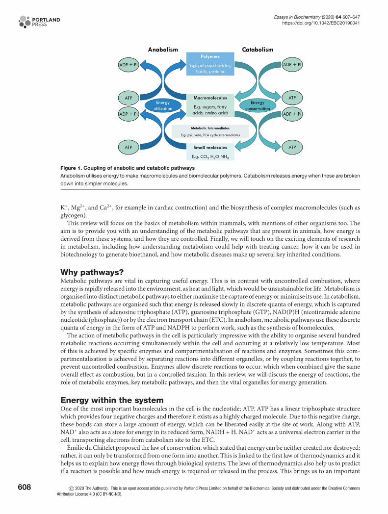
Whilst it can be daunting to think about every metabolic pathway that is occurring, we can break itdown and understand its smaller aspects. Knoop’s words underpin the true meaning of metabolism andone of its central roles in biochemistry and physiological chemistry. Metabolism is derived from the Greekword, metabole meaning ‘to change’ and comprises the total of all chemical reactions that take place inthe cell that are essential for life. These chemical reactions comprise both the synthesis and degradation ofcomplex macromolecules and can be divided into either catabolism or anabolism (Figure 1 – catabolismvs anabolism). Catabolism is the degradation of complex macromolecules into simpler molecules suchas carbon dioxide, water, and ammonia. Anabolism is the biosynthetic pathways that generate complexmacromolecules such as nucleic acids, proteins, polysaccharides, and lipids.
To maintain cellular and whole-body function, living organisms require energy continuously. Energy isrequired for mechanical work (contraction and cellular movement), active transport of ions/substrates (i.e.
© 2020 The Author(s). This is an open access article published by Portland Press Limited on behalf of the Biochemical Society and distributed under the Creative Commons AttributionLicense 4.0 (CC BY-NC-ND).
607
Essays in Biochemistry (2020) 64 607–647https://doi.org/10.1042/EBC20190041
Figure 1. Coupling of anabolic and catabolic pathways
Anabolism utilises energy to make macromolecules and biomolecular polymers. Catabolism releases energy when these are broken
down into simpler molecules.
K+, Mg2+, and Ca2+, for example in cardiac contraction) and the biosynthesis of complex macromolecules (such asglycogen).
This review will focus on the basics of metabolism within mammals, with mentions of other organisms too. Theaim is to provide you with an understanding of the metabolic pathways that are present in animals, how energy isderived from these systems, and how they are controlled. Finally, we will touch on the exciting elements of researchin metabolism, including how understanding metabolism could help with treating cancer, how it can be used inbiotechnology to generate bioethanol, and how metabolic diseases make up several key inherited conditions.
Why pathways?Metabolic pathways are vital in capturing useful energy. This is in contrast with uncontrolled combustion, whereenergy is rapidly released into the environment, as heat and light, which would be unsustainable for life. Metabolism isorganised into distinct metabolic pathways to either maximise the capture of energy or minimise its use. In catabolism,metabolic pathways are organised such that energy is released slowly in discrete quanta of energy, which is capturedby the synthesis of adenosine triphosphate (ATP), guanosine triphosphate (GTP), NAD(P)H (nicotinamide adeninenucleotide (phosphate)) or by the electron transport chain (ETC). In anabolism, metabolic pathways use these discretequanta of energy in the form of ATP and NADPH to perform work, such as the synthesis of biomolecules.
The action of metabolic pathways in the cell is particularly impressive with the ability to organise several hundredmetabolic reactions occurring simultaneously within the cell and occurring at a relatively low temperature. Mostof this is achieved by specific enzymes and compartmentalisation of reactions and enzymes. Sometimes this com-partmentalisation is achieved by separating reactions into different organelles, or by coupling reactions together, toprevent uncontrolled combustion. Enzymes allow discrete reactions to occur, which when combined give the sameoverall effect as combustion, but in a controlled fashion. In this review, we will discuss the energy of reactions, therole of metabolic enzymes, key metabolic pathways, and then the vital organelles for energy generation.
Energy within the systemOne of the most important biomolecules in the cell is the nucleotide; ATP. ATP has a linear triphosphate structurewhich provides four negative charges and therefore it exists as a highly charged molecule. Due to this negative charge,these bonds can store a large amount of energy, which can be liberated easily at the site of work. Along with ATP,NAD+ also acts as a store for energy in its reduced form, NADH + H. NAD+ acts as a universal electron carrier in thecell, transporting electrons from catabolism site to the ETC.
Emilie du Chatelet proposed the law of conservation, which stated that energy can be neither created nor destroyed;rather, it can only be transformed from one form into another. This is linked to the first law of thermodynamics and ithelps us to explain how energy flows through biological systems. The laws of thermodynamics also help us to predictif a reaction is possible and how much energy is required or released in the process. This brings us to an important
608 © 2020 The Author(s). This is an open access article published by Portland Press Limited on behalf of the Biochemical Society and distributed under the Creative CommonsAttribution License 4.0 (CC BY-NC-ND).
Essays in Biochemistry (2020) 64 607–647https://doi.org/10.1042/EBC20190041
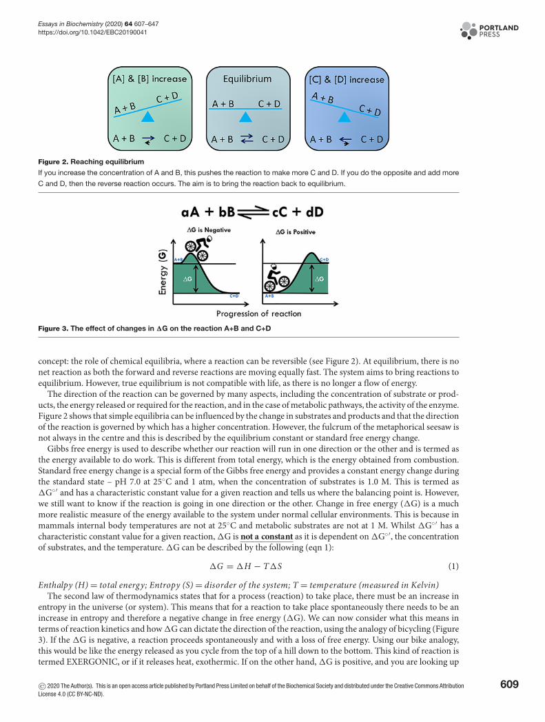
Figure 2. Reaching equilibrium
If you increase the concentration of A and B, this pushes the reaction to make more C and D. If you do the opposite and add more
C and D, then the reverse reaction occurs. The aim is to bring the reaction back to equilibrium.
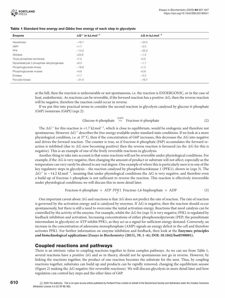
Figure 3. The effect of changes in �G on the reaction A+B and C+D
concept: the role of chemical equilibria, where a reaction can be reversible (see Figure 2). At equilibrium, there is nonet reaction as both the forward and reverse reactions are moving equally fast. The system aims to bring reactions toequilibrium. However, true equilibrium is not compatible with life, as there is no longer a flow of energy.
The direction of the reaction can be governed by many aspects, including the concentration of substrate or prod-ucts, the energy released or required for the reaction, and in the case of metabolic pathways, the activity of the enzyme.Figure 2 shows that simple equilibria can be influenced by the change in substrates and products and that the directionof the reaction is governed by which has a higher concentration. However, the fulcrum of the metaphorical seesaw isnot always in the centre and this is described by the equilibrium constant or standard free energy change.
Gibbs free energy is used to describe whether our reaction will run in one direction or the other and is termed asthe energy available to do work. This is different from total energy, which is the energy obtained from combustion.Standard free energy change is a special form of the Gibbs free energy and provides a constant energy change duringthe standard state – pH 7.0 at 25◦C and 1 atm, when the concentration of substrates is 1.0 M. This is termed as�G◦′ and has a characteristic constant value for a given reaction and tells us where the balancing point is. However,we still want to know if the reaction is going in one direction or the other. Change in free energy (�G) is a muchmore realistic measure of the energy available to the system under normal cellular environments. This is because inmammals internal body temperatures are not at 25◦C and metabolic substrates are not at 1 M. Whilst �G◦′ has acharacteristic constant value for a given reaction, �G is not a constant as it is dependent on �G◦′, the concentrationof substrates, and the temperature. �G can be described by the following (eqn 1):
�G = �H − T�S (1)
Enthalpy (H) = total energy; Entropy (S) = disorder of the system; T = temperature (measured in Kelvin)The second law of thermodynamics states that for a process (reaction) to take place, there must be an increase in
entropy in the universe (or system). This means that for a reaction to take place spontaneously there needs to be anincrease in entropy and therefore a negative change in free energy (�G). We can now consider what this means interms of reaction kinetics and how �G can dictate the direction of the reaction, using the analogy of bicycling (Figure3). If the �G is negative, a reaction proceeds spontaneously and with a loss of free energy. Using our bike analogy,this would be like the energy released as you cycle from the top of a hill down to the bottom. This kind of reaction istermed EXERGONIC, or if it releases heat, exothermic. If on the other hand, �G is positive, and you are looking up
© 2020 The Author(s). This is an open access article published by Portland Press Limited on behalf of the Biochemical Society and distributed under the Creative Commons AttributionLicense 4.0 (CC BY-NC-ND).
609
Essays in Biochemistry (2020) 64 607–647https://doi.org/10.1042/EBC20190041
Table 1 Standard free energy and Gibbs free energy of each step in glycolysis
Enzyme �G◦′ in kJ.mol−1 �G in kJ.mol−1
Hexokinase −16.7 −33.5
G6PI +1.7 −2.5
PFK −14.2 −22.2
Aldolase +23.8 −1.3
Triose phosphate isomerase +7.5 +2.5
Glyceraldehyde 3 phosphate dehydrogenase +6.3 −1.7
Phosphoglycerate kinase −18.8 +1.3
Phosphoglycerate mutase +4.6 +0.8
Enolase +1.7 −3.3
Pyruvate kinase −31.4 −16.7
at the hill, then the reaction is unfavourable or not spontaneous, i.e. the reaction is ENDERGONIC, or in the case ofheat, endothermic. As reactions can be reversible, if the forward reaction has a positive �G, then the reverse reactionwill be negative, therefore the reaction could occur in reverse.
If we put this into practical terms to consider the second reaction in glycolysis catalysed by glucose-6-phosphate(G6P) isomerase (G6PI) (eqn 2):
Glucose-6-phosphateG6PI� Fructose-6-phosphate (2)
The �G◦′ for this reaction is +1.7 kJ.mol−1, which is close to equilibrium, would be endogenic and therefore notspontaneous. However �G◦′ describes the free energy available under standard state conditions. If we look at a morephysiological condition, i.e. at 37◦C, then if the concentration of G6P increases, this decreases the �G into negativeand drives the forward reaction. The counter is true, so if fructose-6-phosphate (F6P) accumulates the forward re-action is inhibited (due to �G now becoming positive) then the reverse reaction is favoured (as the �G for this isnegative). This is an example of one of the freely reversible reactions in glycolysis.
Another thing to take into account is that some reactions will not be reversible under physiological conditions. Forexample, if the �G is very negative, then changing the amount of product or substrate will not affect, especially as thetemperature can very rarely be altered to any real degree. One example of where this is particularly seen is in one of thekey regulatory steps in glycolysis – the reaction catalysed by phosphofructokinase 1 (PFK1), shown in (eqn 3). The�G◦′ is −14.2 kJ.mol−1, meaning that under physiological conditions the �G is very negative, and therefore evena build-up of fructose-1-phosphate is not sufficient to reverse the reaction. This reaction is effectively irreversibleunder physiological conditions; we will discuss this in more detail later.
Fructose-6-phosphate + ATP PFK1→ Fructose-1,6-bisphosphate + ADP (3)
One important caveat about �G and reactions is that �G does not predict the rate of reaction. The rate of reactionis governed by the activation energy and is catalysed by enzymes. If �G is negative, then the reaction should occurspontaneously, but there is still a need to overcome the initial activation energy. Reactions that need catalysis can becontrolled by the activity of the enzyme. For example, whilst the �G for (eqn 3) is very negative, PFK1 is regulated byfeedback inhibition and activation. Increasing concentrations of either phosphoenolpyruvate (PEP; the penultimateintermediate in glycolysis) or ATP inhibit PFK1, as they act as a signal for sufficient energy demand. Conversely, anincrease in the concentration of adenosine monophosphate (AMP) signals an energy deficit in the cell and thereforeactivates PFK1. For further information on enzyme inhibition and feedback, then look at the Enzymes: principlesand biotechnological applications (Essays in Biochemistry (2015), 59, 1–41; DOI: 10.1042/bse0590001).
Coupled reactions and pathwaysThere is an intrinsic value to coupling reactions together to form complex pathways. As we can see from Table 1,several reactions have a positive �G and so in theory, should not be spontaneous nor go in reverse. However, bylinking the reactions together, the product of one reaction becomes the substrate for the next. Thus, by couplingreactions together, substrates can build up and products can be rapidly removed, changing the equilibrium seesaw(Figure 2) making the �G negative (for reversible reactions). We will discuss glycolysis in more detail later and howregulation can control key steps and the other fates of G6P.
610 © 2020 The Author(s). This is an open access article published by Portland Press Limited on behalf of the Biochemical Society and distributed under the Creative CommonsAttribution License 4.0 (CC BY-NC-ND).
Essays in Biochemistry (2020) 64 607–647https://doi.org/10.1042/EBC20190041
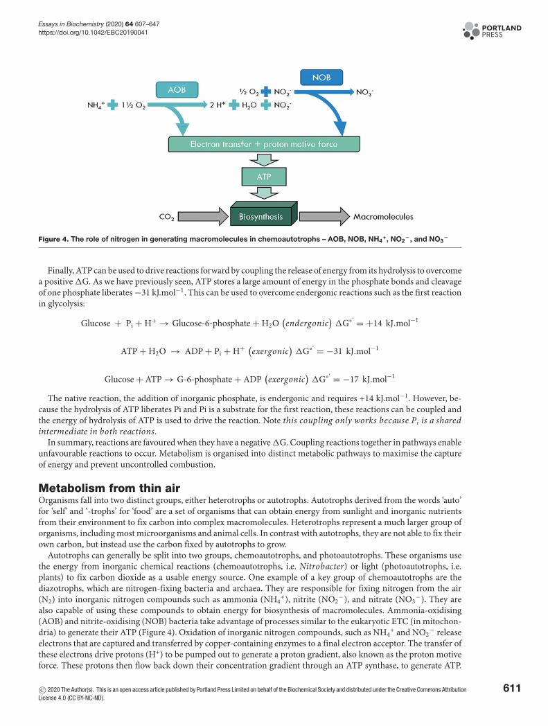
Figure 4. The role of nitrogen in generating macromolecules in chemoautotrophs – AOB, NOB, NH4+, NO2
−, and NO3−
Finally, ATP can be used to drive reactions forward by coupling the release of energy from its hydrolysis to overcomea positive �G. As we have previously seen, ATP stores a large amount of energy in the phosphate bonds and cleavageof one phosphate liberates −31 kJ.mol−1. This can be used to overcome endergonic reactions such as the first reactionin glycolysis:
Glucose + Pi + H+ → Glucose-6-phosphate + H2O(endergonic
)�G°’ = +14 kJ.mol−1
ATP + H2O → ADP + Pi + H+ (exergonic
)�G°’ = −31 kJ.mol−1
Glucose + ATP → G-6-phosphate + ADP(exergonic
)�G°’ = −17 kJ.mol−1
The native reaction, the addition of inorganic phosphate, is endergonic and requires +14 kJ.mol−1. However, be-cause the hydrolysis of ATP liberates Pi and Pi is a substrate for the first reaction, these reactions can be coupled andthe energy of hydrolysis of ATP is used to drive the reaction. Note this coupling only works because Pi is a sharedintermediate in both reactions.
In summary, reactions are favoured when they have a negative �G. Coupling reactions together in pathways enableunfavourable reactions to occur. Metabolism is organised into distinct metabolic pathways to maximise the captureof energy and prevent uncontrolled combustion.
Metabolism from thin airOrganisms fall into two distinct groups, either heterotrophs or autotrophs. Autotrophs derived from the words ‘auto’for ‘self’ and ‘-trophs’ for ‘food’ are a set of organisms that can obtain energy from sunlight and inorganic nutrientsfrom their environment to fix carbon into complex macromolecules. Heterotrophs represent a much larger group oforganisms, including most microorganisms and animal cells. In contrast with autotrophs, they are not able to fix theirown carbon, but instead use the carbon fixed by autotrophs to grow.
Autotrophs can generally be split into two groups, chemoautotrophs, and photoautotrophs. These organisms usethe energy from inorganic chemical reactions (chemoautotrophs, i.e. Nitrobacter) or light (photoautotrophs, i.e.plants) to fix carbon dioxide as a usable energy source. One example of a key group of chemoautotrophs are thediazotrophs, which are nitrogen-fixing bacteria and archaea. They are responsible for fixing nitrogen from the air(N2) into inorganic nitrogen compounds such as ammonia (NH4
+), nitrite (NO2−), and nitrate (NO3
−). They arealso capable of using these compounds to obtain energy for biosynthesis of macromolecules. Ammonia-oxidising(AOB) and nitrite-oxidising (NOB) bacteria take advantage of processes similar to the eukaryotic ETC (in mitochon-dria) to generate their ATP (Figure 4). Oxidation of inorganic nitrogen compounds, such as NH4
+ and NO2− release
electrons that are captured and transferred by copper-containing enzymes to a final electron acceptor. The transfer ofthese electrons drive protons (H+) to be pumped out to generate a proton gradient, also known as the proton motiveforce. These protons then flow back down their concentration gradient through an ATP synthase, to generate ATP.
© 2020 The Author(s). This is an open access article published by Portland Press Limited on behalf of the Biochemical Society and distributed under the Creative Commons AttributionLicense 4.0 (CC BY-NC-ND).
611
Essays in Biochemistry (2020) 64 607–647https://doi.org/10.1042/EBC20190041
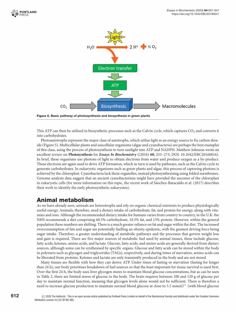
Figure 5. Basic pathway of photosynthesis and biosynthesis in green plants
This ATP can then be utilised in biosynthetic processes such as the Calvin cycle, which captures CO2 and converts itinto carbohydrates.
Photoautotrophs represent the major class of autotrophs, which utilise light as an energy source to fix carbon diox-ide (Figure 5). Multicellular plants and unicellular organisms (algae and cyanobacteria) are perhaps the best examplesof this class, using the process of photosynthesis to turn sunlight into ATP and NADPH. Matthew Johnson wrote anexcellent review on Photosynthesis for Essays In Biochemistry ((2016) 60, 255–273; DOI: 10.1042/EBC20160016).In brief, these organisms use photons of light to obtain electrons from water and produce oxygen as a by-product.These electrons are again used to drive ATP formation, which in turn is used by pathways, such as the Calvin cycle togenerate carbohydrates. In eukaryotic organisms such as green plants and algae, this process of capturing photons isachieved by the chloroplast. Cyanobacteria lack these organelles, instead photosynthesising using folded membranes.Genome analysis data suggest that an ancient cyanobacterium might have provided the ancestor of the chloroplastin eukaryotic cells (for more information on this topic, the recent work of Sanchez-Baracaldo et al. (2017) describestheir work to identify the early photosynthetic eukaryotes).
Animal metabolismAs we have already seen, animals are heterotrophs and rely on organic chemical nutrients to produce physiologicallyuseful energy. Animals, therefore, need a dietary intake of carbohydrate, fat, and protein for energy, along with vita-mins and ions. Although the recommended dietary intake for humans varies from country to country, in the U.K. theNHS recommends a diet comprising 68.5% carbohydrate, 18.5% fat, and 13% protein. However, within the generalpopulation these numbers are shifting. There is a much greater reliance on fat and sugar within the diet. The increasedoverconsumption of fats and sugar are potentially fuelling an obesity epidemic, with the greatest driving force beingsugar intake. Therefore, a greater understanding of metabolic pathways and the processes that govern weight lossand gain is required. There are five major sources of metabolic fuel used by animal tissues, these include glucose,fatty acids, ketones, amino acids, and lactate. Glucose, fatty acids, and amino acids are generally derived from dietarysources, although some can be synthesised by specific organs. Glucose and fatty acids can be stored within the bodyin polymers such as glycogen and triglycerides (TAGs), respectively, and during times of starvation, amino acids canbe liberated from proteins. Ketones and lactate are only transiently produced in the body and are not stored.
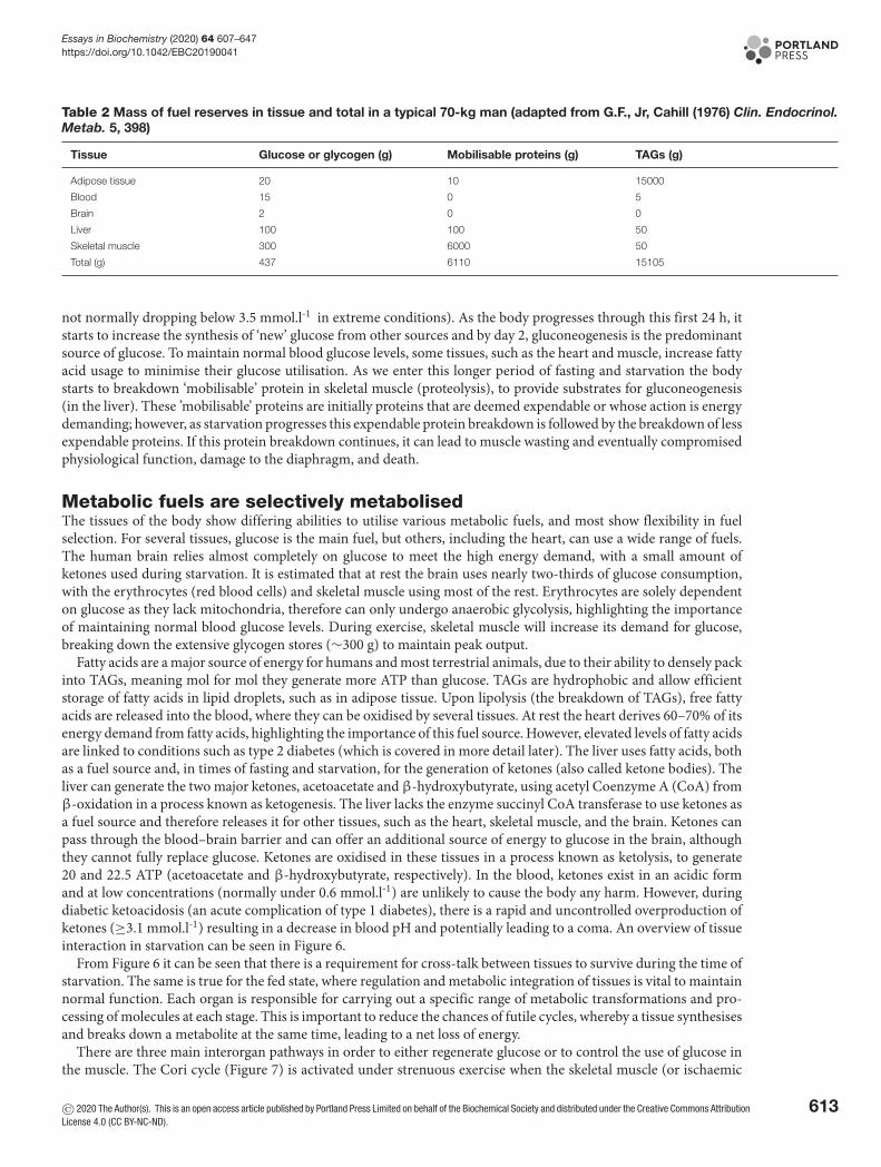
Many tissues are flexible with how they can derive ATP. Under times of fasting or starvation (fasting for longerthan 24 h), our body prioritises breakdown of fuel sources so that the least important for tissue survival is used first.Over the first 24 h, the body uses liver glycogen stores to maintain blood glucose concentrations, but as can be seenin Table 2, there are limited stores of glucose in the body. The brain requires between 100 and 120 g of glucose perday to maintain normal function, meaning that glycogen levels alone would not be sufficient. There is therefore aneed to increase glucose production to maintain normal blood glucose at close to 5.5 mmol.l-1 (with blood glucose
612 © 2020 The Author(s). This is an open access article published by Portland Press Limited on behalf of the Biochemical Society and distributed under the Creative CommonsAttribution License 4.0 (CC BY-NC-ND).
Essays in Biochemistry (2020) 64 607–647https://doi.org/10.1042/EBC20190041
Table 2 Mass of fuel reserves in tissue and total in a typical 70-kg man (adapted from G.F., Jr, Cahill (1976) Clin. Endocrinol.Metab. 5, 398)
Tissue Glucose or glycogen (g) Mobilisable proteins (g) TAGs (g)
Adipose tissue 20 10 15000
Blood 15 0 5
Brain 2 0 0
Liver 100 100 50
Skeletal muscle 300 6000 50
Total (g) 437 6110 15105
not normally dropping below 3.5 mmol.l-1 in extreme conditions). As the body progresses through this first 24 h, itstarts to increase the synthesis of ‘new’ glucose from other sources and by day 2, gluconeogenesis is the predominantsource of glucose. To maintain normal blood glucose levels, some tissues, such as the heart and muscle, increase fattyacid usage to minimise their glucose utilisation. As we enter this longer period of fasting and starvation the bodystarts to breakdown ‘mobilisable’ protein in skeletal muscle (proteolysis), to provide substrates for gluconeogenesis(in the liver). These ’mobilisable’ proteins are initially proteins that are deemed expendable or whose action is energydemanding; however, as starvation progresses this expendable protein breakdown is followed by the breakdown of lessexpendable proteins. If this protein breakdown continues, it can lead to muscle wasting and eventually compromisedphysiological function, damage to the diaphragm, and death.
Metabolic fuels are selectively metabolisedThe tissues of the body show differing abilities to utilise various metabolic fuels, and most show flexibility in fuelselection. For several tissues, glucose is the main fuel, but others, including the heart, can use a wide range of fuels.The human brain relies almost completely on glucose to meet the high energy demand, with a small amount ofketones used during starvation. It is estimated that at rest the brain uses nearly two-thirds of glucose consumption,with the erythrocytes (red blood cells) and skeletal muscle using most of the rest. Erythrocytes are solely dependenton glucose as they lack mitochondria, therefore can only undergo anaerobic glycolysis, highlighting the importanceof maintaining normal blood glucose levels. During exercise, skeletal muscle will increase its demand for glucose,breaking down the extensive glycogen stores (∼300 g) to maintain peak output.
Fatty acids are a major source of energy for humans and most terrestrial animals, due to their ability to densely packinto TAGs, meaning mol for mol they generate more ATP than glucose. TAGs are hydrophobic and allow efficientstorage of fatty acids in lipid droplets, such as in adipose tissue. Upon lipolysis (the breakdown of TAGs), free fattyacids are released into the blood, where they can be oxidised by several tissues. At rest the heart derives 60–70% of itsenergy demand from fatty acids, highlighting the importance of this fuel source. However, elevated levels of fatty acidsare linked to conditions such as type 2 diabetes (which is covered in more detail later). The liver uses fatty acids, bothas a fuel source and, in times of fasting and starvation, for the generation of ketones (also called ketone bodies). Theliver can generate the two major ketones, acetoacetate and β-hydroxybutyrate, using acetyl Coenzyme A (CoA) fromβ-oxidation in a process known as ketogenesis. The liver lacks the enzyme succinyl CoA transferase to use ketones asa fuel source and therefore releases it for other tissues, such as the heart, skeletal muscle, and the brain. Ketones canpass through the blood–brain barrier and can offer an additional source of energy to glucose in the brain, althoughthey cannot fully replace glucose. Ketones are oxidised in these tissues in a process known as ketolysis, to generate20 and 22.5 ATP (acetoacetate and β-hydroxybutyrate, respectively). In the blood, ketones exist in an acidic formand at low concentrations (normally under 0.6 mmol.l-1) are unlikely to cause the body any harm. However, duringdiabetic ketoacidosis (an acute complication of type 1 diabetes), there is a rapid and uncontrolled overproduction ofketones (≥3.1 mmol.l-1) resulting in a decrease in blood pH and potentially leading to a coma. An overview of tissueinteraction in starvation can be seen in Figure 6.
From Figure 6 it can be seen that there is a requirement for cross-talk between tissues to survive during the time ofstarvation. The same is true for the fed state, where regulation and metabolic integration of tissues is vital to maintainnormal function. Each organ is responsible for carrying out a specific range of metabolic transformations and pro-cessing of molecules at each stage. This is important to reduce the chances of futile cycles, whereby a tissue synthesisesand breaks down a metabolite at the same time, leading to a net loss of energy.
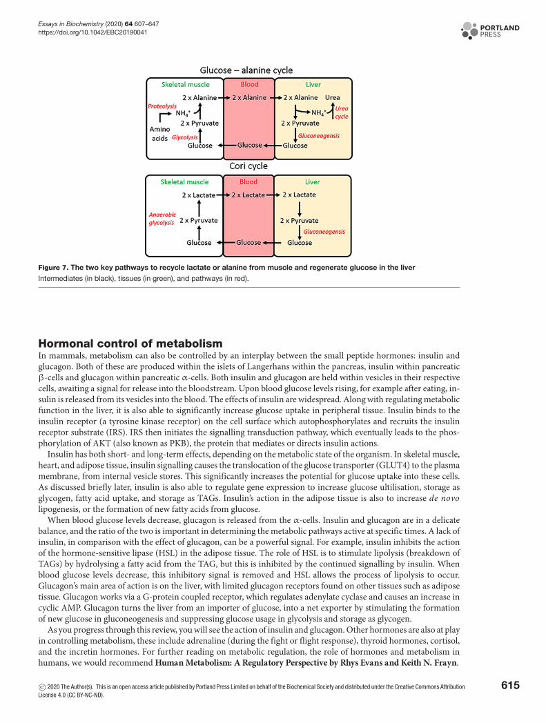
There are three main interorgan pathways in order to either regenerate glucose or to control the use of glucose inthe muscle. The Cori cycle (Figure 7) is activated under strenuous exercise when the skeletal muscle (or ischaemic
© 2020 The Author(s). This is an open access article published by Portland Press Limited on behalf of the Biochemical Society and distributed under the Creative Commons AttributionLicense 4.0 (CC BY-NC-ND).
613

Essays in Biochemistry (2020) 64 607–647https://doi.org/10.1042/EBC20190041
Figure 6. Summary of metabolic pathways active during starvation
During starvation, there is an increase in fatty acid utilisation in the muscle (not shown here for simplicity) and a breakdown of
proteins into amino acids. Intermediates (in black), tissues (in green), and pathways (in red).
heart) are contracting using anaerobic glycolysis, which leads to an accumulation of lactate. Lactate is transportedto the liver where it regenerates glucose (gluconeogenesis), which can then be used by the exercising muscle again.As you will see later, whilst the use of anaerobic glycolysis generates far less ATP than the oxidation of glucose, thisprocess does not require oxygen, which can be limited in strenuous exercise.
During times of starvation, the glucose-alanine cycle can regenerate glucose and remove excess nitrogen formedin the breakdown of amino acids (Figure 7). During proteolysis, amino acids that are liberated can provide carbonskeletons to top up different pathways, but must dispose of the amino group. This amino group is transferred topyruvate by alanine aminotransferase to form alanine. Alanine is the predominant amino acid released by the muscle.In the cycle, glucose taken up by the muscle is used to generate the pyruvate, thereby aiding in proteolysis, without anet loss of glucose. The alanine is released by the muscle and taken up by the liver, where it is converted into pyruvate,and back into glucose to start the cycle again. Finally, the amino group liberated by the conversion of alanine backinto pyruvate enters the urea cycle for disposal.
614 © 2020 The Author(s). This is an open access article published by Portland Press Limited on behalf of the Biochemical Society and distributed under the Creative CommonsAttribution License 4.0 (CC BY-NC-ND).
Essays in Biochemistry (2020) 64 607–647https://doi.org/10.1042/EBC20190041
Figure 7. The two key pathways to recycle lactate or alanine from muscle and regenerate glucose in the liver
Intermediates (in black), tissues (in green), and pathways (in red).
Hormonal control of metabolismIn mammals, metabolism can also be controlled by an interplay between the small peptide hormones: insulin andglucagon. Both of these are produced within the islets of Langerhans within the pancreas, insulin within pancreaticβ-cells and glucagon within pancreatic α-cells. Both insulin and glucagon are held within vesicles in their respectivecells, awaiting a signal for release into the bloodstream. Upon blood glucose levels rising, for example after eating, in-sulin is released from its vesicles into the blood. The effects of insulin are widespread. Along with regulating metabolicfunction in the liver, it is also able to significantly increase glucose uptake in peripheral tissue. Insulin binds to theinsulin receptor (a tyrosine kinase receptor) on the cell surface which autophosphorylates and recruits the insulinreceptor substrate (IRS). IRS then initiates the signalling transduction pathway, which eventually leads to the phos-phorylation of AKT (also known as PKB), the protein that mediates or directs insulin actions.
Insulin has both short- and long-term effects, depending on the metabolic state of the organism. In skeletal muscle,heart, and adipose tissue, insulin signalling causes the translocation of the glucose transporter (GLUT4) to the plasmamembrane, from internal vesicle stores. This significantly increases the potential for glucose uptake into these cells.As discussed briefly later, insulin is also able to regulate gene expression to increase glucose ultilisation, storage asglycogen, fatty acid uptake, and storage as TAGs. Insulin’s action in the adipose tissue is also to increase de novolipogenesis, or the formation of new fatty acids from glucose.
When blood glucose levels decrease, glucagon is released from the α-cells. Insulin and glucagon are in a delicatebalance, and the ratio of the two is important in determining the metabolic pathways active at specific times. A lack ofinsulin, in comparison with the effect of glucagon, can be a powerful signal. For example, insulin inhibits the actionof the hormone-sensitive lipase (HSL) in the adipose tissue. The role of HSL is to stimulate lipolysis (breakdown ofTAGs) by hydrolysing a fatty acid from the TAG, but this is inhibited by the continued signalling by insulin. Whenblood glucose levels decrease, this inhibitory signal is removed and HSL allows the process of lipolysis to occur.Glucagon’s main area of action is on the liver, with limited glucagon receptors found on other tissues such as adiposetissue. Glucagon works via a G-protein coupled receptor, which regulates adenylate cyclase and causes an increase incyclic AMP. Glucagon turns the liver from an importer of glucose, into a net exporter by stimulating the formationof new glucose in gluconeogenesis and suppressing glucose usage in glycolysis and storage as glycogen.
As you progress through this review, you will see the action of insulin and glucagon. Other hormones are also at playin controlling metabolism, these include adrenaline (during the fight or flight response), thyroid hormones, cortisol,and the incretin hormones. For further reading on metabolic regulation, the role of hormones and metabolism inhumans, we would recommend Human Metabolism: A Regulatory Perspective by Rhys Evans and Keith N. Frayn.
© 2020 The Author(s). This is an open access article published by Portland Press Limited on behalf of the Biochemical Society and distributed under the Creative Commons AttributionLicense 4.0 (CC BY-NC-ND).
615
Essays in Biochemistry (2020) 64 607–647https://doi.org/10.1042/EBC20190041
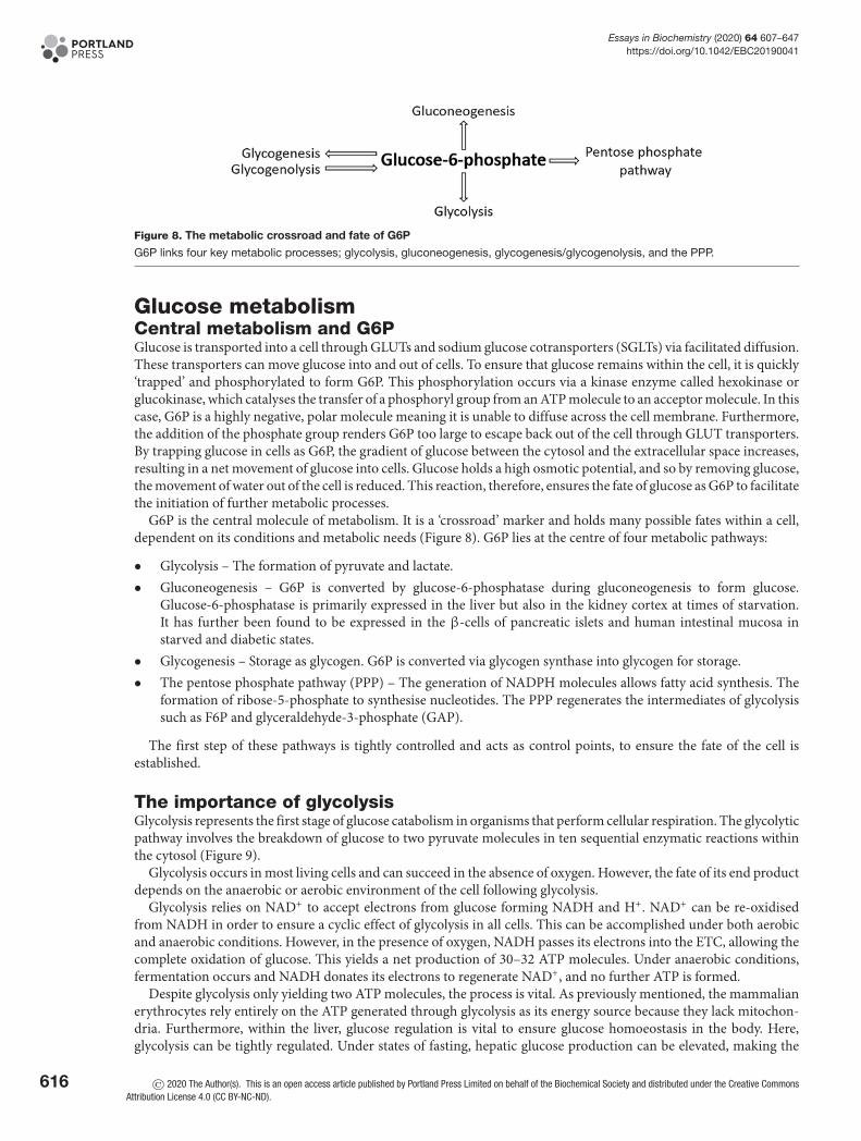
Figure 8. The metabolic crossroad and fate of G6P
G6P links four key metabolic processes; glycolysis, gluconeogenesis, glycogenesis/glycogenolysis, and the PPP.
Glucose metabolismCentral metabolism and G6PGlucose is transported into a cell through GLUTs and sodium glucose cotransporters (SGLTs) via facilitated diffusion.These transporters can move glucose into and out of cells. To ensure that glucose remains within the cell, it is quickly‘trapped’ and phosphorylated to form G6P. This phosphorylation occurs via a kinase enzyme called hexokinase orglucokinase, which catalyses the transfer of a phosphoryl group from an ATP molecule to an acceptor molecule. In thiscase, G6P is a highly negative, polar molecule meaning it is unable to diffuse across the cell membrane. Furthermore,the addition of the phosphate group renders G6P too large to escape back out of the cell through GLUT transporters.By trapping glucose in cells as G6P, the gradient of glucose between the cytosol and the extracellular space increases,resulting in a net movement of glucose into cells. Glucose holds a high osmotic potential, and so by removing glucose,the movement of water out of the cell is reduced. This reaction, therefore, ensures the fate of glucose as G6P to facilitatethe initiation of further metabolic processes.
G6P is the central molecule of metabolism. It is a ‘crossroad’ marker and holds many possible fates within a cell,dependent on its conditions and metabolic needs (Figure 8). G6P lies at the centre of four metabolic pathways:
• Glycolysis – The formation of pyruvate and lactate.• Gluconeogenesis – G6P is converted by glucose-6-phosphatase during gluconeogenesis to form glucose.
Glucose-6-phosphatase is primarily expressed in the liver but also in the kidney cortex at times of starvation.It has further been found to be expressed in the β-cells of pancreatic islets and human intestinal mucosa instarved and diabetic states.
• Glycogenesis – Storage as glycogen. G6P is converted via glycogen synthase into glycogen for storage.• The pentose phosphate pathway (PPP) – The generation of NADPH molecules allows fatty acid synthesis. The
formation of ribose-5-phosphate to synthesise nucleotides. The PPP regenerates the intermediates of glycolysissuch as F6P and glyceraldehyde-3-phosphate (GAP).
The first step of these pathways is tightly controlled and acts as control points, to ensure the fate of the cell isestablished.
The importance of glycolysisGlycolysis represents the first stage of glucose catabolism in organisms that perform cellular respiration. The glycolyticpathway involves the breakdown of glucose to two pyruvate molecules in ten sequential enzymatic reactions withinthe cytosol (Figure 9).
Glycolysis occurs in most living cells and can succeed in the absence of oxygen. However, the fate of its end productdepends on the anaerobic or aerobic environment of the cell following glycolysis.
Glycolysis relies on NAD+ to accept electrons from glucose forming NADH and H+. NAD+ can be re-oxidisedfrom NADH in order to ensure a cyclic effect of glycolysis in all cells. This can be accomplished under both aerobicand anaerobic conditions. However, in the presence of oxygen, NADH passes its electrons into the ETC, allowing thecomplete oxidation of glucose. This yields a net production of 30–32 ATP molecules. Under anaerobic conditions,fermentation occurs and NADH donates its electrons to regenerate NAD+, and no further ATP is formed.
Despite glycolysis only yielding two ATP molecules, the process is vital. As previously mentioned, the mammalianerythrocytes rely entirely on the ATP generated through glycolysis as its energy source because they lack mitochon-dria. Furthermore, within the liver, glucose regulation is vital to ensure glucose homoeostasis in the body. Here,glycolysis can be tightly regulated. Under states of fasting, hepatic glucose production can be elevated, making the
616 © 2020 The Author(s). This is an open access article published by Portland Press Limited on behalf of the Biochemical Society and distributed under the Creative CommonsAttribution License 4.0 (CC BY-NC-ND).

Essays in Biochemistry (2020) 64 607–647https://doi.org/10.1042/EBC20190041
Figure 9. The pathway is split into an initial ‘investment’ phase, where ATP is used and then the ‘payout’ phase, where ATP
is regenerated
Intermediates (in black), by-products (in green), and enzymes (in red).
liver the main source of glucose production at this time. Here, pyruvate can also be used to form precursors for thesynthesis of fats, cholesterol, bile, and plasma proteins. For microorganisms, the glycolytic pathway ensures a sourceof energy for respiration and bacterial photosynthesis, along with necessary biosynthetic precursors.
The glycolytic pathwayThe enzymatic process of glycolysis occurs within the cytosol and can be divided into two definitive stages of energyinvestment and energy recovery (Figure 9).
Stage I energy investment phaseThe first reaction of glycolysis is catalysed by hexokinase (or glucokinase in the liver and pancreas), involving thetransfer of a phosphoryl group from ATP to glucose, forming G6P. G6P is isomerised to F6P by G6PI and is thenfurther phosphorylated by PFK, to form fructose-1,6-bisphosphate (FBP). This phosphorylation step is irreversibleand utilises the second ATP molecule in glycolysis. Aldolase catalyses the cleavage of FBP (6-carbon molecule) toform two 3-carbon molecules GAP and dihydroxyacetone phosphate (DHAP). Another isomerase, triose phosphateisomerase (TIM), catalyses the interconversion between DHAP and GAP, allowing DHAP to convert into GAP, andproceed along the glycolytic pathway. As a result, for every glucose molecule, two molecules of GAP are produced.Therefore, from this stage onwards, all intermediates and by-products are doubled in production.
© 2020 The Author(s). This is an open access article published by Portland Press Limited on behalf of the Biochemical Society and distributed under the Creative Commons AttributionLicense 4.0 (CC BY-NC-ND).
617
Essays in Biochemistry (2020) 64 607–647https://doi.org/10.1042/EBC20190041
Stage II energy payoutGAP is oxidised and phosphorylated by NAD+ and Pi to form 1,3-bisphosphoglycerate (1,3-BPG), catalysed by GAPdehydrogenase (GAPDH). Intermediates NADH and H+ are produced alongside 1,3-BPG. The conversion of 1,3-BPGinto 3-phosphoglycerate (3PG) is catalysed by phosphoglycerate kinase (PGK) and signifies the first step in glycolysisto generate ATP molecules through phosphorylation of adenosine diphosphate (ADP). 3PG can be interconvertedby phosphoglycerate mutase (PGM), to form 2-phosphoglycerate (2PG) that is dehydrated to PEP by enolase. Thefinal reaction of glycolysis generates the final ATP molecule alongside pyruvate in a cleavage reaction catalysed bypyruvate kinase (PK).
The initial energy investment in the form of two ATP molecules is doubly repaid in the later stage of glycolysisdue to the formation of two 3-carbon GAP molecules, which are each transformed to pyruvate and ATP. Therefore,generating four molecules of ATP, a net gain of 2 ATP.
Overall, glycolysis holds a negative �G value of −310 kJ.mol−1. The reaction is as follows:
Glucose + 2 NAD+ + 2 ADP + 2 Pi → 2 Pyruvate + 2 NADH + 2 ATP + 2 H2O + 4 H+
Regulation of glycolysisGlycolysis is regulated by three rate-limiting steps. These are slower, regulated stages and therefore determine theoverall rate of the pathway. Within the glycolytic pathway, these rate-limiting steps are coupled with the hydrolysis ofATP or the phosphorylation of ADP. This ensures these steps are energetically favourable, i.e. holding a very negative�G value and are therefore irreversible under physiological conditions.
Hexokinase/glucokinaseHexokinase and glucokinase are the first regulatory enzymes within the glycolytic pathway. Hexokinase exists inabundance within tissues in our body. It holds a low Km value, thus, ensuring its high affinity for glucose. Due to itslow Km it means that hexokinase is more useful in a state of hypoglycaemia, where glucose levels are low. Hexokinaseis feedback-inhibited by its own product, meaning a build-up of G6P can inhibit hexokinase and therefore, the phos-phorylation of glucose. Hexokinase does ensure the irreversible formation of G6P. In mammalian skeletal muscle,where the major source of energy is glycogen and not glucose, this step is ultimately overcome. Within pancreaticislets, hexokinase allows the control of insulin and glucagon release in the β- and α-cells, respectively.
Glucokinase is an isoenzyme of hexokinase that exists in the liver and pancreatic β-cells. Contrary to hexokinase,glucokinase holds a high Km value and therefore a high Vmax. This means that glucokinase exists with a low affinityfor glucose. As a result of this, glucokinase is utilised in a state of hyperglycaemia or a post-prandial state. Withinpancreatic β-cells, glucokinase acts as a sensor to control the rate of entry of glucose into the glycolytic pathway byphosphorylation. Within the liver, it ensures that glucose is synthesised into glycogen or fatty acids post-prandially,when glucose levels are high. Unlike hexokinase, glucokinase is not inhibited by high levels of G6P and can thereforeremain active to ensure glucose is stored as glycogen when glucose levels are high. The low affinity of glucokinase forglucose ensures that within a state of low glucose, peripheral tissue hexokinase can phosphorylate glucose to G6P forglycolysis and the liver and β-cells stop the uptake of glucose.
PFKPFK is another enzyme that acts as a key regulator of glycolysis. It has a highly negative �G value, thus ensuring thatthe reaction will still occur despite accumulation of FBP. PFK holds two conformational states that exist in equilib-rium, with, ATP acting as both an activator and inhibitor of both the states. When ATP levels are high (e.g. in theliver), ATP acts as an allosteric inhibitor of PFK, shifting its equilibrium and decreasing its affinity for F6P. However,where levels of ATP are low, PFK is activated, shifting its equilibrium and affinity for F6P to form FBP.
PKPK ensures the fate of PEP to form pyruvate in the last step of glycolysis. Pyruvate is an essential intermediate buildingblock for many further metabolic pathways such as fatty acid synthesis, the tricarboxylic acid (TCA) cycle or, underanaerobic conditions, converted into lactic acid or ethanol (in yeast). Therefore, PK is noted as the most importantregulator of glycolysis.
618 © 2020 The Author(s). This is an open access article published by Portland Press Limited on behalf of the Biochemical Society and distributed under the Creative CommonsAttribution License 4.0 (CC BY-NC-ND).
Essays in Biochemistry (2020) 64 607–647https://doi.org/10.1042/EBC20190041
GluconeogenesisThe importance of gluconeogenesisGluconeogenesis is an anabolic process whereby glucose is formed from non-carbohydrate carbon precursors in-cluding pyruvate. The gluconeogenic pathway largely occurs within the liver and kidneys to maintain blood glucoselevels following glycogen depletion, and in the renal cortex during starvation. Gluconeogenesis has also been foundto occur within the β-cells of the islets of Langerhans and intestinal mucosa in starved and diabetic states.
Gluconeogenesis aims to do the reverse of glycolysis; however, due to the presence of irreversible steps within theglycolytic pathway, gluconeogenesis is not simply a reversal of glycolysis. These irreversible steps are overcome ingluconeogenesis, using additional enzymes than those present in the glycolytic pathway. It is crucial that gluconeoge-nesis is not just the reverse of glycolysis. This is because the last step of glycolysis (Figure 9) involves the irreversibleand highly energetically favourable formation of pyruvate. To bypass this, gluconeogenesis is split into a two-stepprocess with specific steps occurring within the mitochondria and the cytosol. Within the mitochondria, pyruvate isconverted into oxaloacetate which, in turn, converts into malate to transport out of the mitochondria into the cytosol.Once here, it is immediately converted back into oxaloacetate and then to PEP by PEP carboxykinase (PEPCK). Thisnot only overcomes the irreversible step within glycolysis but also avoids the cell undergoing a futile cycle wherebypyruvate is immediately converted back into PEP. Furthermore, during steps 1 and 3 of glycolysis (Figure 9) ATP isinvested in order to phosphorylate the product formed. Therefore, if gluconeogenesis were the reverse of glycolysis,it would essentially mean that gluconeogenesis would need to regenerate ATP, a process that is not possible. Gluco-neogenesis is instead ATP-dependent and therefore requires additional enzymes to bypass steps 1 and 3, where ATPis not regenerated.
The importance of gluconeogenesis lies in the fact the brain and erythrocytes rely almost entirely on glucose as aform of energy and, therefore, it is essential that glucose ultimately depleted in glycolysis is restored by gluconeoge-nesis in a cyclic fashion.
Gluconeogenic pathwayThe formation of oxaloacetate from pyruvatePyruvate is carboxylated by pyruvate carboxylase (PC) to oxaloacetate at the expense of 1 ATP molecule. This reactionoccurs inside the mitochondria. PC is activated through increased concentration of acetyl CoA and inhibited in thepresence of glucose and ADP.
Pyruvate + C O2 + ATP PC→
Oxaloacetate + ADP + Pi
Oxaloacetate is reduced to malate in the presence of NADH, to be transported over the mitochondrial mem-brane and into the cytosol. Malate crosses the mitochondrial membranes via the malate-aspartate shuttle, where it isre-oxidised to oxaloacetate.
At the expense of one GTP molecule, oxaloacetate is decarboxylated and phosphorylated by PEPCK.
Oxaloacetate + GTP PEPCK→
Phosphoenolpyruvate + GDP + C O2
Formation of F6PA hydrolysis reaction occurs in a phosphate ester located at carbon 1 of fructose-1,6-bisphosphate, facilitated byfructose-1,6-bisphosphatase (F16BPase).
Fructose − 1, 6 − bisphosphate + H2 O F 16BPase→ Fructose − 6 − phosphate + Pi
G6P and free glucose formationF6P is readily converted into G6P by G6PI.
Fructose − 6 − phosphateG6PI� Glucose − 6 − phosphate
In many scenarios, G6P is utilised to generate glycogen, ending gluconeogenesis. Alternatively, it can be dephos-phorylated to form free glucose molecules.
The site for the formation of glucoseDuring the final step of gluconeogenesis, glucose is formed. This occurs within the lumen of the endoplasmic reticu-lum. The glucose formed is ultimately shuttled into the cytosol by GLUTs, which are readily available and located in
© 2020 The Author(s). This is an open access article published by Portland Press Limited on behalf of the Biochemical Society and distributed under the Creative Commons AttributionLicense 4.0 (CC BY-NC-ND).
619
Essays in Biochemistry (2020) 64 607–647https://doi.org/10.1042/EBC20190041
the endoplasmic reticulum.
Glucose − 6 − phosphate + H2O Glucose−6−phosphatase→
Glucose + Pi
Regulation of glucose metabolism by gene expressionOne of the actions of insulin is to increase glycolysis, whilst suppressing gluconeogenesis (in the liver). In re-sponse to increased insulin and glucose, the mammalian liver, muscle, and peripheral tissue increases the expres-sion of GLUT1–4, hexokinase/glucokinase and key glycolytic genes: GAP dehydrogenase, PK and the bifunctionalenzyme (which stimulates PFK activity). To suppress gluconeogenesis in the liver, insulin decreases the expressionof glucose-6-phosphatase, fructose-1,6-bisphosphatase, and EP carboxylase. This alteration in gene expression pat-tern, increases glucose utilised in the cells, and maintains glycolytic activity. In the mammalian liver, glucose canincrease the expression of PK via the transcription factor known as carbohydrate-responsive element binding protein(ChREBP). Interestingly, the action of glucagon on the liver suppresses this transcription factor, thereby reducing theexpression of PK.
GlycogenWhat is glycogen?Glycogen is a large, multibranched polysaccharide of glucose. It contains α-1,4-glycosidic bonds between adjacentglucose molecules and α-1,6-glycosidic bonds at branching points at every tenth residue within the chain. It is essen-tially the storage form of glucose in animals, fungi, and bacteria. It is also the storage molecule of glucose within thebody and can be broken down to yield glucose when energy is required. Glycogen is stored within muscle and liverin the body. Within the muscle, the breakdown of glycogen serves to supply energy to that muscle, whereas withinthe liver it is degraded to maintain blood glucose levels in the body. It is present within these sites as granules withinthe cytosol that are up to 40 nm in size.
The importance of glycogenGlycogen can be degraded to supply energy to the body. This is specifically important as cells within the brain relyalmost entirely on glucose for energy and therefore glucose released from liver cells can help supply this. Withinperiods of sudden activity, such as sprinting, the glucose obtained from glycogen degradation can produce enoughenergy when no oxygen is initially available.
Furthermore, the question ‘why cannot all excess fuels in the human body be stored as fatty acids?’ is usually probed.There are two main reasons as to why glycogen storage is beneficial over fatty acid storage. Firstly, glycogen is readilymobilised to glucose and therefore can be utilised quickly in situations where glucose is needed immediately. Secondly,the breakdown of glycogen is highly controlled. Therefore, the subsequent release of glucose is also controlled to helpraise or maintain blood glucose levels.
Synthesis and degradationThe synthesis of glycogen requires an activated form of glucose called uridine diphosphate glucose (UDP-glucose).This is formed by the addition of UTP to glucose-1-phosphate. UDP-glucose is added to the non-reducing end ofglycogen, expanding its size.
The degradation of glycogen requires the release of glucose-1-phosphate from glycogen and the remodelling ofglycogen substrates to warrant further degradation. Glucose-1-phosphate is then converted into G6P which has sev-eral fates within metabolism.
PPPThe importance of the PPP and its intermediatesThe PPP is an essential biochemical process that occurs within the cytosol of living organisms (Figure 10). Thispathway runs parallel to glycolysis in the cytosol, as it utilises some similar components of this pathway for its ownuse. It is known to have several important roles.
1. The production of nicotinamide adenine dinucleotide phosphate (NADPH). NADPH is a crucial reducing agentwhich is used in:a. Fatty acid synthesisb. Cholesterol biosynthesisc. Nucleotide synthesis
620 © 2020 The Author(s). This is an open access article published by Portland Press Limited on behalf of the Biochemical Society and distributed under the Creative CommonsAttribution License 4.0 (CC BY-NC-ND).

Essays in Biochemistry (2020) 64 607–647https://doi.org/10.1042/EBC20190041
Figure 10. PPP is split into the oxidative and non-oxidative phases
The oxidative phase represents the conversion of G6P into ribulose-5-phosphtase which generates NADPH molecules. The non-ox-
idative phase shows the generation of ribose-5-phosphate and also glycolysis pathway intermediates. Intermediates (in black),
by-products (in green), and enzymes (in red).
d. Neurotransmitter synthesis.
2. It synthesises pentose sugars which are precursors for nucleotide synthesisa. DNA, RNA, FADH2, ATP, NADH and CoA.
3. It establishes a way to breakdown 5-carbon sugars which are consumed within the diet.
4. It also provides a way to synthesise and break 4- and 7-carbon sugars which are less popular within the body.
The PPPThe PPP consists of two major phases: the oxidative phase, which produces NAPDH molecules, and the non-oxidativephase, which produces the ribose-5-phosphate molecules for nucleotide synthesis.
During the PPP, at various points, the intermediates of glycolysis are available (highlighted in Figure 10). Therefore,this pathway is shown to occur in parallel with glycolysis. This ensures that sufficient amounts of NADPH and pentosesugars are produced for subsequent events such as electron transfer within the electron transfer chain.
© 2020 The Author(s). This is an open access article published by Portland Press Limited on behalf of the Biochemical Society and distributed under the Creative Commons AttributionLicense 4.0 (CC BY-NC-ND).
621
Essays in Biochemistry (2020) 64 607–647https://doi.org/10.1042/EBC20190041
The fate of pyruvate and acetyl CoAPyruvate is the end product of glycolysis and is a key intermediate in numerous metabolic pathways. Its fate is depen-dent on the organism in which it has been synthesised and also the oxygen conditions within the cell.
Anaerobic utilisation of pyruvateThe NADH and H+ molecules that are ultimately generated during glycolysis are re-oxidised to form NAD+
molecules. The recycling of these is a fundamental process that allows glycolysis to continue in a cyclic fashion.The fate of pyruvate and NADH is dependent on the conditions within the cell. In the presence of oxygen, pyruvate
is oxidised completely at the mitochondria, to form carbon dioxide and water to yield ATP molecules. However, whereoxygen is absent, anaerobic respiration occurs. In animal tissues, such as muscle, pyruvate is reduced to lactate byhomolactic fermentation due to lactate dehydrogenase (LDH). This regenerates NAD+ molecules for the continuationof glycolysis and the subsequent formation of 2 ATP molecules. Anaerobic respiration therefore only synthesises 2ATP molecules which, in comparison with the 30–32 ATP molecules yielded in aerobic respiration, is far less efficient.Therefore, energy from anaerobic respiration is not sustainable for whole organism use (in mammals) but is insteadrequired for individual cell survival. For example, erythrocytes lack mitochondria and so rely solely on anaerobicrespiration for energy. In the case of erythrocytes, this is highly advantageous as it means that they do not use theoxygen which they carry. Instead, they use the energy supplied from anaerobic respiration to transport the oxygen toother cells in the body.
In yeast, alcoholic fermentation produces NAD+ and ethanol. This occurs as pyruvate is decarboxylated to carbondioxide and acetaldehyde, which is reduced to NADH to ultimately form NAD+ by yeast alcohol dehydrogenase(YADH).
Aerobic fate of pyruvateFollowing glycolysis, under aerobic conditions, pyruvate is oxidised to form acetyl CoA, which then enters the TCAcycle to further cellular respiration in cells. This reaction is catalysed by pyruvate dehydrogenase (PDH) and is acrucial convergence point between the TCA cycle and glycolysis, lipid, and amino acid metabolic pathways. PDHis regulated based on the demand of the cell for the use of carbohydrates as energy. Where carbohydrate stores aredepleted, PDH activity is down-regulated to diminish the use of glucose via oxidative phosphorylation. Therefore,other sources of energy, such as fatty acids and ketone bodies, can be used in various tissue types such as the heartand muscle.
Pyruvate + CoA + NA D+ → Acetyl CoA + NADH + C O2
Structurally, PDH exists as three subunits: E1, E2, and E3. Regulation of PDH occurs at serine residues withinsubunit E1, where its activity is inhibited through reversible phosphorylation at these sites. PDH kinases (PDK 1–4)catalyse this phosphorylation reaction and therefore inhibits PDH activity. On the other hand, PDH phosphatases(PDP1 and PDP2) catalyse the reverse dephosphorylation reaction to restore PDH activity. The kinases and phos-phatases are respectively differentially expressed in a multitude of tissues within the body. The activity of these en-zymes is tightly controlled, kinases are stimulated by increased NADH and acetyl CoA concentrations (indicative ofhigh energy production) and transcriptionally by peroxisome proliferator-activated receptor α (PPARα – increasedfatty acid uptake), however inhibited by increased pyruvate concentrations. Whereas phosphatases are stimulated byincreased levels of insulin, Mg2+, and Ca2+ (in the case of the heart and muscle). Increasing levels of magnesium arelinked to the breakdown of ATP to ADP, as magnesium ions are found coordinated around ATP, to decrease its highlynegative charge. Calcium on the other hand comes from contraction, which is a highly energy-dependent process,and therefore requires glucose to be fully oxidised.
CoA and acetyl CoACoA is a ubiquitous, indispensable cofactor that is present in all living organisms. CoA functions to carry acyl groupsand is a carbonyl-activating group carrier, which is essential for many metabolic processes such as fatty acid oxidationand the TCA cycle. CoA naturally derives from pantothenic acid, also known as vitamin B5, in a series of steps thatrequire ATP. Pantothenate is synthesised de novo in bacteria and plants and is found in foods such as cereals, meat, andpotatoes. Pantothenate undergoes phosphorylation, its product is then condensed with a cysteine molecule followedby a decarboxylation reaction. AMP is added to form dephospho-CoA, which is then phosphorylated to yield CoA.This pathway is regulated by end-product inhibition as CoA is a competitive inhibitor of pantothenate kinase, thefirst enzyme involved in the phosphorylation of pantothenate.
622 © 2020 The Author(s). This is an open access article published by Portland Press Limited on behalf of the Biochemical Society and distributed under the Creative CommonsAttribution License 4.0 (CC BY-NC-ND).

Essays in Biochemistry (2020) 64 607–647https://doi.org/10.1042/EBC20190041
Figure 11. The chemical structural differences amongst saturated, monosaturated, and polyunsaturated fatty acids
Saturated fatty acids hold no double bond within their structure meaning the carbon atoms are fully ‘saturated’ with hydrogens.
Monounsaturated fats have one carbon–carbon double bond in their structure and polyunsaturated hold two or more.
Acetyl CoA is a molecule that lies at the hub of carbohydrate and fatty acid metabolism. Its main function is todeliver its acetyl group to the TCA cycle for energy production. Here, acetyl CoA readily combines with oxaloacetateto form citrate and begin the TCA cycle. Also, acetyl CoA is formed via fatty acid β-oxidation and therefore acts as anintermediate molecule for the TCA cycle, fatty acid metabolism, and glycolysis. It is known that acetyl CoA is centralto maintain the balance between carbohydrate and fatty acid metabolism for a source of energy. As acetyl CoA caninhibit PDH, an increase in fatty acid uptake into muscle (and heart) causes a build-up of β-oxidation-derived acetylCoA and inhibition of glucose oxidation. This is part of the glucose-fatty acid cycle, also known as the Randle cycle.In the adipose tissue, a counter-reaction occurs, whereby a build-up of glucose (used for making new fatty acids)inhibits lipolysis and reduces fatty acid release from this tissue.
Fatty acidsStructure, function, properties of lipidsLipids are chemically defined as substances that are insoluble in water but are soluble in nonpolar solvents such asacetone. Their insolubility in water is due to the presence of a long hydrophobic, hydrocarbon chain which can beeither saturated or unsaturated. A free fatty acid, made up of lipids, consists of a carboxyl group (–COOH) linked toa straight chain of carbon atoms bound with hydrogen. The carbon chain, which can be up to 24 carbons in length,may be either saturated or unsaturated based on the carbon–carbon bonds they hold (see Figure 11) and may containfunctional groups. If the carbon chain holds a double bond, the fatty acid is unsaturated and can exist in either a cisor trans form.
Lipids can exist as TAGs, an efficient storage solution. TAGs are composed of a glycerol molecule, where the threehydrogen atoms are esterified by fatty acid chains. These TAGs function as energy storage in adipose tissues and are amajor form of energy in both animals and plants. A major function of lipids is to provide an alternative energy sourceto carbohydrates by the hydrolysis of ester bonds between TAGs.
Biologically, lipids are essential components of cellular membranes and the nervous system. Lipids make up adiposetissue, where its role is to protect internal organs and provide insulation. In terms of metabolism, lipids are storedas TAGs for use as energy. TAGs are stored due to their high energy value, providing more energy per gram thancarbohydrates and proteins alone even though carbohydrates are the preferable source of energy in animals.
Where do animals obtain fatty acids from?Fatty acids are the essential building blocks of fat within our bodies. During digestion, the fats that we consume withinour diet are broken down into fatty acid molecules to aid absorption into the blood. Fatty acids are usually formedin groups of three to form TAGs. These reside in the bloodstream to reach capillary beds, which eventually allowdiffusion to muscles where they can be oxidised to form ATP molecules.
There are various sources from where fats can be obtained, as stated below:
• Diet
© 2020 The Author(s). This is an open access article published by Portland Press Limited on behalf of the Biochemical Society and distributed under the Creative Commons AttributionLicense 4.0 (CC BY-NC-ND).
623

Essays in Biochemistry (2020) 64 607–647https://doi.org/10.1042/EBC20190041
Mammals consume TAGs within our diet. As they are consumed, the small intestine packages these fats intoprotein carrier molecules called chylomicrons. These are eventually released into the lymphatic system wherethey reach the bloodstream.
• Adipose cellsAdipose cells are specialised cells that can store large amounts of fat. A few hours after the consumption of a meal,insulin levels decrease. This in turn also diminishes the levels of amylin, a molecule that is secreted with insulin toinhibit glucagon secretion. Due to diminished levels of amylin, glucagon secretion rises. It is at this point, whereinsulin levels are reduced, where the adipose tissues release the stored fatty acids into the bloodstream. Due toits hydrophobic nature, fats usually bind with proteins within the blood such as albumin.
• Liver synthesisThe liver is the main site of fatty acid synthesis. Here, excess glucose that has not been used for ATP synthesis orglycogen, is synthesised into fatty acids. These are packaged in the liver into TAGs alongside cholesterol, to formvery low-density lipoproteins (VLDLs) which can be transported within the bloodstream.
The yin and yang of fatty acidsAs listed above, we consume fats within our diet. Fats exist here as either saturated or unsaturated. Unsaturated fatscan be further divided into monounsaturated or polyunsaturated. The difference between these three groups of fatsis based on their chemical structure, which ultimately determines whether they hold beneficial or harmful effectswithin our body. The structure of fats is ultimately a long hydrocarbon chain bonded to a glycerol backbone.
Saturated fats, such as palmitic acid, are harmful to our body. It is often found in butter, lard, and cheese. Saturatedfats tip the balance between low-density lipoproteins (LDLs) and high-density lipoproteins (HDLs) to favour LDLconcentration, which is harmful. Consuming large amounts of saturated fat within the diet is associated with anincreased risk of heart disease, stroke, and type 2 diabetes. Within their structure, they contain a long single-bondedcarbon chain with lots of hydrogen atoms as shown in Figure 11.
In opposition, unsaturated fats are beneficial when consumed. They are found within vegetables, nuts, and fishand are liquid at room temperature. Their chemical structure contains less hydrogen to carbon bonds due to thepresence of double bonds between carbon atoms within their tail chain. Monounsaturated fats found within olive oil,peanuts, and avocados contain one carbon-to-carbon double bond within their structures. Whereas polyunsaturatedfats, such as sunflower oil and those found within salmon, contain two or more carbon double bonds within theirstructure (Figure 11). These fats can increase levels of HDLs within humans, reducing the chance of heart disease,stroke, and diabetes. Some studies claimed that increasing these fats can treat some of the listed diseases above.
The yin and yang of fatty acids are apparent. Fats live within a balance in the body. As you eat more saturated fats,this diminishes the availability of HDLs within the body, causing harm. The opposite effect is seen when unsaturatedfats are consumed. Therefore, keeping a balance between the two is key to staying healthy and diminishing harshside-effects associated with the overconsumption of saturated fatty acids. One example of this is with the onset oftype 2 diabetes. It is known that the ratio of palmitic acid:oleic acid impacts diabetes risk in humans. In humans, theincreased consumption of saturated fatty acids within the diet, such as palmitic acid, alongside the over consump-tion of carbohydrates, could eventually cause obesity. Chronic obesity and increased visceral fat can cause insulinresistance in insulin target tissues over time, which can manifest as type 2 diabetes. In contrast, the consumption ofmonounsaturated fatty acids such as oleic acid, appears to not only diminish the ability for an individual to developdiabetes but, in diabetic patients, can help to reduce or reverse the disease.
Fatty acid uptake into the cell and activation by acyl synthetaseThe breakdown of TAGs provide twice as much energy per gram compared with the utilisation of carbohydrates andproteins. The heart, the most energy-expensive organ in the body, utilises fatty acids for 50–70% of its energy.
Fats are taken up into the cytosol from the bloodstream, either diffusing across the membrane, or actively by specifictransporters. However, the first step for fatty acid oxidation occurs within the mitochondria.
Fatty acids are initially ‘activated’ in the cytosol and then transported over the outer and inner mitochondrialmembranes for fatty acid oxidation to proceed. The activation of fatty acids begins with the reaction of fatty acidswith CoA to create Acyl CoA, a reaction catalysed by acyl synthetase (thiokinase). This reaction is coupled to ahydrolysis reaction utilising 1 ATP molecule to form AMP and PPi (inorganic pyrophosphate group), which is rapidlyhydrolysed (due to it being unstable in aqueous solution) to inorganic phosphate (PO4
3−). The reverse reaction toform pyrophosphates from this would require heating phosphates. Therefore, the rapid hydrolysis of PPi to inorganic
624 © 2020 The Author(s). This is an open access article published by Portland Press Limited on behalf of the Biochemical Society and distributed under the Creative CommonsAttribution License 4.0 (CC BY-NC-ND).
Essays in Biochemistry (2020) 64 607–647https://doi.org/10.1042/EBC20190041
phosphate renders the cleavage of ATP to AMP and PPi irreversible and thus, the reaction coupled to this hydrolysis,too.
The long-chain fatty-acyl CoA cannot readily pass through the outer mitochondrial membrane. To overcome this,the acyl group is transferred to a carnitine molecule, releasing the CoA group, a reaction catalysed by carnitine palmi-toyl transferase I (CPT1). The acyl-carnitine can readily diffuse through pores in the outer mitochondrial membraneinto the intermembrane space.
Long − chain fatty acid + CoA + ATP Acyl synthetase→
Long − chain fatty acyl − CoA + AMP + PPi
Cartinine + Acyl CoA CPT I→
Acylcarnitine + CoA
Acylcarnitine is then transported via a protein carrier on the inner mitochondrial membrane called the acyl car-nitine translocase, into the mitochondrial matrix. Here, carnitine is substituted for a CoA molecule from the mito-chondrial matrix, forming acyl CoA and carnitine molecules.
Acylcarnitine + CoA CPT II→
Acyl CoA + Carnitine
Here the carnitine is transported back through the carnitine carrier protein to the cytosol and the remaining acylgroup is transferred to a CoA molecule from the mitochondrial pool of CoA. The acyl carnitine translocase proteinpump is efficient in that, for every acyl carnitine it pumps into the mitochondrial matrix, it exchanges it for onemolecule of carnitine. This can then be recycled in the cytosol. The production of long-chain fatty acyl CoA withinthe matrix of the mitochondria marks the start of β-oxidation.
Regulation of fatty acid utilisationFatty acid transport is regulated by CPT1. This allows the formation of acylcarnitine in the cytosol to readily diffuseacross the outer mitochondrial membrane, for subsequent transportation to the matrix. CPTI is a rate-limiting step,thus making it the slowest step in the pathway. Malonyl CoA is an allosteric inhibitor of CPT1 and is formed bycarboxylating acetyl CoA. Therefore, providing a direct relationship with the synthesis of fatty acids and the utilisationof fatty acids for oxidation. If fatty acid synthesis is increased (more malonyl CoA), then we do not need to break downfats. Therefore, inhibiting the rate-limiting step to ensure a net production of fatty acids. It can be deemed that theprocesses of fatty acid synthesis and breakdown are essentially exclusive and limiting to one another.
This is also controlled on a gene level in the mammalian liver and peripheral tissue, where increased fatty aciduptake into cells causes fatty acid binding to the transcription factor PPARα. The PPARα–fatty acid complex formsa heterodimer with retinoid X receptor (RXR), which binds to PPAR response elements and leads to the increasedexpression of CPT1, liver fatty acid binding protein (FABP) and fatty acid β-oxidation genes. As mentioned earlier,PPARα also potentially decreases glucose oxidation by increasing the expression of the PDH inhibitor, PDK4. Theprocess of PPARα activation by fatty acids, means that an increase in fatty acid availability is met by an increase inmetabolism of fatty acids.
Fatty acid β-oxidationFatty acid β-oxidation is the mitochondrial aerobic process of breaking down a fatty acid into acetyl CoA, NADH,and FADH2. As we have just seen, fatty acids are simple lipids and usually have a long hydrocarbon chain with aterminal carboxyl group. Fatty acid β-oxidation involves the break down of long-chain fatty acids by two carbons ata time, starting from the carboxylic acid end. The product formed by its breakdown ultimately feeds into the TCAacid cycle.
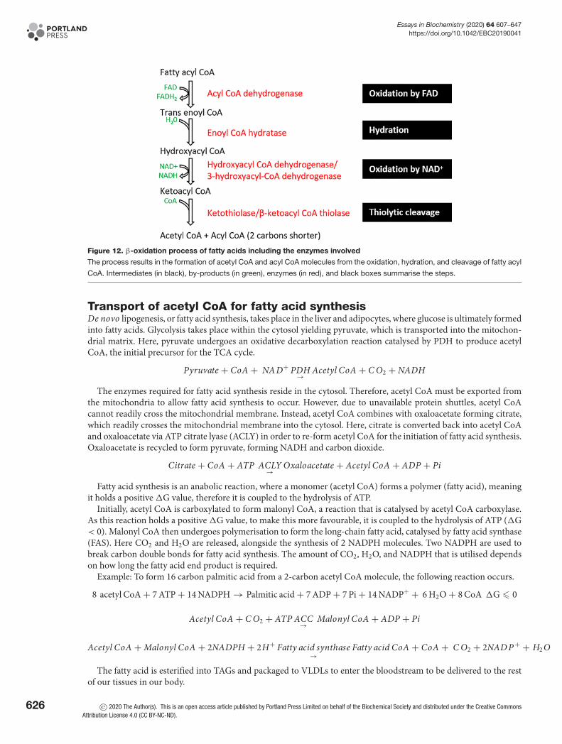
Fatty acid β-oxidation occurs within the mitochondrial matrix (Figure 12). Initially, fatty acyl CoA is oxidised byFAD to form trans-enoyl CoA, where a dehydrogenation reaction removes two hydrogen molecules between carbon2 and 3 of the fatty acid chain. Next, the hydration step adds a water molecule across the double bond forming hy-droxyacyl CoA. The next NAD-dependent dehydrogenation step generates an NADH molecule and ketoacyl CoA.Eventually, a thiolytic cleavage reaction forms an acetyl CoA molecule and acyl CoA that is 2 carbons shorter inlength. This acyl CoA can be recycled and reused cyclically for β-oxidation of fatty acids.
Fatty acid oxidation can also occur within peroxisomes. Peroxisomal oxidation of fatty acids occurs on fats that themitochondria are unable to utilise, such as very long chain fatty acids, pristanic acid, and bile intermediates. Here,fatty acid oxidation proceeds via a similar mechanism; however, enzymes and regulation can differ.
The NADH and FADH2 formed within the β-oxidation steps are utilised during the ETC.
© 2020 The Author(s). This is an open access article published by Portland Press Limited on behalf of the Biochemical Society and distributed under the Creative Commons AttributionLicense 4.0 (CC BY-NC-ND).
625
Essays in Biochemistry (2020) 64 607–647https://doi.org/10.1042/EBC20190041
Figure 12. β-oxidation process of fatty acids including the enzymes involved
The process results in the formation of acetyl CoA and acyl CoA molecules from the oxidation, hydration, and cleavage of fatty acyl
CoA. Intermediates (in black), by-products (in green), enzymes (in red), and black boxes summarise the steps.
Transport of acetyl CoA for fatty acid synthesisDe novo lipogenesis, or fatty acid synthesis, takes place in the liver and adipocytes, where glucose is ultimately formedinto fatty acids. Glycolysis takes place within the cytosol yielding pyruvate, which is transported into the mitochon-drial matrix. Here, pyruvate undergoes an oxidative decarboxylation reaction catalysed by PDH to produce acetylCoA, the initial precursor for the TCA cycle.
Pyruvate + CoA + NA D+ PDH→ Acetyl CoA + C O2 + NADH
The enzymes required for fatty acid synthesis reside in the cytosol. Therefore, acetyl CoA must be exported fromthe mitochondria to allow fatty acid synthesis to occur. However, due to unavailable protein shuttles, acetyl CoAcannot readily cross the mitochondrial membrane. Instead, acetyl CoA combines with oxaloacetate forming citrate,which readily crosses the mitochondrial membrane into the cytosol. Here, citrate is converted back into acetyl CoAand oxaloacetate via ATP citrate lyase (ACLY) in order to re-form acetyl CoA for the initiation of fatty acid synthesis.Oxaloacetate is recycled to form pyruvate, forming NADH and carbon dioxide.
Citrate + CoA + ATP ACLY→
Oxaloacetate + Acetyl CoA + ADP + Pi
Fatty acid synthesis is an anabolic reaction, where a monomer (acetyl CoA) forms a polymer (fatty acid), meaningit holds a positive �G value, therefore it is coupled to the hydrolysis of ATP.
Initially, acetyl CoA is carboxylated to form malonyl CoA, a reaction that is catalysed by acetyl CoA carboxylase.As this reaction holds a positive �G value, to make this more favourable, it is coupled to the hydrolysis of ATP (�G< 0). Malonyl CoA then undergoes polymerisation to form the long-chain fatty acid, catalysed by fatty acid synthase(FAS). Here CO2 and H2O are released, alongside the synthesis of 2 NADPH molecules. Two NADPH are used tobreak carbon double bonds for fatty acid synthesis. The amount of CO2, H2O, and NADPH that is utilised dependson how long the fatty acid end product is required.
Example: To form 16 carbon palmitic acid from a 2-carbon acetyl CoA molecule, the following reaction occurs.
8 acetyl CoA + 7 ATP + 14 NADPH → Palmitic acid + 7 ADP + 7 Pi + 14 NADP+ + 6 H2O + 8 CoA �G � 0
Acetyl CoA + C O2 + ATP ACC→
Malonyl CoA + ADP + Pi
Acetyl CoA + Malonyl CoA + 2NADPH + 2H+ Fatty acid synthase→
Fatty acid CoA + CoA + C O2 + 2NAD P + + H2 O
The fatty acid is esterified into TAGs and packaged to VLDLs to enter the bloodstream to be delivered to the restof our tissues in our body.
626 © 2020 The Author(s). This is an open access article published by Portland Press Limited on behalf of the Biochemical Society and distributed under the Creative CommonsAttribution License 4.0 (CC BY-NC-ND).
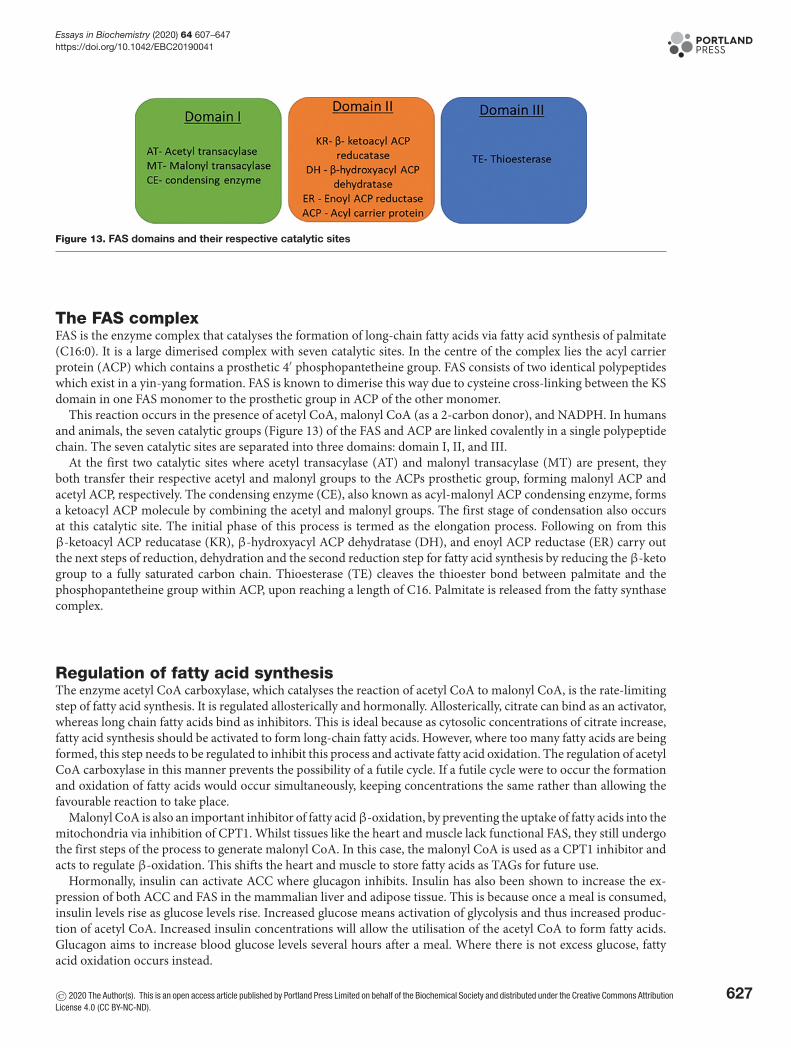
Essays in Biochemistry (2020) 64 607–647https://doi.org/10.1042/EBC20190041
Figure 13. FAS domains and their respective catalytic sites
The FAS complexFAS is the enzyme complex that catalyses the formation of long-chain fatty acids via fatty acid synthesis of palmitate(C16:0). It is a large dimerised complex with seven catalytic sites. In the centre of the complex lies the acyl carrierprotein (ACP) which contains a prosthetic 4′ phosphopantetheine group. FAS consists of two identical polypeptideswhich exist in a yin-yang formation. FAS is known to dimerise this way due to cysteine cross-linking between the KSdomain in one FAS monomer to the prosthetic group in ACP of the other monomer.
This reaction occurs in the presence of acetyl CoA, malonyl CoA (as a 2-carbon donor), and NADPH. In humansand animals, the seven catalytic groups (Figure 13) of the FAS and ACP are linked covalently in a single polypeptidechain. The seven catalytic sites are separated into three domains: domain I, II, and III.
At the first two catalytic sites where acetyl transacylase (AT) and malonyl transacylase (MT) are present, theyboth transfer their respective acetyl and malonyl groups to the ACPs prosthetic group, forming malonyl ACP andacetyl ACP, respectively. The condensing enzyme (CE), also known as acyl-malonyl ACP condensing enzyme, formsa ketoacyl ACP molecule by combining the acetyl and malonyl groups. The first stage of condensation also occursat this catalytic site. The initial phase of this process is termed as the elongation process. Following on from thisβ-ketoacyl ACP reducatase (KR), β-hydroxyacyl ACP dehydratase (DH), and enoyl ACP reductase (ER) carry outthe next steps of reduction, dehydration and the second reduction step for fatty acid synthesis by reducing the β-ketogroup to a fully saturated carbon chain. Thioesterase (TE) cleaves the thioester bond between palmitate and thephosphopantetheine group within ACP, upon reaching a length of C16. Palmitate is released from the fatty synthasecomplex.
Regulation of fatty acid synthesisThe enzyme acetyl CoA carboxylase, which catalyses the reaction of acetyl CoA to malonyl CoA, is the rate-limitingstep of fatty acid synthesis. It is regulated allosterically and hormonally. Allosterically, citrate can bind as an activator,whereas long chain fatty acids bind as inhibitors. This is ideal because as cytosolic concentrations of citrate increase,fatty acid synthesis should be activated to form long-chain fatty acids. However, where too many fatty acids are beingformed, this step needs to be regulated to inhibit this process and activate fatty acid oxidation. The regulation of acetylCoA carboxylase in this manner prevents the possibility of a futile cycle. If a futile cycle were to occur the formationand oxidation of fatty acids would occur simultaneously, keeping concentrations the same rather than allowing thefavourable reaction to take place.
Malonyl CoA is also an important inhibitor of fatty acidβ-oxidation, by preventing the uptake of fatty acids into themitochondria via inhibition of CPT1. Whilst tissues like the heart and muscle lack functional FAS, they still undergothe first steps of the process to generate malonyl CoA. In this case, the malonyl CoA is used as a CPT1 inhibitor andacts to regulate β-oxidation. This shifts the heart and muscle to store fatty acids as TAGs for future use.
Hormonally, insulin can activate ACC where glucagon inhibits. Insulin has also been shown to increase the ex-pression of both ACC and FAS in the mammalian liver and adipose tissue. This is because once a meal is consumed,insulin levels rise as glucose levels rise. Increased glucose means activation of glycolysis and thus increased produc-tion of acetyl CoA. Increased insulin concentrations will allow the utilisation of the acetyl CoA to form fatty acids.Glucagon aims to increase blood glucose levels several hours after a meal. Where there is not excess glucose, fattyacid oxidation occurs instead.
© 2020 The Author(s). This is an open access article published by Portland Press Limited on behalf of the Biochemical Society and distributed under the Creative Commons AttributionLicense 4.0 (CC BY-NC-ND).
627
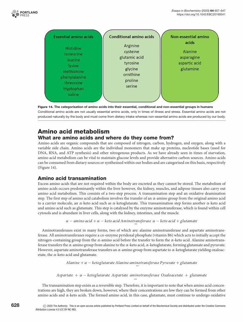
Essays in Biochemistry (2020) 64 607–647https://doi.org/10.1042/EBC20190041
Figure 14. The categorisation of amino acids into their essential, conditional and non-essential groups in humans
Conditional amino acids are not usually essential amino acids, only in times of illness and stress. Essential amino acids are not
produced naturally by the body and must come from dietary intake whereas non-essential amino acids are produced by our body.
Amino acid metabolismWhat are amino acids and where do they come from?Amino acids are organic compounds that are composed of nitrogen, carbon, hydrogen, and oxygen, along with avariable side chain. Amino acids are the individual monomers that make up proteins, nucleotide bases (used forDNA, RNA, and ATP synthesis) and other nitrogenous products. As we have already seen in times of starvation,amino acid metabolism can be vital to maintain glucose levels and provide alternative carbon sources. Amino acidscan be consumed from dietary sources or synthesised within our bodies and are categorised on this basis, respectively(Figure 14).
Amino acid transaminationExcess amino acids that are not required within the body are excreted as they cannot be stored. The metabolism ofamino acids occurs predominantly within the liver however, the kidney, muscles, and adipose tissues also carry outamino acid metabolism. This consists of a two-step process. A transamination step and an oxidative deaminationstep. The first step of amino acid catabolism involves the transfer of an α-amino group from the original amino acidto a carrier molecule, an α-keto acid such as α-ketoglutarate. This transamination step forms another α-keto acidand amino acid such as glutamate. This step is catalysed by the enzyme aminotransferase, which is found within cellcytosols and is abundant in liver cells, along with the kidney, intestines, and the muscle.
α − amino acid + α − keto acid Aminotransferase↔ α − keto acid + glutamate
Aminotransferases exist in many forms, two of which are: alanine aminotransferase and aspartate aminotrans-ferase. All aminotransferases require a co-enzyme pyridoxal phosphate (vitamin B6) which acts to initially accept thenitrogen-containing group from the α-amino acid before the transfer to form the α-keto acid. Alanine aminotrans-ferase transfers theα-amino group from alanine to theα-keto acid,α-ketoglutarate, forming glutamate and pyruvate.However, aspartate aminotransferase transfers an α-amino group from aspartate to α-ketoglutarate yielding oxaloac-etate, the α-keto acid and glutamate.
Alanine + α − ketoglutarate Alanine aminotransferase↔ Pyruvate + glutamate
Aspartate + α − ketoglutarate Aspartate aminotransferase↔
Oxaloacetate + glutamate
The transamination step exists as a reversible step. Therefore, it is important to note that when amino acid concen-trations are high, they are broken down, however, where their concentrations are low they can be formed from otheramino acids and α-keto acids. The formed amino acid, in this case, glutamate, must continue to undergo oxidative
628 © 2020 The Author(s). This is an open access article published by Portland Press Limited on behalf of the Biochemical Society and distributed under the Creative CommonsAttribution License 4.0 (CC BY-NC-ND).
Essays in Biochemistry (2020) 64 607–647https://doi.org/10.1042/EBC20190041
deamination to form ammonia (see Urea cycle later). The formed α-keto acid can be utilised as a form of energy asATP molecules.
Oxidative deaminationOxidative deamination consists of two steps: a dehydrogenation and a hydrolysis step. The deamination step removesthe amino group from glutamate to form an intermediate molecule. The intermediate undergoes a hydrolysis reactionwhere the amino group forms ammonium ions (NH4
+) and regeneration of α-ketoglutarate.
Glutamate + NA D+ Glutamate dehydrogenase↔
Ammonia + α − ketoglutarate + NADH
Glutamate dehydrogenase is predominantly found within the liver and the kidneys, inside mitochondria, for thisreaction to occur. This reaction occurs within the mitochondria to ensure that the toxic ammonium yielded does notcause cytotoxicity within the cell. It utilises two co-enzymes; NAD+ and NADP+. These co-enzymes are respectivelyused based on the conditions for the cell. When driving the forward reaction, in conditions where amino acid con-centrations are high, for example after consuming a protein-rich meal, NAD+ is used as the co-enzyme. However,where amino acid or glutamate levels are low, the reverse reaction occurs to form more glutamate which can aid thesynthesis of other non-essential amino acids. ADP and guanosine diphosphate (GDP) are both allosteric activatorsof glutamate dehydrogenase. Therefore, when energy levels are low (high ADP, GDP but low ATP and GTP), thisreaction occurs to breakdown glutamate and amino acids for energy production.
Following amino acid metabolism within the liver, due to its toxicity, the synthesised ammonia, cannot be simplytransported in the bloodstream. Due to this, the ammonia is transformed into a non-toxic compound, glutamine.Glutamine is transported to the kidneys by the enzyme glutamine synthetase which is present in peripheral tissues.
Ammonia + glutamate + ATP Glutamine synthetase→
glutamine + ADP + Pi
Glutamine is transported via the bloodstream and travels to the liver. Within the liver it is converted back intoglutamate and ammonia by the enzyme glutaminase. In terrestrial vertebrates, ammonia is converted into urea whichis excreted (see the section on Urea later).
Within the kidneys, the proximal tubule is the primary site for ammoniagenesis of predominantly glutaminemetabolism. Ammonia produced here is excreted directly into the urine or returned to the systemic circulation.
Single-step catabolism of amino acidsSome particular amino acids only undergo a single step deamination process. These include serine and threonine.The one step process is catalysed by the enzyme dehydratase.
Serine Dehydratase→
Pyruvate + N H+4
Threonine Dehydratase→
α − ketobutyrate + N H+4
During these reactions, a dehydration reaction occurs to form an unstable, high energy, intermediate moleculesuch as aminoaceylate. This readily converts into a final product and yields ammonium. The ammonium carries intothe urea cycle whereas the carbon skeletons formed, such as pyruvate, can be used for energy purposes.
The TCA cycleThe importance of the TCA cycle and its discoveryThe TCA cycle was initially known as the citric acid cycle by Hans A. Krebs, a German biochemist, in 1937. It is morecommonly known as the Krebs cycle, in recognition of Hans Krebs’ discovery. Krebs’ work was in collaboration withWilliam A. Johnson at the University of Sheffield. However, before their end discovery, a succession of experimentstook place by many other scientists.
Stern carried out his experiments with minced animal tissue. Here, he found that these tissues contained specificsubstances that could transfer H+ atoms from several intracellular organic acids. In particular, fumarate, malate, suc-cinate, and citrate. He studied this using Methylene Blue, where this transfer of H+ atoms resulted in the reduction ofthe dye to a colourless form. This was found to occur within the presence of oxygen at high rates, knowing that activeenzymes existed here. In the 1920s, Thunberg described a respiratory cycle that was present to oxidise acetate whenparticular tissue dehydrogenases were available.
© 2020 The Author(s). This is an open access article published by Portland Press Limited on behalf of the Biochemical Society and distributed under the Creative Commons AttributionLicense 4.0 (CC BY-NC-ND).
629
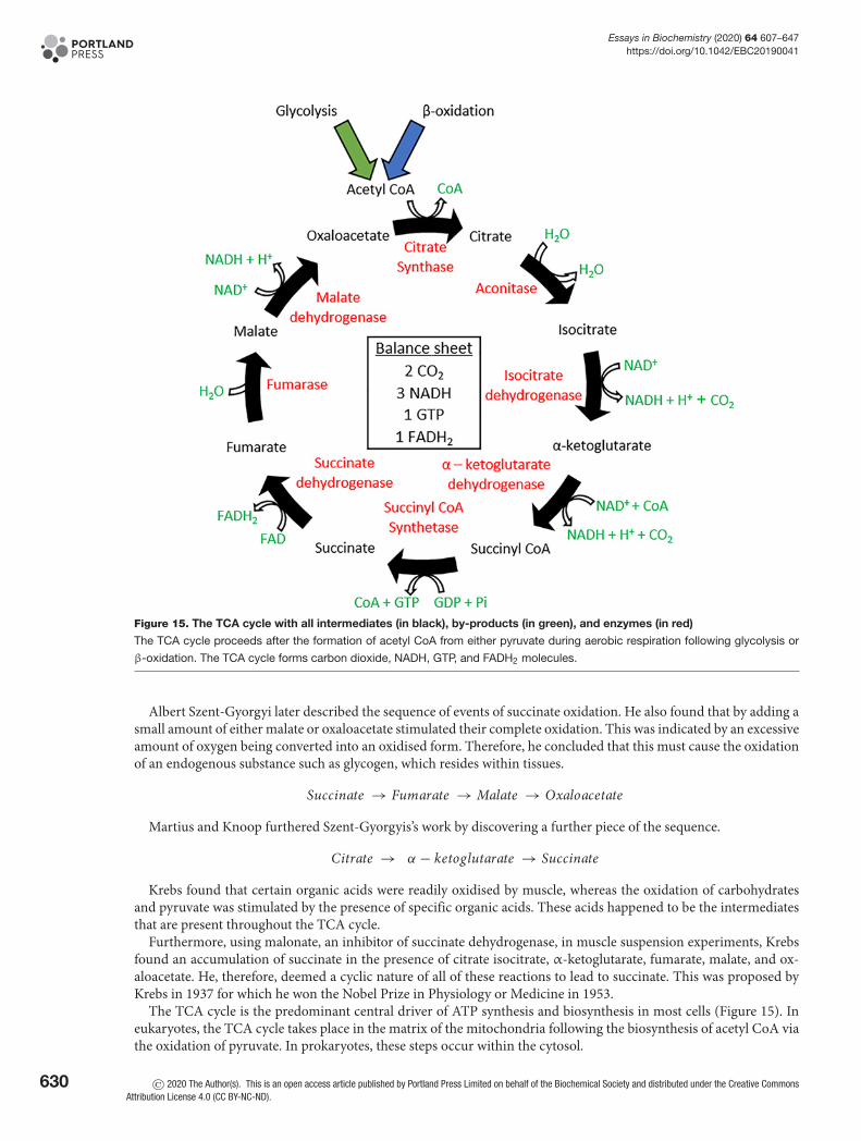
Essays in Biochemistry (2020) 64 607–647https://doi.org/10.1042/EBC20190041
Figure 15. The TCA cycle with all intermediates (in black), by-products (in green), and enzymes (in red)
The TCA cycle proceeds after the formation of acetyl CoA from either pyruvate during aerobic respiration following glycolysis or
β-oxidation. The TCA cycle forms carbon dioxide, NADH, GTP, and FADH2 molecules.
Albert Szent-Gyorgyi later described the sequence of events of succinate oxidation. He also found that by adding asmall amount of either malate or oxaloacetate stimulated their complete oxidation. This was indicated by an excessiveamount of oxygen being converted into an oxidised form. Therefore, he concluded that this must cause the oxidationof an endogenous substance such as glycogen, which resides within tissues.
Succinate → Fumarate → Malate → Oxaloacetate
Martius and Knoop furthered Szent-Gyorgyis’s work by discovering a further piece of the sequence.
Citrate → α − ketoglutarate → Succinate
Krebs found that certain organic acids were readily oxidised by muscle, whereas the oxidation of carbohydratesand pyruvate was stimulated by the presence of specific organic acids. These acids happened to be the intermediatesthat are present throughout the TCA cycle.
Furthermore, using malonate, an inhibitor of succinate dehydrogenase, in muscle suspension experiments, Krebsfound an accumulation of succinate in the presence of citrate isocitrate, α-ketoglutarate, fumarate, malate, and ox-aloacetate. He, therefore, deemed a cyclic nature of all of these reactions to lead to succinate. This was proposed byKrebs in 1937 for which he won the Nobel Prize in Physiology or Medicine in 1953.
The TCA cycle is the predominant central driver of ATP synthesis and biosynthesis in most cells (Figure 15). Ineukaryotes, the TCA cycle takes place in the matrix of the mitochondria following the biosynthesis of acetyl CoA viathe oxidation of pyruvate. In prokaryotes, these steps occur within the cytosol.
630 © 2020 The Author(s). This is an open access article published by Portland Press Limited on behalf of the Biochemical Society and distributed under the Creative CommonsAttribution License 4.0 (CC BY-NC-ND).

Essays in Biochemistry (2020) 64 607–647https://doi.org/10.1042/EBC20190041
Table 3 The reactions of the TCA cycle alongside the standard free energy changes and physiological free energychanges of the reactions
Step Reaction Enzyme Type of reaction�G◦′
kJ.mol −1�G kJ.mol
−1
1 Acetyl CoA + Oxaloacetate → Citrate Citrate synthase Condensation −31.5 Negative
2 Citrate � Isocitrate Aconitase Isomerisation ∼5 ∼0
3 Isocitrate + NAD+ → α-ketoglutarate + NADH + CO2 Isocitratedehydrogenase
Oxidativedecarboxylation
−21 Negative
4 α-ketoglutarate + NAD+ + CoA → Succinyl CoA + NADH + CO2 α-ketoglutaratedehydrogenase
Oxidativedecarboxylation
−33 Negative
5 Succinyl CoA + GDP + Pi � Succinate + GTP + CoA Succinyl CoA synthase Substrate levelphosphorylation
−2.1 ∼ 0
6 Succinate + FAD+ � Fumarate + FADH2 Succinatedehydrogenase
Dehydrogenation +6 ∼ 0
7 Fumarate + H2O � Malate Fumarase Hydration −3.4 ∼ 0
8 Malate + NAD+ � Oxaloacetate + NADH Malate dehydrogenase Dehydrogenation +29.7 ∼0
The pathwayThe cycle is formed of eight major steps, see Table 3.
Important steps of TCAStep 1 – The combination of 2-carbon acetyl CoA and 4-carbon oxaloacetate forms 6-carbon citrate. Here, citrate caneither move into the cytosol to initiate fatty acid synthesis or is destined to carry through the oxidation steps involvedin the TCA cycle. This reaction is irreversible and highly regulated with a highly negative �G.
Step 3 – This step is highly regulated and allows the commitment of citric acid to the TCA cycle instead of fattyacid synthesis. This step is activated and stimulated by NAD+ and ADP but inhibited by NADH and ATP. Here, thefirst NADH molecules are formed along with CO2.
Step 5 – This reaction is coupled to the phosphorylation of GDP to GTP, making it the only reaction in the TCAcycle to generate a high energy phosphate. Following the formation of GTP, this can either be converted to ATP orused in protein synthesis.
Balance sheetGlucoseFollowing glycolysis, 2 pyruvate, 2 ATP, and 2 NADH are formed. The pyruvate molecules are broken down by PDHto 2 acetyl CoA, 2 NADH, and 2 CO2.
These acetyl CoA molecules enter the TCA cycle and generate: 1 molecule of GTP (interchangeable with ATP), 3NADH, and 1 FADH2 co-enzymes. These are doubled as two molecules of acetyl CoA are generated per glucose.
Total : 8 NADHmatrix + 2 NADHcytosol + 4 ATP + 2 FADH2
Palmitate (C16)From β-oxidation: 7 NADH, 7 FADH2, and 8 acetyl CoA
From TCA cycle: 24 NADH, 8 FADH2, 8 GTP/ATP
Anaplerosis and cataplerosisThe importanceAnaplerosis is the act of replenishing intermediates of the TCA cycle that have been used up for biosynthesis. Theseanions must be replaced to retain the function and cyclic fashion of the TCA cycle. However, to ensure that theseintermediates do not over-supply the TCA cycle, anaplerosis is coupled with the exit of intermediates from the TCAcycle, called cataplerosis (Figure 16).
The NADH that is generated from the TCA cycle is used by the cell in the form of ATP. Even though the TCA cycleforms over 70% of the ATP that is used by a cell, it also provides the intermediate building blocks for other processes.As a result of this, the subsequent intermediates within the TCA cycle can be used up excessively and these must bereplenished through the process of anaplerosis.
Intermediates and their use:
© 2020 The Author(s). This is an open access article published by Portland Press Limited on behalf of the Biochemical Society and distributed under the Creative Commons AttributionLicense 4.0 (CC BY-NC-ND).
631
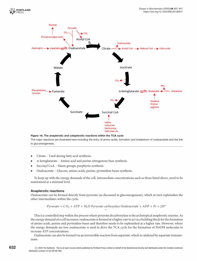
Essays in Biochemistry (2020) 64 607–647https://doi.org/10.1042/EBC20190041
Figure 16. The anaplerotic and cataplerotic reactions within the TCA cycle
The major reactions are illustrated here including the entry of amino acids, formation and breakdown of oxaloacetate and the link
to gluconeogenesis.
• Citrate – Used during fatty acid synthesis.• α-ketoglutarate – Amino acid and purine nitrogenous base synthesis.• Succinyl CoA – Haem groups, porphyrin synthesis.• Oxaloacetate – Glucose, amino acids, purine, pyrimidine bases synthesis.
To keep up with the energy demands of the cell, intermediate concentrations such as those listed above, need to bemaintained at a minimal level.
Anaplerotic reactionsOxaloacetate can be formed directly from pyruvate (as discussed in gluconeogenesis), which in turn replenishes theother intermediates within the cycle.
Pyruvate + C O2 + ATP + H2 O Pyruvate carboxylase→
Oxaloacetate + ADP + Pi + 2H+
This is a controlled step within the process where pyruvate decarboxylase is the archetypical anaplerotic enzyme. Asthe energy demand of a cell increases, oxaloacetate is formed at a higher rate to act as a building block for the formationof amino acids, purine and pyrimidine bases and therefore needs to be replenished at a higher rate. However, wherethe energy demands are low, oxaloacetate is used to drive the TCA cycle for the formation of NADH molecules toincrease ATP concentrations.
Oxaloacetate can also be formed via an irreversible reaction from aspartate, which is catalysed by aspartate transam-inase.
632 © 2020 The Author(s). This is an open access article published by Portland Press Limited on behalf of the Biochemical Society and distributed under the Creative CommonsAttribution License 4.0 (CC BY-NC-ND).
Essays in Biochemistry (2020) 64 607–647https://doi.org/10.1042/EBC20190041
Upon the oxidation of fatty acids, succinyl CoA is formed. Therefore, the process of β-oxidation of fats can allowthe formation of succinyl CoA molecules required in the TCA cycle.α-ketoglutarate is regenerated from glutamate by glutamate dehydrogenase.
Glutamate + NA D+ + H2 O Glutamate dehydrogenase→
α − ketoglutarate + NADH + H+
Fumarate is regenerated from adenylsuccinate during purine synthesis.
Adenylsuccinate Adenylosuccinate lyase→
Fumarate + AMP
CataplerosisDuring the catabolism of amino acids, 4- to 5-carbon intermediates are formed, which ultimately enter the TCA cycle.The TCA cycle is unable to fully oxidise these and therefore are removed by the process of cataplerosis. Cataplerosisaims to remove intermediates, thus ensuring that there is no accumulation of anions in the mitochondrial matrix.Three main cataplerotic enzymes exist; PEPCK, aspartate aminotransferase, and glutamate dehydrogenase. The reac-tions involved within cataplerosis, as described below, form a product that essentially removes specific intermediates.For example, within the liver and kidney, PEPCK forms PEP which can be utilised for gluconeogenesis. However,within the muscle, PEP can be decarboxylated to form acetyl CoA for oxidation within the TCA cycle.
PEPCK
Oxaloacetate PEPCK→
Phosphoenolpyruvate + C O2
Aspartate aminotransferase
L − aspartate + α − ketoglutarate Aspartate aminotransferase→
Oxaloacetate + L − glutamate
Glutamate dehydrogenase
L − glutamate + NA D+ + H2 O Glutamate dehydrogenase→
α − ketoglutarate + N H+4 + NADH + H+
The synthesis of carbohydrates from fatThe glyoxylate cycle is a variation of the TCA cycle that does not exist in animals. In animals, carbohydrates are readilyconverted into fat; however, the reverse process cannot occur. This follows on from the nature of the TCA cycle andthe irreversible conversion of pyruvate into acetyl CoA.
The glyoxylate cycle allows the synthesis of carbohydrates from fat within plants, bacteria, fungi, algae, and protozoawhich grow on acetate as their carbon source for energy and cell components. This is especially important for seedsrich in oil such as peanuts, olives, and sunflowers when they are germinating. The fatty acids stored within the seedsare broken down to form glucose to be used as energy for germination until photosynthesis is established.
The glyoxylate cycle is an anabolic reaction which bypasses the CO2 forming steps of the TCA cycle (isocitrateto succinate), conserving a glyoxylate molecule to form malate. It occurs within specialised peroxisomes called gly-oxysomes.
As represented in Figure 17, within this cycle, isocitrate is formed as per the TCA cycle; however, this is brokendown by isocitrate lyase into glyoxylate (2-carbon molecule) and succinate. Glyoxylate is then combined with acetylCoA forming malate, a reaction catalysed by malate synthase. Malate forms oxaloacetate, which proceeds to generateenergy in the form of glucose from gluconeogenesis.
The carbon skeletons that enter the TCA cycle as acetyl CoA are lost during the decarboxylation steps of the cycle.As oxaloacetate undergoes cataplerosis to make glucose, there is no oxaloacetate remaining for the TCA cycle tocontinue. Due to this, fats cannot produce glucose at a net rate. The glyoxylate cycle bypasses the decarboxylationsteps, creating a compound that can form glucose without depleting the starting compound of the TCA cycle.
Energy production by organellesThere are several common features of the organelles responsible for energy production in eukaryotes – the mitochon-dria and chloroplast. Firstly, both organelles rely on the presence of a highly folded membrane, within a ‘protective’
© 2020 The Author(s). This is an open access article published by Portland Press Limited on behalf of the Biochemical Society and distributed under the Creative Commons AttributionLicense 4.0 (CC BY-NC-ND).
633

Essays in Biochemistry (2020) 64 607–647https://doi.org/10.1042/EBC20190041
Figure 17. The glyoxylate shunt (green) coexisting amongst the TCA cycle (black)
The glyoxylate shunt converts fatty acids into carbohydrates by bypassing decarboxylation steps of the TCA cycle.
outer membrane, which significantly increases surface area for energy generation. Secondly, these folded membranesare studded with enzymes which use Cu and Fe ions, and ubiquinone for the transfer of electrons. Thirdly, theseelectrons are used to pump proton (H+) ions against a concentration gradient, generating a proton motive force. Thisproton motive force is then used to generate ATP via ATP synthase, as protons move back down their concentrationgradient. Finally, both organelles appear to have a prokaryotic origin and one theory is that they have developed anendosymbiotic relationship with an ancient pre-eukaryotic cell, to generate modern-day eukaryotes.
Organisation of organellesWe have already seen that plants are autotrophic and use chloroplasts to capture the energy from sunlight to fix CO2and synthesise useful macromolecules. Chloroplast use their heavily folded membranes to significantly increase theamount of sunlight that can be captured and thus maximise energy generation. As well as enzymes in the mem-branes, the enzymes responsible for CO2 fixation, known as the Calvin cycle, are found within the stroma (largecentral space) which is surrounded by the inner membrane. Unlike mitochondria, chloroplast pump protons into thethylakoids, rather than the intermembrane space, which acts to generate the proton motive force required. For a lotmore detailed understanding of the chloroplast and the process of photosynthesis as a whole, please refer to the Un-derstanding Biochemistry review: Photosynthesis, Matthew P. Johnson (2016), Essays In Biochemistry, 60, 255–273;DOI: 10.1042/EBC20160016.
Unlike chloroplasts, mitochondria use organic chemical nutrients to obtain physiological energy in the form ofATP. We have already seen that ATP can be generated in glycolysis, but per glucose molecule that enters, only twoATP are generated. To capitalise on the energy held in glucose, it needs to be fully oxidised through the TCA cycleand then FAD and NADH enter the ETC. This then liberates 30–32 ATP per glucose molecule, a 15–16-fold increasein energy return. These last two pathways, TCA cycle and ETC, require functioning mitochondria and oxygen to bepresent. In the lumen (known as the matrix), enzymes for the TCA cycle, PDH, and β oxidation reside (Figure 18).Embedded into the cristae (folds in the inner membrane) are the enzymes for the ETC.
One enzyme crosses over between the TCA cycle and the ETC, which is succinate dehydrogenase. This enzymecatalyses the reaction succinate to fumarate and generates FADH2. Succinate dehydrogenase is integral to complex 2and passes electrons from FADH2, directly into the ETC. Generally speaking, the greater the number of cristae, thehigher the respiratory demand of the tissue. This is seen in the heart, where there are a high number of cristae and
634 © 2020 The Author(s). This is an open access article published by Portland Press Limited on behalf of the Biochemical Society and distributed under the Creative CommonsAttribution License 4.0 (CC BY-NC-ND).

Essays in Biochemistry (2020) 64 607–647https://doi.org/10.1042/EBC20190041
Figure 18. Basic structure of the mitochondrion and chloroplast, showing the membrane structures
a large number of mitochondria present. Several studies have shown that one of the consequences of heart failureis mitochondrial dysfunction, where cristae superstructure breaks down and the mitochondria become less able toproduce ATP.
Mitochondria are dynamicMitochondria are often thought of as the static structure presented in Figure 18, but in reality, they can exist in differentforms. Fluorescently tagged neuronal mitochondria have been visualised moving from the neuronal body, along thecytoskeleton of the axon and to the synapse where energy demand is high. Whilst mitochondria can be present assingle mitochondrion, they can also form long connected networks similar to chains seen in some prokaryotes (e.g.cyanobacteria). In these networks, mitochondria can undergo fission (splitting) and then re-join (fusion), dependingon energetic demand and mitochondrial health. This act of fusion is thought to help recycle or refresh damagedmitochondria and allow mutations in mitochondrial DNA and damage to enzymes to be diluted throughout thenetwork. In fact defects in genes regulating fission and fusion (for example PINK1 and Parkin), have been reportedin neurological diseases such as Parkinson’s disease. Mutations in these genes (and others) lead to mitochondrialundergoing higher levels of fission and are less able to fuse, thereby not recycling damaged mtDNA or metabolicenzymes, affecting neuronal health and viability.
Oxidative phosphorylation in the mitochondriaOxidative phosphorylation encompasses both the ETC and ATP synthase. This process occurs within the inner mito-chondria membrane, where the complexes for the ETC and ATP synthase are present (Figure 19). The role of the ETCis, as its name suggests, to transport electrons through a series of complexes to the final electron acceptor: oxygen. Aselectrons flow along this chain, they start with a high energy potential losing energy as they reduce electron carriersas they travel through the chain (Figure 20). The complexes use this energy to pump protons from the matrix, into theintermembrane space. As the inner membrane is impermeable to protons, a concentration and pH gradient developsacross the membrane due to the positive charge of the protons. The matrix becomes negatively charged due to thepumping out of protons and this generates an electrical potential across the membrane, equivalent to ∼150–200 mV.The electrochemical gradient is used to drive ATP synthase to produce ATP. Along with the complexes, there are twoelectron carriers coenzyme Q (also known as quinone and CoQ) and cytochrome c, which are responsible for passingelectrons from complex to complex (Figure 19).
Complex I (also known as NADH dehydrogenase) and complex II (also known as succinate dehydrogenase) areflavoproteins, which means that they contain either FAD or FMN and use a series of iron–sulphur clusters to accepttwo electrons and two protons from NADH + H+ or succinate (eqn 4). The electrons are then passed via coenzymeQ to complex III (Q-cytochrome c oxidoreductase) which is the first cytochrome is the pathway. Complex III, IV,and cytochrome c are all cytochrome proteins, meaning that they use both haem prosthetic groups and iron–sulphurclusters to transfer electrons. Within the haem group, the iron ion alternates between a reduced Fe2+ (ferrous) stateand oxidised Fe3+ (ferric) state as electrons are transferred. At complex IV, the electrons are finally passed to oxygen,which generates water. As the electrons pump through complex I, III, and IV they cause the release of 4H+, 4H+, and2H+ into the intermembrane space, respectively (eqn 4).
© 2020 The Author(s). This is an open access article published by Portland Press Limited on behalf of the Biochemical Society and distributed under the Creative Commons AttributionLicense 4.0 (CC BY-NC-ND).
635

Essays in Biochemistry (2020) 64 607–647https://doi.org/10.1042/EBC20190041
Figure 19. Oxidative phosphorylation – ETC and ATP synthase
Drawn from PDB and OPM codes: 5XTD (Complex I), 1ZOY (Complex II), 1L0L (Complex III), 2DYR (Complex IV) and 6J5I (ATP
synthase).
Figure 20. As electrons (e-) are passed along the chain, they reduce the next carrier and decrease their energy levels, until
they reach the final electron acceptor – oxygen
Summary of equations for the four complexes of the ETC. CoQ = coenzyme Q, Cyt c = cytochrome c.
Complex I: NADH + H+ + CoQ + 4H+matrix → NA D+ + CoQ H2 + 4H+
cytosol
Complex II: FAD H2 + CoQ → FAD + CoQ H2
Complex III: CoQ H2 + 2Cyt cox + 2H+matrix → CoQ + 2Cyt cred + 4H+
cytosol
Complex IV: 2Cyt cred + 4H+matrix + 1
2 O2 → 2Cyt cox + 2H2 O + 2H+cytosol
(4)
There are two entry points into the ETC, this is either via NADH + H+ and complex I or succinate/FADH2 andcomplex II. Ultimately, both pathways deliver electrons into the ETC and generate ATP at the end. However, theamount of ATP generated varies depending on which entry point, complex I or complex II, is used. This difference isdue to the number of protons pumped out into the intermembrane space by the complexes. As can be seen in Figure19, the different complexes pump protons from the matrix into the intermembrane space, thereby generating a protongradient. Roughly four protons are needed for the generation of one ATP molecule, approximately three protons gothrough ATP synthase and one is used for the nucleotide transport into the mitochondria (to import Pi and ADP).
636 © 2020 The Author(s). This is an open access article published by Portland Press Limited on behalf of the Biochemical Society and distributed under the Creative CommonsAttribution License 4.0 (CC BY-NC-ND).
Essays in Biochemistry (2020) 64 607–647https://doi.org/10.1042/EBC20190041
Complex I can pump four protons, whilst complex II does not directly pump any protons out, therefore NADH + H+,via complex I, can generate one more ATP than FADH2, via complex II. In total, 1 NADH + H+ generates 2.5 ATPand 1 FADH2 generates 1.5 ATP. After a breakdown of the ETC process, we will return to the balance sheets that wesaw earlier for the number of NADH + H+ and FADH2 generated from the oxidation of glucose and palmate.
ATP synthaseThe ATP synthase multiprotein complex is a molecular motor powered of protons passing through it. In humans,ATP synthase is made up of 29 subunits of 18 different types that are, interestingly, encoded for by different genomes:some genes are nuclear whilst others are mtDNA. These subunits form the three main structures in ATP synthase:the FO (rotor), F1 (catalytic head and stalk), and FO-stalk arm. The FO is a transmembrane protein complex, which isthe main rotor, accepting protons and turning the F1 head. Protons enter a channel on the intermembrane space sideof FO, these protons then bind into a pocket in the FO and the rotor turns one space. This turning releases a proton onthe matrix side via another channel. The complex can hold ten protons and a single proton turns just under 360◦ as itpasses from the intermembrane space to the matrix. In the F1 subunit, there are 3 catalytic domains each containingbinding sites for ADP and Pi. Within the catalytic domain, ADP and Pi react to form ATP, the action of turning theF1 releases one ATP into the matrix at a time. Therefore, a complete turn of the ATP synthase transfers ten protonsfrom the intermembrane space to the matrix and generates three ATP molecules.
Final balance sheetsNote these numbers are estimation and values differ in different textbooks, depending on if the proton lost for theinput of ADP and Pi is counted, or if you count the number of ATP produced as either 3 or 3.3 protons. There is alsoa difference in the number of protons that can pass through ATP synthase in different organisms. The c-ring, whichis the subunit responsible for binding protons in FO, can vary in the number of subunits and therefore the number ofprotons that it can bind. It has been seen that this can be between 8 and 17, therefore altering the potential numberof protons needed to make a 360◦ transition of the ATP synthase complex. The values we have used here account forthe import of ADP and Pi and use the value of three protons per ATP, therefore 1 NADH = 2.5 ATP and 1 FADH2 =1.5 ATP.
Glucose
8 NADHmatrix + 2 NADHcytosol + 4 ATP + 2 FADH2
NADHcytosol needs to be transported into the mitochondria to be used in the ETC. As NADH cannot be directlytransported, the electrons and protons are transferred to malate, which via the malate-aspartate shuttle, regeneratesNADH in the matrix. The use of the malate-aspartate shuttle however transports one proton from the intermembranespace, into the matrix, reducing the effective ATP production from 2.5 to 2.25.
20 ATP + 4.5 ATP + 4 ATP + 3 ATP = 31.5 ATP per glucose
Palmitate (C16)From β-oxidation: 7 NADH, 7 FADH2, and 8 acetyl CoA
From TCA cycle: 24 NADH, 8 FADH2, 8 GTP/ATP77.5 ATP (from NADH) + 22.5 ATP (from FADH2) + 8 ATP = 108 ATP
Even chained fatty acid with n number of carbons (Cn)Whilst this looks scary, if you input the number of carbons in the fatty acids, then you will get the number of ATPproduced when oxidised.
From β-oxidation: (Cn−2/2) NADH, (Cn−2/2) FADH2, and Cn acetyl CoAFrom TCA cycle: 3 (Cn/2) NADH, (Cn/2) FADH2, (Cn/2) GTP/ATP
2.5((
Cn − 22
)+ 3
(Cn2
))ATP + 1.5
((Cn − 2
2
)+
(Cn2
))ATP +
(Cn2
)ATP
Or more simply for any given fatty acid with a chain length of Cn, this is the number of ATP produced via completeoxidation:
10(
Cn2
)+ 4
(Cn − 2
2
)ATP
© 2020 The Author(s). This is an open access article published by Portland Press Limited on behalf of the Biochemical Society and distributed under the Creative Commons AttributionLicense 4.0 (CC BY-NC-ND).
637
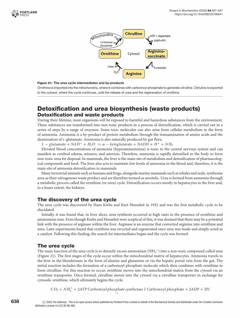
Essays in Biochemistry (2020) 64 607–647https://doi.org/10.1042/EBC20190041
Figure 21. The urea cycle intermediates and by-products
Ornithine is imported into the mitochondria, where it combines with carbomoyl phosphate to generate citrulline. Citriuline is exported
to the cytosol, where the cycle continues, until the release of urea and the regeneration of ornithine.
Detoxification and urea biosynthesis (waste products)Detoxification and waste productsDuring their lifetime, most organisms will be exposed to harmful and hazardous substances from the environment.These substances are transformed into non-toxic products in a process of detoxification, which is carried out in aseries of steps by a range of enzymes. Some toxic molecules can also arise from cellular metabolism in the formof ammonia. Ammonia is a by-product of protein metabolism through the transamination of amino acids and thedeamination of l-glutamate. Ammonia is also naturally produced by gut flora.
L − glutamate + NA D+ + H2 O � α − ketoglutarate + NADH + H+ + N H3Elevated blood concentrations of ammonia (hyperammonemia) is toxic to the central nervous system and can
manifest as cerebral edema, seizures, and asterixis. Therefore, ammonia is rapidly detoxified in the body to formnon-toxic urea for disposal. In mammals, the liver is the main site of metabolism and detoxification of pharmacolog-ical compounds and food. The liver also acts to maintain low levels of ammonia in the blood and, therefore, it is themain site of ammonia detoxification in mammals.
Many terrestrial animals such as humans and frogs, alongside marine mammals such as whales and seals, synthesiseurea as their nitrogenous waste product and are therefore termed as ureotelic. Urea is formed from ammonia througha metabolic process called the ornithine (or urea) cycle. Detoxification occurs mostly in hepatocytes in the liver and,to a lesser extent, the kidneys.
The discovery of the urea cycleThe urea cycle was discovered by Hans Krebs and Kurt Henseleit in 1932 and was the first metabolic cycle to beelucidated.
Initially, it was found that, in liver slices, urea synthesis occurred at high rates in the presence of ornithine andammonium ions. Even though Krebs and Henseleit were sceptical of this, it was deemed that there may be a potentiallink with the presence of arginase within the liver. Arginase is an enzyme that converted arginine into ornithine andurea. Later experiments found that ornithine was recycled and regenerated once urea was made and simply acted asa catalyst. Following this finding, the search for intermediates began and the cycle was formed.
The urea cycleThe main function of the urea cycle is to detoxify excess ammonium (NH4
+) into a non-toxic compound called urea(Figure 21). The first stages of the cycle occur within the mitochondrial matrix of hepatocytes. Ammonia travels tothe liver in the bloodstream in the form of alanine and glutamine or via the hepatic portal vein from the gut. Theinitial reaction includes the formation of a carbomoyl phosphate molecule which then combines with ornithine toform citrulline. For this reaction to occur, ornithine moves into the mitochondrial matrix from the cytosol via anornithine transporter. Once formed, citrulline moves into the cytosol via a citrulline transporter in exchange forcytosolic ornithine, which ultimately begins the cycle.
C O2 + N H+4 + 2ATP Carbomoyl phosphate synthetase I
→Carbomoyl phosphate + 2ADP + 2Pi
638 © 2020 The Author(s). This is an open access article published by Portland Press Limited on behalf of the Biochemical Society and distributed under the Creative CommonsAttribution License 4.0 (CC BY-NC-ND).

Essays in Biochemistry (2020) 64 607–647https://doi.org/10.1042/EBC20190041
Ornithine + Carbomoyl phosphate Ornithine transcarbomoylase→
Citrulline + Pi
In the cytosol, citrulline is condensed with aspartate to form argininosuccinate driven by the hydrolysis of ATP.Argininosuccinate is cleaved to arginine and fumarate, a reaction that is catalysed by argininosuccinate lyase. In astate of fasting when gluconeogenesis occurs, fumarate can be used to form malate which can then go on to formglucose or oxaloacetate. Arginine, on the other hand, proceeds in the ornithine cycle and is cleaved to form urea andregenerate ornithine molecules via arginase.
Citrulline + Aspartate + ATP Argininosuccinate synthetase→
Argininosuccinate + AMP + PPi
Argininosuccinate Argininosuccinate lyase→
Arginine + Fumarate
Arginine + H2 O Arginase→
Ornithine + Urea
The overall reaction of the urea cycle is as follows:
C O2 + N H+4 + H2 O + 3ATP + Aspartate → Urea + 2ADP + AMP + 2Pi + PPi + fumarate
The first step of the urea cycle is a rate-limiting step. The enzyme carbomoyl phosphate synthetase I requires anactivator, N-acetyl-glutamate (NAG). NAG is formed by the addition of glutamate and acetyl CoA occurring underthe influence of NAG synthase, which is regulated by the presence of arginine.
Ammonotelic, uricotelic, and the in-betweenAquatic animals such as fish are termed ammonotelic. Ammonotelic organisms excrete ammonia directly as nitroge-nous waste products. Ammonia is a highly soluble substance and requires large volumes of water to dissolve. There-fore, aquatic animals like fish use their gills to excrete over 80% of ammonia directly into surrounding waters.
Terrestrial organisms such as lizards, snakes, insects, and birds are termed uricotelic organisms. These excrete uricacid as their waste product instead of urea. The complex pathway ultimately stems from the metabolic breakdown ofpurine nucleotides. The metabolic pathway has a higher energy cost, in comparison with the synthesis of urea, butcan reduce water loss in these animals.
In some cases, animals can shift from utilising one form of waste product mechanism to another. In the metamor-phosis of amphibians, you can see the development of the urea cycle as they adapt to terrestrial life. Tadpoles excreteammonia as their primary waste product before metamorphosis. However, during its development to becoming afrog and the ultimate invasion of land, an irreversible change to ureotelism occurs. This, therefore, means that ureais the excretory end-product in comparison.
The reverse is seen in earthworms, which are primarily ammonotelic. Earthworms possess the ability to carry outthe urea cycle in full to synthesise arginine. However, during times of starvation, earthworms shift to become ureotelicwhere they then utilise the urea cycle to detoxify ammonia producing urea. Earthworms can shift between these twostates depending on their state of starvation.
The role of metabolism in inherited diseasesMetabolic processes that have been discussed extensively within this review largely oversimplify and underestimatethe pervasive role of metabolism in all living organisms. The discovery and breakdown of individual metabolic path-ways have been at the forefront of scientific research for many years up until the 1960s. Since then research intothe role of metabolism has been largely driven by the recognition of metabolic perturbations and their manifesta-tion in human diseases. As metabolism is essential for life, disruptions or dysregulation in metabolic processes canbe detrimental. Certain mutations in metabolic enzymes can be embryonically lethal in mouse knockout studies,demonstrating the curial role that metabolic pathways play in life. Metabolic perturbations underlie many humandiseases and here we will briefly discuss metabolic processes that occur within inborn errors of metabolism, whichlead to inherited diseases.
Inborn errors of metabolismIn the U.K. at 6 days of age, a newborn baby will receive a heel prick test (Guthrie test). This test screens for diseasessuch as sickle cell aneamia, congenital hypothyroidism, and inherited metabolic diseases. These metabolic diseases,
© 2020 The Author(s). This is an open access article published by Portland Press Limited on behalf of the Biochemical Society and distributed under the Creative Commons AttributionLicense 4.0 (CC BY-NC-ND).
639
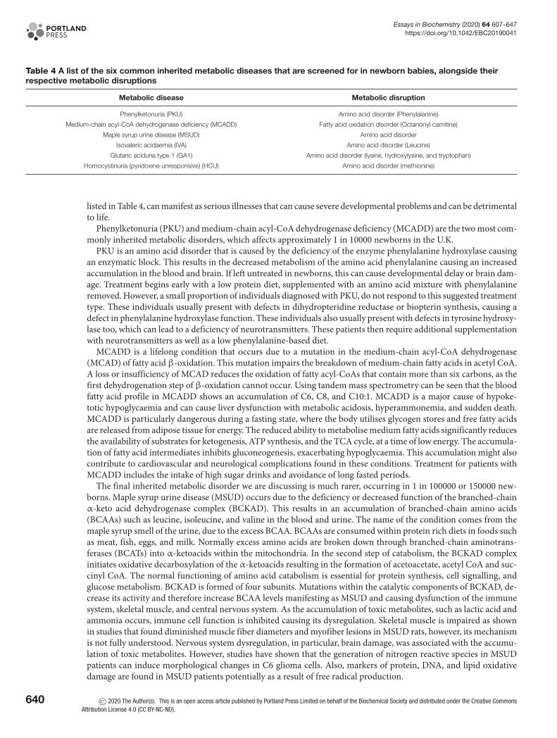
Essays in Biochemistry (2020) 64 607–647https://doi.org/10.1042/EBC20190041
Table 4 A list of the six common inherited metabolic diseases that are screened for in newborn babies, alongside theirrespective metabolic disruptions
Metabolic disease Metabolic disruption
Phenylketonuria (PKU) Amino acid disorder (Phenylalanine)
Medium-chain acyl-CoA dehydrogenase deficiency (MCADD) Fatty acid oxidation disorder (Octanonyl carnitine)
Maple syrup urine disease (MSUD) Amino acid disorder
Isovaleric acidaemia (IVA) Amino acid disorder (Leucine)
Glutaric aciduria type 1 (GA1) Amino acid disorder (lysine, hydroxylysine, and tryptophan)
Homocystinuria (pyridoxine unresponsive) (HCU) Amino acid disorder (methionine)
listed in Table 4, can manifest as serious illnesses that can cause severe developmental problems and can be detrimentalto life.
Phenylketonuria (PKU) and medium-chain acyl-CoA dehydrogenase deficiency (MCADD) are the two most com-monly inherited metabolic disorders, which affects approximately 1 in 10000 newborns in the U.K.
PKU is an amino acid disorder that is caused by the deficiency of the enzyme phenylalanine hydroxylase causingan enzymatic block. This results in the decreased metabolism of the amino acid phenylalanine causing an increasedaccumulation in the blood and brain. If left untreated in newborns, this can cause developmental delay or brain dam-age. Treatment begins early with a low protein diet, supplemented with an amino acid mixture with phenylalanineremoved. However, a small proportion of individuals diagnosed with PKU, do not respond to this suggested treatmenttype. These individuals usually present with defects in dihydropteridine reductase or biopterin synthesis, causing adefect in phenylalanine hydroxylase function. These individuals also usually present with defects in tyrosine hydroxy-lase too, which can lead to a deficiency of neurotransmitters. These patients then require additional supplementationwith neurotransmitters as well as a low phenylalanine-based diet.
MCADD is a lifelong condition that occurs due to a mutation in the medium-chain acyl-CoA dehydrogenase(MCAD) of fatty acid β-oxidation. This mutation impairs the breakdown of medium-chain fatty acids in acetyl CoA.A loss or insufficiency of MCAD reduces the oxidation of fatty acyl-CoAs that contain more than six carbons, as thefirst dehydrogenation step of β-oxidation cannot occur. Using tandem mass spectrometry can be seen that the bloodfatty acid profile in MCADD shows an accumulation of C6, C8, and C10:1. MCADD is a major cause of hypoke-totic hypoglycaemia and can cause liver dysfunction with metabolic acidosis, hyperammonemia, and sudden death.MCADD is particularly dangerous during a fasting state, where the body utilises glycogen stores and free fatty acidsare released from adipose tissue for energy. The reduced ability to metabolise medium fatty acids significantly reducesthe availability of substrates for ketogenesis, ATP synthesis, and the TCA cycle, at a time of low energy. The accumula-tion of fatty acid intermediates inhibits gluconeogenesis, exacerbating hypoglycaemia. This accumulation might alsocontribute to cardiovascular and neurological complications found in these conditions. Treatment for patients withMCADD includes the intake of high sugar drinks and avoidance of long fasted periods.
The final inherited metabolic disorder we are discussing is much rarer, occurring in 1 in 100000 or 150000 new-borns. Maple syrup urine disease (MSUD) occurs due to the deficiency or decreased function of the branched-chainα-keto acid dehydrogenase complex (BCKAD). This results in an accumulation of branched-chain amino acids(BCAAs) such as leucine, isoleucine, and valine in the blood and urine. The name of the condition comes from themaple syrup smell of the urine, due to the excess BCAA. BCAAs are consumed within protein rich diets in foods suchas meat, fish, eggs, and milk. Normally excess amino acids are broken down through branched-chain aminotrans-ferases (BCATs) into α-ketoacids within the mitochondria. In the second step of catabolism, the BCKAD complexinitiates oxidative decarboxylation of the α-ketoacids resulting in the formation of acetoacetate, acetyl CoA and suc-cinyl CoA. The normal functioning of amino acid catabolism is essential for protein synthesis, cell signalling, andglucose metabolism. BCKAD is formed of four subunits. Mutations within the catalytic components of BCKAD, de-crease its activity and therefore increase BCAA levels manifesting as MSUD and causing dysfunction of the immunesystem, skeletal muscle, and central nervous system. As the accumulation of toxic metabolites, such as lactic acid andammonia occurs, immune cell function is inhibited causing its dysregulation. Skeletal muscle is impaired as shownin studies that found diminished muscle fiber diameters and myofiber lesions in MSUD rats, however, its mechanismis not fully understood. Nervous system dysregulation, in particular, brain damage, was associated with the accumu-lation of toxic metabolites. However, studies have shown that the generation of nitrogen reactive species in MSUDpatients can induce morphological changes in C6 glioma cells. Also, markers of protein, DNA, and lipid oxidativedamage are found in MSUD patients potentially as a result of free radical production.
640 © 2020 The Author(s). This is an open access article published by Portland Press Limited on behalf of the Biochemical Society and distributed under the Creative CommonsAttribution License 4.0 (CC BY-NC-ND).
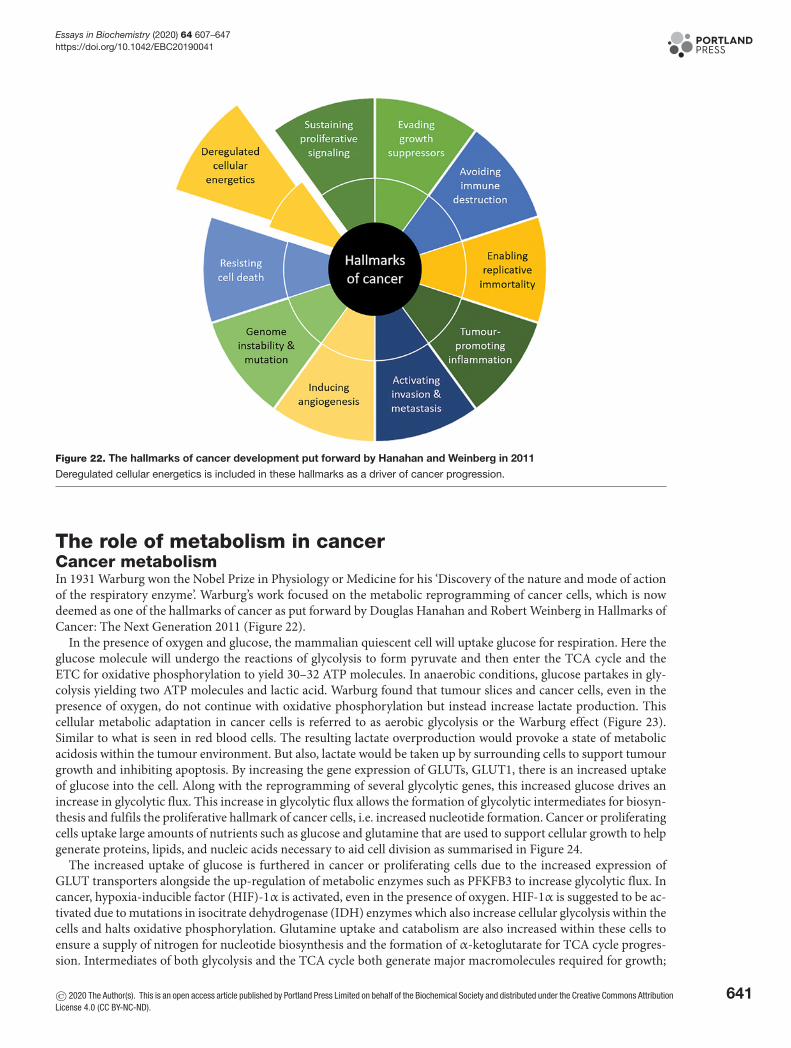
Essays in Biochemistry (2020) 64 607–647https://doi.org/10.1042/EBC20190041
Figure 22. The hallmarks of cancer development put forward by Hanahan and Weinberg in 2011
Deregulated cellular energetics is included in these hallmarks as a driver of cancer progression.
The role of metabolism in cancerCancer metabolismIn 1931 Warburg won the Nobel Prize in Physiology or Medicine for his ‘Discovery of the nature and mode of actionof the respiratory enzyme’. Warburg’s work focused on the metabolic reprogramming of cancer cells, which is nowdeemed as one of the hallmarks of cancer as put forward by Douglas Hanahan and Robert Weinberg in Hallmarks ofCancer: The Next Generation 2011 (Figure 22).
In the presence of oxygen and glucose, the mammalian quiescent cell will uptake glucose for respiration. Here theglucose molecule will undergo the reactions of glycolysis to form pyruvate and then enter the TCA cycle and theETC for oxidative phosphorylation to yield 30–32 ATP molecules. In anaerobic conditions, glucose partakes in gly-colysis yielding two ATP molecules and lactic acid. Warburg found that tumour slices and cancer cells, even in thepresence of oxygen, do not continue with oxidative phosphorylation but instead increase lactate production. Thiscellular metabolic adaptation in cancer cells is referred to as aerobic glycolysis or the Warburg effect (Figure 23).Similar to what is seen in red blood cells. The resulting lactate overproduction would provoke a state of metabolicacidosis within the tumour environment. But also, lactate would be taken up by surrounding cells to support tumourgrowth and inhibiting apoptosis. By increasing the gene expression of GLUTs, GLUT1, there is an increased uptakeof glucose into the cell. Along with the reprogramming of several glycolytic genes, this increased glucose drives anincrease in glycolytic flux. This increase in glycolytic flux allows the formation of glycolytic intermediates for biosyn-thesis and fulfils the proliferative hallmark of cancer cells, i.e. increased nucleotide formation. Cancer or proliferatingcells uptake large amounts of nutrients such as glucose and glutamine that are used to support cellular growth to helpgenerate proteins, lipids, and nucleic acids necessary to aid cell division as summarised in Figure 24.
The increased uptake of glucose is furthered in cancer or proliferating cells due to the increased expression ofGLUT transporters alongside the up-regulation of metabolic enzymes such as PFKFB3 to increase glycolytic flux. Incancer, hypoxia-inducible factor (HIF)-1α is activated, even in the presence of oxygen. HIF-1α is suggested to be ac-tivated due to mutations in isocitrate dehydrogenase (IDH) enzymes which also increase cellular glycolysis within thecells and halts oxidative phosphorylation. Glutamine uptake and catabolism are also increased within these cells toensure a supply of nitrogen for nucleotide biosynthesis and the formation of α-ketoglutarate for TCA cycle progres-sion. Intermediates of both glycolysis and the TCA cycle both generate major macromolecules required for growth;
© 2020 The Author(s). This is an open access article published by Portland Press Limited on behalf of the Biochemical Society and distributed under the Creative Commons AttributionLicense 4.0 (CC BY-NC-ND).
641

Essays in Biochemistry (2020) 64 607–647https://doi.org/10.1042/EBC20190041
Figure 23. The Warburg effect
A cell with no oxygen supply generates lactate as it undergoes glycolysis anaerobically. In aerobic conditions, with oxygen, a cell proceeds with
oxidative phosphorylation yielding a higher number of ATP molecules. However, in a cancer tumour microenvironment, the ability to carry out oxidative
phosphorylation is disrupted, despite oxygen presence. Instead, lactate is formed, and a cell is suggested to undergo aerobic glycolysis.
Figure 24. Increased glucose and glutamine uptake and utilisation drive the increased synthesis of nucleotide, protein, and
lipid synthesis in cancer cells
The up-regulation of glycolysis increases cellular lactate concentrations.
ribose-5-phosphate for nucleotide synthesis, serine, and glycine to fuel protein synthesis and support nucleotide syn-thesis, and citrate which is generated to be exported into the cytoplasm providing acetyl CoA for fatty acid synthesis.
Targeting metabolism in cancer cell drug discoveryAs one of the hallmarks of cancer, targeting metabolic pathways is critical in the development of new therapeutics, andmight serve as useful targets to inhibit cancer cell growth. As cancer cells are rapidly dividing, any disruption in theirability to obtain energy will slow their proliferation. There are two main strategies in targeting glucose metabolism incancer cells, this is to either inhibit anaerobic glycolysis, thereby starving the cells, or by increasing glucose oxidation,increasing the usage of mitochondria and therefore apoptosis.
Examples of targeting glycolysis are found in inhibitors of hexokinase (2-deoxyglucose (2DG)) or enolase (phos-phonoacetohydroxamate (PHAH) or SF-2312). These compounds aim to reduce the flow of glucose within cells thathave a high glycolytic flux, i.e. cancer cells. This would, therefore, reduce the energy available to the cell and slowrapid cell proliferation. The problem is that these compounds might have additional effects in red blood cells, whichare solely reliant on glycolysis.
Another way of reducing anaerobic glycolysis is to inhibit the export of lactic acid from the cell. By targeting themonocarboxylate transporters (MCTs) (AZD3965 or syrosingopine), this increases the intracellular concentration oflactic acid and either increases the acidity of the cell and/or decreases the activity of LDH. Decreased LDH activity,
642 © 2020 The Author(s). This is an open access article published by Portland Press Limited on behalf of the Biochemical Society and distributed under the Creative CommonsAttribution License 4.0 (CC BY-NC-ND).
Essays in Biochemistry (2020) 64 607–647https://doi.org/10.1042/EBC20190041
leads to an accumulation of NADH and pyruvate, which both lead to inhibition of glycolysis by feedback mechanismsdiscussed earlier.
To target glucose oxidation, the drug dichloroacetate (DCA) inhibits PDK, which causes the activation of PDHand an increase in acetyl CoA. PDH activation allows mitochondrial oxidation of pyruvate to occur, disrupting themetabolic glycolytic advantage of cancer cells. Furthermore, DCA acts to counter the acidosis caused by increasedlactate production by decreasing flow through LDH, inhibiting the progression and development of the tumour mi-croenvironment. Therefore, drug therapies have shown the ability to target the altered cellular metabolic pathways incancer cells to slow the uptake of glucose by disrupting and inhibiting glycolysis and the production of lactate.
Imaging metabolism in cancerAlong with targeting metabolism for treating cancer, it is also possible to hijack the importance of metabolism incancer to find and identify tumours. Several techniques take advantage of changes in metabolic state to image where atumour is and also, its response to treatment. Two techniques: positron emission tomography and magnetic resonancespectroscopy (MRS) or imaging (MRI) have demonstrated their ability in this field.
FDG–PET2-deoxy-2-[18F]fluoro-D-glucose (FDG) is a radiotracer that acts similarly to glucose, by being taken up by glucosetransporters, i.e. GLUTs and SGLT. Once inside the cell, FDG goes through the first step in glycolysis and is phos-phorylated to [18F]FDG-6-phosphate, which unlike G6P is unable to continue through glycolysis. Outside of the liver(and fasted kidney), the lack of glucose-6-phosphatase traps [18F]FDG-6-phosphate inside the cells. As this moleculeaccumulates, the 18F radiotracer allows the molecule to be detected using PET, which is overlaid onto an anatomicalimage generated by computed tomography (CT) or MRI. This tracer has been useful in the detection and sizing of tu-mours, as an increase in [18F]FDG-6-phosphate detection compared with the surrounding tissue, is indicative of highglucose uptake and rapidly proliferating cells and possibly cancer. This is particularly useful, as metabolic imaging ofcancerous cells as the potential to detect tumours, before they are visible using conventional imaging, such as X-ray,CT, or MRI. However, there are still limits to the sensitivity of this technique for the detection of tumours, it can alsobe difficult to detect changes in the brain (where there is a high background glucose uptake) and it only measuresglucose uptake and not directly glycolysis.
Hyperpolarised 13C MRI/MRSWhilst MRI and MRS have been around for many years, monitoring real-time metabolism using hyperpolarisedcarbon 13 (13C) is relatively new. 13C MRS generates a set of peaks depending on the molecular environment that the13C molecule is found in, this is called a spectrum. The new technique relies on an external machine (a polariser) tosignificantly enhance the signal from a 13C containing compound. This enhanced state is temporary (between 1 and 5min), so the compound needs to be rapidly injected into a patient inside an MRI machine to enable detection. UnlikePET, 13C tracers are non-radioactive and can also monitor real-time metabolism. The most common tracer has been[1-13C]pyruvate, which can be rapidly taken up by cells and has three possible fates; through PDH, ALT, or LDH.When [1-13C]pyruvate is metabolised by one of these three enzymes, it produces a new 13C compound, which has adifferent peak on the spectrum. Meaning you can trace the conversion of pyruvate into CO2/bicarbonate (acetyl-CoAis not seen as a carbon is lost as CO2), alanine, and lactate, respectively. These can be visualised on a spectrum as aseries of peaks, with a new spectrum being produced every 0.1–1 s, thereby generating a time course to work outenzyme kinetics. Or the peaks can have spatial information added and an image of where metabolism is occurringcan be produced for a specific tissue.
The first in man trial for using this technique in patients with prostate cancer was only completed in 2013. Theymonitored the conversion of [1-13C]pyruvate into [1-13C]lactate and produced images of where this conversion oc-curred in the human prostate. This was used as a marker of elevated glycolytic flux (although it only measures thelast step in anaerobic glycolysis) and showed cancerous tissue amongst healthy tissue. In rodent models of cancer thistechnique can also monitor the effectiveness of treatment in tumours. This technique has also been applied to thestudy of cardiac metabolism, where it was able to image real-time metabolism in the human heart.
Both PET and hyperpolarised 13C MRS/MRI have their limitations, these are mainly due to the injection of tracers.These tracers only provide you with a snapshot of the processes occurring, as they only make up a very small percent-age of the endogenous metabolites. They also only show you a limited number of steps within metabolic pathwaysand several assumptions are required to enable analysis and understanding of the pathways at work. Having said that,these are exciting areas and have shown an ability to visualise metabolism occurring in real-time, within the human
© 2020 The Author(s). This is an open access article published by Portland Press Limited on behalf of the Biochemical Society and distributed under the Creative Commons AttributionLicense 4.0 (CC BY-NC-ND).
643
Essays in Biochemistry (2020) 64 607–647https://doi.org/10.1042/EBC20190041
body, which takes us from perfused animal organ and tissue slices of last century to live metabolic imaging of thehuman body.
The importance of metabolism in biotechnologyThe potential of metabolismAlong with drug discovery and diagnosis of human diseases, metabolism plays an exciting role in the developmentof biotechnology. Whether that is bio-engineering of Escherichia coli to meet the growing demand for bioethanol ormanipulating plants to generate better defence against pests, there are lots of possibilities. Advancements of knowl-edge in these particular fields have allowed the development of more specific biotechnological advancements. Theseadvancements allow us to overcome hurdles that once caused issues with yield and even resistance. These ideasare discussed below as how biotechnology has ensured efficient and elaborate systems to target particular areas ofmetabolism.
Bioethanol production from E. coliEthanol holds a variety of applications in the food, chemical, medical, and health industry and is commonly usedin beverages, fuels, and paint. Ethanol is largely produced from coal and petroleum using non-renewable processes;however, ethanol can be produced by fermentation of sugars and starch. Microorganisms hold the capacity to pro-duce ethanol via fermentation and due to this, have been identified and studied to produce ethanol in high capacity.Microorganisms can be metabolically engineered to redirect carbon sources to desired fuel products. One of thesemicroorganisms is E. coli.
E. coli can naturally utilise a variety of carbon sources such as sugars and sugar alcohols, under both aerobic andanaerobic conditions. All biofuels that are derived from E. coli are derived from the modification of central carboncatabolism including the conversion of pentose/hexose sugar molecules into diatomic carbon.
E. coli can produce ethanol under anaerobic conditions to produce 1 mole of ethanol for every 1 mole of glu-cose used, alongside formate and acetate. The reduction reaction that occurs to synthesise these, utilises two NADHmolecules but the initial stages of glycolysis only yield one. This causes a redox imbalance. This redox imbalance isovercome by E. coli by balancing the production of ethanol by oxidation of acetyl CoA into acetate, which requiresno NADH. These E. coli were engineered to express pyruvate decarboxylase and alcohol dehydrogenase II genes ofZymomonas mobilis. This pathway produces 95% ethanol, without a redox imbalance as it only requires 1 NADHmolecule.
Modification of metabolism in plantsPlant metabolism is highly organised and controlled by innate regulations alongside homoeostatic mechanisms. Theirmetabolic procedures are highly compartmentalised where each organelle carries out a specific metabolic function.
Within nature, plants are constantly challenged by insect herbivores. In response to this, as a defence to reduceinsect-related damage to the plant, they produce toxins and small deterrent molecules for protection. Some of thesedefence responses are inducible where these are related to the up-regulation or down-regulation of metabolites andproteins in response to an attack.
For example, the tobacco plant naturally synthesises nicotine to deter insects from eating them. However, if anattack does occur on a tobacco plant, this response is up-regulated. Here, the roots synthesise large amounts of nico-tine, and this is transported to the shoot. This response is regulated by jasmonic acid and auxin, both signals that aretriggered by wounding. This increase in nicotine production is to avoid further herbivore attack.
In addition to defences such as those described above, plants can also induce changes in their metabolism in re-sponse to attacks. These include elevating or decreasing their photosynthetic efficiency, remobilisation of carbon andnitrogen resources alongside altering their plant growth.
Amino acids are the form of nitrogen found in plants. They can regulate plant growth and are precursors fordefence-related metabolites. When plants are infested by herbivore insects, studies found that amino acid produc-tion increased to synthesise defensive metabolites to ensure a second attack does not occur. This increase in aminoacid production occurs as genes involved in amino acid synthesis are up-regulated and sulphur assimilation, which isultimately required for the biosynthesis of cysteine and methionine, are all up-regulated upon caterpillar feeding onArabidopsis thaliana. Furthering this, a similar study further found that the increased expression of genes involvedin amino acid biosynthesis, in particular, related to methionine and tryptophan synthesis, led to a greater accumula-tion of glucosinolates. These are defence-related molecules that are derived from methionine and tryptophan in A.
644 © 2020 The Author(s). This is an open access article published by Portland Press Limited on behalf of the Biochemical Society and distributed under the Creative CommonsAttribution License 4.0 (CC BY-NC-ND).

Essays in Biochemistry (2020) 64 607–647https://doi.org/10.1042/EBC20190041
thaliana. Interestingly, this was also found in tomatoes. Upon attack, tomato plants accumulate tryptophan withintheir stems and apex of tomatoes, to serve as a precursor for the production of defensive metabolites.
Plant biotechnology is a set of techniques that are used to adapt plants for survival and defence. One of the mosteffective ways to manage plant pathogenicity is to use genetic modification alongside genome editing. This is par-ticularly useful in the agriculture industry to ensure increased food supply. Globally, up to 30% of food is damagedby pathogens and disease pests. Therefore, the adaptation of plants to pathogens and pests is imperative for survival.Gene modification and genome editing can help manage plant pathogens to ensure plant cell survival in times ofattack, these solutions currently include bacterial, viral, fungal, and oomycete pathogens.
As knowledge regarding the plant immune system has expanded, it has been shown that plants can distinguish be-tween ‘self’ and ‘non-self’ through monitoring the extracellular and intracellular environment. However, pathogenscan overcome this system by producing proteins and effectors to suppress plant host immunity and colonise it. Abacterial pathogen can hijack the coronatine-insensitive protein 1 jasmonate receptor, altering the plant’s defence re-sponse and activating jasmonate responses and suppressing salicylic acid defence pathways. Manipulation like thesehas allowed the development of biotechnology to intervene at the point of pathogen perception. These can be cre-ated by introducing receptors from other plants with novel recognition specificity, reactivation of genes disabled inNOD-like receptors, and modification of domains in NOD-like receptors that are targeted by pathogens. This knowl-edge reveals which components of the host can be manipulated to promote a disease state. Therefore, by removingthese vulnerable points or replacing them with variants that are immune to specific effectors, the natural functional-ity of the plant can be retained. Bacterial pathogens, that express the transcription activate like effectors that activatesusceptibility genes within the host, can be overcome by the deletion of TAL DNA-binding sites in the promoter.Another approach includes engineering resistance to bacterial pathogens by adding TAL effector binding sites to anexecutor gene.
Another way to regulate plant pathogen perception is by the intervention of the defence signalling pathways. Themajor hormones involved in defence signalling are salicylic acid, jasmonate, and ethylene. In tomato plants, a loss offunction allele was engineered for Downy mildew resistance 6 (DMR6) to ensure resistance to biotrophic pathogens.DMR6 is widely conserved and encodes a salicylate-5-hydrozylase enzyme around infection sites.
Biotechnology advancements regarding plants and plant metabolism help ensure defence and healthy plant growth.In conclusion, biotechnology has allowed the intervention of defence signalling and regulation, pathogen recognition,effector recognition, and allowed targeting of genes associated with plant pathogenicity.
ConclusionAt least 25 Nobel Prizes have been awarded for work related to metabolism, with the most recent for theoxygen-sensing role of HIF1α in 2019. This review has introduced the basic metabolic pathways of life and demon-strated how a series of reactions can combine to sustain life. Over the last 150–200 years, the understanding ofmetabolism has moved from individual enzymes and metabolites to the complicated network we see today. This hasallowed us to visualise metabolism occurring in real-time within the human body, to help us understand metabolicalterations in human disease. Manipulation of metabolic pathways in both microorganisms and plants has also ledthe way in the development of new biotechnological techniques.
Competing InterestsThe authors declare that there are no competing interests associated with the manuscript.
FundingAyesha Judge is funded by a PhD studentship from Coventry University with additional funding and support from the UniversityAlliance Doctoral Training Alliance. Michael Dodd is funded by Coventry University and the Centre for Sport, Exercise and LifeSciences.
AcknowledgementsWe would like to thank Jennifer Greaves (Coventry University) for her useful discussions on this article and A.R. Teal (Faculty ofScience and Technology, North East Surrey College of Technology) for the original Metabolism BASC article (1994) on which thisessay is partly based.
© 2020 The Author(s). This is an open access article published by Portland Press Limited on behalf of the Biochemical Society and distributed under the Creative CommonsAttribution License 4.0 (CC BY-NC-ND).
645
Essays in Biochemistry (2020) 64 607–647https://doi.org/10.1042/EBC20190041
AbbreviationsACP, acyl carrier protein; ADP, adenosine diphosphate; AMP, adenosine monophosphate; ATP, adenosine triphosphate; BCAA,branched-chain amino acid; BCAT, branched-chain aminotransferase; BCKAD, branched-chain α-keto acid dehydrogenasecomplex; CoA, coenzyme A; CPT1, carnitine palmitoyl transferase I; CT, computed tomography; DCA, dichloroacetate; DHAP,dihydroxyacetone phosphate; DMR6, Downy mildew resistance 6; ER, enoyl ACP reductase; ETC, electron transport chain; FAS,fatty acid synthase; FDG, 2-deoxy-2-[18F]fluoro-D-glucose; F6P, fructose-6-phosphate; GAP, glyceraldehyde-3-phosphate;GDP, guanosine diphosphate; GLUT, glucose transporter; GTP, guanine triphosphate; G6P, glucose-6-phosphate; G6PI, G6Pisomerase; HDL, high-density lipoprotein; HIF, hypoxia-inducible factor; HSL, hormone-sensitive lipase; IRS, insulin receptorsubstrate; LDH, lactate dehydrogenase; LDL, low-density lipoprotein; MCAD, medium-chain acyl-CoA dehydrogenase; MCADD,medium-chain acyl-CoA dehydrogenase deficiency; MRI, magnetic resonance imaging; MRS, magnetic resonance spec-troscopy; MSUD, maple syrup urine disease; NAD(P)H, nicotinamide adenine nucleotide (phosphate); NAG, N-acetyl-glutamate;NH4
+, ammonium ions; NO2−, nitrite; PC, pyruvate carboxylase; PDH, pyruvate dehydrogenase; PEP, phosphoenolpyruvate;
PEPCK, PEP carboxykinase; PFK, phosphofructokinase; PK, pyruvate kinase; PKU, phenylketonuria; PPi, inorganic pyrophos-phate group; PPARα, peroxisome proliferator-activated receptor α; PPP, pentose phosphate pathway; SGLT, sodium glucosecotransporter; TAG, triglyceride; TCA, tricarboxylic acid; UDP-glucose, uridine diphosphate glucose; VLDL, very low-densitylipoprotein; 1,3-BPG, 1,3-bisphosphoglycerate; 3PG, 3-phosphoglycerate.
References and further readingAlbrecht, T. and Baron, S. (1996) Medical Microbiology, University of Texas Medical Branch at Galveston, Dept. of Microbiology & Immunology, Galveston,
TX, U.S.A., Chapter 4Ameer, F., Scandiuzzi, L., Hasnain, S., Kalbacher, H. and Zaidi, N. (2014) De novo lipogenesis in health and disease. Metabolism 63, 895–902,
https://doi.org/10.1016/j.metabol.2014.04.003Berg, J., Tymoczko, J. and Stryer, L. (2002) The first step in amino acid degradation is the removal of nitrogen.
https://www.ncbi.nlm.nih.gov/books/NBK22475Bishop, S. and Campbell, J. (1965) Arginine and urea biosynthesis in the earthworm Lumbricus terrestris. Comp. Biochem. Physiol. 15, 51–71,
https://doi.org/10.1016/0010-406X(65)90240-9Brown, G., Brown, W. and Cohen, P. (1962) Comparative biochemistry of urea synthesis IV. [14C]Urea synthesis by liver slices of the metamorphosing
tadpole. Biochim. Biophys. Acta 60, 185–186, https://doi.org/10.1016/0006-3002(62)90387-6Chirala, S., Jayakumar, A., Gu, Z. and Wakil, S. (2001) Human fatty acid synthase: Role of interdomain in the formation of catalytically active synthase
dimer. Proc. Natl. Acad. Sci. U.S.A. 98, 3104–3108, https://doi.org/10.1073/pnas.051635998DeBerardinis, R. and Chandel, N. (2016) Fundamentals of cancer metabolism. Sci. Adv. 2, e1600200, https://doi.org/10.1126/sciadv.1600200Esse, H., Reuber, T. and Does, D. (2019) Genetic modification to improve disease resistance in crops. New Phytol. 225, 70–86,
https://doi.org/10.1111/nph.15967Frayn, K. and Evans, R. (2018) Human Metabolism: A Regulatory Perspective, 4th , Wiley BlackwellGartner, V., McGuire, P. and Lee, P. (2015) Child Neurology: medium-chain acyl-coenzyme A dehydrogenase deficiency. Neurology 85, e37–e40,
https://doi.org/10.1212/WNL.0000000000001786Gibala, M., Young, M. and Taegtmeyer, H. (2000) Anaplerosis of the citric acid cycle: role in energy metabolism of heart and skeletal muscle. Acta Physiol.
Scand. 168, 657–665, https://doi.org/10.1046/j.1365-201x.2000.00717.xGuo, X., Li, H., Xu, H., Woo, S., Dong, H., Lu, F. et al. (2012) Glycolysis in the control of blood glucose homeostasis. Acta Pharm. Sin. B 2, 358–367,
https://doi.org/10.1016/j.apsb.2012.06.002Hanahan, D. and Weinberg, R. (2011) Hallmarks of cancer: the next generation. Cell 144, 646–674, https://doi.org/10.1016/j.cell.2011.02.013He, J., Ford, H., Carroll, J., Douglas, C., Gonzales, E., Ding, S. et al. (2018) Assembly of the membrane domain of ATP synthase in human mitochondria.
Proc. Natl. Acad. Sci. U.S.A. 115, 2988–2993, https://doi.org/10.1073/pnas.1722086115Klotz, M. and Stein, L. (2008) Nitrifier genomics and evolution of the nitrogen cycle. FEMS Microbiol. Lett. 278, 146–156,
https://doi.org/10.1111/j.1574-6968.2007.00970.xKoppolu, V. and Vasigala, V. (2016) Role of Escherichia coli in biofuel production. Microbiol. Insights 9, 29–35, https://doi.org/10.4137/MBI.S10878Kornberg, H. and Beevers, H. (1957) A mechanism of conversion of fat to carbohydrate in castor beans. Nature 180, 35–36,
https://doi.org/10.1038/180035a0Krebs, H. (1973) The discovery of the ornithine cycle of urea synthesis. Biochem. Educ. 1, 19–23, https://doi.org/10.1016/0307-4412(73)90048-4Leonardi, R., Zhang, Y., Rock, C. and Jackowski, S. (2005) Coenzyme A: back in action. Prog. Lipid Res. 44, 125–153,
https://doi.org/10.1016/j.plipres.2005.04.001Li, C., Teng, W., Shi, Q. and Zhang, F. (2007) Multiple signals regulate nicotine synthesis in tobacco plant. Plant Signal. Behav. 2, 280–281Navale, A. and Paranjape, A. (2016) Glucose transporters: physiological and pathological roles. Biophys. Rev. 8, 5–9,
https://doi.org/10.1007/s12551-015-0186-2Nelson, S., Kurhanewicz, J., Vigneron, D., Larson, P., Harzstark, A., Ferrone, M. et al. (2013) Metabolic imaging of patients with prostate cancer using
hyperpolarized [1-13C]pyruvate. Sci. Transl. Med. 5, 198.ra108, https://doi.org/10.1126/scitranslmed.3006070Owen, O., Kalhan, S. and Hanson, R. (2002) The key role of anaplerosis and cataplerosis for citric acid cycle function. J. Biol. Chem. 277, 30409–30412,
https://doi.org/10.1074/jbc.R200006200
646 © 2020 The Author(s). This is an open access article published by Portland Press Limited on behalf of the Biochemical Society and distributed under the Creative CommonsAttribution License 4.0 (CC BY-NC-ND).

Essays in Biochemistry (2020) 64 607–647https://doi.org/10.1042/EBC20190041
Palomer, X., Pizarro-Delgado, J., Barroso, E. and Vazquez-Carrera, M. (2018) Palmitic and oleic acid: the yin and yang of fatty acids in type 2 diabetesmellitus. Trends Endocrinol. Metab. 29, 178–190
Park, J., Davis, R. and Sue, C. (2018) Mitochondrial dysfunction in Parkinson’s disease: new mechanistic insights and therapeutic perspectives. Curr.Neurol. Neurosci. Rep. 18, https://doi.org/10.1007/s11910-018-0829-3
Rider, O., Apps, A., Miller, J., Lau, J., Lewis, A., Peterzan, M. et al. (2020) Noninvasive in vivo assessment of cardiac metabolism in the healthy and diabetichuman heart using hyperpolarized 13 C MRI. Circ. Res. 126, 725–736, https://doi.org/10.1161/CIRCRESAHA.119.316260
Robinson, P. (2015) Enzymes: principles and biotechnological applications. Essays Biochem. 59, 1–41, https://doi.org/10.1042/bse0590001Roger, A., Munoz-Gomez, S. and Kamikawa, R. (2017) The origin and diversification of mitochondria. Curr. Biol. 27, R1177–R1192,
https://doi.org/10.1016/j.cub.2017.09.015Sanchez-Baracaldo, P., Raven, J.A., Pisani, D. and Knoll, A.H. (2017) Early photosynthetic eukaryotes inhabited low-salinity habitats. Proc. Natl. Acad. Sci.
U.S.A. 114, E7737–E7745, https://doi.org/10.1073/pnas.1620089114Schmitz, O., Brock, B. and Rungby, J. (2004) Amylin agonists: a novel approach in the treatment of diabetes. Diabetes 53, S233–S238,
https://doi.org/10.2337/diabetes.53.suppl˙3.S233Sitta, A., Ribas, G., Mescka, C., Barschak, A., Wajner, M. and Vargas, C. (2013) Neurological damage in MSUD: the role of oxidative stress. Cell. Mol.
Neurobiol. 34, 157–165, https://doi.org/10.1007/s10571-013-0002-0Sonnet, D., O’Leary, M., Gutierrez, A., Nguyen, S.M., Mateen, S., Hsu, Y. et al. (2016) Metformin inhibits branched chain amino acid (BCAA) derived
ketoacidosis and promotes metabolic homeostasis in MSUD. Sci. Rep. 6, https://doi.org/10.1038/srep28775Stumpf, P., Conn, E., Miflin, B. and Lea, P. (1987) The Biochemistry of Plants, 11th , pp. 29–30, Academic Press, San DiegoVoet, D., Voet, J. and Pratt, C. (2013) Principles of Biochemistry, 4th , (Hoboken, N.J., ed.), John Wiley & SonsWang, Z., Ohliger, M., Larson, P., Gordon, J., Bok, R., Slater, J. et al. (2019) Hyperpolarized 13C MRI: state of the art and future directions. Radiology 291,
273–284, https://doi.org/10.1148/radiol.2019182391Weiner, I. and Verlander, J. (2013) Renal ammonia metabolism and transport. Compr. Physiol. 3 (1)), 201–220, https://doi.org/10.1002/cphy.c120010Wilson, B., Schisler, J. and Willis, M. (2010) Sir Hans Adolf Krebs: architect of metabolic cycles. Lab. Med. 41, 377–380,
https://doi.org/10.1309/LMZ5ZLAC85GFMGHUYudkoff, M. (1999) Urea cycle. https://www.ncbi.nlm.nih.gov/books/NBK27982Zhang, S., Hulver, M., McMillan, R., Cline, M. and Gilbert, E. (2014) The pivotal role of pyruvate dehydrogenase kinases in metabolic flexibility. Nutr. Metab.
11, 10Zhou, S., Lou, Y., Tzin, V. and Jander, G. (2015) Alteration of plant primary metabolism in response to insect herbivory. Plant Physiol. 169 (3)), 1488–98,
https://doi.org/10.1104/pp.15.01405
© 2020 The Author(s). This is an open access article published by Portland Press Limited on behalf of the Biochemical Society and distributed under the Creative Commons AttributionLicense 4.0 (CC BY-NC-ND).
647
Related Documents