Meta-analytic evidence of differential prefrontal and early sensory cortex activity during non-social sensory perception in autism Nazia Jassim 1* , Simon Baron-Cohen 1,# and John Suckling 1,2,# Affiliations: 1. Autism Research Centre, Department of Psychiatry, University of Cambridge, UK Douglas House, 18B Trumpington Road, Cambridge CB2 8AH, United Kingdom 2. Department of Psychiatry, University of Cambridge, UK Herchel Smith Building for Brain and Mind Sciences, Forvie Site, Robinson Way, Cambridge, CB2 0SZ, United Kingdom # Joint last authors. Open access published version now available: Jassim, N., Baron-Cohen, S., & Suckling, J. (2021). Meta-analytic evidence of differential prefrontal and early sensory cortex activity during non-social sensory perception in autism. Neuroscience & Biobehavioral Reviews. https://doi.org/10.1016/j.neubiorev.2021.04.014

Welcome message from author
This document is posted to help you gain knowledge. Please leave a comment to let me know what you think about it! Share it to your friends and learn new things together.
Transcript

Meta-analytic evidence of differential prefrontal and early
sensory cortex activity during non-social sensory
perception in autism
Nazia Jassim1*, Simon Baron-Cohen1,# and John Suckling1,2,#
Affiliations:
1. Autism Research Centre, Department of Psychiatry, University of Cambridge, UK
Douglas House, 18B Trumpington Road, Cambridge CB2 8AH, United Kingdom
2. Department of Psychiatry, University of Cambridge, UK
Herchel Smith Building for Brain and Mind Sciences, Forvie Site, Robinson Way,
Cambridge, CB2 0SZ, United Kingdom
# Joint last authors.
Open access published version now available:
Jassim, N., Baron-Cohen, S., & Suckling, J. (2021). Meta-analytic evidence of differential
prefrontal and early sensory cortex activity during non-social sensory perception in autism.
Neuroscience & Biobehavioral Reviews. https://doi.org/10.1016/j.neubiorev.2021.04.014
Sensory perception in autism: an fMRI ALE meta-analysis
2
Sensory perception in autism: an fMRI ALE meta-analysis
Nazia Jassim1*, Simon Baron-Cohen1,# and John Suckling1,2,#
Affiliations:
3. Autism Research Centre, Department of Psychiatry, University of Cambridge, UK
Douglas House, 18B Trumpington Road, Cambridge CB2 8AH, United Kingdom
4. Department of Psychiatry, University of Cambridge, UK
Herchel Smith Building for Brain and Mind Sciences, Forvie Site, Robinson Way,
Cambridge, CB2 0SZ, United Kingdom
# Joint last authors.
Word Count: 3718
*Correspondence to Nazia Jassim
Present Address: Autism Research Centre, Douglas House, 18B Trumpington Road,
Cambridge CB2 8AH, United Kingdom
Phone: (+44) 01223 465223
Email: [email protected]
ORCID: orcid.org/0000-0001-9761-7784
Sensory perception in autism: an fMRI ALE meta-analysis
3
Abstract:
Sensory sensitivities occur in up to 90% of autistic individuals. With the recent inclusion of
sensory symptoms in the diagnostic criteria for autism, there is a current need to develop
neural hypotheses related to autistic sensory perception. Using activation likelihood
estimation (ALE), we meta-analysed 52 task-based fMRI studies investigating differences
between autistic (n=891) and control (n=967) participants during non-social sensory
perception. During complex perception, autistic groups showed more activity in the
secondary somatosensory and occipital cortices, insula, caudate, superior temporal gyrus, and
inferior parietal lobule, while control groups showed more activity in the frontal and parietal
regions. During basic sensory processing, autistic groups showed hyperactivity in the lateral
occipital cortex, primary somatosensory and motor cortices, insula, caudate, and thalamus,
while controls showed heightened activity in the precentral gyrus, middle frontal gyrus,
precuneus, and anterior cingulate cortex. We conclude that autistic individuals, on average,
show distinct engagement of sensory-related brain networks during sensory perception. These
findings may help guide future research to focus on relevant neurobiological mechanisms
underpinning the autistic experience.
Keywords: autistic perception; sensory processing; non-social; activation likelihood
estimation (ALE); fMRI; meta-analysis; somatosensory; primary sensory cortex
1. Introduction
Autism spectrum conditions (henceforth autism) are neurodevelopmental conditions
diagnosed by social and non-social symptoms; namely, difficulties in communication and
relationships, unusually narrow interests, and strongly repetitive, restrictive patterns of
behaviour (American Psychiatric Association, 2013). Autism is also commonly associated

Sensory perception in autism: an fMRI ALE meta-analysis
4
with sensory perception, a feature occurring in up to 90% of autistic individuals (Tavassoli et
al., 2013). With the inclusion of sensory sensitivities as one of the core diagnostic criteria for
autism in the latest Diagnostic and Statistical Manual of Mental Disorders (Fifth Edition)
(American Psychiatric Association, 2013), there is considerable interest in understanding its
neurobiological substrates.
Until the recent revision of the diagnostic criteria for autism, the perspective of autism as
primarily a “social” condition was prevalent, thus causing sensory symptoms to be largely
overlooked. While it has been hypothesized that sensory perception may contribute to
“talent” due to superior perceptual abilities in autism (Baron-Cohen & Lombardo, 2017), it is
also widely recognized that it may lead to high levels of anxiety due to “sensory overload”
(Ben-Sasson et al., 2009; Green & Ben-Sasson, 2010). There is a growing body of research
suggesting that atypical sensory perception may be deserving of core phenotype status in
autism due to its potential to serve as early diagnostic markers and to illuminate fundamental
mechanistic explanations for autism (Robertson & Baron-Cohen, 2017).
To date, neuroimaging research has had a limited focus on the non-social symptoms of
autism. As a result, the neurobiology of autistic sensory perception, and the neural
mechanisms driving the co-occurrence of social and sensory symptoms, remain poorly
understood. A number of theories posit that the core domains of autism may be dissociable
(Happé et al., 2006; Happé & Ronald, 2008), a view recently substantiated by findings from a
genome-wide association study of more than 50,000 individuals (Warrier et al., 2019).
Meanwhile, computational theories propose a unifying framework for the social and sensory
symptoms, suggesting that the two may share common neural mechanisms (Pellicano & Burr,
2012; Lawson et al., 2014, 2015; Van de Cruys et al., 2014). To unravel the link between the
social and non-social domains of autistic symptomatology, it is important to have a clear
domain-specific understanding of these processes at the neurobiological level.

Sensory perception in autism: an fMRI ALE meta-analysis
5
With the recent marked shift in the understanding of autism, we aimed to quantitatively
summarize information from the current non-social sensory perception neuroimaging
literature on autism. First, we condensed findings from task-based functional Magnetic
Resonance Imaging (fMRI) studies on non-social sensory perception in autistic compared to
neurotypical control groups. Next, based on the available literature, we made efforts to
disentangle the neural substrates of basic and complex perceptual processes in autistic
compared to neurotypical control groups. The present study provides an in-depth overview of
the autism task-based non-social neuroimaging data published to date and highlights
important considerations for future functional neuroimaging work on sensory perception in
autism.
2. Methods
2.1 Study selection:
Based on the recommended best-practice guidelines for neuroimaging meta-analyses (Müller
et al., 2018), we first pre-registered the study on PROSPERO
(https://www.crd.york.ac.uk/PROSPERO/).
We conducted a comprehensive literature search in accordance with the Preferred Reporting
Items for Systematic Review and Meta-Analysis (PRISMA) statement (Moher et al., 2009).
A Pubmed search on the following keywords was conducted: (("autism" OR "autistic" OR
"Asperger*") AND ("fMRI" OR "functional magnetic resonance imaging")). Filters were set
to limit the search to English-language articles of research conducted on humans.
The following inclusion criteria were used:
1) Empirical research with original data presented
2) Task fMRI studies

Sensory perception in autism: an fMRI ALE meta-analysis
6
3) Autism vs Control group comparisons
4) Whole-brain fMRI analyses
5) No interventional clinical trials/treatment effects
6) Conducted on human participants
7) English-language articles
Following the initial literature search, whole-brain task fMRI studies were categorized as
either social or non-social. Studies with social paradigms were checked for non-social
contrasts (such as neutral/control/baseline contrasts). We recorded the following details for
each included study: first author and year of publication, number of participants per group,
age, sex, task details (domain, sensory modality, and contrasts), location and direction of
effects, and standard stereotactic space used to spatially align imaging data for group
comparisons.
As of December 2019, a total of 52 task fMRI studies met inclusion criteria for our ALE
meta-analysis examining differences in sensory perception between autistic and control
participants. A flowchart of the literature search and study selection process can be seen in
Fig. 1.
2.2 Activation Likelihood Estimation:
The meta-analyses were conducted using GingerALE v3.0.2 (www.brainmap.org/ale) (Laird
et al., 2005; Eickhoff et al., 2009).
Activation Likelihood Estimation (ALE) models the spatial agreement of foci across studies
or experiments by means of a random-effects approach (Eickhoff et al., 2009, 2012;
Turkeltaub et al., 2012). The algorithm treats foci as 3D spatial probability distributions and
estimates the Full-Width Half Maximum (FWHM) of the Gaussian distribution, which is
dependent on the number of participants in each study. The spatial probability distributions

Sensory perception in autism: an fMRI ALE meta-analysis
7
are merged to create “Modelled activation” (MA) maps. By taking the union of each MA
map, the algorithm computes an ALE value at each voxel in the brain. These are tested
against the null hypothesis of random spatial convergence across studies. The ALE maps
were thresholded using the simplest uncorrected p-value method (Polyanska et al., 2017; R.
Ding et al., 2020). Based on the recommendation of the GingerALE user manual
(brainmap.org/ale/manual.pdf), the uncorrected maps were threshholded at p<0.001 with a
minimum cluster volume of 100 mm3.
Peak coordinates from each study were entered into GingerALE. Coordinates in Talairach
space were converted to MNI space using the GingerALE ‘convert foci’ tool. These foci were
organized according to study, with different experiments/contrasts from the same study being
grouped under their respective main studies. Some studies indicated the use of duplicate
samples (i.e., same participant groups from previously published papers), and in these cases
the studies were grouped together (R.-A. Müller et al., 2001, 2004; Gomot et al., 2006, 2008).
A study which separately compared two different autism sub-groups (that is, autism with and
without Speech Onset Delay) with a neurotypical control group was treated as two separate
entries (F. Samson et al., 2015). Studies that found no group differences were included with
empty coordinates. In order to gauge the direction of group differences, separate analyses
were computed for the comparisons Autism>Control and Control>Autism. For each of these
comparisons, the number of participants per group were appropriately coded. We included
ANOVA results, main effects, and interaction effects only when group differences and
direction of the effects were clearly reported.
The results were visualized using the stereotactic coordinate system and template of the
Montreal Neurological Institute (MNI) in MRICron (www.mccauslandcenter.sc.edu/crnl).
Anatomical labelling was done with the help of in-built FSL atlases, namely the Harvard-
Sensory perception in autism: an fMRI ALE meta-analysis
8
Oxford Cortical Atlas, Juelich Histological Atlas, and MNI Structural Atlas
(https://fsl.fmrib.ox.ac.uk/fsl/fslwiki/Atlases).
To investigate the neural substrates associated with different levels of perception, we
conducted two sub-analyses:
“Complex” perception: To investigate group differences in overall perception, we first
meta-analysed our complete list of non-social task fMRI studies. The tasks ranged from basic
sensory processing tasks, such as visuospatial reasoning, visual/auditory/tactile stimulation,
and target detection, to higher-level executive function paradigms, such as learning, reward
anticipation, and response inhibition. A total of 52 studies, encompassing 1,858 participants
(891 Autism and 967 Control) were included in this analysis. Analyses were computed on
307 and 369 foci for the Autism>Control and Control>Autism comparisons respectively.
“Basic” sensory processing: Next, to identify group differences in the more basic aspects of
sensory processing, we conducted a sub-analysis after constraining the sample to simple
active/passive sensory processing tasks. We excluded the higher-level executive function
paradigms of learning, reward anticipation, and response inhibition. The remaining studies
comprised simple sensory stimulation tasks, visuospatial reasoning, visual search, target
detection, oddball tasks, and attention paradigms. We meta-analysed 34 studies,
encompassing 1,219 participants (592 Autism and 627 Control) on 229 and 233 foci for the
Autism>Control and Control>Autism comparisons respectively.
Due to the limited number of studies, we chose to include task fMRI studies of different
sensory modalities in the same analysis. However, we recognize that these analyses are
limited by the heterogeneity of tasks and sensory domains across studies. Therefore, we also
conducted two smaller exploratory sub-analyses after segregating studies according to

Sensory perception in autism: an fMRI ALE meta-analysis
9
sensory domain: i) Visual (24 studies on 458 autism and 486 control participants) and ii)
Auditory and Tactile (12 studies on 175 autistic and 182 control participants).
3 Results
3.1 Complex perception:
Autism>Control: We identified 11 clusters that showed significantly more activity in autism
compared to neurotypical groups across all studies. The clusters primarily covered the
secondary somatosensory cortex, insula, right caudate, superior temporal gyrus, and inferior
parietal lobule. Out of the 11 clusters, 3 were located in the occipital lobe, specifically in the
lateral occipital cortex, occipital pole, and visual cortex. A total of 307 foci from 25 studies
on 417 autistic participants contributed to these clusters.
Control> Autism: This meta-analysis comparison yielded 10 clusters of hypoactivity in
autistic compared to control groups. Compared to autism, control groups showed
significantly more activity in the frontal, parietal, and sub-cortical regions, with clusters
located in the frontal pole, middle frontal gyrus, precuneus, primary somatosensory cortex,
and insula. The paracingulate gyrus featured in 3 clusters. Smaller clusters were found in the
fusiform area and cerebellum. 369 foci from 22 studies on 606 control participants
contributed to these results.
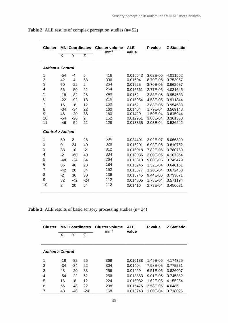
Coordinates, size, and significance values of the clusters can be found in Table 2 and
visualized in Fig. 2.
3.2 Basic sensory processing:
Autism > Control: The meta-analysis of foci showing greater activity in autistic compared to
control groups yielded 12 clusters primarily in the occipital, parietal, and striatal regions. The

Sensory perception in autism: an fMRI ALE meta-analysis
10
main clusters were found in the occipital lobe, specifically the lateral occipital cortex,
occipital pole and visual cortices. Parietal clusters were found in the primary somatosensory
and motor cortices, while sub-cortical clusters were identified in the insula, caudate, and
thalamus. The meta-analysis also revealed heightened cerebellar activity in autism compared
to controls. Cluster contributions were from 229 foci from 20 studies on 357 autistic
individuals.
Control > Autism: On the other hand, control groups showed greater involvement of the
frontal areas during sensory processing tasks. Control participants showed significantly more
activity than autism in 9 clusters, of which 7 were located in the frontal lobe, encompassing
the precentral gyrus, middle frontal gyrus, and precuneus. Control groups were also found to
engage the anterior cingulate cortex more than autistic groups. A total of 233 foci from 20
studies on 369 control participants contributed to these results.
Coordinates, size, and significance values of the clusters can be found in Table 3 and
visualized in Fig. 3.
3.2.1 Exploratory domain-specific analyses:
Visual: Autism groups showed greater activity than controls in 6 clusters, primarily in the
occipital and temporal areas. The clusters spanned the lateral occipital cortex, visual cortex,
inferior parietal lobule, cerebellum, premotor and primary motor cortices, and the secondary
somatosensory cortex. Meanwhile, control groups showed greater engagement of frontal and
parietal areas, with 5 clusters located in the middle and inferior frontal gyrus, precuneus,
precentral gyrus, central opercular cortex, and superior parietal lobule. Autism> Control and
Control> Autism clusters were attributed to 13 experiments with 252 autistic (106 foci) and
268 control (84 foci) participants, respectively. Details of the significant clusters can be
found in the Supplementary Material (Table 1 and Fig. 1).
Sensory perception in autism: an fMRI ALE meta-analysis
11
Auditory and Tactile: Compared to control participants, on average, autistic individuals
showed significantly greater engagement of parietal, frontal, and sub-cortical regions, with 6
clusters found in the parietal operculum, inferior parietal lobule, premotor and primary
sensory cortices, thalamus and posterior cingulate gyrus. Cluster contributions were from
110 foci from 8 experiments with 130 autistic participants. Meanwhile, compared to autistic
groups, control groups showed distinct engagement of the frontal areas. Thirteen clusters
spanning the frontal pole, paracingulate gyrus, anterior cingulate gyrus, corticospinal tract,
precentral gyrus and primary somatosensory cortex were identified from 139 foci from 8
experiments with 126 control subjects. Details of the significant clusters can be found in the
Supplementary Material (Table 2 and Fig. 2).
4 Discussion
4.1 Summary:
We quantitatively summarized evidence from task-based fMRI studies of non-social sensory
perception in autistic compared to neurotypical control participants by conducting a series of
meta-analyses. First, we investigated the neural substrates of complex perceptual processes,
including learning and expectation. Next, by confining the analyses to a more homogenous
set of tasks, we examined task activation patterns of basic sensory processing. Finally, we
conducted exploratory domain-specific sub-analyses on visual and auditory/ tactile studies.
Overall, we found that, compared to control groups, autistic participants showed distinct
engagement of somatosensory and occipital cortices during sensory perception. Conversely,
control groups showed significantly greater recruitment of frontal areas during perceptual
processing.
4.2 Distinct sensory perception in autism:
Sensory perception in autism: an fMRI ALE meta-analysis
12
These neural meta-analytic findings lend support to a large body of evidence of distinct
autistic perception derived from decades of historical reports, first-hand accounts, and
behavioural research (Simmons et al., 2009). Autism is associated with superior attention to
detail (Shah & Frith, 1983), heightened ability to “systemize” (i.e, to identify if-and-then
rules in a system) (Baron-Cohen et al., 2003; 2009), enhanced perceptual functioning
(Mottron et al., 2006) and perceptual load (Remington et al., 2009). Autistic individuals have
consistently shown superior performance on tasks related to visual search (Plaisted et al.,
1998) and identifying hidden figures in complex scenes (Jolliffe & Baron‐Cohen, 1997;
Happé & Frith, 2006). Despite the relatively limited research, similar perceptual differences
have been found to extend to the auditory and tactile domains (O’Riordan & Passetti, 2006;
Kwakye et al., 2011; Tavassoli et al., 2016; Remington & Fairnie, 2017; Mikkelsen et al.,
2018).
Furthermore, our ALE results of hypoactivity of frontal areas and hyperactivity in occipital
and sensorimotor cortices in autism supplement findings from neuroimaging meta-analyses
on autistic perception published in the past decade. An fMRI meta-analysis of visual
processing tasks with words, objects and faces as stimuli found similar patterns of neural
activity in autistic compared to neurotypical control groups (Samson et al., 2012). Philip et al.
(2012) conducted systematic meta-analyses on different task domains: visual processing tasks
showed hyperactivity of thalamus and medial frontal gyrus and hypoactivity of the cingulate
and occipital cortex, while auditory and language tasks yielded hyperactivity of the precentral
gyrus and posterior cingulate and hypoactivity of the superior temporal gyrus. In addition,
Yang & Hofmann (2016) meta-analysed thirteen fMRI studies on action observation in
autism compared to controls; they found hyperactivity in the frontal and parietal cortices, and
hypoactivity in the occipital and temporal areas in autistic groups. Although the direction of
the effects is unclear, a consistent finding in autism is aberrant neural activity in frontal and

Sensory perception in autism: an fMRI ALE meta-analysis
13
sensorimotor cortices. However, these meta-analyses made no distinction between social and
non-social perception, rendering it possible that findings may have been weighted by the high
prevalence of social stimuli in the published literature. By focusing solely on non-social
experimental contrasts, our results provide a meaningful account of differential neural
activity between autistic and control individuals during non-social sensory perception.
4.3 Neural substrates of basic and complex sensory perception in autism:
Perception is a dynamic, hierarchical process involving bottom-up sensory data and top-down
expectations. Empirical research on the effects of stimuli complexity on task performance
have found that, when compared to controls, autistic individuals are better at processing
“simple” stimuli and poorer at processing “complex” stimuli (Bertone et al., 2005; Koolen et
al., 2014; Stevenson et al., 2014). We attempted to disentangle the different levels of
perceptual processing by means of a broad meta-analysis followed by a narrower sub-
analysis.
First, we meta-analysed all fMRI studies of non-social perceptual tasks, including executive
function tasks of learning, reward anticipation, and cognitive control. Aside from heightened
activity in sensorimotor and occipital cortices, autistic groups showed greater engagement of
the caudate, a region implicated in neuro-computational accounts of learning, perceptual
decision-making and reward-processing, among other functions (L. Ding & Gold, 2013;
Keuken et al., 2014). Second, after narrowing the list of included studies down to more basic
sensory processing tasks, we observed more pronounced effects of occipital, somatosensory,
caudate, thalamic, and cerebellar activity in autism. In addition, this sub-analysis of basic
sensory processing tasks showed evidence of low-level neural activity in the primary sensory
regions of the autistic brain. These results are in line with previous neuroimaging empirical

Sensory perception in autism: an fMRI ALE meta-analysis
14
studies which have consistently traced the neural origins of autistic sensory perception to the
primary sensory cortices (for a review, see Robertson & Baron-Cohen, 2017).
Although the neuroanatomical hierarchy is still under investigation, there have been attempts
to formulate the relationship between autistic perception and low-level sensory processing
through neurocomputational models. According to Bayesian inference and predictive coding,
autistic individuals may: rely more on bottom-up sensory input than top-down expectations
(Pellicano & Burr, 2012); show heightened precision of sensory evidence (Friston et al.,
2013; Lawson et al., 2014, 2015); form imprecise sensory representations due to inflexible
perceptual processing (Brock, 2012); have difficulties in disentangling signal from noise
(Van de Cruys et al., 2017), or show aberrant updating of prior beliefs (Haker et al., 2016).
Another computational perspective on autistic perception is based on altered neural
computations, or a failure of divisive normalization (Rosenberg et al., 2015). In divisive
normalization, the activity of an individual neuron is divided by the total activity of the
surrounding neuronal population, thus making them context-sensitive. This has been linked to
an imbalance in the excitation-inhibition (E/I) neural circuitry in autism (Gogolla et al., 2009;
Rubenstein & Merzenich, 2003).
Our approach of conducting a broad meta-analysis on complex perception followed by a sub-
analysis of basic sensory processing highlighted that, when compared to neurotypical
controls, autistic individuals not only differed in neural activity during complex perception,
but also in low-level neural circuitry during basic sensory processing. As delineating the
hierarchy of sensory perception is beyond the scope of meta-analysis, future empirical
experiments using sophisticated paradigms, computational approaches, and novel imaging
methods may shed light on the intricacies of these processes.
4.4 Limitations:

Sensory perception in autism: an fMRI ALE meta-analysis
15
A number of limitations are pertinent to the interpretation of our ALE results.
First, a general challenge of ALE meta-analyses is the issue of heterogeneity across included
studies. Despite our use of stringent, pre-registered inclusion criteria, we had to make some
compromises in homogeneity at the expense of sample size. The recommended number of
studies to yield sufficient statistical power for ALE meta-analyses is 17-20 (Eickhoff et al.,
2016; Müller et al., 2018). Our comprehensive literature search resulted in 52 task-based
fMRI studies of non-social sensory perception. However, as cluster contribution is based on
group differences, only 25 studies and 20 studies contributed to our ALE results of complex
and basic sensory processing, respectively. Although it would have been ideal to restrict our
inclusion criteria to specific sensory modalities and paradigms, our decisions were driven by
the need for sufficient statistical power to draw reliable inferences. In addition, we
acknowledge that our categorization of task contrasts as basic and complex is somewhat
arbitrary due to the overlap between various perceptual processes. Another factor worth
addressing is the sampling bias of the population being studied; i.e, autistic individuals who
were eligible for the MRI environment.
Due to our focus on whole-brain fMRI studies, these findings are not representative of the
entire task-based fMRI literature on non-social sensory perception in autism. We were
limited by whole-brain analyses as the inclusion of region-specific analyses would violate the
assumptions of the coordinate-based voxel-wise meta-analysis (Radua & Mataix-Cols, 2009;
Wager et al., 2007, p. 20; Eickhoff et al., 2012) By excluding hypothesis-driven fMRI studies
employing ROI analyses, we may be missing out on subtle, low-level neural differences
identified in the primary sensory cortices. Using ROI-based approaches, studies have
identified early, autism-specific neural responses in a number of regions including: the
primary visual cortex and middle temporal gyrus during visual global motion perception
(Robertson et al., 2014) ; intraparietal sulcus, primary and secondary visual cortex,

Sensory perception in autism: an fMRI ALE meta-analysis
16
precuneus, cerebellum and middle temporal gyrus during passive and active visual movement
tracking (Takarae et al., 2014) ; extrastriate population receptive fields during visual
stimulation (Schwarzkopf et al., 2014); and the primary auditory cortices as a result of audio-
visual adaptation (Millin et al., 2018) . Although these regions feature in our meta-analysis
findings, we note that the exclusion of such studies may have attenuated the effects of some
regions commonly activated during autistic perception.
Finally, we exert caution while interpreting our results as cognitive neuroimaging findings
are largely based on reverse inferences (Poldrack, 2006, 2011). Moreover, the meta-analytic
results reflect the quality of the fMRI literature in general. Factors contributing to quality
range from data acquisition parameters to the pre-processing and statistical approaches
employed for the fMRI analyses. Important considerations include publication bias,
reproducibility issues, and the need for standardized analysis pipelines and best-practice
guidelines for fMRI research (Nichols et al., 2017).
4.5 Conclusions:
Using ALE, we found that autistic individuals, compared to the neurotypical population, on
average show distinct engagement of sensory-related neural circuits during sensory
perception. These neural differences were found to extend to both basic and complex levels
of perceptual processing, with greater involvement of the primary sensory cortices during
basic sensory processing in autism. Furthermore, this work quantitatively summarizes
information from task-based fMRI studies on sensory perception in autism and highlights
some of the limitations of fMRI research. Our findings may help guide future research to
utilize novel neuroimaging methods and psychophysics paradigms to focus on relevant brain
mechanisms associated with perception in autism.

Sensory perception in autism: an fMRI ALE meta-analysis
17
Acknowledgements:
We thank Liliana Polyanska for her helpful comments on the methods during the design stage
of the study. We are grateful to Rebecca Lawson, Paul Fletcher, Richard Bethlehem, and
Varun Warrier for the discussion.
Funding:
NJ was supported by the April Trust PhD Studentship awarded by Newnham College. SBC
was funded by the Autism Research Trust, the Wellcome Trust, the Templeton World
Charitable Foundation, and the NIHR Biomedical Research Centre in Cambridge, during the
period of this work. SBC also received funding from the Innovative Medicines Initiative 2
Joint Undertaking (JU) under grant agreement No 777394. The JU receives support from the
European Union’s Horizon 2020 research and innovation programme and EFPIA and
AUTISM SPEAKS, Autistica, SFARI. His research was also supported by the National
Institute of Health Research (NIHR) Applied Research Collaboration East of England (ARC
EoE) programme. The views expressed are those of the authors, and not necessarily those of
the NIHR, NHS or Department of Health and Social Care. The funding sources had no role in
the study design; collection, analysis and interpretation of data; writing of the manuscript;
and the decision to submit the article for publication.
Conflicts of interest:
None
Data availability:
Jassim, Nazia (2020) (under moderation), “Data for: Sensory perception in autism: An fMRI
ALE meta-analysis ”, Mendeley Data, V1, doi: 10.17632/pwgdfd88cy.1

Sensory perception in autism: an fMRI ALE meta-analysis
18
References:
American Psychiatric Association. (2013). Diagnostic and Statistical Manual of Mental Disorders
(Fifth Edition). American Psychiatric Association.
https://doi.org/10.1176/appi.books.9780890425596
Baron-Cohen, S., & Lombardo, M. V. (2017). Autism and talent: The cognitive and neural basis of
systemizing. Dialogues in Clinical Neuroscience, 19(4), 345–353.
Bertone, A., Mottron, L., Jelenic, P., & Faubert, J. (2005). Enhanced and diminished visuo-spatial
information processing in autism depends on stimulus complexity. Brain, 128(10), 2430–
2441. https://doi.org/10.1093/brain/awh561
Brock, J. (2012). Alternative Bayesian accounts of autistic perception: Comment on Pellicano and
Burr. Trends in Cognitive Sciences, 16(12), 573–574.
https://doi.org/10.1016/j.tics.2012.10.005
Ding, L., & Gold, J. I. (2013). The basal ganglia’s contributions to perceptual decision-making.
Neuron, 79(4), 640–649. https://doi.org/10.1016/j.neuron.2013.07.042
Ding, R., Ren, J., Li, S., Zhu, X., Zhang, K., & Luo, W. (2020). Domain-general and domain-
preferential neural correlates underlying empathy towards physical pain, emotional situation
and emotional faces: An ALE meta-analysis. Neuropsychologia, 137, 107286.
https://doi.org/10.1016/j.neuropsychologia.2019.107286
Eickhoff, S. B., Bzdok, D., Laird, A. R., Kurth, F., & Fox, P. T. (2012). Activation likelihood
estimation meta-analysis revisited. NeuroImage, 59(3), 2349–2361.
https://doi.org/10.1016/j.neuroimage.2011.09.017
Eickhoff, S. B., Laird, A. R., Grefkes, C., Wang, L. E., Zilles, K., & Fox, P. T. (2009). Coordinate-
based activation likelihood estimation meta-analysis of neuroimaging data: A random-effects
approach based on empirical estimates of spatial uncertainty. Human Brain Mapping, 30(9),
2907–2926. https://doi.org/10.1002/hbm.20718
Eickhoff, S. B., Nichols, T. E., Laird, A. R., Hoffstaedter, F., Amunts, K., Fox, P. T., Bzdok, D., &
Eickhoff, C. R. (2016). Behavior, Sensitivity, and power of activation likelihood estimation
characterized by massive empirical simulation. NeuroImage, 137, 70–85.
https://doi.org/10.1016/j.neuroimage.2016.04.072
Friston, K. J., Lawson, R., & Frith, C. D. (2013). On hyperpriors and hypopriors: Comment on
Pellicano and Burr. Trends in Cognitive Sciences, 17(1), 1.
https://doi.org/10.1016/j.tics.2012.11.003
Gogolla, N., LeBlanc, J. J., Quast, K. B., Südhof, T. C., Fagiolini, M., & Hensch, T. K. (2009).
Common circuit defect of excitatory-inhibitory balance in mouse models of autism. Journal
of Neurodevelopmental Disorders, 1(2), 172–181. https://doi.org/10.1007/s11689-009-9023-x

Sensory perception in autism: an fMRI ALE meta-analysis
19
Haker, H., Schneebeli, M., & Stephan, K. E. (2016). Can Bayesian Theories of Autism Spectrum
Disorder Help Improve Clinical Practice? Frontiers in Psychiatry, 7.
https://doi.org/10.3389/fpsyt.2016.00107
Happé, F., & Frith, U. (2006). The Weak Coherence Account: Detail-focused Cognitive Style in
Autism Spectrum Disorders. Journal of Autism and Developmental Disorders, 36(1), 5–25.
https://doi.org/10.1007/s10803-005-0039-0
Happé, F., & Ronald, A. (2008). The ‘Fractionable Autism Triad’: A Review of Evidence from
Behavioural, Genetic, Cognitive and Neural Research. Neuropsychology Review, 18(4), 287–
304. https://doi.org/10.1007/s11065-008-9076-8
Happé, F., Ronald, A., & Plomin, R. (2006). Time to give up on a single explanation for autism.
Nature Neuroscience, 9(10), 1218–1220. https://doi.org/10.1038/nn1770
Jolliffe, T., & Baron‐Cohen, S. (1997). Are People with Autism and Asperger Syndrome Faster Than
Normal on the Embedded Figures Test? Journal of Child Psychology and Psychiatry, 38(5),
527–534. https://doi.org/10.1111/j.1469-7610.1997.tb01539.x
Keuken, M. C., Müller-Axt, C., Langner, R., Eickhoff, S. B., Forstmann, B. U., & Neumann, J.
(2014). Brain networks of perceptual decision-making: An fMRI ALE meta-analysis.
Frontiers in Human Neuroscience, 8. https://doi.org/10.3389/fnhum.2014.00445
Koolen, S., Vissers, C. Th. W. M., Egger, J. I. M., & Verhoeven, L. (2014). How Stimulus and Task
Complexity Affect Monitoring in High-Functioning Adults with Autism Spectrum Disorder.
Journal of Autism and Developmental Disorders, 44(10), 2499–2513.
https://doi.org/10.1007/s10803-014-2119-5
Kwakye, L. D., Foss-Feig, J. H., Cascio, C. J., Stone, W. L., & Wallace, M. T. (2011). Altered
Auditory and Multisensory Temporal Processing in Autism Spectrum Disorders. Frontiers in
Integrative Neuroscience, 4. https://doi.org/10.3389/fnint.2010.00129
Laird, A. R., Fox, P. M., Price, C. J., Glahn, D. C., Uecker, A. M., Lancaster, J. L., Turkeltaub, P. E.,
Kochunov, P., & Fox, P. T. (2005). ALE meta-analysis: Controlling the false discovery rate
and performing statistical contrasts. Human Brain Mapping, 25(1), 155–164.
https://doi.org/10.1002/hbm.20136
Lawson, R. P., Friston, K. J., & Rees, G. (2015). A more precise look at context in autism.
Proceedings of the National Academy of Sciences of the United States of America, 112(38),
E5226. https://doi.org/10.1073/pnas.1514212112
Lawson, R. P., Rees, G., & Friston, K. J. (2014). An aberrant precision account of autism. Frontiers
in Human Neuroscience, 8. https://doi.org/10.3389/fnhum.2014.00302
Mikkelsen, M., Wodka, E. L., Mostofsky, S. H., & Puts, N. A. J. (2018). Autism spectrum disorder in
the scope of tactile processing. Developmental Cognitive Neuroscience, 29, 140–150.
https://doi.org/10.1016/j.dcn.2016.12.005
Sensory perception in autism: an fMRI ALE meta-analysis
20
Millin, R., Kolodny, T., Flevaris, A. V., Kale, A. M., Schallmo, M.-P., Gerdts, J., Bernier, R. A., &
Murray, S. (2018). Reduced auditory cortical adaptation in autism spectrum disorder. ELife,
7, e36493. https://doi.org/10.7554/eLife.36493
Moher, D., Liberati, A., Tetzlaff, J., Altman, D. G., & Group, T. P. (2009). Preferred Reporting Items
for Systematic Reviews and Meta-Analyses: The PRISMA Statement. PLOS Medicine, 6(7),
e1000097. https://doi.org/10.1371/journal.pmed.1000097
Mottron, L., Dawson, M., Soulières, I., Hubert, B., & Burack, J. (2006). Enhanced Perceptual
Functioning in Autism: An Update, and Eight Principles of Autistic Perception. Journal of
Autism and Developmental Disorders, 36(1), 27–43. https://doi.org/10.1007/s10803-005-
0040-7
Müller, V. I., Cieslik, E. C., Laird, A. R., Fox, P. T., Radua, J., Mataix-Cols, D., Tench, C. R.,
Yarkoni, T., Nichols, T. E., Turkeltaub, P. E., Wager, T. D., & Eickhoff, S. B. (2018). Ten
simple rules for neuroimaging meta-analysis. Neuroscience & Biobehavioral Reviews, 84,
151–161. https://doi.org/10.1016/j.neubiorev.2017.11.012
Nichols, T. E., Das, S., Eickhoff, S. B., Evans, A. C., Glatard, T., Hanke, M., Kriegeskorte, N.,
Milham, M. P., Poldrack, R. A., Poline, J.-B., Proal, E., Thirion, B., Van Essen, D. C., White,
T., & Yeo, B. T. T. (2017). Best practices in data analysis and sharing in neuroimaging using
MRI. Nature Neuroscience, 20(3), 299–303. https://doi.org/10.1038/nn.4500
O’Riordan, M., & Passetti, F. (2006). Discrimination in Autism Within Different Sensory Modalities.
Journal of Autism and Developmental Disorders, 36(5), 665–675.
https://doi.org/10.1007/s10803-006-0106-1
Pellicano, E., & Burr, D. (2012). When the world becomes ‘too real’: A Bayesian explanation of
autistic perception. Trends in Cognitive Sciences, 16(10), 504–510.
https://doi.org/10.1016/j.tics.2012.08.009
Philip, R. C. M., Dauvermann, M. R., Whalley, H. C., Baynham, K., Lawrie, S. M., & Stanfield, A. C.
(2012). A systematic review and meta-analysis of the fMRI investigation of autism spectrum
disorders. Neuroscience & Biobehavioral Reviews, 36(2), 901–942.
https://doi.org/10.1016/j.neubiorev.2011.10.008
Poldrack, R. A. (2006). Can cognitive processes be inferred from neuroimaging data? Trends in
Cognitive Sciences, 10(2), 59–63. https://doi.org/10.1016/j.tics.2005.12.004
Poldrack, R. A. (2011). Inferring mental states from neuroimaging data: From reverse inference to
large-scale decoding. Neuron, 72(5), 692–697. https://doi.org/10.1016/j.neuron.2011.11.001
Polyanska, L., Critchley, H. D., & Rae, C. L. (2017). Centrality of prefrontal and motor preparation
cortices to Tourette Syndrome revealed by meta-analysis of task-based neuroimaging studies.
NeuroImage: Clinical, 16, 257–267. https://doi.org/10.1016/j.nicl.2017.08.004

Sensory perception in autism: an fMRI ALE meta-analysis
21
Radua, J., & Mataix-Cols, D. (2009). Voxel-wise meta-analysis of grey matter changes in obsessive–
compulsive disorder. The British Journal of Psychiatry, 195(5), 393–402.
https://doi.org/10.1192/bjp.bp.108.055046
Remington, A., & Fairnie, J. (2017). A sound advantage: Increased auditory capacity in autism.
Cognition, 166, 459–465. https://doi.org/10.1016/j.cognition.2017.04.002
Remington, A., Swettenham, J., Campbell, R., & Coleman, M. (2009). Selective Attention and
Perceptual Load in Autism Spectrum Disorder: Psychological Science.
https://journals.sagepub.com/doi/10.1111/j.1467-9280.2009.02454.x
Robertson, C. E., & Baron-Cohen, S. (2017). Sensory perception in autism. Nature Reviews
Neuroscience, 18(11), 671–684. https://doi.org/10.1038/nrn.2017.112
Robertson, C. E., Thomas, C., Kravitz, D. J., Wallace, G. L., Baron-Cohen, S., Martin, A., & Baker,
C. I. (2014). Global motion perception deficits in autism are reflected as early as primary
visual cortex. Brain, 137(9), 2588–2599. https://doi.org/10.1093/brain/awu189
Rubenstein, J. L. R., & Merzenich, M. M. (2003). Model of autism: Increased ratio of
excitation/inhibition in key neural systems. Genes, Brain and Behavior, 2(5), 255–267.
https://doi.org/10.1034/j.1601-183X.2003.00037.x
Samson, F., Zeffiro, T. A., Doyon, J., Benali, H., & Mottron, L. (2015). Speech acquisition predicts
regions of enhanced cortical response to auditory stimulation in autism spectrum individuals.
Journal of Psychiatric Research, 68, 285–292.
https://doi.org/10.1016/j.jpsychires.2015.05.011
Samson, Fabienne, Mottron, L., Soulières, I., & Zeffiro, T. A. (2012). Enhanced visual functioning in
autism: An ALE meta-analysis. Human Brain Mapping, 33(7), 1553–1581.
https://doi.org/10.1002/hbm.21307
Schwarzkopf, D. S., Anderson, E. J., Haas, B. de, White, S. J., & Rees, G. (2014). Larger Extrastriate
Population Receptive Fields in Autism Spectrum Disorders. Journal of Neuroscience, 34(7),
2713–2724. https://doi.org/10.1523/JNEUROSCI.4416-13.2014
Shah, A., & Frith, U. (1983). An Islet of Ability in Autistic Children: A Research Note. Journal of
Child Psychology and Psychiatry, 24(4), 613–620. https://doi.org/10.1111/j.1469-
7610.1983.tb00137.x
Simmons, D. R., Robertson, A. E., McKay, L. S., Toal, E., McAleer, P., & Pollick, F. E. (2009).
Vision in autism spectrum disorders. Vision Research, 49(22), 2705–2739.
https://doi.org/10.1016/j.visres.2009.08.005
Stevenson, R. A., Siemann, J. K., Schneider, B. C., Eberly, H. E., Woynaroski, T. G., Camarata, S.
M., & Wallace, M. T. (2014). Multisensory Temporal Integration in Autism Spectrum
Disorders. Journal of Neuroscience, 34(3), 691–697.
https://doi.org/10.1523/JNEUROSCI.3615-13.2014

Sensory perception in autism: an fMRI ALE meta-analysis
22
Takarae, Y., Luna, B., Minshew, N. J., & Sweeney, J. A. (2014). Visual Motion Processing and
Visual Sensorimotor Control in Autism. Journal of the International Neuropsychological
Society, 20(1), 113–122. https://doi.org/10.1017/S1355617713001203
Tavassoli, T., Bellesheim, K., Tommerdahl, M., Holden, J. M., Kolevzon, A., & Buxbaum, J. D.
(2016). Altered tactile processing in children with autism spectrum disorder: Tactile
processing, inhibition, ASD. Autism Research, 9(6), 616–620.
https://doi.org/10.1002/aur.1563
Tavassoli, T., Miller, L. J., Schoen, S. A., Nielsen, D. M., & Baron-Cohen, S. (2013). Sensory over-
responsivity in adults with autism spectrum conditions: Autism.
https://doi.org/10.1177/1362361313477246
Turkeltaub, P. E., Eickhoff, S. B., Laird, A. R., Fox, M., Wiener, M., & Fox, P. (2012). Minimizing
within-experiment and within-group effects in activation likelihood estimation meta-analyses.
Human Brain Mapping, 33(1), 1–13. https://doi.org/10.1002/hbm.21186
Van de Cruys, S., Evers, K., Van der Hallen, R., Van Eylen, L., Boets, B., de-Wit, L., & Wagemans,
J. (2014). Precise minds in uncertain worlds: Predictive coding in autism. Psychological
Review, 121(4), 649–675. https://doi.org/10.1037/a0037665
Van de Cruys, S., Van der Hallen, R., & Wagemans, J. (2017). Disentangling signal and noise in
autism spectrum disorder. Brain and Cognition, 112, 78–83.
https://doi.org/10.1016/j.bandc.2016.08.004
Wager, T. D., Lindquist, M., & Kaplan, L. (2007). Meta-analysis of functional neuroimaging data:
Current and future directions. Social Cognitive and Affective Neuroscience, 2(2), 150–158.
https://doi.org/10.1093/scan/nsm015
Warrier, V., Toro, R., Won, H., Leblond, C. S., Cliquet, F., Delorme, R., De Witte, W., Bralten, J.,
Chakrabarti, B., Børglum, A. D., Grove, J., Poelmans, G., Hinds, D. A., Bourgeron, T., &
Baron-Cohen, S. (2019). Social and non-social autism symptoms and trait domains are
genetically dissociable. Communications Biology, 2(1), 1–13. https://doi.org/10.1038/s42003-
019-0558-4
Yang, J., & Hofmann, J. (2016). Action observation and imitation in autism spectrum disorders: An
ALE meta-analysis of fMRI studies. Brain Imaging and Behavior, 10(4), 960–969.
https://doi.org/10.1007/s11682-015-9456-7
Sensory perception in autism: an fMRI ALE meta-analysis
23
Figure and Table captions:
Fig 1. Flowchart of the literature search and study selection process
Fig 2. Neural differences between autistic and control groups during complex perception
tasks
Fig 3. Neural differences between autistic and control groups during basic sensory
processing tasks
Table 1. Complete list and relevant characteristics of whole-brain fMRI studies included in
the ALE analyses
Table 2. ALE results of complex perception studies (n=52)
Table 3. ALE results of basic sensory processing studies (n=34)
Figures (In colour):
Fig.1. Flowchart representing the literature search process. n = number of publications;
ROI= Region-of-interest.
Studies identified
(n = 741)
Inc
lud
ed
Sc
ree
nin
g
Eli
gib
ilit
y
Ide
nti
fica
tio
n
Abstracts screened
(n = 599)
Full-text papers read
(n= 326)
Studies chosen for
meta-analysis
(n=52)
Excluded (n=142) 71 meta-
analyses/reviews
71 methods/ repositories
Excluded (n=273)
136 no autism vs control
group comparison 115 resting-state fMRI
22 other methods
Excluded (n=274) 227 social contrasts 33 ROI/other analyses 4 inaccessible coordinates 8 irrelevant contrasts

Sensory perception in autism: an fMRI ALE meta-analysis
24
Fig.2. Neural differences between autistic and control groups during complex perception
tasks. ALE meta-analysis results of 52 complex perception fMRI studies for the comparisons
Autism>Control and Control>Autism (p <0.001, min. cluster size 100 mm3). Coordinates are
in MNI space. Colour bars represent the ALE values.

Sensory perception in autism: an fMRI ALE meta-analysis
25
Fig.3. Neural differences between autistic and control groups during basic sensory
processing tasks. ALE meta-analysis results of 34 basic sensory processing fMRI studies for
the comparisons Autism>Control and Control>Autism (p <0.001, min. cluster size 100
mm3). Coordinates are in MNI space. Colour bars represent the ALE values.

Sensory perception in autism: an fMRI ALE meta-analysis
26
Table 1. Complete list and relevant characteristics of whole-brain fMRI studies included in the ALE analyses.
Study
First
Author &
Year
Experiment Participants fMRI
Sensory Domain
Task Contrast(s) N Age Range / Mean (SD)
Autism Sex (M:F)
Toolbox Statistical threshhold
Schuetze 2019*
Visual Implicit reinforcement learning
Choice behaviour to infer reward value: liked, non-liked, neutral images
32 ASC
31 Con
14 – 20 28:4 SPM FWE-corrected, p<0.05
Velasquez 2019
Visual Response inhibition: Go/No Go
Letter NoGo vs Go 19 ASC
22 Con
18 – 46 13:6 FSL FWE- corrected, p<0.05
Green 2018 Auditory & Tactile
Auditory sarcasm task with and without tactile stimulation & instructions
No Instructions- Tactile vs baseline, Instructions- Tactile vs baseline, Instructions- Tactile vs No Instructions- No Tactile, No Instructions-Tactile vs No Instructions- No Tactile
15 ASC
16 Con
9 - 17.6 11:4 FSL FWE- corrected, p<0.05
Murphy 2017 Visual Attention orienting
Patterned vs neutral stimuli 23 ASC
35 Con
8 – 23 17:6 AFNI FWE - corrected, p<0.05
Keehn 2017* Auditory & Visual
Auditory- high & low pitch detection,
Auditory vs null condition, Visual vs null condition
16 ASC
16 Con
8 – 18 14:2 AFNI FWE - corrected, p<0.05

Sensory perception in autism: an fMRI ALE meta-analysis
27
Visual- high & low spatial dot location
Schelinksi 2016*
Auditory Sound processing
Non vocal sounds (cars, nature music) vs silence baseline
16 ASC
16 Con
18 – 52 13:3 SPM Uncorrected,
P<0.001
D’Cruz 2017 Visual Reversal learning: 4-choice visuospatial location
Unexpected reversal (no reinforcement) vs Expected positive reinforcement
17 ASC
23 Con
7 – 44 12:5 FSL Corrected, FSL Randomize v2.1, TFCE Type 1 error rate p<0.01
Prat 2016* Visual Response inhibition: Go / No Go
Letter No Go vs Go 16 ASC
17 Con
25.3± 5 (ASC), 25.6±7.2(Con)
10:6 SPM Uncorrected, p<0.001
Rahko 2016 Visual Working memory: N-back
0-back vs baseline, 0-back vs 2-back
28 ASC
22 Con
11.4 - 17.6 20:8 FSL FWE-corrected, p<0.05
Kaiser 2016 Tactile Arm and palm touch
Arm vs Palm 19 ASC
19 Con
6.43–20.26 (ASC), 5.56–17.05 (Con)
16:3 FSL FWE-corrected, p<0.05
Keehn 2016 Visual Rapid Serial Visual Presentation
Target Present/Absent vs Target-Coloured/Neutral Distractors, Control condition: Target- Absent + Neutral-Distractors
16 ASC
21 Con
12 – 17 14:2 AFNI Cluster-wise corrected (p<0.05), voxel-wise uncorrected (p<0.01), Monte Carlo simulation
Schipul 2016 Visual Dot pattern learning
Encoding vs fixation 16 ASC
16 Con
16 – 42 14:2 SPM Uncorrected, p < 0.005, spatial extent of 10 voxels

Sensory perception in autism: an fMRI ALE meta-analysis
28
Kleinhans 2016
Visual Habituation to houses
House 1 vs House 2 27 ASC
25 Con
18 – 44 25:2 FSL Cluster-wise corrected (p<0.05), voxel-wise (z>2.3) Monte Carlo simulation
Sharer 2015 Visual Visuomotor learning: Serial Reaction Time task
Sequence vs random 17 ASC
32 Con
10.5±1.36, (ASC) 10.46±1.3, (Con)
14:3 SPM FWE-corrected,
P<0.05
Solomon 2015 Visual Transitive inference learning: Stimulus hierarchy of coloured ovals
Training phase: learning pairs, Testing phase : generalization to new pairs
21 ASC
23 Con
12.2 – 17 17:4 SPM FWE – corrected, p<0.05
Samson 2015 Auditory Listening to sounds of pure tone, harmonic tone, varying levels of frequency modulation
All sound conditions vs silence baseline
27 ASC (14+13)
13 Con
14 – 39 11:2 SPM FWE – corrected, p<0.05
Green 2015 Auditory & Tactile
Auditory stimulation: Traffic noises, Tactile stimulation: rough fabric
Auditory vs baseline, tactile vs baseline, joint auditory + tactile vs baseline
19 ASC
19 Con
9 – 17 16:3 FSL FWE – corrected, p<0.05

Sensory perception in autism: an fMRI ALE meta-analysis
29
Shafritz 2015 Visual Response inhibition: Go/No Go
Letter No Go vs Go 15 ASC
18 Con
13 – 23 12:3 SPM p <0.001, cluster-filter of 10 contiguous voxels
Simhard 2015 Visual Visuospatial reasoning: Raven’s Standard Progressive Matrices
Figural vs Analytical vs Complex Analytical stimuli
15 ASC
18 Con
14 – 36 13:2 SPM p<0.001 uncorrected, extent threshold of 50 contiguous voxels
Barbeau 2015 Visual Visuomotor Poffenberger task
Hand response: Left & Right, Stimulated visual field: Left & Right
34 ASC
33 Con
14 – 37 31:3 SPM FWE-corrected, p<0.05
Yerys 2015 Visual Set shifting: Text display “STAY” or “CHANGE” with a circle and a square on either the left
or right of the word
Stay+Switch vs Fixation
20 ASC
19 Con
7.17 - 13.33 16:4 FSL FWE-corrected, p<0.05
Travers 2015 Visual Visuomotor learning: Serial Reaction Time task
Sequence vs non-sequence learning
15 ASC
15 Con
20.81±3.98 (ASC),
21.41±2.85 (Con)
All male SPM
Uncorrected p<0.001, extent threshold of 72 contiguous voxels
Solomon 2014 Visual Cognitive control:
High-control vs low-control cue 27 ASC 12 – 18 17:10 SPM FWE-corrected, p<0.05
Sensory perception in autism: an fMRI ALE meta-analysis
30
Preparing to overcome prepotency (POP) task
27 Con
Sabatino 2013 Visual Oddball target detection
High Autism Interest images vs baseline
15 ASC
17 Con
16.9 – 45.3 13:2 FSL FWE-corrected, p<0.05
Green 2013 Auditory & Visual
Auditory stimulation: White noise, Visual stimulation: Rotating colour wheel
Auditory vs baseline, visual vs baseline, joint auditory + visual vs baseline
25 ASC
25 Con
9 – 17 21:4 FSL Uncorrected, thresholded at z>2.3
Gadgil 2013 Visual Shape processing: Local vs global hierarchical shape recognition task
Global vs control stimulus, local vs control stimulus, global vs local
17 ASC
16 Con
18 – 55 14:3 SPM FWE- corrected, p<0.05
Spencer 2012 Visual Visuospatial reasoning: Embedded Figures Task
Embedded Figures vs Control Task
38 ASC
40 Con
12 – 18 34:4 SPMs Uncorrected, p<0.001
Yamada 2012 Visual Visuospatial reasoning: Raven’s Standard Progressive Matrices
Easy analytical vs baseline, difficult analytical vs baseline
25 ASC
26 Con
30.7±7.78 (ASC), 32.2±7.7 (Con)
22:3 SPM Uncorrected,p<0.001

Sensory perception in autism: an fMRI ALE meta-analysis
31
Ohta 2012* Visual Selective attention/ perceptual load: Rapid Serial Visual Presentation vs checkerboard
Low vs high load, distractor vs no distractor
24 ASC
25 Con
22 – 40 21:3 SPM Uncorrected, p<0.001, voxel extent threshold=70
Beacher 2012*
Visual Visuospatial reasoning: Mental rotation
Rotated letters vs control condition
29 ASC
32 Con
32.8(9.1) (ASC), 30.48(7.7) (Con)
15:14 SPM P<0.001, cluster extent k=7 voxels
Dichter 2012 Visual Reward anticipation
Anticipation of monetary reward and autism interest object reward
15 ASC
16 Con
30±11.6 (ASC), 27.5±7.5 (Con)
All male FSL Uncorrected, cluster voxels extent k=10, z >2.5, P < 0.005
McGrath 2012 Visual Visuospatial reasoning: Mental rotation
3D cube stimuli: same vs mirror trials
22 ASC
22 Con
13 – 21 All male AFNI Uncorrected, voxel-wise statistical threshold (t = 2.96, P< 0.005)
Cascio 2012 Tactile Tactile stimulation with textures
Brush vs rest, burlap vs rest, mesh vs rest
13 ASC
14 Con
28.3(10.7) (ASC), 30.8(12) (Con)
12:1 SPM Uncorrected, P<0.005 , z>2.3, cluster voxel extent k=10
Caria 2011 Auditory Passive listening to classical music
Happy vs baseline, sad vs baseline
8 ASC
14 Con
19 – 37 6:2 SPM FDR- corrected, p<0.05
Goldberg 2011
Visual Response inhibition: Go/ No Go
Green and red spaceships: Error vs correct inhibition
11 ASC
15 Con
8 – 12 8:3 SPM Corrected p<0.05
Koldewyn 2011*
Visual Dot motion Static vs coherent dot motion 16 ASC 11.41 -19.53 14:2 SPM Voxel-wise (t = 2.95, p < .005, uncorrected)
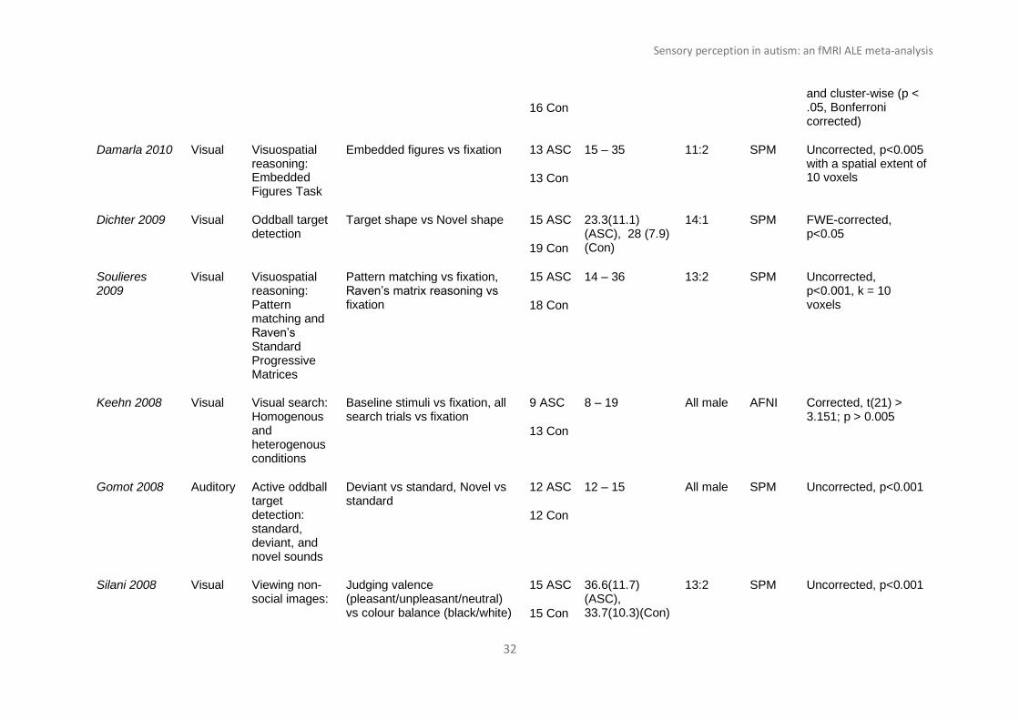
Sensory perception in autism: an fMRI ALE meta-analysis
32
16 Con
and cluster-wise (p < .05, Bonferroni corrected)
Damarla 2010 Visual Visuospatial reasoning: Embedded Figures Task
Embedded figures vs fixation 13 ASC
13 Con
15 – 35 11:2 SPM Uncorrected, p<0.005 with a spatial extent of 10 voxels
Dichter 2009 Visual Oddball target detection
Target shape vs Novel shape 15 ASC
19 Con
23.3(11.1) (ASC), 28 (7.9) (Con)
14:1 SPM FWE-corrected, p<0.05
Soulieres 2009
Visual Visuospatial reasoning: Pattern matching and Raven’s Standard Progressive Matrices
Pattern matching vs fixation, Raven’s matrix reasoning vs fixation
15 ASC
18 Con
14 – 36 13:2 SPM Uncorrected, p<0.001, k = 10 voxels
Keehn 2008 Visual Visual search: Homogenous and heterogenous conditions
Baseline stimuli vs fixation, all search trials vs fixation
9 ASC
13 Con
8 – 19 All male AFNI Corrected, t(21) > 3.151; p > 0.005
Gomot 2008 Auditory Active oddball target detection: standard, deviant, and novel sounds
Deviant vs standard, Novel vs standard
12 ASC
12 Con
12 – 15 All male SPM Uncorrected, p<0.001
Silani 2008 Visual Viewing non-social images:
Judging valence (pleasant/unpleasant/neutral) vs colour balance (black/white)
15 ASC
15 Con
36.6(11.7) (ASC), 33.7(10.3)(Con)
13:2 SPM Uncorrected, p<0.001

Sensory perception in autism: an fMRI ALE meta-analysis
33
valence and colour
Shafritz 2008 Visual Target detection and set-shifting with geometric shapes
All target trials vs fixation, novel trials vs fixation
18 ASC
15 Con
22.3(8.7) (ASC), 24.3(6.2) (Con)
16:2 SPM Uncorrected, p<0.001
Kana 2007 Visual Response inhibition/ working memory: Simple inhibition and letter 1-back
Simple inhibition, 1-back 12 ASC
12 Con
26.8(7.77) (ASC), 22.5(3.2) (Con)
11:1 SPM Uncorrected, p<0.005
Manjaly 2007* Visual Visuospatial reasoning: Embedded Figures Task
Embedded figures vs control task
12 ASC
12 Con
10 – 18 - SPM Corrected, p<0.05
Gomot 2006 Auditory Passive oddball target detection: standard, deviant, and novel sounds
Deviant vs standard, Novel vs standard
12 ASC
12 Con
12 – 15 All male SPM Uncorrected, p<0.001
Schmitz 2006 Visual Response inhibition: Go/No Go, Stroop, and set shifting
No Go vs Go, correct Stroop, SWITCH responses
10 ASC
12 Con
18 – 52 All male SPM Corrected, p<0.05
Haist 2006 Visual Spatial attention:
Short cue-to-target ISI, long cue-to-target-ISI
8 ASC
8 Con
14 – 43 All male AFNI Corrected, p<0.05

Sensory perception in autism: an fMRI ALE meta-analysis
34
N= number of participants; ASC= Autism Spectrum Conditions; Con = Controls; FWE= Family Wise Error; FDR = False Discovery Rate. Italicized studies
indicate basic sensory processing studies. Studies which found no group differences are indicated by an asterisk (*). Unreported items are indicated by a
hyphen. Experimental contrasts, participants age and sex, and fMRI statistical thresholds are entered as reported.
Cued target detection
Mueller 2004 Visual Visuomotor learning: 8-digit sequence learning
Early learning and late learning 8 ASC
8 Con
15 – 41 All male - Corrected, p<0.05, and uncorrected, p<0.01
Belmonte 2004
Visual Spatial attention: Target detection
Task vs fixation 8 ASC
6 Con
24 – 50 7:1 AFNI & SPM
-
Gervais 2004* Auditory Passive listening
Non-vocal sounds vs silence 5 ASC
5 Con
25.8(5.9)(ASC), 27.9(2.9)(Con)
All male SPM Random effect analysis, P < 0.001
Corrected
Mueller 2003 Visual Visuomotor learning: 6-digit sequence learning
Task vs blue dot control 8 ASC
8 Con
15 – 41 All male - Bonferroni-corrected, p<0.05
Sensory perception in autism: an fMRI ALE meta-analysis
35
Table 2. ALE results of complex perception studies (n= 52)
Cluster
MNI Coordinates
Cluster volume
mm3
ALE value
P value
Z Statistic
X Y Z
Autism > Control
1 -54 -4 6 416 0.016543 3.02E-05 4.011552 2 42 -4 58 336 0.01504 8.70E-05 3.753957 3 60 -22 2 264 0.01625 3.70E-05 3.962957 4 56 -50 22 264 0.016661 2.77E-05 4.031645 5 -18 -82 26 248 0.0162 3.83E-05 3.954633 6 -22 -92 18 216 0.015954 4.58E-05 3.911844 7 16 18 12 160 0.0162 3.83E-05 3.954633 8 -34 -34 22 160 0.01404 1.79E-04 3.569143 9 48 -20 38 160 0.01429 1.50E-04 3.615944 10 -54 -26 2 152 0.012951 3.88E-04 3.361358 11 -46 -54 22 128 0.013855 2.03E-04 3.536242
Control > Autism
1 50 2 26 696 0.024401 2.02E-07 5.066899 2 0 24 40 328 0.016201 6.93E-05 3.810752 3 38 10 -2 312 0.016018 7.82E-05 3.780769 4 -2 -60 40 304 0.018036 2.00E-05 4.107364 5 -48 -24 54 264 0.015813 9.00E-05 3.745479 6 36 46 28 184 0.015245 1.32E-04 3.648161 7 -42 20 34 152 0.015377 1.20E-04 3.672463 8 -2 36 30 136 0.015745 9.44E-05 3.733671 9 32 -42 -24 112 0.014805 1.78E-04 3.571194 10 2 20 54 112 0.01416 2.73E-04 3.456621
Table 3. ALE results of basic sensory processing studies (n= 34)
Cluster
MNI Coordinates
Cluster volume
mm3
ALE value
P value
Z Statistic
X Y Z
Autism > Control
1 -18 -82 26 368 0.016188 1.49E-05 4.174325
2 -34 -34 22 304 0.01404 7.98E-05 3.775551
3 48 -20 38 256 0.01429 6.51E-05 3.826007
4 -54 -22 52 256 0.013883 9.01E-05 3.745382
5 16 18 12 224 0.016082 1.62E-05 4.155254
6 56 -48 22 208 0.015475 2.58E-05 4.0486
7 48 -46 -24 168 0.013743 1.00E-04 3.718026

Sensory perception in autism: an fMRI ALE meta-analysis
36
8 14 -46 -14 168 0.01386 9.12E-05 3.742139
9 -22 -90 20 160 0.013737 1.00E-04 3.718026
10 -56 0 20 160 0.013695 1.03E-04 3.71102
11 -18 -28 4 144 0.013193 1.54E-04 3.608243
12 -32 -48 50 104 0.012498 2.56E-04 3.474702
Control > Autism
1 50 2 26 800 0.024128 2.45E-08 5.4553
2a 36 46 28 712 0.015245 2.85E-05 4.024902
2b 46 42 16 712 0.013346 1.24E-04 3.664035
2c 42 46 20 712 0.011963 3.38E-04 3.399287
3 0 24 40 424 0.015299 2.73E-05 4.035437
4 -50 -22 54 336 0.014669 4.45E-05 3.918659
5 -42 20 34 288 0.015248 2.83E-05 4.026852
6 -2 -60 40 160 0.01445 5.27E-05 3.878024
7 -44 6 28 136 0.012082 3.11E-04 3.421643
8 -52 -22 26 128 0.01304 1.57E-04 3.603902
9 -16 -26 56 120 0.012832 1.84E-04 3.561517

Sensory perception in autism: an fMRI ALE meta-analysis
37
Supplementary Material
Supplementary Table 1. ALE results of visual processing studies
Supplementary Table 2. ALE results of auditory and tactile studies
Cluster
MNI Coordinates
Cluster volume
mm3
ALE
value
P
value
Z Statistic
X Y Z
Autism> Control
1 -18 -82 26 728 0.016188 1.49E-05 4.706512
2 56 -48 22 424 0.015475 7.98E-05 4.570058
3 14 -46 -14 384 0.01386 6.51E-05 4.240931
4 -22 -90 20 384 0.013737 9.01E-05 4.21044
5 2 -16 50 208 0.010537 1.62E-05 3.593646
6 52 4 10 104 0.009877 2.58E-05 3.474651
Control > Autism
1 -42 2 26 416 0.024128 2.45E-08 5.4553 2 -2 46 28 416 0.015245 2.85E-05 4.024902 3 50 42 16 336 0.013346 1.24E-04 3.664035 4 -46 46 20 256 0.011963 3.38E-04 3.399287 5 -38 -40 44 114 0.010332 1.11E-04 3.692705
Cluster
MNI Coordinates
Cluster volume
mm3
ALE
value
P
value
Z Statistic
X Y Z
Autism> Control
1 -34 -34 22 848 0.01404 8.07E-06 4.312621
1 -42 -40 24 0.009694 3.01E-04 3.430335
2 48 -20 38 624 0.014289 5.84E-06 4.383307
3 36 -4 56 504 0.011814 6.40E-05 3.830206
3 34 -8 60 0.009558 3.27E-04 3.407907
4 -52 -24 54 432 0.013881 9.82E-06 4.268924
5 -18 -28 4 384 0.013188 1.87E-05 4.123173 6 14 -26 36 176 0.009739 2.90E-04 3.441182 6 16 -22 38 0.009293 3.84E-04 3.363792
Control> Autism
1 36 46 28 1480 0.015245 4.19E-06 4.455027
1 46 42 16 0.013346 2.50E-05 4.056004
1 42 46 20 0.011963 8.51E-05 3.759673
2 0 24 40 768 0.015269 4.07E-06 4.461631
3 -50 -22 54 536 0.014577 7.65E-06 4.32437 4 64 -40 6 384 0.011293 1.46E-04 3.621839

Sensory perception in autism: an fMRI ALE meta-analysis
38
Supplementary Fig. 1. ALE results of visual processing studies (n=13) showing group
differences between autism and control participants (p <0.001, min. cluster size 100 mm3).
Coordinates are in MNI space. Colour bars represents the ALE values.
5 -16 -26 56 352 0.012832 4.12E-05 3.937583 6 50 4 24 232 0.011813 9.82E-05 3.723606 7 -52 -22 26 224 0.01304 3.42E-05 3.98173 8 -40 48 14 192 0.011511 1.24E-04 3.663595 9 46 -56 -10 192 0.011265 1.49E-04 3.617773
10 54 -20 52 184 0.011589 1.17E-04 3.678719 11 -34 -28 66 160 0.010931 1.90E-04 3.55341 12 24 56 14 128 0.010389 2.83E-04 3.447441 13 -4 -10 46 104 0.009614 4.83E-04 3.300214

Sensory perception in autism: an fMRI ALE meta-analysis
39
Supplementary Fig. 2. ALE results of auditory and tactile processing studies (n=8) showing
group differences between autism and control participants (p <0.001, min. cluster size 100
mm3). Coordinates are in MNI space. Colour bars represent the ALE values
Related Documents