Membrane proteins of human fetal primitive nucleated red blood cells Sukumar Ponnusamy a, 1 , Huoming Zhang a, 1 , Priya Kadam a , Qingsong Lin b , Teck Kwang Lim b , Jaspal Singh Sandhu a , Narasimhan Kothandaraman a, 2 , Aniza Puteri Mahyuddin a , Arijit Biswas c , Annapoorna Venkat c , Choy-Leong Hew b , Shashikant B. Joshi b , Maxey Ching Ming Chung b, d , Mahesh Choolani a, ⁎ a Department of Obstetrics and Gynecology, Yong Loo Lin School of Medicine, National University of Singapore, National University Health System, Singapore b Department of Biological Sciences, National University of Singapore, Singapore c Department of Obstetrics and Gynecology, National University Hospital, National University Health System, Singapore d Department of Biochemistry, Yong Loo Lin School of Medicine, National University of Singapore, National University Health System, Singapore ARTICLE INFO ABSTRACT Article history: Received 21 March 2012 Accepted 15 July 2012 Available online 25 July 2012 In humans, primitive fetal nucleated red blood cells (FNRBCs) are thought to be as vital for embryonic life as their counterpart, adult red blood cells (adult RBCs) are in later-gestation fetuses and adults. Unlike adult RBCs, the identity and functions of FNRBC proteins are poorly understood owing to a scarcity of FNRBCs for proteomic investigations. The study aimed to investigate membrane proteins of this unique cell type. We present here, the first report on the membrane proteome of human primitive FNRBCs investigated by two-dimensional liquid chromatography coupled with mass-spectrometry (2D-LCMS/MS) and bioinformatics analysis. A total of 273 proteins were identified, of which 133 (48.7%) were membrane proteins. We compared our data with membrane proteins of adult RBCs to identify common, and unique, surface membrane proteins. Twelve plasma membrane proteins with transmembrane domains and eight proteins with transmembrane domains but without known sub-cellular location were identified as unique-to-FNRBCs. Except for the transferrin receptor, all other 19 unique-to-FNRBC membrane proteins have never been described in RBCs. Reverse- transcriptase PCR (RT-PCR) and immunocytochemistry validated the 2D-LCMS/MS data. Our findings provide potential surface antigens for separation of primitive FNRBCs from maternal blood for noninvasive prenatal diagnosis, and to understand the biology of these rare cells. © 2012 Elsevier B.V. All rights reserved. Keywords: Membrane protein profiling Human fetal nucleated red blood cells 2D-LC-MALDITOF/TOF-MS Noninvasive prenatal diagnosis Erythrocytes RBC 1. Introduction In humans, primitive fetal nucleated red blood cells (FNRBCs, fetal primitive erythroblasts) generated in the yolk sac meso- derm remain the predominant blood cell type in the embryonic circulation [1] until 10 weeks post-conception [2]. Studies on this cell type in humans have been limited owing to limited access to pure populations of these cells for laboratory investigations; JOURNAL OF PROTEOMICS 75 (2012) 5762 – 5773 ⁎ Corresponding author at: Department of Obstetrics and Gynecology, Yong Loo Lin School of Medicine, National University of Singapore, National University Health System, 1E Kent Ridge Road, Singapore, 119228. Fax: + 65 6779 4753. E-mail address: [email protected] (M. Choolani). 1 S.P. and H.Z. co-first authors contributed equally to this study. 2 Present address: Centre for Excellence in Genomic Medicine Research, King Abdul Aziz University, PO Box 80216, Jeddah 21589, Saudi Arabia. 1874-3919/$ – see front matter © 2012 Elsevier B.V. All rights reserved. doi:10.1016/j.jprot.2012.07.020 Available online at www.sciencedirect.com www.elsevier.com/locate/jprot

Welcome message from author
This document is posted to help you gain knowledge. Please leave a comment to let me know what you think about it! Share it to your friends and learn new things together.
Transcript
J O U R N A L O F P R O T E O M I C S 7 5 ( 2 0 1 2 ) 5 7 6 2 – 5 7 7 3
Ava i l ab l e on l i ne a t www.sc i enced i r ec t . com
www.e l sev i e r . com/ loca te / j p ro t
Membrane proteins of human fetal primitive nucleated redblood cells
Sukumar Ponnusamya, 1, Huoming Zhanga, 1, Priya Kadama, Qingsong Linb,Teck Kwang Limb, Jaspal Singh Sandhua, Narasimhan Kothandaramana, 2,Aniza Puteri Mahyuddina, Arijit Biswasc, Annapoorna Venkatc, Choy-Leong Hewb,Shashikant B. Joshib, Maxey Ching Ming Chungb, d, Mahesh Choolania,⁎aDepartment of Obstetrics and Gynecology, Yong Loo Lin School of Medicine, National University of Singapore,National University Health System, SingaporebDepartment of Biological Sciences, National University of Singapore, SingaporecDepartment of Obstetrics and Gynecology, National University Hospital, National University Health System, SingaporedDepartment of Biochemistry, Yong Loo Lin School of Medicine, National University of Singapore, National University Health System, Singapore
A R T I C L E I N F O
⁎ Corresponding author at: Department of ObsNational University Health System, 1E Kent R
E-mail address: [email protected] (M. C1 S.P. and H.Z. co-first authors contributed2 Present address: Centre for Excellence in
Arabia.
1874-3919/$ – see front matter © 2012 Elseviedoi:10.1016/j.jprot.2012.07.020
A B S T R A C T
Article history:Received 21 March 2012Accepted 15 July 2012Available online 25 July 2012
In humans, primitive fetal nucleated red blood cells (FNRBCs) are thought to be as vital forembryonic life as their counterpart, adult red blood cells (adult RBCs) are in later-gestationfetuses and adults. Unlike adult RBCs, the identity and functions of FNRBC proteins are poorlyunderstood owing to a scarcity of FNRBCs for proteomic investigations. The study aimed toinvestigatemembrane proteins of this unique cell type.We present here, the first report on themembrane proteome of human primitive FNRBCs investigated by two-dimensional liquidchromatography coupled withmass-spectrometry (2D-LCMS/MS) and bioinformatics analysis.A total of 273 proteins were identified, of which 133 (48.7%) were membrane proteins. Wecompared our data with membrane proteins of adult RBCs to identify common, and unique,surface membrane proteins. Twelve plasma membrane proteins with transmembranedomains and eight proteins with transmembrane domains but without known sub-cellularlocation were identified as unique-to-FNRBCs. Except for the transferrin receptor, all other 19unique-to-FNRBC membrane proteins have never been described in RBCs. Reverse-transcriptase PCR (RT-PCR) and immunocytochemistry validated the 2D-LCMS/MS data. Ourfindings provide potential surface antigens for separation of primitive FNRBCs from maternalblood for noninvasive prenatal diagnosis, and to understand the biology of these rare cells.
© 2012 Elsevier B.V. All rights reserved.
Keywords:Membrane protein profilingHuman fetal nucleated red blood cells2D-LC-MALDITOF/TOF-MSNoninvasive prenatal diagnosisErythrocytesRBC
1. Introduction
In humans, primitive fetal nucleated red blood cells (FNRBCs,fetal primitive erythroblasts) generated in the yolk sac meso-
tetrics and Gynecology, Yidge Road, Singapore, 11hoolani).equally to this study.Genomic Medicine Resear
r B.V. All rights reserved
derm remain the predominant blood cell type in the embryoniccirculation [1] until 10 weeks post-conception [2]. Studies on thiscell type in humanshave been limited owing to limited access topure populations of these cells for laboratory investigations;
ong Loo Lin School of Medicine, National University of Singapore,9228. Fax: +65 6779 4753.
ch, King Abdul Aziz University, PO Box 80216, Jeddah 21589, Saudi
.
5763J O U R N A L O F P R O T E O M I C S 7 5 ( 2 0 1 2 ) 5 7 6 2 – 5 7 7 3
only recently has it been shown that these cells may enucleatewithin the first trimester human placenta [3], suggesting thatmay be terminally differentiated. Primitive erythroblasts differfrom fetal definitive erythroblasts not only in their anatomicalsite of origin, but also in the types of hemoglobins containedwithin them [4]. Whereas, much is already known aboutembryonic, fetal and adult-type hemoglobins within red bloodcells, and developmental hemoglobin switching [5], very little isknown about all other proteins within FNRBCs.
Adult red blood cells (adult RBCs) are smaller, discoid, readilydeformable cells that are produced in the long bone marrow.Owing to their ready availability, these cells have beenextensively studied in recent years. Using mass spectrometry,adult RBC membrane and cytoplasmic proteins have beencharacterized [6–12], and differences demonstrated betweenmouse and human adult RBCs [13].
Cell surface membrane proteins have an integral role inmaintaining health: when altered structurally or functionally,they are responsible for the more commonly known diseasedstates such as spherocytosis [14] and sickle cell disease [15],and also the less commonly recognized conditions suchas elliptocytosis [16], familial pseudohyperkalaemia [17],dehydrated hereditary stomatocytosis [18,19] and membranedefects in β-thalassemia [20]. Knowledge about cell membraneproteins and their functions in health and disease could lead tounderstanding mechanisms of disease processes such as theinvasion of the malaria parasite into human erythrocytes [21]and the possibility of developing therapeutic interventions [22].
In contrast to the large amount of information alreadyavailable on the adult RBC membrane proteome [23,24], noinformation is currently available on the proteome of humanfetal primitive erythroblasts. Only very limited data on theircell surface antigens such as CD71 [25] and Glycophorin A [2]and some information on their cytoplasmic hemoglobin areknown. We anticipate that knowledge on the membraneproteome of the FNRBC would be useful in two ways: tofacilitate a deeper understanding of primitive erythropoiesisin humans, and to identify specific surface antigen(s) for theenrichment of ε-globin-positive fetal primitive erythroblastsfrom maternal blood for noninvasive prenatal diagnosis. Ithas been suggested that the ε-globin-positive fetal primitiveerythroblast is the ideal fetal cell type for non-invasiveprenatal diagnosis [2,26–30] and we hope to identify uniquemembrane proteins on either FNRBC or adult RBC that couldbe exploited for noninvasive prenatal diagnosis in the future.To our knowledge, differences between human FNRBCs andadult RBCs have never been studied systematically to date.
We planned to profile membrane proteins of FNRBCs bymass spectrometry, and compare this profile with that of theadult RBC membrane proteome as previously published byothers [6–10] and ourselves [11]. A shot-gun proteomicsapproach, two-dimensional liquid chromatography coupledwith MALDI-TOF/TOF-MS (2D-LCMS/MS) was used to charac-terize the membrane proteome of fetal primitive erythroblasts.This is the first report on the membrane proteome of the fetalprimitive erythroblasts. We present details of all 273 proteinsidentified including their annotated sub-cellular locations,molecular functions and number of transmembrane domains.133 (48.7%) proteins weremembrane proteins, of which 37wereplasma membrane proteins. The biological significance of
plasma membrane proteins unique to fetal primitive erythro-blasts is discussed.
2. Materials and methods
2.1. Tissues
Placental tissue collection from women undergoing electivefirst trimester surgical termination of pregnancy was approvedby the Institutional Review Board, and all patients gave writteninformed consent.
2.2. Extraction of FNRBCs from placental villi
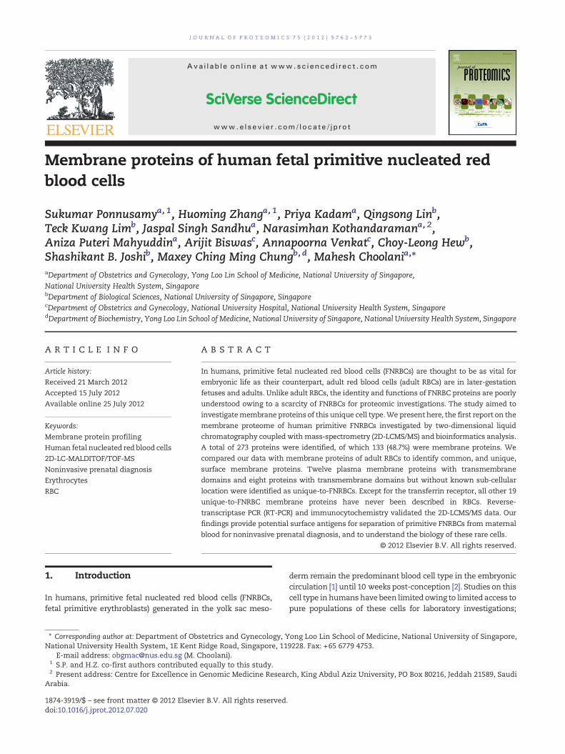
Placental tissueswere collected at the termination of pregnancy(7+0 to 9+3 weeks amenorrhea). FNRBCs were extracted fromplacental villi as per our protocol [30]: placental villi weredigested in trophoblast digestion buffer (146.3 ml HBSScontaining 0.182 g trypsin and 3.75 ml 1 M Hepes (Gibco®-Invitrogen-Life-Technologies, NY, USA)) for 30 min at 37 °C in ashaking-water-bath, and digestion was stopped using fetal calfserum (Pierce, IL, USA) (5 ml/45 ml digestion buffer). Single cellsuspensions were centrifuged (3000 rpm, 20 °C, 10 min). Redcell pellets containing FNRBCs were suspended in PBS, andseparated using Percoll 1083 (GE Healthcare, Uppsala, Sweden)(3000 rpm, 20 °C, 20 min). FNRBC purity was determined bybasic staining of cytospun slides. Samples were stored formembrane preparation (if purity ≥90% FNRBCs) in HES buffer(20 mM HEPES, pH 7.4, 1 mM EDTA and 250 mM sucrose) withprotease-inhibitor cocktail (Roche Diagnostics, Mannheim,Germany) at −80 °C. Morphologies of FNRBCs and adult RBCsare shown in Fig. 1.
2.3. Membrane protein preparation and digestion
Membranes from pooled FNRBCs (5×10⁷ cells) were prepared asdescribed earlier by us [11]: cells stored in HES buffer were lysedby thawing and sonication, and ultra-centrifuged at 100,000 xg4 °C (1 h) to obtain themembrane pelletwhichwas thenwashedusing high pH solution (0.1 M Na2CO3, pH11), and twice withMilli-Q water. Proteins were extracted from FNRBC membranesusing methanol (MeOH)/50 mM NH4HCO3 (60:40,vol/vol), andprotein reduction, alkylation and digestion were carried out asdescribed by Blonder and others [31]. Tryptic digestion wascarried out using sequencing grade modified trypsin (Promega,Southampton, UK). Digested sample was centrifuged and thepellet washed inMeOH solution (60%MeOH in 50 mMNH4HCO3)twice. Supernatants were pooled (MeOH-derived digests), whilethe pellet was re-suspended in Trifluoroethanol (TFE)/50 mMNH4HCO3 (50:50vol/vol) and the proteins extracted were thendiluted 10 times with 50 mM NH4HCO3 for a second trypsindigestion to obtain supernatants (TFE-derived digests). Bothdigests were lyophilized and stored at −80 °C.
2.4. Two-dimensional liquid chromatography and massspectrometry (2D-LCMS/MS)
2D-LCMS/MS was essentially the same as described earlier byus [11]. Briefly, lyophilized digests were re-suspended in
BA
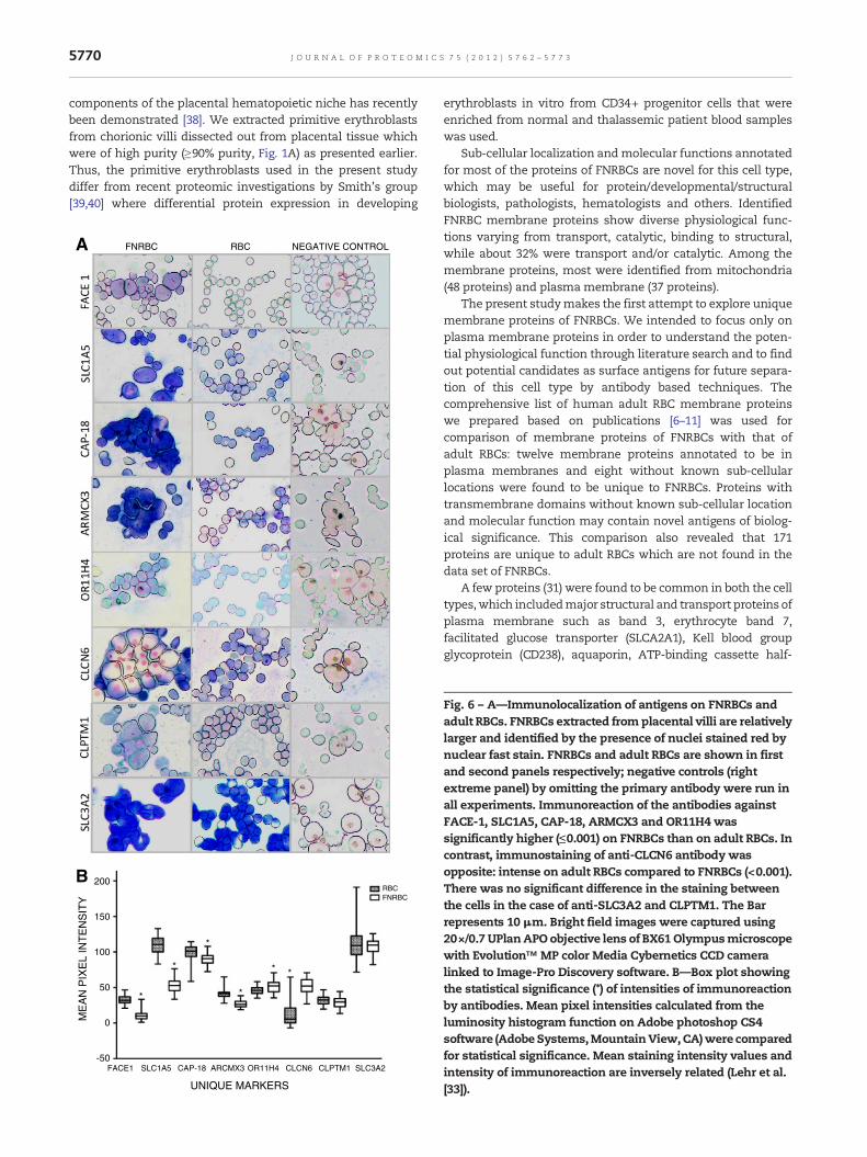
Fig. 1 –Wright's stained FNRBCs and adult RBCs. FNRBCs extracted from placental villi, and adult RBCs prepared from volunteerblood sample. (A) FNRBCs (nucleated). (B) adult RBCs without nuclei. Bright field images were captured using20×/0.40PhPobjective lens of CKX41 Olympus microscope. Bar represents 10 μm.
5764 J O U R N A L O F P R O T E O M I C S 7 5 ( 2 0 1 2 ) 5 7 6 2 – 5 7 7 3
solvent [(98%H2O, 2% acetonitrile (CAN) and 0.05%trifluoroacetic acid (TFA))], and after centrifugation superna-tants were separated using an Ultimate-Dual-HPLC system(Dionex, Sunnyvale, CA, USA). All samples were first separatedon a strong cation exchange (SCX) column (300 μm i.d.,×15 cm,packed with 10 μm POROS 10S) and eluted fractions werecaptured on the PepMap trap column (300 μm i.d.,×1 mm,packedwith 5 μmC18100 Å), and eluted by gradient elution to areversed‐phase column (Monolithic Capillary Column, 200 μmi.d.,×5 cm). LC fractions were mixed with matrix-assisted laserdesorption/ionization (MALDI) matrix (7 mg/ml α-cyano-4-hydroxycinnamic acid and 130 μg/ml ammonium citrate in75% CAN) at a flow rate of 5.4 μl/min through a 25 nlmixing-tee(Upchurch Scientific, Oak Harbor, WA, USA) before beingspotted onto 192-well stainless steel MALDI target plates (ABSCIEX, Foster City, CA, USA), at a rate of one well per 5 s, using aProbot Micro Fraction collector (Dionex).
Samples on the MALDI target plates were analyzed usingan ABI 4700 Proteomics Analyzer (AB SCIEX) with a MALDIsource and time of flight analyzer TOF/TOF™ optics. For MSanalysis, typically 1000 shots were accumulated for eachsample well. Tandem-MS_(MS/MS) analyses were performedusing nitrogen, at collision energy of 1 kV and a collision gaspressure of ~3.0×10⁻⁷ Torr. 3000 to 6000 shots were combinedfor each spectrum depending on the quality of the data.
2.5. Database searching
MASCOT search engine (v2.0;Matrix Science)wasused to searchtandem mass spectra. GPS Explorer™ software (v3.6; AB SCIEX)was used to create and search files with the MASCOT searchengine for peptide and protein identifications. The InternationalProtein Index (IPI) human protein database (v3.10) was used forthe search of tryptic peptides [32] and 57,478 entries weresearched. All MS/MS spectra from the LC runs were combinedfor the search. Cysteine carbamidomethylation, N-terminalacetylation and pyroglutamination, and methionine oxidationwere selected as variable modifications. Two missed cleavageswere allowed. Precursor error tolerance was set to 200 ppm andMS/MS fragment error tolerance was 0.4 Da.
2.6. Estimation of false positive rate
The false positive rate was calculated by comparing the searchresults from a randomized database versus the actual database.The minimum ion score C.I. percent was set such that no morethan 5% false discovery rate (FDR) was achieved and was usedas the cut-off threshold at the peptide level. All the proteinsidentified from random database search were single peptide-matched. Proteins identified by this method from IPI humandatabase were color coded as red, green or black: those redcolored proteins arematched to at least twopeptides and henceare statistically confident (FDR is zero); proteins that are greencolored are identified by single peptide where match scores arehigher than the highest score in the decoy database andessentially the FDR is zero; black colored proteins wereidentified based on single peptide match fall within the setthreshold of 5% FDR. Top ranked peptides with Best Ion scores≥33 and ≥36 for TFE and MeOH extractions, respectively, wereincluded for analysis as peptides counted for each protein. Allthe MS/MS spectra were further validated manually.
2.7. Annotation
Sub cellular and functional categories of the identified proteinswere obtained based on annotations of Gene Ontology usingGoFig.(http://udgenome.ags.udel.edu/gofigure/index.html).Swiss-prot and TrEMBL data base were also used for functionalannotation of unique proteins of FNRBCs. The number oftransmembrane domains (TMD) of the identified proteins waspredicted using TMHMM Server (v2.0) (http://www.cbs.dtu.dk/services/TMHMM/).
2.8. Evaluation of the identified unique proteins
2.8.1. Reverse transcriptase PCR (RT-PCR) for mRNAexpression of unique proteins
2.8.1.1. RNA extraction. RNA from FNRBCs was isolatedusing an RNeasy Mini Kit (Qiagen, Germany) according tomanufacturer's instructions. Briefly, FNRBCs (3×10⁶ cells) were
12974 70MeOH
144TFE199
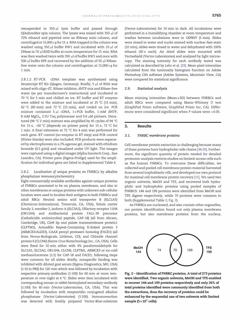
Fig. 2 – Identification of FNRBC proteins. A total of 273 proteinswere identified. Two organic solvents, MeOH and TFE enabledto recover 144 and 199 proteins respectively and only 26% oftotal proteins identified were commonly identified from boththe extractions, thus the recovery of proteins could beenhanced by the sequential use of two solvents with limitedsample (5×10⁷ cells).
5765J O U R N A L O F P R O T E O M I C S 7 5 ( 2 0 1 2 ) 5 7 6 2 – 5 7 7 3
resuspended in 350 μl lysis buffer and passed throughQIAshredder spin column. The lysate was mixed with 350 μl of70% ethanol and pipetted onto an RNeasy mini column, andcentrifugedat 15,000×g for 15 s. RNA trapped in the columnwaswashed using 350 μl buffer RW1 and incubated with 10 μl ofDNase in 70 μl RDD buffer at room temperature for 15 min. RNAwas thenwashed twicewith 350 μl of buffer RW1 and oncewith500 μl buffer RPE and recovered by the addition of 50 μl RNase-free water onto the column and centrifugation at 15,000×g for1 min.
2.8.1.2. RT-PCR. cDNA template was synthesized usingSensiscript RT Kit (Qiagen, Germany). Briefly, 5 μl of RNA wasmixedwith oligo-dT, RNase inhibitor, dNTPmix and RNase-freewater (as per manufacturer's instructions) and incubated at70 °C for 5 min and chilled on ice. RT buffer and RT enzymewere added to the mixture and incubated at 25 °C (15 min),42 °C (60 min) and 72 °C (15 min), and cooled on ice. PCRmixture contained 5 μl cDNA, 1×PCR buffer, 1 mM dNTP,8 mM MgCl2, 2.5U Taq polymerase and 0.6 μM primers. Dena-tured (94 °C 2 min) mixture was amplified by 45 cycles of 94 °Cfor 15 s, ~60 °C (depends on primer pairs) for 15 s, 72 °C for1 min. A final extension at 72 °C for 4 min was performed foreach gene. RT control (no enzyme in RT step) and PCR control(Water-blanks) were also included. PCR products were separat-ed by electrophoresis in a 2%agarose gel, stainedwith ethidiumbromide (0.5 g/ml) and visualized under UV light. The imageswere captured using a digital imager (Alpha InnotechCorp., SanLeandro, CA). Primer pairs (Sigma-Proligo) used for the ampli-fication for individual gene are listed in Supplemental Table 6.
2.8.2. Localisation of unique proteins on FNRBCs by alkalinephosphatase immunocytochemistryEight commercially available antibodies against unique proteinsof FNRBCs annotated to be on plasma membrane, and also inothermembranes or unique proteinswith unknown sub-cellularlocationwere used to localize their antigens in both FNRBCs andadult RBCs: Neutral amino acid transporter B (SLC1A5)(Chemicon-International, Temecula, CA, USA), Solute carrierfamily 3, member 2, isoform A (SLC3A2), Olfactory receptor 11H4(OR11H4) and Antibacterial protein FALL-39 precursor(Cathelicidin antimicrobial peptide, CAP-18) (all from Abcam,Cambridge, UK), Cleft lip and palate transmembrane protein1(CLPTM1), Armadillo Repeat-Containing X-linked protein 3(ARMCX3/ALEX3), CAAX prenyl protease1 homolog (FACE1) (allfrom Novus-Biologicals, Littleton, CO), and Chloride channelprotein 6 (CLCN6) (Santa-Cruz Biotechnology,Inc., CA, USA). Cellswere fixed for 10 min either with 4% paraformaldehyde forSLC1A5, SLC3A2, OR11H4, CLCN6, CLPTM1, ARMCX3 or ice-coldmethanol:acetone (1:1) for CAP-18 and FACE1; following stepswere common for all slides: Briefly, nonspecific binding wasinhibited with diluted goat serum (Sigma-Diagnostics, MO, USA)(1:10 in PBS) for 120 min which was followed by incubation withrespective primary-antibodies (1:100) for 60 min at room tem-perature or over night at 4 °C. Slides were then incubated withcorresponding mouse or rabbit biotinylated secondary-antibody(1:100) for 60 min (Vector-Laboratories, CA, USA). This wasfollowed by incubation with streptavidin conjugated alkalinephosphatase (Vector-Laboratories) (1:100). Immunoreactionwas detected with freshly prepared Vector-Blue-substrate
(Vector-Laboratories) for 10 min in dark. All incubations wereperformed in a humidifying chamber at room temperature andwashes between incubations were in 1XPBST (5 min). Slideswere rinsed in water and nuclei stained with nuclear-fast-stain(10 min), slides were rinsed in water and dehydrated with 100%ethanol (30 s each). Air dried slides were mounted withVectashield (Vector-Laboratories) and analyzed by light micros-copy. The staining intensity for each antibody tested wascalculated as described by Lehr et al. [33]. Mean pixel intensitiescalculated from the luminosity histogram function on AdobePhotoshop CS4 software (Adobe Systems, Mountain View, CA)were compared for statistical significance.
2.9. Statistical analysis
Mean staining intensities (Mean±SD) between FNRBCs andadult RBCs were compared using Mann–Whitney U test(GraphPad Prism software, GraphPad Prism Inc, CA). Differ-ences were considered significant when P values were <0.05.
3. Results
3.1. FNRBC membrane proteins
Cell membrane protein extraction is challenging because manyof these proteins have hydrophobic side chains [34,35]. Further-more, the significant quantity of protein needed for detailedproteomic analysis restricts studies on limited-access cells suchas the human FNRBCs. To overcome these difficulties, wecollected and pooled cell membrane proteinmaterial harvestedfromseveral trophoblastic villi, anddeveloped our ownprotocolfor maximal cell membrane protein recovery [11]. We used twoorganic solvents, MeOH and TFE, and recovered both hydro-philic and hydrophobic proteins using pooled samples ofFNRBCS. 144 and 199 proteins were identified from MeOH andTFE digests respectively, while 70 proteins were common toboth (Supplemental Table 1; Fig. 2).
As FNRBCs are nucleated, and also contain other organelles,our protein identification found not only plasma membraneproteins, but also membrane proteins from the nucleus,
5766 J O U R N A L O F P R O T E O M I C S 7 5 ( 2 0 1 2 ) 5 7 6 2 – 5 7 7 3
mitochondria, endoplasmic reticulum, Golgi, microsomes andperoxisomes.
3.2. Location annotation of identified proteins
A total of 273 proteins were identified, and their locationswithin the cell annotated (Supplemental Table 2): 133 weremembrane proteins (Supplemental Table 2) while 132 werenon-membrane proteins including 16 that have been describedas exclusively cytoplasmic (Supplemental Table 3). Locations ofthe remaining 8 are as yet unclassified (Supplemental Table 4).
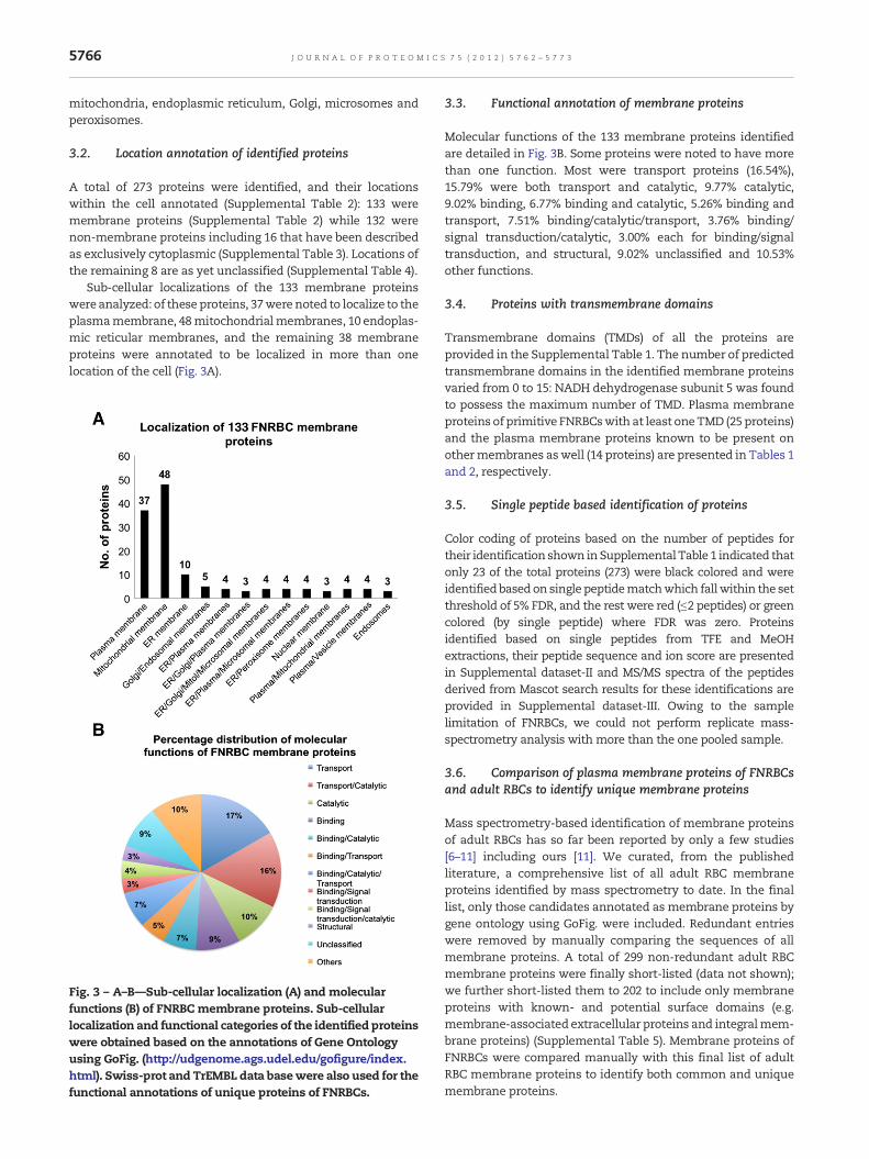
Sub-cellular localizations of the 133 membrane proteinswere analyzed: of these proteins, 37were noted to localize to theplasmamembrane, 48mitochondrialmembranes, 10 endoplas-mic reticular membranes, and the remaining 38 membraneproteins were annotated to be localized in more than onelocation of the cell (Fig. 3A).
Fig. 3 – A–B—Sub-cellular localization (A) and molecularfunctions (B) of FNRBC membrane proteins. Sub-cellularlocalization and functional categories of the identified proteinswere obtained based on the annotations of Gene Ontologyusing GoFig. (http://udgenome.ags.udel.edu/gofigure/index.html). Swiss-prot and TrEMBL data basewere also used for thefunctional annotations of unique proteins of FNRBCs.
3.3. Functional annotation of membrane proteins
Molecular functions of the 133 membrane proteins identifiedare detailed in Fig. 3B. Some proteins were noted to have morethan one function. Most were transport proteins (16.54%),15.79% were both transport and catalytic, 9.77% catalytic,9.02% binding, 6.77% binding and catalytic, 5.26% binding andtransport, 7.51% binding/catalytic/transport, 3.76% binding/signal transduction/catalytic, 3.00% each for binding/signaltransduction, and structural, 9.02% unclassified and 10.53%other functions.
3.4. Proteins with transmembrane domains
Transmembrane domains (TMDs) of all the proteins areprovided in the Supplemental Table 1. The number of predictedtransmembrane domains in the identified membrane proteinsvaried from 0 to 15: NADH dehydrogenase subunit 5 was foundto possess the maximum number of TMD. Plasma membraneproteins of primitive FNRBCswith at least oneTMD (25 proteins)and the plasma membrane proteins known to be present onothermembranes aswell (14 proteins) are presented in Tables 1and 2, respectively.
3.5. Single peptide based identification of proteins
Color coding of proteins based on the number of peptides fortheir identification shown inSupplemental Table 1 indicated thatonly 23 of the total proteins (273) were black colored and wereidentified basedon single peptidematchwhich fallwithin the setthreshold of 5% FDR, and the rest were red (≤2 peptides) or greencolored (by single peptide) where FDR was zero. Proteinsidentified based on single peptides from TFE and MeOHextractions, their peptide sequence and ion score are presentedin Supplemental dataset-ΙΙ and MS/MS spectra of the peptidesderived from Mascot search results for these identifications areprovided in Supplemental dataset-ΙΙΙ. Owing to the samplelimitation of FNRBCs, we could not perform replicate mass-spectrometry analysis with more than the one pooled sample.
3.6. Comparison of plasma membrane proteins of FNRBCsand adult RBCs to identify unique membrane proteins
Mass spectrometry-based identification of membrane proteinsof adult RBCs has so far been reported by only a few studies[6–11] including ours [11]. We curated, from the publishedliterature, a comprehensive list of all adult RBC membraneproteins identified by mass spectrometry to date. In the finallist, only those candidates annotated as membrane proteins bygene ontology using GoFig. were included. Redundant entrieswere removed by manually comparing the sequences of allmembrane proteins. A total of 299 non-redundant adult RBCmembrane proteins were finally short-listed (data not shown);we further short-listed them to 202 to include only membraneproteins with known- and potential surface domains (e.g.membrane-associated extracellular proteins and integralmem-brane proteins) (Supplemental Table 5). Membrane proteins ofFNRBCs were compared manually with this final list of adultRBC membrane proteins to identify both common and uniquemembrane proteins.
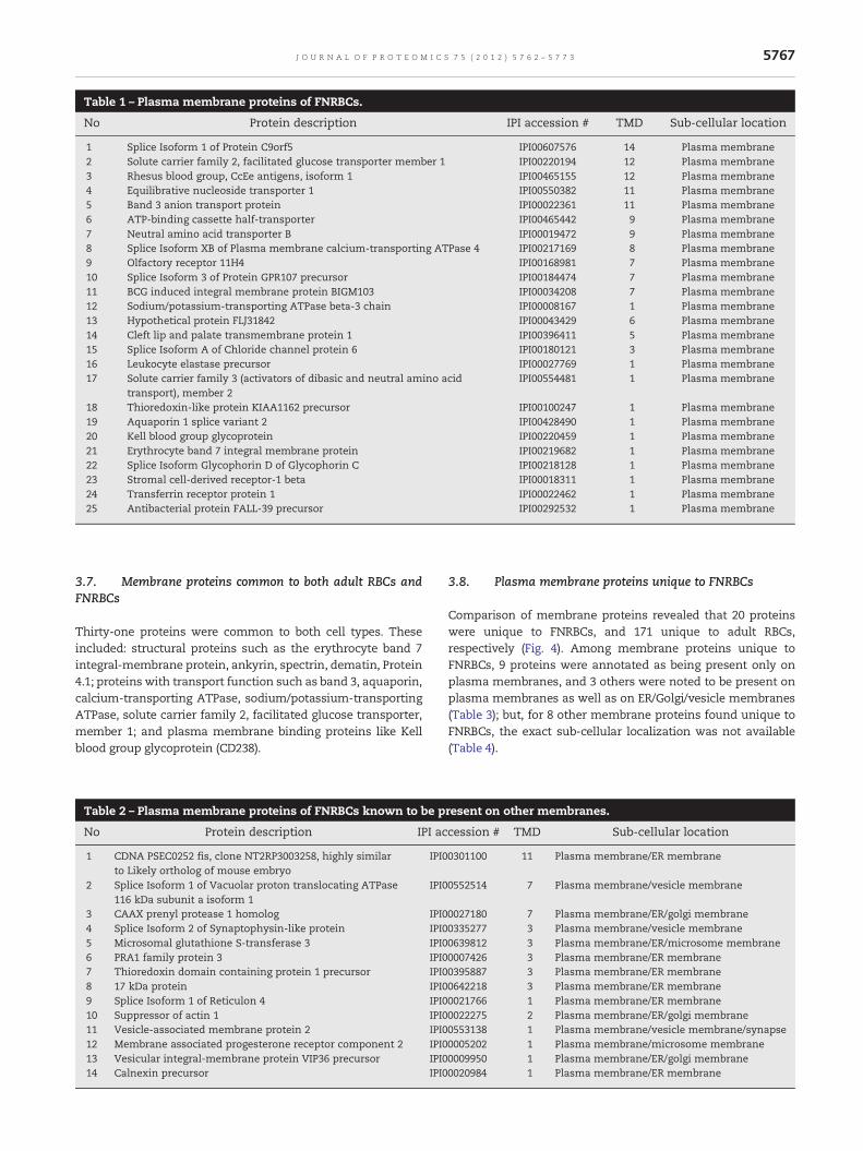
Table 1 – Plasma membrane proteins of FNRBCs.
No Protein description IPI accession # TMD Sub-cellular location
1 Splice Isoform 1 of Protein C9orf5 IPI00607576 14 Plasma membrane2 Solute carrier family 2, facilitated glucose transporter member 1 IPI00220194 12 Plasma membrane3 Rhesus blood group, CcEe antigens, isoform 1 IPI00465155 12 Plasma membrane4 Equilibrative nucleoside transporter 1 IPI00550382 11 Plasma membrane5 Band 3 anion transport protein IPI00022361 11 Plasma membrane6 ATP-binding cassette half-transporter IPI00465442 9 Plasma membrane7 Neutral amino acid transporter B IPI00019472 9 Plasma membrane8 Splice Isoform XB of Plasma membrane calcium-transporting ATPase 4 IPI00217169 8 Plasma membrane9 Olfactory receptor 11H4 IPI00168981 7 Plasma membrane10 Splice Isoform 3 of Protein GPR107 precursor IPI00184474 7 Plasma membrane11 BCG induced integral membrane protein BIGM103 IPI00034208 7 Plasma membrane12 Sodium/potassium-transporting ATPase beta-3 chain IPI00008167 1 Plasma membrane13 Hypothetical protein FLJ31842 IPI00043429 6 Plasma membrane14 Cleft lip and palate transmembrane protein 1 IPI00396411 5 Plasma membrane15 Splice Isoform A of Chloride channel protein 6 IPI00180121 3 Plasma membrane16 Leukocyte elastase precursor IPI00027769 1 Plasma membrane17 Solute carrier family 3 (activators of dibasic and neutral amino acid
transport), member 2IPI00554481 1 Plasma membrane
18 Thioredoxin-like protein KIAA1162 precursor IPI00100247 1 Plasma membrane19 Aquaporin 1 splice variant 2 IPI00428490 1 Plasma membrane20 Kell blood group glycoprotein IPI00220459 1 Plasma membrane21 Erythrocyte band 7 integral membrane protein IPI00219682 1 Plasma membrane22 Splice Isoform Glycophorin D of Glycophorin C IPI00218128 1 Plasma membrane23 Stromal cell-derived receptor-1 beta IPI00018311 1 Plasma membrane24 Transferrin receptor protein 1 IPI00022462 1 Plasma membrane25 Antibacterial protein FALL-39 precursor IPI00292532 1 Plasma membrane
5767J O U R N A L O F P R O T E O M I C S 7 5 ( 2 0 1 2 ) 5 7 6 2 – 5 7 7 3
3.7. Membrane proteins common to both adult RBCs andFNRBCs
Thirty-one proteins were common to both cell types. Theseincluded: structural proteins such as the erythrocyte band 7integral-membrane protein, ankyrin, spectrin, dematin, Protein4.1; proteins with transport function such as band 3, aquaporin,calcium-transporting ATPase, sodium/potassium-transportingATPase, solute carrier family 2, facilitated glucose transporter,member 1; and plasma membrane binding proteins like Kellblood group glycoprotein (CD238).
Table 2 – Plasma membrane proteins of FNRBCs known to be p
No Protein description IPI ac
1 CDNA PSEC0252 fis, clone NT2RP3003258, highly similarto Likely ortholog of mouse embryo
IPI0
2 Splice Isoform 1 of Vacuolar proton translocating ATPase116 kDa subunit a isoform 1
IPI0
3 CAAX prenyl protease 1 homolog IPI04 Splice Isoform 2 of Synaptophysin-like protein IPI05 Microsomal glutathione S-transferase 3 IPI06 PRA1 family protein 3 IPI07 Thioredoxin domain containing protein 1 precursor IPI08 17 kDa protein IPI09 Splice Isoform 1 of Reticulon 4 IPI010 Suppressor of actin 1 IPI011 Vesicle-associated membrane protein 2 IPI012 Membrane associated progesterone receptor component 2 IPI013 Vesicular integral-membrane protein VIP36 precursor IPI014 Calnexin precursor IPI0
3.8. Plasma membrane proteins unique to FNRBCs
Comparison of membrane proteins revealed that 20 proteinswere unique to FNRBCs, and 171 unique to adult RBCs,respectively (Fig. 4). Among membrane proteins unique toFNRBCs, 9 proteins were annotated as being present only onplasma membranes, and 3 others were noted to be present onplasma membranes as well as on ER/Golgi/vesicle membranes(Table 3); but, for 8 other membrane proteins found unique toFNRBCs, the exact sub-cellular localization was not available(Table 4).
resent on other membranes.
cession # TMD Sub-cellular location
0301100 11 Plasma membrane/ER membrane
0552514 7 Plasma membrane/vesicle membrane
0027180 7 Plasma membrane/ER/golgi membrane0335277 3 Plasma membrane/vesicle membrane0639812 3 Plasma membrane/ER/microsome membrane0007426 3 Plasma membrane/ER membrane0395887 3 Plasma membrane/ER membrane0642218 3 Plasma membrane/ER membrane0021766 1 Plasma membrane/ER membrane0022275 2 Plasma membrane/ER/golgi membrane0553138 1 Plasma membrane/vesicle membrane/synapse0005202 1 Plasma membrane/microsome membrane0009950 1 Plasma membrane/ER/golgi membrane0020984 1 Plasma membrane/ER membrane
2020171171 3131
Adult RBCs FNRBCs
Fig. 4 – Membrane proteins with potential surface domains ofadult RBCs and FNRBCs. A comparison of membrane proteinswith potential surface domains (as annotated) indicated thatonly 31 proteins were commonmembrane proteins to adultRBCs and FNRBCs, and 171 and 20 were unique to themrespectively.
5768 J O U R N A L O F P R O T E O M I C S 7 5 ( 2 0 1 2 ) 5 7 6 2 – 5 7 7 3
Membrane proteins unique to FNRBCs fall mainly underbroad functional groups such as (a) transporter proteins: neutralamino acid transporter B, solute carrier family 3 (activators ofdibasic and neutral amino acid transport), splice isoform A ofchloride channel protein 6 (chloride ion transport); (b) bindingproteins: transferrin receptor protein, splice isoform3 of ProteinGPR107 precursor, olfactory receptor 11H4; and (c) catalyticproteins: CAAX prenyl protease 1 homolog, Vitamin K epoxidereductase complex subunit 1-like protein 1 (VKORC1 L1), SpliceIsoform 1 of Protein C20orf22.
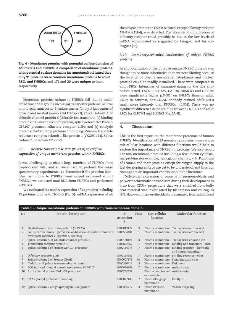
3.9. Reverse transcriptase PCR (RT-PCR) to confirmexpression of unique membrane proteins within FNRBCs
It was challenging to obtain large numbers of FNRBCs fromtrophoblastic villi, and all were used to perform the massspectrometry experiments. To determine if the proteins iden-tified as unique to FNRBCs were indeed expressed withinFNRBCs, we extracted total RNA from FNRBCs and performeda RT-PCR.
We evaluated themRNA expression of 23 proteins including13 proteins unique to FNRBCs (Fig. 5). mRNA expression of all
Table 3 – Unique membrane proteins of FNRBCs with transmem
No Protein descriptiona
1 Neutral amino acid transporter B (SLC1A5) IP2 Solute carrier family 3 (activators of dibasic and neutral amino acid
transport), member 2, isoform A (SLC3A2)IP
3 Splice Isoform A of Chloride channel protein 6 IP4 Transferrin receptor protein 1 IP5 Splice Isoform 3 of Protein GPR107 precursor IP
6 Olfactory receptor 11H4 IP7 Splice Isoform 1 of Protein C9orf5 IP8 Cleft lip and palate transmembrane protein 1 IP9 BCG induced integral membrane protein BIGM103 IP10 Antibacterial protein FALL-39 precursor IP
11 CAAX prenyl protease 1 homolog IP
12 Splice Isoform 2 of Synaptophysin-like protein IP
the unique proteins on FNRBCs tested, except olfactory receptor11H4 (OR11H4), was detected. The absence of amplification ofolfactory receptor could probably be due to the low levels ofmRNA accumulated as suggested by Feingold and his col-leagues [36].
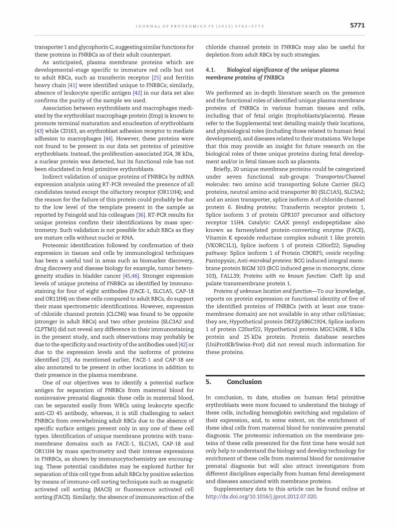
3.10. Immunocytochemical localization of unique FNRBCproteins
In situ localization of the putative unique FNRBC proteins wasthought to be more informative than western blotting becausethe location of plasma membrane, cytoplasmic and nuclearproteins could be readily visualized. These were compared toadult RBCs. Intensities of immunostaining for the five anti-bodies tested, FACE-1, SLC1A5, CAP-18, ARMCX3 and OR11H4were significantly higher (≤0.001) on FNRBCs than on adultRBCs; in contrast, anti-CLCN6 antibody stained adult RBCsmuch more intensely than FNRBCs (<0.001). There was nosignificant difference in the staining between FNRBCs and adultRBCs for CLPTM1 and SLC3A2 (Fig. 6A–B).
4. Discussion
This is the first report on the membrane proteome of humanFNRBCs. Identification of 133 membrane proteins from varioussub-cellular locations with different functions would help toexplore the importance of FNRBC in medicine. We also report132 non-membrane proteins including a few known cytoplas-mic proteins (for example, hemoglobin chains ε, γ, δ). Functionsof FNRBCs and their proteins except for oxygen supply to thefast developing embryo are yet to be understood, and thus ourfindings are an important contribution to the literature.
Differential expression of proteins in pronormoblasts andpoly/orthochromatic normoblasts during their development invitro from CD34+ progenitors that were enriched from buffycoat material was investigated by Richardson and colleagues[37]. However, these erythroblasts presumably from adult blood
brane domain.
IPIccession
#
TMD Sub-cellularlocation
Molecular function
I00019472 9 Plasma membrane Transporter-amino acidI00554481 1 Plasma membrane Transporter-amino acid
I00180121 3 Plasma membrane Transporter-chloride ionI00022462 1 Plasma membrane Binding and transport —ironI00184474 7 Plasma membrane Binding receptor —hormone
and neurotransmitterI00168981 7 Plasma membrane Binding receptor—odorI00607576 14 Plasma membrane Signaling pathwaysI00396411 5 Plasma membrane UnknownI00034208 7 Plasma membrane AntimicrobialI00292532 1 Plasmamembrane/
extracellularAntibacterial
I00027180 7 Plasma/ER/golgimembrane
Catalytic
I00335277 3 Plasma/vesiclemembrane
Vesicle recycling
Table 4 – Unique membrane proteins of FNRBCs with transmembrane domain but location unknown.
No Protein description IPI accession # TMD Sub-cellular location Molecular function
1 Vitamin K epoxide reductase complexsubunit 1-like protein 1
IPI00166079 2 Unclassified membrane (potential) Catalytic
2 Splice Isoform 1 of Protein C20orf22 IPI00394779 1 Unclassified membrane (by similarity) Catalytic3 Hypothetical protein DKFZp564K247
(hypoxia induced gene 1 protein)IPI00295621 2 Unclassified membrane (potential) Unclassified
4 Hypothetical protein DKFZp586C1924 IPI00031064 2 Unclassified membrane (potential) Unclassified5 ALEX3 protein variant IPI00604615 1 Unclassified single pass membrane (potential) Unclassified6 Hypothetical protein MGC14288 IPI00176708 1 Unclassified membrane (potential) Unclassified7 8 kDa protein IPI00639803 2 Unclassified Unclassified8 25 kDa protein IPI00646289 1 Unclassified Unclassified
5769J O U R N A L O F P R O T E O M I C S 7 5 ( 2 0 1 2 ) 5 7 6 2 – 5 7 7 3
samples differ from the yolk-sac derived fetal primitiveerythroblasts extracted from chorionic villi in our study,although both types of erythroblasts may appear morphologi-cally identical with higher cytoplasmic-nuclear ratio. In addi-tion, of the 21 differentially expressed proteins in two stages oferythroblasts reported by them, only one protein (cytochromeb5) appears to closely match with our protein identification ifnot identical (similar to cytochrome b5 outer mitochondrialmembrane isoform precursor). We presume that this observa-tion may probably due to the difference in the origin oferythroblasts in addition to the proteomic strategies followedin both studies.
PC
R c
on
tro
l
SL
C4A
1
HB
G2
HB
E1
SL
C39
A8
AZ
U1
CL
CN
6
RT
co
ntr
ol
CL
PT
M1
CA
MP
SL
C22
A11
SL
C3A
2
VA
MP
2
TF
RC
RT
co
ntr
ol
PC
R c
on
tro
l
Mar
ker
Mar
ker
Mar
ker
Mar
ker
Fig. 5 – Validation of uniquemembrane proteins of FNRBCs by revproteins of FNRBCs using total RNA extracted from FNRBCs and(Supplemental Table 6). RT control: no RT enzyme; PCR control: wepsilon chain; HBG2, hemoglobin gamma-2 chain; SLC4A1, solutemember 8; CLCN6, chloride channel protein 6; AZU1, Azurocidinsubunit 1-like protein 1; GPR107, protein GPR107 precursor; SLC1Glyceraldehyde-3-phosphate dehydrogenase; Bottom panel: SLCsolute carrier family 22 member 11, isoform 2; CAMP, antibactermembrane protein 2; TFRC transferrin receptor protein 1; CLPTMCAAX prenyl protease 1 homolog; ATP6VOA1, vacuolar proton trsteroid dehydrogenase homolog; OR11H4, olfactory receptor 11Hsynaptophysin-like protein; and C9orf5, protein C9orf5.
Proteomic analyses of FNRBCs had not been attemptedpreviously owing to the difficulty to obtain sufficient number ofcells. Our access to placental villi from patients undergoingtermination of pregnancy enabled to pool cells for 2D-LCMS/MSanalysis. In addition, the extraction ofmembrane proteins is yetanother challenge in proteomics; recovery of more membraneproteins (48.7% of total) froma limited sample (5×10⁷ cells) thanthose fromadult RBCsusing similar protocol (11) is encouraging,which also explains the structural complexity of these nucleat-ed cells. Yolk-sac derived fetal primitive erythroblasts enterfetal circulation and they do enucleate in the first trimesterhuman placenta [3]. Trophoblast and PDGF-B signaling as key
GP
R10
7
SL
C1A
5
RT
co
ntr
ol
PC
R c
on
tro
l
VK
OR
C1L
1
GA
PD
H
AT
P6
V0A
1
ZM
PS
TE
24
HS
D17
ββ12
SL
C43
A3
OR
11H
4
SY
PL
1
C9o
rf5
PC
R c
on
tro
l
RT
co
ntr
ol
erse transcriptase-PCR (RT-PCR). mRNA expression of uniqueby RT-PCR using primers specific for genes testedater blank in place of template. Top panel: HBE1, hemoglobincarrier family 4 member 1; SLC39A8, solute carrier family 39precursor; VKORC1L1, vitamin K epoxide reductase complexA5 neutral amino acid transporter B; GAPDH,3A2, solute carrier family 3 member 2, isoform A; SLC22A11,ial protein FALL-39 precursor; VAMP2, vesicle-associated1, cleft lip and palate transmembrane protein 1; ZMPSTE24,anslocating ATPase 116 kDa subunit a isoform 1; HSD17β12,4; SLC43A3, solute carrier family 43 member 3; SYPL1,
5770 J O U R N A L O F P R O T E O M I C S 7 5 ( 2 0 1 2 ) 5 7 6 2 – 5 7 7 3
components of the placental hematopoietic niche has recentlybeen demonstrated [38]. We extracted primitive erythroblastsfrom chorionic villi dissected out from placental tissue whichwere of high purity (≥90% purity, Fig. 1A) as presented earlier.Thus, the primitive erythroblasts used in the present studydiffer from recent proteomic investigations by Smith's group[39,40] where differential protein expression in developing
*
*
*
*
**
FACE1 SLC1A5 CAP-18 ARCMX3 OR11H4 CLCN6 CLPTM1 SLC3A2
UNIQUE MARKERS
ME
AN
PIX
EL
INT
EN
SIT
Y
200
150
100
50
0
-50
FNRBC RBC NEGATIVE CONTROL
RBCFNRBC
A
B
Fig. 6 – A—Immunolocalization of antigens on FNRBCs andadult RBCs. FNRBCs extracted from placental villi are relativelylarger and identified by the presence of nuclei stained red bynuclear fast stain. FNRBCs and adult RBCs are shown in firstand second panels respectively; negative controls (rightextreme panel) by omitting the primary antibody were run inall experiments. Immunoreaction of the antibodies againstFACE-1, SLC1A5, CAP-18, ARMCX3 and OR11H4 wassignificantly higher (≤0.001) on FNRBCs than on adult RBCs. Incontrast, immunostaining of anti-CLCN6 antibody wasopposite: intense on adult RBCs compared to FNRBCs (<0.001).There was no significant difference in the staining betweenthe cells in the case of anti-SLC3A2 and CLPTM1. The Barrepresents 10 μm. Bright field images were captured using20×/0.7 UPlanAPO objective lens of BX61Olympusmicroscopewith Evolution™ MP color Media Cybernetics CCD cameralinked to Image-Pro Discovery software. B—Box plot showingthe statistical significance (*) of intensities of immunoreactionby antibodies. Mean pixel intensities calculated from theluminosity histogram function on Adobe photoshop CS4software (AdobeSystems,MountainView,CA)were comparedfor statistical significance. Mean staining intensity values andintensity of immunoreaction are inversely related (Lehr et al.[33]).
erythroblasts in vitro from CD34+ progenitor cells that wereenriched from normal and thalassemic patient blood sampleswas used.
Sub-cellular localization andmolecular functions annotatedfor most of the proteins of FNRBCs are novel for this cell type,which may be useful for protein/developmental/structuralbiologists, pathologists, hematologists and others. IdentifiedFNRBC membrane proteins show diverse physiological func-tions varying from transport, catalytic, binding to structural,while about 32% were transport and/or catalytic. Among themembrane proteins, most were identified from mitochondria(48 proteins) and plasmamembrane (37 proteins).
The present studymakes the first attempt to explore uniquemembrane proteins of FNRBCs. We intended to focus only onplasma membrane proteins in order to understand the poten-tial physiological function through literature search and to findout potential candidates as surface antigens for future separa-tion of this cell type by antibody based techniques. Thecomprehensive list of human adult RBC membrane proteinswe prepared based on publications [6–11] was used forcomparison of membrane proteins of FNRBCs with that ofadult RBCs: twelve membrane proteins annotated to be inplasma membranes and eight without known sub-cellularlocations were found to be unique to FNRBCs. Proteins withtransmembrane domains without known sub-cellular locationand molecular function may contain novel antigens of biolog-ical significance. This comparison also revealed that 171proteins are unique to adult RBCs which are not found in thedata set of FNRBCs.
A few proteins (31) were found to be common in both the celltypes,which includedmajor structural and transport proteins ofplasma membrane such as band 3, erythrocyte band 7,facilitated glucose transporter (SLCA2A1), Kell blood groupglycoprotein (CD238), aquaporin, ATP-binding cassette half-
5771J O U R N A L O F P R O T E O M I C S 7 5 ( 2 0 1 2 ) 5 7 6 2 – 5 7 7 3
transporter 1 and glycophorinC, suggesting similar functions forthese proteins in FNRBCs as of their adult counterpart.
As anticipated, plasma membrane proteins which aredevelopmental-stage specific to immature red cells but notto adult RBCs, such as transferrin receptor [25] and ferritinheavy chain [41] were identified unique to FNRBCs; similarly,absence of leukocyte specific antigen [42] in our data set alsoconfirms the purity of the sample we used.
Association between erythroblasts and macrophages medi-ated by the erythroblast macrophage protein (Emp) is known topromote terminal maturation and enucleation of erythroblasts[43] while CD163, an erythroblast adhesion receptor to mediateadhesion to macrophages [44]. However, these proteins werenot found to be present in our data set proteins of primitiveerythroblasts. Instead, the proliferation-associated 2G4, 38 kDa,a nuclear protein was detected, but its functional role has notbeen elucidated in fetal primitive erythroblasts.
Indirect validation of unique proteins of FNRBCs by mRNAexpression analysis using RT-PCR revealed the presence of allcandidates tested except the olfactory receptor (OR11H4); andthe reason for the failure of this protein could probably be dueto the low level of the template present in the sample asreported by Feingold and his colleagues [36]. RT-PCR results forunique proteins confirm their identifications by mass spec-trometry. Such validation is not possible for adult RBCs as theyare mature cells without nuclei or RNA.
Proteomic identification followed by confirmation of theirexpression in tissues and cells by immunological techniqueshas been a useful tool in areas such as biomarker discovery,drug discovery and disease biology for example, tumor hetero-geneity studies in bladder cancer [45,46]. Stronger expressionlevels of unique proteins of FNRBCs as identified by immuno-staining for four of eight antibodies (FACE-1, SLC1A5, CAP-18and OR11H4) on these cells compared to adult RBCs, do supporttheir mass spectrometric identifications. However, expressionof chloride channel protein (CLCN6) was found to be opposite(stronger in adult RBCs) and two other proteins (SLC3A2 andCLPTM1) did not reveal any difference in their immunostainingin the present study, and such observations may probably bedue to the specificity and reactivity of the antibodies used [42] ordue to the expression levels and the isoforms of proteinsidentified [23]. As mentioned earlier, FACE-1 and CAP-18 arealso annotated to be present in other locations in addition totheir presence in the plasmamembrane.
One of our objectives was to identify a potential surfaceantigen for separation of FNRBCs from maternal blood fornoninvasive prenatal diagnosis: these cells in maternal blood,can be separated easily from WBCs using leukocyte specificanti-CD 45 antibody, whereas, it is still challenging to selectFNRBCs from overwhelming adult RBCs due to the absence ofspecific surface antigen present only in any one of these celltypes. Identification of unique membrane proteins with trans-membrane domains such as FACE-1, SLC1A5, CAP-18 andOR11H4 by mass spectrometry and their intense expressionsin FNRBCs, as shown by immunocytochemistry are encourag-ing. These potential candidates may be explored further forseparation of this cell type fromadult RBCs by positive selectionby means of immuno-cell sorting techniques such as magneticactivated cell sorting (MACS) or fluorescence activated cellsorting (FACS). Similarly, the absence of immunoreaction of the
chloride channel protein in FNRBCs may also be useful fordepletion from adult RBCs by such strategies.
4.1. Biological significance of the unique plasmamembrane proteins of FNRBCs
We performed an in-depth literature search on the presenceand the functional roles of identified unique plasmamembraneproteins of FNRBCs in various human tissues and cells,including that of fetal origin (trophoblasts/placenta). Pleaserefer to the Supplemental text detailing mainly their locations,and physiological roles (including those related to human fetaldevelopment), anddiseases related to theirmutations.Wehopethat this may provide an insight for future research on thebiological roles of these unique proteins during fetal develop-ment and/or in fetal tissues such as placenta.
Briefly, 20 unique membrane proteins could be categorizedunder seven functional sub-groups: Transportes/Channelmolecules: two amino acid transporting Solute Carrier (SLC)proteins, neutral amino acid transporter B0 (SLC1A5), SLC3A2;and an anion transporter, splice isoform A of chloride channelprotein 6. Binding proteins: Transferrin receptor protein 1,Splice isoform 3 of protein GPR107 precursor and olfactoryreceptor 11H4. Catalytic: CAAX prenyl endopeptidase alsoknown as farnesylated protein-converting enzyme (FACE),Vitamin K epoxide reductase complex subunit 1 like protein(VKORC1L1), Splice isoform 1 of protein C20orf22; Signalingpathway: Splice isoform 1 of Protein C9ORF5; vesicle recycling:Pantopysin; Anti-microbial proteins: BCG induced integral mem-brane protein BIGM 103 (BCG induced gene in monocyte, clone103), FALL39; Proteins with no known function: Cleft lip andpalate transmembrane protein 1.
Proteins of unknown location and function—To our knowledge,reports on protein expression or functional identity of five ofthe identified proteins of FNRBCs (with at least one trans-membrane domain) are not available in any other cell/tissue;they are, Hypothetical protein DKFZp586C1924, Splice isoform1 of protein C20orf22, Hypothetical protein MGC14288, 8 kDaprotein and 25 kDa protein. Protein database searches(UniProtKB/Swiss-Prot) did not reveal much information forthese proteins.
5. Conclusion
In conclusion, to date, studies on human fetal primitiveerythroblasts were more focused to understand the biology ofthese cells, including hemoglobin switching and regulation oftheir expression, and, to some extent, on the enrichment ofthese ideal cells from maternal blood for noninvasive prenataldiagnosis. The proteomic information on the membrane pro-teins of these cells presented for the first time here would notonly help to understand the biology and develop technology forenrichment of these cells frommaternal blood for noninvasiveprenatal diagnosis but will also attract investigators fromdifferent disciplines especially from human fetal developmentand diseases associated with membrane proteins.
Supplementary data to this article can be found online athttp://dx.doi.org/10.1016/j.jprot.2012.07.020.

5772 J O U R N A L O F P R O T E O M I C S 7 5 ( 2 0 1 2 ) 5 7 6 2 – 5 7 7 3
Author contribution
S.P. wrote the manuscript and participated in analysis andinterpretation of data; H.Z. performed the proteomics, RT-PCRstudies and analyzed the data, P.K. carried out immunocyto-chemistry; Q.L., T.K.L., helped in 2DLCMS studies and MS-dataanalyses; J.S.S., assisted in data analysis; M.C.M.C., N.K.,A.P.M. S.B.J. and C-L.H. helped in proteomics study; A.B. andA.V. provided clinical samples; M.C. designed the research,provided overall direction and corrected the manuscript.
Conflicts of interest statement
All authors declare no conflicts of interest. A patent applicationhas been submitted.
Acknowledgments
Research scholarship to H.Z. from the National University ofSingapore is gratefully acknowledged. This research wassupported by the National Medical Research Council (NMRCGrants: NMRC/0561/2001; NMRC/1080/2006 and NMRC/CSA/007/2009), and Biomedical Research Council (BMRC), Agencyfor Science, Technology and Research (A*STAR), Singapore(Grant no. 04/1/21/19/343). Facilities at Protein ProteomicsCentre, Department of Biological Sciences, National Universityof Singapore availed are gratefully acknowledged.
R E F E R E N C E S
[1] Palis J. Ontogeny of erythropoiesis. Curr Opin Hematol2008;15:155-61.
[2] Choolani M, O'Donoghue K, Talbert D, Kumar S, Roberts I,Letsky E, et al. Characterization of first trimester fetalerythroblasts for non-invasive prenatal diagnosis. Mol HumReprod 2003;9:227-35.
[3] Van Handel B, Prashad SL, Hassanzadeh-Kiabi N, Huang A,Magnusson M, Atanassova B, et al. The first trimester humanplacenta is a site for terminal maturation of primitiveerythroid cells. Blood 2010;116:3321-30.
[4] Peschle C, Mavilio F, Care A, Migliaccio G, Migliaccio AR, SalvoG, et al. Haemoglobin switching in human embryos:asynchrony of zeta – alpha and epsilon – gamma-globinswitches in primitive and definite erythropoietic lineage.Nature 1985;313:235-8.
[5] Qiu C, Olivier EN, Velho M, Bouhassira EE. Globin switches inyolk sac-like primitive and fetal-like definitive red blood cellsproduced from human embryonic stem cells. Blood 2008;111:2400-8.
[6] Kakhniashvili DG, Bulla Jr LA, Goodman SR. The humanerythrocyte proteome: analysis by ion trapmass spectrometry.Mol Cell Proteomics 2004;3:501-9.
[7] Low TY, SeowTK, ChungMC. Separation of human erythrocytemembrane associated proteins with one-dimensional andtwo-dimensional gel electrophoresis followed by identificationwith matrix-assisted laser desorption/ionization-time of flightmass spectrometry. Proteomics 2002;2:1229-39.
[8] Bruschi M, Seppi C, Arena S, Musante L, Santucci L, BalduiniC, et al. Proteomic analysis of erythrocyte membranes by soft
Immobiline gels combined with differential proteinextraction. J Proteome Res 2005;4:1304-9.
[9] Tyan YC, Jong SB, Liao JD, Liao PC, Yang MH, Liu CY, et al.Proteomic profiling of erythrocyte proteins by proteolyticdigestion chip and identification using two-dimensionalelectrospray ionization tandemmass spectrometry. J ProteomeRes 2005;4:748-57.
[10] Pasini EM, Kirkegaard M, Mortensen P, Lutz HU, Thomas AW,Mann M. In-depth analysis of the membrane and cytosolicproteome of red blood cells. Blood 2006;108:791-801.
[11] Zhang H, Lin Q, Ponnusamy S, Kothandaraman N, Lim TK,Zhao C, et al. Differential recovery of membrane proteinsafter extraction by aqueous methanol and trifluoroethanol.Proteomics 2007;7:1654-63.
[12] Roux-Dalvai F, Gonzalez de Peredo A, Simo C, Guerrier L,Bouyssie D, Zanella A, et al. Extensive analysis of thecytoplasmic proteome of human erythrocytes using thepeptide ligand library technology and advanced massspectrometry. Mol Cell Proteomics 2008;7:2254-69.
[13] Pasini EM, Kirkegaard M, Salerno D, Mortensen P, Mann M,Thomas AW. Deep coveragemouse red blood cell proteome: afirst comparison with the human red blood cell. Mol CellProteomics 2008;7:1317-30.
[14] Eber S, Lux SE. Hereditary spherocytosis—defects in proteinsthat connect the membrane skeleton to the lipid bilayer.Semin Hematol 2004;41:118-41.
[15] Kakhniashvili DG, Griko NB, Bulla Jr LA, Goodman SR. Theproteomics of sickle cell disease: profiling of erythrocytemembrane proteins by 2D-DIGE and tandem massspectrometry. Exp Biol Med (Maywood) 2005;230:787-92.
[16] Lane PA, Shew RL, Iarocci TA, Mohandas N, Hays T, MentzerWC. Unique alpha-spectrin mutant in a kindred with commonhereditary elliptocytosis. J Clin Invest 1987;79:989-96.
[17] Stewart GW, Corrall RJ, Fyffe JA, Stockdill G, Strong JA.Familial pseudohyperkalaemia. A new syndrome. Lancet1979;2:175-7.
[18] Delaunay J. The hereditary stomatocytoses: genetic disordersof the red cell membrane permeability to monovalentcations. Semin Hematol 2004;41:165-72.
[19] Wilkinson DK, Turner EJ, Parkin ET, Garner AE, Harrison PJ,Crawford M, et al. Membrane raft actin deficiency and alteredCa2+− induced vesiculation in stomatin-deficientoverhydrated hereditary stomatocytosis. Biochim BiophysActa 2008;1778:125-32.
[20] Schrier SL, Rachmilewitz E, Mohandas N. Cellular andmembrane properties of alpha and beta thalassemicerythrocytes are different: implication for differences inclinical manifestations. Blood 1989;74:2194-202.
[21] Roggwiller E, Betoulle ME, Blisnick T, Braun Breton C. A rolefor erythrocyte band 3 degradation by the parasite gp76serine protease in the formation of the parasitophorousvacuole during invasion of erythrocytes by Plasmodiumfalciparum. Mol Biochem Parasitol 1996;82:13-24.
[22] Cooper RA, Carucci DJ. Proteomic approaches to studyingdrug targets and resistance in Plasmodium. Curr DrugTargets Infect Disord 2004;4:41-51.
[23] Pasini EM, Lutz HU, Mann M, Thomas AW. Red blood cell(RBC) membrane proteomics—Part I: proteomics and RBCphysiology. J Proteomics 2010;73:403-20.
[24] Pasini EM, Lutz HU, MannM, Thomas AW. Red blood cell (RBC)membrane proteomics—Part II: comparative proteomics andRBC patho-physiology. J Proteomics 2010;73:421-35.
[25] Mathew A, Bell A, Johnstone RM. Hsp-70 is closely associatedwith the transferrin receptor in exosomes from maturingreticulocytes. Biochem J 1995;308(Pt 3):823-30.
[26] Voullaire L, Ioannou P, Nouri S, Williamson R. Fetal nucleatedred blood cells from CVS washings: an aid to development offirst trimester non-invasive prenatal diagnosis. Prenat Diagn2001;21:827-34.

5773J O U R N A L O F P R O T E O M I C S 7 5 ( 2 0 1 2 ) 5 7 6 2 – 5 7 7 3
[27] Christensen B, Kolvraa S, Lykke-Hansen L, Lorch T, Gohel D,Smidt-Jensen S, et al. Studies on the isolation andidentification of fetal nucleated red blood cells in thecirculation of pregnant women before and after chorion villussampling. Fetal Diagn Ther 2003;18:376-84.
[28] Nagy GR, Ban Z, Sipos F, Beke A, Papp C, Papp Z. Isolation ofepsilon-haemoglobin-chain positive fetal cells withmicromanipulation for prenatal diagnosis. Prenat Diagn2005;25:398-402.
[29] Choolani M, O'Donnell H, Campagnoli C, Kumar S, Roberts I,Bennett PR, et al. Simultaneous fetal cell identification anddiagnosis by epsilon-globin chain immunophenotyping andchromosomal fluorescence in situ hybridization. Blood2001;98:554-7.
[30] Ponnusamy S, Mohammed N, Ho SS, Zhang HM, Chan YH, NgYW, et al. In vivo model to determine fetal-cell enrichmentefficiency of novel noninvasive prenatal diagnosis methods.Prenat Diagn 2008;28:494-502.
[31] Blonder J, Goshe MB, Moore RJ, Pasa-Tolic L, Masselon CD,Lipton MS, et al. Enrichment of integral membrane proteinsfor proteomic analysis using liquid chromatography-tandemmass spectrometry. J Proteome Res 2002;1:351-60.
[32] Kersey PJ, Duarte J, Williams A, Karavidopoulou Y, Birney E,Apweiler R. The International Protein Index: an integrateddatabase for proteomics experiments. Proteomics2004;4:1985-8.
[33] Lehr HA, Mankoff DA, Corwin D, Santeusanio G, Gown AM.Application of photoshop-based image analysis toquantification of hormone receptor expression in breastcancer. J Histochem Cytochem 1997;45:1559-65.
[34] Santoni V, Molloy M, Rabilloud T. Membrane proteins andproteomics: un amour impossible? Electrophoresis2000;21:1054-70.
[35] Tan S, Tan HT, Chung MC. Membrane proteins andmembrane proteomics. Proteomics 2008;8:3924-32.
[36] Feingold EA, Penny LA, Nienhuis AW, Forget BG. An olfactoryreceptor gene is located in the extended human beta-globingene cluster and is expressed in erythroid cells. Genomics1999;61:15-23.
[37] Richardson BM, Heesom KJ, Parsons SF, Anstee DJ, Frayne J.Analysis of the differential proteome of human erythroblastsduring in vitro erythropoiesis by 2-D DIGE. Proteomics ClinAppl 2009;3:1123-34.
[38] Chhabra A, Lechner AJ, Ueno M, Acharya A, Van Handel B,Wang Y, et al. Trophoblasts regulate the placentalhematopoietic niche through PDGF-B signaling. Dev Cell2012;22:651-9.
[39] Leecharoenkiat A, Wannatung T, Lithanatudom P, Svasti S,Fucharoen S, Chokchaichamnankit D, et al. Increased oxida-tive metabolism is associated with erythroid precursorexpansion in beta0-thalassaemia/Hb E disease. Blood CellsMol Dis 2011;47:143-57.
[40] Sriiam S, Leecharoenkiat A, Lithanatudom P, Wannatung T,Svasti S, Fucharoen S, et al. Proteomic analysis of hemoglobinH-constant spring (Hb H-CS) erythroblasts. Blood Cells MolDis 2012;48:77-85.
[41] Pollack S, Campana T. Immature red cells have ferritinreceptors. Biochem Biophys Res Commun 1981;100:1667-72.
[42] Rider DA, Young SP. Measuring the specific activity of theCD45 protein tyrosine phosphatase. J Immunol Methods2003;277:127-34.
[43] Hanspal M, Hanspal JS. The association of erythroblasts withmacrophages promotes erythroid proliferation andmaturation: a 30-kD heparin-binding protein is involved inthis contact. Blood 1994;84:3494-504.
[44] Fabriek BO, Polfliet MM, Vloet RP, van der Schors RC,Ligtenberg AJ, Weaver LK, et al. The macrophage CD163surface glycoprotein is an erythroblast adhesion receptor.Blood 2007;109:5223-9.
[45] Celis JE, Celis P, Palsdottir H, Ostergaard M, Gromov P,Primdahl H, et al. Proteomic strategies to reveal tumorheterogeneity among urothelial papillomas. Mol CellProteomics 2002;1:269-79.
[46] Ohlsson G, Moreira JM, Gromov P, Sauter G, Celis JE. Loss ofexpression of the adipocyte-type fatty acid-binding protein(A-FABP) is associated with progression of human urothelialcarcinomas. Mol Cell Proteomics 2005;4:570-81.
Related Documents


![Multi-nucleated cells use ROS to induce breast cancer ......and have been reported to resume proliferation in certain contexts [31]. A novel type of cell division where multi-nucleated](https://static.cupdf.com/doc/110x72/5ec7cb93a3fbe17ed30cbdab/multi-nucleated-cells-use-ros-to-induce-breast-cancer-and-have-been-reported.jpg)


