MEF2C Silencing Attenuates Load-Induced Left Ventricular Hypertrophy by Modulating mTOR/S6K Pathway in Mice Ana Helena M. Pereira 1 , Carolina F. M. Z. Clemente 1 , Alisson C. Cardoso 1 , Thais H. Theizen 1 , Silvana A. Rocco 1 , Carla C. Judice 1 , Maria Carolina Guido 1 , Vinı´cius D. B. Pascoal 2 , Iscia Lopes-Cendes 2 , Jose ´ Roberto M. Souza 1 , Kleber G. Franchini 1 * 1 Department of Internal Medicine, School of Medicine, State University of Campinas, Campinas, Sa ˜o Paulo, Brazil, 2 Department of Medical Genetics, School of Medicine, State University of Campinas, Campinas, Sa ˜o Paulo, Brazil Abstract Background: The activation of the members of the myocyte enhancer factor-2 family (MEF2A, B, C and D) of transcription factors promotes cardiac hypertrophy and failure. However, the role of its individual components in the pathogenesis of cardiac hypertrophy remains unclear. Methodology/Principal Findings: In this study, we investigated whether MEF2C plays a role in mediating the left ventricular hypertrophy by pressure overload in mice. The knockdown of myocardial MEF2C induced by specific small interfering RNA (siRNA) has been shown to attenuate hypertrophy, interstitial fibrosis and the rise of ANP levels in aortic banded mice. We detected that the depletion of MEF2C also results in lowered levels of both PGC-1a and mitochondrial DNA in the overloaded left ventricle, associated with enhanced AMP:ATP ratio. Additionally, MEF2C depletion was accompanied by defective activation of S6K in response to pressure overload. Treatment with the amino acid leucine stimulated S6K and suppressed the attenuation of left ventricular hypertrophy and fibrosis in the aforementioned aortic banded mice. Conclusion/Significance: These findings represent new evidences that MEF2C depletion attenuates the hypertrophic responses to mechanical stress and highlight the potential of MEF2C to be a target for new therapies to cardiac hypertrophy and failure. Citation: Pereira AHM, Clemente CFMZ, Cardoso AC, Theizen TH, Rocco SA, et al. (2009) MEF2C Silencing Attenuates Load-Induced Left Ventricular Hypertrophy by Modulating mTOR/S6K Pathway in Mice. PLoS ONE 4(12): e8472. doi:10.1371/journal.pone.0008472 Editor: Arnold Schwartz, University of Cincinnati, United States of America Received September 23, 2009; Accepted November 17, 2009; Published December 29, 2009 Copyright: ß 2009 Pereira et al. This is an open-access article distributed under the terms of the Creative Commons Attribution License, which permits unrestricted use, distribution, and reproduction in any medium, provided the original author and source are credited. Funding: Funding for this work was provided by Fundac ¸a ˜o de Amparo a ` Pesquisa do Estado de Sa ˜o Paulo (FAPESP) grants 2006/54878-3, 2008/53583-5, 2008/ 53519-5, Conselho Nacional de Desenvolvimento Cientı ´-fico e Tecnolo ´ gico (CNPq) grants 305604/2006-6, 501160/2008-6 and Laborato ´ rio Crista ´ lia. The funders had no role in study design, data collection and analysis, decision to publish, or preparation of the manuscript. Competing Interests: The authors have declared that no competing interests exist. * E-mail: [email protected] Introduction Hypertrophy is a common feature of many forms of heart disease. While initially an adaptive response to increased workload and injury, in the long term cardiac hypertrophy predisposes to heart failure[1,2,3]. At the cellular level, myocardial hypertrophy is characterized by distinct accumulation of myofibrillar proteins and organelles in cardiomyocytes, while the development of heart failure is accompanied by degeneration and loss of hypertrophic cardiomyocytes as well as interstitial fibrosis[4]. In a current view, the hypertrophy and degeneration of cardiomyocytes represent a continuum governed by patterns of beneficial and adverse signaling triggered by stimuli such as mechanical stress and neurohumoral factors[5,6,7]. Among the intracellular pathways that integrate mechanical and hormonal signals, MEF2 (myocyte enhancer factors-2, members A to D) transcription factors play prominent roles in the regulation of cardiac hypertrophy and remodeling[8,9,10]. In this context, many studies have shown that overall MEF2 DNA-binding activity is enhanced in cardiomyocytes in response to biomechanical and neurohormonal stimuli[11,12,13]. Overexpression of MEF2A or MEF2C in cultured cardiomyocytes induces sarcomere degener- ation and cardiomyocytes elongation, suggesting that activation of these members may compose signaling pathways responsible for pathologic hypertrophy [8]. Accordingly, forced expressions of MEF2A, C and D in mice heart were demonstrated to be sufficient to drive intolerance to pressure overload, ventricular chamber dilation and contractile dysfunction[8,9,10]. There is also evidence associating MEF2 transcription factors with common forms of human heart failure[14]. Furthermore, the transgenic expression of negative dominants of MEF2 was shown to prevent chamber dilation and mechanical dysfunction, with minor effects on cardiac growth in calcineurin-induced hypertrophy[9]. Recent studies performed in mice with dominant-negative MEF2D suggested that this factor is an important mediator of the pathologic left ventricular hypertrophy, as these mice displayed no cardiac hypertrophy, fibrosis or fetal gene activation in response to pressure overload[10]. Altogether, these evidences support the PLoS ONE | www.plosone.org 1 December 2009 | Volume 4 | Issue 12 | e8472

Welcome message from author
This document is posted to help you gain knowledge. Please leave a comment to let me know what you think about it! Share it to your friends and learn new things together.
Transcript
MEF2C Silencing Attenuates Load-Induced LeftVentricular Hypertrophy by Modulating mTOR/S6KPathway in MiceAna Helena M. Pereira1, Carolina F. M. Z. Clemente1, Alisson C. Cardoso1, Thais H. Theizen1, Silvana A.
Rocco1, Carla C. Judice1, Maria Carolina Guido1, Vinıcius D. B. Pascoal2, Iscia Lopes-Cendes2, Jose
Roberto M. Souza1, Kleber G. Franchini1*
1 Department of Internal Medicine, School of Medicine, State University of Campinas, Campinas, Sao Paulo, Brazil, 2 Department of Medical Genetics, School of Medicine,
State University of Campinas, Campinas, Sao Paulo, Brazil
Abstract
Background: The activation of the members of the myocyte enhancer factor-2 family (MEF2A, B, C and D) of transcriptionfactors promotes cardiac hypertrophy and failure. However, the role of its individual components in the pathogenesis ofcardiac hypertrophy remains unclear.
Methodology/Principal Findings: In this study, we investigated whether MEF2C plays a role in mediating the left ventricularhypertrophy by pressure overload in mice. The knockdown of myocardial MEF2C induced by specific small interfering RNA(siRNA) has been shown to attenuate hypertrophy, interstitial fibrosis and the rise of ANP levels in aortic banded mice. Wedetected that the depletion of MEF2C also results in lowered levels of both PGC-1a and mitochondrial DNA in theoverloaded left ventricle, associated with enhanced AMP:ATP ratio. Additionally, MEF2C depletion was accompanied bydefective activation of S6K in response to pressure overload. Treatment with the amino acid leucine stimulated S6K andsuppressed the attenuation of left ventricular hypertrophy and fibrosis in the aforementioned aortic banded mice.
Conclusion/Significance: These findings represent new evidences that MEF2C depletion attenuates the hypertrophicresponses to mechanical stress and highlight the potential of MEF2C to be a target for new therapies to cardiac hypertrophyand failure.
Citation: Pereira AHM, Clemente CFMZ, Cardoso AC, Theizen TH, Rocco SA, et al. (2009) MEF2C Silencing Attenuates Load-Induced Left Ventricular Hypertrophyby Modulating mTOR/S6K Pathway in Mice. PLoS ONE 4(12): e8472. doi:10.1371/journal.pone.0008472
Editor: Arnold Schwartz, University of Cincinnati, United States of America
Received September 23, 2009; Accepted November 17, 2009; Published December 29, 2009
Copyright: � 2009 Pereira et al. This is an open-access article distributed under the terms of the Creative Commons Attribution License, which permitsunrestricted use, distribution, and reproduction in any medium, provided the original author and source are credited.
Funding: Funding for this work was provided by Fundacao de Amparo a Pesquisa do Estado de Sao Paulo (FAPESP) grants 2006/54878-3, 2008/53583-5, 2008/53519-5, Conselho Nacional de Desenvolvimento Cientı-fico e Tecnologico (CNPq) grants 305604/2006-6, 501160/2008-6 and Laboratorio Cristalia. The fundershad no role in study design, data collection and analysis, decision to publish, or preparation of the manuscript.
Competing Interests: The authors have declared that no competing interests exist.
* E-mail: [email protected]
Introduction
Hypertrophy is a common feature of many forms of heart
disease. While initially an adaptive response to increased workload
and injury, in the long term cardiac hypertrophy predisposes to
heart failure[1,2,3]. At the cellular level, myocardial hypertrophy
is characterized by distinct accumulation of myofibrillar proteins
and organelles in cardiomyocytes, while the development of heart
failure is accompanied by degeneration and loss of hypertrophic
cardiomyocytes as well as interstitial fibrosis[4]. In a current view,
the hypertrophy and degeneration of cardiomyocytes represent a
continuum governed by patterns of beneficial and adverse
signaling triggered by stimuli such as mechanical stress and
neurohumoral factors[5,6,7].
Among the intracellular pathways that integrate mechanical
and hormonal signals, MEF2 (myocyte enhancer factors-2, members A
to D) transcription factors play prominent roles in the regulation of
cardiac hypertrophy and remodeling[8,9,10]. In this context,
many studies have shown that overall MEF2 DNA-binding activity
is enhanced in cardiomyocytes in response to biomechanical and
neurohormonal stimuli[11,12,13]. Overexpression of MEF2A or
MEF2C in cultured cardiomyocytes induces sarcomere degener-
ation and cardiomyocytes elongation, suggesting that activation of
these members may compose signaling pathways responsible for
pathologic hypertrophy [8]. Accordingly, forced expressions of
MEF2A, C and D in mice heart were demonstrated to be sufficient
to drive intolerance to pressure overload, ventricular chamber
dilation and contractile dysfunction[8,9,10]. There is also evidence
associating MEF2 transcription factors with common forms of
human heart failure[14]. Furthermore, the transgenic expression
of negative dominants of MEF2 was shown to prevent chamber
dilation and mechanical dysfunction, with minor effects on cardiac
growth in calcineurin-induced hypertrophy[9]. Recent studies
performed in mice with dominant-negative MEF2D suggested that
this factor is an important mediator of the pathologic left
ventricular hypertrophy, as these mice displayed no cardiac
hypertrophy, fibrosis or fetal gene activation in response to
pressure overload[10]. Altogether, these evidences support the
PLoS ONE | www.plosone.org 1 December 2009 | Volume 4 | Issue 12 | e8472
idea that MEF2 factors mediate the effects of detrimental signaling
pathways in response to hypertrophic stimuli. Nevertheless, the
role of each specific MEF2 member, such as MEF2A or MEF2C,
in cardiac hypertrophy remains unclear mainly because of the
lethal cardiac phenotypes resulting from genetic deletions of these
members[15,16,17].
To define the potential function of MEF2C in the cardiac
responses to pressure overload we conceived a strategy to deplete
this factor in mouse heart by in vivo delivery of small interfering
(si)RNA. Left ventricular hypertrophy induced by aortic banding
in mice was used as a model system in this study.
Results
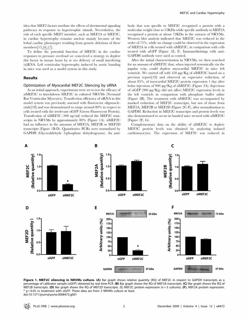
Optimization of Myocardial MEF2C Silencing by siRNAAs an initial approach, experiments were set to test the efficacy of
siMEF2C to knockdown MEF2C in cultured NRVMs (Neonatal
Rat Ventricular Myocytes). Transfection efficiency of siRNA in this
model system was previously assessed with fluorescent oligonucle-
otide[18] and was demonstrated to range around 80% in respect to
cells treated with the irrelevant siGFP (Green Fluorescent Protein).
Transfection of siMEF2C (300 ng/ml) reduced the MEF2C tran-
scripts in NRVMs by approximately 80% (Figure 1A). siMEF2C
had no influence in the amounts of MEF2A, MEF2B or MEF2D
transcripts (Figure 1B-D). Quantitative PCRs were normalized by
GAPDH (Glyceraldehyde 3-phosphate dehydrogenase). An anti-
body that was specific to MEF2C recognized a protein with a
molecular weight close to 55KDa while specific antibody to MEF2A
recognized a protein at about 72KDa in the extracts of NRVMs.
Western blot analysis indicated that MEF2C was reduced in the
order of 75%, while no change could be observed in the expression
of MEF2A in cells treated with siMEF2C, in comparison with cells
treated with siGFP (Figure 1E, F). Immunoblottings with anti-
GAPDH antibody were used as control.
After the initial characterization in NRVMs, we then searched
for an amount of siMEF2C that, when injected systemically via the
jugular vein, could deplete myocardial MEF2C in mice left
ventricle. We started off with 450 mg/Kg of siMEF2C based on a
previous report[19] and observed an expressive reduction, of
about 85%, of myocardial MEF2C protein expression 1 day after
bolus injections of 900 mg/Kg of siMEF2C (Figure 2A). Injections
of siGFP (900 mg/Kg) did not affect MEF2C expression levels in
the left ventricle in comparison with phosphate buffer saline
(Figure 2B). The treatment with siMEF2C was accompanied by
marked reduction of MEF2C transcripts, but not of those from
MEF2A, MEF2B or MEF2D (Figure 2C-F), after normalization to
GAPDH. Reduction in MEF2C transcripts and protein levels was
also demonstrated to occur in banded mice treated with siMEF2C
(Figure 2F, G).
Complementary data on the ability of siMEF2C to deplete
MEF2C protein levels was obtained by analyzing isolated
cardiomyocytes. The expression of MEF2C was reduced in
Figure 1. MEF2C silencing in NRVMs culture. (A) Bar graph shows relative quantity (RQ) of MEF2C in respect to GAPDH transcripts as apercentage of calibrator sample (siGFP) obtained by real time-PCR. (B) Bar graph shows the RQ of MEF2A transcripts. (C) Bar graph shows the RQ ofMEF2B transcripts. (D) Bar graph shows the RQ of MEF2D transcripts. (E) MEF2C protein expression (n = 3 cultures). (F), MEF2A protein expression.* p,0.05 vs treatment with siGFP. These data are from 3 NRVMs culture at least.doi:10.1371/journal.pone.0008472.g001
MEF2C and Cardiac Hypertrophy
PLoS ONE | www.plosone.org 2 December 2009 | Volume 4 | Issue 12 | e8472
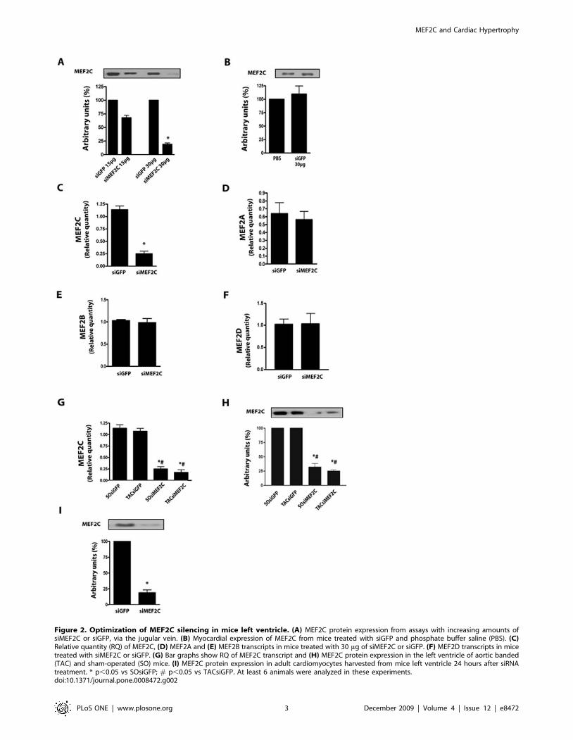
Figure 2. Optimization of MEF2C silencing in mice left ventricle. (A) MEF2C protein expression from assays with increasing amounts ofsiMEF2C or siGFP, via the jugular vein. (B) Myocardial expression of MEF2C from mice treated with siGFP and phosphate buffer saline (PBS). (C)Relative quantity (RQ) of MEF2C, (D) MEF2A and (E) MEF2B transcripts in mice treated with 30 mg of siMEF2C or siGFP. (F) MEF2D transcripts in micetreated with siMEF2C or siGFP. (G) Bar graphs show RQ of MEF2C transcript and (H) MEF2C protein expression in the left ventricle of aortic banded(TAC) and sham-operated (SO) mice. (I) MEF2C protein expression in adult cardiomyocytes harvested from mice left ventricle 24 hours after siRNAtreatment. * p,0.05 vs SOsiGFP; # p,0.05 vs TACsiGFP. At least 6 animals were analyzed in these experiments.doi:10.1371/journal.pone.0008472.g002
MEF2C and Cardiac Hypertrophy
PLoS ONE | www.plosone.org 3 December 2009 | Volume 4 | Issue 12 | e8472
cardiomyocytes harvested from the left ventricle 24 hours after the
treatment with siMEF2C to similar levels of myocardial extracts
(,75%), as depicted in Figure 2H.
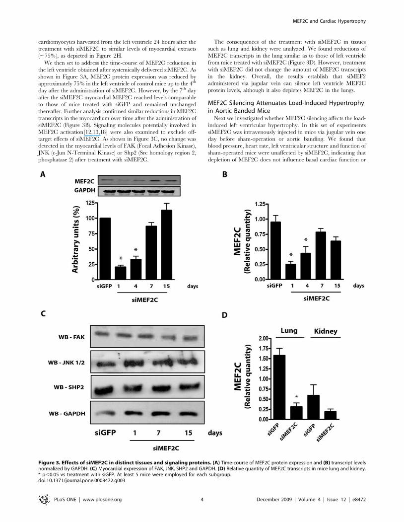
We then set to address the time-course of MEF2C reduction in
the left ventricle obtained after systemically delivered siMEF2C. As
shown in Figure 3A, MEF2C protein expression was reduced by
approximately 75% in the left ventricle of control mice up to the 4th
day after the administration of siMEF2C. However, by the 7th day
after the siMEF2C myocardial MEF2C reached levels comparable
to those of mice treated with siGFP and remained unchanged
thereafter. Further analysis confirmed similar reductions in MEF2C
transcripts in the myocardium over time after the administration of
siMEF2C (Figure 3B). Signaling molecules potentially involved in
MEF2C activation[12,13,18] were also examined to exclude off-
target effects of siMEF2C. As shown in Figure 3C, no change was
detected in the myocardial levels of FAK (Focal Adhesion Kinase),
JNK (c-Jun N-Terminal Kinase) or Shp2 (Src homology region 2,
phosphatase 2) after treatment with siMEF2C.
The consequences of the treatment with siMEF2C in tissues
such as lung and kidney were analyzed. We found reductions of
MEF2C transcripts in the lung similar as to those of left ventricle
from mice treated with siMEF2C (Figure 3D). However, treatment
with siMEF2C did not change the amount of MEF2C transcripts
in the kidney. Overall, the results establish that siMEF2
administered via jugular vein can silence left ventricle MEF2C
protein levels, although it also depletes MEF2C in the lungs.
MEF2C Silencing Attenuates Load-Induced Hypertrophyin Aortic Banded Mice
Next we investigated whether MEF2C silencing affects the load-
induced left ventricular hypertrophy. In this set of experiments
siMEF2C was intravenously injected in mice via jugular vein one
day before sham-operation or aortic banding. We found that
blood pressure, heart rate, left ventricular structure and function of
sham-operated mice were unaffected by siMEF2C, indicating that
depletion of MEF2C does not influence basal cardiac function or
Figure 3. Effects of siMEF2C in distinct tissues and signaling proteins. (A) Time-course of MEF2C protein expression and (B) transcript levelsnormalized by GAPDH. (C) Myocardial expression of FAK, JNK, SHP2 and GAPDH. (D) Relative quantity of MEF2C transcripts in mice lung and kidney.* p,0.05 vs treatment with siGFP. At least 5 mice were employed for each subgroup.doi:10.1371/journal.pone.0008472.g003
MEF2C and Cardiac Hypertrophy
PLoS ONE | www.plosone.org 4 December 2009 | Volume 4 | Issue 12 | e8472
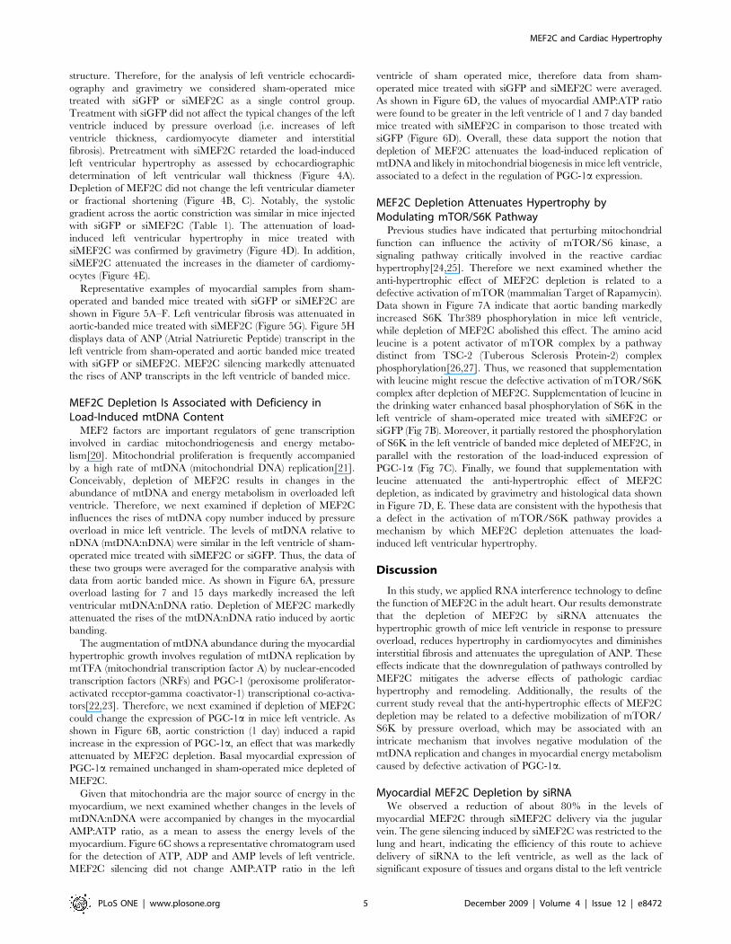
structure. Therefore, for the analysis of left ventricle echocardi-
ography and gravimetry we considered sham-operated mice
treated with siGFP or siMEF2C as a single control group.
Treatment with siGFP did not affect the typical changes of the left
ventricle induced by pressure overload (i.e. increases of left
ventricle thickness, cardiomyocyte diameter and interstitial
fibrosis). Pretreatment with siMEF2C retarded the load-induced
left ventricular hypertrophy as assessed by echocardiographic
determination of left ventricular wall thickness (Figure 4A).
Depletion of MEF2C did not change the left ventricular diameter
or fractional shortening (Figure 4B, C). Notably, the systolic
gradient across the aortic constriction was similar in mice injected
with siGFP or siMEF2C (Table 1). The attenuation of load-
induced left ventricular hypertrophy in mice treated with
siMEF2C was confirmed by gravimetry (Figure 4D). In addition,
siMEF2C attenuated the increases in the diameter of cardiomy-
ocytes (Figure 4E).
Representative examples of myocardial samples from sham-
operated and banded mice treated with siGFP or siMEF2C are
shown in Figure 5A–F. Left ventricular fibrosis was attenuated in
aortic-banded mice treated with siMEF2C (Figure 5G). Figure 5H
displays data of ANP (Atrial Natriuretic Peptide) transcript in the
left ventricle from sham-operated and aortic banded mice treated
with siGFP or siMEF2C. MEF2C silencing markedly attenuated
the rises of ANP transcripts in the left ventricle of banded mice.
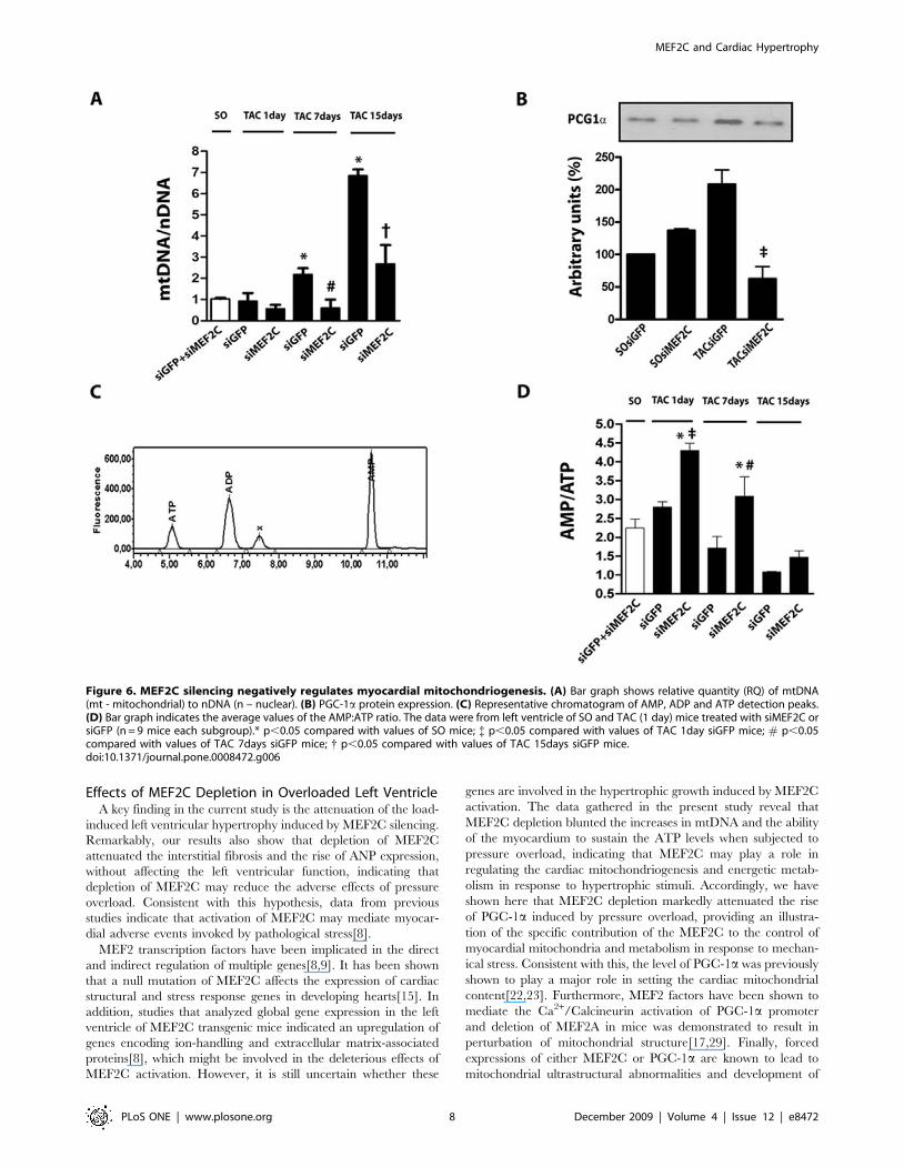
MEF2C Depletion Is Associated with Deficiency inLoad-Induced mtDNA Content
MEF2 factors are important regulators of gene transcription
involved in cardiac mitochondriogenesis and energy metabo-
lism[20]. Mitochondrial proliferation is frequently accompanied
by a high rate of mtDNA (mitochondrial DNA) replication[21].
Conceivably, depletion of MEF2C results in changes in the
abundance of mtDNA and energy metabolism in overloaded left
ventricle. Therefore, we next examined if depletion of MEF2C
influences the rises of mtDNA copy number induced by pressure
overload in mice left ventricle. The levels of mtDNA relative to
nDNA (mtDNA:nDNA) were similar in the left ventricle of sham-
operated mice treated with siMEF2C or siGFP. Thus, the data of
these two groups were averaged for the comparative analysis with
data from aortic banded mice. As shown in Figure 6A, pressure
overload lasting for 7 and 15 days markedly increased the left
ventricular mtDNA:nDNA ratio. Depletion of MEF2C markedly
attenuated the rises of the mtDNA:nDNA ratio induced by aortic
banding.
The augmentation of mtDNA abundance during the myocardial
hypertrophic growth involves regulation of mtDNA replication by
mtTFA (mitochondrial transcription factor A) by nuclear-encoded
transcription factors (NRFs) and PGC-1 (peroxisome proliferator-
activated receptor-gamma coactivator-1) transcriptional co-activa-
tors[22,23]. Therefore, we next examined if depletion of MEF2C
could change the expression of PGC-1a in mice left ventricle. As
shown in Figure 6B, aortic constriction (1 day) induced a rapid
increase in the expression of PGC-1a, an effect that was markedly
attenuated by MEF2C depletion. Basal myocardial expression of
PGC-1a remained unchanged in sham-operated mice depleted of
MEF2C.
Given that mitochondria are the major source of energy in the
myocardium, we next examined whether changes in the levels of
mtDNA:nDNA were accompanied by changes in the myocardial
AMP:ATP ratio, as a mean to assess the energy levels of the
myocardium. Figure 6C shows a representative chromatogram used
for the detection of ATP, ADP and AMP levels of left ventricle.
MEF2C silencing did not change AMP:ATP ratio in the left
ventricle of sham operated mice, therefore data from sham-
operated mice treated with siGFP and siMEF2C were averaged.
As shown in Figure 6D, the values of myocardial AMP:ATP ratio
were found to be greater in the left ventricle of 1 and 7 day banded
mice treated with siMEF2C in comparison to those treated with
siGFP (Figure 6D). Overall, these data support the notion that
depletion of MEF2C attenuates the load-induced replication of
mtDNA and likely in mitochondrial biogenesis in mice left ventricle,
associated to a defect in the regulation of PGC-1a expression.
MEF2C Depletion Attenuates Hypertrophy byModulating mTOR/S6K Pathway
Previous studies have indicated that perturbing mitochondrial
function can influence the activity of mTOR/S6 kinase, a
signaling pathway critically involved in the reactive cardiac
hypertrophy[24,25]. Therefore we next examined whether the
anti-hypertrophic effect of MEF2C depletion is related to a
defective activation of mTOR (mammalian Target of Rapamycin).
Data shown in Figure 7A indicate that aortic banding markedly
increased S6K Thr389 phosphorylation in mice left ventricle,
while depletion of MEF2C abolished this effect. The amino acid
leucine is a potent activator of mTOR complex by a pathway
distinct from TSC-2 (Tuberous Sclerosis Protein-2) complex
phosphorylation[26,27]. Thus, we reasoned that supplementation
with leucine might rescue the defective activation of mTOR/S6K
complex after depletion of MEF2C. Supplementation of leucine in
the drinking water enhanced basal phosphorylation of S6K in the
left ventricle of sham-operated mice treated with siMEF2C or
siGFP (Fig 7B). Moreover, it partially restored the phosphorylation
of S6K in the left ventricle of banded mice depleted of MEF2C, in
parallel with the restoration of the load-induced expression of
PGC-1a (Fig 7C). Finally, we found that supplementation with
leucine attenuated the anti-hypertrophic effect of MEF2C
depletion, as indicated by gravimetry and histological data shown
in Figure 7D, E. These data are consistent with the hypothesis that
a defect in the activation of mTOR/S6K pathway provides a
mechanism by which MEF2C depletion attenuates the load-
induced left ventricular hypertrophy.
Discussion
In this study, we applied RNA interference technology to define
the function of MEF2C in the adult heart. Our results demonstrate
that the depletion of MEF2C by siRNA attenuates the
hypertrophic growth of mice left ventricle in response to pressure
overload, reduces hypertrophy in cardiomyocytes and diminishes
interstitial fibrosis and attenuates the upregulation of ANP. These
effects indicate that the downregulation of pathways controlled by
MEF2C mitigates the adverse effects of pathologic cardiac
hypertrophy and remodeling. Additionally, the results of the
current study reveal that the anti-hypertrophic effects of MEF2C
depletion may be related to a defective mobilization of mTOR/
S6K by pressure overload, which may be associated with an
intricate mechanism that involves negative modulation of the
mtDNA replication and changes in myocardial energy metabolism
caused by defective activation of PGC-1a.
Myocardial MEF2C Depletion by siRNAWe observed a reduction of about 80% in the levels of
myocardial MEF2C through siMEF2C delivery via the jugular
vein. The gene silencing induced by siMEF2C was restricted to the
lung and heart, indicating the efficiency of this route to achieve
delivery of siRNA to the left ventricle, as well as the lack of
significant exposure of tissues and organs distal to the left ventricle
MEF2C and Cardiac Hypertrophy
PLoS ONE | www.plosone.org 5 December 2009 | Volume 4 | Issue 12 | e8472
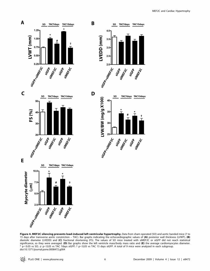
Figure 4. MEF2C silencing prevents load-induced left ventricular hypertrophy. Data from sham operated (SO) and aortic banded mice (7 to15 days after transverse aortic constriction – TAC). Bar graphs indicating the echocardiographic values of (A) posterior wall thickness (LVWT), (B)diastolic diameter (LVEDD) and (C) fractional shortening (FS). The values of SO mice treated with siMEF2C or siGFP did not reach statisticalsignificance, so they were averaged. (D) Bar graphs show the left ventricle mass/body mass ratio and (E) the average cardiomyocytes diameter.* p,0.05 vs SO; # p,0.05 vs TAC 7days siGFP; { p,0,05 vs TAC 15 days siGFP. A total of 9 mice were analyzed in each subgroup.doi:10.1371/journal.pone.0008472.g004
MEF2C and Cardiac Hypertrophy
PLoS ONE | www.plosone.org 6 December 2009 | Volume 4 | Issue 12 | e8472
to siRNA. Besides the left ventricle, depletion of MEF2C was
confirmed to occur in cardiomyocytes harvested from mice treated
with siMEF2C, thereby demonstrating the susceptibility of this cell
type to the effects of systemically delivered siRNA. Moreover, the
specificity of MEF2C silencing was substantiated by the lack of
effects of siMEF2C on MEF2A, MEF2B or MEF2D, as well as on
the unrelated proteins FAK, JNK, SHP2 and GAPDH. The
MEF2C depletion induced by the RNAi strategy used in the
present study lasted for approximately 4 days. Such a prolonged
effect of systemically delivered siRNA was previously observed for
distinct targets in various tissues[19,28], although the underlying
mechanism remains unclear.
Table 1. Mice hemodynamics.
Vehicle Leucine
siRNAGFP siRNAMEF2C siRNAGFP siRNAMEF2C
SO TAC 7days TAC 15 days SO TAC 7 days TAC 15 days SO TAC 7days SO TAC 7days
N 11 12 7 11 7 10 9 8 7 9
SBPa(mmHg) 12363 17163* 17764* 12163 17163* 17166* 13863 17166# 14564 16066#
SBPf (mmHg) 11962 12864 12463 12565 12362 12964
SGr 5262 4963 4661 4665 4865 4362
HR (beats/min) 397622 420616 377626 436615 365619 365626 415615 422612 443611 40269
SO. Sham operated; TAC. transverse aorta constricted; SBPa. systolic blood pressure in ascending aorta; SBPf. systolic blood pressure in femoral artery; SGr. systolicgradient; HR. heart rate.*P,0.05 indicated statistical significance compared to values of SO mice treated whit vehicle.# P,0.05 indicated statistical significance compared to values of SO mice treated whith leucine.doi:10.1371/journal.pone.0008472.t001
Figure 5. MEF2C silencing prevents the myocardial deleterious effects of pressure overload. Myocardial samples of SO and TAC micetreated with siMEF2C or siGFP stained with Masson trichrome (X400): (A) - SO- siGFP, (B) – SO- siMEF2C, (C) – TAC(7days)- SO- siGFP, (D) -TAC(7days)- siMEF2C, (E) - TAC(15days)- SO- siGFP, (F) - TAC(15days)- siMEF2C. (G) Bar graph indicates the average fraction volume of collagen(CVF%) (n = 9 mice each subgroup). (H) Relative quantity (RQ) of ANP transcript (n = 9 mice each subgroup). * p,0.05 compared with values of SOmice; # p,0.05 compared with values of TAC 7days siGFP mice, { p,0,05 compared to values of TAC 15days siGFP mice.doi:10.1371/journal.pone.0008472.g005
MEF2C and Cardiac Hypertrophy
PLoS ONE | www.plosone.org 7 December 2009 | Volume 4 | Issue 12 | e8472
Effects of MEF2C Depletion in Overloaded Left VentricleA key finding in the current study is the attenuation of the load-
induced left ventricular hypertrophy induced by MEF2C silencing.
Remarkably, our results also show that depletion of MEF2C
attenuated the interstitial fibrosis and the rise of ANP expression,
without affecting the left ventricular function, indicating that
depletion of MEF2C may reduce the adverse effects of pressure
overload. Consistent with this hypothesis, data from previous
studies indicate that activation of MEF2C may mediate myocar-
dial adverse events invoked by pathological stress[8].
MEF2 transcription factors have been implicated in the direct
and indirect regulation of multiple genes[8,9]. It has been shown
that a null mutation of MEF2C affects the expression of cardiac
structural and stress response genes in developing hearts[15]. In
addition, studies that analyzed global gene expression in the left
ventricle of MEF2C transgenic mice indicated an upregulation of
genes encoding ion-handling and extracellular matrix-associated
proteins[8], which might be involved in the deleterious effects of
MEF2C activation. However, it is still uncertain whether these
genes are involved in the hypertrophic growth induced by MEF2C
activation. The data gathered in the present study reveal that
MEF2C depletion blunted the increases in mtDNA and the ability
of the myocardium to sustain the ATP levels when subjected to
pressure overload, indicating that MEF2C may play a role in
regulating the cardiac mitochondriogenesis and energetic metab-
olism in response to hypertrophic stimuli. Accordingly, we have
shown here that MEF2C depletion markedly attenuated the rise
of PGC-1a induced by pressure overload, providing an illustra-
tion of the specific contribution of the MEF2C to the control of
myocardial mitochondria and metabolism in response to mechan-
ical stress. Consistent with this, the level of PGC-1a was previously
shown to play a major role in setting the cardiac mitochondrial
content[22,23]. Furthermore, MEF2 factors have been shown to
mediate the Ca2+/Calcineurin activation of PGC-1a promoter
and deletion of MEF2A in mice was demonstrated to result in
perturbation of mitochondrial structure[17,29]. Finally, forced
expressions of either MEF2C or PGC-1a are known to lead to
mitochondrial ultrastructural abnormalities and development of
Figure 6. MEF2C silencing negatively regulates myocardial mitochondriogenesis. (A) Bar graph shows relative quantity (RQ) of mtDNA(mt - mitochondrial) to nDNA (n – nuclear). (B) PGC-1a protein expression. (C) Representative chromatogram of AMP, ADP and ATP detection peaks.(D) Bar graph indicates the average values of the AMP:ATP ratio. The data were from left ventricle of SO and TAC (1 day) mice treated with siMEF2C orsiGFP (n = 9 mice each subgroup).* p,0.05 compared with values of SO mice; { p,0.05 compared with values of TAC 1day siGFP mice; # p,0.05compared with values of TAC 7days siGFP mice; { p,0.05 compared with values of TAC 15days siGFP mice.doi:10.1371/journal.pone.0008472.g006
MEF2C and Cardiac Hypertrophy
PLoS ONE | www.plosone.org 8 December 2009 | Volume 4 | Issue 12 | e8472
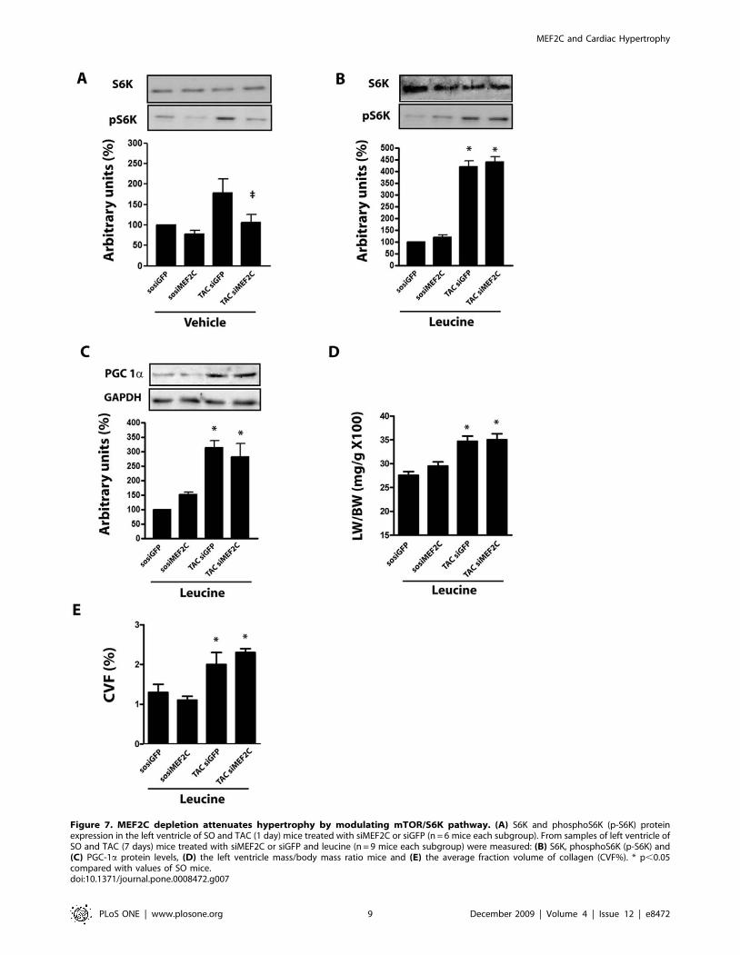
Figure 7. MEF2C depletion attenuates hypertrophy by modulating mTOR/S6K pathway. (A) S6K and phosphoS6K (p-S6K) proteinexpression in the left ventricle of SO and TAC (1 day) mice treated with siMEF2C or siGFP (n = 6 mice each subgroup). From samples of left ventricle ofSO and TAC (7 days) mice treated with siMEF2C or siGFP and leucine (n = 9 mice each subgroup) were measured: (B) S6K, phosphoS6K (p-S6K) and(C) PGC-1a protein levels, (D) the left ventricle mass/body mass ratio mice and (E) the average fraction volume of collagen (CVF%). * p,0.05compared with values of SO mice.doi:10.1371/journal.pone.0008472.g007
MEF2C and Cardiac Hypertrophy
PLoS ONE | www.plosone.org 9 December 2009 | Volume 4 | Issue 12 | e8472
cardiomyopathy[8,22,23]. Taken together, these data imply that
modulation of load-induced mitochondrial biogenesis may contrib-
ute to the beneficial effects of MEF2C depletion in overloaded
myocardium. It was somewhat surprising to find that the improved
cardiac phenotype after MEF2C, i.e. with less fibrosis and less
pathological hypertrophy, was accompanied by a transient worse
energetic profile including higher AMP/ATP levels. Although we
have no clear explanation for these effects, one could speculate they
are consistent with an expected increase in the myocardial energy
consumption induced by pressure overload in the absence of
appropriate hypertrophic growth. Maintenance of normal cardiac
function in the face of increased workload but without changes in
the left ventricle wall thickness, end-diastolic or end-systolic volumes
implies a positive inotropic effect, which is paralleled by increased
energy consumption per unit of myocardial mass. This would
explain the higher AMP/ATP ratio of overloaded mice left ventricle
silenced for MEF2C. Moreover, these data may imply that the
activation of mitochondrial biogenesis in the early hypertrophic
responses to pressure overload might have detrimental influence on
the heart. Accordingly, increased mitochondrial mass could induce
abnormally high myocardial oxidative stress that in turn might
induce cell loss in the early period of pressure overload[30].
Alternatively, functional and structural abnormalities of mitochon-
dria from overloaded heart might be related solely to increases in
mitochondrial mass which might affect myofibrils, compromising
the contractile function[31]. Thus it is conceivable that lessening
MEF2C levels by modulating the excessive mitochondrial biogen-
esis of overloaded myocardium, may contribute to mitigate the
degeneration of overloaded myocardium. However, this issue needs
further studies.
The data from the present study indicate that MEF2C depletion is
accompanied by downregulation of the mTOR/S6K pathway
[32,33] [34]. Remarkably, the restoration of the activity of this
signaling complex after supplementation with leucine was accompa-
nied by suppression of the anti-hypertrophic of MEF2C depletion,
indicating that the defective activation of mTOR/S6K pathway is a
critical component of the beneficial effects of MEF2C depletion in the
cardiac phenotype of TAC mice. Conversely, the activation of this
pathway might be involved, together with changes in mitochondrial
biogenesis and function, to the detrimental effects of MEF2C
activation in cardiac hypertrophy. These assumptions are in
agreement with previous data indicating that mTOR/S6K complex
plays a crucial role in the hypertrophic growth of cardiomyocytes and
left ventricle invoked by mechanical stress[18,35]. Moreover, mTOR
overactivation has been shown to cause increased mitochondrial
biogenesis and accumulation of reactive oxygen species in distinct
model systems[36], suggesting that the activation of mTOR pathway
might be responsible for the detrimental effects of MEF2C activation
and vice-versa to the beneficial effects of MEF2C depletion in the
mechanically overloaded hearts. However, the mechanisms that
connect the MEF2C to mTOR/S6K pathway were not explored in
the present study. It is possible that the activation of AMPK induced
by the raise in the AMP:ATP relative amount may inhibit the
mTOR/S6K complex in the overloaded myocardium[32]. Alterna-
tively, reduced mitochondrial function may also inactivate mTOR
activity[37], however further studies are needed to clarify this issue.
In conclusion, we used RNAi strategy to deplete MEF2C in the
left ventricle of adult mice and understand the role of this factor in
the responses of the left ventricle to mechanical stress. The data
indicate that depletion of MEF2C is sufficient to markedly
attenuate the hypertrophic growth and myocardial fibrosis of
overloaded left ventricle by a mechanism dependent on defective
activation of mTOR/S6K pathway. Overall, these data highlight
the potential of MEF2C in the pathogenesis of cardiac
hypertrophy and remodeling, and provide insights into novel
therapeutic targets to heart disease.
Materials and Methods
Expanded Materials and Methods are available in the
Supporting Information Text S1 file of this manuscript.
Ethics StatementAnimals were handled in compliance with the principles of
laboratory animal care formulated by the Animal Care and Use
Committee of University of Campinas. Procedures such as jugular
vein catheterization, aortic banding, echocardiographic examina-
tion and arterial vessels catheterization for blood pressure
monitoring were performed under anesthesia with a mixture of
ketamine (100 mg/Kg) and xylazine (5 mg/Kg).
Antibodies and ChemicalsPolyclonal mouse antibody against MEF2C was from Abcam
(ab43796-100). Polyclonal rabbit antibodies against FAK (sc558),
SHP2 (sc7384), JNK (sc571), GAPDH (sc25778), S6K p70 (sc230),
pS6K p70 Thr389 (sc11759R) and PGC1-a (sc13067), were
purchased from Santa Cruz Biotechnology (USA). Antibody
against MEF2A was from Cell Signaling (9736). Antibodies were
used following the manufacturer’s recommendation. Colagenase
type IA and trypsin were from Sigma (USA). Trizol, Phenol and
Super Script II were from Invitrogen. Super Signal west Pico
Cheluminescent Substract and Ampliscribe T7 high yield
transcription were from Epicentre. L-Leucine was from Ajinomoto
Aminoscience LLC.
Experimental Models and AnimalsSwiss mice (6–8 week old) and neonatal Wistar rats were obtained
from the animal facility center of State University of Campinas.
Animals were handled in compliance with the principles of
laboratory animal care formulated by the university’s Animal Care
and Use Committee. Procedures such as jugular vein catheterization,
aortic banding, echocardiographic examination and arterial vessels
catheterization for blood pressure monitoring were performed under
anesthesia with a mixture of ketamine (100 mg/Kg) and xylazine
(5 mg/Kg) as previously reported[19]. Primary cultures of neonatal
rat ventricular myocytes (NRVMs) were prepared from 1- to 2-day-
old Wistar rats and plated on type I collagen Bioflex plates at
56105 cells/well, as previously reported[38].
Isolation of Adult Mouse Ventricular MyocytesMouse cardiac myocytes were isolated from mice ventricle one
day after treatment with siRNA targeted to MEF2C (siMEF2C) or
GFP (siGFP), as previously reported[19].
Echocardiography2D M-mode echocardiography was performed with a 12-MHz
probe connected to a Toshiba Power Vision system in anesthetized
mice by a blinded observer at 15 minutes after the induction of
anesthesia, as previously reported[19]. The short axis measure-
ments were taken at the level of the midpapillary muscle. Three
measurements were taken at end-systole and end-diastole to
determine left ventricular diastolic and systolic diameter, wall
thickness and fractional shortening.
HemodynamicsFor blood pressure monitoring the right carotid and the right
femoral arteries were cannulated with flame stretched PE-50
MEF2C and Cardiac Hypertrophy
PLoS ONE | www.plosone.org 10 December 2009 | Volume 4 | Issue 12 | e8472
polyethylene tube. Blood pressure in the carotid and femoral
arteries were simultaneously recorded for a 10 minute period to
determine the transconstriction systolic gradient. The following
parameters were computed: systemic systolic, mean and diastolic
blood pressure (mmHg) and heart rate (bpm).
Histological ExaminationHearts were rapidly excised from fully anesthetized mice and
washed in PBS. The left ventricles were fixed in 10%
paraformaldehyde, embedded in paraffin and cut into 5 mm
sections. Tissue sections stained with haematoxylin and eosin (HE)
and Masson’s trichrome underwent morphometric studies using
an image analysis system (Leica Q500 iW; Leica Imaging Systems,
Cambridge, UK).
siRNA Design and SynthesissiRNA targeted to mouse MEF2C gene was designed and
synthesized as previously published[18,19]. DNA oligonucleotides
(IDT-USA) were as follow: (i) T7: 59-GGTAATACGACTCAC-
TATAG-39. (ii): MEF2C 1187 sense: 59-CCCACCUGGCAG-
CAAGAACAC-39 (iii): MEF2C 1187 antisense: 59-GUUCUUG-
CUGCCAGGUGGGAU-39 (iv): GFP sense: 59-GTGTCTTG-
TAGTTCCCGTCTATAGTGAGTCGTATTACC-39. (v): GFP
antisense: 59-ATGACGGGAACTACAAACACCTATAGTGA-
GTCGTATTACC-39 were ordered from IDT (USA). The
oligonucleotide-directed production of small RNA transcripts with
T7 RNA polymerase were made with AmpliscribeTM T7
transcription kit (Epicentre Biotechnologies; Madison WI, USA)
according to manufacturer’s instructions.
Transfection of NRVMs with siRNANRVMs were transfected with siRNA as previously reported
[18].
Western BlottingTissue or cell extracts containing equal amounts of total protein
(50 mg) were resolved by 8% SDS-PAGE. The membranes were
incubated with primary antibodies. Detection was accomplished
by using an enhanced chemiluminescence detection system.
Gene Transcripts and Mitochondrial DNA QuantificationMyocardial MEF2A, MEF2C, MEF2D and ANP transcripts
were quantified after reverse transcription to cDNA followed by
real-time PCR. Primers are shown in Table S1 of Supporting
Information. Results were evaluated by the comparative CT
method in respect to GAPDH, used as the normalizer. For
mithocondrial and nuclear DNA quantification, were compared
the expression of D-loop (EU194676.1) and 18S rRNA
(NR_003278.1) genes, respectively.
Myocardial AMP and ATPSamples of myocardium were analyzed for AMP and ATP
quantification by a method based on high performance liquid
chromatography.
Statistical AnalysisData are presented as means6SEM. Student’s t-test and 1-way
repeated-measures ANOVA were used to compare groups. Post
hoc analysis was performed with Bonferroni multiple-range test. A
value of P,0.05 indicated statistical significance.
Supporting Information
Text S1
Found at: doi:10.1371/journal.pone.0008472.s001 (0.08 MB
DOC)
Table S1
Found at: doi:10.1371/journal.pone.0008472.s002 (0.04 MB
DOC)
Author Contributions
Conceived and designed the experiments: AHMP CFMZC ACC ILC
KGF. Performed the experiments: AHMP CFMZC ACC THT SAR CCJ
MCG VDBP JRMS. Analyzed the data: AHMP CFMZC ACC THT SAR
CCJ MCG VDBP JRMS KGF. Contributed reagents/materials/analysis
tools: ILC. Wrote the paper: AHMP CFMZC KGF.
References
1. Katz AM (1994) The cardiomyopathy of overload: an unnatural growthresponse in the hypertrophied heart. Ann Intern Med 121: 363–371.
2. Opie LH, Commerford PJ, Gersh BJ, Pfeffer MA (2006) Controversies inventricular remodelling. Lancet 367: 356–367.
3. Vakili BA, Okin PM, Devereux RB (2001) Prognostic implications of leftventricular hypertrophy. Am Heart J 141: 334–341.
4. Diwan A, Dorn GW 2nd (2007) Decompensation of cardiac hypertrophy:cellular mechanisms and novel therapeutic targets. Physiology (Bethesda) 22:
56–64.
5. Chien KR (1999) Stress pathways and heart failure. Cell 98: 555–558.
6. Heineke J, Molkentin JD (2006) Regulation of cardiac hypertrophy by
intracellular signalling pathways. Nat Rev Mol Cell Biol 7: 589–600.
7. Dorn GW 2nd (2007) The fuzzy logic of physiological cardiac hypertrophy.
Hypertension 49: 962–970.
8. Xu J, Gong NL, Bodi I, Aronow BJ, Backx PH, et al. (2006) Myocyte enhancer
factors 2A and 2C induce dilated cardiomyopathy in transgenic mice. J BiolChem 281: 9152–9162.
9. van Oort RJ, van Rooij E, Bourajjaj M, Schimmel J, Jansen MA, et al. (2006)MEF2 activates a genetic program promoting chamber dilation and contractile
dysfunction in calcineurin-induced heart failure. Circulation 114: 298–308.
10. Kim Y, Phan D, van Rooij E, Wang DZ, McAnally J, et al. (2008) The MEF2D
transcription factor mediates stress-dependent cardiac remodeling in mice. J ClinInvest 118: 124–132.
11. Molkentin JD, Markham BE (1993) Myocyte-specific enhancer-binding factor(MEF-2) regulates alpha-cardiac myosin heavy chain gene expression in vitro
and in vivo. J Biol Chem 268: 19512–19520.
12. Nadruz W Jr, Kobarg CB, Constancio SS, Corat PD, Franchini KG (2003)
Load-induced transcriptional activation of c-jun in rat myocardium: regulation
by myocyte enhancer factor 2. Circ Res 92: 243–251.
13. Nadruz W Jr, Corat MA, Marin TM, Guimaraes Pereira GA, Franchini KG (2005)Focal adhesion kinase mediates MEF2 and c-Jun activation by stretch: role in the
activation of the cardiac hypertrophic genetic program. Cardiovasc Res 68: 87–97.
14. Hannenhalli S, Putt ME, Gilmore JM, Wang J, Parmacek MS, et al. (2006)Transcriptional genomics associates FOX transcription factors with human
heart failure. Circulation 114: 1269–1276.
15. Lin Q, Schwarz J, Bucana C, Olson EN (1997) Control of mouse cardiac
morphogenesis and myogenesis by transcription factor MEF2C. Science 276:1404–1407.
16. Bi W, Drake CJ, Schwarz JJ (1999) The transcription factor MEF2C-null mouseexhibits complex vascular malformations and reduced cardiac expression of
angiopoietin 1 and VEGF. Dev Biol 211: 255–267.
17. Naya FJ, Black BL, Wu H, Bassel-Duby R, Richardson JA, et al. (2002)Mitochondrial deficiency and cardiac sudden death in mice lacking the MEF2A
transcription factor. Nat Med 8: 1303–1309.
18. Marin TM, Clemente CF, Santos AM, Picardi PK, Pascoal VD, et al. (2008)
Shp2 negatively regulates growth in cardiomyocytes by controlling focaladhesion kinase/Src and mTOR pathways. Circ Res 103: 813–824.
19. Clemente CF, Tornatore TF, Theizen TH, Deckmann AC, Pereira TC, et al.(2007) Targeting focal adhesion kinase with small interfering RNA prevents and
reverses load-induced cardiac hypertrophy in mice. Circ Res 101: 1339–1348.
20. Czubryt MP, Olson EN (2004) Balancing contractility and energy production:the role of myocyte enhancer factor 2 (MEF2) in cardiac hypertrophy. Recent
Prog Horm Res 59: 105–124.
21. Attardi G, Schatz G (1988) Biogenesis of mitochondria. Annu Rev Cell Biol 4:
289–333.
22. Lehman JJ, Barger PM, Kovacs A, Saffitz JE, Medeiros DM, et al. (2000)
Peroxisome proliferator-activated receptor gamma coactivator-1 promotes
cardiac mitochondrial biogenesis. J Clin Invest 106: 847–856.
MEF2C and Cardiac Hypertrophy
PLoS ONE | www.plosone.org 11 December 2009 | Volume 4 | Issue 12 | e8472
23. Russell LK, Mansfield CM, Lehman JJ, Kovacs A, Courtois M, et al. (2004)
Cardiac-specific induction of the transcriptional coactivator peroxisomeproliferator-activated receptor gamma coactivator-1alpha promotes mitochon-
drial biogenesis and reversible cardiomyopathy in a developmental stage-
dependent manner. Circ Res 94: 525–533.24. Sarbassov DD, Ali SM, Sabatini DM (2005) Growing roles for the mTOR
pathway. Curr Opin Cell Biol 17: 596–603.25. Wullschleger S, Loewith R, Hall MN (2006) TOR signaling in growth and
metabolism. Cell 124: 471–484.
26. Hara K, Yonezawa K, Weng QP, Kozlowski MT, Belham C, et al. (1998)Amino acid sufficiency and mTOR regulate p70 S6 kinase and eIF-4E BP1
through a common effector mechanism. J Biol Chem 273: 14484–14494.27. Kim E, Goraksha-Hicks P, Li L, Neufeld TP, Guan KL (2008) Regulation of
TORC1 by Rag GTPases in nutrient response. Nat Cell Biol 10: 935–945.28. Zimmermann TS, Lee AC, Akinc A, Bramlage B, Bumcrot D, et al. (2006)
RNAi-mediated gene silencing in non-human primates. Nature 441: 111–114.
29. Handschin C, Rhee J, Lin J, Tarr PT, Spiegelman BM (2003) An autoregulatoryloop controls peroxisome proliferator-activated receptor gamma coactivator
1alpha expression in muscle. Proc Natl Acad Sci U S A 100: 7111–7116.30. Sebastiani M, Giordano C, Nediani C, Travaglini C, Borchi E, et al. (2007)
Induction of mitochondrial biogenesis is a maladaptive mechanism in
mitochondrial cardiomyopathies. J Am Coll Cardiol 50: 1362–1369.31. Ventura-Clapier R, Garnier A, Veksler V (2008) Transcriptional control of
mitochondrial biogenesis: the central role of PGC-1alpha. Cardiovasc Res 79:208–217.
32. Chan AY, Soltys CL, Young ME, Proud CG, Dyck JR (2004) Activation of
AMP-activated protein kinase inhibits protein synthesis associated with
hypertrophy in the cardiac myocyte. J Biol Chem 279: 32771–32779.
33. Noga AA, Soltys CL, Barr AJ, Kovacic S, Lopaschuk GD, et al. (2007)
Expression of an active LKB1 complex in cardiac myocytes results in decreased
protein synthesis associated with phenylephrine-induced hypertrophy.
Am J Physiol Heart Circ Physiol 292: H1460–1469.
34. Motoshima H, Goldstein BJ, Igata M, Araki E (2006) AMPK and cell
proliferation–AMPK as a therapeutic target for atherosclerosis and cancer.
J Physiol 574: 63–71.
35. Shioi T, McMullen JR, Tarnavski O, Converso K, Sherwood MC, et al. (2003)
Rapamycin attenuates load-induced cardiac hypertrophy in mice. Circulation
107: 1664–1670.
36. Chen C, Liu Y, Liu R, Ikenoue T, Guan KL, et al. (2008) TSC-mTOR
maintains quiescence and function of hematopoietic stem cells by repressing
mitochondrial biogenesis and reactive oxygen species. J Exp Med 205:
2397–2408.
37. Schieke SM, Phillips D, McCoy JP Jr, Aponte AM, Shen RF, et al. (2006) The
mammalian target of rapamycin (mTOR) pathway regulates mitochondrial
oxygen consumption and oxidative capacity. J Biol Chem 281: 27643–27652.
38. Torsoni AS, Constancio SS, Nadruz W Jr, Hanks SK, Franchini KG (2003)
Focal adhesion kinase is activated and mediates the early hypertrophic response
to stretch in cardiac myocytes. Circ Res 93: 140–147.
MEF2C and Cardiac Hypertrophy
PLoS ONE | www.plosone.org 12 December 2009 | Volume 4 | Issue 12 | e8472
Related Documents


![MEF2C Ablation in Endothelial Cells Reduces Retinal Vessel ... · MEF2C Ablation in Endothelial Cells Reduces Retinal ... (VO)] results in hypoxic retina, which secretes growth factors](https://static.cupdf.com/doc/110x72/6074652eae791d1ccd15870e/mef2c-ablation-in-endothelial-cells-reduces-retinal-vessel-mef2c-ablation-in.jpg)