The Macaque Gut Microbiome in Health, Lentiviral Infection, and Chronic Enterocolitis Philip McKenna 1 , Christian Hoffmann 1 , Nana Minkah 1 , Pyone Pyone Aye 2 , Andrew Lackner 2 , Zongzhi Liu 3 , Catherine A. Lozupone 4 , Micah Hamady 5 , Rob Knight 3 , Frederic D. Bushman 1* 1 Department of Microbiology, University of Pennsylvania School of Medicine, Philadelphia, Pennsylvania, United States of America, 2 Tulane National Primate Research Center, Tulane University Health Science Center, Covington, Louisiana, United States of America, 3 Department of Chemistry and Biochemistry, University of Colorado at Boulder, Boulder, Colorado, United States of America, 4 Department of Molecular, Cellular and Developmental Biology, University of Colorado at Boulder, Boulder, Colorado, United States of America, 5 Department of Computer Science, University of Colorado at Boulder, Boulder, Colorado, United States of America The vertebrate gut harbors a vast community of bacterial mutualists, the composition of which is modulated by the host immune system. Many gastrointestinal (GI) diseases are expected to be associated with disruptions of host- bacterial interactions, but relatively few comprehensive studies have been reported. We have used the rhesus macaque model to investigate forces shaping GI bacterial communities. We used DNA bar coding and pyrosequencing to characterize 141,000 sequences of 16S rRNA genes obtained from 100 uncultured GI bacterial samples, allowing quantitative analysis of community composition in health and disease. Microbial communities of macaques were distinct from those of mice and humans in both abundance and types of taxa present. The macaque communities differed among samples from intestinal mucosa, colonic contents, and stool, paralleling studies of humans. Communities also differed among animals, over time within individual animals, and between males and females. To investigate changes associated with disease, samples of colonic contents taken at necropsy were compared between healthy animals and animals with colitis and undergoing antibiotic therapy. Communities from diseased and healthy animals also differed significantly in composition. This work provides comprehensive data and improved methods for studying the role of commensal microbiota in macaque models of GI diseases and provides a model for the large-scale screening of the human gut microbiome. Citation: McKenna P, Hoffmann C, Minkah N, Aye PP, Lackner A, et al. (2008) The macaque gut microbiome in health, lentiviral infection, and chronic enterocolitis. PLoS Pathog 4(2): e20. doi:10.1371/journal.ppat.0040020 Introduction The human intestine is home to some 100 trillion micro- organisms of at least 400 species. The density of bacterial cells in the colon has been estimated at 10 11 to 10 12 per ml, which makes it one of the most densely populated microbial habitats known [1,2]. The number of unique genes in the microbial pool is estimated to outnumber the genes in the human nuclear genome by two orders of magnitude [1,2], and these genes contribute many essential metabolic functions to the host. The great majority of gut bacterial species have not been cultured outside the human host and are known only by fragments of their DNA sequences. A few pioneering reports have begun to survey the intestinal microbiota of humans and mice using DNA sequencing of uncultured communities [1,3,4] or using microarray-based methods [5,6]. It is widely expected that human disease states will be linked to characteristic transitions in the intestinal microbiota, and connections have been proposed between GI bacterial communities and obesity [7,8] and Crohn’s disease [9,10], but studies in this area are just beginning. Here we report characterization of GI microbial communities in rhesus macaques and their alteration accompanying colitis associ- ated with SIV infection or in animals with chronic enter- ocolitis. The mammalian GI tract is a major locus of immune tissues responsible for blocking invasion by pathogens, and more recently, these tissues have been implicated in normal homeostasis of the gut microbiota as well. For example, B- cells of the gut associated lymphoid tissues (GALT) synthesize IgA, which is secreted in large amounts into the lumen of the gut, and mice genetically incapable of normal IgA synthesis have abnormally large proportions of anaerobes in the small intestine [11,12]. Secreted antibacterial peptides have also been implicated in regulating the composition of the gut microbiota [13,14]. Effects of host genotype are also docu- mented by the finding that genetically obese mice have detectably different gut microbiota compared to wild-type controls [8]. HIV infection causes rapid and massive destruction of GALT [15–20], and HIV infection is also frequently associated with gastrointestinal disorders, many of which are of unexplained etiology [21]. Destruction of GALT and gastro- intestinal disorders are also a well-characterized consequence Editor: Frederick M. Ausubel, Harvard Medical School, United States of America Received April 30, 2007; Accepted December 19, 2007; Published February 1, 2008 Copyright: Ó 2008 McKenna et al. This is an open-access article distributed under the terms of the Creative Commons Attribution License, which permits unrestricted use, distribution, and reproduction in any medium, provided the original author and source are credited. * To whom correspondence should be addressed. E-mail: [email protected]. upenn.edu PLoS Pathogens | www.plospathogens.org February 2008 | Volume 4 | Issue 2 | e20 0001

McKenna Et Al. - 2008 - The Macaque Gut Microbiome in Health, Lentiviral Infection, And Chronic Enterocolitis
Dec 02, 2015
The vertebrate gut harbors a vast community of bacterial mutualists, the composition of which is modulated by the
host immune system. Many gastrointestinal (GI) diseases are expected to be associated with disruptions of hostbacterial
interactions, but relatively few comprehensive studies have been reported. We have used the rhesus
macaque model to investigate forces shaping GI bacterial communities. We used DNA bar coding and pyrosequencing
to characterize 141,000 sequences of 16S rRNA genes obtained from 100 uncultured GI bacterial samples, allowing
quantitative analysis of community composition in health and disease. Microbial communities of macaques were
distinct from those of mice and humans in both abundance and types of taxa present. The macaque communities
differed among samples from intestinal mucosa, colonic contents, and stool, paralleling studies of humans.
Communities also differed among animals, over time within individual animals, and between males and females. To
investigate changes associated with disease, samples of colonic contents taken at necropsy were compared between
healthy animals and animals with colitis and undergoing antibiotic therapy. Communities from diseased and healthy
animals also differed significantly in composition. This work provides comprehensive data and improved methods for
studying the role of commensal microbiota in macaque models of GI diseases and provides a model for the large-scale
screening of the human gut microbiome.
Citation: McKenna P, Hoffmann C, Minkah N, Aye PP, Lackner A, et al. (2008) The
Welcome message from author
This document is posted to help you gain knowledge. Please leave a comment to let me know what you think about it! Share it to your friends and learn new things together.
Transcript

The Macaque Gut Microbiomein Health, Lentiviral Infection,and Chronic EnterocolitisPhilip McKenna
1, Christian Hoffmann
1, Nana Minkah
1, Pyone Pyone Aye
2, Andrew Lackner
2, Zongzhi Liu
3,
Catherine A. Lozupone4
, Micah Hamady5
, Rob Knight3
, Frederic D. Bushman1*
1 Department of Microbiology, University of Pennsylvania School of Medicine, Philadelphia, Pennsylvania, United States of America, 2 Tulane National Primate Research
Center, Tulane University Health Science Center, Covington, Louisiana, United States of America, 3 Department of Chemistry and Biochemistry, University of Colorado at
Boulder, Boulder, Colorado, United States of America, 4 Department of Molecular, Cellular and Developmental Biology, University of Colorado at Boulder, Boulder, Colorado,
United States of America, 5 Department of Computer Science, University of Colorado at Boulder, Boulder, Colorado, United States of America
The vertebrate gut harbors a vast community of bacterial mutualists, the composition of which is modulated by thehost immune system. Many gastrointestinal (GI) diseases are expected to be associated with disruptions of host-bacterial interactions, but relatively few comprehensive studies have been reported. We have used the rhesusmacaque model to investigate forces shaping GI bacterial communities. We used DNA bar coding and pyrosequencingto characterize 141,000 sequences of 16S rRNA genes obtained from 100 uncultured GI bacterial samples, allowingquantitative analysis of community composition in health and disease. Microbial communities of macaques weredistinct from those of mice and humans in both abundance and types of taxa present. The macaque communitiesdiffered among samples from intestinal mucosa, colonic contents, and stool, paralleling studies of humans.Communities also differed among animals, over time within individual animals, and between males and females. Toinvestigate changes associated with disease, samples of colonic contents taken at necropsy were compared betweenhealthy animals and animals with colitis and undergoing antibiotic therapy. Communities from diseased and healthyanimals also differed significantly in composition. This work provides comprehensive data and improved methods forstudying the role of commensal microbiota in macaque models of GI diseases and provides a model for the large-scalescreening of the human gut microbiome.
Citation: McKenna P, Hoffmann C, Minkah N, Aye PP, Lackner A, et al. (2008) The macaque gut microbiome in health, lentiviral infection, and chronic enterocolitis. PLoSPathog 4(2): e20. doi:10.1371/journal.ppat.0040020
Introduction
The human intestine is home to some 100 trillion micro-organisms of at least 400 species. The density of bacterial cellsin the colon has been estimated at 1011 to 1012 per ml, whichmakes it one of the most densely populated microbialhabitats known [1,2]. The number of unique genes in themicrobial pool is estimated to outnumber the genes in thehuman nuclear genome by two orders of magnitude [1,2], andthese genes contribute many essential metabolic functions tothe host. The great majority of gut bacterial species have notbeen cultured outside the human host and are known only byfragments of their DNA sequences. A few pioneering reportshave begun to survey the intestinal microbiota of humans andmice using DNA sequencing of uncultured communities[1,3,4] or using microarray-based methods [5,6]. It is widelyexpected that human disease states will be linked tocharacteristic transitions in the intestinal microbiota, andconnections have been proposed between GI bacterialcommunities and obesity [7,8] and Crohn’s disease [9,10],but studies in this area are just beginning. Here we reportcharacterization of GI microbial communities in rhesusmacaques and their alteration accompanying colitis associ-ated with SIV infection or in animals with chronic enter-ocolitis.
The mammalian GI tract is a major locus of immune tissuesresponsible for blocking invasion by pathogens, and more
recently, these tissues have been implicated in normalhomeostasis of the gut microbiota as well. For example, B-cells of the gut associated lymphoid tissues (GALT) synthesizeIgA, which is secreted in large amounts into the lumen of thegut, and mice genetically incapable of normal IgA synthesishave abnormally large proportions of anaerobes in the smallintestine [11,12]. Secreted antibacterial peptides have alsobeen implicated in regulating the composition of the gutmicrobiota [13,14]. Effects of host genotype are also docu-mented by the finding that genetically obese mice havedetectably different gut microbiota compared to wild-typecontrols [8].HIV infection causes rapid and massive destruction of
GALT [15–20], and HIV infection is also frequently associatedwith gastrointestinal disorders, many of which are ofunexplained etiology [21]. Destruction of GALT and gastro-intestinal disorders are also a well-characterized consequence
Editor: Frederick M. Ausubel, Harvard Medical School, United States of America
Received April 30, 2007; Accepted December 19, 2007; Published February 1,2008
Copyright: � 2008 McKenna et al. This is an open-access article distributed underthe terms of the Creative Commons Attribution License, which permits unrestricteduse, distribution, and reproduction in any medium, provided the original authorand source are credited.
* To whom correspondence should be addressed. E-mail: [email protected]
PLoS Pathogens | www.plospathogens.org February 2008 | Volume 4 | Issue 2 | e200001

of simian immunodeficiency virus (SIV) infection in mac-aques [15,16,22–24]. A role for the GI microbiota in AIDSdisease progression has recently been suggested–bacterialantigens are proposed to pass through the damaged GImucosa and promote immune activation, which in turnpromotes viral replication and disease progression [20].
Chronic enterocolitis is fairly common in rhesus macaqueseven in the absence of SIV infection or other knowninfectious or parasitic agents. Analysis of the clinical courseand histopathology of idiopathic chronic enterocolitis showsmany parallels with human inflammatory bowel disease (IBD),and indeed the macaque disease has been studied as a modelfor the human disorder [24–26]. Evidence of proinflammatorydysfunction of the IL6-JAK-STAT3-SOCS3 pathway has beenreported [24]. A role for the gut microbiota in human IBD haslong been suspected, and several studies have profileduncultured GI bacteria from healthy and diseased patients(e. g. [9,27]). Such studies have not yet yielded a clear-cutpicture of the relationship of GI microbiota to pathogenesis,though a reduction in microbial diversity has been proposed.Studies of the macaque disease have suggested that several GImicrobes may be slightly more common in macaques with anIBD-like disease, but the macaque GI communities have notbeen comprehensively analyzed [20,25].
Here we characterize the macaque GI microbial commun-ities and compare community composition in health and GIdisease. To profile the bacterial taxa present, we purifiedbacterial DNA from samples of intestinal contents, amplifiedsegments of the 16S rRNA gene, determined the sequencesusing massively parallel pyrosequencing, then used these datato identify and quantify the types of bacteria present [28].The approach used here was based on extensive reconstruc-tion studies [29], which showed that known clustering ofmicrobial communities could be recaptured using 16S rRNAgene sequences of lengths generated by pyrosequencing usingtechnology commercialized by 454 Life Sciences [30]. Thesepreliminary bioinformatic studies also disclosed that someshort segments of the 16S rRNA gene sequence wereespecially useful for phylogenetic reconstruction, allowingoptimized primers to be chosen for the study reported here.
We found that the macaque microbiota was distinct from
other vertebrates studied previously. Even in healthy animalsthe taxa present in the gut microbiota differed betweenindividuals and changed substantially within individuals overtime. Unexpectedly, communities from males and femalesalso differed. Distinctive GI microbial communities were alsoobtained in samples of colonic contents taken at necropsyfrom animals with GI disease. Most of these animals were alsotreated with antibiotics to ameliorate their symptoms, so ouranalysis models human cases of colitis accompanied byantibiotic therapy. These data indicate that colitis and itstreatment are associated with transitions in GI microbiota inthe macaque, providing a model that may be useful inunderstanding the human GI microbiota in health anddisease.
Results
Monitoring Macaque Intestinal MicrobiotaWe surveyed a range of sample types and disease states for
possible effects on the macaque GI microbiota. We analyzed atotal of 100 samples, including healthy animals, SIV infectedanimals, and animals with chronic enterocolitis. For thecolitic samples, some of the animals were SIV infected andhad colitis as a result of simian AIDS, while others were coliticbut not SIV infected. Sample types included colonic contentscollected at necropsy, bacterial communities adhered tobiopsy specimens from the upper and lower GI mucosa(jejunum and colon respectively), and stool (Table 1; detaileddata for each animal is in Table S1).DNA was isolated from all 100 samples and amplified by
PCR using primers BSR357-A and BSF8-B, which anneal toconserved regions of bacterial 16S rRNA gene. All sequencereads extended from the BSR357-A primer. The median readlength was 264 nt (Figure 1A). These primers were chosenbased on a series of simulations carried out to investigate theoptimal region of the 16S rRNA gene to query using the shortsequence reads expected from pyrosequencing. Use of amoderately conserved region yielded relatively stable phylo-genetic placements, though at the expense of reduced abilityto discriminate low-level taxa. Biased amplification of 16SrRNA gene sequences from mixed bacterial populations canlead to distortions in abundance estimates, but these aretypically in the range of only a few fold [6,31–33]. To facilitatecomparison among samples, only a single region of the 16SrRNA gene was amplified, and uni-directional reads wereused for the analysis, so that any biases introduced duringamplification are common among all samples.The primers for the 16S rRNA gene sequences were each
marked with a unique DNA bar code by including distinctive4 base sequences in the primers between the 16S rRNA genecomplementary region and the binding sites for thepyrosequencing primers (Figure 1B). This allowed the PCRproducts from many samples to be sequenced using the 454Life Sciences [30] technology, then indexed afterwards. Afterremoval of low quality sequences, a total of 140,356 useablesequence reads were obtained. All bar codes were wellpopulated, with an average of 1404 sequences per communitytested. The error rate for the bar coding procedure could beestimated by cataloging all those sequence reads with barcodes that were not among those used for labeling. Theanalysis indicated that only 0.01% of sequences were likely tobe miscataloged due to errors parsing the bar codes.
PLoS Pathogens | www.plospathogens.org February 2008 | Volume 4 | Issue 2 | e200002
Macaque Microbiota in Health and Disease
Author Summary
Bacterial mutualists within the gastrointestinal tract aid digestion,promote development of the gut immune system, and providecompetitive barriers to pathogen invasion. The host, in return,provides bacteria with safe housing and food during lean times. Thecomposition of the gut microbiota is controlled in part by the hostimmune system. In a variety of disease states, immune function canbe altered, and gut morbidity is often associated, leading to thehypothesis that alterations in the GI microbiota may contribute todisease. In this study, the gut microbiota was characterized in 100samples from rhesus macaques using pyrosequencing, whichallowed 141,000 sequences from 16S rRNA genes to be generatedand analyzed. Healthy animals were compared to animals with gutdisorders, induced, for example by advanced simian AIDS. Manyfactors contributed to changes in the microbiota, including the sexof the animal of origin. Animals with chronic colitis showeddifferences in composition of the GI microbiota compared tohealthy animals, providing an association between altered micro-biota and disease.

One DNA sample was sequenced twice to assess reprodu-cibility. To determine the bacterial taxa present, the 16SrRNA gene sequences were aligned using NAST and GREEN-GENES and then inserted into pre-established phylogenetictrees of full length 16S rRNA gene sequences [34,35] usingARB. Over all the sequences analyzed in this study, 99.94%sequences aligned with previously determined 16S rRNA genesequences (80 total sequences failed to align). The bacterialtaxa in each sample were then tabulated. Comparison of thetwo independent sequencing experiments showed excellentreproducibility of the phylogenetic assignments (Figure 1C).
Ninety-four near-full length macaque bacterial 16S rRNAgene sequences from two communities were also determinedby conventional Sanger sequencing to provide a check on thepyrosequencing data (Figure 1D; Table S2). As can be seenfrom Figure 1D, the major types and relative numbers of taxawere closely similar in the Sanger and pyrosequencinganalysis for each sample, indicating that the pyrosequencingdata yielded an accurate reflection of the species detected byconventional sequencing, though the minor taxa detected bypyrosequencing could not be detected in the Sanger readsdue to the lower number of the latter.
Microbial Diversity in the Macaque Intestinal MicrobiotaWe first investigated the bacterial diversity present in our
16S rRNA gene sequence data. Sequence reads were alignedusing NAST and compiled in OTUPicker. When sequenceswere condensed under conditions demanding 99% identity,about 20,000 different operational taxonomic units (OTUs;groups defined by pairwise sequence identity) were found(Figure 2A). When OTUs were defined using a threshold of97% identity or greater, a criteria that in previous studies wasjudged to match roughly the species level [36,37], about 5,000OTUs were identified. Errors introduced during pyrose-quencing may influence this value, but effects are expected tobe small (discussed further in the Methods section). In aneffort to determine whether all the OTUs present in the dataset had been recovered in the pyrosequencing study, ararefaction analysis was carried out (Figure 2B). Increasinglylarge random subsets of the initial group of OTUs wereanalyzed for OTU number, and the totals plotted. If all the
OTUs in the sample had been sequenced multiple times, astable estimate would be reached at OTU values less than thenumber present in the full data set. As can be seen in Figure2B, the estimates are still climbing even at the highestnumbers of OTUs analyzed, indicating that substantialnumbers of unseen OTUs exist in the samples and wouldonly be detected after determining larger numbers ofsequences.In an attempt to estimate the total number of OTUs in
each data set, the Chao 1 estimator was used, which usesfrequency of isolation information to estimate the number ofunseen OTUs present in the original sample. For most of thesamples, the rarefaction curves on the Chao 1 estimates didnot reached a stable value, indicating that the true numbersof OTUs in the samples are larger even than the Chao 1estimates (53–1185 OTUs per sample; 97% identity criteria).Overall the richness of the bacterial taxa in the macaque GImicrobiota was very high.A comparison of the estimated diversity in all 100 samples
was carried out by computing the Shannon Diversity Indexfrom the OTU data for each sample (Figure 2C). Toinvestigate the relative diversity at different anatomical sites,the 100 communities were grouped by sample type and theirrelative diversity compared. Rarefaction analysis indicatedthat most of the Shannon Diversity estimates had reachedstable values. Separating the communities by sample typeindicated that the upper GI mucosal samples from thejejunum were notably less diverse than the other groups.
Comparison of the Macaque, Human, and Mouse GIMicrobiotaA comparison of the macaque GI microbiota to that of
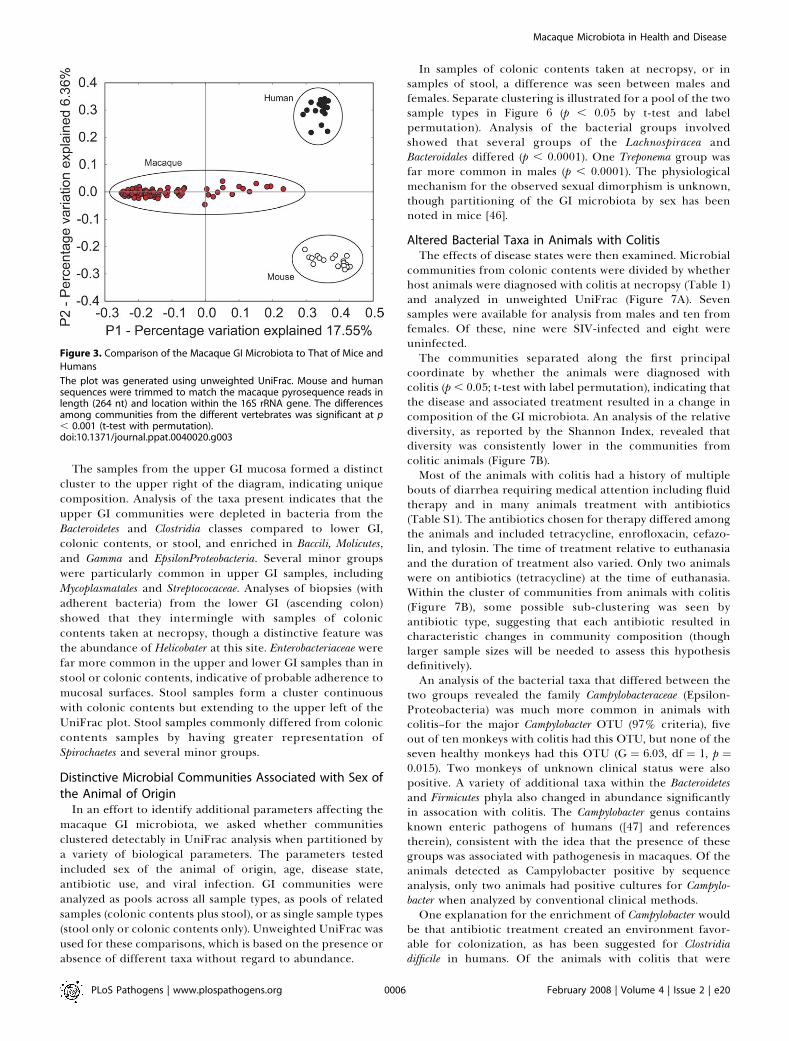
humans [4] and mice [8] is shown in Figure 3. To compare theglobal compositions of microbial communities, we usedUniFrac [38–40], which measures the similarity amongbacterial communities based on phylogenetic distances. Tocarry out a UniFrac analysis, we used the augmented ARBdatabase described above. To compare two communitiesusing UniFrac, sequences from the two communities aremarked on a common phylogenetic tree that contains all thesequences from the communities to be analyzed, and the
Table 1. Samples of GI Microbiota Analyzed in This Study
Gender Infected Colitis Colonic Contents Stool Upper Lower Total
Male Yes Yes 1 0 0 0 1
Yes No 3 0 0 0 3
Yes NA/unknown 3 29 8 12 52
No Yes 3 0 0 0 3
No No 0 0 0 0 0
No NA/unknown 0 4 0 4
Total Male 10 33 8 12 63
Female Yes Yes 2 0 0 0 2
Yes No 3 0 0 0 3
Yes NA/unknown 6 0 0 0 6
No Yes 4 0 0 0 4
No No 1 0 0 0 1
No NA/unknown 1 20 0 0 21
Total Female 17 20 0 0 37
Total Male and Female 27 53 8 12 100
doi:10.1371/journal.ppat.0040020.t001
PLoS Pathogens | www.plospathogens.org February 2008 | Volume 4 | Issue 2 | e200003
Macaque Microbiota in Health and Disease

fraction of the branch length on the tree unique to eachcommunity is then computed. This procedure provides ameasure of the similarity between the two communities interms of the total amount of evolutionary history thatseparates the sequences in the two communities. UniFracassigns only a small difference to changes in representation ofclosely-related taxa, but larger value for changes in repre-sentation of more distant taxa, in contrast to OTU-basedmethods that assume that all taxa are equally distinct. Tocompare multiple communities, all the pair-wise distancesbetween communities were computed, then Principal Coor-dinate Analysis (PCoA) was used to cluster the communitiesalong axes of maximal variance (Figure 3).
To compare human and mouse samples to the macaque
pyrosequencing data, sequence reads determined by theSanger method from human and mouse were first truncatedto match the length and position of the macaque 16S rRNAgene sequences. The UniFrac comparison showed strongclustering by species of origin. Similar separation by specieswas obtained when pyrosequencing data was used for boththe rhesus and murine samples (unpublished data). For thehuman and macaque samples, the communities clustered byspecies of origin even though samples from diverse anatom-ical sites were included for each species.The taxonomic groups from GI communities of each
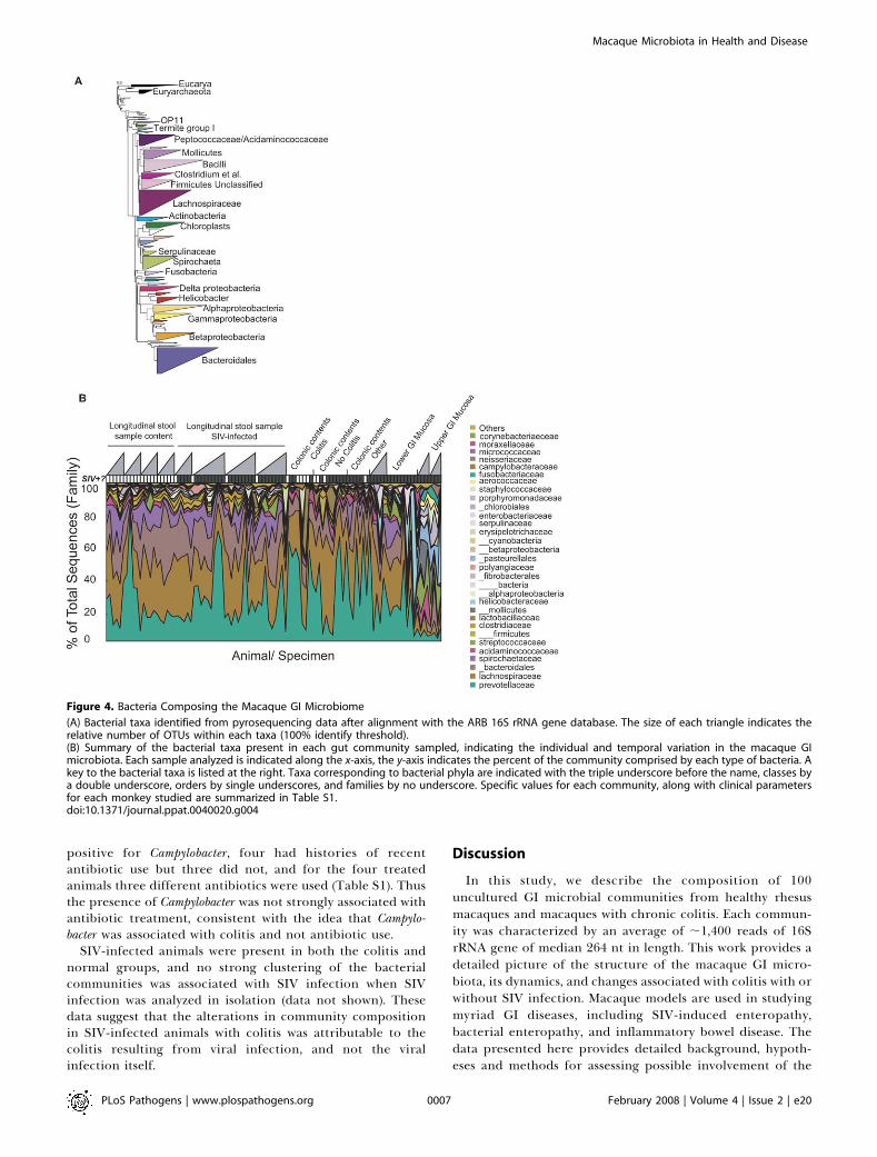
species were then compared. The bacterial taxa detectedare summarized in Figure 4A. The most prominent bacterialclasses were Clostridia (Phylum Firmicutes), Bacteroidetes (Phylum
Figure 1. Use of DNA Bar-coding and Pyrosequencing to Analyze Uncultured Bacterial Communities
(A) Length distribution of the pyrosequence reads used in this study. The median length was 264 nt.(B) The DNA bar-coding strategy. PCR primers are indicated by the arrows, DNA 59 ends are shown as balls. Each primer contains a regioncomplementary to the 454 sequencing primers (either A or B) and the 16S rRNA gene DNA (either BSR357 or BSF8) separated by a unique 4 base barcode (bold).(C) Reproducibility of the pyrosequencing method. DNA from a single specimen was analyzed by pyrosequencing at two different centers. Bacterialorders are indicated by the color code.(D) Comparison of results with pyrosequencing and conventional Sanger sequencing. Bacterial orders are indicated by the color code. Numbers ofsequences are as follows: M3T1 pyrosequence, 1382; M3T1 Sanger sequence, 47; M3T6 pyrosequence, 1360; M3T6 Sanger sequence, 47.doi:10.1371/journal.ppat.0040020.g001
PLoS Pathogens | www.plospathogens.org February 2008 | Volume 4 | Issue 2 | e200004
Macaque Microbiota in Health and Disease

Bacteroidetes) and Spirochaetes (Phylum Spirochaetes). Present inlesser amounts are Bacilli and Molicutes (Phylum Firmicutes),Alpha, Beta, Gamma, and Epsilon Proteobacteria, and a collectionof additional classes. Several of the minor classes were foundrepeatedly in specific individual macaques (e. g. Fibrobacteres,Gemmatimonadetes, Deferribacteres). All of the animals showedvariation over time, in both the classes detected and in theirrelative abundance. Many of the bacterial taxa identified werenot previously known to be present in the macaque intestinalmicrobiota.The predominance of the phyla Firmicutes and Bacteroidetes
were similar in all three vertebrates, and several lower-abundance phyla also overlapped. For example, Proteobacteriaand Actinobacteria were found in both macaques and humans.Verrucomicrobia were detected in humans but were raremacaques.A distinctive feature of the macaques was the density of
Phyla Spirochaetes, particularly members of the genus Trepone-ma, which were present in abundance in macaques (Figure 4)but mostly absent in the samples from in mice and humans.The abundance of flagellated Helicobater (EpsilonProteobacteria)has previously been noted [41], and Spirochaetes have beenidentified in the gut microbiota of many vertebrates includ-ing humans and non-human primates [42,43]. However, theabundance of Treponema in macaques was unexpected and fargreater than in human.In humans, within the Class Bacteroidetes, members of the
genus Bacteroides have been reported to be a major andfunctionally significant component of the human intestinalmicrobiota [4,44,45], but of the 94 near full length 16S rRNAgene macaque sequences, only one was genus Bacteroides. Morecommon were genus Prevotella (16/94 sequences), which is alsocommon in humans, and Rikenella (18/94 sequences), which israre or absent in humans [4]. These proportions ofBacteroidaceae and Prevotellaceae were similar in the shorterpyrosequencing reads.In macaques, comparison of microbial communities among
animals showed considerable variation among individuals,both in the relative abundance of the major taxonomicgroups and in the presence of minor groups (Figure 4B). Forsome animals, longitudinal samples were available, showingthat the composition of the GI microbiota was quite dynamicover the period of sampling.
Distinctive Microbial Communities Associated withDifferent Anatomical SitesFigure 5 shows a UniFrac clustering diagram comparing
the communities from different anatomical sites. Possibleclustering by sample type on the first two principalcoordinates was assessed using a t-test to compare thewithin-group and between-group distances, then 1,000 labelpermutations were used to assess significance. Clustering forall four sample types was found to be significant at the p ,
0.01 level.
Figure 2. Diversity of the Macaque GI Microbiota
(A) The numbers of operational taxonomic units (OTUs) present in thecollection of pyrosequence reads was analyzed by condensing sequen-ces at several percent identity thresholds. The x-axis shows the percentidentity, the y-axis the number of OTUs detected.(B) Collectors curves analysis of the completeness of sampling. Repeatedsamples of OTU subsets were used to evaluate whether further samplingwould likely yield additional taxa (rarefaction analysis), as indicated bywhether the curve has reached a plateau value. The y-axis indicates thenumber of OTUs detected, the x-axis the number of taxa in the sequencesubset analyzed. The color codes are as follows: green, stool samples;
yellow, colonic contents; red, lower GI mucosal surface; blue, upper GImucosal surface.(C) Rarefaction curves to estimate the diversity of taxa present inindividual samples, using the Shannon Index. Color code as in (B). Theupper GI mucosal samples were significantly less diverse than the othergroups (p , 0.004 for pairwise comparisons of upper gut samples toeach of the other three; Mann-Whitney comparison of means).doi:10.1371/journal.ppat.0040020.g002
PLoS Pathogens | www.plospathogens.org February 2008 | Volume 4 | Issue 2 | e200005
Macaque Microbiota in Health and Disease
The samples from the upper GI mucosa formed a distinctcluster to the upper right of the diagram, indicating uniquecomposition. Analysis of the taxa present indicates that theupper GI communities were depleted in bacteria from theBacteroidetes and Clostridia classes compared to lower GI,colonic contents, or stool, and enriched in Baccili, Molicutes,and Gamma and EpsilonProteobacteria. Several minor groupswere particularly common in upper GI samples, includingMycoplasmatales and Streptococaceae. Analyses of biopsies (withadherent bacteria) from the lower GI (ascending colon)showed that they intermingle with samples of coloniccontents taken at necropsy, though a distinctive feature wasthe abundance of Helicobater at this site. Enterobacteriaceae werefar more common in the upper and lower GI samples than instool or colonic contents, indicative of probable adherence tomucosal surfaces. Stool samples form a cluster continuouswith colonic contents but extending to the upper left of theUniFrac plot. Stool samples commonly differed from coloniccontents samples by having greater representation ofSpirochaetes and several minor groups.
Distinctive Microbial Communities Associated with Sex ofthe Animal of Origin
In an effort to identify additional parameters affecting themacaque GI microbiota, we asked whether communitiesclustered detectably in UniFrac analysis when partitioned bya variety of biological parameters. The parameters testedincluded sex of the animal of origin, age, disease state,antibiotic use, and viral infection. GI communities wereanalyzed as pools across all sample types, as pools of relatedsamples (colonic contents plus stool), or as single sample types(stool only or colonic contents only). Unweighted UniFrac wasused for these comparisons, which is based on the presence orabsence of different taxa without regard to abundance.
In samples of colonic contents taken at necropsy, or insamples of stool, a difference was seen between males andfemales. Separate clustering is illustrated for a pool of the twosample types in Figure 6 (p , 0.05 by t-test and labelpermutation). Analysis of the bacterial groups involvedshowed that several groups of the Lachnospiracea andBacteroidales differed (p , 0.0001). One Treponema group wasfar more common in males (p , 0.0001). The physiologicalmechanism for the observed sexual dimorphism is unknown,though partitioning of the GI microbiota by sex has beennoted in mice [46].
Altered Bacterial Taxa in Animals with ColitisThe effects of disease states were then examined. Microbial
communities from colonic contents were divided by whetherhost animals were diagnosed with colitis at necropsy (Table 1)and analyzed in unweighted UniFrac (Figure 7A). Sevensamples were available for analysis from males and ten fromfemales. Of these, nine were SIV-infected and eight wereuninfected.The communities separated along the first principal
coordinate by whether the animals were diagnosed withcolitis (p , 0.05; t-test with label permutation), indicating thatthe disease and associated treatment resulted in a change incomposition of the GI microbiota. An analysis of the relativediversity, as reported by the Shannon Index, revealed thatdiversity was consistently lower in the communities fromcolitic animals (Figure 7B).Most of the animals with colitis had a history of multiple
bouts of diarrhea requiring medical attention including fluidtherapy and in many animals treatment with antibiotics(Table S1). The antibiotics chosen for therapy differed amongthe animals and included tetracycline, enrofloxacin, cefazo-lin, and tylosin. The time of treatment relative to euthanasiaand the duration of treatment also varied. Only two animalswere on antibiotics (tetracycline) at the time of euthanasia.Within the cluster of communities from animals with colitis(Figure 7B), some possible sub-clustering was seen byantibiotic type, suggesting that each antibiotic resulted incharacteristic changes in community composition (thoughlarger sample sizes will be needed to assess this hypothesisdefinitively).An analysis of the bacterial taxa that differed between the
two groups revealed the family Campylobacteraceae (Epsilon-Proteobacteria) was much more common in animals withcolitis–for the major Campylobacter OTU (97% criteria), fiveout of ten monkeys with colitis had this OTU, but none of theseven healthy monkeys had this OTU (G ¼ 6.03, df ¼ 1, p ¼0.015). Two monkeys of unknown clinical status were alsopositive. A variety of additional taxa within the Bacteroidetesand Firmicutes phyla also changed in abundance significantlyin assocation with colitis. The Campylobacter genus containsknown enteric pathogens of humans ([47] and referencestherein), consistent with the idea that the presence of thesegroups was associated with pathogenesis in macaques. Of theanimals detected as Campylobacter positive by sequenceanalysis, only two animals had positive cultures for Campylo-bacter when analyzed by conventional clinical methods.One explanation for the enrichment of Campylobacter would
be that antibiotic treatment created an environment favor-able for colonization, as has been suggested for Clostridiadifficile in humans. Of the animals with colitis that were
Figure 3. Comparison of the Macaque GI Microbiota to That of Mice and
Humans
The plot was generated using unweighted UniFrac. Mouse and humansequences were trimmed to match the macaque pyrosequence reads inlength (264 nt) and location within the 16S rRNA gene. The differencesamong communities from the different vertebrates was significant at p, 0.001 (t-test with permutation).doi:10.1371/journal.ppat.0040020.g003
PLoS Pathogens | www.plospathogens.org February 2008 | Volume 4 | Issue 2 | e200006
Macaque Microbiota in Health and Disease
positive for Campylobacter, four had histories of recentantibiotic use but three did not, and for the four treatedanimals three different antibiotics were used (Table S1). Thusthe presence of Campylobacter was not strongly associated withantibiotic treatment, consistent with the idea that Campylo-bacter was associated with colitis and not antibiotic use.
SIV-infected animals were present in both the colitis andnormal groups, and no strong clustering of the bacterialcommunities was associated with SIV infection when SIVinfection was analyzed in isolation (data not shown). Thesedata suggest that the alterations in community compositionin SIV-infected animals with colitis was attributable to thecolitis resulting from viral infection, and not the viralinfection itself.
Discussion
In this study, we describe the composition of 100uncultured GI microbial communities from healthy rhesusmacaques and macaques with chronic colitis. Each commun-ity was characterized by an average of ;1,400 reads of 16SrRNA gene of median 264 nt in length. This work provides adetailed picture of the structure of the macaque GI micro-biota, its dynamics, and changes associated with colitis with orwithout SIV infection. Macaque models are used in studyingmyriad GI diseases, including SIV-induced enteropathy,bacterial enteropathy, and inflammatory bowel disease. Thedata presented here provides detailed background, hypoth-eses and methods for assessing possible involvement of the
Figure 4. Bacteria Composing the Macaque GI Microbiome
(A) Bacterial taxa identified from pyrosequencing data after alignment with the ARB 16S rRNA gene database. The size of each triangle indicates therelative number of OTUs within each taxa (100% identify threshold).(B) Summary of the bacterial taxa present in each gut community sampled, indicating the individual and temporal variation in the macaque GImicrobiota. Each sample analyzed is indicated along the x-axis, the y-axis indicates the percent of the community comprised by each type of bacteria. Akey to the bacterial taxa is listed at the right. Taxa corresponding to bacterial phyla are indicated with the triple underscore before the name, classes bya double underscore, orders by single underscores, and families by no underscore. Specific values for each community, along with clinical parametersfor each monkey studied are summarized in Table S1.doi:10.1371/journal.ppat.0040020.g004
PLoS Pathogens | www.plospathogens.org February 2008 | Volume 4 | Issue 2 | e200007
Macaque Microbiota in Health and Disease

full GI microbiota, and provides a model for investigatingchanges in the human GI microbiota in healthy and diseasedindividuals.
The pyrosequencing method [30] allows large numbers of16S rRNA gene sequence reads to be obtained whilecontrolling the costs of data acquisition, greatly increasingthe number of bacterial communities and species accessibleto analysis compared to culture-based methods. In thebioinformatic approach used here, the pyrosequencing readswere analyzed after first inserting them into pre-existingphylogenetic trees formed from full-length 16S rRNA genesequences, allowing relatively accurate phylogenetic place-ment despite the short sequences lengths [29]. Aligningpyrosequencing reads to a pre-existing tree also serves tominimize the effects of pyrosequencing errors, since singlenucleotide substitutions that cause a sequence read to alignwith an incorrect full length sequence will be rare.
Communities characterized by 16S rRNA gene sequencereads were compared to each other using UniFrac [38,40],which evaluates the distance between pairs of samples afteralignment on phylogenetic trees based on the unique branchlength leading to members of each community. Oneadvantage of this approach is that the collection of pair-wisedistances between communities can be subject to PCoA,allowing communities to be clustered along orthogonal axesof maximal variance. In a successful study of this type,clustering on each axis can report the effects of differentbiological variables. Previous studies of the vertebrate GImicrobiota have indicated that many factors influencemicrobial populations, including host genotype [8,48], geog-raphy [49], antibiotic use [50], and diet [51]. Using UniFracand PCoA, in combination with case-controlled samples, it is
potentially possible to extract the effects of these and othervariables and analyze each independently.Our analysis showed that the macaque microbiota differed
significantly from that of mouse or human. Even whencommunities from different anatomical sites were consid-ered, or when samples from healthy hosts were mixed withdiseased hosts, the effect of species of origin was stillpredominant. For all three vertebrates, the Firmicutes andBacteroidetes comprised the most abundant phyla, but thecomposition of minor groups differed and the taxa within theFirmicutes and Bacteroidetes also differed. A distinctive featureof the macaque samples was the abundance of Spirochaetesfrom the Treponema lineage. These Treponema differ from thespiral-shaped Helicobacter reported previously [41], which werealso detected here. Analysis of full-length 16S rRNA geneclones (Figure 1D) showed closest matches to Treponemabrennaborense and Treponema saccharophilum. T. brennaborense hasbeen associated with digital dermatitis in dairy cows [52]. T.saccharophilum has been identified as a component of therumen GI flora that aids in digestion of pectin [53], suggestinga possible role in digesting vegetable matter in the macaqueGI tract.The analysis of healthy animals emphasized the many
factors affecting composition of GI communities in mac-aques. The number of types of bacteria involved is very large–when macaque 16S rRNA gene sequences are grouped intoOTUs at 97% or greater similarity, a threshold that has beensuggested to correspond roughly to the species level, about
Figure 5. Distinctive GI Microbiota in Samples from Communities from
Different Anatomical Sites
Unweighted UniFrac was used in the comparison. The types of samplesstudied are indicated by the key at lower right.doi:10.1371/journal.ppat.0040020.g005
Figure 6. Sexual Dimorphism in the Macaque GI Microbiota
Samples of stool and colonic contents are combined for this analysis.Cluster analysis was carried out using unweighted UniFrac. Separationbetween male communities (green) and female communities (pink) wassignificant (p , 0.05, t-test with permutation; analysis over all variationbetween samples). Note that with the simplest null model, we expecteach Principal Coordinate to explain 100/number of samples, which is100/100 communities ¼ 1% of the variation. Thus the fourth PrincipalCoordinate, which separates males and females, is expected to containmeaningful information.doi:10.1371/journal.ppat.0040020.g006
PLoS Pathogens | www.plospathogens.org February 2008 | Volume 4 | Issue 2 | e200008
Macaque Microbiota in Health and Disease

5,000 OTUs were identified. Microbial communities ofindividual animals differed from one another, and all animalsfollowed longitudinally showed changes in communitycomposition over time. Similarly in humans, GI microbialcommunities have been reported to differ among individualsand at different anatomical sites [4]. The macaque GIcommunities also clustered by the sex of the host animal,paralleling a proposal for sexual dimorphism in the GImicrobiota in mice [46].
Samples from colonic contents of animals euthanized dueto advanced colitis showed distinctive communities com-pared to similar samples from healthy controls, linkingalterations in the GI microbial communities and GI patho-genesis. Samples from animals with colitis, whether associatedwith SIV infection or not, were indistinguishable. Thisemphasized that colitis itself (and associated therapeuticinterventions) and not the cause of colitis was most tightlylinked with altered GI microbiota. The presence of Campylo-bacter was strongly associated with colitis. The majorCampylobacter OTU (97% threshold) was present in five outof ten animals with colitis, but in zero out of seven free ofcolitis (p¼ 0.014). Cultureable C. jejuni or C. coli were obtainedonly from two animals, indicating that the Campylobacterspecies detected were either too rare to detect by culture, ordid not grow under the culture conditions used.
Most of the macaques euthanized due to GI-disease weretreated with antibiotics at some point during diseaseprogression. Thus these findings model human clinical caseswhere antibiotic therapy can be indicated in the treatment ofcolitis, but antibiotic treatment complicates analysis of effectsof GI disease alone. For the samples of colonic contents takenat necropsy, there were indications of clustering due to typeof antibiotic used for treatment within the larger cluster of
samples from animals with colitis, though the number ofsamples in each antibiotic group was too low for detailedanalysis by antibiotic type. Our data are consistent with theidea that the disease state caused a shift in bacterialcommunities that was further shaped by the antibiotics usedfor treatment.The sequence-based approach described here has the
potential to identify candidate pathogens involved inpreviously obscure disease conditions. Animal FH09 (TableS1) provides a case study. This animal suffered fromprolonged chronic diarhhea of unknown cause. Exhaustivesearches for a microbial pathogen by conventional culturingmethods were negative. For unexplained reasons, placing theanimal on a gluten-free diet helped ameliorate the condition,but eventually the animal declined and was euthanized forhumanitarian reasons. Analysis of colonic contents taken atnecropsy revealed a substantial number of 16S rRNA genesequences (51 reads) that clustered with a group containingCampylobacter fetus and Campylobacter hyointestinalis. EvidentlyCampylobacters of this group are not detected in the usualculture assays. C. fetus has been implicated as an emergingpathogen and could well have been involved in the GI diseaseof FH09. These findings suggest that further analysis of therelationship between diet and C. fetus pathogenesis might beuseful, and illustrate how the methods described here couldbe applied in diagnosis of human GI diseases of unknownetiology.In summary, this study presents the first use of DNA bar
coding and pyrosequencing to analyze uncultured bacterialcommunities from the primate gut, and provides the deepestview into the gut microbiome from the largest sample of anynon-human species to date. Using the macaque model andthe methods reported here, it will be possible to investigate
Figure 7. Colitis Is Associated with Distinctive GI Microbiota in Samples of Colonic Contents Taken at Necropsy
The analysis was restricted to samples of colonic contents taken at necropsy that allowed unambiguous assignment to the ‘‘colitis’’ or ‘‘healthy’’categories.(A) Analysis of communities in unweighted UniFrac. Samples in the colitis and healthy categories showed significant separation along the first principalcoordinate (p , 0.05, t-test with permutation). For an additional four animals, insufficient clinical histories were available, so these were not included inthe analysis (Table 1).(B) Diversity in samples from healthy animals or those with colitis were analyzed using the Shannon Index on OTUs condensed at 97% identity. Thediversity in the samples from animals with colitis was significantly lower (p , 0.05; Mann-Whitney comparison of means).doi:10.1371/journal.ppat.0040020.g007
PLoS Pathogens | www.plospathogens.org February 2008 | Volume 4 | Issue 2 | e200009
Macaque Microbiota in Health and Disease

how the interaction among bacterial community members,together with alterations in the GI environment, leads tooutgrowth of pathogenic forms and resultant disease. Thisstudy also paves the way for broader application ofpyrosequencing to characterize the human microbiota inhealth and disease, which could potentially allow large-scalecharacterization of thousands of human samples with ordersof magnitude less expense and effort than traditional Sangersequencing. We will thus soon be able to identify thosefeatures of the microbiota (if any) that are common to allhealthy individuals, and to assess the extent to which changesin the microbiota in animal models can help guide thedevelopment of therapy for human diseases.
Materials and Methods
Sample collection. Rhesus macaques (Macaca mulatta) were housedsingly at the Tulane National Primate Research Center. Forlongitudinal studies of stool samples, four animals (CC47, FH40,CT64, DD05; here M1-M4) were infected intravenously with 100TCID50 SIVmac251 on study day 0. Fecal samples were collectedprior to infection (t1), at day 7 (t3), day 14 (t4), day 28 (t6) and day 56(t10) post infection. These are standard time points for examinationof early events in the pathogenesis of AIDS and are associated withpeak viremia (day 14) and establishment of viral set point (by day 56).Stool samples for control animals (AM87, DG23, CC79, BA02; hereC1-C4) were collected similarly over an eight week period. Forsamples of colonic contents, each was collected from the ascendingcolon at necropsy within one hour of euthanasia with an intravenousoverdose of phenobarbital. All samples were immediately frozen to�80 8C. Samples were shipped on dry ice and stored at �80 8C untilprocessing. In addition, intestinal biopsies of the upper (jejunum) andlower (ascending colon) were obtained by standard techniques. Thesebiopsies were immediately frozen as for colonic contents. Housingand handling of animals were in accordance with the Guide for theCare and Use of Laboratory Animals (U.S. Public Health Service) andthe Animal Welfare Act. All protocols and procedures were reviewedand approved by the Tulane University Institutional Animal Care andUse Committee. Additional animals studied, their clinical conditions,and detailed ecological descriptions of samples are in Table S1.
Extraction and purification of DNA. Total DNA was extracted fromfrozen stool using the QIAampt DNA Stool Mini Kit (Qiagen, Inc.,Valencia CA), following the manufacturer’s protocol for pathogendetection.
PCR amplification of bacterial 16S rRNA gene sequences. Forsamples from each animal and at each time-point, the 16S rRNA genewas amplified from extracted DNA using the composite forwardprimer 59-GCCTCCCTCGCGCCATCAGNNNNCTGCTGCCTYCCG-TA-39 where the underlined sequence is that of 454 Life Sciencestprimer A and in italics is the broad range bacterial primer BSR357.The reverse primer was 59-GCCTTGCCAGCCCGCTCAGNNNNAGAGTTTGATCCTGGCTCAG-93, where the underlined sequence isthat of 454 Life Sciencest primer B and in italics is the broad rangebacterial primer BSF8. The NNNN designates the unique four basebar code used to tag each PCR product. Reaction conditions were asfollows: 5.0 ll 103 PCR buffer II (Applied Biosystems, Foster City,CA), 3.0 ll MgCl2 (25 mM; Applied Biosystems), 2.5 ll Triton X-100(1%), 2.0 ll deoxyribonucleoside triphosphates (10 mM), 1.0 llforward primer and 1.0 ll reverse primer (20 pmol/ll each) and 0.5 llAmpliTaqt DNA polymerase (5U/ll; Applied Biosystems) and 100 ngof template DNA in a total reaction volume of 50 ll. Reactions wererun in a GeneAmpt PCR System 9700 cycler (Applied Biosystems)using the following cycling parameters: 5 minutes denaturing at 95 8Cfollowed by 20 cycles of 30 secs at 95 8C (denaturing), 30 secs at 56 8C(annealing) and 90 secs at 72 8C (elongation), with a final extension at72 8C for 7 minutes. Four independent PCR reactions wereperformed for each sample along with a no template negativecontrol.
Gel purification and pyrosequencing. Each PCR product was gelpurified from a 0.8% agarose gel. DNA was isolated using theQIAquickt Gel extraction kit (Qiagen, Inc., Valencia CA). 100 ng ofeach of the 100 gel purified DNAs was added to a master pool of DNAwhich was sent for pyrosequencing with primer A as described[30,54]. Several studies have analyzed sources of error in 454sequencing runs, which informed our choices for quality control
here [37,54,55]. For a sequence to pass quality control, it needed to (1)show a perfect match to the bar code and 16S rRNA gene primer, (2)be at least 50 nt in length, (3) have no more than two undeterminedbases in the sequence read, and (4) find at least a 75% match to apreviously determined 16S rRNA gene sequence after alignment withNAST (http://greengenes.lbl.gov/). The sequences were inserted intothe 16S rRNA gene tree constructed by Hugenholz et al. [56] using theparsimony insertion tool from ARB software (http://www.arb-home.de/). A ‘‘termini’’ filter was used for the parsimony insertion. Afterapplying this criteria, 36,652,141 bases of sequence were available foranalysis. All sequence data will be deposited at NCBI upon accept-ance of this manuscript for publication.
Bioinformatic analysis. OTU clustering and analysis was carriedout using OTUPicker (M. Hamady and R. Knight, unpublished).Clustering and principal coordinate analysis were conducted usingUniFrac [29,38,39]. UniFrac analysis can be carried out based on thepresence and absence of bacterial taxa (unweighted UniFrac), ortaking into account abundance information on each group (weightedUniFrac); Figures 3, 4, 6, and 7 report unweighted UniFrac results. Toperform permutation tests within UniFrac, we randomized the labelsof each group and repeated the cluster analysis. We then comparedall distances between points that both came from the same group toall distances between points that came from different groups using at-test. In the permutation test, we obtained a nonparametricdistribution for the t statistic that takes into account the correlationsintroduced by the distance matrix structure. We used 1,000permutations, so we cannot specify p-value more precisely than‘‘,0.001’’ if none of the permuted sets gave a more extreme resultthan the actual set. We note that the principal coordinate analysisassumes that the relationships between taxon abundance andenvironmental gradients is linear. In choosing the Monte Carlomethods used for significance testing, we accepted reduced power toavoid using parametric methods, which assume random distributionin the error terms. The taxonomy assignments were based on thegroup names in Arb. Ecological parameters in Table S1 werecalculated using OTUPicker and PAST [57].
Errors in pyrosequencing may occur at a rate of about 0.25% [37],suggesting that the most of the 260-nucleotide sequences that remainafter filtering will contain either 0 or 1 errors. Single- nucleotideerrors will not affect either of the analyses we present (high-leveltaxonomic breakdowns or UniFrac) substantially, as they are unlikelyto cause assignment of pyrosequence reads to the wrong taxonomicgroup and contribute almost no branch length to the phylogenetictree used for UniFrac analyses. However, these sequencing errorscould affect estimates of the total number of OTUs at a giventhreshold, so some caution in interpreting the total number ofspecies-level taxa in the samples is required. Using the Poisson model,we would expect only 4.4 3 10�5% of the reads to contain the sevenerrors that would be required to form a new species-level at the 97%OTU threshold. Thus, it is unlikely that a single OTU in the analysiswas generated through that mechanism.
Supporting Information
Table S1. Characteristics of Samples of Uncultured Macaque GICommunities Used in This Study
This table provides the description of monkeys sampled, clinicalparameters for disease states, and ecological statistics describing thecommunities sampled.
Found at doi:10.1371/journal.ppat.0040020.st001 (93 KB XLS).
Table S2. Near-full-length 16S rRNA Gene Sequences Determined bythe Sanger Method, and Their Taxonomic Positions
These sequences allow finer discrimination of the major macaque GIbacterial taxa.
Found at doi:10.1371/journal.ppat.0040020.st002 (404 KB XLS).
Acknowledgments
We thank the Keck Foundation for a pilot grant to RK and FDB. Wethank Paul Sniegowski, Dustin Britton, and Jeff Weiser for helpfuldiscussions; Dan Frank for critical reading of the manuscript; andSteven Holland for creating a command-line version of DOTUR.
Author contributions. PM, CH, NM, PPA, AL, ZL, CAL, MH, RK,and FDB contributed to the design of experiments and interpretationof data. Animal work was carried out at the Tulane National PrimateResearch Center by PPA and AL. Sequencing was carried out by PM,
PLoS Pathogens | www.plospathogens.org February 2008 | Volume 4 | Issue 2 | e200010
Macaque Microbiota in Health and Disease
CH, NM, and FDB. Data analysis was carried out by PM, CH, AL, ZL,CAL, MH, RK, and FDB.
Funding. This work was supported in part by Public Health ServiceGrants RR00164, RR016930, and AI045008; NIH grantsP01DK078669, T32GM065103, and GM08759; grants from the
University of Pennsylvania Center for AIDS Research and theColorado Center for AIDS Research; and by the Keck RNABioinformatics Initiative at the University of Colorado, Boulder.
Competing interests. The authors have declared that no competinginterests exist.
References1. Ley RE, Peterson DA, Gordon JI (2006) Ecological and evolutionary forces
shaping microbial diversity in the human intestine. Cell 124: 837–484.2. Whitman WB, Coleman DC, Wiebe WJ (1998) Prokaryotes: the unseen
majority. Proc Natl Acad Sci U S A 95: 6578–6583.3. Gill SR, Pop M, Deboy RT, Eckburg PB, Turnbaugh PJ, et al. (2006)
Metagenomic analysis of the human distal gut microbiome. Science 312:1355–1359.
4. Eckburg PB, Bik EM, Bernstein CN, Purdom E, Dethlefsen L, et al. (2005)Diversity of the human intestinal microbial flora. Science 308: 1635–1638.
5. Palmer C, Bik EM, Eisen MB, Eckburg PB, Sana TR, et al. (2006) Rapidquantitative profiling of complex microbial populations. Nucleic Acids Res10: e5.
6. Palmer C, Bik EM, Digiulio DB, Relman DA, Brown PO (2007) Developmentof the human infant intestinal microbiota. PLoS Biol 5: e177. doi: 10.1371/journal.pbio.0050177
7. Turnbaugh PJ, Ley RE, Mahowald MA, Magrini V, Mardis ER, et al. (2006)An obesity-associated gut microbiome with increased capacity for energyharvest. Nature 444: 1027–1031.
8. Ley RE, Backhed F, Turnbaugh P, Lozupone CA, Knight RD, et al. (2005)Obesity alters gut microbial ecology. Proc Natl Acad Sci U S A 102: 11070–11075.
9. Scanlan PD, Shanahan F, O’Mahony C, Marchesi JR (2006) Culture-independent analyses of temporal variation of the dominant fecal micro-biota and targeted bacterial subgroups in crohn’s disease. J Clin Microbiol44: 3980–3988.
10. Manichanh C, Rigottier-Gois L, Bonnaud E, Gloux K, Pelletier E, et al.(2006) Reduced diversity of faecal microbiota in crohn’s disease revealed bya metagenomic approach. Gut 55: 205–211.
11. Fagarasan S, Muramatsu M, Suzuki K, Nagaoka H, Hiai H, et al. (2002)Critical roles of activation-induced cytidine deaminase in the homeostasisof gut flora. Science 298: 1424–1427.
12. Suzuki K, Meek B, Doi Y, Muramatsu M, Chiba T, et al. (2004) Aberrantexpansion of segmented filamentous bacteria in IgA-deficient gut. ProcNatl Acad Sci U S A 101: 1981–1986.
13. Ayabe T, Satchell DP, Wilson CL, Parks WC, Selsted ME, et al. (2000)Secretion of microbicidal alpha-defensins by intestinal paneth cells inresponse to bacteria. Nat Immunol 1: 113–118.
14. Hooper LV, Stappenbeck TS, Hong CV, Gordon JI (2003) Angiogenins: anew class of microbicidal proteins involved in innate immunity. NatImmunol 4: 269–273.
15. Li Q, Duan L, Estes JD, Ma ZM, Rourke T, et al. (2005) Peak SIV replicationin resting memory CD4þ T cells depletes gut lamina propria CD4þ T cells.Nature 434: 1148–1152.
16. Veazey RS, DeMaria M, Chalifoux LV, Shvetz DE, Pauley DR, et al. (1998)Gastrointestinal tract as a major site of CD4þ T cell depletion and viralreplication in SIV infection. Science 280: 427–431.
17. Mehandru S, Poles MA, Tenner-Racz K, Horowitz A, Hurley A, et al. (2004)Primary HIV-1 infection is associated with preferential depletion of CD4þT lymphocytes from effector sites in the gastrointestinal tract. J Exp Med200: 761–770.
18. Guadalupe M, Reay E, Sankaran S, Prindiville T, Flamm J, et al. (2003)Severe CD4þ T-cell depletion in gut lymphoid tissue during primaryhuman immunodeficiency virus type 1 infection and substantial delay inrestoration following highly active antiretroviral therapy. J Virol 77: 11708–11717.
19. Guadalupe M, Sankaran S, George MD, Reay E, Verhoeven D, et al. (2006)Viral suppression and immune restoration in the gastrointestinal mucosaof human immunodeficiency virus type 1-infected patients initiatingtherapy during primary or chronic infection. J Virol 80: 8236–8247.
20. Brenchley JM, Price DA, Schacker TW, Asher TE, Silvestri G, et al. (2006)Microbial translocation is a cause of systemic immune activation in chronicHIV infection. Nat Med 12: 1365–1371.
21. Knox TA, Spiegelman D, Skinner SC, Gorbach S (2000) Diarrhea andabnormalities of gastrointestinal function in a cohort of men and womenwith HIV infection. Am J Gastroenterol 95: 3482–3489.
22. Heise C, Miller CJ, Lackner A, Dandekar S (1994) Primary acute simianimmunodeficiency virus infection of intestinal lymphoid tissue is associ-ated with gastrointestinal dysfunction. J Infect Dis 169: 1116–1120.
23. Stone JD, Heise CC, Miller CJ, Halsted CH, Dandekar S (1994) Developmentof malabsorption and nutritional complications in simian immunodefi-ciency virus-infected rhesus macaques. Aids 8: 1245–1256.
24. Mohan M, Aye PP, Borda JT, Alvarez XJ, Lackner AA (2007) Gastro-intestinal disease in SIV-infected rhesus macaques is characterized byproinflammatory dysregulation of the IL-6-JAK-STAT3 pathway. Am J Path171: 1952–1965.
25. Sestak K, Merritt CK, Borda J, Saylor E, Schwamberger SR, et al. (2003)
Infectious agent and immune response characteristics of chronic enter-ocolitis in captive rhesus macaques. Infect Immun 71: 4079–4086.
26. Ramesh G, Alvarez X, Borda JT, Aye PP, Lackner AA, et al. (2005)Visualizing cytokine-secreting cells in situ in the rhesus macaque model ofchronic gut inflammation. Clin Diagn Lab Immunol 12: 192–197.
27. De Hertogh G, Aerssens J, de Hoogt R, Peeters P, Verhasselt P, et al. (2006)Validation of 16S rDNA sequencing in microdissected bowel biopsies fromcrohn’s disease patients to assess bacterial flora diversity. J Pathol 209: 532–539.
28. Pace NR, Olsen GJ, Woese CR (1986) Ribosomal RNA phylogeny and theprimary lines of evolutionary descent. Cell 45: 325–326.
29. Liu Z, Lozupone C, Hamady M, Bushman FD, Knight R (2007) Shortpyrosequencing reads suffice for accurate microbial community analysis.Nucleic Acids Res 35: e120.
30. Margulies M, Egholm M, Altman WE, Attiya S, Bader JS, et al. (2005)Genome sequencing in microfabricated high-density picolitre reactors.Nature 437: 376–380.
31. Dedysh SN, Pankratov TA, Belova SE, Kulichevskaya IS, Liesack W (2006)Phylogenetic analysis and in situ identification of bacteria communitycomposition in an acidic sphagnum peat bog. Appl Environ Microbiol 72:2110–2117.
32. Schmitt-Wagner D, Friedrich MW, Wagner B, Brune A (2003) Phylogeneticdiversity, abundance, and axial distribution of bacteria in the intestinaltract of two soil-feeding termites (cubitermes spp.). Appl Environ Microbiol69: 6007–6017.
33. Salzman NH, de Jong H, Paterson Y, Harmsen HJ, Welling GW, et al. (2002)Analysis of 16S libraries of mouse gastrointestinal microflora reveals a largenew group of mouse intestinal bacteria. Microbiology 148: 3651–3660.
34. Ludwig W (2004) ARB: a software environment for sequence data. NucleicAcids Res 32: 1363–1371.
35. DeSantis TZ Jr, Hugenholtz P, Keller K, Brodie EL, Larsen N, et al. (2006)NAST: a multiple sequence alignment server for comparative analysis of16S rRNA genes. Nucleic Acids Res 34: W394–W399.
36. Stackebrandt E, Goebal B (1994) Taxonomic note: a place for DNA-DNAreassociation and 16s rRNA sequence analysis in the present speciesdefinition in bacteriology. Int Syst Bacteriol 44: 846–849.
37. Huse SM, Huber JA, Morrison HG, Sogin ML, Welch DM (2007) Accuracyand quality of massively parallel DNA pyrosequencing. Genome Biol 8:R143.
38. Lozupone C, Hamady M, Knight R (2006) UniFrac–an online tool forcomparing microbial community diversity in a phylogenetic context. BMCBioinformatics 7: 371.
39. Lozupone CA, Hamady M, Kelley ST, Knight R (2007) Quantitative andqualitative fbetag diversity measures lead to different insights into factorsthat structure microbial communities. Appl Environ Microbiol 73: 1576–1585.
40. Lozupone C, Knight R (2005) UniFrac: a new phylogenetic method forcomparing microbial communities. Appl Environ Microbiol 71: 8228–8235.
41. Duhamel GE, Stryker CJ, Lu G, Wong VJ, Tarara RP (2003) Colonicspirochetosis of colony-raised rhesus macaques associated with brachyspiraand helicobacter. Anaerobe 9: 45–55.
42. Duhamel GE, Elder RO, Muniappa N, Mathiesen MR, Wong VJ, et al. (1997)Colonic spirochetal infections in nonhuman primates that were associatedwith brachyspira aalborgi, serpulina pilosicoli, and unclassified flagellatedbacteria. Clin Infect Dis 25 Suppl 2: S186–S188.
43. Munshi MA, Taylor NM, Mikosza AS, Spencer PB, Hampson DJ (2003)Detection by PCR and isolation assays of the anaerobic intestinalspirochete brachyspira aalborgi from the feces of captive nonhumanprimates. J Clin Microbiol 41: 1187–1191.
44. Xu J, Bjursell MK, Himrod J, Deng S, Carmichael LK, et al. (2003) Agenomic view of the human-bacteriodetes thetaiotaomicron symbiosis.Science 299: 2074–2076.
45. Xu J, Mahowald MA, Ley RE, Lozupone CA, Hamady M, et al. (2007)Evolution of symbiotic bacteria in the distal human intestine. PLoS Biol 5:e156. doi: 10.1371/journal.pbio.0050156
46. Schloss PD, Handelsman J (2006) Introducing SONS, a tool for operationaltaxonomic unit-based comparisons of microbial community membershipsand structures. Appl Environ Microbiol 72: 6773–6779.
47. Parkhill J (2000) The genome sequence of the food-borne pathogencampylobacter jejuni reveals hypervariable sequences. Nature 403: 665–668.
48. Zoetendal EG, Akkermans AD, Akkermansvan Vliet WM, De Visser AGM,De Vos WM (2001) The host genotype affects the bacterial community inthe human gastrointestinal tract. Microbial Ecol Health Dis 13: 129–134.
49. Lay C, Rigottier-Gois L, Holmstrom K, Rajilic M, Vaughan EE, et al. (2005)Colonic microbiota signatures across five northern european countries.Appl Environ Microbiol 71: 4153–4155.
PLoS Pathogens | www.plospathogens.org February 2008 | Volume 4 | Issue 2 | e200011
Macaque Microbiota in Health and Disease

50. Young VB, Schmidt TM (2004) Antibiotic-associated diarrhea accompaniedby large-scale alterations in the composition of the fecal microbiota. J ClinMicrobiol 42: 1203–1206.
51. Tannock GW, Munro K, Bibiloni R, Simon MA, Hargreaves P, et al. (2004)Impact of consumption of oligosaccharide-containing biscuits on the fecalmicrobiota of humans. Appl Environ Microbiol 70: 2129–2136.
52. Dermirkan I, Williams HF, Dhawi A, Carter SD, Winstanley C, et al. (2006)Characterization of a spirochaete isolated from a case of bovine digitaldermatitis. J Appl Microbiol 101: 948–955.
53. Paster BJ, Canale-Parola E (1985) Treponema saccharophilum sp. nov., alarge pectinolytic spirochete from the bovine rumen. Appl EnvironMicrobiol 50: 212–219.
54. Sogin ML, Morrison HG, Huber JA, Welch DM, Huse SM, et al. (2006)Microbial diversity in the deep sea and the underexplored ‘‘rare bio-sphere’’. Proc Natl Acad Sci U S A 103: 12115–12120.
55. Hoffmann C, Minkah N, Leipzig J, Wang G, Arens MQ, et al. (2007) DNAbar coding and pyrosequencing to identify rare HIV drug resistancemutations. Nucleic Acids Res 35: e91.
56. Hugenholtz P (2002) Exploring prokaryotic diversity in the genomic era.Genome Biol 3: REVIEWS0003.
57. Hammer O, Harper DAT, Ryan PD (2001) PAST: paleontological statisticssoftware package for education and data analysis. Paleontologica Elec-tronica 4: 9.
PLoS Pathogens | www.plospathogens.org February 2008 | Volume 4 | Issue 2 | e200012
Macaque Microbiota in Health and Disease
Related Documents