Maternal Obesity Induced by Diet in Rats Permanently Influences Central Processes Regulating Food Intake in Offspring Shona L. Kirk, Anne-Maj Samuelsson, Marco Argenton, Hannah Dhonye, Theodosis Kalamatianos, Lucilla Poston, Paul D. Taylor*, Clive W. Coen Division of Reproduction and Endocrinology, King’s College London, London, United Kingdom Abstract Hypothalamic systems which regulate appetite may be permanently modified during early development. We have previously reported hyperphagia and increased adiposity in the adult offspring of rodents fed an obesogenic diet prior to and throughout pregnancy and lactation. We now report that offspring of obese (OffOb) rats display an amplified and prolonged neonatal leptin surge, which is accompanied by elevated leptin mRNA expression in their abdominal white adipose tissue. At postnatal Day 30, before the onset of hyperphagia in these animals, serum leptin is normal, but leptin- induced appetite suppression and phosphorylation of STAT3 in the arcuate nucleus (ARC) are attenuated; the level of AgRP- immunoreactivity in the hypothalamic paraventricular nucleus (PVH), which derives from neurones in the ARC and is developmentally dependent on leptin, is also diminished. We hypothesise that prolonged release of abnormally high levels of leptin by neonatal OffOb rats leads to leptin resistance and permanently affects hypothalamic functions involving the ARC and PVH. Such effects may underlie the developmental programming of hyperphagia and obesity in these rats. Citation: Kirk SL, Samuelsson A-M, Argenton M, Dhonye H, Kalamatianos T, et al. (2009) Maternal Obesity Induced by Diet in Rats Permanently Influences Central Processes Regulating Food Intake in Offspring. PLoS ONE 4(6): e5870. doi:10.1371/journal.pone.0005870 Editor: Paul A. Bartell, Pennsylvania State University, United States of America Received February 15, 2009; Accepted May 12, 2009; Published June 11, 2009 Copyright: ß 2009 Kirk et al. This is an open-access article distributed under the terms of the Creative Commons Attribution License, which permits unrestricted use, distribution, and reproduction in any medium, provided the original author and source are credited. Funding: This study was supported by the British Heart Foundation (PG/06/067; http://www.bhf.org.uk/) and the BBSRC (BBD5231861; http://www.bbsrc.ac.uk/). LP is funded by Tommy’s The Baby Charity. The funders had no role in study design, data collection and analysis, decision to publish, or preparation of the manuscript. Competing Interests: The authors have declared that no competing interests exist. * E-mail: [email protected] Introduction The developmental overnutrition hypothesis suggests that maternal obesity and/or gestational diabetes in humans may predispose offspring to altered energy balance and increased adiposity in adulthood [1–4]. This hypothesis has gained strength with recent reports of an association between excessive weight gain in pregnancy and the BMI of the adolescent child and of a greater influence of maternal BMI than paternal BMI on offspring adiposity [5–7]. Animal models have proven invaluable in understanding developmental programming of adult disease. We have recently reported that offspring of mice or rats in which obesity had been induced by prolonged consumption of an obesogenic diet display hyperphagia, increased fat mass and hyperleptinaemia in adulthood [8,9]. Other studies on experimental animals indicate that nutritional imbalance during pregnancy and lactation may lead to permanent modification of food intake due to developmental plasticity in the hypothalamus [10–18]. Studies on genetically hyperphagic (ob/ob) mice have demonstrated a neurotrophic role for leptin in the development of projections from the arcuate nucleus (ARC) to the hypothalamic paraventricular (PVH) nucleus [19]. The timing and magnitude of the neonatal leptin surge, normally present during the second postnatal week in rodents [20], can be perturbed by maternal undernutrition, thereby altering hypothalamic development, with persistent effects on energy balance [21–23]. In contrast, the present study addresses the hypothesis that a maternal calorie-rich diet and consequent obesity lead to hyperpha- gia in adult offspring through processes involving impaired leptin- signalling and altered neuronal development. We have characterised the neonatal profiles of serum leptin and adipose leptin mRNA and the composition of ingested milk in the offspring of obese dams (OffOb rats). To our knowledge, this is the first study to investigate the effects of maternal obesity on the neonatal leptin surge. We have also assessed behavioural and cell-signalling responses to exogenous leptin and the density of immunoreactivity for orexigenic and anorexigenic peptides in the hypothalamic paraventricular nucleus (PVH) prior to the onset of hyperphagia. This study thereby investigates the processes underlying the non-genetic transmission of an obesogenic trait from mother to offspring. Results Development of Maternal Obesity Female Sprague Dawley rats consuming the highly palatable fat- and sugar-rich diet became significantly heavier than control animals after 10 days (Fig. 1A). After 6 weeks on this obesogenic diet, they were 20% heavier than controls, at which point they were mated. The weight difference was maintained throughout pregnancy by significantly increased calorific intake of both fat and simple sugars (Fig. 1A–D). During lactation, dams on the obesogenic diet continued to show a significantly higher calorific intake from fat and simple sugars than the control dams (Fig. 1E). PLoS ONE | www.plosone.org 1 June 2009 | Volume 4 | Issue 6 | e5870

Welcome message from author
This document is posted to help you gain knowledge. Please leave a comment to let me know what you think about it! Share it to your friends and learn new things together.
Transcript

Maternal Obesity Induced by Diet in Rats PermanentlyInfluences Central Processes Regulating Food Intake inOffspringShona L. Kirk, Anne-Maj Samuelsson, Marco Argenton, Hannah Dhonye, Theodosis Kalamatianos,
Lucilla Poston, Paul D. Taylor*, Clive W. Coen
Division of Reproduction and Endocrinology, King’s College London, London, United Kingdom
Abstract
Hypothalamic systems which regulate appetite may be permanently modified during early development. We havepreviously reported hyperphagia and increased adiposity in the adult offspring of rodents fed an obesogenic diet prior toand throughout pregnancy and lactation. We now report that offspring of obese (OffOb) rats display an amplified andprolonged neonatal leptin surge, which is accompanied by elevated leptin mRNA expression in their abdominal whiteadipose tissue. At postnatal Day 30, before the onset of hyperphagia in these animals, serum leptin is normal, but leptin-induced appetite suppression and phosphorylation of STAT3 in the arcuate nucleus (ARC) are attenuated; the level of AgRP-immunoreactivity in the hypothalamic paraventricular nucleus (PVH), which derives from neurones in the ARC and isdevelopmentally dependent on leptin, is also diminished. We hypothesise that prolonged release of abnormally high levelsof leptin by neonatal OffOb rats leads to leptin resistance and permanently affects hypothalamic functions involving theARC and PVH. Such effects may underlie the developmental programming of hyperphagia and obesity in these rats.
Citation: Kirk SL, Samuelsson A-M, Argenton M, Dhonye H, Kalamatianos T, et al. (2009) Maternal Obesity Induced by Diet in Rats Permanently Influences CentralProcesses Regulating Food Intake in Offspring. PLoS ONE 4(6): e5870. doi:10.1371/journal.pone.0005870
Editor: Paul A. Bartell, Pennsylvania State University, United States of America
Received February 15, 2009; Accepted May 12, 2009; Published June 11, 2009
Copyright: � 2009 Kirk et al. This is an open-access article distributed under the terms of the Creative Commons Attribution License, which permits unrestricteduse, distribution, and reproduction in any medium, provided the original author and source are credited.
Funding: This study was supported by the British Heart Foundation (PG/06/067; http://www.bhf.org.uk/) and the BBSRC (BBD5231861; http://www.bbsrc.ac.uk/).LP is funded by Tommy’s The Baby Charity. The funders had no role in study design, data collection and analysis, decision to publish, or preparation of themanuscript.
Competing Interests: The authors have declared that no competing interests exist.
* E-mail: [email protected]
Introduction
The developmental overnutrition hypothesis suggests that
maternal obesity and/or gestational diabetes in humans may
predispose offspring to altered energy balance and increased
adiposity in adulthood [1–4]. This hypothesis has gained strength
with recent reports of an association between excessive weight gain
in pregnancy and the BMI of the adolescent child and of a greater
influence of maternal BMI than paternal BMI on offspring adiposity
[5–7]. Animal models have proven invaluable in understanding
developmental programming of adult disease. We have recently
reported that offspring of mice or rats in which obesity had been
induced by prolonged consumption of an obesogenic diet display
hyperphagia, increased fat mass and hyperleptinaemia in adulthood
[8,9]. Other studies on experimental animals indicate that
nutritional imbalance during pregnancy and lactation may lead to
permanent modification of food intake due to developmental
plasticity in the hypothalamus [10–18].
Studies on genetically hyperphagic (ob/ob) mice have demonstrated
a neurotrophic role for leptin in the development of projections from
the arcuate nucleus (ARC) to the hypothalamic paraventricular
(PVH) nucleus [19]. The timing and magnitude of the neonatal leptin
surge, normally present during the second postnatal week in rodents
[20], can be perturbed by maternal undernutrition, thereby altering
hypothalamic development, with persistent effects on energy balance
[21–23]. In contrast, the present study addresses the hypothesis that a
maternal calorie-rich diet and consequent obesity lead to hyperpha-
gia in adult offspring through processes involving impaired leptin-
signalling and altered neuronal development. We have characterised
the neonatal profiles of serum leptin and adipose leptin mRNA and
the composition of ingested milk in the offspring of obese dams
(OffOb rats). To our knowledge, this is the first study to investigate the
effects of maternal obesity on the neonatal leptin surge. We have also
assessed behavioural and cell-signalling responses to exogenous leptin
and the density of immunoreactivity for orexigenic and anorexigenic
peptides in the hypothalamic paraventricular nucleus (PVH) prior to
the onset of hyperphagia. This study thereby investigates the
processes underlying the non-genetic transmission of an obesogenic
trait from mother to offspring.
Results
Development of Maternal ObesityFemale Sprague Dawley rats consuming the highly palatable
fat- and sugar-rich diet became significantly heavier than control
animals after 10 days (Fig. 1A). After 6 weeks on this obesogenic
diet, they were 20% heavier than controls, at which point they
were mated. The weight difference was maintained throughout
pregnancy by significantly increased calorific intake of both fat and
simple sugars (Fig. 1A–D). During lactation, dams on the
obesogenic diet continued to show a significantly higher calorific
intake from fat and simple sugars than the control dams (Fig. 1E).
PLoS ONE | www.plosone.org 1 June 2009 | Volume 4 | Issue 6 | e5870
Figure 1. Maternal body weight and food intake in rats fed control or obesogenic diet. Body weight (A) was recorded for 6 weeks prior topregnancy and throughout pregnancy and lactation for the animals on the control (open symbols) or obesogenic (closed symbols) diet; calorificintake was recorded throughout pregnancy and lactation (B). Average daily calorific intake from all sources during pregnancy (C) and average dailycalorific intake from fat or simple sugars during pregnancy (D) or lactation (E) for the animals on the control (Con) or obesogenic (Ob) diet.Macronutrient content of ingested food (expressed as percentage by weight) for control (F) or obese (G) dams during lactation; ‘‘other’’ includescellulose, ash, water etc. * p,0.05 and ** p,0.01 versus control dams (n = 11–12).doi:10.1371/journal.pone.0005870.g001
Obesity & Juvenile Hyperphagia
PLoS ONE | www.plosone.org 2 June 2009 | Volume 4 | Issue 6 | e5870
The obese dams consumed approximately 4 times more fat and 5
times more simple sugars than the control dams during pregnancy
and lactation (Fig. 1F,G; data presented for lactation; similar data
for the pre-conditioning period and during pregnancy not shown).
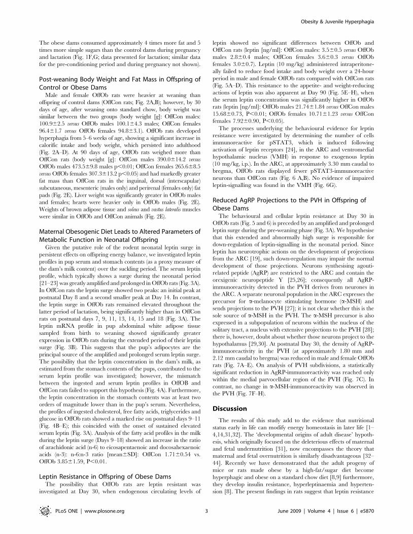
Post-weaning Body Weight and Fat Mass in Offspring ofControl or Obese Dams
Male and female OffOb rats were heavier at weaning than
offspring of control dams (OffCon rats; Fig. 2A,B); however, by 30
days of age, after weaning onto standard chow, body weight was
similar between the two groups (body weight [g]: OffCon males:
100.962.5 versus OffOb males 100.164.3 males; OffCon females
96.461.7 versus OffOb females 94.863.1). OffOb rats developed
hyperphagia from 5–6 weeks of age, showing a significant increase in
calorific intake and body weight, which persisted into adulthood
(Fig. 2A–D). At 90 days of age, OffOb rats weighed more than
OffCon rats (body weight [g]: OffCon males 390.0614.2 versus
OffOb males 473.569.8 males p,0.01; OffCon females 265.668.5
versus OffOb females 307.3613.2 p,0.05) and had markedly greater
fat mass than OffCon rats in the inguinal, dorsal (interscapular)
subcutaneous, mesenteric (males only) and perirenal (females only) fat
pads (Fig. 2E). Liver weight was significantly greater in OffOb males
and females; hearts were heavier only in OffOb males (Fig. 2E).
Weights of brown adipose tissue and soleus and vastus lateralis muscles
were similar in OffOb and OffCon animals (Fig. 2E).
Maternal Obesogenic Diet Leads to Altered Parameters ofMetabolic Function in Neonatal Offspring
Given the putative role of the rodent neonatal leptin surge in
persistent effects on offspring energy balance, we investigated leptin
profiles in pup serum and stomach contents (as a proxy measure of
the dam’s milk content) over the suckling period. The serum leptin
profile, which typically shows a surge during the neonatal period
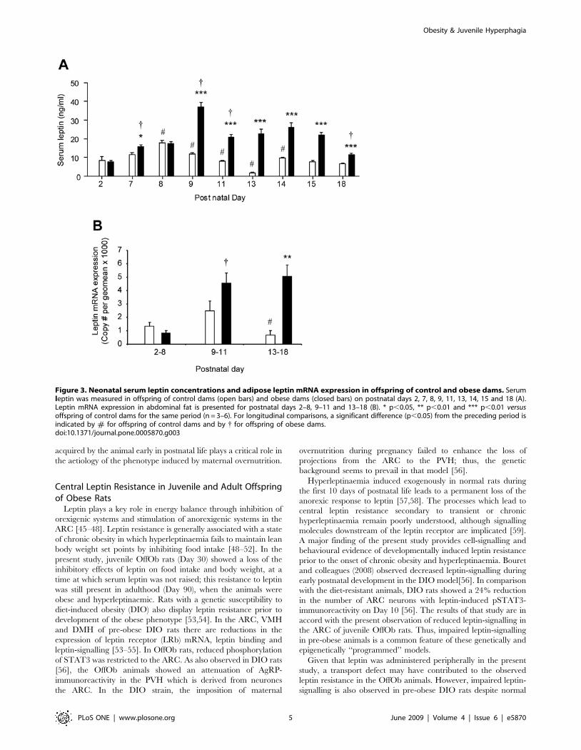
[21–23] was greatly amplified and prolonged in OffOb rats (Fig. 3A).
In OffCon rats the leptin surge showed two peaks: an initial peak at
postnatal Day 8 and a second smaller peak at Day 14. In contrast,
the leptin surge in OffOb rats remained elevated throughout the
latter period of lactation, being significantly higher than in OffCon
rats on postnatal days 7, 9, 11, 13, 14, 15 and 18 (Fig. 3A). The
leptin mRNA profile in pup abdominal white adipose tissue
sampled from birth to weaning showed significantly greater
expression in OffOb rats during the extended period of their leptin
surge (Fig. 3B). This suggests that the pup’s adipocytes are the
principal source of the amplified and prolonged serum leptin surge.
The possibility that the leptin concentration in the dam’s milk, as
estimated from the stomach contents of the pups, contributed to the
serum leptin profile was investigated; however, the mismatch
between the ingested and serum leptin profiles in OffOB and
OffCon rats failed to support this hypothesis (Fig. 4A). Furthermore,
the leptin concentration in the stomach contents was at least two
orders of magnitude lower than in the pup’s serum. Nevertheless,
the profiles of ingested cholesterol, free fatty acids, triglycerides and
glucose in OffOb rats showed a marked rise on postnatal days 9–11
(Fig. 4B–E); this coincided with the onset of sustained elevated
serum leptin (Fig. 3A). Analysis of the fatty acid profiles in the milk
during the leptin surge (Days 9–18) showed an increase in the ratio
of arachidonic acid (n-6) to eicosapentaenoic and docosahexaenoic
acids (n-3): n-6:n-3 ratio [mean6SD]: OffCon 1.7160.54 vs.
OffOb 3.8561.59, P,0.01.
Leptin Resistance in Offspring of Obese DamsThe possibility that OffOb rats are leptin resistant was
investigated at Day 30, when endogenous circulating levels of
leptin showed no significant differences between OffOb and
OffCon rats (leptin [ng/ml]: OffCon males: 3.560.5 versus OffOb
males 2.860.4 males; OffCon females 3.660.3 versus OffOb
females 3.060.7). Leptin (10 mg/kg) administered intraperitone-
ally failed to reduce food intake and body weight over a 24-hour
period in male and female OffOb rats compared with OffCon rats
(Fig. 5A–D). This resistance to the appetite- and weight-reducing
actions of leptin was also apparent at Day 90 (Fig. 5E–H), when
the serum leptin concentration was significantly higher in OffOb
rats (leptin [ng/ml]: OffOb males 21.7461.84 versus OffCon males
15.6860.73, P,0.01; OffOb females 10.7161.23 versus OffCon
females 7.9260.90, P,0.05).
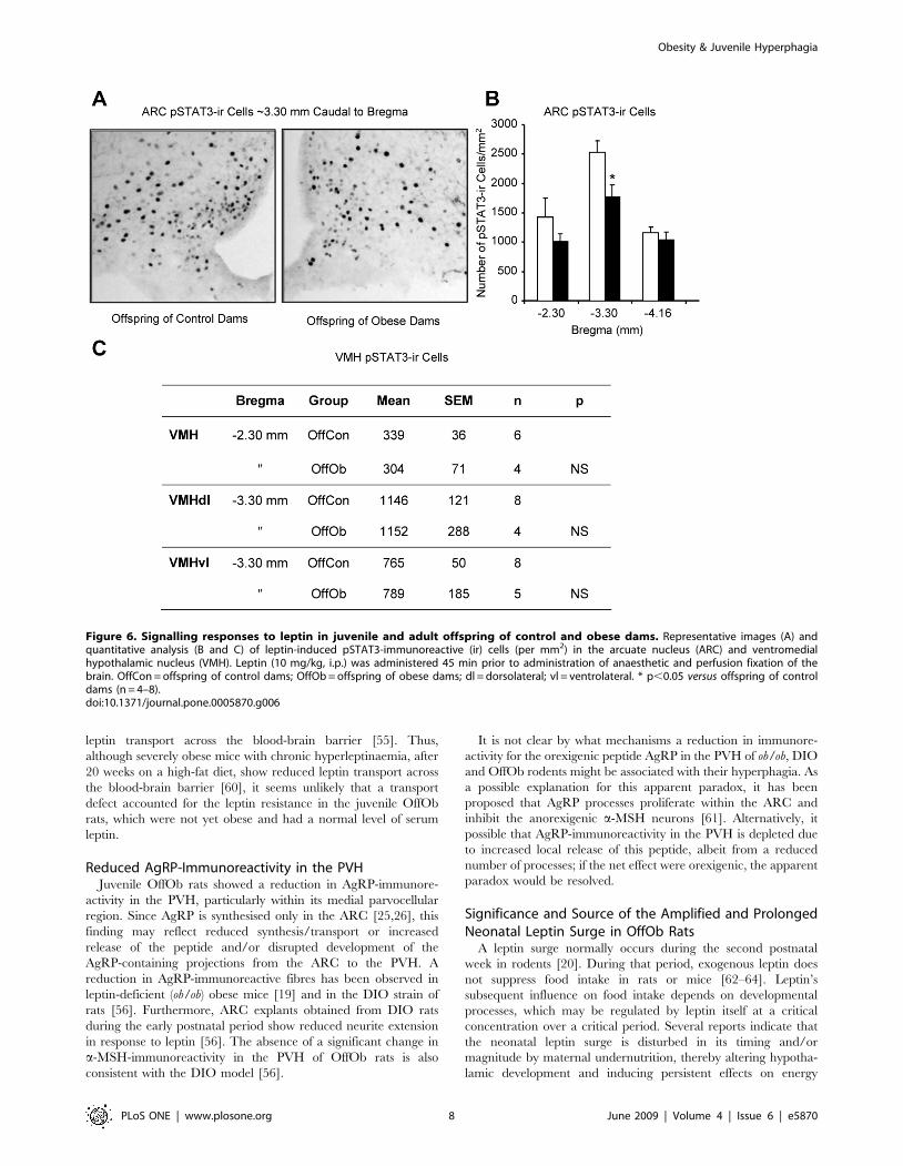
The processes underlying the behavioural evidence for leptin
resistance were investigated by determining the number of cells
immunoreactive for pSTAT3, which is induced following
activation of leptin receptors [24], in the ARC and ventromedial
hypothalamic nucleus (VMH) in response to exogenous leptin
(10 mg/kg, i.p.). In the ARC, at approximately 3.30 mm caudal to
bregma, OffOb rats displayed fewer pSTAT3-immunoreactive
neurons than OffCon rats (Fig. 6 A,B). No evidence of impaired
leptin-signalling was found in the VMH (Fig. 6G).
Reduced AgRP Projections to the PVH in Offspring ofObese Dams
The behavioural and cellular leptin resistance at Day 30 in
OffOb rats (Fig. 5 and 6) is preceded by an amplified and prolonged
leptin surge during the pre-weaning phase (Fig. 3A). We hypothesise
that this extended and abnormally high surge is responsible for
down-regulation of leptin-signalling in the neonatal period. Since
leptin has neurotrophic actions on the development of projections
from the ARC [19], such down-regulation may impair the normal
development of those projections. Neurons synthesising agouti-
related peptide (AgRP) are restricted to the ARC and contain the
orexigenic neuropeptide Y [25,26]; consequently all AgRP-
immunoreactivity detected in the PVH derives from neurones in
the ARC. A separate neuronal population in the ARC expresses the
precursor for a-melanocyte stimulating hormone (a-MSH) and
sends projections to the PVH [27]; it is not clear whether this is the
sole source of a-MSH in the PVH. The a-MSH precursor is also
expressed in a subpopulation of neurons within the nucleus of the
solitary tract, a nucleus with extensive projections to the PVH [28];
there is, however, doubt about whether those neurons project to the
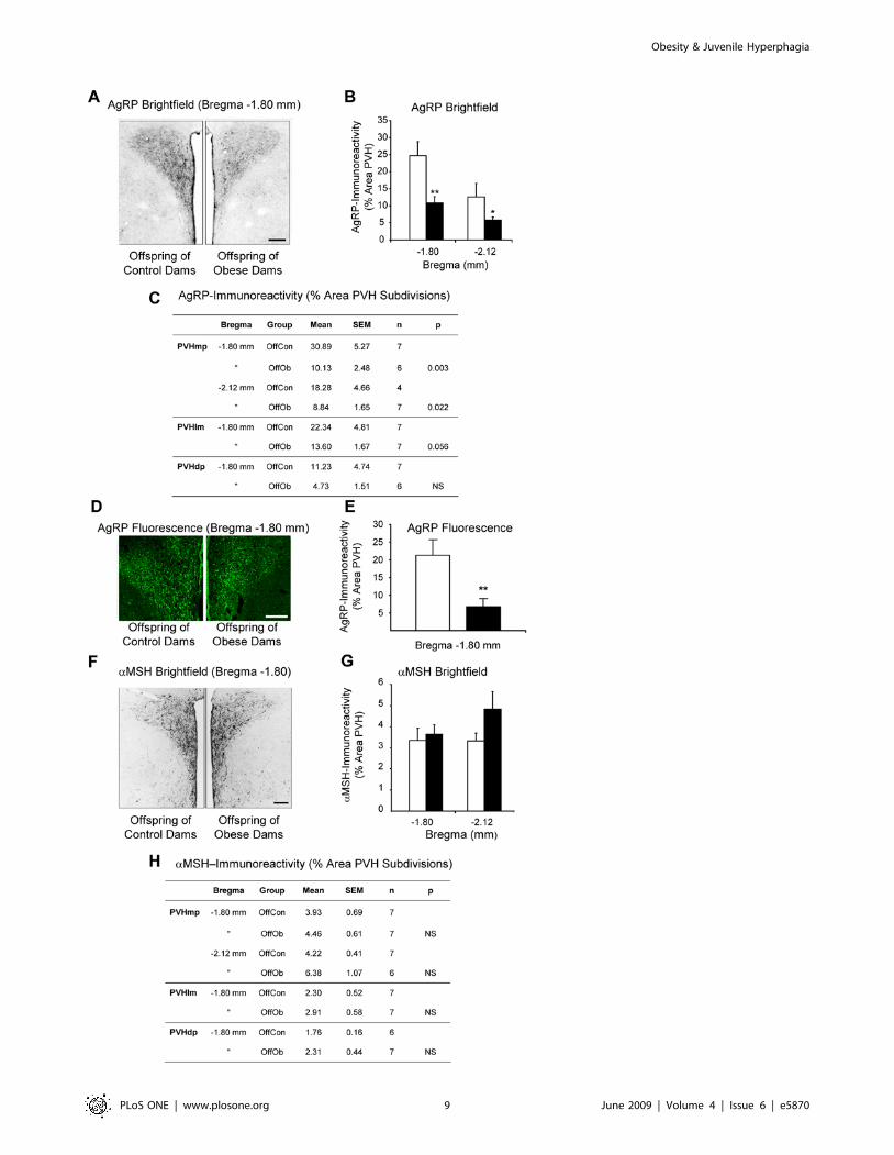
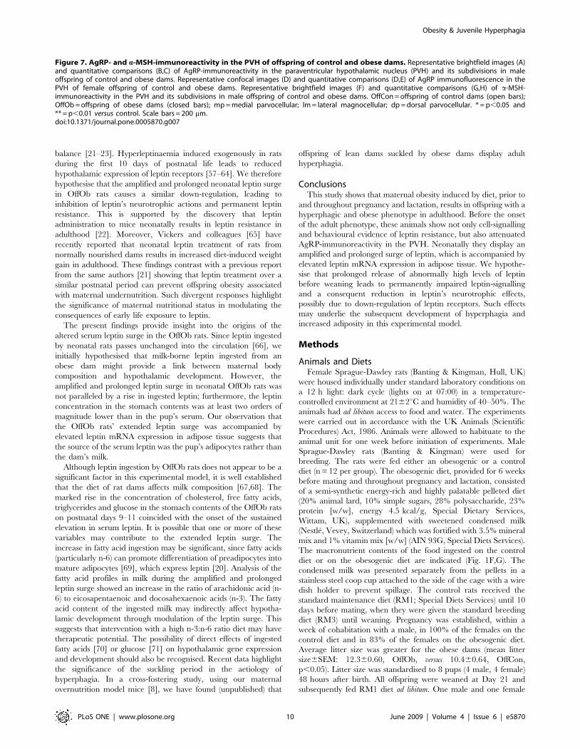
hypothalamus [29,30]. At postnatal Day 30, the density of AgRP-
immunoreactivity in the PVH (at approximately 1.80 mm and
2.12 mm caudal to bregma) was reduced in male and female OffOb
rats (Fig. 7A–E). On analysis of PVH subdivisions, a statistically
significant reduction in AgRP-immunoreactivity was reached only
within the medial parvocellular region of the PVH (Fig. 7C). In
contrast, no change in a-MSH-immunoreactivity was observed in
the PVH (Fig. 7F–H).
Discussion
The results of this study add to the evidence that nutritional
status early in life can modify energy homeostasis in later life [1–
4,14,31,32]. The ‘developmental origins of adult disease’ hypoth-
esis, which originally focused on the deleterious effects of maternal
and fetal undernutrition [31], now encompasses the theory that
maternal and fetal overnutrition is similarly disadvantageous [32–
44]. Recently we have demonstrated that the adult progeny of
mice or rats made obese by a high-fat/sugar diet become
hyperphagic and obese on a standard chow diet [8,9] furthermore,
they develop insulin resistance, hyperleptinaemia and hyperten-
sion [8]. The present findings in rats suggest that leptin resistance
Obesity & Juvenile Hyperphagia
PLoS ONE | www.plosone.org 3 June 2009 | Volume 4 | Issue 6 | e5870
Figure 2. Post-weaning characteristics of offspring of control and obese dams. Body weight for males (A) and females (B) and calorificintake for males (C) and females (D) were recorded post-weaning for offspring of control (open symbols) or obese (closed symbols) dams. Offspringbody and tissue weights were recorded at postnatal Day 90 (E). OffCon = offspring of control dams; OffOb = offspring of obese dams; WAT = whiteadipose tissue; BAT = brown adipose tissue; s.c. = subcutaneous. * p,0.05 and ** p,0.01 versus offspring of control dams (n = 8–11).doi:10.1371/journal.pone.0005870.g002
Obesity & Juvenile Hyperphagia
PLoS ONE | www.plosone.org 4 June 2009 | Volume 4 | Issue 6 | e5870
acquired by the animal early in postnatal life plays a critical role in
the aetiology of the phenotype induced by maternal overnutrition.
Central Leptin Resistance in Juvenile and Adult Offspringof Obese Rats
Leptin plays a key role in energy balance through inhibition of
orexigenic systems and stimulation of anorexigenic systems in the
ARC [45–48]. Leptin resistance is generally associated with a state
of chronic obesity in which hyperleptinaemia fails to maintain lean
body weight set points by inhibiting food intake [48–52]. In the
present study, juvenile OffOb rats (Day 30) showed a loss of the
inhibitory effects of leptin on food intake and body weight, at a
time at which serum leptin was not raised; this resistance to leptin
was still present in adulthood (Day 90), when the animals were
obese and hyperleptinaemic. Rats with a genetic susceptibility to
diet-induced obesity (DIO) also display leptin resistance prior to
development of the obese phenotype [53,54]. In the ARC, VMH
and DMH of pre-obese DIO rats there are reductions in the
expression of leptin receptor (LRb) mRNA, leptin binding and
leptin-signalling [53–55]. In OffOb rats, reduced phosphorylation
of STAT3 was restricted to the ARC. As also observed in DIO rats
[56], the OffOb animals showed an attenuation of AgRP-
immunoreactivity in the PVH which is derived from neurones
the ARC. In the DIO strain, the imposition of maternal
overnutrition during pregnancy failed to enhance the loss of
projections from the ARC to the PVH; thus, the genetic
background seems to prevail in that model [56].
Hyperleptinaemia induced exogenously in normal rats during
the first 10 days of postnatal life leads to a permanent loss of the
anorexic response to leptin [57,58]. The processes which lead to
central leptin resistance secondary to transient or chronic
hyperleptinaemia remain poorly understood, although signalling
molecules downstream of the leptin receptor are implicated [59].
A major finding of the present study provides cell-signalling and
behavioural evidence of developmentally induced leptin resistance
prior to the onset of chronic obesity and hyperleptinaemia. Bouret
and colleagues (2008) observed decreased leptin-signalling during
early postnatal development in the DIO model[56]. In comparison
with the diet-resistant animals, DIO rats showed a 24% reduction
in the number of ARC neurons with leptin-induced pSTAT3-
immunoreactivity on Day 10 [56]. The results of that study are in
accord with the present observation of reduced leptin-signalling in
the ARC of juvenile OffOb rats. Thus, impaired leptin-signalling
in pre-obese animals is a common feature of these genetically and
epigenetically ‘‘programmed’’ models.
Given that leptin was administered peripherally in the present
study, a transport defect may have contributed to the observed
leptin resistance in the OffOb animals. However, impaired leptin-
signalling is also observed in pre-obese DIO rats despite normal
Figure 3. Neonatal serum leptin concentrations and adipose leptin mRNA expression in offspring of control and obese dams. Serumleptin was measured in offspring of control dams (open bars) and obese dams (closed bars) on postnatal days 2, 7, 8, 9, 11, 13, 14, 15 and 18 (A).Leptin mRNA expression in abdominal fat is presented for postnatal days 2–8, 9–11 and 13–18 (B). * p,0.05, ** p,0.01 and *** p,0.01 versusoffspring of control dams for the same period (n = 3–6). For longitudinal comparisons, a significant difference (p,0.05) from the preceding period isindicated by # for offspring of control dams and by { for offspring of obese dams.doi:10.1371/journal.pone.0005870.g003
Obesity & Juvenile Hyperphagia
PLoS ONE | www.plosone.org 5 June 2009 | Volume 4 | Issue 6 | e5870
Figure 4. Constituents of milk from control and obese dams by analysis of pups’ stomach contents. The concentration of leptin (A),cholesterol (B), free fatty acids (FFA) (C), triglycerides (D) and glucose (E) was assayed in stomach contents (as a proxy measure of the dams’ milkcontent) from offspring of control dams (open bars; n = 4–8) and obese dams (closed bars; n = 4–8) throughout suckling. * p,0.05 and ** p,0.01versus offspring of control dams at the same period. For longitudinal comparisons, a significant difference (p,0.05) from the preceding period isindicated by # for offspring of control dams and by { for offspring of obese dams.doi:10.1371/journal.pone.0005870.g004
Obesity & Juvenile Hyperphagia
PLoS ONE | www.plosone.org 6 June 2009 | Volume 4 | Issue 6 | e5870

Figure 5. Behavioural responses to leptin in juvenile and adult offspring of control and obese dams. Food intake for males (A) andfemales (B) and change in body weight for males (C) and females (D) recorded over 24 hours following administration of leptin (10 mg/kg, i.p.) in 30day-old offspring of control or obese dams. Food intake for males (E) and females (F) and change in body weight for males (G) and females (H) in 90day-old offspring of control or obese dams. OffCon = offspring of control dams; OffOb = offspring of obese dams; * p,0.05 and ** p,0.01***p,0.001, versus offspring of control dams (n = 6).doi:10.1371/journal.pone.0005870.g005
Obesity & Juvenile Hyperphagia
PLoS ONE | www.plosone.org 7 June 2009 | Volume 4 | Issue 6 | e5870
leptin transport across the blood-brain barrier [55]. Thus,
although severely obese mice with chronic hyperleptinaemia, after
20 weeks on a high-fat diet, show reduced leptin transport across
the blood-brain barrier [60], it seems unlikely that a transport
defect accounted for the leptin resistance in the juvenile OffOb
rats, which were not yet obese and had a normal level of serum
leptin.
Reduced AgRP-Immunoreactivity in the PVHJuvenile OffOb rats showed a reduction in AgRP-immunore-
activity in the PVH, particularly within its medial parvocellular
region. Since AgRP is synthesised only in the ARC [25,26], this
finding may reflect reduced synthesis/transport or increased
release of the peptide and/or disrupted development of the
AgRP-containing projections from the ARC to the PVH. A
reduction in AgRP-immunoreactive fibres has been observed in
leptin-deficient (ob/ob) obese mice [19] and in the DIO strain of
rats [56]. Furthermore, ARC explants obtained from DIO rats
during the early postnatal period show reduced neurite extension
in response to leptin [56]. The absence of a significant change in
a-MSH-immunoreactivity in the PVH of OffOb rats is also
consistent with the DIO model [56].
It is not clear by what mechanisms a reduction in immunore-
activity for the orexigenic peptide AgRP in the PVH of ob/ob, DIO
and OffOb rodents might be associated with their hyperphagia. As
a possible explanation for this apparent paradox, it has been
proposed that AgRP processes proliferate within the ARC and
inhibit the anorexigenic a-MSH neurons [61]. Alternatively, it
possible that AgRP-immunoreactivity in the PVH is depleted due
to increased local release of this peptide, albeit from a reduced
number of processes; if the net effect were orexigenic, the apparent
paradox would be resolved.
Significance and Source of the Amplified and ProlongedNeonatal Leptin Surge in OffOb Rats
A leptin surge normally occurs during the second postnatal
week in rodents [20]. During that period, exogenous leptin does
not suppress food intake in rats or mice [62–64]. Leptin’s
subsequent influence on food intake depends on developmental
processes, which may be regulated by leptin itself at a critical
concentration over a critical period. Several reports indicate that
the neonatal leptin surge is disturbed in its timing and/or
magnitude by maternal undernutrition, thereby altering hypotha-
lamic development and inducing persistent effects on energy
Figure 6. Signalling responses to leptin in juvenile and adult offspring of control and obese dams. Representative images (A) andquantitative analysis (B and C) of leptin-induced pSTAT3-immunoreactive (ir) cells (per mm2) in the arcuate nucleus (ARC) and ventromedialhypothalamic nucleus (VMH). Leptin (10 mg/kg, i.p.) was administered 45 min prior to administration of anaesthetic and perfusion fixation of thebrain. OffCon = offspring of control dams; OffOb = offspring of obese dams; dl = dorsolateral; vl = ventrolateral. * p,0.05 versus offspring of controldams (n = 4–8).doi:10.1371/journal.pone.0005870.g006
Obesity & Juvenile Hyperphagia
PLoS ONE | www.plosone.org 8 June 2009 | Volume 4 | Issue 6 | e5870
Obesity & Juvenile Hyperphagia
PLoS ONE | www.plosone.org 9 June 2009 | Volume 4 | Issue 6 | e5870
balance [21–23]. Hyperleptinaemia induced exogenously in rats
during the first 10 days of postnatal life leads to reduced
hypothalamic expression of leptin receptors [57–64]. We therefore
hypothesise that the amplified and prolonged neonatal leptin surge
in OffOb rats causes a similar down-regulation, leading to
inhibition of leptin’s neurotrophic actions and permanent leptin
resistance. This is supported by the discovery that leptin
administration to mice neonatally results in leptin resistance in
adulthood [22]. Moreover, Vickers and colleagues [65] have
recently reported that neonatal leptin treatment of rats from
normally nourished dams results in increased diet-induced weight
gain in adulthood. These findings contrast with a previous report
from the same authors [21] showing that leptin treatment over a
similar postnatal period can prevent offspring obesity associated
with maternal undernutrition. Such divergent responses highlight
the significance of maternal nutritional status in modulating the
consequences of early life exposure to leptin.
The present findings provide insight into the origins of the
altered serum leptin surge in the OffOb rats. Since leptin ingested
by neonatal rats passes unchanged into the circulation [66], we
initially hypothesised that milk-borne leptin ingested from an
obese dam might provide a link between maternal body
composition and hypothalamic development. However, the
amplified and prolonged leptin surge in neonatal OffOb rats was
not paralleled by a rise in ingested leptin; furthermore, the leptin
concentration in the stomach contents was at least two orders of
magnitude lower than in the pup’s serum. Our observation that
the OffOb rats’ extended leptin surge was accompanied by
elevated leptin mRNA expression in adipose tissue suggests that
the source of the serum leptin was the pup’s adipocytes rather than
the dam’s milk.
Although leptin ingestion by OffOb rats does not appear to be a
significant factor in this experimental model, it is well established
that the diet of rat dams affects milk composition [67,68]. The
marked rise in the concentration of cholesterol, free fatty acids,
triglycerides and glucose in the stomach contents of the OffOb rats
on postnatal days 9–11 coincided with the onset of the sustained
elevation in serum leptin. It is possible that one or more of these
variables may contribute to the extended leptin surge. The
increase in fatty acid ingestion may be significant, since fatty acids
(particularly n-6) can promote differentiation of preadipocytes into
mature adipocytes [69], which express leptin [20]. Analysis of the
fatty acid profiles in milk during the amplified and prolonged
leptin surge showed an increase in the ratio of arachidonic acid (n-
6) to eicosapentaenoic and docosahexaenoic acids (n-3). The fatty
acid content of the ingested milk may indirectly affect hypotha-
lamic development through modulation of the leptin surge. This
suggests that intervention with a high n-3:n-6 ratio diet may have
therapeutic potential. The possibility of direct effects of ingested
fatty acids [70] or glucose [71] on hypothalamic gene expression
and development should also be recognised. Recent data highlight
the significance of the suckling period in the aetiology of
hyperphagia. In a cross-fostering study, using our maternal
overnutrition model mice [8], we have found (unpublished) that
offspring of lean dams suckled by obese dams display adult
hyperphagia.
ConclusionsThis study shows that maternal obesity induced by diet, prior to
and throughout pregnancy and lactation, results in offspring with a
hyperphagic and obese phenotype in adulthood. Before the onset
of the adult phenotype, these animals show not only cell-signalling
and behavioural evidence of leptin resistance, but also attenuated
AgRP-immunoreactivity in the PVH. Neonatally they display an
amplified and prolonged surge of leptin, which is accompanied by
elevated leptin mRNA expression in adipose tissue. We hypothe-
sise that prolonged release of abnormally high levels of leptin
before weaning leads to permanently impaired leptin-signalling
and a consequent reduction in leptin’s neurotrophic effects,
possibly due to down-regulation of leptin receptors. Such effects
may underlie the subsequent development of hyperphagia and
increased adiposity in this experimental model.
Methods
Animals and DietsFemale Sprague-Dawley rats (Banting & Kingman, Hull, UK)
were housed individually under standard laboratory conditions on
a 12 h light: dark cycle (lights on at 07:00) in a temperature-
controlled environment at 2162uC and humidity of 40–50%. The
animals had ad libitum access to food and water. The experiments
were carried out in accordance with the UK Animals (Scientific
Procedures) Act, 1986. Animals were allowed to habituate to the
animal unit for one week before initiation of experiments. Male
Sprague-Dawley rats (Banting & Kingman) were used for
breeding. The rats were fed either an obesogenic or a control
diet (n = 12 per group). The obesogenic diet, provided for 6 weeks
before mating and throughout pregnancy and lactation, consisted
of a semi-synthetic energy-rich and highly palatable pelleted diet
(20% animal lard, 10% simple sugars, 28% polysaccharide, 23%
protein [w/w], energy 4.5 kcal/g, Special Dietary Services,
Wittam, UK), supplemented with sweetened condensed milk
(Nestle, Vevey, Switzerland) which was fortified with 3.5% mineral
mix and 1% vitamin mix [w/w] (AIN 93G, Special Diets Services).
The macronutrient contents of the food ingested on the control
diet or on the obesogenic diet are indicated (Fig. 1F,G). The
condensed milk was presented separately from the pellets in a
stainless steel coop cup attached to the side of the cage with a wire
dish holder to prevent spillage. The control rats received the
standard maintenance diet (RM1; Special Diets Services) until 10
days before mating, when they were given the standard breeding
diet (RM3) until weaning. Pregnancy was established, within a
week of cohabitation with a male, in 100% of the females on the
control diet and in 83% of the females on the obesogenic diet.
Average litter size was greater for the obese dams (mean litter
size6SEM: 12.360.60, OffOb, versus 10.460.64, OffCon,
p,0.05). Litter size was standardised to 8 pups (4 male, 4 female)
48 hours after birth. All offspring were weaned at Day 21 and
subsequently fed RM1 diet ad libitum. One male and one female
Figure 7. AgRP- and a-MSH-immunoreactivity in the PVH of offspring of control and obese dams. Representative brightfield images (A)and quantitative comparisons (B,C) of AgRP-immunoreactivity in the paraventricular hypothalamic nucleus (PVH) and its subdivisions in maleoffspring of control and obese dams. Representative confocal images (D) and quantitative comparisons (D,E) of AgRP immunofluorescence in thePVH of female offspring of control and obese dams. Representative brightfield images (F) and quantitative comparisons (G,H) of a-MSH-immunoreactivity in the PVH and its subdivisions in male offspring of control and obese dams. OffCon = offspring of control dams (open bars);OffOb = offspring of obese dams (closed bars); mp = medial parvocellular; lm = lateral magnocellular; dp = dorsal parvocellular. * = p,0.05 and** = p,0.01 versus control. Scale bars = 200 mm.doi:10.1371/journal.pone.0005870.g007
Obesity & Juvenile Hyperphagia
PLoS ONE | www.plosone.org 10 June 2009 | Volume 4 | Issue 6 | e5870

from each litter were then sacrificed for blood and tissue collection
at Day 30 and Day 90; remaining littermates were used in other
studies. Organs weights were recorded and serum stored at 280uCfor future analysis. After weaning of their pups, dams were
sacrificed following an overnight fast; blood was collected and
serum stored at 280uC. Animals tested for anorexic responses to
leptin at Day 30 were fasted during the preceding night; the leptin
was administered at 10.00 h.
In a separate cohort of animals, litters of control and obese
dams were killed at several postnatal stages from Day 2 to Day 18.
At each time-point (Days 2, 7, 8, 9, 11, 13, 14, 15 and 18), litters
were killed by decapitation between 0800 and 1100 h. Blood
samples were collected (trunk blood) and abdominal fat pads and
stomach content collected and stored at 280uC until analyzed.
Assessing Anorexic Responses to a Leptin challenge inYoung Offspring of Obese Dams
At 30 or 90 days of age, the rats were tested for food intake in
response to either leptin or saline after being fasted for 18 hours.
Recombinant rat leptin (PeproTech, Inc., Rocky hill, NJ, USA)
was dissolved in saline vehicle (0.9% w/v) and given as a bolus
injection at the dose of 10 mg/kg body weight i.p. After the
intraperitoneal treatment, the animals were housed singly, and
food intake was measured over the 24 hour post-challenge period.
Change in body weight was also recorded over the same period (Dweight in grams).
Biochemical AssaysNeonatal stomach contents were used as an indirect measure-
ment of the milk contents. Stomach contents were extracted in an
equal volume of water, employing an ultrasonicator and
centrifuged for 15 min. The supernatant was then used for
glucose and leptin analysis. Remaining sample was further
extracted in ethanol, shaken and centrifuged for 15 min. The
concentrations of leptin (in stomach contents and serum) were
measured by ELISA (RD291001200 kit; Biovendor, Modrice,
Czech Republic); glucose, triglycerides, cholesterol and free fatty
acids were determined by an autoanalyzer (Hitachi 912, Roche
Diagnostics, Almere, The Netherlands) using commercial kits
(Gluco-Quant/HK, Triglycerides-GPO-PAP, Cholesterol-
CHOD-PAP from Roche Diagnostics, Brussels, Belgium; NEFA-
C from Wako Chemicals, Neuss, Germany).
Real-time PCRTotal RNA was extracted from abdominal fat from neonatal
rats on Days 2, 7, 8, 9, 11, 13, 14, 15 and 18 by standard Trizol
(Sigma-Aldrich Ltd., Poole, UK) method. RNA quantity and
integrity were assessed by optical density using a Nanodrop-1000
spectrophotometer (NanoDrop Products, Wilmington, USA).
Reverse transcription was carried out from 1 mg of RNA sample
with QuantiTect Reverse Transcription Kit (cat. no. 205311;
Qiagen, Crawley, UK) according to instructions; cDNA was stored
at 280uC. Intron-spanning primers for leptin (accession number
NM_013076) for real-time PCR were designed using Universal
Probelibrary (Roche Diagnostics Ltd., Burgess Hill, UK); primers
(F: 59-CCA GGA TCA ATG ACA TTT CAC-39 and R: 59-AAT
GAA GTC CAA ACC GGT GA-39) were obtained from Operon
Biotechnologies GmbH (Cologne, Germany). Two microlitres of
cDNA was used in 10 ml amplification reactions, containing 5 ml
SYBR green fluorescent dye (QuantiFast SYBR Green PCR Kit;
Qiagen Crawley, UK), 0.5 ml 10 mM forward and reverse primers
and 2 ml RNA-ase free water, with the following cycling
conditions: initial activation for 5 minutes at 95uC, followed by
40 cycles of denaturation at 95uC for 10 seconds and combined
annealing/extension at 60uC for 30 seconds. Sample copy
numbers for leptin, 28S and b-actin were determined by standard
curves (leptin, R2 = 0.997; 28S R2 = 0.996; b-actin R2 = 0.94) and
used to calculate the concentrations of leptin mRNA relative to the
geometric mean of 28S and b-actin mRNAs, with Rotorgene 6000
series software (Corbett Research, Mortlake, Australia).
ImmunohistochemistryOn postnatal Day 30, one male rat from each litter was given
saline or leptin 45 minutes before anaesthesia (Pentoject, 50 mg/
kg, i.p.; Animalcare Ltd., York, UK) and transcardiac perfusion
with 4% paraformaldehyde. Coronal cryostat sections (30 mm)
containing the ARC and VMH were processed for pSTAT3
immunohistochemistry according to a method modified from
Levin and colleagues [55]. Floating sections were immersed in
phosphate buffered saline (PBS) containing 1% NaOH and 1%
hydrogen peroxide for 20 minutes and then in PBS containing
0.3% glycine for 10 minutes, followed by PBS containing 0.15%
SDS for 10 minutes. After incubation in 2% donkey serum
containing 0.4% Triton X-100 for 2 hours, the sections were
immersed in monoclonal rabbit anti-pSTAT3 (9145L, 1:2000;
Cell Signaling Technologies Inc., Boston, MA, USA) for 7 days at
4uC. The sections were then incubated for 1 hour in biotinylated
donkey anti-rabbit IgG (1:1000; Stratech Scientific Ltd., New-
market, UK) and subsequently in avidin-biotin-peroxidase com-
plex (ABC, 1:1000; Vector Laboratories, Peterborough, UK) for
1 hour. Immunoreactivity was detected by incubation in Tris
buffer (pH 7.6) containing 0.05% 3,39-diaminobenzidine-4HCl
(DAB), 0.15% nickel ammonium sulphate (NAS) and 0.005%
H2O2.
Additional untreated male and female OffCon and OffOb rats
were perfused transcardially with 4% PFA on postnatal Day 30 to
determine AgRP- and a-MSH-immunoreactivity in the PVH.
Coronal cryostat sections (30 mm) were treated with 0.5% Triton
X-100 for 30 minutes, 0.5% hydrogen peroxide for 15 minutes
and 2% normal donkey serum for 1 hour. For brightfield
immunohistochemistry, sections were then incubated for 4 days
at 4uC in rabbit anti-AgRP (H-003-57, 1:20,000; Phoenix Europe
GmbH, Karlsruhe, Germany) or sheep anti-a-MSH (1:50,000,
AB5087; Millipore, Livingstone, UK). Following a 2-hour
incubation in biotinylated donkey anti-rabbit IgG (1:800; Stratech
Scientific Ltd.) or biotinylated donkey anti-sheep IgG (1:1000;
Stratech Scientific Ltd.), sections were incubated in ABC (1:2000;
Vector Laboratories) for 90 minutes. Immunoreactivity was
detected as for pSTAT3. For confocal immunohistochemistry,
sections were incubated in rabbit anti-AgRP (H-003-57, 1:4,000;
Phoenix Europe GmbH) for 4 days at 4uC. They were
subsequently incubated in biotinylated donkey anti-rabbit IgG
(1:1000; Stratech Scientific Ltd.) for 2 hours, followed by ABC
(1:3000; Vector Laboratories) for 90 minutes, before being
exposed to biotinylated tyramide (1:500; PerkinElmer Laborato-
ries, Waltham, USA) for 30 minutes in the presence of 0.005%
H2O2. The sections were then immersed in AlexaFluor 488-
conjugated streptavidin (1:500; Invitrogen Ltd., Paisley, UK) for
24 hours at 4uC. For these sections, coverslips were applied over
an anti-fade reagent (Prolong; Invitrogen Ltd.).
Image analysisFollowing immunohistochemical visualisation of pSTAT3,
AgRP or a-MSH, computerised image analysis, blind to
treatment, was used to determine the number of pSTAT3-
immunoreactive cells within the ARC and VMH and the density
of AgRP- and a-MSH- immunoreactivity within the PVH.
Obesity & Juvenile Hyperphagia
PLoS ONE | www.plosone.org 11 June 2009 | Volume 4 | Issue 6 | e5870
pSTAT3-immunoreactive cells in the ARC and VMH were
counted (MCID software, version 7.0; Interfocus Imaging Ltd.,
Cambridge, UK) and expressed as cells/mm2. The mean number
of pSTAT3-immunoreactive cells on each side in 3–6 sections
from each rat, within approximately 60.20 mm of the rostro-
caudal level cited [72] was used for statistical analysis. The
percentage of the area containing immunoreactivity for AgRP or
a-MSH within the whole PVH or its subdivisions was quantified
(MCID software, version 7.0); thresholding was optimised blind to
treatment for each animal. For brightfield immunoreactivity, the
mean immunoreactive density on each side in 3 sections from each
rat, within approximately 60.10 mm of the rostro-caudal level
cited [72], was used for statistical analysis. Analysis of AgRP-
immunofluorescence within the PVH was undertaken following
collection of confocal images at 1 mm intervals (10–15 optical
sections) through each section. For immunofluorescence, the mean
immunoreactive density on each side in 2 sections from each rat,
within approximately 60.10 mm of the rostro-caudal level cited
[72], was used for statistical analysis.
Analysis of fatty acidsFatty acid composition of milk extracted from pup stomach
contents was analyzed by gas-liquid chromatography, using a
modified version of the direct one step transesterification method
by Lepage & Roy (1986)[73].
Statistical analysisData are expressed as mean6SEM, unless stated, and
compared using 2-way ANOVA followed by a post hoc Dunnett’s
test, or Student’s t test. p#0.05 was regarded as significant.
Acknowledgments
The authors would like to thank Dr Simon Wheeler for assistance in the
analysis of milk fatty acids.
Author Contributions
Conceived and designed the experiments: TK LP PDT CWC. Performed
the experiments: SLK AMS MA HD TK. Analyzed the data: SLK AMS
MA PDT. Wrote the paper: SLK PDT CWC.
References
1. Catalano PM (2003) Obesity and pregnancy–the propagation of a viscous cycle?
J Clin Endocrinol Metab 88: 3505–3506.
2. Catalano PM, Kirwan JP, Haugel-de Mouzon S, King J (2003) Gestational
diabetes and insulin resistance: role in short- and long-term implications formother and fetus. J Nutr 133: 1674S–1683S.
3. Catalano PM, Thomas A, Huston-Presley L, Amini SB (2003) Increased fetal
adiposity: a very sensitive marker of abnormal in utero development.
AmJObstetGynecol 189: 1698–1704.
4. Oken E, Gillman MW (2003) Fetal origins of obesity. Obes Res 11: 496–506.
5. Harvey NC, Poole JR, Javaid MK, Dennison EM, Robinson S, et al. (2007)Parental determinants of neonatal body composition. J Clin Endocrinol Metab
92: 523–526.
6. Lawlor DA, Smith GD, O’Callaghan M, Alati R, Mamun AA, et al. (2007)
Epidemiologic evidence for the fetal overnutrition hypothesis: findings from themater-university study of pregnancy and its outcomes. Am J Epidemiol 165:
418–424.
7. Oken E (2008) Excess Gestational Weight Gain Amplifies Risks Among Obese
Mothers. Epidemiology.
8. Samuelsson AM, Matthews PA, Argenton M, Christie MR, McConnell JM, etal. (2008) Diet-induced obesity in female mice leads to offspring hyperphagia,
adiposity, hypertension, and insulin resistance: a novel murine model ofdevelopmental programming. Hypertension 51: 383–392.
9. Nivoit P, Morens C, Van Assche FA, Jansen E, Poston L, et al. (2009)Established diet-induced obesity in female rats leads to offspring hyperphagia,
adiposity and insulin resistance. Diabetologia 52: 1133–1142.
10. Plagemann A, Harder T, Rake A, Melchior K, Rohde W, et al. (1999) Increasednumber of galanin-neurons in the paraventricular hypothalamic nucleus of
neonatally overfed weanling rats. Brain Res 818: 160–163.
11. Schmidt I, Fritz A, Scholch C, Schneider D, Simon E, et al. (2001) The effect of
leptin treatment on the development of obesity in overfed suckling Wistar rats.Int J Obes Relat Metab Disord 25: 1168–1174.
12. Plagemann A (2005) Perinatal programming and functional teratogenesis:
Impact on body weight regulation and obesity. Physiol Behav 86: 661–668.
13. Cripps RL, Martin-Gronert MS, Ozanne SE (2005) Fetal and perinatal
programming of appetite. Clin Sci (Lond) 109: 1–11.
14. McMillen IC, Robinson JS (2005) Developmental origins of the metabolicsyndrome: prediction, plasticity, and programming. Physiol Rev 85: 571–633.
15. Muhlhausler BS, Adam CL, Marrocco EM, Findlay PA, Roberts CT, et al.(2005) Impact of glucose infusion on the structural and functional characteristics
of adipose tissue and on hypothalamic gene expression for appetite regulatoryneuropeptides in the sheep fetus during late gestation. J Physiol 565: 185–195.
16. Grove KL, Grayson BE, Glavas MM, Xiao XQ, Smith MS (2005) Development
of metabolic systems. Physiol Behav 86: 646–660.
17. Elmquist JK, Flier JS (2004) Neuroscience. The fat-brain axis enters a new
dimension. Science 304: 63–64.
18. Ferezou-Viala J, Roy AF, Serougne C, Gripois D, Parquet M, et al. (2007) Long-term consequences of maternal high-fat feeding on hypothalamic leptin
sensitivity and diet-induced obesity in the offspring. Am J Physiol Regul Integr
Comp Physiol 293: R1056–1062.
19. Bouret SG, Draper SJ, Simerly RB (2004) Trophic action of leptin onhypothalamic neurons that regulate feeding. Science 304: 108–110.
20. Ahima RS, Prabakaran D, Flier JS (1998) Postnatal leptin surge and regulation
of circadian rhythm of leptin by feeding. Implications for energy homeostasis
and neuroendocrine function. J Clin Invest 101: 1020–1027.
21. Vickers MH, Gluckman PD, Coveny AH, Hofman PL, Cutfield WS, et al.(2005) Neonatal leptin treatment reverses developmental programming.
Endocrinology 146: 4211–4216.
22. Yura S, Itoh H, Sagawa N, Yamamoto H, Masuzaki H, et al. (2005) Role of
premature leptin surge in obesity resulting from intrauterine undernutrition. CellMetab 1: 371–378.
23. Delahaye F, Breton C, Risold PY, Enache M, Dutriez-Casteloot I, et al. (2008)
Maternal perinatal undernutrition drastically reduces postnatal leptin surge andaffects the development of arcuate nucleus proopiomelanocortin neurons in
neonatal male rat pups. Endocrinology 149: 470–475.
24. Vaisse C, Halaas JL, Horvath CM, Darnell JE Jr, Stoffel M, et al. (1996) Leptin
activation of Stat3 in the hypothalamus of wild-type and ob/ob mice but not db/db mice. Nat Genet 14: 95–97.
25. Broberger C, Johansen J, Johansson C, Schalling M, Hokfelt T (1998) Theneuropeptide Y/agouti gene-related protein (AGRP) brain circuitry in normal,
anorectic, and monosodium glutamate-treated mice. Proc Natl Acad Sci U S A
95: 15043–15048.
26. Grove KL, Smith MS (2003) Ontogeny of the hypothalamic neuropeptide Y
system. Physiol Behav 79: 47–63.
27. Baker RA, Herkenham M (1995) Arcuate nucleus neurons that project to the
hypothalamic paraventricular nucleus: neuropeptidergic identity and consequenc-es of adrenalectomy on mRNA levels in the rat. J Comp Neurol 358: 518–530.
28. Sawchenko PE, Swanson LW (1983) The organization of forebrain afferents to theparaventricular and supraoptic nuclei of the rat. J Comp Neurol 218: 121–144.
29. Pilcher WH, Joseph SA (1986) Differential sensitivity of hypothalamic andmedullary opiocortin and tyrosine hydroxylase neurons to the neurotoxic effects
of monosodium glutamate (MSG). Peptides 7: 783–789.
30. Joseph SA, Michael GJ (1988) Efferent ACTH-IR opiocortin projections from
nucleus tractus solitarius: a hypothalamic deafferentation study. Peptides 9:
193–201.
31. Barker DJ (1994) The fetal origins of adult disease. Fetal and maternal medicine
review 6: 71–80.
32. Taylor PD, Poston L (2007) Developmental programming of obesity in
mammals. Exp Physiol 92: 287–298.
33. Armitage JA, Taylor PD, Poston L (2005) Experimental models of develop-
mental programming; Consequences of exposure to an energy rich diet duringdevelopment. J Physiol.
34. Guo F, Jen KL (1995) High-fat feeding during pregnancy and lactation affectsoffspring metabolism in rats. Physiol Behav 57: 681–686.
35. Koukkou E, Ghosh P, Lowy C, Poston L (1998) Offspring of normal anddiabetic rats fed saturated fat in pregnancy demonstrate vascular dysfunction.
Circulation 98: 2899–2904.
36. Ghebremeskel K, Bitsanis D, Koukkou E, Lowy C, Poston L, et al. (1999)
Maternal diet high in fat reduces docosahexaenoic acid in liver lipids of newborn
and sucking rat pups. Br J Nutr 81: 395–404.
37. Khan IY, Dekou V, Douglas G, Jensen R, Hanson MA, et al. (2005) A high-fat
diet during rat pregnancy or suckling induces cardiovascular dysfunction in adultoffspring. Am J Physiol Regul Integr Comp Physiol 288: R127–133.
38. Khan IY, Dekou V, Hanson M, Poston L, Taylor PD (2004) Predictive adaptiveresponses to maternal high fat diet prevent endothelial dysfunction but not
hypertension in adult rat offspring. Circulation 110: 1097–1102.
39. Khan IY, Taylor PD, Dekou V, Seed PT, Lakasing L, et al. (2003) Gender-
linked hypertension in offspring of lard-fed pregnant rats. Hypertension 41:168–175.
Obesity & Juvenile Hyperphagia
PLoS ONE | www.plosone.org 12 June 2009 | Volume 4 | Issue 6 | e5870

40. Taylor PD, Khan IY, Hanson MA, Poston L (2004) Impaired EDHF-mediated
vasodilatation in adult offspring of rats exposed to a fat-rich diet in pregnancy.J Physiol 558: 943–951.
41. Taylor PD, McConnell J, Khan IY, Holemans K, Lawrence KM, et al. (2005)
Impaired glucose homeostasis and mitochondrial abnormalities in offspring ofrats fed a fat-rich diet in pregnancy. Am J Physiol Regul Integr Comp Physiol
288: R134–139.42. Korotkova M, Gabrielsson BG, Holmang A, Larsson BM, Hanson LA, et al.
(2005) Gender-related long-term effects in adult rats by perinatal dietary ratio of
n-6/n-3 fatty acids. Am J Physiol Regul Integr Comp Physiol 288: R575–579.43. Zhang J, Wang C, Terroni PL, Cagampang FR, Hanson M, et al. (2005) High-
unsaturated-fat, high-protein, and low-carbohydrate diet during pregnancy andlactation modulates hepatic lipid metabolism in female adult offspring.
Am J Physiol Regul Integr Comp Physiol 288: R112–118.44. Srinivasan M, Aalinkeel R, Song F, Mitrani P, Pandya JD, et al. (2006) Maternal
hyperinsulinemia predisposes rat fetuses for hyperinsulinemia, and adult-onset
obesity and maternal mild food restriction reverses this phenotype. Am J PhysiolEndocrinol Metab 290: E129–E134.
45. Elias CF, Aschkenasi C, Lee C, Kelly J, Ahima RS, et al. (1999) Leptindifferentially regulates NPY and POMC neurons projecting to the lateral
hypothalamic area. Neuron 23: 775–786.
46. Cowley MA, Smart JL, Rubinstein M, Cerdan MG, Diano S, et al. (2001) Leptinactivates anorexigenic POMC neurons through a neural network in the arcuate
nucleus. Nature 411: 480–484.47. Pinto S, Roseberry AG, Liu H, Diano S, Shanabrough M, et al. (2004) Rapid
rewiring of arcuate nucleus feeding circuits by leptin. Science 304: 110–115.48. Wynne K, Stanley S, McGowan B, Bloom S (2005) Appetite control.
J Endocrinol 184: 291–318.
49. Neary NM, Goldstone AP, Bloom SR (2004) Appetite regulation: from the gut tothe hypothalamus. Clin Endocrinol (Oxf) 60: 153–160.
50. Levin BE, Keesey RE (1998) Defense of differing body weight set points in diet-induced obese and resistant rats. Am J Physiol 274: R412–419.
51. Keesey RE, Hirvonen MD (1997) Body weight set-points: determination and
adjustment. J Nutr 127: 1875S–1883S.52. Plagemann A, Harder T (2008) Hormonal programming in perinatal life: leptin
and beyond. Br J Nutr. pp 1–2.53. Levin BE, Dunn-Meynell AA, Ricci MR, Cummings DE (2003) Abnormalities
of leptin and ghrelin regulation in obesity-prone juvenile rats. Am J PhysiolEndocrinol Metab 285: E949–957.
54. Irani BG, Dunn-Meynell AA, Levin BE (2007) Altered hypothalamic leptin,
insulin, and melanocortin binding associated with moderate-fat diet andpredisposition to obesity. Endocrinology 148: 310–316.
55. Levin BE, Dunn-Meynell AA, Banks WA (2004) Obesity-prone rats have normalblood-brain barrier transport but defective central leptin signaling before obesity
onset. Am J Physiol Regul Integr Comp Physiol 286: R143–150.
56. Bouret SG, Gorski JN, Patterson CM, Chen S, Levin BE, et al. (2008)Hypothalamic neural projections are permanently disrupted in diet-induced
obese rats. Cell Metab 7: 179–185.
57. Toste FP, de Moura EG, Lisboa PC, Fagundes AT, de Oliveira E, et al. (2006)
Neonatal leptin treatment programmes leptin hypothalamic resistance andintermediary metabolic parameters in adult rats. Br J Nutr 95: 830–837.
58. Passos MC, Toste FP, Dutra SC, Trotta PA, Lisboa PC, et al. (2009) Role of
neonatal hyperleptinaemia on serum adiponectin and suppressor of cytokinesignalling-3 expression in young rats. Br J Nutr 101: 250–256.
59. Howard JK, Flier JS (2006) Attenuation of leptin and insulin signaling by SOCSproteins. Trends Endocrinol Metab 17: 365–371.
60. Morgan DA, Thedens DR, Weiss R, Rahmouni K (2008) Mechanisms
mediating renal sympathetic activation to leptin in obesity. Am J Physiol RegulIntegr Comp Physiol 295: R1730–1736.
61. Horvath TL, Bruning JC (2006) Developmental programming of thehypothalamus: a matter of fat. Nat Med 12: 52–53.
62. Mistry AM, Swick A, Romsos DR (1999) Leptin alters metabolic rates beforeacquisition of its anorectic effect in developing neonatal mice. Am J Physiol 277:
R742–747.
63. Ahima RS, Hileman SM (2000) Postnatal regulation of hypothalamicneuropeptide expression by leptin: implications for energy balance and body
weight regulation. Regul Pept 92: 1–7.64. Proulx K, Richard D, Walker CD (2002) Leptin regulates appetite-related
neuropeptides in the hypothalamus of developing rats without affecting food
intake. Endocrinology 143: 4683–4692.65. Vickers MH, Gluckman PD, Coveny AH, Hofman PL, Cutfield WS, et al.
(2008) The effect of neonatal leptin treatment on postnatal weight gain in malerats is dependent on maternal nutritional status during pregnancy. Endocrinol-
ogy 149: 1906–1913.66. Casabiell X, Pineiro V, Tome MA, Peino R, Dieguez C, et al. (1997) Presence of
leptin in colostrum and/or breast milk from lactating mothers: a potential role in
the regulation of neonatal food intake. J Clin Endocrinol Metab 82: 4270–4273.67. Del Prado M, Delgado G, Villalpando S (1997) Maternal lipid intake during
pregnancy and lactation alters milk composition and production and littergrowth in rats. J Nutr 127: 458–462.
68. Green MH, Dohner EL, Green JB (1981) Influence of dietary fat and cholesterol
on milk lipids and on cholesterol metabolism in the rat. J Nutr 111: 276–286.69. Ailhaud G, Massiera F, Weill P, Legrand P, Alessandri JM, et al. (2006)
Temporal changes in dietary fats: role of n-6 polyunsaturated fatty acids inexcessive adipose tissue development and relationship to obesity. Prog Lipid Res
45: 203–236.70. Morgan K, Obici S, Rossetti L (2004) Hypothalamic responses to long-chain
fatty acids are nutritionally regulated. J Biol Chem 279: 31139–31148.
71. Cai F, Gyulkhandanyan AV, Wheeler MB, Belsham DD (2007) Glucoseregulates AMP-activated protein kinase activity and gene expression in clonal,
hypothalamic neurons expressing proopiomelanocortin: additive effects of leptinor insulin. J Endocrinol 192: 605–614.
72. Paxinos G, Watson C (1989) The rat brain in stereotaxic coordinates Academic
Press.73. Lepage G, Roy CC (1986) Direct transesterification of all classes of lipids in a
one-step reaction. J Lipid Res 27: 114–120.
Obesity & Juvenile Hyperphagia
PLoS ONE | www.plosone.org 13 June 2009 | Volume 4 | Issue 6 | e5870
Related Documents