Moderate Exercise during Pregnancy in Wistar Rats Alters Bone and Body Composition of the Adult Offspring in a Sex-Dependent Manner Brielle V. Rosa 1* , Hugh T. Blair 1 , Mark H. Vickers 2 , Keren E. Dittmer 3 , Patrick C. H. Morel 3 , Cameron G. Knight 4 , Elwyn C. Firth 1 1 GRAVIDA: National Centre for Growth and Development, Institute of Veterinary, Animal and Biomedical Sciences, Massey University, Palmerston North, New Zealand, 2 GRAVIDA: National Centre for Growth and Development, The Liggins Institute, The University of Auckland, Auckland, New Zealand, 3 Institute of Veterinary, Animal and Biomedical Sciences, Massey University, Palmerston North, New Zealand, 4 Department of Ecosystem and Public Health, Faculty of Veterinary Medicine, University of Calgary, Calgary, Canada Abstract Exercise during pregnancy may have long-lasting effects on offspring health. Musculoskeletal growth and development, metabolism, and later-life disease risk can all be impacted by the maternal environment during pregnancy. The skeleton influences glucose handling through the actions of the bone-derived hormone osteocalcin. The purpose of this study was to test the effects of moderate maternal exercise during pregnancy on the bone and body composition of the offspring in adult life, and to investigate the role of osteocalcin in these effects. Groups of pregnant Wistar rats either performed bipedal standing exercise to obtain food/water throughout gestation but not lactation, or were fed conventionally. Litters were reduced to 8/dam and pups were raised to maturity under control conditions. Whole body dual-energy x-ray absorptiometry, and ex vivo peripheral quantitative computed tomography scans of the right tibia were performed. At study termination blood and tissue samples were collected. Serum concentrations of fully and undercarboxylated osteocalcin were measured, and the relative expression levels of osteocalcin, insulin receptor, Forkhead box transcription factor O1, and osteotesticular protein tyrosine phosphatase mRNA were quantified. Body mass did not differ between the offspring of exercised and control dams, but the male offspring of exercised dams had a greater % fat and lower % lean than controls (p=0.001 and p=0.0008, respectively). At the mid-tibial diaphysis, offspring of exercised dams had a lower volumetric bone mineral density than controls (p=0.01) and in the male offspring of exercised dams the bone: muscle relationship was fundamentally altered. Serum concentrations of undercarboxylated osteocalcin were significantly greater in the male offspring of exercised dams than in controls (p=0.02); however, the relative expression of the measured genes did not differ between groups. These results suggest that moderate exercise during pregnancy can result in lasting changes to the musculoskeletal system and adiposity in offspring, in a sex-specific manner. Citation: Rosa BV, Blair HT, Vickers MH, Dittmer KE, Morel PCH, et al. (2013) Moderate Exercise during Pregnancy in Wistar Rats Alters Bone and Body Composition of the Adult Offspring in a Sex-Dependent Manner. PLoS ONE 8(12): e82378. doi:10.1371/journal.pone.0082378 Editor: Nick Ashton, The University of Manchester, United Kingdom Received August 16, 2013; Accepted October 26, 2013; Published December 5, 2013 Copyright: © 2013 Rosa et al. This is an open-access article distributed under the terms of the Creative Commons Attribution License, which permits unrestricted use, distribution, and reproduction in any medium, provided the original author and source are credited. Funding: This study was supported by Gravida: National Research Centre for Growth and Development, New Zealand (www.gravida.org.nz) and by the Institute of Veterinary, Animal and Biomedical Sciences Postgraduate Research Fund, Massey University, New Zealand. The funders had no role in study design, data collection and analysis, decision to publish, or preparation of the manuscript. Competing interests: The authors have declared that no competing interests exist. * E-mail: [email protected] Introduction The Developmental Origins of Health and Disease (DOHaD) paradigm suggests that the environment to which an organism is exposed during prenatal development and early life can have lasting health consequences [1]. Maternal undernutrition during pregnancy has been widely studied in relation to later-life health, and animal models used in controlled studies have confirmed the long-lasting effects of undernutrition during development. The pups of undernourished pregnant rats are hypertensive, hyperphagic, and obese in mature life [2], and both under- and over-nutrition during gestation result in earlier reproductive maturation in female rat pups [3]. However, while the effects of nutritional stress during fetal development on long-term health have been well-proven, the effects on offspring development of other environmental influences during pregnancy have not been clearly defined. Exercise during pregnancy may also affect later offspring health. In humans, exercise during pregnancy affects fetal growth through effects on placental size and blood flow in a PLOS ONE | www.plosone.org 1 December 2013 | Volume 8 | Issue 12 | e82378

Welcome message from author
This document is posted to help you gain knowledge. Please leave a comment to let me know what you think about it! Share it to your friends and learn new things together.
Transcript
Moderate Exercise during Pregnancy in Wistar RatsAlters Bone and Body Composition of the Adult Offspringin a Sex-Dependent MannerBrielle V. Rosa1*, Hugh T. Blair1, Mark H. Vickers2, Keren E. Dittmer3, Patrick C. H. Morel3, Cameron G.Knight4, Elwyn C. Firth1
1 GRAVIDA: National Centre for Growth and Development, Institute of Veterinary, Animal and Biomedical Sciences, Massey University, Palmerston North, NewZealand, 2 GRAVIDA: National Centre for Growth and Development, The Liggins Institute, The University of Auckland, Auckland, New Zealand, 3 Institute ofVeterinary, Animal and Biomedical Sciences, Massey University, Palmerston North, New Zealand, 4 Department of Ecosystem and Public Health, Faculty ofVeterinary Medicine, University of Calgary, Calgary, Canada
Abstract
Exercise during pregnancy may have long-lasting effects on offspring health. Musculoskeletal growth anddevelopment, metabolism, and later-life disease risk can all be impacted by the maternal environment duringpregnancy. The skeleton influences glucose handling through the actions of the bone-derived hormone osteocalcin.The purpose of this study was to test the effects of moderate maternal exercise during pregnancy on the bone andbody composition of the offspring in adult life, and to investigate the role of osteocalcin in these effects. Groups ofpregnant Wistar rats either performed bipedal standing exercise to obtain food/water throughout gestation but notlactation, or were fed conventionally. Litters were reduced to 8/dam and pups were raised to maturity under controlconditions. Whole body dual-energy x-ray absorptiometry, and ex vivo peripheral quantitative computed tomographyscans of the right tibia were performed. At study termination blood and tissue samples were collected. Serumconcentrations of fully and undercarboxylated osteocalcin were measured, and the relative expression levels ofosteocalcin, insulin receptor, Forkhead box transcription factor O1, and osteotesticular protein tyrosine phosphatasemRNA were quantified. Body mass did not differ between the offspring of exercised and control dams, but the maleoffspring of exercised dams had a greater % fat and lower % lean than controls (p=0.001 and p=0.0008,respectively). At the mid-tibial diaphysis, offspring of exercised dams had a lower volumetric bone mineral densitythan controls (p=0.01) and in the male offspring of exercised dams the bone: muscle relationship was fundamentallyaltered. Serum concentrations of undercarboxylated osteocalcin were significantly greater in the male offspring ofexercised dams than in controls (p=0.02); however, the relative expression of the measured genes did not differbetween groups. These results suggest that moderate exercise during pregnancy can result in lasting changes to themusculoskeletal system and adiposity in offspring, in a sex-specific manner.
Citation: Rosa BV, Blair HT, Vickers MH, Dittmer KE, Morel PCH, et al. (2013) Moderate Exercise during Pregnancy in Wistar Rats Alters Bone and BodyComposition of the Adult Offspring in a Sex-Dependent Manner. PLoS ONE 8(12): e82378. doi:10.1371/journal.pone.0082378
Editor: Nick Ashton, The University of Manchester, United Kingdom
Received August 16, 2013; Accepted October 26, 2013; Published December 5, 2013
Copyright: © 2013 Rosa et al. This is an open-access article distributed under the terms of the Creative Commons Attribution License, which permitsunrestricted use, distribution, and reproduction in any medium, provided the original author and source are credited.
Funding: This study was supported by Gravida: National Research Centre for Growth and Development, New Zealand (www.gravida.org.nz) and by theInstitute of Veterinary, Animal and Biomedical Sciences Postgraduate Research Fund, Massey University, New Zealand. The funders had no role in studydesign, data collection and analysis, decision to publish, or preparation of the manuscript.
Competing interests: The authors have declared that no competing interests exist.
* E-mail: [email protected]
Introduction
The Developmental Origins of Health and Disease (DOHaD)paradigm suggests that the environment to which an organismis exposed during prenatal development and early life can havelasting health consequences [1]. Maternal undernutrition duringpregnancy has been widely studied in relation to later-lifehealth, and animal models used in controlled studies haveconfirmed the long-lasting effects of undernutrition duringdevelopment. The pups of undernourished pregnant rats are
hypertensive, hyperphagic, and obese in mature life [2], andboth under- and over-nutrition during gestation result in earlierreproductive maturation in female rat pups [3]. However, whilethe effects of nutritional stress during fetal development onlong-term health have been well-proven, the effects onoffspring development of other environmental influences duringpregnancy have not been clearly defined.
Exercise during pregnancy may also affect later offspringhealth. In humans, exercise during pregnancy affects fetalgrowth through effects on placental size and blood flow in a
PLOS ONE | www.plosone.org 1 December 2013 | Volume 8 | Issue 12 | e82378
type-, frequency-, and intensity-dependent manner [4]. Thusmothers who begin a moderate exercise program in earlypregnancy will have larger babies [5], while those who continuerigorous exercise throughout pregnancy have thinner, lighteroffspring [6]. Whether these effects on birth size result in later-life health effects is not yet known, as no data are currentlyavailable on the effects of exercise during pregnancy on humanoffspring during adulthood. The results of studies thatinvestigated the effects of exercise during pregnancy onchildhood outcomes suggest that the children of exercisingmothers are cognitively advanced relative to controls and at 5years old are slightly lighter than, but otherwise similar to,controls in terms of physical development [7–9].
Research using animal models has shown a dose-responseeffect of exercise on uteroplacental blood flow similar to thatseen in humans [10]. The effects of maternal exercise duringpregnancy on birth weight in rats have also varied with the typeand intensity of the exercise performed, with results rangingfrom no change [11–13] to significant decreases [14–16] inbirth weight, the latter being associated with higher intensitymaternal exercise. Studies examining the effects of exerciseduring pregnancy on factors other than birth weight haveshown increased brain-derived neurotrophic factor andimproved learning and memory in the pups of exercised ratdams relative to those of controls [11,17]. Studies examiningthe effects of maternal exercise during pregnancy on adulthealth in rats are few, but the available data suggest thatmaternal exercise can indeed have a long-term effect on thehealth of the offspring: a recent study showed that maternaltreadmill running during pregnancy enhances insulin sensitivityin the adult offspring [18].
Although there has been great interest in the long-termmetabolic effects of early life influences, the musculoskeletalsystem may also be affected by environmental factors duringdevelopment. Recent research in mice has revealed that boneis a key regulator of metabolism, and thus changes to theskeleton may also affect metabolic health. Osteoblasts produceosteocalcin (OC), the undercarboxylated form of which can actas an endocrine hormone and regulate glucose handling bystimulating insulin production and sensitivity [19,20].Developmental influences that alter osteoblast activity mayresult in changes to whole body glucose handling. It has alsobeen suggested that the skeletal disorder osteoporosis may fitthe DOHaD concept [21] and in humans there is a relationshipbetween birth weight and adult bone mass [23]; this may reflectthe associations of birth weight and infant growth with musclesize and strength in later life [24,25]. However, there has beenvery little research examining the effects of exercise duringpregnancy on the musculoskeletal system of the offspring inadulthood. To our knowledge, the only data available on thistopic are those of Monteiro et al. [22], who investigated theeffects of both exercise during pregnancy and proteinmalnutrition during pregnancy and lactation, and found thatonly protein malnutrition, and not maternal exercise, affectedfemur length in adult offspring.
Since maternal exercise during pregnancy may havebeneficial effects on the metabolism and musculoskeletalsystem of the offspring in later life, we hypothesized that the
adult offspring of dams that exercised during pregnancy wouldhave improved musculoskeletal and metabolic health relative tothe adult offspring of control rats. Since bone and metabolismare linked by the endocrine actions of the undercarboxylatedform of OC, we further surmised that serum concentrations ofthis bone-derived hormone would differ in the adult offspring ofexercised and control dams. In this study, we utilized rising toan erect bipedal stance [23], which we have previously shownto be a non-stressful exercise for pregnant rats [13,24], to testthe long-term effects of maternal exercise during pregnancy onthe musculoskeletal and metabolic health of the offspring, andto investigate the role of OC in these effects.
Materials and Methods
AnimalsTwenty virgin female Wistar rats were randomly assigned to
one of two age- and weight-matched groups (exercise andcontrol). Rats in the exercise group (DAMEX) were habituatedto their bipedal stance exercise over a 5 day period, duringwhich the height of their cages was raised incrementally eachday until they had to stand on extended hindlimbs to reach thefeeder and water bottle in the cage lid, and then mated. Matingtook place in standard control height cages with a wire meshfloor to facilitate identification of the semen plug. Once the plugwas observed the DAMEX rats were housed in raised cagesthroughout gestation so that they had to achieve an erectbipedal stance to obtain food and water as describedpreviously [25]. Rats in the control group (DAMCON) werehoused in cages of conventional height for the duration of thetrial. Fifteen (7 control and 8 exercised) of the 20 females gavebirth to litters of 8 or more live pups. Of the remaining 5 matedfemales, three (1 DAMCON and 2 DAMEX) did not becomepregnant, and two (both DAMCON) had less than eight livepups each. A detailed description of the dam exercise regimeand early-life pup outcomes from birth until weaning is providedin Rosa et al. (2012) [13].
On the day after parturition litter sizes were reduced to 8 perlitter and bipedal stance exercise was stopped. All mothers andpups were then housed in control housing throughout thelactation period. Pups were weaned at lactation day 25. Malepups were pair housed from weaning until day 98 ± 2, at whichtime they were separated into individual cages to allow morespace for their larger body size and to prevent fighting. Femalepups were pair housed from weaning until study termination. Allrats were housed in a climate-controlled dedicated animalfacility with a 12:12 hour light:dark cycle. Feed (Research DietsAIN-93G) and water were provided ad libitum. Rats werebedded on kiln-dried wood shavings, and after weaning alloffspring were provided with PVC tubes to allow shelteringbehaviour and with marbles for enrichment.
Ethics StatementThis study was carried out in strict accordance with the
recommendations in the Guide for the Care and Use ofLaboratory Animals of the National Institutes of Health. TheMassey University Animal Ethics Committee approved thestudy protocol and all animal procedures (permit number:
Maternal Exercise Alters Offspring Bone and Fat
PLOS ONE | www.plosone.org 2 December 2013 | Volume 8 | Issue 12 | e82378
10/72). All rats were anesthetized prior to imaging procedureswith a mixture consisting of 0.5 mL ketamine (100 mg/mL), 0.2mL acepromazine (2 mg/mL), 0.1 mL xylazine (100 mg/mL),and 0.2 mL sterile water injected intraperitoneally at a doserate of 0.6 mL/100 g live weight via a 25 g needle. Every effortwas made to minimize animal suffering.
Feed Intake and Puberty AssessmentThe female offspring were visually inspected once daily from
day 27 until vaginal opening (indicating the onset of puberty)was observed. The rats were weighed weekly except when thedaily feed intake and live weight gain of all rats was recordedover a one week period beginning when the rats were 98-101days old; measurement of food intake was started two daysafter the males were moved to single housing to allow them toadjust to the cage change. Since the females were pair-housed, individual feed efficiencies were obtained for only malerats.
Grip StrengthForelimb grip strength was measured using a grip strength
meter for rats (Columbus Instruments, Columbus, Ohio, USA)when the rats were 167 - 172 days old. Rats were allowed tograsp a metal bar connected to the force meter and were thenheld at the base of the tail and pulled slowly backwards untilthey released their grasp. Grip strength testing was repeated 5times with a rest period of 15-30 seconds between each test.All tests were performed by the same handler, and gripstrength was defined as the mean value of all successfulmeasurements.
ImagingDual-energy X-ray absorptiometry (DXA) was used to
measure bone and soft tissue parameters as describedpreviously [25]. Male rats were scanned twice during the trial,once at 114–118 days old and a second time 2 weeks prior toeuthanasia (age 187–193 days). Female rats underwent onlyone DXA scan at age 227–232 days; also 2 weeks prior toeuthanasia. Peripheral quantitative computed tomography(pQCT) scans of the right tibia were performed ex vivo. Scanswere made at the proximal tibial metaphysis 5 mm distal to thetibial plateau, and at the midpoint of the tibia as describedpreviously [13]. The CV for ex vivo pQCT bone parametersranged from 0.5%–2.6%.
Sample collectionMale rats were euthanized at 201–207 days of age, and
female rats were euthanized at 242–249 days of age. The ratswere fasted for 12 hours prior to euthanasia and were killed byanesthetic overdose followed by terminal blood collection asdescribed previously [13]. All sample collection was performedbetween 0800 and 1200 hours to minimize the effects ofcircadian variation on measured parameters. Blood glucosewas immediately assessed using an Accu-check Advantageblood glucose meter (Roche Diagnostics GmbH, Mannheim,Germany). Blood samples were collected into plain and Ca-ethylenediaminetetraacetic acid vacutainer tubes and
immediately placed on ice; both serum and plasma wereseparated and stored at -80°C within one hour of collection.Immediately following euthanasia, nose-tail, head and spinelength (from the occipital condyles to the palpable ischiatictuberosity of the pelvis) were measured, and samples of boneand soft tissues were collected for future gene expressionanalysis; these were snap frozen in liquid nitrogen and storedat -80°C. The right tibia and both kidneys were fixed in 4%paraformaldehyde for one week and then stored in 70%ethanol. All tissue samples were collected within 20 minutes ofdeath. Because several rats had evidence of renal pathologyon post mortem examination, the kidneys of all rats wereexamined by a veterinary pathologist and scored from 1–4 fordegree of nephropathy where 1 = 0–10% glomeruli affected, 2= 10–40% glomeruli affected, 3 = 40–80% glomeruli affected, 4= end-stage renal disease (80–100% glomeruli affected). Ratswith nephropathy scores ≥ 3 were excluded from all post-weaning analyses.
Osteocalcin assaySerum levels of carboxylated (cOC) and undercarboxylated
(uOC) osteocalcin were measured using commerciallyavailable, highly sensitive, rat-specific EIA kits (MK 126 and146, Takara Bio Inc., Otsu, Japan). All samples were assayedin duplicate and the average result for each sample was usedfor statistical analysis. The intra-assay CVs were 5.3% and3.5% for the cOC and uOC assays, respectively.
Gene ExpressionAt euthanasia the left femur was removed, cleaned of soft
tissue, cut into equal length thirds with a small hacksaw,flushed clean of bone marrow using saline, and snap frozen inliquid nitrogen. Prior to RNA extraction the cleaned femoraldiaphyses were pre-crushed using a MicroCryoCrusher(BioSpec, Oklahoma, USA) with liquid nitrogen cooling, andthen 50 mg samples were homogenized by agitation in a Mini-Beadbeater-16 (BioSpec, Oklahoma, USA) in 1 ml of Tri-Reagent (T9424, Sigma-Aldrich, Auckland, New Zealand).RNA was extracted using chloroform with isopropanolprecipitation and the RNA pellet resuspended in 50 µL ofdiethylpyrocarbonate treated water. The extracted RNA wasDNase-treated with TURBO-DNA free (Ambion, LifeTechnologies, Texas, USA) according to the manufacturer’sinstructions. RNA concentration and purity was determinedusing a Nanodrop ND-1000 Spectrophotometer(Thermoscientific, Wilmington, USA) and Qubit 2.0 (LifeTechnologies, Carlsbad, USA) followed by storage at -80°Cprior to further analysis.
The RNA was converted to cDNA using the RocheTranscriptor cDNA synthesis kit (Roche Applied Science,Mannheim, Germany) as per the manufacturer’s instructions. Amix of 2.5 µM oligo(dT), 60 µM random hexamers, 1 mM eachdNTP, 20 U RNase Inhibitor, 10 U reverse transcriptase, 5Xreaction buffer, 250 ng RNA, and water, up to a final volume of20 µL, was added to each tube. Samples were incubated at25°C for 10 min, 55°C for 30 min and 85°C for 5 min. Real timequantitative polymerase chain reaction (qPCR) analyses forhydroxymethylbilane synthase (Hmbs), ß-actin (ActB),
Maternal Exercise Alters Offspring Bone and Fat
PLOS ONE | www.plosone.org 3 December 2013 | Volume 8 | Issue 12 | e82378
osteocalcin (OC), insulin receptor (InsR), Forkhead boxtranscription factor O1 (FoxO1), and osteotesticular proteintyrosine phosphatase (Esp) were performed using the StepOneReal-Time PCR system (Applied Biosystems, California, USA)and Taqman® primer/ probe sets (Applied Biosystems, LifeTechnologies, Texas, USA). Each qPCR reaction mixcontained 1X Taqman® gene expression assay (AppliedBiosystems, Life Technologies, Texas, USA), 1X Taqman®gene expression master mix (Applied Biosystems, LifeTechnologies, Texas, USA), 3 µL cDNA and H2O, up to a finalvolume of 10 µL. Thermal cycling conditions included an initialhold at 50°C for 2 minutes, 95°C for 10 minutes, and then 40cycles of 95°C for 15 seconds and 60°C for 1 minute. Allsamples were assayed in duplicate. Standard curves wereperformed to determine the efficiency and R2 of the primer/probe combinations, which were as follows: OC (assay IDRn00566386_g1) 95.6% efficiency, R2=1.00; InsR (assay IDRn00690703_m1) 94.0% efficiency, R2=0.98, FoxO1 (assay IDRn01494868_m1) 95.1% efficiency, R2=0.98; Esp (assay IDRn00583620_m1) 101.6% efficiency, R2=0.99; Hmbs (assay IDRn00565886_m1) 95.1% efficiency, R2=0.99; and ActB (assayID Rn00667869_m1) 95.4% efficiency, R2=1.00. Target geneswere normalized to the reference genes Hmbs and ActB. Thereal-time data were analysed using StepOne plus software(Applied Biosystems, Life Technologies Corp., Carlsbad CA,USA) to produce relative expression ratios.
Statistical analysisOffspring were excluded from analysis if they had a
nephropathy score ≥3 or any health problems that might haveadversely influenced growth. Several rats also died during orafter anesthesia. For the statistical analysis of body weight, gripstrength, and imaging data, 1–4 male offspring per dam and 2–4 female offspring per dam were used; the number of animalsused in the analyses is given below each table. For serum OCtesting, 1 male and 2 females per litter were randomly selectedfrom the eligible animals, females were in any stage of theirestrous cycle as this has been shown to have little effect onserum concentrations of uOC and cOC [26]. The same maleand one of the females used for OC testing were selected forgene expression testing. Between-group differences in pQCT,DXA, serum OC and gene expression results were assessedusing a nested model, with sex, dam exercise group, and theirinteraction as fixed effects and dam nested within exercisegroup as a random effect. Log transformation was performedprior to analysis when required to achieve a normal distribution.Day of puberty attainment and the cOC:uOC ratio were notnormally distributed, even after transformation, and for thesevariables the differences between dam exercise groups foreach sex were assessed using the non-parametric Wilcoxontest. Pearson’s correlation coefficients were determined usingthe residuals of the variables after fitting the linear model. Alldata are expressed as lsmeans ± SE unless otherwiseindicated. Differences are considered significant if p≤ 0.05.
Results
Body composition and sizeWe previously reported that there were no between dam
exercise-group differences in the body weights of theseoffspring at birth or weaning [13]. This initial lack of differencein body weight persisted throughout the study. At studytermination, the weights of the DAMCON offspring versus theDAMEX offspring were 434.37 ± 47.08 g versus 421.68 ± 73.52g (p=0.14) and 709.93 ± 55.76 g versus 699.45 ± 62.08 g(p=0.97) for female and male animals, respectively. However,body size did differ significantly between the female offspring ofexercised and control dams: the mean spine length of theDAMCON female offspring was 16.54 ± 1.61 cm and of theDAMEX female offspring was 15.88 ± 0.53 cm (p=0.005);whereas the mean spine lengths of the males were 18.19 ±0.52 and 17.99 ± 0.49 cm (p=0.25) for the DAMCON andDAMEX offspring, respectively. Analysis of the DXA scansperformed 2 weeks prior to euthanasia revealed significantdifferences in the body composition of the male DAMEX andDAMCON offspring, but not the females; male DAMEXoffspring had a greater percent body fat (p=0.001) and lesserpercent lean tissue (p=0.0008) than the male DAMCONoffspring (Table 1). Dam exercise group did not significantlyinfluence whole body bone mineral content (BMC), bonemineral density, or bone area as assessed by DXA scanning.The first DXA scan (performed at 114–118 days of age inmales only) showed no significant differences between theDAMEX and DAMCON offspring in bone or body composition.Spine length also did not differ between the male DAMCONand DAMEX offspring at that time.
Puberty attainment and feed efficiencyThe age at which the female offspring attained puberty
(vaginal opening) was almost significantly different betweengroups (DAMCON 31.67 ± 2.04 days vs. DAMEX 30.59 ± 1.59days, p=0.06). Individual feed intake and feed efficiency of the
Table 1. Body composition of the offspring of control andexercised dams.
Male Female P-values
DAMCON DAMEX DAMCON DAMEX Ex Sex Ex*Sex
% Fat32.48 ±1.06
37.55 ±1.08
35.47 ±0.98
36.27 ±0.93
0.25 0.41 0.04
% Lean64.78 ±1.03
59.74 ±1.04
61.47 ±0.94
60.51 ±0.90
0.23 0.20 0.04
Data are lsmeans ± SE.N = 95 offspring from 15 dams.DAMCON = offspring of control dams, DAMEX = offspring of exercised dams, Ex =dam exercise group.Significance of difference between % Fat in male and female offspring, p = 0.001and 0.56, respectively.Significance of difference between % Lean in male and female offspring, p =0.0008 and 0.46, respectively.doi: 10.1371/journal.pone.0082378.t001
Maternal Exercise Alters Offspring Bone and Fat
PLOS ONE | www.plosone.org 4 December 2013 | Volume 8 | Issue 12 | e82378
male rats did not differ between the DAMEX and DAMCONoffspring at week 16 of life (p=0.41 and 0.42 for intake andefficiency, respectively). Since the female rats were pair-housed individual feed intake and efficiency were notmeasured.
Peripheral quantitative computed tomographyThe results of analysis of ex vivo pQCT images of the right
proximal tibial metaphysis and mid-diaphysis are shown inTables 2 and 3. At the proximal tibial metaphysis there were nosignificant effects of exercise on any parameters, but total BMCtrended lower in both male and female DAMEX offspring thanin DAMCON offspring (p=0.10). At the mid-tibial diaphysis, theDAMEX offspring had lower cortical volumetric bone mineraldensity (BMDv) than the DAMCON offspring (p=0.01) of bothgenders. Correcting for body weight or spine length in themodel did not change the significance of the values obtained.
Because the CT scans were done after the tibias had beencleaned of soft tissue we were unable to compare the BMC atthe tibial diaphysis directly with the muscle mass in that region.Therefore, we used the DXA values for total body BMC andlean mass to examine the relationship between bone mineralcontent and muscle. When the head was excluded from theregion of interest, the BMC: lean mass ratio of the maleDAMEX offspring was significantly greater than that of the maleDAMCON offspring (p=0.0003), but the relationship betweenBMC and lean mass did not differ between female DAMEX andDAMCON offspring (p=0.23).
Grip strengthMean forelimb grip strength differed significantly between
genders (p<0.0001), but did not differ between dam exercisegroups (p=0.24). The average mean grip strengths were618.87 ± 15.02 g for females and 757.43 ±16.05 g for males.Forelimb grip strength was correlated with total BMC of thetibial diaphysis (R=0.22, p=0.03), total BMDv of the tibialmetaphysis (R=0.22, p=0.03), and total body lean mass(R=0.20, p=0.05). Total body lean mass was also correlatedwith BMC of the tibial diaphysis (R=0.53, p<0.0001).
Table 2. pQCT results at the right proximal tibialmetaphysis.
Male Female P-values
DAMCON DAMEX DAMCON DAMEX Ex Sex Ex*Sex
Total BMC(mg)
14.59 ±0.22
13.61 ±0.22
11.22 ±0.20
10.79 ±0.23
0.10 <0.0001 0.19
Total area(mm2)
24.65 ±0.52
22.81 ±0.53
16.23 ±0.48
15.85 ±0.54
0.16 <0.0001 0.15
Total BMDv(mg/cm3)
595.87 ±8.20
596.79± 8.36
693.97 ±7.50
686.21± 8.40
0.77 <0.0001 0.59
LogTrabecularBMC (mg)
0.62 ±0.07
0.46 ±0.07
0.44 ±0.07
0.36 ±0.07
0.21 0.04 0.52
LogTrabeculararea (mm2)
2.39 ±0.04
2.31 ±0.04
1.82 ±0.04
1.81 ±0.04
0.42 <0.0001 0.33
TrabecularBMDv(mg/cm3)
174.55 ±9.93
158.97± 9.98
257.57 ±9.14
241.93± 10.23
0.37 <0.0001 0.998
Cort/subcortBMC (mg)
12.52 ±0.14
12.01 ±0.14
9.60 ±0.12
9.26 ±0.14
0.20 <0.0001 0.53
Cort/subcortarea (mm2)
8.05 ±0.23
7.51±0.21
7.93 ±0.23
8.12 ±0.21
0.11 <0.0001 0.38
Cort/subcortBMDv(mg/cm3)
934.30 ±12.22
944.82± 12.29
964.48 ±11.26
972.41± 12.60
0.55 0.02 0.91
Data are lsmeans ± SE.N = 94 offspring from 15 dams.DAMCON = offspring of control dams, DAMEX = offspring of exercised dams, Ex =dam exercise group, BMC = bone mineral content, BMDv = volumetric bonemineral density.doi: 10.1371/journal.pone.0082378.t002
Table 3. pQCT results at the right mid-tibial diaphysis.
Male Female P-values
DAMCON DAMEX DAMCON DAMEX Ex Sex Ex*Sex
Cortical BMC (mg) 10.24 ± 0.14 9.72 ± 0.14 7.08 ± 0.13 6.87 ± 0.14 0.15 <0.0001 0.23
Cortical area (mm2) 7.50 ± 0.10 7.20 ± 0.10 5.23 ± 0.09 5.15 ± 0.10 0.26 <0.0001 0.25
Cortical BMDv (mg/cm3) 1363.96 ± 3.45 1350.17 ± 3.52 1352.29 ± 3.18 1337.33 ± 3.56 0.01 0.0004 0.86
Endosteal circumference (mm) 6.28 ± 0.08 6.17 ± 0.08 5.00 ± 0.07 5.07 ± 0.08 0.89 <0.0001 0.21
Periosteal circumference (mm) 11.56 ± 0.08 11.34 ± 0.09 9.52 ± 0.08 9.51 ± 0.09 0.47 <0.0001 0.21
Log SSI 2.08 ± 0.02 2.01 ± 0.02 1.52 ± 0.02 1.51 ± 0.03 0.17 <0.0001 0.33
Data are lsmeans ± SE.N = 94 offspring from 15 dams.DAMCON = offspring of control dams, DAMEX = offspring of exercised dams, Ex = dam exercise group, BMC = bone mineral content, BMDv = volumetric bone mineraldensity.doi: 10.1371/journal.pone.0082378.t003
Maternal Exercise Alters Offspring Bone and Fat
PLOS ONE | www.plosone.org 5 December 2013 | Volume 8 | Issue 12 | e82378
Serum osteocalcinThere was large variation between individuals in serum OC
concentrations (Table 4), with coefficients of variation of 21.0and 38.6% for uOC, and 20.7 and 27.8% for cOC, in femaleand male animals, respectively. However, dam exercisesignificantly affected offspring serum uOC (p=0.02), withconcentrations markedly greater in the male DAMEX offspringrelative to male DAMCON offspring.
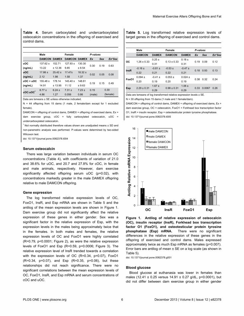
Gene expressionThe log transformed relative expression levels of OC,
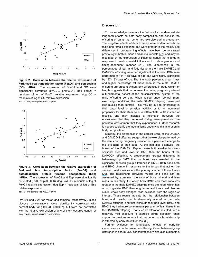
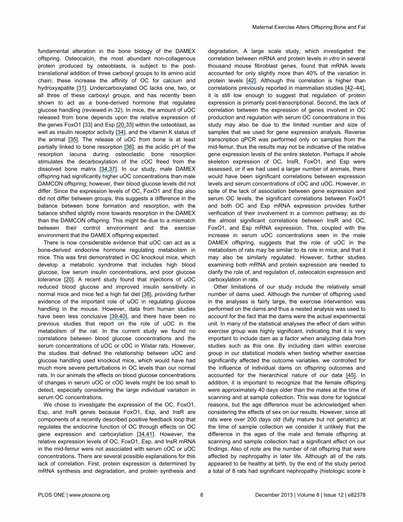
FoxO1, InsR, and Esp mRNA are shown in Table 5 and theantilog of the mean expression levels are shown in Figure 1.Dam exercise group did not significantly affect the relativeexpression of these genes in either gender. Sex was asignificant factor in the relative expression of Esp, with theexpression levels in the males being approximately twice thatin the females. In both males and females, the relativeexpression levels of OC and FoxO1 were highly correlated(R=0.79, p<0.0001; Figure 2), as were the relative expressionlevels of FoxO1 and Esp (R=0.59, p=0.0006; Figure 3). Therelative expression level of InsR trended towards a correlationwith the expression levels of OC (R=0.34, p=0.07), FoxO1(R=0.34, p=0.07), and Esp (R=0.35, p=0.06), but theserelationships did not reach significance. There were nosignificant correlations between the mean expression levels ofOC, FoxO1, InsR, and Esp mRNA and serum concentrations ofcOC and uOC.
Table 4. Serum carboxylated and undercarboxylatedosteocalcin concentrations in the offspring of exercised andcontrol dams.
Male Female P-values DAMCON DAMEX DAMCON DAMEX Ex Sex Ex*Sex
cOC(ng/mL)
137.62 ±13.22
152.71± 12.36
127.93 ±9.85
130.30± 8.54
0.30 0.19 0.63
uOC(ng/mL)
17.86 ±2.12
25.42 ±1.98
17.47±1.58
18.32 ±1.37
0.02 0.05 0.08
cOC + uOC(ng/mL)
155.48 ±14.91
178.14± 13.95
145.40 ±11.12
148.61± 9.63
0.19 0.15 0.49
cOC:uOC* 8.77 ±4.86
6.24 ±1.27
7.31 ±0.056
7.23 ±0.86
0.15(male)
0.30(female)
Data are lsmeans ± SE unless otherwise indicated.N = 44 offspring from 15 dams (1 male, 2 female/dam except for 1 excludedfemale).DAMCON = offspring of control dams, DAMEX = offspring of exercised dams, Ex =dam exercise group, cOC = fully carboxylated osteocalcin, uOC =undercarboxylated osteocalcin.* Not normally distributed therefore values shown are unadjusted means ± SD andnon-parametric analysis was performed. P-values were determined by two-sidedWilcoxon test.doi: 10.1371/journal.pone.0082378.t004
Blood glucoseBlood glucose at euthanasia was lower in females than
males (12.41 ± 0.25 versus 14.91 ± 0.27 g/dL, p<0.0001), butdid not differ between dam exercise group in either gender
Table 5. Log transformed relative expression levels oftarget genes in the offspring of exercised and control dams.
Male Female P-values
DAMCON DAMEX DAMCON DAMEX Ex Sex Ex*Sex
OC 1.26 ± 0.330.25 ±0.31
0.13 ± 0.330.18 ±0.31
0.19 0.09 0.12
InsR-0.16 ±0.22
-0.81 ±0.21
-0.53 ±0.22
-0.47 ±0.21
0.18 0.93 0.13
FoxO10.094 ±0.20
-0.41 ±0.19
0.053 ±0.20
0.024 ±0.19
0.38 0.32 0.24
Esp 2.29 ± 0.311.67 ±0.29
0.98 ± 0.311.06 ±0.29
0.33 0.0067 0.26
Data are lsmeans of log transformed relative expression levels ± SE.N = 30 offspring from 15 dams (1 male and 1 female/dam).DAMCON = offspring of control dams, DAMEX = offspring of exercised dams, Ex =dam exercise group, OC = osteocalcin, FoxO1 = Forkhead box transcription factorO1, InsR = insulin receptor, Esp = osteotesticular protein tyrosine phosphatase.doi: 10.1371/journal.pone.0082378.t005
Figure 1. Antilog of relative expression of osteocalcin(OC), insulin receptor (InsR), Forkhead box transcriptionfactor O1 (FoxO1), and osteotesticular protein tyrosinephosphatase (Esp) mRNA. There were no significantdifferences in the relative expression of these genes in theoffspring of exercised and control dams. Males expressedapproximately twice as much Esp mRNA as females (p=0.007).Error bars are antilog of mean ± SE on a log scale (as shown inTable 5).doi: 10.1371/journal.pone.0082378.g001
Maternal Exercise Alters Offspring Bone and Fat
PLOS ONE | www.plosone.org 6 December 2013 | Volume 8 | Issue 12 | e82378
(p=0.91 and 0.26 for males and females, respectively). Bloodglucose concentrations were significantly correlated withpercent body fat (R=0.28, p=0.007), but were not correlatedwith the relative expression of any of the measured genes, orany measure of serum osteocalcin.
Figure 2. Correlation between the relative expression ofForkhead box transcription factor (FoxO1) and osteocalcin(OC) mRNA. The expression of FoxO1 and OC weresignificantly correlated (R=0.79, p<0.0001). rlog FoxO1 =residuals of log of FoxO1 relative expression. rlog OC =residuals of log of OC relative expression.doi: 10.1371/journal.pone.0082378.g002
Figure 3. Correlation between the relative expression ofForkhead box transcription factor (FoxO1) andosteotesticular protein tyrosine phosphatase (Esp)mRNA. The expression of FoxO1 and Esp were significantlycorrelated (R=0.59, p=0.0006). rlog FoxO1 = residuals of log ofFoxO1 relative expression. rlog Esp = residuals of log of Esprelative expression.doi: 10.1371/journal.pone.0082378.g003
Discussion
To our knowledge these are the first results that demonstratelong-term effects on both body composition and bone in theoffspring of dams that performed exercise during pregnancy.The long-term effects of dam exercise were evident in both themale and female offspring, but were greater in the males. Sexdifferences in programming effects have been demonstratedpreviously in both humans and animal models [27], and may bemediated by the expression of placental genes that change inresponse to environmental influences in both a gender- andtiming-dependent manner [28]. The differences in thepercentages of lean and fatty tissue in the male DAMEX andDAMCON offspring were not significant at the initial DXA scanperformed at 114–118 days of age, but were highly significantby 187–193 days of age. That the lower percentage lean massand higher percentage fat mass seen in the male DAMEXoffspring are present without any differences in body weight orlength, suggests that our intervention during pregnancy altereda fundamental aspect of the musculoskeletal system of themale offspring so that, when raised under control (non-exercising) conditions, the male DAMEX offspring developedless muscle than controls. This may be due to differences intheir basal level of physical activity, or to an increasedpropensity for their stem cells to differentiate to fat instead ofmuscle, and may indicate a mismatch between theenvironment that they perceived during development and thepostnatal environment that they experienced. Further researchis needed to clarify the mechanisms underlying this alteration inbody composition.
Similarly, the differences in the cortical BMDv of the DAMEXand DAMCON offspring suggest that the exercise performed bythe dams during pregnancy resulted in a persistent change tothe skeletons of their pups. At the mid-tibial diaphysis, thebones of the DAMEX offspring were both smaller in cross-sectional area and lower in BMC than the bones of theDAMCON offspring. A proportionally greater difference inbetween-group BMC than in bone area resulted in thesignificant between-group difference in BMDv. Both bone areaand BMC change in response to the forces that act on theskeleton, and muscles are the primary source of these forces[29]. The relationship between muscle and bone can beassessed by examining the ratio of bone mineral and leanmass. In this study, the whole body BMC: lean mass ratio wasgreater in the male DAMEX offspring once the head, which hasa much greater BMD than long bones and thus could obscuresubtle whole-body changes, was excluded from the region ofinterest. These results indicate that the relationship betweenbone and muscle was fundamentally altered in the maleDAMEX offspring, and that (although they had lower BMDv andBMC) they had more bone mineral per gram of lean tissue thanthe DAMCON offspring. That such an alteration resulted from arelatively mild exposure to exercise during gestation lendssupport to previous reports that the bone: muscle relationshipis affected by early-life influences [30].
Further evidence for long-lasting effects of early-lifecircumstances on the skeleton is the significant between-groupdifference in serum uOC concentrations, which also suggests a
Maternal Exercise Alters Offspring Bone and Fat
PLOS ONE | www.plosone.org 7 December 2013 | Volume 8 | Issue 12 | e82378
fundamental alteration in the bone biology of the DAMEXoffspring. Osteocalcin, the most abundant non-collagenousprotein produced by osteoblasts, is subject to the post-translational addition of three carboxyl groups to its amino acidchain; these increase the affinity of OC for calcium andhydroxyapatite [31]. Undercarboxylated OC lacks one, two, orall three of these carboxyl groups, and has recently beenshown to act as a bone-derived hormone that regulatesglucose handling (reviewed in 32). In mice, the amount of uOCreleased from bone depends upon the relative expression ofthe genes FoxO1 [33] and Esp [20,33] within the osteoblast, aswell as insulin receptor activity [34], and the vitamin K status ofthe animal [35]. The release of uOC from bone is at leastpartially linked to bone resorption [36], as the acidic pH of theresorption lacuna during osteoclastic bone resorptionstimulates the decarboxylation of the cOC freed from thedissolved bone matrix [34,37]. In our study, male DAMEXoffspring had significantly higher uOC concentrations than maleDAMCON offspring; however, their blood glucose levels did notdiffer. Since the expression levels of OC, FoxO1 and Esp alsodid not differ between groups, this suggests a difference in thebalance between bone formation and resorption, with thebalance shifted slightly more towards resorption in the DAMEXthan the DAMCON offspring. This might be due to a mismatchbetween their control environment and the exerciseenvironment that the DAMEX offspring expected.
There is now considerable evidence that uOC can act as abone-derived endocrine hormone regulating metabolism inmice. This was first demonstrated in OC knockout mice, whichdevelop a metabolic syndrome that includes high bloodglucose, low serum insulin concentrations, and poor glucosetolerance [20]. A recent study found that injections of uOCreduced blood glucose and improved insulin sensitivity innormal mice and mice fed a high fat diet [38], providing furtherevidence of the important role of uOC in regulating glucosehandling in the mouse. However, data from human studieshave been less conclusive [39,40], and there have been noprevious studies that report on the role of uOC in themetabolism of the rat. In the current study we found nocorrelations between blood glucose concentrations and theserum concentrations of uOC or cOC in Wistar rats. However,the studies that defined the relationship between uOC andglucose handling used knockout mice, which would have hadmuch more severe perturbations in OC levels than our normalrats. In our animals the effects on blood glucose concentrationsof changes in serum uOC or cOC levels might be too small todetect, especially considering the large individual variation inserum OC concentrations.
We chose to investigate the expression of the OC, FoxO1,Esp, and InsR genes because FoxO1, Esp, and InsR arecomponents of a recently described positive feedback loop thatregulates the endocrine function of OC through effects on OCgene expression and carboxylation [34,41]. However, therelative expression levels of OC, FoxO1, Esp, and InsR mRNAin the mid-femur were not associated with serum cOC or uOCconcentrations. There are several possible explanations for thislack of correlation. First, protein expression is determined bymRNA synthesis and degradation, and protein synthesis and
degradation. A large scale study, which investigated thecorrelation between mRNA and protein levels in vitro in severalthousand mouse fibroblast genes, found that mRNA levelsaccounted for only slightly more than 40% of the variation inprotein levels [42]. Although this correlation is higher thancorrelations previously reported in mammalian studies [42–44],it is still low enough to suggest that regulation of proteinexpression is primarily post-transcriptional. Second, the lack ofcorrelation between the expression of genes involved in OCproduction and regulation with serum OC concentrations in thisstudy may also be due to the limited number and size ofsamples that we used for gene expression analysis. Reversetranscription qPCR was performed only on samples from themid-femur, thus the results may not be indicative of the relativegene expression levels of the entire skeleton. Perhaps if wholeskeleton expression of OC, InsR, FoxO1, and Esp wereassessed, or if we had used a larger number of animals, therewould have been significant correlations between expressionlevels and serum concentrations of cOC and uOC. However, inspite of the lack of association between gene expression andserum OC levels, the significant correlations between FoxO1and both OC and Esp mRNA expression provides furtherverification of their involvement in a common pathway; as dothe almost significant correlations between InsR and OC,FoxO1, and Esp mRNA expression. This, coupled with theincrease in serum uOC concentrations seen in the maleDAMEX offspring, suggests that the role of uOC in themetabolism of rats may be similar to its role in mice, and that itmay also be similarly regulated. However, further studiesexamining both mRNA and protein expression are needed toclarify the role of, and regulation of, osteocalcin expression andcarboxylation in rats.
Other limitations of our study include the relatively smallnumber of dams used. Although the number of offspring usedin the analyses is fairly large, the exercise intervention wasperformed on the dams and thus a nested analysis was used toaccount for the fact that the dams were the actual experimentalunit. In many of the statistical analyses the effect of dam withinexercise group was highly significant, indicating that it is veryimportant to include dam as a factor when analyzing data fromstudies such as this one. By including dam within exercisegroup in our statistical models when testing whether exercisesignificantly affected the outcome variables, we controlled forthe influence of individual dams on offspring outcomes andaccounted for the hierarchical nature of our data [45]. Inaddition, it is important to recognize that the female offspringwere approximately 40 days older than the males at the time ofscanning and at sample collection. This was done for logisticalreasons, but the age difference must be acknowledged whenconsidering the effects of sex on our results. However, since allrats were over 200 days old (fully mature but not geriatric) atthe time of sample collection we consider it unlikely that thedifference in the ages of the male and female offspring atscanning and sample collection had a significant effect on ourfindings. Also of note are the number of rat offspring that wereaffected by nephropathy in later life. Although all of the ratsappeared to be healthy at birth, by the end of the study perioda total of 8 rats had significant nephropathy (histologic score ≥
Maternal Exercise Alters Offspring Bone and Fat
PLOS ONE | www.plosone.org 8 December 2013 | Volume 8 | Issue 12 | e82378
3). Of these, 6 were male, 2 were female, and all were DAMEXoffspring from one of three dams. Chronic progressivenephropathy in laboratory rats is a known entity in many strainsof laboratory rats, and is most commonly seen in aging males[46]. Whether the occurrence of this condition in only theDAMEX offspring in our study is related to the maternalexercise or to genetic predisposition of the dams whohappened to be in the exercise group is difficult to determinewith the number of animals per group that we used. Certainlynephron development occurs during gestation and the earlypostnatal period, and the number of nephrons that the offspringhave is affected by environmental conditions during pregnancy,effects which may not be evident in altered birth weight [47].However, most of the DAMEX offspring in our study did nothave significant nephropathy, suggesting that there are otherfactors underlying the development of this condition. Becauseof the relationships between kidney disease, growth,parathyroid hormone and OC concentrations, and bonemineralization, we decided to screen all animals for histologicevidence of nephropathy and to exclude from the analysis anywith a nephropathy score of ≥ 3. Thus, the animals that wehave included in our analyses were physiologically “normal” tothe best of our knowledge, and our results reflect the effects ofmaternal exercise during pregnancy on these normal offspring.Whether the maternal exercise itself predisposes the rats todevelop nephropathy is a topic for future research.
The differences that we observed between the DAMEX andDAMCON offspring were triggered by a minor interventionduring development, but the actual factor or factors associatedwith the exercise performed by the dams in this study thatresulted in these changes is difficult to determine. The increasein fatness seen in the DAMEX offspring is similar to thechanges seen in the offspring of rat dams that wereundernourished during pregnancy. Our exercising dams didhave to stand to reach their food and water, and did havenumerically (but not significantly) lower food intakes duringpregnancy than did their control counterparts [13]. Perhaps thissmall reduction in food intake may have resulted in a subtlenutritional stress on the developing offspring that, over time,resulted in relatively mild but detectable changes in bodycomposition. Nutritional stress during pregnancy and aroundthe time of conception can also result in a reduced activity levelin the offspring, as has been demonstrated in both rats [48] andsheep [49].
It is also possible that the between-group differences in ourstudy resulted from predictive adaptive responses of theDAMEX offspring to an anticipated living situation that includedexercise, and that the subsequent lack of exercise in their
postnatal environment (control conditions for laboratory rats)resulted in a mismatch. A mismatch situation is one in which anorganism makes predictive adaptive changes during itsdevelopment in response to environmental cues, responsesthat would aid its survival in the environment that it expects toencounter, but then is born into an environment in which itsdevelopmental adaptations are no longer advantageous andmay even be deleterious [50]. In the current study, possibly theDAMEX offspring developed anticipating a greater amount ofexercise than they were allowed during postnatal life, and thusbecame fatter and had lower bone density than the DAMCONoffspring in identical living conditions; in the latter group, theenvironmental conditions perceived during gestation andexperienced postnatally were the same. Interpreting thedifferences in uOC in the male offspring, and the altered bone:muscle relationship (also more pronounced in males), isdifficult at this time. Future studies that examine the effects ofdam exercise during pregnancy on long-term outcomes inoffspring that are also exercised postnatally will provide moreinsight into the specific factors underlying the effects ofmaternal exercise during pregnancy on adult offspring health.
In conclusion, the results of our study provide the firstevidence that very moderate voluntary exercise duringpregnancy can result in lasting changes to the musculoskeletalsystem and adiposity in the offspring without differences in birthweight. Our results provide further support to the concept of theskeleton as an organ that can be permanently altered by fetalprogramming, and suggest a link between uOC andmetabolism in the rat.
Acknowledgements
We thank Janice Rhodes, Natalie Thomson, Nikita Stowers,and Jasmine Tanner for their help with animal care, datacollection, and sample processing. We are grateful to MarlenaKruger, Anne Broomfield, Kim Wylie, Wei-Hang Chua, LaryssaHowe, Indira Rasiah and Gaya Gopakumar for their expertiseand assistance with technical aspects of the study. We alsothank the staff at the Massey University Small AnimalProduction Unit for their help with animal care and handling.
Author Contributions
Conceived and designed the experiments: BVR ECF HTBMHV. Performed the experiments: BVR KED CGK. Analyzedthe data: BVR PCHM. Contributed reagents/materials/analysistools: CGK KED. Wrote the manuscript: BVR HTB MHV KEDPCHM CGK ECF.
References
1. Low FM, Gluckman PD, Hanson MA (2012) Developmental Plasticity,Epigenetics and Human. Health - Evol Biol 39: 650-665. doi:10.1007/s11692-011-9157-0.
2. Vickers MH, Breier BH, Cutfield WS, Hofman PL, Gluckman PD (2000)Fetal origins of hyperphagia, obesity, and hypertension and postnatalamplification by hypercaloric nutrition. Am J Physiol Endocrinol Metab279: E83-E87. PubMed: 10893326.
3. Sloboda DM, Howie GJ, Pleasants A, Gluckman PD, Vickers MH(2009) Pre- and postnatal nutritional histories influence reproductive
maturation and ovarian function in the rat. PLOS ONE 4: e6744. doi:10.1371/journal.pone.0006744. PubMed: 19707592.
4. Clapp JF (2003) The effects of maternal exercise on fetal oxygenationand feto-placental growth. Eur J Obstet Gynecol Reprod Biol 110: S80-S85. doi:10.1016/S0301-2115(03)00176-3. PubMed: 12965094.
5. Clapp JF, Kim H, Burciu B, Lopez B (2000) Beginning regular exercisein early pregnancy: Effect on fetoplacental growth. Am J ObstetGynecol 183: 1484-1488. doi:10.1067/mob.2000.107096. PubMed:11120515.
Maternal Exercise Alters Offspring Bone and Fat
PLOS ONE | www.plosone.org 9 December 2013 | Volume 8 | Issue 12 | e82378
6. Clapp JF, Kim H, Burciu B, Schmidt S, Petry K et al. (2002) Continuingregular exercise during pregnancy: Effect of exercise volume onfetoplacental growth. Am J Obstet Gynecol 186: 142-147. doi:10.1067/mob.2002.119109. PubMed: 11810100.
7. Clapp JF, Simonian S, Lopez B, Appleby-Wineberg S, Harcar-Sevcik R(1998) The one-year morphometric and neurodevelopmental outcomeof the offspring of women who continued to exercise regularlythroughout pregnancy. Am J Obstet Gynecol 178: 594-599. doi:10.1016/S0002-9378(98)70444-2. PubMed: 9539531.
8. Clapp JF (1996) Morphometric and neurodevelopmental outcome atage five years of the offspring of women who continued to exerciseregularly throughout pregnancy. J Pediatr 129: 856-863. doi:10.1016/S0022-3476(96)70029-X. PubMed: 8969727.
9. Pivarnik JM, Chambliss HO, Clapp JF, Dugan SA, Hatch MC et al.(2006) Impact of physical activity during pregnancy and postpartum onchronic disease risk. Med Sci Sports Exerc 38: 989-1006. doi:10.1249/01.mss.0000218147.51025.8a. PubMed: 16672855.
10. Mottola MF (1996) The use of animal models in exercise andpregnancy research. Semin Perinatol 20: 222-231. doi:10.1016/S0146-0005(96)80015-2. PubMed: 8888448.
11. Akhavan MM, Emami-Abarghoie M, Safari M, Sadighi-Moghaddam B,Vafaei AA et al. (2008) Serotonergic and noradrenergic lesionssuppress the enhancing effect of maternal exercise during pregnancyon learning and memory in rat pups. Neuroscience 151: 1173-1183.
12. Mottola M, Bagnall KM, McFadden KD (1983) The effects of maternalexercise on developing rat fetuses. Br J Sports Med 17: 117-121. doi:10.1136/bjsm.17.2.117. PubMed: 6883019.
13. Rosa BV, Blair HT, Vickers MH, Morel PCH, Cockrem JF et al. (2012)Voluntary exercise in pregnant rats improves post-lactation maternalbone parameters but does not affect offspring outcomes in early life. JMusculoskelet Neuronal Interact 12: 199-208. PubMed: 23196262.
14. Denadai BS, Picarro Ida C, Madjian S, Bergamaschi CT, Santos VC etal. (1994) High intensity exercise during pregnancy of rats. Effects onmother and offspring. Comp Biochem Physiol A Physiol 109: 727-740.doi:10.1016/0300-9629(94)90216-X. PubMed: 8529013.
15. Houghton PE, Mottola MF, Plust JH, Schachter CL (2000) Effect ofmaternal exercise on fetal and placental glycogen storage in the maturerat. Can J Appl Physiol 25: 443-452. doi:10.1139/h00-029. PubMed:11098156.
16. Treadway J, Dover EV, Morse W, Newcomer L, Craig BW (1986)Influence of exercise training on maternal and fetal morphologicalcharacteristics in the rat. Journal of Appl Physiol 60: 1700-1703.PubMed: 3710987.
17. Lee H-H, Kim H, Lee J-W, Kim Y-S, Yang H-Y et al. (2006) Maternalswimming during pregnancy enhances short-term memory andneurogenesis in the hippocampus of rat pups. Brain Dev 28: 147-154.doi:10.1016/j.braindev.2005.05.007. PubMed: 16368211.
18. Carter LG, Qi NR, De Cabo R, Pearson KJ (2013) Maternal exerciseimproves insulin sensitivity in mature rat offspring. Med Sci SportsExerc 45: 832-840. doi:10.1249/MSS.0b013e31827de953. PubMed:23247711.
19. Confavreux CB, Levine RL, Karsenty G (2009) A paradigm ofintegrative physiology, the crosstalk between bone and energymetabolisms. Mol Cell Endocrinol 310: 21-29. doi:10.1016/j.mce.2009.04.004. PubMed: 19376193.
20. Lee NK, Sowa H, Hinoi E, Ferron M, Ahn JD et al. (2007) Endocrineregulation of energy metabolism by the skeleton. Cell 130: 456-469.doi:10.1016/j.cell.2007.05.047. PubMed: 17693256.
21. Cooper C, Westlake S, Harvey N, Javaid K, Dennison E et al. (2006)Review: developmental origins of osteoporotic fracture. Osteoporos Int17: 337-347. doi:10.1007/s00198-005-2039-5. PubMed: 16331359.
22. Monteiro ACT, Paes ST, dos Santos JA, de Lira KDS, de Moraes SRA(2010) Effects of physical exercise during pregnancy and proteinmalnutrition during pregnancy and lactation on the development andgrowth of the offspring's femur. J Pediatr (Rio J) 86: 233-238. doi:10.1590/S0021-75572010000300012. PubMed: 20440444.
23. Yao W, Jee WS, Chen JL, Li CY, Frost HM (2001) A novel method to'exercise' rats: making rats rise to erect bipedal stance for feeding -raised cage model. J Musculoskelet Neuronal Interact 1: 241-247.PubMed: 15758498.
24. Rosa BV, Firth EC, Blair HT, Vickers MH, Morel PCH (2011) Voluntaryexercise in pregnant rats positively influences fetal growth withoutinitiating a maternal physiological stress response. Am J Physiol RegulIntegr Comp Physiol 300: R1134-R1141. doi:10.1152/ajpregu.00683.2010. PubMed: 21307360.
25. Rosa BV, Firth EC, Blair HT, Vickers MH, Morel PCH et al. (2010)Short-term voluntary exercise in the rat causes bone modeling withoutinitiating a physiological stress response. Am J Physiol Regul Integr
Comp Physiol 299: R1037-R1043. doi:10.1152/ajpregu.00112.2010.PubMed: 20668232.
26. Rosa BV, Blair HT, Vickers MH, Knight CG, Morel PCH et al. (2013)Serum concentrations of fully and undercarboxylated osteocalcin do notvary between estrous cycle stages in Sprague-Dawley rats. Endocrine.doi:10.1007/s12020-013-0008-x.
27. Gilbert JS, Nijland MJ (2008) Sex differences in the developmentalorigins of hypertension and cardiorenal disease. Am J Physiol RegulIntegr Comp Physiol 295: R1941-R1952. doi:10.1152/ajpregu.90724.2008. PubMed: 18971349.
28. Gabory A, Ferry L, Fajardy I, Jouneau L, Gothié JD et al. (2012)Maternal diets trigger sex-specific divergent trajectories of geneexpression and epigenetic systems in mouse placenta. PLOS ONE 7:e47986. PubMed: 23144842.
29. Frost HM (2001) From Wolff's law to the Utah paradigm: insights aboutbone physiology and its clinical applications. Anat Rec 262: 398-419.doi:10.1002/ar.1049. PubMed: 11275971.
30. Firth EC, Rogers CW, Vickers M, Kenyon PR, Jenkinson CM et al.(2008) The bone-muscle ratio of fetal lambs is affected more bymaternal nutrition during pregnancy than by maternal size. Am JPhysiol Regul Integr Comp Physiol 294: R1890-R1894. doi:10.1152/ajpregu.00805.2007. PubMed: 18385462.
31. Lee AJ, Hodges S, Eastell R (2000) Measurement of osteocalcin. AnnClin Biochem 37: 432-446. doi:10.1258/0004563001899573. PubMed:10902858.
32. Ducy P (2011) The role of osteocalcin in the endocrine cross-talkbetween bone remodelling and energy metabolism. Diabetologia 54:1291-1297. doi:10.1007/s00125-011-2155-z. PubMed: 21503740.
33. Rached MT, Kode A, Silva BC, Jung DY, Gray S et al. (2010) FoxO1expression in osteoblasts regulates glucose homeostasis throughregulation of osteocalcin in mice. J Clin Invest 120: 357-368. doi:10.1172/JCI39901. PubMed: 20038793.
34. Ferron M, Wei JW, Yoshizawa T, Del Fattore A, DePinho RA et al.(2010) Insulin signaling in osteoblasts integrates bone remodeling andenergy metabolism. Cell 142: 296-308. doi:10.1016/j.cell.2010.06.003.PubMed: 20655470.
35. Vermeer C, Gijsbers B, Craciun AM, GroenenvanDooren M, KnapenMHJ (1996) Effects of vitamin K on bone mass and bone metabolism. JNutr 126: S1187-S1191.
36. Ivaska KK, Hentunen TA, Vääräniemi J, Ylipahkala H, Pettersson K etal. (2004) Release of intact and fragmented osteocalcin molecules frombone matrix during bone resorption in vitro. J Biol Chem 279:18361-18369. doi:10.1074/jbc.M314324200. PubMed: 14970229.
37. Clemens TL, Karsenty G (2011) The osteoblast: An insulin target cellcontrolling glucose homeostasis. J Bone Miner Res 26: 677-680. doi:10.1002/jbmr.321. PubMed: 21433069.
38. Ferron M, McKee MD, Levine RL, Ducy P, Karsenty G (2012)Intermittent injections of osteocalcin improve glucose metabolism andprevent type 2 diabetes in mice. Bone 50: 568-575. doi:10.1016/j.bone.2011.04.017. PubMed: 21550430.
39. Motyl KJ, McCabe LR, Schwartz AV (2010) Bone and glucosemetabolism: A two-way street. Arch Biochem Biophys 503: 2-10. doi:10.1016/j.abb.2010.07.030. PubMed: 20682281.
40. Booth SL, Centi A, Smith SR, Gundberg C (2013) The role ofosteocalcin in human glucose metabolism: marker or mediator? NatRev Endocrinol 9: 43-55. PubMed: 23147574.
41. Karsenty G, Oury F (2012) Biology Without Walls: The NovelEndocrinology of Bone. In: D JuliusDE Clapham. Annu Rev Physiol 74:87-105.
42. Schwanhäusser B, Busse D, Li N, Dittmar G, Schuchhardt J et al.(2011) Global quantification of mammalian gene expression control.Nature 473: 337-342. doi:10.1038/nature10098. PubMed: 21593866.
43. Tian Q, Stepaniants SB, Mao M, Weng L, Feetham MC et al. (2004)Integrated genomic and proteomic analyses of gene expression inmammalian cells. Mol Cell Proteomics 3: 960-969. PubMed: 15238602.
44. Maier T, Güell M, Serrano L (2009) Correlation of mRNA and protein incomplex biological samples. FEBS Lett 583: 3966-3973. doi:10.1016/j.febslet.2009.10.036. PubMed: 19850042.
45. Kwong WY, Osmond C, Fleming TP (2004) Support for Barkerhypothesis upheld in rat model of maternal undernutrition during thepreimplantation period: application of integrated 'random effects'.Statistical Model - Reprod Biomed Online 8: 574-576. doi:10.1016/S1472-6483(10)61105-4.
46. Hard GC, Khan KN (2004) A contemporary overview of chronicprogressive nephropathy in the laboratory rat, and its significance forhuman risk assessment. Toxicol Pathol 32: 171-180. doi:10.1080/01926230490422574. PubMed: 15200155.
47. Moritz KM, Singh RR, Probyn ME, Denton KM (2009) Developmentalprogramming of a reduced nephron endowment: more than just a
Maternal Exercise Alters Offspring Bone and Fat
PLOS ONE | www.plosone.org 10 December 2013 | Volume 8 | Issue 12 | e82378
baby's birth weight. Am J Physiol Renal Physiol 296: F1-F9. PubMed:18653482.
48. Vickers MH, Breier BH, McCarthy D, Gluckman PD (2003) Sedentarybehavior during postnatal life is determined by the prenatalenvironment and exacerbated by postnatal hypercaloric nutrition. Am J
Physiol Regul Integr Comp Physiol 285: R271-R273. PubMed:12794001.
49. Donovan EL, Hernandez CE, Matthews LR, Oliver MH, Jaquiery AL etal. (2013) Periconceptional undernutrition in sheep leads to decreasedlocomotor activity in a natural environment. J Dohad 4: 296-299.
50. Gluckman P, Hanson M (2006) Mismatch: How Our World No LongerFits Our Bodies. Oxford: Oxford University Press.
Maternal Exercise Alters Offspring Bone and Fat
PLOS ONE | www.plosone.org 11 December 2013 | Volume 8 | Issue 12 | e82378
Related Documents