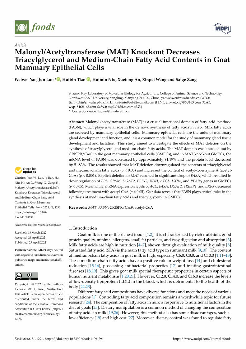
Citation: Yao, W.; Luo, J.; Tian, H.; Niu, H.; An, X.; Wang, X.; Zang, S. Malonyl/Acetyltransferase (MAT) Knockout Decreases Triacylglycerol and Medium-Chain Fatty Acid Contents in Goat Mammary Epithelial Cells. Foods 2022, 11, 1291. https://doi.org/10.3390/ foods11091291 Academic Editor: Michelle Colgrave Received: 18 March 2022 Accepted: 26 April 2022 Published: 29 April 2022 Publisher’s Note: MDPI stays neutral with regard to jurisdictional claims in published maps and institutional affil- iations. Copyright: © 2022 by the authors. Licensee MDPI, Basel, Switzerland. This article is an open access article distributed under the terms and conditions of the Creative Commons Attribution (CC BY) license (https:// creativecommons.org/licenses/by/ 4.0/). foods Article Malonyl/Acetyltransferase (MAT) Knockout Decreases Triacylglycerol and Medium-Chain Fatty Acid Contents in Goat Mammary Epithelial Cells Weiwei Yao, Jun Luo * , Huibin Tian , Huimin Niu, Xuetong An, Xinpei Wang and Saige Zang Shaanxi Key Laboratory of Molecular Biology for Agriculture, College of Animal Science and Technology, Northwest A&F University, Yangling, Xianyang 712100, China; [email protected] (W.Y.); [email protected] (H.T.); [email protected] (H.N.); [email protected] (X.A.); [email protected] (X.W.); [email protected] (S.Z.) * Correspondence: [email protected] Abstract: Malonyl/acetyltransferase (MAT) is a crucial functional domain of fatty acid synthase (FASN), which plays a vital role in the de novo synthesis of fatty acids in vivo. Milk fatty acids are secreted by mammary epithelial cells. Mammary epithelial cells are the units of mammary gland development and function, and it is a common model for the study of mammary gland tissue development and lactation. This study aimed to investigate the effects of MAT deletion on the synthesis of triacylglycerol and medium-chain fatty acids. The MAT domain was knocked out by CRISPR/Cas9 in the goat mammary epithelial cells (GMECs), and in MAT knockout GMECs, the mRNA level of FASN was decreased by approximately 91.19% and the protein level decreased by 51.83%. The results showed that MAT deletion downregulated the contents of triacylglycerol and medium-chain fatty acids (p < 0.05) and increased the content of acetyl-Coenzyme A (acetyl- CoA) (p < 0.001). Explicit deletion of MAT resulted in significant drop of FASN, which resulted in downregulation of LPL, GPAM, DGAT2, PLIN2, XDH, ATGL, LXRα, and PPARγ genes in GMECs (p < 0.05). Meanwhile, mRNA expression levels of ACC, FASN, DGAT2, SREBP1, and LXRα decreased following treatment with acetyl-CoA (p < 0.05). Our data reveals that FASN plays critical roles in the synthesis of medium-chain fatty acids and triacylglycerol in GMECs. Keywords: MAT; FASN; CRISPR/Cas9; acetyl-CoA 1. Introduction Goat milk is one of the richest foods [1,2]; it is characterized by rich nutrition, good protein quality, minimal allergens, small fat particles, and easy digestion and absorption [3]. Milk fatty acids are high in nutrition [4–7], shown through evaluation of milk quality [8]. Saturated fatty acid (SFA) is the main fatty acid type in ruminant milk [9,10]. The content of medium-chain fatty acids in goat milk is high, especially C6:0, C8:0, and C10:0 [1,11–13]. These medium-chain fatty acids have a positive role in weight loss [14] and cholesterol reduction [15,16], possessing antibacterial properties [17] and treating gastrointestinal diseases [18,19]. This gives goat milk special therapeutic properties in certain aspects of human nutrient metabolism [1,20,21]. However, C12:0, C14:0, and C16:0 increase the levels of low-density lipoprotein (LDL) in the blood, which is detrimental to the health of the body [22,23]. Different fatty acid compositions have diverse functions and meet the needs of various populations [1]. Controlling fatty acid composition remains a worthwhile topic for future research [24]. The composition of fatty acids in milk is responsive to nutritional factors in the diet of goats [25]. Dietary manipulation is a common method of changing the composition of fatty acids in milk [19,26]. However, this method also has some disadvantages, such as low efficiency [19] and high cost [27]. Moreover, dietary control was found to regulate fatty Foods 2022, 11, 1291. https://doi.org/10.3390/foods11091291 https://www.mdpi.com/journal/foods

Welcome message from author
This document is posted to help you gain knowledge. Please leave a comment to let me know what you think about it! Share it to your friends and learn new things together.
Transcript
Citation: Yao, W.; Luo, J.; Tian, H.;
Niu, H.; An, X.; Wang, X.; Zang, S.
Malonyl/Acetyltransferase (MAT)
Knockout Decreases Triacylglycerol
and Medium-Chain Fatty Acid
Contents in Goat Mammary
Epithelial Cells. Foods 2022, 11, 1291.
https://doi.org/10.3390/
foods11091291
Academic Editor: Michelle Colgrave
Received: 18 March 2022
Accepted: 26 April 2022
Published: 29 April 2022
Publisher’s Note: MDPI stays neutral
with regard to jurisdictional claims in
published maps and institutional affil-
iations.
Copyright: © 2022 by the authors.
Licensee MDPI, Basel, Switzerland.
This article is an open access article
distributed under the terms and
conditions of the Creative Commons
Attribution (CC BY) license (https://
creativecommons.org/licenses/by/
4.0/).
foods
Article
Malonyl/Acetyltransferase (MAT) Knockout DecreasesTriacylglycerol and Medium-Chain Fatty Acid Contents in GoatMammary Epithelial CellsWeiwei Yao, Jun Luo * , Huibin Tian , Huimin Niu, Xuetong An, Xinpei Wang and Saige Zang
Shaanxi Key Laboratory of Molecular Biology for Agriculture, College of Animal Science and Technology,Northwest A&F University, Yangling, Xianyang 712100, China; [email protected] (W.Y.);[email protected] (H.T.); [email protected] (H.N.); [email protected] (X.A.);[email protected] (X.W.); [email protected] (S.Z.)* Correspondence: [email protected]
Abstract: Malonyl/acetyltransferase (MAT) is a crucial functional domain of fatty acid synthase(FASN), which plays a vital role in the de novo synthesis of fatty acids in vivo. Milk fatty acidsare secreted by mammary epithelial cells. Mammary epithelial cells are the units of mammarygland development and function, and it is a common model for the study of mammary gland tissuedevelopment and lactation. This study aimed to investigate the effects of MAT deletion on thesynthesis of triacylglycerol and medium-chain fatty acids. The MAT domain was knocked out byCRISPR/Cas9 in the goat mammary epithelial cells (GMECs), and in MAT knockout GMECs, themRNA level of FASN was decreased by approximately 91.19% and the protein level decreasedby 51.83%. The results showed that MAT deletion downregulated the contents of triacylglyceroland medium-chain fatty acids (p < 0.05) and increased the content of acetyl-Coenzyme A (acetyl-CoA) (p < 0.001). Explicit deletion of MAT resulted in significant drop of FASN, which resulted indownregulation of LPL, GPAM, DGAT2, PLIN2, XDH, ATGL, LXRα, and PPARγ genes in GMECs(p < 0.05). Meanwhile, mRNA expression levels of ACC, FASN, DGAT2, SREBP1, and LXRα decreasedfollowing treatment with acetyl-CoA (p < 0.05). Our data reveals that FASN plays critical roles in thesynthesis of medium-chain fatty acids and triacylglycerol in GMECs.
Keywords: MAT; FASN; CRISPR/Cas9; acetyl-CoA
1. Introduction
Goat milk is one of the richest foods [1,2]; it is characterized by rich nutrition, goodprotein quality, minimal allergens, small fat particles, and easy digestion and absorption [3].Milk fatty acids are high in nutrition [4–7], shown through evaluation of milk quality [8].Saturated fatty acid (SFA) is the main fatty acid type in ruminant milk [9,10]. The contentof medium-chain fatty acids in goat milk is high, especially C6:0, C8:0, and C10:0 [1,11–13].These medium-chain fatty acids have a positive role in weight loss [14] and cholesterolreduction [15,16], possessing antibacterial properties [17] and treating gastrointestinaldiseases [18,19]. This gives goat milk special therapeutic properties in certain aspects ofhuman nutrient metabolism [1,20,21]. However, C12:0, C14:0, and C16:0 increase the levelsof low-density lipoprotein (LDL) in the blood, which is detrimental to the health of thebody [22,23].
Different fatty acid compositions have diverse functions and meet the needs of variouspopulations [1]. Controlling fatty acid composition remains a worthwhile topic for futureresearch [24]. The composition of fatty acids in milk is responsive to nutritional factors in thediet of goats [25]. Dietary manipulation is a common method of changing the compositionof fatty acids in milk [19,26]. However, this method also has some disadvantages, such aslow efficiency [19] and high cost [27]. Moreover, dietary control was found to regulate fatty
Foods 2022, 11, 1291. https://doi.org/10.3390/foods11091291 https://www.mdpi.com/journal/foods
Foods 2022, 11, 1291 2 of 14
acid composition through affecting fatty acid metabolism gene function [28]. Therefore,regulation of fatty acid composition by fatty acid metabolism genes should be considered.
Compared with plant breeding, gene editing breeding is seldom used in animalhusbandry [27]. Large livestock such as goats have high breeding costs, long breedingcycles, and complex breeding processes [27]. Meanwhile, fatty acids in milk are synthesizedby mammary epithelial cells [29]. Understanding the effect of key genes on fatty acidcomposition of mammary epithelial cells is the basis of molecular breeding [7].
The effects of FASN on de novo fatty acid synthesis have been confirmed in hu-mans [30], cattle [27], mice [31], and goats [32]. Several studies reported that the expressionlevel of FASN is related to the fatty acids composition in milk [31–33]. Most short- andmedium-chain fatty acid synthesis in goat milk comes from the de novo fatty acid syn-thesis pathway [13,34], and FASN is a key enzyme in de novo synthesis of fatty acids inGMECs [35]. Deletion of FASN in mammary epithelial cells of non-ruminants leads tothe decrease in the contents of medium- and long-chain fatty acids and total fatty acidsin milk [31]. Inhibition of FASN expression in mammary epithelial cells in ruminantsreduces the content of medium-chain fatty acids (C10:0, C12:0, C14:0) in milk [31,36,37].The overexpression of FASN increases the percentage of C12:0 [21].
The nutrition and flavor could be altered by modifying fatty acid composition of milk.In this study, MAT-knockout goat mammary epithelial cells (GMECs) were generated usingCRISPR/Cas9, aiming to investigate the effects of MAT on fatty acid composition. Here,we found that the contents of medium-chain fatty acids and triacylglycerol decreased, andthe unsaturated fatty acids increased, in MAT-knockout cells. These data provide a usefulway to regulate the composition of milk fatty acids through gene handling.
2. Materials and Methods2.1. Ethics Statement
All the experimental procedures were permitted by the Animal Care and Use Commit-tee of the College of Animal Science and Technology in Northwest A&F University, YangLing, China (permit number: 15-516).
2.2. sgRNA Design and Vector Construction
The goat FASN sequence was downloaded from NCBI website (https://www.ncbi.nlm.nih.gov/, accessed on 24 November 2019), the MAT fragment included part of exonsfrom 9 to 15. Then, sgRNA was designed according to CHOPCHOP website (https://zlab.bio/guide-design-resources, accessed on 24 November 2019) [38]. BbsI and protectivesequence were added to both ends of the sgRNA sequence, and double-stranded DNAwas generated by annealing. Then, the all-in-one vector pSpCas9 (BB)-2A-Puro (plasmid62988, PX459 V2.0, Addgene, Cambridge, MA, USA), a gift from Feng Zhang [39], wasdigested by BbsI enzyme (R3539S, NEB, Ipswich, MA, USA) and linked to double-strandedDNA. The connect product was transferred into E. coli (CB171211, TIANGEN, Beijing,China) for amplification. Single colonies on the solid medium (tryptone 10 g/L, yeastextract 5 g/L, NaCl 10 g/L, Agaragar 15 g/L) were picked and placed in a liquid LBmedium (tryptone 10 g/L, yeast extract 5 g/L, NaCl 10 g/L) for overnight propagation,and the plasmids were extracted for sequencing (Tsingke Biotechnology Co Ltd., Beijing,China). The vector successfully ligated with sgRNA was screened, the PX459-sgRNAplasmid vector was extracted with HighPure Mini Plasmid Kit (DP201101X, TIANGEN,Beijing, China). The plasmid was stored at −20 ◦C and its concentration and quality weredetected by spectrophotometer (Nanodrop 2000, Thermo Fisher Scientific, Rockford, IL,USA) before use.
2.3. Isolation of GMECs
Dairy goats during the peak lactation period (60 days after parturition) were anes-thetized by xylazine hydrochloride (0.01 mL/kg, Huamu Animal Health Products Cor-poration, Shandong, China) before surgery [40], then operated according to the previous
Foods 2022, 11, 1291 3 of 14
purification studies [41]. The breast tissues were cut into small pieces and cultured inan incubator at 37 ◦C and 5% CO2, with the medium changed every two days until themammary epithelial cells were isolated from the breast tissues block. The basal growthmedium contained 90% DMEM/F12 medium (SH30023−01, Hyclone, Logan, UT, USA),and 10% fetal bovine serum (10099-141, Invitrogen, Waltham, MA, USA), with 5 mg/Lbovine insulin (16634, Sigma, St. Louis, MO, USA), 5 µg/mL hydrocortisone (H0888, Sigma,St. Louis, MO, USA), 100 U/mL penicillin/streptomycin (080092569, Harbin Pharma-ceutical Group, Harbin, PR China), and 10 ng/mL epidermal growth factor (PHG0311,Invitrogen, Waltham, MA, USA) added. Then, GMECs were stored in liquid nitrogen andresuscitated before use.
2.4. Cells Culture, Transfection, and Screening
The frozen primary mammary epithelial cells were thawed in a 37 ◦C wet bath, quicklyadded into a 37 ◦C preheated culture medium, centrifuged at 1000 rpm for 4 min, and thesupernatant was discarded. After being blown evenly with the fresh culture medium, thecells were evenly spread into a 60 mm dish and cultured in a 5% CO2 and 37 ◦C incubator,the fresh culture medium was replaced every day for culture and passage until the cellstate was stable. The cells were digested by the ATV (digestive fluid, Trypsin 2.5 g/L, NaCl8 g/L, KCl 0.4 g/L, D-G 1 g/L, NaHCO3 0.58 g/L, EDTA-Na2 g/L) and evenly spread inthe six-well plate. When the cells confluency reached approximately 70% to 80%, the cellswere transfected with LipofectamineTM 2000 (11668019, Invitrogen, Waltham, MA, USA)transfection reagent, and the control group was set. The plasmid and LipofectamineTM
2000 were diluted simultaneously with DMEM/F12 (SH30023−01, Hyclone, Logan, UT,USA). Plasmids and transfection reagents were mixed and incubated for 15 min, thendropped into a six-well plate for gentle mixing. After 48 h of transfection, 1.0 µg/mLpuromycin (P8833, Sigma, St. Louis, MO, USA) was added for screening. About 96 h later,the cells death status was examined through a microscope. After all the normal cells died,the normal medium was replaced for continuous culture until cell clusters appeared. Allthe dead cells except a single round cells cluster was scraped off with a gun head andcarefully digested to the new medium.
2.5. Cells Culture and T7EN1 Assay
The GMECs were digested, half were used for T7 Endonuclease I (M0302L, NEB,Ipswich, MA, USA) cleavage assay [42], and the other half was cultured continuously. Thecells genome was extracted using a Universal Genomic DNA Kit (CW2298S, CW Biotech,Beijing, China). Extraction of genomes from cells with a Universal Genomic DNA Kit(CW2298S, CW Biotech, Beijing, China). Utilizing the genome as the template, the genomefragment of about 500 bp near the sgRNA locus was amplified by PCR using PrimeS-TAR Max DNA Polymerase (R045A, Takara Bio Inc., Otsu, Japan), then detected it in a1% agarose gel. If the band was single, the solution be purified by a PCR Clean-Up Kit(AP-PCR-50, Axygen, Union City, CA, USA) according to the manufacturers instructions.The purified DNA fragments were annealed in NEB buffer 2 (B7202S, NEB, Ipswich, MA,USA), followed by the addition of 0.3 µL T7 Endonuclease I (M0302L, NEB, Ipswich, MA,USA), reaction at 37 ◦C for 30 min, and results of enzyme digestion were identified by 2%agarose electrophoresis. The selected DNA fragments were ligated into Pmd19-T vector(CB35526014, takara Bio Inc., Otsu, Japan), transferred to E. coli (CB171211, TIANGEN, Bei-jing, China), and monoclonal colonies were selected and sequenced, which were comparedwith the gene sequence of the control group.
2.6. Genotypic Structure Prediction
The DNA sequence obtained by sequencing was translated into an amino acid se-quence using BioXM2.6 software (http://nome.njau.edu.cn/biom, accessed on 24 July 2020).The amino acid sequence was input to Protparam online website (https://web.expasy.org/protparam, accessed on 24 July 2020) for protein physical property analysis. Entered NPS@
Foods 2022, 11, 1291 4 of 14
online (http://npsa-pbil.ibcp.fr, accessed on 24 July 2020) for secondary structure analy-sis. Entered SWISS-MODEL online website (https://swissmodel.expasy.org/interactive,accessed on 24 July 2020) for tertiary structure prediction.
2.7. RNA Extraction and Real-Time Quantitative PCR (RT-qPCR)
Quantitative primers were designed for MAT region using AlleleID 6 software (http://www.premierbiosoft.com/, accessed on 25 July 2020), Sense Primer: ATGGCGGCTG-TAGGCTTGAC, Anti-Sense Primer: GCACCTCCTTGGCGAACACG. RNAiso Plus (9109,Takara Bio Inc., Otsu, Japan) was added to the cells, and RNA in the cells was extractedaccording to the manufacturer’s protocol. The RNA was reversely transcribed into cDNAwith the PrimeScript RT Reagent Kit (RR820A, Perfect Real Time, Takara Bio Inc., Otsu,Japan), and the mRNA expression levels of MAT gene in each group were detected quantita-tively with SYBR Premix Ex TaqII (RR820A, Perfect Real Time, Takara Bio Inc., Otsu, Japan)by Light Cycler 96 Real-Time PCR system (Roche Diagnostics Ltd., Mannheim, Germany).Using UXT and GAPDH as internal reference genes, the relative expression values werecalculated by the 2−∆∆Ct method. Each set of tests consisted of three technical and threebiological replicates. The primers of target genes are listed in Table S1.
2.8. Protein Extraction and Western Blot Assay
The total protein of NC-GMECs and MAT-KO-GMECs were, respectively, extractedand lysed in ice-cold RIPA buffer (R0010, Solarbio, Beijing, China) with protease inhibitor(04693132001, Roche Diagnostics Ltd., Mannheim, Germany). Protein concentration wasmeasured by BCA Protein Assay Kit (23227, Thermo Fisher Scientific, Rockford, IL, USA).The size of FASN protein was about 272 kD, and that of β-actin protein was about 42 kD.The amount of protein loading was 20 ng. After SDS-PAGE electrophoresis, the bands weretransferred to FVDF membrane (HATF00010, Millipore, Burlington, MA, USA) by semi-drytransfer method. After the transfer membrane was completed, blocking was performedwith 5% skim milk (232100, BD, Franklin Lakes, NJ, USA), and antibodies (FASN, bs-1498R;β-actin, CW0096) were incubated. The horseradish–peroxidase (HRP)-conjugated goatanti-rabbit-IgG (CW0103, CW Biotech, Beijing, China) and goat anti-mouse-IgG (CW0102)were used as secondary antibodies, and the target band signals were exposed by ECLWestern blot system (1705061, Bio-Rad, Hercules, CA, USA).
2.9. Off-Target Analysis
The online website Cas-OFFinder (http://www.rgenome.net/cas-offinder/, accessedon 8 August 2020) was utilized to predict the off-target sites [43]. The choices were: SpCas9,5′-NGG-3′, Capra hircus, and mismatch number (eq or less than) 3 [44]. Ten high-probabilitytarget sites were screened, and corresponding sequences were found at genomic positions.Primers (Table S2) were designed (www.PremierBiosoft.com, accessed on 8 August 2020)to amplify the fragment on the genome, and T7EN1 (M0302L, NEB, Ipswich, MA, USA)digestion test was performed.
2.10. Measurement of Total Cellular Triacylglycerol
MAT-KO-GMECs and NC-GMECs were spread on a 6-well-plate, and collected afterthe cells upon reaching 90% confluence. The triacylglycerol in the cells were extracted by theTissue Triacylglycerol Assay Kit (E1013, Applypen Technologies Inc., Beijing, China). Thesample contents were read at 550 nm by a Biotek Microplate Reader (Winooski, VT, USA).The relative content of triacylglycerol was corrected by intracellular protein levels anddisplayed as micrograms per milligram of protein (µg/mg protein) [45]. Protein contentwas determined by a BCA Protein Assay Kit (23227, Thermo Fisher Scientific, Rockford,IL, USA).
Foods 2022, 11, 1291 5 of 14
2.11. Measurement of Intracellular Fatty Acid Composition
The MAT-KO-GMECs and NC-GMECs were spread on a 6-well-plate. The sampleswere methylated with 2.5% (v/v) methanol sulfate, collected into a glass tube, sonicated for10 min, and sealed at 80 ◦C for 1 h. Then, added hydrochloric acid and normal hexane. Thesupernatant was charged with anhydrous sodium sulfate, shaken, and allowed to standovernight [46]. The supernatant was centrifuged and placed in a vial for GC analysis bygas chromatography (Agilent 7890A; Agilent Technologies Inc., CA, USA) with 100 m HP-5column (Agilent Technologies Inc.).
2.12. Measurement of Genes Related to Intracellular Fatty Acid Metabolism
Using UXT and GAPDH as internal reference genes, the expression levels of fattyacid synthesis genes (ACC, FASN), triacylglycerol synthesis genes (GPAM, DGAT1, DGAT2,AGPAT6), lipid droplet secretion genes (PLIN2, XDH, TIP47), triacylglycerol hydrolysisgenes (LPL, ATGL), and regulatory factors (LXRα, PPARγ, SREBP1) were detected withRT-qPCR. The primers of target genes are listed in Table S1.
2.13. Measurement of Intracellular Acetyl-CoA
Two groups of knockout cells and two groups of wild cells were cultured at the sametime, with three replicates in each group. When the cells reached 70% confluence, one groupof knockout cells and one group of wild cells were randomly treated with 100 µM acetyl-CoA salt (32140-51-5, Sigma, St. Louis, MO, USA, ≥93%) addition. After 48 h, the cellswere collected, acetyl-CoA in the cells was extracted by using an acetyl-coenzyme assay kit(MAK039, Sigma, St. Louis, MO, USA) according to the manufacturer’s instructions. Then,the fluorescence intensity was measured (λex = 535/λem = 587 nm) by a Biotek MicroplateReader (Winooski, VT, USA).
2.14. Measurement of Fatty Acid Metabolism Genes with Acetyl-CoA Addition
Acetyl-CoA salt (32140-51-5, Sigma, St. Louis, MO, USA, ≥93%) was added accordingto the above method, and the cells were collected. The mRNA expression levels of fatty acidsynthesis genes (ACC, FASN), triacylglycerol synthesis genes (DGAT1, DGAT2, AGPAT6),lipid droplet formation and secretion genes (PLIN2, TIP47), and fatty acid regulatory factorgenes (SREBP1, LXRα) were detected by using UXT and GAPDH as internal referencegenes. The primers of target genes are listed in Table S1.
2.15. Statistical Analysis
The MAT-KO (n = 3) group and the NC group (n = 3) were taken as the experimen-tal subjects, and the experimental data are shown as mean ± SEM. T-test analysis wasperformed on the data of the two groups using SPSS 19.0 statistical software (SPSS, Inc.,Chicago, IL, USA), the difference was significant when p < 0.05 and extremely significantwhen p < 0.01.
3. Results3.1. Evaluation of sgRNA for CRISPR-Mediated Repression of MAT
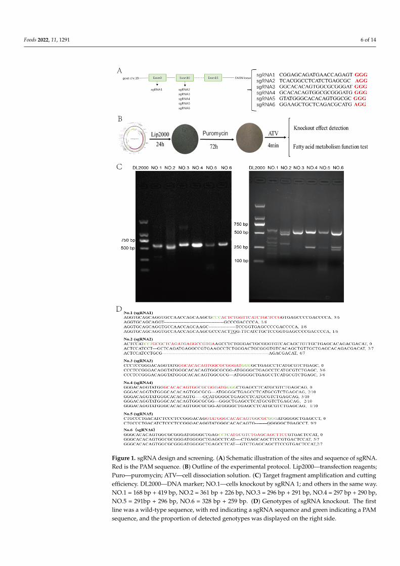
SgRNA vectors were successfully constructed and transfected into GMECs(Figure 1A,B). Thirty-one independent cells groups were obtained by puromycin screening.The efficiency of each sgRNA was detected by T7EN1 digestion (Figure 1C) and sequencing(Figure 1D), all six sgRNAs had gene editing efficiency.
Foods 2022, 11, 1291 6 of 14Foods 2022, 11, x FOR PEER REVIEW 6 of 15
Figure 1. sgRNA design and screening. (A) Schematic illustration of the sites and sequence of sgRNA. Red is the PAM sequence. (B) Outline of the experimental protocol. Lip2000—transfection reagents; Puro—puromycin; ATV—cell dissociation solution. (C) Target fragment amplification and cutting efficiency. DL2000—DNA marker; NO.1—cells knockout by sgRNA 1; and others in the same way. NO.1 = 168 bp + 419 bp, NO.2 = 361 bp + 226 bp, NO.3 = 296 bp + 291 bp, NO.4 = 297 bp + 290 bp, NO.5 = 291bp + 296 bp, NO.6 = 328 bp + 259 bp. (D) Genotypes of sgRNA knockout. The first line was a wild-type sequence, with red indicating a sgRNA sequence and green indicating a PAM sequence, and the proportion of detected genotypes was displayed on the right side.
Figure 1. sgRNA design and screening. (A) Schematic illustration of the sites and sequence of sgRNA.Red is the PAM sequence. (B) Outline of the experimental protocol. Lip2000—transfection reagents;Puro—puromycin; ATV—cell dissociation solution. (C) Target fragment amplification and cuttingefficiency. DL2000—DNA marker; NO.1—cells knockout by sgRNA 1; and others in the same way.NO.1 = 168 bp + 419 bp, NO.2 = 361 bp + 226 bp, NO.3 = 296 bp + 291 bp, NO.4 = 297 bp + 290 bp,NO.5 = 291bp + 296 bp, NO.6 = 328 bp + 259 bp. (D) Genotypes of sgRNA knockout. The firstline was a wild-type sequence, with red indicating a sgRNA sequence and green indicating a PAMsequence, and the proportion of detected genotypes was displayed on the right side.
Foods 2022, 11, 1291 7 of 14
3.2. Decreased Expression of MAT in Genome-Modified GMECs
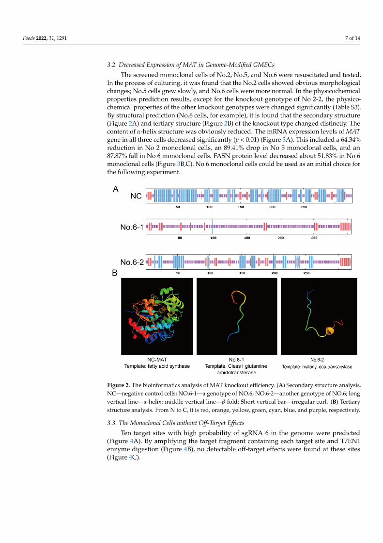
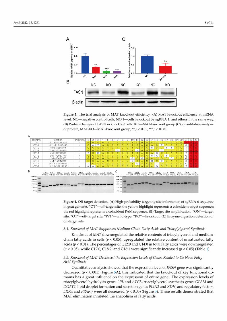
The screened monoclonal cells of No.2, No.5, and No.6 were resuscitated and tested.In the process of culturing, it was found that the No.2 cells showed obvious morphologicalchanges; No.5 cells grew slowly, and No.6 cells were more normal. In the physicochemicalproperties prediction results, except for the knockout genotype of No 2-2, the physico-chemical properties of the other knockout genotypes were changed significantly (Table S3).By structural prediction (No.6 cells, for example), it is found that the secondary structure(Figure 2A) and tertiary structure (Figure 2B) of the knockout type changed distinctly. Thecontent of α-helix structure was obviously reduced. The mRNA expression levels of MATgene in all three cells decreased significantly (p < 0.01) (Figure 3A). This included a 64.34%reduction in No 2 monoclonal cells, an 89.41% drop in No 5 monoclonal cells, and an87.87% fall in No 6 monoclonal cells. FASN protein level decreased about 51.83% in No 6monoclonal cells (Figure 3B,C). No 6 monoclonal cells could be used as an initial choice forthe following experiment.
Foods 2022, 11, x FOR PEER REVIEW 7 of 15
3.2. Decreased Expression of MAT in Genome-Modified GMECs The screened monoclonal cells of No.2, No.5, and No.6 were resuscitated and tested.
In the process of culturing, it was found that the No.2 cells showed obvious morphological changes; No.5 cells grew slowly, and No.6 cells were more normal. In the physicochemical properties prediction results, except for the knockout genotype of No 2-2, the physico-chemical properties of the other knockout genotypes were changed significantly (Table S3). By structural prediction (No.6 cells, for example), it is found that the secondary struc-ture (Figure 2A) and tertiary structure (Figure 2B) of the knockout type changed distinctly. The content of α-helix structure was obviously reduced. The mRNA expression levels of MAT gene in all three cells decreased significantly (p < 0.01) (Figure 3A). This included a 64.34% reduction in No 2 monoclonal cells, an 89.41% drop in No 5 monoclonal cells, and an 87.87% fall in No 6 monoclonal cells. FASN protein level decreased about 51.83% in No 6 monoclonal cells (Figure 3B,C). No 6 monoclonal cells could be used as an initial choice for the following experiment.
Figure 2. The bioinformatics analysis of MAT knockout efficiency. (A) Secondary structure analysis. NC—negative control cells; NO.6-1—a genotype of NO.6; NO.6-2—another genotype of NO.6; long vertical line—α-helix; middle vertical line—β-fold; Short vertical bar—irregular curl. (B) Tertiary structure analysis. From N to C, it is red, orange, yellow, green, cyan, blue, and purple, respectively.
Figure 2. The bioinformatics analysis of MAT knockout efficiency. (A) Secondary structure analysis.NC—negative control cells; NO.6-1—a genotype of NO.6; NO.6-2—another genotype of NO.6; longvertical line—α-helix; middle vertical line—β-fold; Short vertical bar—irregular curl. (B) Tertiarystructure analysis. From N to C, it is red, orange, yellow, green, cyan, blue, and purple, respectively.
3.3. The Monoclonal Cells without Off-Target Effects
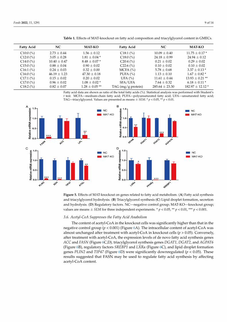
Ten target sites with high probability of sgRNA 6 in the genome were predicted(Figure 4A). By amplifying the target fragment containing each target site and T7EN1enzyme digestion (Figure 4B), no detectable off-target effects were found at these sites(Figure 4C).
Foods 2022, 11, 1291 8 of 14Foods 2022, 11, x FOR PEER REVIEW 8 of 15
Figure 3. The trial analysis of MAT knockout efficiency. (A) MAT knockout efficiency at mRNA level. NC—negative control cells; NO.1—cells knockout by sgRNA 1; and others in the same way. (B) Protein changes of FASN in knockout cells. KO—MAT-knockout group (C); quantitative analy-sis of protein; MAT-KO—MAT-knockout group; ** p < 0.01, *** p < 0.001.
3.3. The Monoclonal Cells without Off-Target Effects Ten target sites with high probability of sgRNA 6 in the genome were predicted (Fig-
ure 4A). By amplifying the target fragment containing each target site and T7EN1 enzyme digestion (Figure 4B), no detectable off-target effects were found at these sites (Figure 4C).
Figure 4. Off-target detection. (A) High-probability targeting site information of sgRNA 6 sequence in goat genome. “OT”—off-target site; the yellow highlight represents a coincident target sequence; the red highlight represents a coincident PAM sequence. (B) Target site amplification. “ON”—target site; “OT”—off-target site; “WT”—wild-type; “KO”—knockout. (C) Enzyme digestion detection of off-target site.
3.4. Knockout of MAT Suppresses Medium-Chain Fatty Acids and Triacylglycerol Synthesis Knockout of MAT downregulated the relative contents of triacylglycerol and me-
dium-chain fatty acids in cells (p < 0.05), upregulated the relative content of unsaturated fatty acids (p < 0.01). The percentages of C12:0 and C14:0 in total fatty acids were down-regulated (p < 0.05), while C17:0, C18:2, and C18:1 were significantly increased (p < 0.05) (Table 1).
Figure 3. The trial analysis of MAT knockout efficiency. (A) MAT knockout efficiency at mRNAlevel. NC—negative control cells; NO.1—cells knockout by sgRNA 1; and others in the same way.(B) Protein changes of FASN in knockout cells. KO—MAT-knockout group (C); quantitative analysisof protein; MAT-KO—MAT-knockout group; ** p < 0.01, *** p < 0.001.
Foods 2022, 11, x FOR PEER REVIEW 8 of 15
Figure 3. The trial analysis of MAT knockout efficiency. (A) MAT knockout efficiency at mRNA level. NC—negative control cells; NO.1—cells knockout by sgRNA 1; and others in the same way. (B) Protein changes of FASN in knockout cells. KO—MAT-knockout group (C); quantitative analy-sis of protein; MAT-KO—MAT-knockout group; ** p < 0.01, *** p < 0.001.
3.3. The Monoclonal Cells without Off-Target Effects Ten target sites with high probability of sgRNA 6 in the genome were predicted (Fig-
ure 4A). By amplifying the target fragment containing each target site and T7EN1 enzyme digestion (Figure 4B), no detectable off-target effects were found at these sites (Figure 4C).
Figure 4. Off-target detection. (A) High-probability targeting site information of sgRNA 6 sequence in goat genome. “OT”—off-target site; the yellow highlight represents a coincident target sequence; the red highlight represents a coincident PAM sequence. (B) Target site amplification. “ON”—target site; “OT”—off-target site; “WT”—wild-type; “KO”—knockout. (C) Enzyme digestion detection of off-target site.
3.4. Knockout of MAT Suppresses Medium-Chain Fatty Acids and Triacylglycerol Synthesis Knockout of MAT downregulated the relative contents of triacylglycerol and me-
dium-chain fatty acids in cells (p < 0.05), upregulated the relative content of unsaturated fatty acids (p < 0.01). The percentages of C12:0 and C14:0 in total fatty acids were down-regulated (p < 0.05), while C17:0, C18:2, and C18:1 were significantly increased (p < 0.05) (Table 1).
Figure 4. Off-target detection. (A) High-probability targeting site information of sgRNA 6 sequencein goat genome. “OT”—off-target site; the yellow highlight represents a coincident target sequence;the red highlight represents a coincident PAM sequence. (B) Target site amplification. “ON”—targetsite; “OT”—off-target site; “WT”—wild-type; “KO”—knockout. (C) Enzyme digestion detection ofoff-target site.
3.4. Knockout of MAT Suppresses Medium-Chain Fatty Acids and Triacylglycerol Synthesis
Knockout of MAT downregulated the relative contents of triacylglycerol and medium-chain fatty acids in cells (p < 0.05), upregulated the relative content of unsaturated fattyacids (p < 0.01). The percentages of C12:0 and C14:0 in total fatty acids were downregulated(p < 0.05), while C17:0, C18:2, and C18:1 were significantly increased (p < 0.05) (Table 1).
3.5. Knockout of MAT Decreased the Expression Levels of Genes Related to De Novo FattyAcid Synthesis
Quantitative analysis showed that the expression level of FASN gene was significantlydecreased (p < 0.001) (Figure 5A), this indicated that the knockout of key functional do-mains has a great influence on the expression of entire gene. The expression levels oftriacylglycerol hydrolysis genes LPL and ATGL, triacylglycerol synthesis genes GPAM andDGAT2, lipid droplet formation and secretion genes PLIN2 and XDH, and regulatory factorsLXRα and PPARγ were all decreased (p < 0.05) (Figure 5). These results demonstrated thatMAT elimination inhibited the anabolism of fatty acids.
Foods 2022, 11, 1291 9 of 14
Table 1. Effects of MAT-knockout on fatty acid composition and triacylglycerol content in GMECs.
Fatty Acid NC MAT-KO Fatty Acid NC MAT-KO
C10:0 (%) 2.73 ± 0.44 1.56 ± 0.12 C18:1 (%) 10.09 ± 0.40 11.75 ± 0.17 *C12:0 (%) 3.05 ± 0.28 1.81 ± 0.04 * C18:0 (%) 24.18 ± 0.99 24.94 ± 0.12C14:0 (%) 10.40 ± 0.47 8.48 ± 0.07 * C20:4 (%) 0.21 ± 0.02 0.29 ± 0.02C15:0 (%) 0.88 ± 0.04 0.90 ± 0.02 C22:6 (%) 0.10 ± 0.02 0.10 ± 0.02C16:1 (%) 0.24 ± 0.03 0.32 ± 0.00 MCFA (%) 5.78 ± 0.68 3.37 ± 0.13 *C16:0 (%) 46.19 ± 1.23 47.30 ± 0.18 PUFA (%) 1.13 ± 0.10 1.67 ± 0.82 *C17:1 (%) 0.15 ± 0.02 0.20 ± 0.02 UFA (%) 11.61 ± 0.44 13.93 ± 0.21 **C17:0 (%) 0.96 ± 0.02 1.08 ± 0.02 * SFA/UFA 7.64 ± 0.32 6.18 ± 0.11 *C18:2 (%) 0.82 ± 0.07 1.28 ± 0.05 ** TAG (mg/g protein) 285.64 ± 23.30 182.97 ± 12.12 *
Fatty acid data are shown as ratio of the total fatty acids (%). Statistical analysis was performed with Student’st test. MCFA—medium-chain fatty acid; PUFA—polyunsaturated fatty acid; UFA—unsaturated fatty acid;TAG—triacylglycerol. Values are presented as means ± SEM. * p < 0.05, ** p < 0.01.
Foods 2022, 11, x FOR PEER REVIEW 9 of 15
Table 1. Effects of MAT-knockout on fatty acid composition and triacylglycerol content in GMECs.
Fatty Acid NC MAT-KO Fatty Acid NC MAT-KO C10:0 (%) 2.73 ± 0.44 1.56 ± 0.12 C18:1 (%) 10.09 ± 0.40 11.75 ± 0.17 * C12:0 (%) 3.05 ± 0.28 1.81 ± 0.04 * C18:0 (%) 24.18± 0.99 24.94 ± 0.12 C14:0 (%) 10.40 ± 0.47 8.48 ±0.07 * C20:4 (%) 0.21 ± 0.02 0.29 ± 0.02 C15:0 (%) 0.88 ± 0.04 0.90 ± 0.02 C22:6 (%) 0.10 ± 0.02 0.10 ± 0.02 C16:1 (%) 0.24 ± 0.03 0.32 ± 0.00 MCFA (%) 5.78 ± 0.68 3.37 ± 0.13 * C16:0 (%) 46.19 ± 1.23 47.30 ± 0.18 PUFA (%) 1.13 ± 0.10 1.67 ± 0.82 * C17:1 (%) 0.15± 0.02 0.20 ± 0.02 UFA (%) 11.61 ± 0.44 13.93 ± 0.21 ** C17:0 (%) 0.96 ± 0.02 1.08 ± 0.02 * SFA/UFA 7.64 ± 0.32 6.18 ± 0.11 * C18:2 (%) 0.82 ± 0.07 1.28 ± 0.05 ** TAG (mg/g protein) 285.64 ±23.30 182.97 ± 12.12 *
Fatty acid data are shown as ratio of the total fatty acids (%). Statistical analysis was performed with Student’s t test. MCFA—medium-chain fatty acid; PUFA—polyunsaturated fatty acid; UFA—un-saturated fatty acid; TAG—triacylglycerol. Values are presented as means ± SEM. * p < 0.05, ** p < 0.01.
3.5. Knockout of MAT Decreased the Expression Levels of Genes Related to De Novo Fatty Acid Synthesis
Quantitative analysis showed that the expression level of FASN gene was signifi-cantly decreased (p < 0.001) (Figure 5A), this indicated that the knockout of key functional domains has a great influence on the expression of entire gene. The expression levels of triacylglycerol hydrolysis genes LPL and ATGL, triacylglycerol synthesis genes GPAM and DGAT2, lipid droplet formation and secretion genes PLIN2 and XDH, and regulatory factors LXRα and PPARγ were all decreased (p < 0.05) (Figure 5). These results demon-strated that MAT elimination inhibited the anabolism of fatty acids.
Figure 5. Effects of MAT-knockout on genes related to fatty acid metabolism. (A) Fatty acid synthe-sis and triacylglycerol hydrolysis. (B) Triacylglycerol synthesis (C) Lipid droplet formation, secre-tion and hydrolysis. (D) Regulatory factors. NC—negative control group; MAT-KO—knockout group; values are means ± SEM for three independent experiments. * p < 0.05, ** p < 0.01, *** p < 0.001.
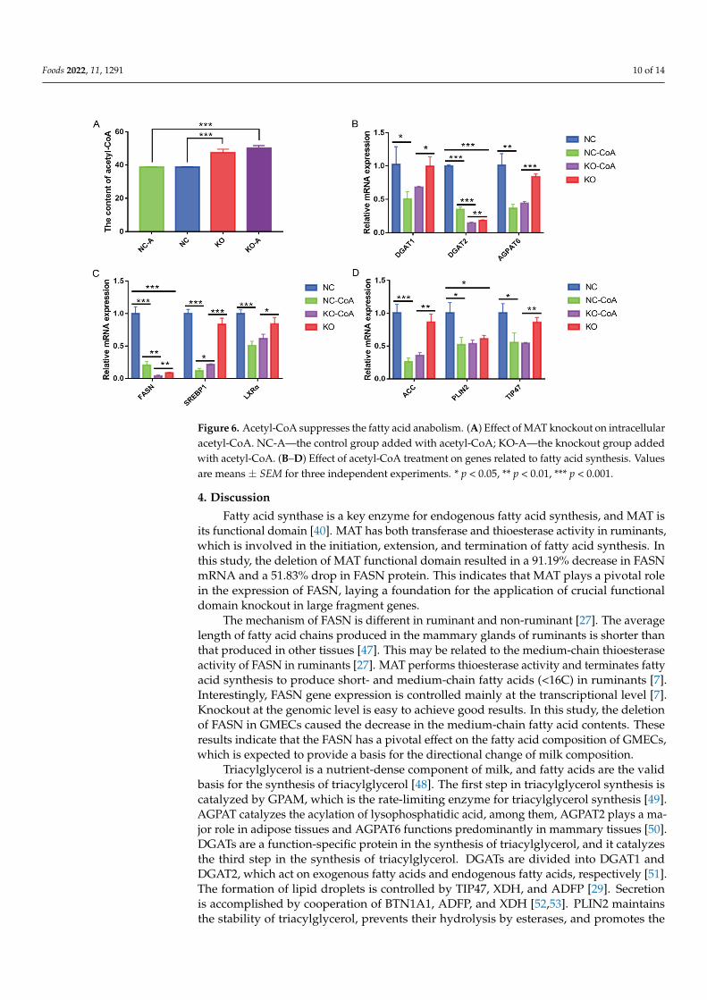
3.6. Acetyl-CoA Suppresses the Fatty Acid Anabolism The content of acetyl-CoA in the knockout cells was significantly higher than that in
the negative control group (p < 0.001) (Figure 6A). The intracellular content of acetyl-CoA was almost unchanged after treatment with acetyl-CoA in knockout cells (p > 0.05).
Figure 5. Effects of MAT-knockout on genes related to fatty acid metabolism. (A) Fatty acid synthesisand triacylglycerol hydrolysis. (B) Triacylglycerol synthesis (C) Lipid droplet formation, secretionand hydrolysis. (D) Regulatory factors. NC—negative control group; MAT-KO—knockout group;values are means ± SEM for three independent experiments. * p < 0.05, ** p < 0.01, *** p < 0.001.
3.6. Acetyl-CoA Suppresses the Fatty Acid Anabolism
The content of acetyl-CoA in the knockout cells was significantly higher than that in thenegative control group (p < 0.001) (Figure 6A). The intracellular content of acetyl-CoA wasalmost unchanged after treatment with acetyl-CoA in knockout cells (p > 0.05). Conversely,after treatment with acetyl-CoA, the expression levels of de novo fatty acid synthesis genesACC and FASN (Figure 6C,D), triacylglycerol synthesis genes DGAT1, DGAT2, and AGPAT6(Figure 6B), regulatory factors SREBP1 and LXRα (Figure 6C), and lipid droplet formationgenes PLIN2 and TIP47 (Figure 6D) were significantly downregulated (p < 0.05). Theseresults suggested that FASN may be used to regulate fatty acid synthesis by affectingacetyl-CoA content.
Foods 2022, 11, 1291 10 of 14
Foods 2022, 11, x FOR PEER REVIEW 10 of 15
Conversely, after treatment with acetyl-CoA, the expression levels of de novo fatty acid synthesis genes ACC and FASN (Figure 6C,D), triacylglycerol synthesis genes DGAT1, DGAT2, and AGPAT6 (Figure 6B), regulatory factors SREBP1 and LXRα (Figure 6C), and lipid droplet formation genes PLIN2 and TIP47 (Figure 6D) were significantly downregu-lated (p < 0.05). These results suggested that FASN may be used to regulate fatty acid synthesis by affecting acetyl-CoA content.
Figure 6. Acetyl-CoA suppresses the fatty acid anabolism. (A) Effect of MAT knockout on intracel-lular acetyl-CoA. NC-A—the control group added with acetyl-CoA; KO-A—the knockout group added with acetyl-CoA. (B–D) Effect of acetyl-CoA treatment on genes related to fatty acid synthe-sis. Values are means ± SEM for three independent experiments. * p < 0.05, ** p < 0.01, *** p < 0.001.
4. Discussion Fatty acid synthase is a key enzyme for endogenous fatty acid synthesis, and MAT is
its functional domain [40]. MAT has both transferase and thioesterase activity in rumi-nants, which is involved in the initiation, extension, and termination of fatty acid synthe-sis. In this study, the deletion of MAT functional domain resulted in a 91.19% decrease in FASN mRNA and a 51.83% drop in FASN protein. This indicates that MAT plays a pivotal role in the expression of FASN, laying a foundation for the application of crucial func-tional domain knockout in large fragment genes.
The mechanism of FASN is different in ruminant and non-ruminant [27]. The average length of fatty acid chains produced in the mammary glands of ruminants is shorter than that produced in other tissues [47]. This may be related to the medium-chain thioesterase activity of FASN in ruminants [27]. MAT performs thioesterase activity and terminates fatty acid synthesis to produce short- and medium-chain fatty acids (<16C) in ruminants [7]. Interestingly, FASN gene expression is controlled mainly at the transcriptional level [7]. Knockout at the genomic level is easy to achieve good results. In this study, the dele-tion of FASN in GMECs caused the decrease in the medium-chain fatty acid contents. These results indicate that the FASN has a pivotal effect on the fatty acid composition of GMECs, which is expected to provide a basis for the directional change of milk composi-tion.
Triacylglycerol is a nutrient-dense component of milk, and fatty acids are the valid basis for the synthesis of triacylglycerol [48]. The first step in triacylglycerol synthesis is catalyzed by GPAM, which is the rate-limiting enzyme for triacylglycerol synthesis [49].
Figure 6. Acetyl-CoA suppresses the fatty acid anabolism. (A) Effect of MAT knockout on intracellularacetyl-CoA. NC-A—the control group added with acetyl-CoA; KO-A—the knockout group addedwith acetyl-CoA. (B–D) Effect of acetyl-CoA treatment on genes related to fatty acid synthesis. Valuesare means ± SEM for three independent experiments. * p < 0.05, ** p < 0.01, *** p < 0.001.
4. Discussion
Fatty acid synthase is a key enzyme for endogenous fatty acid synthesis, and MAT isits functional domain [40]. MAT has both transferase and thioesterase activity in ruminants,which is involved in the initiation, extension, and termination of fatty acid synthesis. Inthis study, the deletion of MAT functional domain resulted in a 91.19% decrease in FASNmRNA and a 51.83% drop in FASN protein. This indicates that MAT plays a pivotal rolein the expression of FASN, laying a foundation for the application of crucial functionaldomain knockout in large fragment genes.
The mechanism of FASN is different in ruminant and non-ruminant [27]. The averagelength of fatty acid chains produced in the mammary glands of ruminants is shorter thanthat produced in other tissues [47]. This may be related to the medium-chain thioesteraseactivity of FASN in ruminants [27]. MAT performs thioesterase activity and terminates fattyacid synthesis to produce short- and medium-chain fatty acids (<16C) in ruminants [7].Interestingly, FASN gene expression is controlled mainly at the transcriptional level [7].Knockout at the genomic level is easy to achieve good results. In this study, the deletionof FASN in GMECs caused the decrease in the medium-chain fatty acid contents. Theseresults indicate that the FASN has a pivotal effect on the fatty acid composition of GMECs,which is expected to provide a basis for the directional change of milk composition.
Triacylglycerol is a nutrient-dense component of milk, and fatty acids are the validbasis for the synthesis of triacylglycerol [48]. The first step in triacylglycerol synthesis iscatalyzed by GPAM, which is the rate-limiting enzyme for triacylglycerol synthesis [49].AGPAT catalyzes the acylation of lysophosphatidic acid, among them, AGPAT2 plays a ma-jor role in adipose tissues and AGPAT6 functions predominantly in mammary tissues [50].DGATs are a function-specific protein in the synthesis of triacylglycerol, and it catalyzesthe third step in the synthesis of triacylglycerol. DGATs are divided into DGAT1 andDGAT2, which act on exogenous fatty acids and endogenous fatty acids, respectively [51].The formation of lipid droplets is controlled by TIP47, XDH, and ADFP [29]. Secretionis accomplished by cooperation of BTN1A1, ADFP, and XDH [52,53]. PLIN2 maintainsthe stability of triacylglycerol, prevents their hydrolysis by esterases, and promotes the
Foods 2022, 11, 1291 11 of 14
formation of cytoplasmic lipid droplets in GMECs [54]. ATGL, HSL, and LPL play crucialroles in the metabolism of triacylglycerol into free fatty acids [55]. Regulatory factors suchas PPAR, LXR, and SREBP1 [56] affect milk fat content by regulating the expression of fattyacid synthesis genes, and the regulatory role of LXR is dependent on SREBP1 [57]. In thisstudy, MAT knockout inhibited the expression of the above genes, slowed down the wholeprocess of triacylglycerol synthesis, and finally reduced the triacylglycerol content (p < 0.05),indicating that MAT functional domain plays a pivotal role in triacylglycerol synthesis.
Acetyl-CoA is a powerful intracellular metabolic intermediate that determines thebalance of catabolism and anabolism in lipid metabolism [58]. Normally, mitochondrialacetyl-CoA is taken as a relatively independent part, and acetyl-CoA in cytoplasm andnucleus is taken as a whole [59]. Acetyl-CoA in mitochondria is produced by β-oxidationof fatty acids. Acetyl-CoA in cytoplasm is converted from acetic acid or produced bydecomposition of citric acid in mitochondria with ATP-citrate lyase [60]. Acetyl-CoA in thenucleus is converted by citric acid or pyruvic acid and interacts with the acetyl-CoA contentin the cytoplasm. Acetyl-CoA in cytoplasm forms malonyl-CoA under the catalysis of ACC,then forms fatty acid under the catalysis of FASN. In the nucleus, acetyl-CoA regulates geneexpression by affecting the acetylation of transcription factors such as PPARγ and histoneacetylation [61,62]. Among them, it is established in ruminants that PPARs are capable ofutilizing milk fatty acids as ligands and modulate the expression of genes involved in lipidmetabolism [63]. In this study, the treatment of acetyl-CoA in cells significantly reduced theexpression of genes related to fatty acid synthesis, and MAT knockout accumulated acetyl-CoA content in cells, indicating that FASN regulates fatty acid synthesis by regulating thecontent of acetyl-CoA.
The limitation of this work is that our study only explored the knockout of FASNin GMECs. In particular, total FASN knockout in mice is fatal to embryos, and evenheterozygous mice show impaired growth and survival [64]. Mammary specific FASNdeletion hinders breast development and induces premature breast degeneration [31].Therefore, the toxic effect of FASN knockout on tissue must be considered, which is likelyto affect the observed phenotype.
5. Conclusions
FASN is a core enzyme in de novo synthesis of fatty acids, and MAT is a pivotalfunctional domain of FASN. Moreover, FASN plays a crucial role in the synthesis of medium-chain fatty acids in ruminant, which may be related to the MAT functional domain’stransferase and thioesterase activities. This study proves that MAT knockout in GMECsleads to a decrease in medium-chain fatty acids and triacylglycerol. Our findings providethe basis for altering the composition of milk fatty acids by gene editing.
Supplementary Materials: The following supporting information can be downloaded at: https://www.mdpi.com/article/10.3390/foods11091291/s1, Table S1: Characteristics of Primers Used inthe RT-qPCR Reaction; Table S2: Amplification primer of sgRNA 6 target site sequence; Table S3:Physicochemical properties of knockout genotypes.
Author Contributions: Conceptualization, W.Y. and J.L.; methodology, W.Y. and H.T.; formal anal-ysis, W.Y.; visualization, W.Y.; validation, W.Y., H.N., X.A., X.W. and S.Z.; writing—original draftpreparation, W.Y.; funding acquisition, J.L.; resources, J.L.; supervision, J.L.; writing—review andediting J.L. and H.T. All authors have read and agreed to the published version of the manuscript.
Funding: This research was supported by the Transgenic New Species Breeding Program of China(2018ZX0800802B) and the National Natural Science Foundation of China (Beijing, China; 31772575).
Institutional Review Board Statement: The animal study protocol was approved by the InstitutionalAnimal Care and Use Committee of Northwest A&F University, YangLing, China (protocol code15-516, 13 September 2015).
Informed Consent Statement: Not applicable.
Data Availability Statement: Data is contained within the article or supplementary material.
Foods 2022, 11, 1291 12 of 14
Conflicts of Interest: The authors declare no conflict of interest.
AbbreviationsACC acetyl-coenzyme A carboxylase alphaAGPAT6 1-acylglycerol-3-phosphate O-acyltransferase 6ATGL adipose triacylglycerol lipaseDGAT1 diacylglycerol acyl transferase 1DGAT2 diacylglycerol acyl transferase 2FASN fatty acid synthaseGAPDH glyceraldehyde-3-phosphate dehydrogenaseGPAM glycerol-3-phosphate acyltransferase 1LPL lipoprotein lipaseLXRα liver X receptors alphaMAT malonyl/acetyltransferasePPARγ Peroxisome-proliferator-activated receptor gammaSREBP1 sterol regulatory element-binding transcription protein 1TIP47 tail-interacting protein 47UXT ubiquitously expressed transcriptXDH Xanthine dehydrogenase
References1. Zhao, W.; Adjei, M.; Wang, H.; Yangliu, Y.; Zhu, J.; Wu, H. Adipor1 regulates genes involved in milk fat metabolism in goat
mammary epithelial cells. Res. Vet. Sci. 2021, 137, 194–200. [CrossRef]2. Jorgensen, S.B.; Richter, E.A.; Wojtaszewski, J.F. Role of AMPK in skeletal muscle metabolic regulation and adaptation in relation
to exercise. J. Physiol. 2006, 574 Pt 1, 17–31. [CrossRef]3. Prosser, C.G. Compositional and functional characteristics of goat milk and relevance as a base for infant formula. J. Food Sci.
2021, 86, 257–265. [CrossRef]4. Li, M.; Lu, X.; Gao, Q.; Wang, M.; Arbab, A.; Sun, Y.; Chen, Z.; Zhang, H.; Karrow, N.A.; Yang, Z.; et al. A Functional 3′ UTR
Polymorphism of FADS2 Affects Cow Milk Composition through Modifying Mir-744 Binding. Animals 2019, 9, 1090. [CrossRef]5. Yao, D.; Luo, J.; He, Q.; Shi, H.; Li, J.; Wang, H.; Xu, H.; Chen, Z.; Yi, Y.; Loor, J.J. SCD1 Alters Long-Chain Fatty Acid (LCFA)
Composition and Its Expression Is Directly Regulated by SREBP-1 and PPARgamma 1 in Dairy Goat Mammary Cells. J. CellPhysiol. 2017, 232, 635–649. [CrossRef]
6. Wang, H.; Luo, J.; Chen, Z.; Cao, W.T.; Xu, H.F.; Gou, D.M.; Zhu, J.J. MicroRNA-24 can control triacylglycerol synthesis in goatmammary epithelial cells by targeting the fatty acid synthase gene. J. Dairy Sci. 2015, 98, 9001–9014. [CrossRef]
7. Li, J.; Luo, J.; Xu, H.; Wang, M.; Zhu, J.; Shi, H.; Haile, A.B.; Wang, H.; Sun, Y. Fatty acid synthase promoter: Characterization,and transcriptional regulation by sterol regulatory element binding protein-1 in goat mammary epithelial cells. Gene 2015, 561,157–164. [CrossRef]
8. Liu, Q.; Wang, C.; Guo, G.; Huo, W.J.; Zhang, S.L.; Pei, C.X.; Zhang, Y.L.; Wang, H. Effects of branched-chain volatile fatty acids onlactation performance and mRNA expression of genes related to fatty acid synthesis in mammary gland of dairy cows. Animals2018, 12, 2071–2079. [CrossRef]
9. Mayer, H.K.; Fiechter, G. Physical and chemical characteristics of sheep and goat milk in Austria. Int. Dairy J. 2012, 24, 57–63.[CrossRef]
10. Schmidely, P.; Andrade, P. Dairy performance and milk fatty acid composition of dairy goats fed high or low concentrate diet incombination with soybeans or canola seed supplementation. Small Rumin. Res. 2011, 99, 135–142. [CrossRef]
11. Wang, L.; Li, X.; Liu, L.; Da, Z.H.; Zhang, Y.; Hao, C.Y.; Zhu, Q.P. Comparative lipidomics analysis of human, bovine and caprinemilk by UHPLC-Q-TOF-MS. Food Chem. 2020, 310, 125865. [CrossRef]
12. He, Q.; Luo, J.; Wu, J.; Yao, W.; Li, Z.; Wang, H.; Xu, H. FoxO1 Knockdown Promotes Fatty Acid Synthesis via Modulating SREBP1Activities in the Dairy Goat Mammary Epithelial Cells. J. Agric. Food Chem. 2020, 68, 12067–12078. [CrossRef]
13. Zhang, T.; Huang, J.; Yi, Y.; Zhang, X.; Loor, J.J.; Cao, Y.; Shi, H.; Luo, J. Akt Serine/Threonine Kinase 1 Regulates de Novo FattyAcid Synthesis through the Mammalian Target of Rapamycin/Sterol Regulatory Element Binding Protein 1 Axis in Dairy GoatMammary Epithelial Cells. J. Agric. Food Chem. 2018, 66, 1197–1205. [CrossRef]
14. Maher, T.; Clegg, M.E. A systematic review and meta-analysis of medium-chain triglycerides effects on acute satiety and foodintake. Crit. Rev. Food Sci. Nutr. 2021, 61, 636–648. [CrossRef]
15. Sung, M.H.; Liao, F.H.; Chien, Y.W. Medium-Chain Triglycerides Lower Blood Lipids and Body Weight in Streptozotocin-InducedType 2 Diabetes Rats. Nutrients 2018, 10, 963. [CrossRef]
16. Lee, R.; Corley, M.J.; Pang, A.; Arakaki, G.; Abbott, L.; Nishimoto, M.; Miyamoto, R.; Lee, E.; Yamamoto, S.; Maunakea, A.K.; et al.A modified ketogenic gluten-free diet with MCT improves behavior in children with autism spectrum disorder. Physiol. Behav.2018, 188, 205–211. [CrossRef]
Foods 2022, 11, 1291 13 of 14
17. Strzałkowska, N.; Józwik, A.; Bagnicka, E.; Krzyzewski, J.; Horbanczuk, K.; Pyzel, B.; Horbanczuk, J.O. Chemical composition,physical traits and fatty acid profile of goat milk as related to the stage of lactation. Anim. Sci. Pap. Rep. 2009, 27, 311–320.
18. Rial, S.A.; Karelis, A.D.; Bergeron, K.F.; Mounier, C. Gut Microbiota and Metabolic Health: The Potential Beneficial Effects of aMedium Chain Triglyceride Diet in Obese Individuals. Nutrients 2016, 8, 281. [CrossRef]
19. Lin, X.; Luo, J.; Zhang, L.; Wang, W.; Gou, D. MiR-103 controls milk fat accumulation in goat (Capra hircus) mammary glandduring lactation. PLoS ONE. 2013, 8, e79258. [CrossRef]
20. Xu, H.; Luo, J.; Tian, H.; Li, J.; Zhang, X.; Chen, Z.; Li, M.; Loor, J.J. Rapid communication: Lipid metabolic gene expression andtriacylglycerol accumulation in goat mammary epithelial cells are decreased by inhibition of SREBP-1. J. Anim. Sci. 2018, 96,2399–2407. [CrossRef] [PubMed]
21. Yao, D.W.; Luo, J.; He, Q.Y.; Wu, M.; Shi, H.B.; Wang, H.; Wang, M.; Xu, H.F.; Loor, J.J. Thyroid hormone responsive (THRSP)promotes the synthesis of medium-chain fatty acids in goat mammary epithelial cells. J. Dairy Sci. 2016, 99, 3124–3133. [CrossRef][PubMed]
22. Markiewicz-Keszycka, M.; Czyzak-Runowska, G.; Lipinska, P.; Wójtowski, J. Fatty acid profile of milk-a review. B Vet. I Pulawy.2013, 57, 135–139. [CrossRef]
23. Parodi, P.W. Has the association between saturated fatty acids, serum cholesterol and coronary heart disease been over empha-sized? Int. Dairy J. 2009, 19, 345–361. [CrossRef]
24. Fan, Y.; Arbab, A.A.I.; Zhang, H.; Yang, Y.; Lu, X.; Han, Z.; Yang, Z. MicroRNA-193a-5p Regulates the Synthesis of PolyunsaturatedFatty Acids by Targeting Fatty Acid Desaturase 1 (FADS1) in Bovine Mammary Epithelial Cells. Biomolecules 2021, 11, 157.[CrossRef]
25. Harvatine, K.J.; Boisclair, Y.R.; Bauman, D.E. Liver x receptors stimulate lipogenesis in bovine mammary epithelial cell culturebut do not appear to be involved in diet-induced milk fat depression in cows. Physiol. Rep. 2014, 2, e00266. [CrossRef]
26. Serment, A.; Schmidely, P.; Giger-Reverdin, S.; Chapoutot, P.; Sauvant, D. Effects of the percentage of concentrate on rumenfermentation, nutrient digestibility, plasma metabolites, and milk composition in mid-lactation goats. J. Dairy Sci. 2011, 94,3960–3972. [CrossRef]
27. Morris, C.A.; Cullen, N.G.; Glass, B.C.; Hyndman, D.L.; Manley, T.R.; Hickey, S.M.; Lee, M.A. Fatty acid synthase effects onbovine adipose fat and milk fat. Mamm. Genome 2007, 18, 64–74. [CrossRef]
28. Rudolph, M.C.; Monks, J.; Burns, V.; Phistry, M.; Marians, R.; Foote, M.R.; Bauman, D.E.; Anderson, S.M.; Neville, M.C. Sterolregulatory element binding protein and dietary lipid regulation of fatty acid synthesis in the mammary epithelium. Am. J. Physiol.Endocrinol. Metab. 2010, 299, E918–E927. [CrossRef]
29. Chong, B.M.; Reigan, P.; Mayle-Combs, K.D.; Orlicky, D.J.; Mcmanaman, J.L. Determinants of adipophilin function in milk lipidformation and secretion. Trends Endocrinol. Metab. 2011, 22, 211–217. [CrossRef]
30. Chirala, S.S.; Jayakumar, A.; Gu, Z.W.; Wakil, S.J. Human fatty acid synthase: Role of interdomain in the formation of catalyticallyactive synthase dimer. Proc. Natl. Acad. Sci. USA 2001, 98, 3104–3108. [CrossRef]
31. Suburu, J.; Shi, L.; Wu, J.; Wang, S.; Samuel, M.; Thomas, M.J.; Kock, N.D.; Yang, G.; Kridel, S.; Chen, Y.Q. Fatty acid synthase isrequired for mammary gland development and milk production during lactation. Am. J. Physiol. Endocrinol. Metab. 2014, 306,E1132–E1143. [CrossRef] [PubMed]
32. Zhu, J.J.; Luo, J.; Sun, Y.T.; Shi, H.B.; Li, J.; Wu, M.; Yu, K.; Haile, A.B.; Loor, J.J. Short communication: Effect of inhibition of fattyacid synthase on triglyceride accumulation and effect on lipid metabolism genes in goat mammary epithelial cells. J. Dairy Sci.2015, 98, 3485–3491. [CrossRef] [PubMed]
33. Li, C.; Sun, D.; Zhang, S.; Yang, S.; Alim, M.A.; Zhang, Q.; Li, Y.; Liu, L. Genetic effects of FASN, PPARGC1A, ABCG2 and IGF1revealing the association with milk fatty acids in a Chinese Holstein cattle population based on a post genome-wide associationstudy. BMC. Genet. 2016, 17, 110. [CrossRef] [PubMed]
34. Shi, H.B.; Wu, M.; Zhu, J.J.; Zhang, C.H.; Yao, D.W.; Luo, J.; Loor, J.J. Fatty acid elongase 6 plays a role in the synthesis oflong-chain fatty acids in goat mammary epithelial cells. J. Dairy Sci. 2017, 100, 4987–4995. [CrossRef]
35. Matsuzaka, T.; Shimano, H.; Yahagi, N.; Yoshikawa, T.; Amemiya-Kudo, M.; Hasty, A.H.; Okazaki, H.; Tamura, Y.; Iizuka, Y.;Ohashi, K.; et al. Cloning and characterization of a mammalian fatty acyl-CoA elongase as a lipogenic enzyme regulated bySREBPs. J. Lipid Res. 2002, 43, 911–920. [CrossRef]
36. Lv, Y.; Zhang, S.; Guan, W.; Chen, F.; Zhang, Y.; Chen, J.; Liu, Y. Metabolic transition of milk triacylglycerol synthesis in responseto varying levels of palmitate in porcine mammary epithelial cells. Genes Nutr. 2018, 13, 18. [CrossRef]
37. Zhu, J.J.; Luo, J.; Wang, W.; Yu, K.; Wang, H.B.; Shi, H.B.; Sun, Y.T.; Lin, X.Z.; Li, J. Inhibition of FASN reduces the synthesis ofmedium-chain fatty acids in goat mammary gland. Animal 2014, 8, 1469–1478. [CrossRef]
38. Montague, T.G.; Cruz, J.M.; Gagnon, J.A.; Church, G.M.; Valen, E. Chopchop: A Crispr/Cas9 and TALEN web tool for genomeediting. Nucleic Acids Res. 2014, 42, W401–W407. [CrossRef]
39. Ran, F.A.; Hsu, P.D.; Wright, J.; Agarwala, V.; Scott, D.A.; Zhang, F. Genome engineering using the CRISPR-Cas9 system. Nat.Protoc. 2013, 8, 2281–2308. [CrossRef]
40. Zhu, J.; Sun, Y.; Luo, J.; Wu, M.; Li, J.; Cao, Y. Specificity protein 1 regulates gene expression related to fatty acid metabolism ingoat mammary epithelial cells. Int. J. Mol. Sci. 2015, 16, 1806–1820. [CrossRef]
41. Wang, Z.; Luo, J.; Wang, W.; Zhao, W.; Lin, X. Characterization and culture of isolated primary dairy goat mammary glandepithelial cells. Sheng Wu Gong Cheng Xue Bao = Chin. J. Biotechnol. 2010, 26, 1123–1127.
Foods 2022, 11, 1291 14 of 14
42. Vouillot, L.; Thelie, A.; Pollet, N. Comparison of T7E1 and surveyor mismatch cleavage assays to detect mutations triggered byengineered nucleases. G3 2015, 5, 407–415. [CrossRef] [PubMed]
43. Bae, S.; Park, J.; Kim, J.S. Cas-OFFinder: A fast and versatile algorithm that searches for potential off-target sites of Cas9RNA-guided endonucleases. Bioinformatics 2014, 30, 1473–1475. [CrossRef] [PubMed]
44. Kim, Y.; Cheong, S.A.; Lee, J.G.; Lee, S.W.; Lee, M.S.; Baek, I.J.; Sung, Y.H. Generation of knockout mice by Cpf1-mediated genetargeting. Nat. Biotechnol. 2016, 34, 808–810. [CrossRef]
45. Luo, J.; Zhu, J.J.; Sun, Y.T.; Shi, H.B.; Li, J. Inhibitions of FASN suppress triglyceride synthesis via the control of malonyl-CoA ingoat mammary epithelial cells. Anim. Prod. Sci. 2017, 57, 1624–1630. [CrossRef]
46. Xu, H.F.; Luo, J.; Zhao, W.S.; Yang, Y.C.; Tian, H.B.; Shi, H.B.; Bionaz, M. Overexpression of SREBP1 (sterol regulatory elementbinding protein 1) promotes de novo fatty acid synthesis and triacylglycerol accumulation in goat mammary epithelial cells. J.Dairy Sci. 2016, 99, 783–795. [CrossRef]
47. Smith, S.; Gagne, H.T.; Pitelka, D.R.; Abraham, S. The effect of dietary fat on lipogenesis in mammary gland and liver fromlactating and virgin mice. Biochem. J. 1969, 115, 807–815. [CrossRef]
48. Strucken, E.M.; Laurenson, Y.C.; Brockmann, G.A. Go with the flow-biology and genetics of the lactation cycle. Front. Genet. 2015,6, 118. [CrossRef]
49. Mu, T.; Hu, H.; Ma, Y.; Feng, X.; Zhang, J.; Gu, Y. Regulation of Key Genes for Milk Fat Synthesis in Ruminants. Front. Nutr. 2021,8, 765147. [CrossRef]
50. Agarwal, A.K.; Garg, A. Enzymatic activity of the human 1-acylglycerol-3-phosphate-O-acyltransferase isoform 11: Upregulatedin breast and cervical cancers. J. Lipid Res. 2010, 51, 2143–2152. [CrossRef]
51. Harris, C.A.; Haas, J.T.; Streeper, R.S.; Stone, S.J.; Kumari, M.; Yang, K.; Han, X.; Brownell, N.; Gross, R.W.; Zechner, R.; et al. DGATenzymes are required for triacylglycerol synthesis and lipid droplets in adipocytes. J. Lipid Res. 2011, 52, 657–667. [CrossRef][PubMed]
52. Han, L.; Zhang, M.; Xing, Z.; Coleman, D.N.; Liang, Y.; Loor, J.J.; Yang, G. Knockout of butyrophilin subfamily 1 memberA1 (BTN1A1) alters lipid droplet formation and phospholipid composition in bovine mammary epithelial cells. J. Anim. Sci.Biotechnol. 2020, 11, 72. [CrossRef] [PubMed]
53. Lee, H.; Padhi, E.; Hasegawa, Y.; Larke, J.; Parenti, M.; Wang, A.; Hernell, O.; Lonnerdal, B.; Slupsky, C. Compositional Dynamicsof the Milk Fat Globule and Its Role in Infant Development. Front. Pediatr. 2018, 6, 313. [CrossRef] [PubMed]
54. Shi, H.B.; Yu, K.; Luo, J.; Li, J.; Tian, H.B.; Zhu, J.J.; Sun, Y.T.; Yao, D.W.; Xu, H.F.; Shi, H.P.; et al. Adipocyte differentiation-relatedprotein promotes lipid accumulation in goat mammary epithelial cells. J. Dairy Sci. 2015, 98, 6954–6964. [CrossRef]
55. Li, J.; Luo, J.; Wang, H.; Shi, H.; Zhu, J.; Sun, Y.; Yu, K.; Yao, D. Adipose triglyceride lipase regulates lipid metabolism in dairygoat mammary epithelial cells. Gene 2015, 554, 125–130. [CrossRef]
56. Xu, H.F.; Luo, J.; Zhang, X.Y.; Li, J.; Bionaz, M. Activation of liver X receptor promotes fatty acid synthesis in goat mammaryepithelial cells via modulation of SREBP1 expression. J. Dairy Sci. 2019, 102, 3544–3555. [CrossRef]
57. Yao, D.; Yang, C.; Ma, J.; Chen, L.; Luo, J.; Ma, Y.; Loor, J.J. cAMP Response Element Binding Protein 1 (CREB1) PromotesMonounsaturated Fatty Acid Synthesis and Triacylglycerol Accumulation in Goat Mammary Epithelial Cells. Animals 2020,10, 1871. [CrossRef]
58. Shi, L.; Tu, B.P. Acetyl-CoA and the regulation of metabolism: Mechanisms and consequences. Curr. Opin. Cell Biol. 2015, 33,125–131. [CrossRef]
59. Wente, S.R.; Rout, M.P. The nuclear pore complex and nuclear transport. Cold Spring Harb. Perspect. Biol. 2010, 2, a000562.[CrossRef]
60. Zaidi, N.; Swinnen, J.V.; Smans, K. ATP-citrate lyase: A key player in cancer metabolism. Cancer Res. 2012, 72, 3709–3714.61. Menzies, K.J.; Zhang, H.; Katsyuba, E.; Auwerx, J. Protein acetylation in metabolism—Metabolites and cofactors. Nat. Rev.
Endocrinol. 2016, 12, 43–60. [CrossRef] [PubMed]62. Sivanand, S.; Viney, I.; Wellen, K.E. Spatiotemporal Control of Acetyl-CoA Metabolism in Chromatin Regulation. Trends Biochem.
Sci. 2018, 43, 61–74. [CrossRef] [PubMed]63. Busato, S.; Bionaz, M. The interplay between non-esterified fatty acids and bovine peroxisome proliferator-activated receptors:
Results of an in vitro hybrid approach. J. Anim. Sci. Biotechnol. 2020, 11, 91. [CrossRef] [PubMed]64. Chilliard, Y.; Ferlay, A.; Rouel, J.; Lamberet, G. A review of nutritional and physiological factors affecting goat milk lipid synthesis
and lipolysis. J. Dairy Sci. 2003, 86, 1751–1770. [CrossRef]
Related Documents