Summary. Despite the fact that the Senegalese sole Solea senegalensis is a target species for the aquaculture industry in Spain and Portugal, very little is known about its gametogenesis and especially about testicular development. Therefore, the male reproductive system in adult S. senegalensis was described using a histological and histochemical approach. Mean gonadosomatic index was very low (0.094±0.004%) and suffered slight changes throughout the experimental period. In transverse sections, the testis presents a reniform structure with two main regions, a cortical one with seminiferous lobules where germ cells proliferate in spermatocysts (germ /Sertoli cells units), and a medullar one with spermatic ducts that collect and store the produced sperm. The germinal compartment is organized into branching lobules of the unrestricted spermatogonial type, although the majority of type A spermatogonia are located at the distal part of the lobules. Spermatogenesis seems to be semi-cystic, since spermatids are released into the lobule lumen, where they are transformed into spermatozoa. Proteins in general, especially those rich in arginine, and carboxylated mucosubstances/glyco- proteins increased from spermatogonia to spermatozoa. 3ß-Hydroxysteroid dehydrogenase enzymatic activity was exclusively observed in Leydig cells. The present study provides the first precise description of male reproductive apparatus in S. senegalensis. Key words: Teleost, Solea senegalensi s, Testis, spermatogenesis, Semi-cystic Introduction In Spain and Portugal, the Senegalese sole Solea senegalensis (Kaup) is a target species for marine fish aquaculture diversification, because it is well adapted to warm climates and it has been traditionally raised in extensive earthen ponds (Drake et al., 1984; Dinis, 1992). Its good market price and the recent solving of numerous zootechnical problems in its culture are renewing the interest in its commercial farming. Although its reproduction in captivity has been the focus of research since the early 80’s (Rodríguez, 1984; Dinis, 1992), there are numerous unanswered questions about the factors controlling the reproductive process. Current production (approximately 57.6 Tm in Spain during 2004; JACUMAR, 2005) is exclusively based on natural spawning during spring and /or autumn of captive wild broodstocks (Anguis and Cañavate, 2005). However, breeders often fail to spawn or spawning is erratic producing a small quantity of eggs, sometimes unfertilized or of poor quality (unpublished data). Very few light and electron microscopy studies have been published on S. senegalensis reproductive system. Its ovarian cycle in the Gulf of Cadiz has been characterized histologically and histochemically by Rodríguez (1984) and Gutiérrez et al. (1985). Arellano (1999) described the histology, histochemistry and ultrastructure of their major organs and tissues, but little attention was focused on male reproductive apparatus. Finally, Medina et al. (2000) studied the spermatozoon ultrastructure. The testis of teleost fish is usually a whitish organ that comprises two lobes separated by a septum or independent by themselves. Within the testis, there are two compartments: the germinal and the interstitial one, being separated from each other by a basement membrane (Lo Nostro et al., 2003). In the germinal compartment, Sertoli cells envelop with their cytoplasmatic extensions a clone of germ cells all at the same stage of development, forming the spermatocyst (Pudney, 1995). Depending on the spermatogenetic events that occur within them, Mattei et al. (1993) distinguished two types of spermatogenesis in teleosts: cystic and semi-cystic. In the cystic type, spermatogenesis is completed within spermatocysts that break down at the end of spermiogenesis releasing spermatozoa into the lobules or tubules lumen. In the Male reproductive system in Senegalese sole Solea senegalensis (Kaup): Anatomy, histology and histochemistry Á. García-López, G. Martínez-Rodríguez and C. Sarasquete Institute of Marine Sciences of Andalucía (CSIC), Puerto Real, Cádiz, Spain Histol Histopathol (2005) 20: 1179-1189 Offprint requests to: Dra. Carmen Sarasquete, Instituto de Ciencias Marinas de Andalucía, Consejo Superior de Investigaciones Científicas (CSIC), Avenida República Saharaui, nº 2, Apartado de correos 11510, Puerto Real (Cádiz), Spain. e-mail: [email protected] http://www.hh.um.es Histology and Histopathology Cellular and Molecular Biology

Welcome message from author
This document is posted to help you gain knowledge. Please leave a comment to let me know what you think about it! Share it to your friends and learn new things together.
Transcript

Summary. Despite the fact that the Senegalese soleSolea senegalensis is a target species for the aquacultureindustry in Spain and Portugal, very little is known aboutits gametogenesis and especially about testiculardevelopment. Therefore, the male reproductive system inadult S. senegalensis was described using a histologicaland histochemical approach. Mean gonadosomatic indexwas very low (0.094±0.004%) and suffered slightchanges throughout the experimental period. Intransverse sections, the testis presents a reniformstructure with two main regions, a cortical one withseminiferous lobules where germ cells proliferate inspermatocysts (germ /Sertoli cells units), and a medullarone with spermatic ducts that collect and store theproduced sperm. The germinal compartment is organizedinto branching lobules of the unrestricted spermatogonialtype, although the majority of type A spermatogonia arelocated at the distal part of the lobules. Spermatogenesisseems to be semi-cystic, since spermatids are releasedinto the lobule lumen, where they are transformed intospermatozoa. Proteins in general, especially those rich inarginine, and carboxylated mucosubstances/glyco-proteins increased from spermatogonia to spermatozoa.3ß-Hydroxysteroid dehydrogenase enzymatic activitywas exclusively observed in Leydig cells. The presentstudy provides the first precise description of malereproductive apparatus in S. senegalensis.
Key words: Teleost, Solea senegalensis, Testis,spermatogenesis, Semi-cystic
Introduction
In Spain and Portugal, the Senegalese sole Soleasenegalensis (Kaup) is a target species for marine fishaquaculture diversification, because it is well adapted to
warm climates and it has been traditionally raised inextensive earthen ponds (Drake et al., 1984; Dinis,1992). Its good market price and the recent solving ofnumerous zootechnical problems in its culture arerenewing the interest in its commercial farming.Although its reproduction in captivity has been the focusof research since the early 80’s (Rodríguez, 1984; Dinis,1992), there are numerous unanswered questions aboutthe factors controlling the reproductive process. Currentproduction (approximately 57.6 Tm in Spain during2004; JACUMAR, 2005) is exclusively based on naturalspawning during spring and /or autumn of captive wildbroodstocks (Anguis and Cañavate, 2005). However,breeders often fail to spawn or spawning is erraticproducing a small quantity of eggs, sometimesunfertilized or of poor quality (unpublished data).
Very few light and electron microscopy studies havebeen published on S. senegalensis reproductive system.Its ovarian cycle in the Gulf of Cadiz has beencharacterized histologically and histochemically byRodríguez (1984) and Gutiérrez et al. (1985). Arellano(1999) described the histology, histochemistry andultrastructure of their major organs and tissues, but littleattention was focused on male reproductive apparatus.Finally, Medina et al. (2000) studied the spermatozoonultrastructure.
The testis of teleost fish is usually a whitish organthat comprises two lobes separated by a septum orindependent by themselves. Within the testis, there aretwo compartments: the germinal and the interstitial one,being separated from each other by a basementmembrane (Lo Nostro et al., 2003). In the germinalcompartment, Sertoli cells envelop with theircytoplasmatic extensions a clone of germ cells all at thesame stage of development, forming the spermatocyst(Pudney, 1995). Depending on the spermatogeneticevents that occur within them, Mattei et al. (1993)distinguished two types of spermatogenesis in teleosts:cystic and semi-cystic. In the cystic type,spermatogenesis is completed within spermatocysts thatbreak down at the end of spermiogenesis releasingspermatozoa into the lobules or tubules lumen. In the
Male reproductive system in Senegalese sole Solea senegalensis (Kaup): Anatomy, histology and histochemistryÁ. García-López, G. Martínez-Rodríguez and C. SarasqueteInstitute of Marine Sciences of Andalucía (CSIC), Puerto Real, Cádiz, Spain
Histol Histopathol (2005) 20: 1179-1189
Offprint requests to: Dra. Carmen Sarasquete, Instituto de CienciasMarinas de Andalucía, Consejo Superior de Investigaciones Científicas(CSIC), Avenida República Saharaui, nº 2, Apartado de correos 11510,Puerto Real (Cádiz), Spain. e-mail: [email protected]
http://www.hh.um.es
Histology andHistopathology
Cellular and Molecular Biology

semi-cystic type, the cysts open at a certain momentbefore the end of spermatogenesis which is thencompleted in the lobule lumen. Structurally, vertebratetestis can be classified according to the morphology ofthe germinal compartment and the distribution of germcells within it (Weltzien et al., 2002). In lower teleosts,the germinal compartment is organized intoanastomosing tubules, while those in higher teleosts areorganized into branching lobules (Grier, 1993). Bothtypes of testis can be further divided into an unrestrictedspermatogonial testis-type, where spermatogonia can befound all along the tubules or lobules, and a restrictedspermatogonial testis-type, where spermatogonia arerestricted to the distal part of them (Selman and Wallace,1986; Grier, 1993; Weltzien et al., 2004). The germinalcompartment is surrounded and structurally supported bythe interstitial compartment, constituted by connectivetissue where myoid cells, fibroblasts, collagen fibres,blood vessels and Leydig cells can be found.
During spermatogenesis, male germ cells passthrough three major phases (Schulz and Miura, 2002;Weltzien et al., 2004): mitotic proliferation(spermatogonial stem cells to differentiatedspermatogonia), meiosis (spermatocytes to spermatids)and spermiogenesis (spermatids to spermatozoa).
The aim of this study was to describe the anatomy,histology, histochemistry and histoenzymatic propertiesof the male reproductive system of S. senegalensisduring its annual cycle in captivity, in order to provideuseful information on the biological aspects ofreproduction for this species.
Materials and methods
Fish and rearing conditions
Senegalese sole adults (hatched in captivity during1999) were held at PROMAN S.L. aquaculture station(Carchuna, Granada, Spain) under natural photoperiod ina circular pond (8 m diameter, 2.5 m water depth).Number of males/females was approximately 1.5throughout all the experimental period. The pond wassupplied with running sea water (5-8 renewals/day) froma submarine emissary located at 150 meters from thecoast and at 5 meters depth. Water temperaturefluctuated according to season between 14 and 24°C.Salinity ranged between 36 and 38 and pH between 7.7and 8.1. The pond was partially covered with a blackmesh in order to reduce light incidence. The fish werefed to satiation 5 days per week on a commerciallyavailable dry diet specific for breeders (Trow España,Spain) at a daily ration of 0.1-0.4 % of body mass.
Sampling
From November 2003 to July 2004, 4-6 males weresacrificed monthly by an anaesthetic overdose (2-phenoxiethanol, 600 ppm; Panreac, Spain). A total of 35males (average total mass: 1212±35 g) were sampled
and investigated. For each fish, the total body mass (MB;precision: 1 g) was measured. The testis was excised andweighted (MT) to nearest 0.01 g, and transversefragments from the central part of both lobes (in thiszone the whole cross section of the gonad can beobserved) were taken for analysis.
Histology and histochemistry
For morphological examination, fragments oftesticular tissue were fixed in 4% phosphate buffered(0.1 M, pH 7.2) formalin for 48-96 hours at roomtemperature. After rinsing in running tap water (16hours) and dehydration in ascending concentrations ofethanol, samples were infiltrated and embedded in LeicaHistoresin (2-hydroxi-ethyl-metacrylate; Reichert-Jung,Germany). Sections were cut at 3 µm on a Supercut2065 microtome (Reichert-Jung, Germany) and stainedwith Harris’ Haematoxylin and Eosin (H-E).Histological and histochemical sections were examinedand photographed on a Leitz Diaplan light microscope.Cell nuclei were measured microscopically.
For histochemical evaluation of carbohydrates andproteins, fragments of testicular tissue (fixed, rinsed anddehydrated as above) were cleared in xylene, infiltratedand embedded in paraffin. Sections were cut at 6 µm ona Leica RM 2025 microtome (Leica Instruments,Germany). Specific reactions for carbohydrates were:diastase-PAS (glycogen), PAS (neutral mucosubstances/glycoproteins), alcian blue pH 2.5 (carboxylatedmucosubstances) and alcian blue pH 1 and 0.5(sulphated mucosubstances) and their respectivecontrols. Histochemical reactions for proteins andaminoacids were: bromophenol blue (proteins ingeneral), ninhydrin-Schiff (lysine), thioglicolate-potassium ferricianide Fe (III) (cystine), NQS (arginine),Millon´s reaction (tyrosine), ferric ferricianide Fe (III)(cysteine) and p-dimethylaminobenzaldehyde(tryptophan) and their respective controls. For lipid andenzymatic activity detection, testicular tissue wasdirectly embedded and frozen in Tissue-Tek® and storedat -20°C. Samples were cut at 9-10 µm on a Leica CM1510 cryostat (Leica Instruments, Germany) at -20°C.Lipid detection reactions were: Sudan black B (generallipids) and aqueous Nile blue (neutral and acidic lipids).The method of 3ß-hydroxysteroid dehydrogenase (3ß-HSD) enzymatic activity was performed for thedetection of steroid-producing cells using epi-androsterone (5α-androstan-3ß-ol-17-one) as substrate.References for the histochemical methods are quoted inMartoja and Martoja-Pierson (1970), Pearse (1985),Bancroft and Stevens (1990), and Lo Nostro et al.(2004).
Calculations and statistics
The gonadosomatic index (IG) was calculated as: IG(%) = 100 MT MB
-1. Data, expressed as mean±standarderror of mean (S.E.M.), were analysed for statistical
1180
Male reproductive system in sole

differences by one-way ANOVA followed by Fisher’smultiple range test with a significance level (p) of 0.05.When necessary, data were log-transformed in order tocomply with normality and homogeneity of variance,which were tested by Kolmogorov-Smirnov’s andBartlett’s methods, respectively.
Results
Anatomy of male reproductive system
The testis of S. senegalensis is a bilobulated, whitishand asymmetric organ that is attached in the visceralcavity by mesenteric tissue (Fig. 1). Testicular lobescould be named depending on their position in relationto the side in which eyes are located. Thus, the lobe fromthe ocular side is significantly larger (mass: 0.670±0.047g; p<0.01), flat-shaped, and located over the anteriorregion of the central skeletal portion. The lobe locatednear the blind side is smaller (mass: 0.470±0.031 g;p<0.01), conic-shaped, and situated in a ventral positionfrom the beginning of the central skeletal portion, with acertain angle in relation to the other lobe (Fig. 1).
The deferent duct emerges in ventral position fromthe ocular side lobe, and from the caudal region in theblind side lobe. Both ducts fuse in the anterior limit ofthe central skeletal portion constituting the spermaticduct that goes along this limit in ventro-cephalic sense.The spermatic duct opens through the urogenital pore,which is located in the ocular side (dorsal surface) veryclose to the pelvic fins (Fig. 1).
The mean IG was 0.094±0.004 %, ranging between0.045 and 0.149%. Throughout the sampling period(data not shown), IG was lower (0.067±0.006%; p< 0.01)in November and July (beginning and end of thespermatogenetic development) and higher (0.099±0.005%; p<0.01) during the rest of the months.
Structural organization of the testis
In transverse sections, the testis of S. senegalensis isreniform (Fig. 2), with two main regions: the corticaland medullar one. In the cortical region, the tunicaalbuginea, a thin connective capsule that covers all theorgan, can be observed, as well as the seminiferouslobules, the main location of spermatocysts, formed bygerm cells associated with somatic Sertoli cells. In themedullar region, developing spermatocysts are scarce orinexistent and it is possible to find the efferent ductsystem that collects and stores the spermatozoa (SPZ).Central efferent ducts leave the testicular lobe and openinto the main deferent duct, that canalises the spermoutwards.
Both seminiferous lobules and ducts are surroundedand structurally supported by the testicular interstitialtissue (or stroma), based on branches of connectivetissue that run from the tunica albuginea into the cortexand medulla. Within the interstitial tissue, myoid cells,collagen fibres, Leydig cells, amyelinic nerves, melano-macrophage centres (MMC’s), and capillaries can beobserved.
The seminiferous lobules radiate from the medullar
1181
Male reproductive system in sole
Fig. 1. Anatomy of male reproductivesystem in S. senegalensis. Specimen of1.27 kg and 45.5 cm. a, ocular sidetesticular lobe; b, blind side testicularlobe; c, spermatic duct; d, urogenitalpore; e, visceral cavity; f, central skeletalportion; g, pelvic fins; h, pectoral fin fromthe ocular side; i, operculum; j, head.
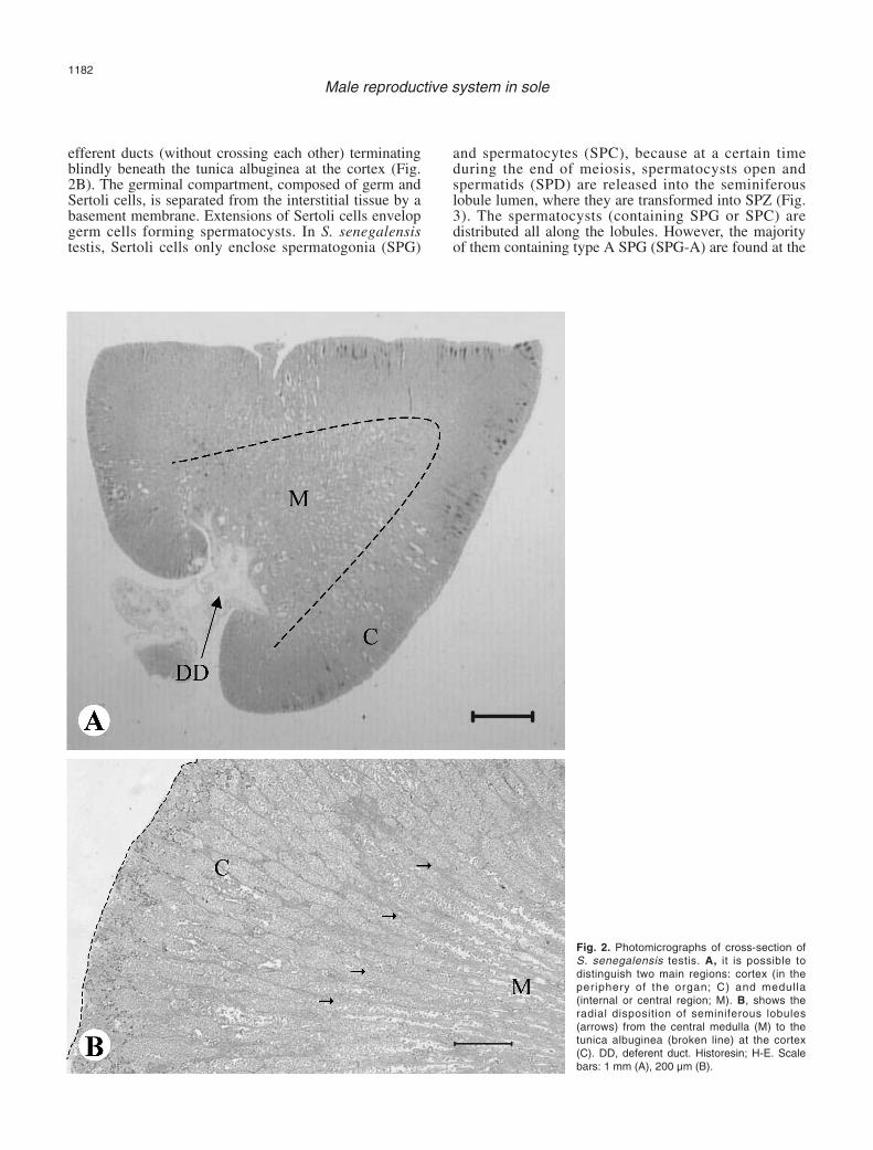
efferent ducts (without crossing each other) terminatingblindly beneath the tunica albuginea at the cortex (Fig.2B). The germinal compartment, composed of germ andSertoli cells, is separated from the interstitial tissue by abasement membrane. Extensions of Sertoli cells envelopgerm cells forming spermatocysts. In S. senegalensistestis, Sertoli cells only enclose spermatogonia (SPG)
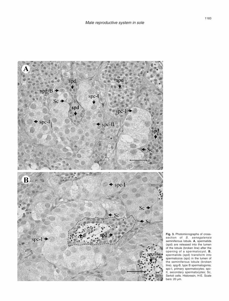
and spermatocytes (SPC), because at a certain timeduring the end of meiosis, spermatocysts open andspermatids (SPD) are released into the seminiferouslobule lumen, where they are transformed into SPZ (Fig.3). The spermatocysts (containing SPG or SPC) aredistributed all along the lobules. However, the majorityof them containing type A SPG (SPG-A) are found at the
1182
Male reproductive system in sole
Fig. 2. Photomicrographs of cross-section ofS. senegalensis testis. A, it is possible todistinguish two main regions: cortex (in theperiphery of the organ; C) and medulla(internal or central region; M). B, shows theradial disposition of seminiferous lobules(arrows) from the central medulla (M) to thetunica albuginea (broken line) at the cortex(C). DD, deferent duct. Historesin; H-E. Scalebars: 1 mm (A), 200 µm (B).
1183
Male reproductive system in sole
Fig. 3. Photomicrographs of cross-section of S. senegalensisseminiferous lobule. A, spermatids(spd) are released into the lumenof the lobule (broken line) after theopening of a spermatocyst. B,spermatids (spd) transform intospermatozoa (spz) in the lumen ofthe seminiferous lobule (brokenline). spg-B, type B spermatogonia;spc-I, primary spermatocytes; spc-II, secondary spermatocytes; Sc,Sertoli cells. Historesin, H-E. Scalebars: 20 µm.

distal part of the lobules.
Cellular components of the interstitial compartment
Myoid cells are elongated and thin, presenting afusiform and slightly basophilic nucleus and anacidophilic cytoplasm (Fig. 4A). They are distributedbordering the seminiferous lobules and spermatic ducts,and are randomly surrounded by collagen fibres (Fig.4A), which are very abundant in the interstitial tissue.
Leydig cells (Fig. 4B) are round shaped, have aspherical and basophilic nucleus (diameter: 3.18±0.14µm; n=8) with one or several nucleoli, and a clearcytoplasm in which a fine acidophilic dotted pattern canbe seen. Leydig cells are mainly located near bloodcapillaries in small groups.
In the interstitial tissue of the medulla it is possibleto distinguish some PAS-positive structures calledMMC’s (Fig. 4C). These groups of cells are yellowish tobrown coloured (unstained sections) and commonly ofnodular shape.
Testicular tissue is respectively irrigated andinnervated by numerous capillaries and amyelinic nervessituated within the interstitial tissue (Fig. 4B).
Germ and associated cells: the germinal compartment
The Sertoli cells (Figs. 3, 5) have a triangular andslightly basophilic nucleus with one nucleolus and areusually in contact with the basement membrane thatseparates germinal and interstitial compartments. Theirclear cytoplasm is difficult to distinguish because it doesnot stain differentially, and its expansions surround thedeveloping germ cells (SPG or SPC).
SPG-A (Fig. 5) are large and oval-shaped cells.Their spherical nucleus, 8.44±0.48 µm in diameter(n=8), is slightly basophilic and has a single, prominentand strongly basophilic centrally located nucleolus. Thenucleus occupies the vast majority of the cytoplasm andis located in a central or slightly eccentric position. SPG-A are found individually surrounded by Sertoli cellprocesses.
Type B SPG (SPG-B), resulting from successivemitoses of SPG-A, are found in small groups enclosedby Sertoli cells (Figs. 3A, 5). SPG-B have a similaraspect to SPG-A, though with a smaller size. Theirnuclei (diameter: 5.86±0.21 µm; n=7) have severalnucleoli, are located in a central position and are slightlymore basophilic than the nuclei of SPG-A, due toincreasing chromatin condensation. After adifferentiation process, SPG-B transform into primarySPC (SPC-I) which enter in meiosis.
SPC-I (Fig. 3) are similar to SPG-B in shape andsize (nuclear diameter: 5.65±0.11 µm; n=26), being alsowithin spermatocysts. Their cytoplasm is clear andcellular limits are hard to distinguish. At the beginningof the first meiotic division prophase, the nucleus has anirregular or granular texture, due to the distribution ofthe genetic material. After this division, a new germ cell,
1184
Male reproductive system in sole
Fig. 4. Photomicrographs of S. senegalensis testis showing the cellularcomponents of the interstitial compartment. Some spermatozoa (spz)are found in the lumen of the efferent ducts (ED). A, shows some myoidcells (Mc) and collagen fibres (Cf) bordering the efferent ducts (ED). B,shows two groups of Leydig cells (Lc) located near a blood vessel (BV).C, shows a melano-macrophage centre (MMC). Historesin, H-E. Scalebars: 20 µm.

the secondary SPC (SPC-II), is formed.SPC-II (Fig. 3A) are smaller in size compared to
SPC-I. They possess a spherical nucleus, 3.46±0.09 µmin diameter (n=19), with condensed and basophilicchromatin. SPC-II are rarely observed throughout thetestis since they enter rapidly in the second meioticdivision forming haploid and genetically unique SPD.
SPD are released into the lobule lumen anddifferentiate into SPZ in this space (Figs. 3, 5). SPD areround-shaped with a scarce cytoplasm and posses ahighly basophilic nucleus (diameter: 2.31±0.05 µm;n=40). SPD are the most abundant germ cell type in thetesticular tissue of S. senegalensis. SPD undergo a sizereduction and shape remodelling giving rise to SPZ.
The highly specialized SPZ can be morphologicallydistinguished under the light microscope into head andtail (Figs. 3B, 4, 5). Their small and rounded head,1.04±0.04 mm in diameter (n=13), is stronglybasophilic, while the tail, formed by one large flagellum,is acidophilic.
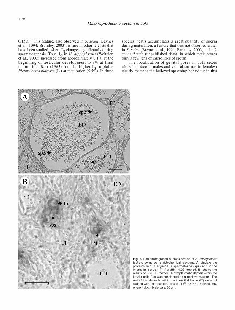
Histochemical characterisation of testicular tissue
Proteins in general, and especially those rich in basicaminoacids like arginine, increased as spermatogenesisproceeded (Fig. 6A). SPG were moderately rich in lysineand tyrosine, while in the rest of the germ cells thequantity of these aminoacids was low. Proteins rich intryptophan, cysteine and cystine had a low presence in
all germ cell types. Leydig and Sertoli cells weremoderately rich in proteins.
In general, carbohydrate quantity was low in allgerm cells types and in Sertoli cells, except carboxilatedmucosubstances /glycoproteins, which were abundant inSPZ and moderately abundant in Sertoli cells. Leydigcells were moderately stained with diastase-PAS reactionindicating the presence of glycogen.
Testicular tissue was unstained with Sudan black Band aqueous Nile blue, indicating the absence of lipids.
3ß-HSD activity was exclusively observed withinthe cytoplasm of Leydig cells from the interstitial tissue(Fig. 6B). Germ and Sertoli cells were not reactive.
Discussion
The shape and size of the testis of S. senegalensisare quite different to those of many Perciformes andPleuronectiformes that have been studied. Concerningshape, S. senegalensis presents round testis, while thetestis of the vast majority of teleosts (like English soleParophrys vetulus (Girard) [Sol et al., 1998] andAtlantic halibut Hippoglossus hippoglossus (L.)[Weltzien et al., 2002]) usually have an elongatedconformation. The testis morphology of S. solea (Bayneset al., 1994; Bromley, 2003) is similar to that of S.senegalensis, so it could be characteristic of this genus.Testis size and IG in S. senegalensis were very reducedthroughout all the spermatogenetic development (IG <
1185
Male reproductive system in sole
Fig. 5. Photomicrograph of cross-section of S. senegalensis testis,showing type A (spg-A) and B (spg-B)spermatogonia enclosed by Sertolicells (Sc). Some spermatids (spd) andspermatozoa (spz) are occupying thelumen of the lobules (L). Historesin, H-E. Scale bar: 20 µm.
0.15%). This feature, also observed in S. solea (Bayneset al., 1994; Bromley, 2003), is rare in other teleosts thathave been studied, where IG changes significantly duringspermatogenesis. Thus, IG in H. hippoglossus (Weltzienet al., 2002) increased from approximately 0.1% at thebeginning of testicular development to 3% at finalmaturation. Barr (1963) found a higher IG in plaicePleuronectes platessa (L.) at maturation (5.5%). In these
species, testis accumulates a great quantity of spermduring maturation, a feature that was not observed eitherin S. solea (Baynes et al., 1994; Bromley, 2003) or in S.senegalensis (unpublished data), in which testis storesonly a few tens of microlitres of sperm.
The localization of genital pores in both sexes(dorsal surface in males and ventral surface in females)clearly matches the believed spawning behaviour in this
1186
Male reproductive system in sole
Fig. 6. Photomicrographs of cross-section of S. senegalensistestis showing some histochemical reactions. A, displays theproteins rich in arginine in spermatozoa (spz) and in theinterstitial tissue (IT). Paraffin, NQS method. B, shows theresults of 3ß-HSD method. A cytoplasmatic deposit within theLeydig cells (Lc) was considered as a positive reaction. Therest of the elements within the interstitial tissue (IT) were notstained with this reaction. Tissue-Tek®, 3ß-HSD method. ED,efferent duct. Scale bars: 20 µm.

species that is supposed to be quite similar to thatreported in captive S. solea (Baynes et al., 1994). In thisspecies, fish form pairs during spawning, with the malealways positioned under the female, so that the genitalpores were close together. In this paired swimmingperiod, gamete release would take place, thus providingthe most suitable opportunity for a large quantity ofoocytes to be fertilized by spermatozoa.
Despite the differences in external morphologybetween S. senegalensis testis and the vast majority ofteleosts that have been studied, transverse testis sectionsshow more similarities. Testis of S. senegalensis presentsa radial or reniform structure with two main regions, thecortical one where germ cells are produced, and themedullar one, which consists of the efferent duct systemthat collects and stores the sperm. Therefore, thefunction changes from germ cell production in the outerregion to sperm collection and storage in the centralmedulla. This structure has been reported in many otherteleosts species, like anglerfish Lophiomus setigerus(Vahl) (Yoneda et al., 1998), cobia Rachycentroncanadum (L.) (Brown-Peterson et al., 2002), and bluefintuna Thunnus thynnus (L.) (Abascal et al., 2004).
In S. senegalensis only SPG and SPC are inside thespermatocysts, because SPD are released into the lobulelumen, where they transform into SPZ. This unusualfeature has been described in very few fishes likeOphidion sp. (Mattei et al., 1993), L. setigerus (Yonedaet al., 1998), and small red scorpionfish Scorpaenanotata (Rafinesque) (Muñoz et al., 2002). In thesespecies, several types of developing germ cells fromSPC to SPD are released from the spermatocysts into thelobule or tubule lumen (Yoneda et al., 1998). Mattei etal. (1993) concluded that this type of spermatogenesisshould be termed semi-cystic and distinguished from thecystic type which applies to most teleosts, like P.americanus (Harmin et al., 1995) and H. hippoglossus(Weltzien et al., 2002).
Spermatocysts are distributed all along theseminiferous lobules, as corresponds to unrestrictedspermatogonial type (Grier, 1993). Despite this, themajority of them containing SPG-A are found at thedistal part of the lobules, which may be reported as anintermediate type between the restricted spermatogonialtype, reported in Atheriniformes and someCyprinodontids like mummichog Fundulus heteroclitus(L.) (Selman and Wallace, 1986), and the unrestrictedspermatogonial type, found in all other teleosts to date.Muñoz et al. (2002) described a similar situation forSPG in S. notata, though they pointed out that thedifferent spermatogenetic stages appeared to develop asthe germ cells approach the centre of the gonad, similarto the process in the restricted-type testis. This featurewas not observed in S. senegalensis, where SPD can bereleased relatively near the distal part of the lobule.
S. senegalensis testis, as in all other vertebrates (LoNostro et al., 2003), is composed of a germinalcompartment, where germ cells associated with Sertolicells are located, and a non-germinal or interstitial
compartment, in which myoid cells, collagen fibres,Leydig cells, amyelinic nerves, MMC´s, and capillariescan be observed.
Myoid cells, first described in teleosts testis byBillard (1970), border the lobules and ducts forming anincomplete layer, as other authors have reported (Grau,1992; Lo Nostro et al., 2004). Grier et al. (1989)suggested that they could form a contractile network,which would facilitate the expulsion of sperm duringspawning.
Leydig cells have been reported as typicalcomponents of testicular tissue in the studied teleostspecies (Billard, 1983; Nagahama, 1983). The positivereaction of 3ß-HSD demonstrated in S. senegalensisLeydig cells seems to confirm the steroidogenic capacityof these cells, as in Leydig cells from common gobyPadogobius martensi (Günther) (Cinquetti and Dramis,2003) and swamp eel Synbranchus marmotatus (Bloch)(Lo Nostro et al., 2004). These authors also reported thatthese cells are the only ones that display ultrastructuralfeatures of steroidogenic cells: extensive smoothendoplasmic reticulum and mitochondria with tubularcistrae. Arellano (1999) reported the same ultrastructuralcharacteristics in S. senegalensis Leydig cells. All thesefeatures, together with the absence of reactivity of germand Sertoli cells to the 3‚-HSD enzymatic method,confirm that Leydig cells are the only cell type thatproduces androgens inside the testis of S. senegalensis.However, it remains to be investigated in flatfishwhether the testis is the main androgen producer orwhether extra-testicular tissues (Cavaco et al., 1997) alsohave the capacity of androgen production or conversion(Weltzien et al., 2004). In African catfish Clariasgariepinus (Burchell), Cavaco et al. (1997) concludedthat the hepatic production of 11-Ketotestosterone, themain androgen in teleosts (Borg, 1994), was of primaryrelevance in vivo. Preliminary data indicate that S.senegalensis liver does not have this capacity, since 3ß-HSD enzymatic activity was not detected within thistissue (unpublished data).
MMC’s are groups of different types of cells, mainlymacrophages (Agius and Roberts, 2003; Lo Nostro et al.,2004) and granulocytes (Grier and Taylor, 1998). Thesestructures have been reported as common components offish spleen, kidney and liver (Haaparanta et al., 1996;Arellano, 1999; Simpson et al., 2000; Mizuno et al.,2002). Their presence in the testis has been described inseveral teleosts like common snook Centropomusundecimalis (Bloch) (Grier and Taylor, 1998) and P.martensi (Cinquetti and Dramis, 2003). Grier and Taylor(1998) pointed out that occurrence of MMC´s in gonadscould be a generalized mechanism of gonadal regressionin animals with an annual reproductive cycle. Normally,MMC´s accumulate pigments like hemosiderin,lipofuscin, ceroids and melanin (Sarasquete andGutiérrez, 1984; Wolke, 1992).
In the germinal compartment, a specific number ofSertoli cells surround a clone of germ cells all at thesame stage of development (Pudney, 1995). In this unit
1187
Male reproductive system in sole

(spermatocyst), Sertoli cells attach to one another byspecialized junctional complexes resulting in a blood-testis barrier, that provide the germ cells with physicalsupport and factors needed for its survival, proliferationand differentiation (Weltzien et al., 2004). Functions ofsomatic Sertoli cells seem to be metabolites transport(Grier, 1975; Billard et al., 1982) and phagocytosis ofresidual bodies and degenerating germ cells includingresidual sperm (Grier, 1993; Grier and Taylor, 1998).
Morphological features of S. senegalensis germ cellsare similar to those described previously (Arellano,1999) and comparable to those extensively reported inteleosts like flounder Platichthys flesus (L.) (Gill et al.,2002) and H. hippoglossus (Weltzien et al., 2002).
Our histochemical study showed an increase in totalproteins, and especially in those rich in basic aminoacidslike arginine, as spermatogenesis progresses. Similarresults have been reported in toadfish Halobatrachusdidactylus (Schneider) (Blanco, 1991) and amberjack(Risso) Seriola dumerili (Grau, 1992). In teleosts, it hasbeen demonstrated that concomitantly with nuclearcondensation during spermatogenesis, nuclear histonesare replaced by protamins (Alfert, 1956; Louie andDixon, 1972). Protamins are strongly basic proteins andcontain abundant rests of lysine and arginine. Therefore,the increase in proteins rich in basic aminoacids duringS. senegalensis spermatogenesis would be related toprotamins synthesis and chromatin package. S.senegalensis SPZ contain a small quantity of reserves,mainly carboxylated mucosubstances /glycoproteins, andthey lack lipids like SPZ from other teleosts (Blanco,1991). Billard (1986) pointed out that these scarcereserves were typical of primitive SPZ that are present inspecies with external fertilization.
This study has precisely described the anatomy,histology and histochemical characteristics of the malereproductive system in S. senegalensis, a highlyattractive flatfish for the aquaculture industry in Spainand Portugal. Gonadosomatic index values and the typeof spermatogenesis, differ greatly from otherPleuronectiformes that have been studied. Therefore, thereproductive strategy in this species may also bedifferent, a hypothesis that requires further investigation.The present work contributes to improve the knowledgeof the reproductive biology of Solea senegalensis in ouraim to control its reproduction in captivity.
Acknowledgements. This research has been funded by the SpanishMCYT project no. AGL2000-0179-P4-02 to G. Martínez-Rodríguez andby the CSIC grant (I3P programme, ESF) to A. García-López. Theauthors thank V. Fernández Pasquier, from PROMAN S.L., for providingthe broodstock and F. Antonelli for her help in histological observations.Thanks are also given to J. L. Palazón, I. Viaña and F. Lo Nostro fortheir technical assistance in histochemical methods.
References
Abascal F.J., Megina C. and Medina A. (2004). Testicular development
in migrant and spawning bluefin tuna (Thunnus thynnus (L.)) fromthe eastern Atlantic and Mediterranean. Fish. Bull. 102, 407-417.
Agius C. and Roberts R.J. (2003) Melano-macrophage centres and theirrole in fish pathology. J. Fish Dis. 26, 499-509.
Alfert M. (1956). Chemical differentiation of nuclear proteins duringspermatogenesis in the salmon. J. Biophys. Biochem. Cytol. 2, 109-114.
Anguis V. and Cañavate J.P. (2005). Spawning of captive Senegal sole(Solea senegalensis) under a naturally fluctuating temperatureregime. Aquaculture 243, 133-145.
Arellano J.M. (1999). Descripción morfoestructural e histoquímica en ellenguado senegalés, Solea senegalensis (Kaup, 1858).Cuantificación y efectos histopatológicos del cobre. Ph.D. Thesis.Universidad de Cádiz. Spain.
Bancroft J.D. and Stevens A. (1990). Theory and practice of histologicaltechniques. 2nd edition. Churchill Livingstone. Edinburgh.
Barr W.A. (1963). The endocrine control of the sexual cycle in theplaice, Pleuronectes platessa (L.). III. The endocrine control ofspermatogenesis. Gen. Comp. Endocrinol. 3, 216-225.
Baynes S.M., Howell B.R., Beard T.W. and Hallam J.D. (1994). Adescription of spawning behaviour of captive dover sole, Solea solea(L.). Neth. J. Sea Res. 32, 271-275.
Billard R. (1970). Ultrastructure compareé de spermatozoïdes delquelques poissons Téléostéens. In: Comparative Spermatology.Baccetti B. (ed). Academic Press. New York. pp 71-79.
Billard R. (1983). A quantitative analysis of spermatogenesis in the trout,Salmo trutta fario. Cell. Tissue Res. 230, 495-502.
Billard R. (1986). Spermatogenesis and spermatology of some teleostfish species. Reprod. Nutr. Dev. 26, 877-920.
Billard R., Fostier A., Weil C. and Breton B. (1982). Endocrine control ofspermatogenesis in Teleost fish. Can. J. Fish. Aquat. Sci. 39, 65-79.
Blanco M.A. (1991). Estudio histológico y bioquímico durante lareproducción de Halobatrachus didactylus en la Bahía de Cádiz.Ph.D. Thesis. Universidad de Cádiz. Spain.
Borg B. (1994). Androgens in teleost fishes. Comp. Biochem. Physiol. C109, 219-245.
Bromley P.J. (2003). The use of market sampling to generate maturityogives and to investigate growth, sexual dimorphism andreproductive strategy in central and south-western North Sea sole(Solea solea L.). ICES J. Mar. Sci. 60, 52-65.
Brown-Peterson N.J., Grier H.J. and Overstreet R.M. (2002). Annualchanges in germinal epithelium determine male reproductive classesof the cobia. J. Fish Biol. 60, 178-202.
Cavaco J.E.B., Vischer H.F., Lambert J.G.D., Goos H.J.Th. and SchulzR.W. (1997). Mismatch between patterns of circulating and testicularandrogens in African catfish, Clarias gariepinus. Fish Physiol.Biochem. 17, 155-162.
Cinquetti R. and Dramis L. (2003). Histological, histochemical, enzymehistochemical and ultrastructural investigations of the testis ofPadogobius martensi between annual breeding seasons. J. FishBiol. 63, 1402-1428.
Dinis M.T. (1992). Aspects of the potential of Solea senegalensis Kaupfor aquaculture: larval rearing and weaning to artificial diets.Aquacult. Fish. Manag. 23, 515-520.
Drake P., Arias A.M. and Rodríguez, A. (1984). Cultivo extensivo depeces marinos en los esteros de las Salinas de San Fernando(Cádiz): II. Características de la producción de peces. Inf. Tec. Inst.Inv. Pesq. 116, 1-23.
Gill M.E., Spiropoulos J. and Moss C. (2002). Testicular structure and
1188
Male reproductive system in sole

sperm production in flounders from a polluted estuary: a preliminarystudy. J. Exp. Mar. Biol. Ecol. 281, 41-51.
Grau A. (1992). Aspectos histológicos, ciclo reproductor y principalesprocesos patológicos de Seriola dumerili, Risso 1810 (Carangidae).Ph.D. Thesis. Universidad Autónoma de Barcelona. Spain.
Grier H.J. (1975). Aspects of the germinal cyst and sperm developmentin Poecilia latipinna (Teleostei: Poeciilidae). J. Morphol. 146, 229-250.
Grier H.J. (1993). Comparative organization of Sertoli cells including theSertoli cell barrier. In: The sertoli cell. Russel L.D. and Griswold M.D.(eds). Cache River Press. Clearwater (Florida). pp 704-739.
Grier H.J. and Taylor R.G. (1998). Testicular maturation and regressionin the common snook. J. Fish Biol. 53, 521-542.
Grier H.J., Van der Hurk R. and Billard R. (1989). Cytologicalidentification of cell types in the testis of Esox lucius and E. niger.Cell Tissue Res. 257, 491-496.
Gutiérrez M., Sarasquete C. and Rodríguez R.B. (1985). Caracterescitohistoquímicos de carbohidratos y proteínas durante laovogénesis del lenguado, Solea senegalensis Kaup, 1858. Inv.Pesq. 49, 353-363.
Haaparanta A., Valtonen E.T., Hoffmann R. and Holmes, J. (1996). Domacrophage centres in freshwater fishes reflect the differences inwater quality?. Aquat. Toxicol. 34, 253-272.
Harmin S.A., Crim L.W. and Wiegand M.D. (1995). Plasma sex steroidprofiles and the seasonal reproductive cycle in male and femaleswinter flounder, Pleuronectes americanus. Mar. Biol. 121, 601-610.
JACUMAR (Junta Asesora de Cultivos Marinos) (2005). Estadísticas deproducción del año 2004. Ministerio de Agricultura, Pesca yAlimentación. Spain.
Lo Nostro F.L., Grier H.J., Meijide F.J. and Guerrero G.A. (2003).Ultrastructure of the testis in Synbranchus marmoratus (Teleostei,Synbranchidae): the germinal compartment. Tissue Cell 35, 121-132.
Lo Nostro F.L., Antonelli F.N., Quagio-Grassiotto I. and Guerrero G.A.(2004). Testicular interstitial cells, and steroidogenic detection in theprotogynous fish, Synbranchus marmoratus (Teleostei,Synbranchidae). Tissue Cell 36, 221-231.
Louie A.J. and Dixon G.H. (1972). Trout testis cells I. Characterizationby deoxyribonucleic acid and protein analysis of cells separated byvelocity sedimentation. J. Biol. Chem. 247, 5490-5497.
Martoja R. and Martoja–Pierson M. (1970). Técnicas de histologíaanimal. 1st edition. Toray Masson. Barcelona.
Mattei X., Siau Y., Thiaw O.T. and Thiam D. (1993). Peculiarities in theorganization of tesis of Ophidion sp. (Pisces, Teleostei). Evidence oftwo types of spermatogenesis in teleost fish. J. Fish Biol. 43, 931-937.
Medina A., Megina C., Abascal F.J. and Calzada A. (2000). Thespermatozoon morphology of Solea senegalensis (Kaup, 1858)(Teleostei, Pleuronectiformes). J. Submicrosc. Cytol. Pathol. 32,
645-650.Mizuno S., Misaka N., Miyakoshi Y., Takeuchi K. and Kasahara N.
(2002). Effects of starvation on melano-macrophages in the kidneyof masu salmon (Oncorhynchus masou). Aquaculture 209, 247-255.
Muñoz M., Casadevall M. and Bonet S. (2002). Testicular structure andsemicystic spermatogénesis in a specialized ovuliparous species:Scorpaena notata (Pisces, Scorpaenidae). Acta Zool. 83, 213-219.
Nagahama Y. (1983). The functional morphology of teleost gonads. In:Fish physiology. Vol. IX. Part A. Hoar W.S., Randall D.J. andDonaldson E.M. (eds). Academic Press. New York. pp 223-275.
Pearse A.G.E. (1985). Histochemistry. Theoretical and Applied. Vol. II:Analytical Technology. 4th edition. Churchill Livingstone. Edinburgh.
Pudney J. (1995). Spermatogenesis in nonmammalian vertebrates.Microsc. Res. Tech. 32, 459-497.
Rodríguez R.B. (1984). Biología y cultivo de Solea senegalensis Kaup1858 en el Golfo de Cádiz. Ph.D. Thesis. Universidad de Sevilla.Spain.
Sarasquete C. and Gutiérrez M. (1984). Caracteres histoquímicos delos granos de pigmento marrón-negro del riñón de dorada, Sparusaurata L. Inv. Pesq. 48, 539-547.
Schulz R.W. and Miura T. (2002). Spermatogenesis and its endocrineregulation. Fish Physiol. Biochem. 26, 43-56.
Selman K. and Wallace R.A. (1986). Gametogenesis in Fundulusheteroclitus. Am. Zool. 26, 173-192.
Simpson M.G., Parry M., Kleinkauf A., Swarbreck D., Walker P. andLeah R.T. (2000). Pathology of the liver, kidney and gonad offlounder (Platichthys flesus) from a UK estuary impacted byendocrine disrupting chemicals. Mar. Environ. Res. 50, 283-287.
Sol S.Y., Olson O.P., Lomax D.P. and Johnson L.L. (1998). Gonadaldevelopment and associated changes in plasma in plasmareproductive steroids in English sole, Pleuronectes vetulus, fromPuget Sound, Washington. Fish. Bull. 96, 859-870.
Weltzien F.A., Taranger G.L., Karlsen Ø. and Norberg B. (2002).Spermatogenesis and related plasma androgen levels in Atlantichalibut (Hippoglossus hippoglossus L.). Comp. Biochem. Physiol. A132, 567-575.
Weltzien F.A., Andersson E., Andersen Ø., Shalchian-Tabrizi K. andNorberg B. (2004). The brain-pituitary-gonad axis in male teleost,with special emphasis on flatfish (Pleuronectiformes). Comp.Biochem. Physiol. A 137, 447-477.
Wolke R.E. (1992). Piscine macrophage aggregates: A review. Ann.Rev. Fish Dis. 2, 91-108.
Yoneda M., Tokimura M., Fujita H., Takeshita N., Takeshita K.,Matsuyama M. and Matsuura S. (1998). Reproductive cycle andsexual maturity of the anglerfish Lophiomus setigerus in the EastChina Sea with a note on specialized spermatogenesis. J. Fish Biol.53, 164-178.
Accepted July 11, 2005
1189
Male reproductive system in sole
Related Documents


