Lungs and AIDS Dr Etienne Leroy Terquem – Pr Pierre L’Her SPI / ISP Soutien Pneumologique Internationa / International Support for Pulmonology

Welcome message from author
This document is posted to help you gain knowledge. Please leave a comment to let me know what you think about it! Share it to your friends and learn new things together.
Transcript

Lungs and AIDS
Dr Etienne Leroy Terquem – Pr Pierre L’Her
SPI / ISP Soutien Pneumologique Internationa / International Support for Pulmonology

Incidence of TB: HIV (+) vs HIV (-)
TB Infection
3-13%
every year
5%
first 2 years
>30% (40/60%)
lifetime
10%
lifetime
HIV (+) HIV (-)
World Health Organization
Increased risk of
TB disease in HIV
Active Tb disease after TB infection

More difficult to treat TB disease
• Adverse drug reactions
• May increase default rates in TB
programs
• May increase overall mortality rate in TB
programs

More difficult to diagnose
TB in HIV
• TB infection
– False positives and false negatives from
tuberculin skin test in HIV
• TB disease
– Classical symptoms may be missing
– Sputum smear may be negative
– Chest x-rays may be normal or atypical
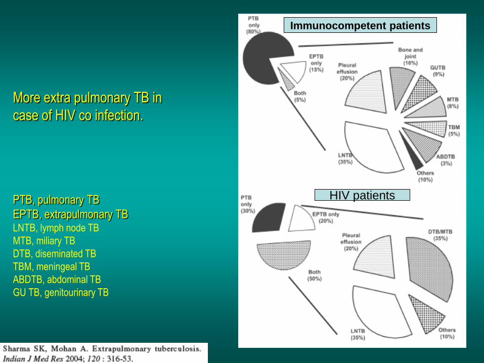
More extra pulmonary TB in
case of HIV co infection.
PTB, pulmonary TB
EPTB, extrapulmonary TB LNTB, lymph node TB
MTB, miliary TB
DTB, diseminated TB
TBM, meningeal TB
ABDTB, abdominal TB
GU TB, genitourinary TB
Immunocompetent patients
HIV patients

The global answer to TB/HIV:
Collaborative activities

Cascade of infections and cancers that develop as immune function is depleted HIV/AIDS prevention and treatment.NIH Stefano Bertozzi and coll.
Tuberculosis typical appearance
PCP
Mycobacterium avium complex
cytomegalovirus
Bacterial pneumoniae
TB: atypical appearance
Fungal infections


39% TB
30% PCP
16% Bacterial inf.
6% Mycosis
5% atypical mycobac.
4.7% Strongyloïdiasis
0.3% Cancer
Cambodia :
Vietnam : similar but very few fungal infections,
no atypical mycobacteriae or anguillulosis
Dakar and Bangui : very few PCP
more pneumoniae with S pneumoniae and H influenzae,
Kaposi, more severe illnesses with no diagnosis…
ANRS* study on lung diseases and AIDS in East Asia and Africa
*French national agency for scientific research in AIDS
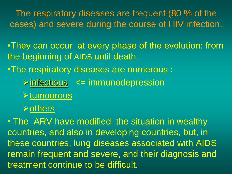
The respiratory diseases are frequent (80 % of the
cases) and severe during the course of HIV infection.
•They can occur at every phase of the evolution: from
the beginning of AIDS until death.
•The respiratory diseases are numerous :
infectious <= immunodepression
tumourous
others
• The ARV have modified the situation in wealthy
countries, and also in developing countries, but, in
these countries, lung diseases associated with AIDS
remain frequent and severe, and their diagnosis and
treatment continue to be difficult.

HIV and lungs: infections are the most
important problem
Lung = target for many and severe infections with high
incidence of death
• This natural evolution can be modified by :
– prophylactic treatment => effective on some
pathologies (ex: cotrimoxazole and pneumocystosis or
toxoplasmosis)
– The use of antiretroviral treatments: they are very
effective against HIV and can remain effective for a long
time if the treatment is correctly adapted and if the patient
is compliant.

VIH and lungs : 3 situations
• No prophylaxy against lung diseases and no ARV treatment
• No ARV treatment but possible access to prophylaxy (ex: prophylaxy of pneumocystosis by cotrimoxazole)
• ARV treatment is possible: mortality by infectious
disease drastically decreases

3 pathologies for 80% of pulmonary
infectious diseases in AIDS:
• Tuberculosis
• Pneumocystosis
• Bacterial pneumoniae

Respiratory diseases in
patients not receiving ARV
Pneumocystosis (PCP)
Tuberculosis
Bacterial Pneumoniae
Parasitic pneumoniae
Fungal pulmonary diseases
Atypical mycobacteriae
Viral diseases
Infectious diseases

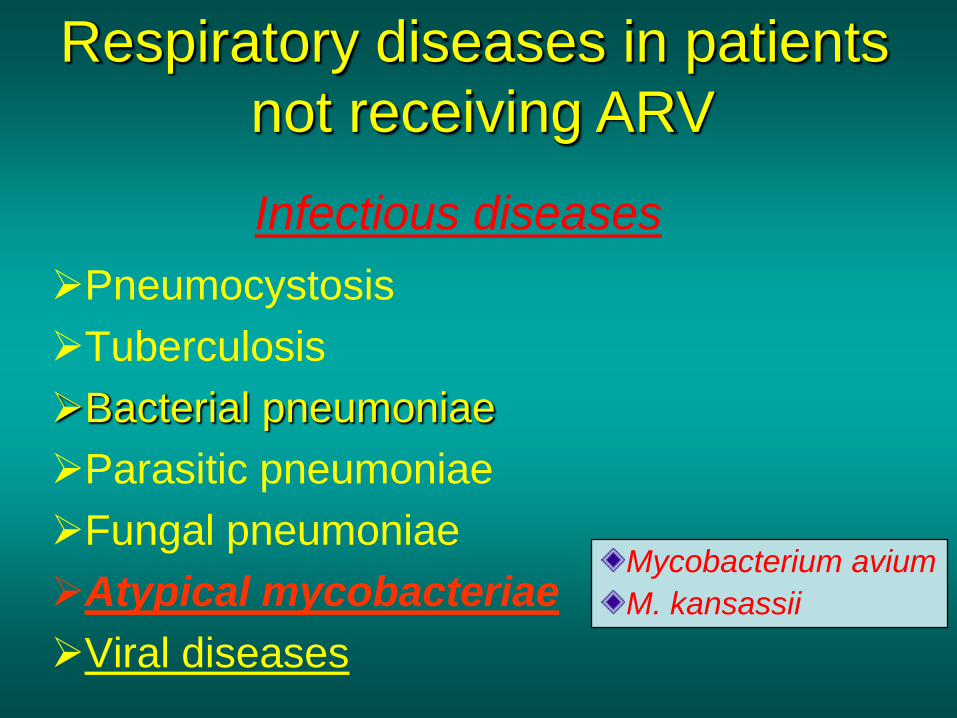
Respiratory diseases in patients
not receiving ARV
Pneumocystosis
Tuberculosis
Bacterial pneumoniae
Parasitic pneumoniae
Fungal pneumoniae
Atypical mycobacteriae
Viral diseases
Strepto pneumoniae
H. influenzae
others
Staph. aureus
Ps. aeruginosa
Legionnaires’ disease
Nocardia asteroides
Rhodococcus equi….
Infectious diseases

Toxoplasmosis
Anguillulosis
Leishmaniosis
Cryptosporidiosis
Strongiloïdiasis…
Strongyloidiasis
Pneumocystosis
Tuberculosis
Bacterial pneumonia
Parasitic pneumoniae
Fungal pneumoniae
Atypical mycobacteriae
Viral diseases
Infectious diseases
Respiratory diseases in
patients not receiving ARV

Pneumocystosis
Tuberculosis
Bacterial pneumonia
Parasitic pneumoniae
Fungal pneumoniae
Atypical mycobacteriae
Viral diseases
Cryptococcosis
Aspergillosis
Histoplasmosis
Coccidioïdomycosis
Penicilliosis
Coccidioidomycosis
Respiratory diseases in patients
not receiving ARV
Infectious diseases
Pneumocystosis
Tuberculosis
Bacterial pneumoniae
Parasitic pneumoniae
Fungal pneumoniae
Atypical mycobacteriae
Viral diseases
Respiratory diseases in patients
not receiving ARV
Infectious diseases
Mycobacterium avium
M. kansassii

Pneumocystosis
Tuberculosis
Bacterial pneumoniae
Parasitic pneumoniae
Fungal pneumoniae
Atypical mycobacteriae
Viral diseases
Respiratory diseases in patients
not receiving ARV
Infectious diseases
CMV

Possible etiologies according to radiological appearance:
Focalised condensation
Frequent pathology - common bacterial infection
possible pathology - Tuberculosis
- Mycosis (aspergillosis, cryptococcosis…)
- Non TB mycobacteria
- others bacterial infections (Nocardia, Actinomyces,
Rhodococcus equii.. )
rare pathology - lymphoma
- toxoplasmosis
differential diagnosis -lung cancer
courtesy of Mayaud in Girard, Katlama, Pialoux
“VIH 2001 “, éd. Douin Paris

Possible etiologies according to radiological appearance
Diffuse lesions
frequent pathology - pneumocystosis
- Kaposi’s disease
- tuberculosis
possible pathology - mycosis (aspergillosis,histoplasmosis, cryptococcosis)
- mycobactérioses atypical mycobacteries
- others infections (toxoplasmosis... )
- usual bacterial infections
rare pathology - interstitial lymphoïd pneumonia
Différential diagnosis - pulmonary œdema
- iatrogenic pneumopathy
courtesy of Mayaud in Girard, Katlama, Pialoux
“VIH 2001 “, éd. Douin Paris
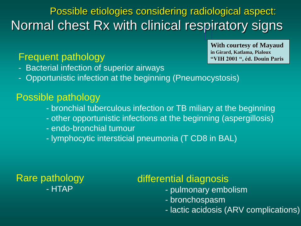
Possible etiologies considering radiological aspect:
Normal chest Rx with clinical respiratory signs
Frequent pathology - Bacterial infection of superior airways
- Opportunistic infection at the beginning (Pneumocystosis)
Possible pathology - bronchial tuberculous infection or TB miliary at the beginning
- other opportunistic infections at the beginning (aspergillosis)
- endo-bronchial tumour
- lymphocytic intersticial pneumonia (T CD8 in BAL)
Rare pathology - HTAP
differential diagnosis - pulmonary embolism
- bronchospasm
- lactic acidosis (ARV complications)
With courtesy of Mayaud in Girard, Katlama, Pialoux
“VIH 2001 “, éd. Douin Paris

Chest X ray TB HIV(-)
and HIV+ CD4>200
• cavitation is rare
• Frequency of TB pneumoniae
and adenopathies (often
associated)
• Lesions in inferior and
superior lobes
• Frequency of miliaries
Frequency of extra
pulmonary TB
• more frequent in
superior lobes
• caverns
• typical nodular
infiltrates (in the apex
and more or less
excavated)
Chest X ray TB HIV+
( CD4 < 200 )

not too severe immunodepression
CD4>200
Severe immunodepression
CXR in case of patients TB/ HIV+

Male 30 years old
Soldier HIV +
Pneumonia of right
superior and middle
Lobes
Hilar adenopathies
AFB x3 negative
Bronchial aspiration
and BAL : AFB+ +
Bronchial endoscopy:
Aspect of fistula from
adenopathy
© OFCP

TB bilateral pneumonia and mediastinal adenopathies in a
patient with AIDS. CD4 level: 50/mm3.
No excavation.

TB, HIV+: double tuberculous pneumonia; middle lobe and left
superior lobe. Mediastinal adenopathies

Cambodian national TB program
Bilateral pneumonia + mediastinum and hilar adenopathies + HIV context = TB

Bilateral tuberculous
pneumonia, in a
patient with AIDS.
Rapidly deteriorating
condition.
CD4 level: 35/mm3

HIV+ AFB pos.
TB pneumonia associated with
mediastinum adenopathies Courtesy Dr Peo Setha Cambodia

Left lower lobe TB
pneumonia
(negative silhouette sign
with cardiac left edge)
Bulky hilar adenopathy (positive silhouette sign
with Aortic arch)
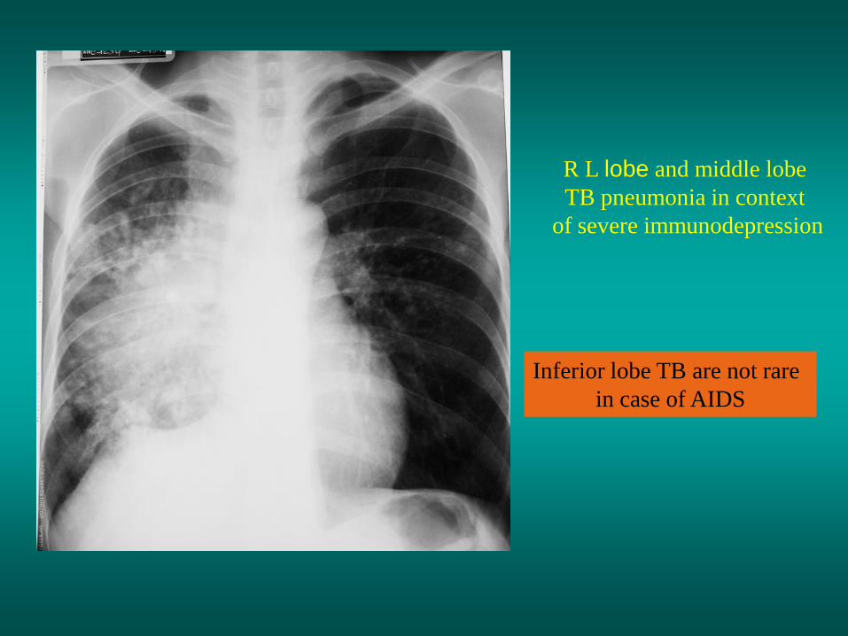
Inferior lobe TB are not rare
in case of AIDS
R L lobe and middle lobe
TB pneumonia in context
of severe immunodepression

Middle lobe, right upper lobe and left upper lobe
pneumonia. Mediastinum enlargment (probable
mediastinum adenopathies. IN HIV context, TB is highly
probable

TB of middle or
inferior lobes pneumoniae
are common in cases of AIDS
External segment
of middle lobe pneumonia
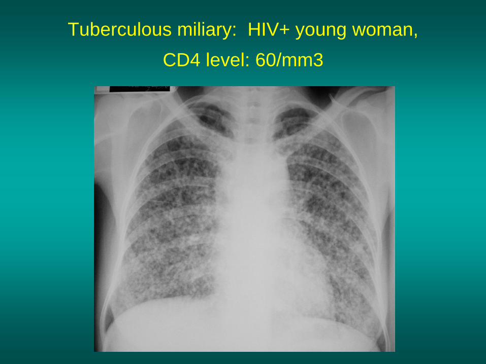
Tuberculous miliary: HIV+ young woman,
CD4 level: 60/mm3

Mediastinal adenopathies are frequent in AIDS cases
Endobronchial fistula with bronchogenic dissemination is possible

Immune reconstitution
inflammatory syndrome:
4 clinical exemples

Male HIV +, CD4 level: 50/mm3
October 2006. AFB (-)
Case 1

Dec 2006: AFB + in sputum .Beginning of TB
treatment
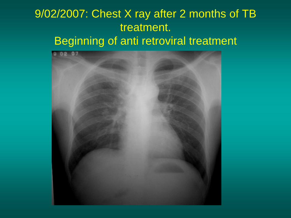
9/02/2007: Chest X ray after 2 months of TB
treatment.
Beginning of anti retroviral treatment

Chest X ray on 28/02/2007 (After 3 weeks of ARV
treatment)

Chest X ray on 04/04/2007: 7 weeks of
antiretroviral and TB treatment.(Favourable issue
after few weeks of associated cortico-steroïd)
treatment)

TB, VIH+, beginning of TB treatment
© OFCP
Case 2

Beginning of ARV treatment after 2 months
of TB treatment
© OFCP

severe pericarditis few weeks later
© OFCP

Pericardic drainage and continuation of the TB
and ARV treatment © OFCP

Male, HIV +. Left hilar tb adenopathy.TB treatment
for 2 months.
Chest X ray on the first day of ARV treatment.
Case 3

D12 of ARV treatment
Left increase opacities and pneumothorax

D 20 after drainage of the pneumothorax

Man, 37 years old, refugee from Congo. Diarrhea,
worsening condition, cough and weight loss.
HIV positive. CD4 level: 14/ mm3.
Beginning of ARV treatment the 30/12/2008 Case 4

X chest radio 3 weeks later. Dyspnea, cough,
fever, delirium and headache…
TB miliary with BK positive in sputum (PCR technique)
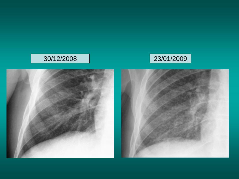
30/12/2008 23/01/2009


intra-cérebral tuberculous granulomas

Tuberculous abdominal adenopathies

Paradoxical reactions in the immune
reconstitution inflammatory syndrome
• Fever
• Adenopathies
• Ascites
• Pleural or pericardic effusion
• Pulmonary infiltrate or
pneumoniae
• Encephalic diseases
(tuberculoma)
-Beginning soon after introduction of ARV -The severity is correlated with the initial Immunodepression (CD4 level)
Frequency of pneumocystosis
Several micro-organisms are responsible for lung diseases
in AIDS. Therefore, differential diagnosis of TB in HIV patients are many,
and especially pneumocystosis.

Pneumocystoses
which clinical data ?
• HIV infection not known before (80% of cases )
• No prophylaxy with bactrim (100% of cases)
• Fever: 38° - 40°C
• Normal pulmonary auscultation (90% of cases)
• No extra-pulmonary signs (90% of cases)
• interstial/ alveolar diffuse opacities (100% of cases)
• Hypoxemy (SaO2 < 90%) 100% of cases
Courtesy of Chan Sarin ANRS1260

Interstitial picture: ground glass attenuation image

Male, HIV +, severe dyspnea, normal auscultation, SaO2 86%
interstitial and alveolar diffuse lesions

Bilateral alveolar and
Interstitial opacities
without excavation
© OFCP

© OFCP
Bilateral alveolar and interstitial opacities without excavation

Man25 years old. Increasing dyspnea and non productive cough. Fever 38° C.
First line antibiotic by doxycycline: no improvment.
Emergency room: Sa O2 88%, normal auscultation. Positive test for HIV.
Bronchio alveolar lavage: pneumocystosis

Male 42 years old, cough, exertional dyspnea, SaO2 92 %;HIV+ BAL: pneumocystosis Chest X ray: could be considered as normal. Possible ground glass attenuation image
Normal chest X ray

HIV+ context, exertional dyspnea, non-productive cough,
normal pulmonary auscultation, CD4 level 150/ mm3.
Endoscopy with BAL: P. jirovecii
Pneumocystosis at the beginning of the evolution
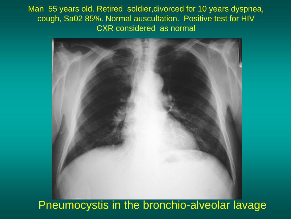
Man 55 years old. Retired soldier,divorced for 10 years dyspnea,
cough, Sa02 85%. Normal auscultation. Positive test for HIV
CXR considered as normal
Pneumocystis in the bronchio-alveolar lavage

CT scan of the previous patient
Normal CT scan

interstitial and diffuse pneumonia
with ground glass attenuation
+ Hypoxemia
SaO2 < 90 %
The pulse oxymeter is a very useful tool, yet expensive
But cheaper and cheaper (less than 100 dollars)
Without
cotrim.
prophylaxy
= PCP
If no oxymeter, remember that polypnea is proportional
to hypoxemia
Cotrimoxazole +/-cortisone
+ oxygen
are mandatory to prevent
death

National TB Program strategy for TB case finding
Respiratory +/- general symptoms
AFB-sputum X 3 (2 days)
If negative antibiotic (amoxycillin) X 10 days
If patient not improved and new smears negative
CXR (after 2 or 3 weeks)
In HIV infected patients, CXr must be performed early
If it was PCP, the patient is dead

non TB bacterial pneumoniae are fréquent
in case of HIV infection
Mild Immunodépression
Severe immunodepression

Non TB bacterial pneumonia are frequent in Hiv infection with
moderate immunodepression: Str. Pneumoniae, hemophilus….
They are often bilateral

Pneumopathy to pseudomonas aeruginosa.context of
worsening condition and cachexia. (CD4 level: 40/mm3)

bilateral opacities
With excavated nodules
Nocardiosis
Infectious disease and aids ward. khmero russian hospital
PhnomPenh
© OFCP© OFCP
One can also see fungal infections:
Cryptococcosis
Histoplasmosis
Penicillium marneffei
Invasive aspergillosis

© OFCP
© OFCP MGG X630
Grocott X630
Disseminated histoplasmosis
to H. capsulatum in an HIV+ patient BAL : fungal micro-
organisms in
the macrophages

© OFCP
BAL : Histoplasmosis
W. 20y. HIV+,
cough, dyspnea,
t° 38°5C
Miliary
AFB -
© OFCP

Sometimes in AIDS: poly-pathology

Soldier 25 y. old
Confusion, obnubilation
with quick onset,
Vomiting then coma
t° 40°C. HIV+
Bronchio alveolar
lavage : P. carinii
and S. aureus

L
Very severe dyspnea in HIV context
Not able to produce sputum. Endoscopy with BAL…
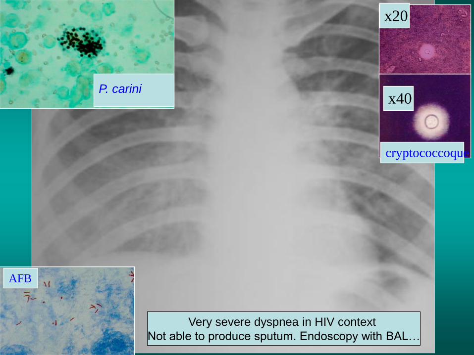
x20
x40
P. carini
cryptococcoque
AFB
L
Very severe dyspnea in HIV context
Not able to produce sputum. Endoscopy with BAL…
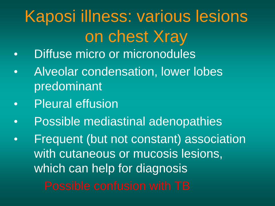
Kaposi illness: various lesions
on chest Xray • Diffuse micro or micronodules
• Alveolar condensation, lower lobes
predominant
• Pleural effusion
• Possible mediastinal adenopathies
• Frequent (but not constant) association
with cutaneous or mucosis lesions,
which can help for diagnosis
Possible confusion with TB

Kaposi illness Courtesy of Dr Difenthal. Tanzania

LIP
Lymphocitic interstitial pneumoniae:
- 2 to 5 years old HIV children (20% of HIV+ children in developed countries)
- Less frequent in adults. The diagnosis is difficult: One must eliminate
opportunistic infection (Bronchio-alveolar lavage and lung biopsy)

Lymphoma Courtesy Dr jaffer Dharsee . Tanzania

Lymphoma
• Rarely confined to chest only
• When seen in the chest it presents as
typical mediastinum nodal enlargement, or
mass in the anterior mediastinum (as in the
previous slide) pleural or pericardial effusion,
pulmonary infiltrates or pulmonary mass

In cases of acute respiratory
disease in AIDS with AFB(-)
in sputum,
Bronchial endoscopy and
BAL (broncho alveolar
lavage) are useful for
diagnosis if a reliable
bacteriological laboratory is
available…
Conclusion (1) :

BAL is feasible even in
low income countries
100 cc Slowly injection
Slowly aspiration > 50 cc collected

Conclusions (2)
VIH infection increases risk of developping very
severe TB
TB treatment is the same in HIV(+) et HIV(-) cases
but with more risk of complications and more risk of
associated opportunistic infections
Collaboration beetwen National TB program and
HIV/AIDS program is crcial in countries with high
TB/VIH prevalence
Mortality rate of lung disease in AIDS stay at a
high level

TB is yet the more frequent lung disease in AIDS and the more frequent cause of death
CXR can give informations for diagnosis especially if AFB neg
Diagnostic of opportunistic infections can be difficult and needs sophisticated explorations (need of financement and training)
Reference hospital should have special pulmonology ward with bronchoscopy and BAL available
Physicians working in TB program or in TB field must be correctly trained to CXR interpretation
Conclusions (3) CXR and TB / HIV
Related Documents