Low-Intensity Pulsed Ultrasound Enhances Bone Formation around Miniscrew Implants Khaliunaa Ganzorig a , Shingo Kuroda b , Yuichi Maeda a , Karima Mansjur a , Minami Sato a , Kumiko Nagata b , Eiji Tanaka b a Department of Orthodontics and Dentofacial Orthopedics, Tokushima University Graduate School of Oral Sciences, Tokushima, Japan b Department of Orthodontics and Dentofacial Orthopedics, Institute of Health Biosciences, Tokushima University Graduate School, Tokushima, Japan All authors certify that they have no conflict of interest. Correspondence to: Eiji Tanaka Department of Orthodontics and Dentofacial Orthopedics, Institute of Health Biosciences, Tokushima University Graduate School, 3-18-15 Kuramoto-Cho, Tokushima 770-8504, Japan. Tel: +81 88 6337357 / Fax: +81 88 6339139. E-mail: [email protected] Keywords: Low-intensity pulsed ultrasound, miniscrew implant, bone formation, stainless steel and titanium

Welcome message from author
This document is posted to help you gain knowledge. Please leave a comment to let me know what you think about it! Share it to your friends and learn new things together.
Transcript
Low-Intensity Pulsed Ultrasound Enhances Bone Formation around Miniscrew Implants
Khaliunaa Ganzoriga, Shingo Kuroda
b, Yuichi Maeda
a, Karima Mansjur
a, Minami Sato
a, Kumiko
Nagatab, Eiji Tanaka
b
aDepartment of Orthodontics and Dentofacial Orthopedics, Tokushima University Graduate
School of Oral Sciences, Tokushima, Japan
bDepartment of Orthodontics and Dentofacial Orthopedics, Institute of Health Biosciences,
Tokushima University Graduate School, Tokushima, Japan
All authors certify that they have no conflict of interest.
Correspondence to: Eiji Tanaka
Department of Orthodontics and Dentofacial Orthopedics, Institute of Health Biosciences,
Tokushima University Graduate School, 3-18-15 Kuramoto-Cho, Tokushima 770-8504, Japan.
Tel: +81 88 6337357 / Fax: +81 88 6339139.
E-mail: [email protected]
Keywords: Low-intensity pulsed ultrasound, miniscrew implant, bone formation, stainless steel
and titanium
ABSTRACT
Miniscrew implants (MSIs) are currently used to provide absolute anchorage in orthodontics;
however, their initial stability is an issue of concern. Application of low-intensity pulsed
ultrasound (LIPUS) can promote bone healing. Therefore, LIPUS application may stimulate
bone formation around MSIs and enhance their initial stability. Aim. To investigate the effect of
LIPUS exposure on bone formation after implantation of titanium (Ti) and stainless steel (SS)
MSIs. Methods. MSIs made of Ti-6Al-4V and 316L SS were placed on rat tibiae and treated with
LIPUS. The bone morphology around MSIs was evaluated by scanning electron microscopy and
three-dimensional micro-computed tomography. MC3T3-E1 cells cultured on Ti and SS discs
were treated with LIPUS, and the temporary expression of alkaline phosphatase (ALP) was
examined. Results. Bone-implant contact increased gradually from day 3 to day 14 after MSI
insertion. LIPUS application increased the cortical bone density, cortical bone thickness, and
cortical bone rate after implantation of Ti and SS MSIs (P < 0.05). LIPUS exposure induced
ALP upregulation in MC3T3-E1 cells at day 3 (P < 0.05). Conclusion. LIPUS enhanced bone
formation around Ti and SS MSIs, enhancing the initial stability of MSIs.
1. INTRODUCTION
Anchorage control is a key component in clinical orthodontic success. Numerous anchorage
devices have been proposed and used for more than a century. However, most of these devices
have disadvantages, in that their effectiveness depends on patient compliance and they cannot
provide absolute anchorage. The concept of skeletal anchorage was initially introduced to the
orthodontic field in the 1980s, reaching worldwide acceptance by the year 2000.1-5
In the skeletal
anchorage approach, screws or miniplates are fixed directly onto the bone and provide absolute
anchorage for several kinds of tooth movements.
Miniscrew implants (MSIs) made of Ti-6Al-4V alloy offer biocompatibility, improved
comfort, relative noninvasiveness, and fewer limitations in placement compared to other skeletal
anchorage devices.6,7
For these reasons, MSI use is generally accepted by orthodontists and
patients. However, the clinical use of MSIs has been associated with some risks and
complications, particularly screw failure.8 A recent systematic review found an overall success
rate of 86.5% among 4,987 MSIs placed in 2,281 patients. This rate is significantly lower than
the success rate of dental implants for prosthetic restorations.9 Therefore, increasing the success
rate of MSIs in clinical orthodontics is an urgent issue.
Low-intensity pulsed ultrasound (LIPUS) is a form of physical energy that can be
delivered to living tissue as acoustic waves. Used extensively as a therapeutic, operative, and
diagnostic tool in medicine, LIPUS does not have any known deleterious, carcinogenic, or
thermal effects on living tissues. LIPUS is well accepted as a noninvasive and safe tool for the
treatment of bone fractures.10
In previous reports, LIPUS increased the rate of repair of bone
fractures at all stages of the healing process11–14
and increased the mechanical properties of
callus.15,16
Radical changes in density are inherent in a healing tissue, which may lead to
gradients in physical strain.17
Ultrasound can be generated through several possible mechanisms.
Microbubble compression and acoustic streaming can have direct effects on cell membrane
permeability.18
Physical force serves as an extracellular signal to various cell types, including
bone cells. For example, BMP-2–induced bone formation19
and cellular mineralization20
were
enhanced after various types of biophysical stimulation of bone cells. Additionally, ultrasound
has been shown to enhance protein synthesis.21,22
Most MSI failures occur within a week after implant placement. This fact implies that
early bone metabolism around the inserted screws might be related to the screw stability. If
LIPUS can stimulate bone formation around the MSI, then LIPUS application after MSI
implantation may be able to enhance the initial implant stability. Therefore, the aim of this study
was to evaluate the effect of LIPUS application on bone formation after placement of MSIs made
of Ti-6Al-4V alloy or stainless steel (SS).
2. MATERIAL AND METHODS
2.1. Animals
Forty 6-week-old Sprague-Dawley rats (body weight: 190.0–210.5 g) were used in this study. All
animals were treated in accordance with the Guidelines for Animal Experiments at the
Laboratory Animal Centre of Tokushima University. Animals were caged individually under
automatically controlled conditions, with a temperature of 23 °C, humidity of 50%, and a 12
h:12 h light: dark cycle. Animals were given free access to tap water and rodent chow. All of the
protocols of the study were approved by the Ethics Committee of Tokushima University.
2.2. Surgical Procedure
Each animal was anesthetized with an intra-abdominal injection of 50 mg/kg sodium
pentobarbital (Kyoritsu, Tokyo, Japan). The skin was cleaned and incised with a scalpel blade.
The tibia surface was exposed, and the implant site was prepared by a standard surgical
technique with sharp drills. All drilling procedures were done under profuse irrigation with
sterile saline. For each animal, four MSIs were inserted with a miniature jewelry screwdriver.
MSIs measured 1.0 mm in inner diameter, 1.5 mm in outer diameter, and 1.6 mm in length. MSIs
were made of Ti-6Al-4V or 316L SS (Nishimura Metal, Sabae, Japan).
2.3. In Vivo LIPUS Application
The LIPUS exposure system used in this study was modified from a clinical device (Osteotron-D
IV; Ito Co, Tokyo, Japan) and was used in both the in vitro and in vivo experiments. Pulsed
ultrasound signal was transmitted at a frequency of 1.5 MHz with a spatially averaged intensity
of 30 mW/cm2 and 1:4 pulse rate (2 ms on to 8 ms off). LIPUS exposure was initiated 24 h after
MSI implantation. Tibiae on the right side were irradiated with LIPUS for 20 min/d. Tibiae on
the other side served as a sham-irradiated control.
2.4. Micro-Computed Tomography (μ-CT) Analysis
Animals were perfusion-fixed with 4% paraformaldehyde (Wako, Osaka, Japan) 0, 3, 7, and 14
days after MSI implantation. The tibiae were resected and dehydrated by incubation in a graded
ethanol series (70%, 80%, 90%, 99%, 100%, and 100% ethanol, v/v) for 12 h at each
concentration. Tibiae were embedded in methyl methacrylate resin (Technovit 9100; Kulzer,
Wehrheim, Germany). The resin blocks were scanned by µ-CT (Latheta LCT-200; Hitachi Aloka
Medical, Tokyo, Japan). Images consisted of 936 slices with a voxel size of 24 µm in all three
axes. Cortical bone and MSIs in the specimens were imaged and reconstructed in three
dimensions. Regions of interest adjacent to the implants were analyzed to determine the cortical
bone density (CBD; mg hydroxyapatite/cm3), cortical bone thickness (CBT; mm) and cortical
bone ratio (CBR; %). CBD was defined as the volumetric density of calcium hydroxyapatite.
CBR was calculated as the amount of cortical bone area divided by the total area in the tread.
2.5. Scanning Electron Microscopy (SEM) Analysis
After µ-CT analysis, the resin blocks were trimmed and prepared for analysis by SEM (Carry
Scope JCM-5700; JOEL, Tokyo, Japan; Fig. 1). Acquired images were analyzed with a digital
image analysis software (ImageJ version 1.44; US National Institutes of Health, Bethesda, MD).
2.6. Cell Culture
Ti-6Al-4V and 316L SS discs measuring 33 mm in diameter and 1 mm in thickness were used.
These discs were perfectly fitted to the bottom of wells in six-well cell culture plates. The discs
were sterilized, cleaned with ethanol and double-distilled water (ultrasonication, 15 min per wash
step), and autoclaved. Newborn mouse calvaria-derived osteoblastic precursor cells (MC3T3-E1)
were seeded at a density of 2 × 10⁴ cells in growth medium containing Dulbecco’s modified
Eagle’s medium (Invitrogen, Carlsbad, CA), 10% fetal bovine serum (JRH Biosciences, Kansas,
MO), and 1% penicillin G (Meiji Seika, Tokyo, Japan). Cells were grown in an incubator at
37 °C and 5% CO2. The complete medium was replaced every 2 to 3 d.
2.7. In Vitro LIPUS Application
The pulsed ultrasound signal was transmitted at a frequency of 3 MHz with a spatially averaged
intensity of 30 mW/cm2 and a pulse rate of 1:4 (2 ms on and 8 ms off, Osteotron-D IV). Cells
began to be treated with LIPUS 24 h after cell seeding. A six-well plate was held in place, and
the transducers were applied such that they directly touched the cell-containing medium.23,24
The
distance between the transducer and the cells was less than 4 mm. The cell culture was exposed
to a single treatment of ultrasound for 15 min.25,26
Ultrasound treatment was performed with the
culture dishes in an incubator (37 °C, 5% CO2-95% air). Control samples were subjected to the
same procedures under the same conditions but without ultrasound stimulation.
2.8. Count of Cell Number
MC3T3-E1 cells were seeded onto Ti and SS discs in six-well plates. Then, 2 × 10⁴ cells were
cultured with or without daily LIPUS stimulation for 1, 3 and 7 days. Cells in the four 1mm
corner squares were counted by a hemocytometer under a light microscope. The total cell
numbers were determined with the following calculation: Total cells = cell density (cells / ml) x
original volume of sample (ml)
2.9. Determination of Alkaline Phosphatase (ALP) Activity
To measure the ALP activity, MC3T3-E1 cells were seeded onto Ti and SS discs in six-well
dishes. Then, 2 × 10⁴ cells were cultured in the presence (LIPUS group) or absence (Control
group) of daily LIPUS stimulation for 7 or 14 days. ALP contents in the control and LIPUS
groups were analyzed by the ALP kit (Wako), according to the manufacturer’s instructions.
2.10. Determination of Mineralized Nodule Formation
MC3T3-E1 cells were seeded onto Ti and SS discs in six-well plates. Then, 2 × 10⁴ cells were
cultured with or without daily LIPUS stimulation for 21 days. The condition of the cells and
presence of nodule formation were checked routinely under stereomicroscopy. The presence of
mineralized nodules was determined by alizarin red (AR) staining (Wako).
2.11. Real-Time Polymerase Chain Reaction (PCR) Analysis
Cells were cultured with or without daily LIPUS stimulation for 7 days. All cultures were
terminated 24 h after the last ultrasound exposure. Total RNA was extracted from each culture
dish by using Trizol reagent according to the manufacturer’s instructions. The mRNA was
reverse-transcribed to yield cDNA, which served as a template for real-time PCR, to determine
the expression level of osteogenic factors. For this purpose, a 7500 real-time PCR system and
SYBR Green Master Mix (Applied Biosystems, Foster City, CA) were used. Real-time PCR was
performed with the following cycle conditions: 2 min at 50 °C, 10 min at 95 °C, 40 cycles of 15
s at 95 °C, and 1 min at 60 °C. The primer pairs for the mouse genes are listed in Table 1. The
PCR products were electrophoresed and quantified by scanning with an optical densitometer.
Expression levels of all genes were normalized to the expression level of β-actin (as a
housekeeping gene) within the same sample. Ratios of target genes to β-actin were calculated by
a digital image analysis software (ImageJ).
2.12. Statistical Analysis
All data are presented as the mean ± standard deviation (SD). Mean differences between the
groups were assessed with the Statistical Package for the Social Sciences (SPSS version 15;
Chicago, IL) by one-way analysis of variance (ANOVA). Comparisons between the LIPUS and
control groups were performed using the Mann-Whitney U test. In all cases, differences were
considered statistically significant at the 5% level of significance.
3. RESULTS
3.1. μ-CT Analysis
The CBD and CBT around Ti MSIs were enhanced on the LIPUS-treated side on days 7 and 14
(P < 0.05; Fig. 2(A) and Fig. 3(A) for CBD and CBT, respectively, Table 2). The CBR around Ti
MSIs was higher on the LIPUS-treated side than on the control side on day 14 (P < 0.05, Fig.
4(A)). The CBD around the SS MSIs showed no differences between the LIPUS-treated and
control sides from day 0 to day 7 after implantation, but the CBD was enhanced on the LIPUS-
treated side on day 14 (P < 0.05, Fig. 2(B)). The CBT around the SS MSIs was increased by
LIPUS application on day 14 (P < 0.05, Fig. 3(B)).The CBR around SS MSIs was higher on the
LIPUS-treated side than on the control side on days 7 and 14 (P < 0.05, Fig. 4(B)).
3.2. SEM Analysis
The bone-implant contact (BIC) around the Ti MSIs was higher on the LIPUS-treated side
compared to the control side on days 7 and 14 (P < 0.05, Fig. 5(A), Table 2). The BIC around the
SS MSIs was enhanced by LIPUS application on day 14 (P < 0.05, Fig. 5(B)). There was no
significant difference in BIC between the Ti and SS MSIs on day 3. However, on days 7 and 14,
the Ti MSIs showed more BIC than the SS MSIs (Fig. 6).
3.3. In Vitro Bone Formation
The numbers of MC3T3-E1 cells were increased by LIPUS application on both Ti and SS discs
on days 3 and 7 (P < 0.05, Fig. 7). For osteogenic cells on both Ti and SS discs, the ALP activity
was enhanced by LIPUS stimulation for 14th
d (Fig. 8). Mineralized nodule formation in the cells
was detected after LIPUS stimulation for 21 d. The AR staining of mineralized nodules was
more intense in the LIPUS-stimulated cells compared to the control cells, on both the Ti and SS
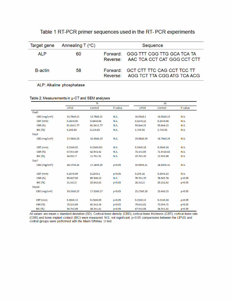
discs (Fig. 9). Expression of ALP mRNA was significantly increased in osteogenic cells on both
Ti and SS discs by 3 d after LIPUS application (P < 0.05, Fig. 10).
4. DISCUSSION
The purpose of the present study was to investigate whether LIPUS exposure could enhance
bone formation around Ti and SS MSIs in in vivo and in vitro experiments. Bone formation on
the LIPUS-treated side started earlier and became more extensive than bone formation on the
control side. The findings of this study provide a new strategy for increasing the initial stability
of MSIs after implantation. To the authors’ knowledge, this study is the first to examine the
effect of LIPUS exposure on bone formation around MSIs.
The BIC between the Ti MSI and surrounding bone was 23% ± 8% on day 7 and 38% ±
16% on day 14. For the SS MSI, these values were 20% ± 7% and 34.5% ± 13%, respectively. A
previous report suggested that a BIC of 10–58% can successfully resist orthodontic loads.27
In
addition, the CBR increased gradually after the implantation of MSIs of either material from day
3 to day 14. Therefore, both types of MSIs might be suitable as orthodontic anchorage devices.
A limitation of the present study was the use of tibiae instead of maxillofacial bones. It is
technically difficult to implant MSIs in the maxillofacial bones of rats and to confine the LIPUS
energy to the experimental side. Most reports evaluating the effect of LIPUS on bone healing
have chosen tibiae for implantation sites.28,29
However, two studies evaluated the BIC of Ti
implants placed on the maxillofacial bone, reporting BIC values of 23.4% ± 17.9% to 33.9% ±
3.3%.30,31
These values are similar to the present results, suggesting that there may not be a
significant difference in BIC values obtained with tibiae versus maxillofacial bones.
To the best of the authors’ knowledge, no study has evaluated the potential utility of
LIPUS in promoting bone formation after SS MSI placement. 316L SS was chosen as a material
for study because its composition, microstructure, and tensile properties are standardized in ISO
and ASTM material specifications. The Ti and SS MSIs showed osseointegration with the
surrounding bone in vivo. Bone formation and mineralization were promoted on the discs. With
the increased demand for Ti and its use in various fields, this metal is becoming more
expensive.32
Moreover, a major concern in MSI usage is screw breakage during insertion or
removal.8 Using MSIs made of SS might reduce the possibility of screw breakage because SS is
stronger than Ti-6Al-4V alloy.33
Recent reports have suggested that SS MSIs could be used as
temporary anchorage devices.34,35
In previous reports, SS MSIs showed no significant
differences from Ti MSIs in terms of stress distribution,36
rate of screw failure until removal,37
microdamage in the surrounding bones, rate of BIC,35
or stability.38
Moreover, SS MSIs had
higher insertion torque values than Ti MSIs.35
In terms of biocompatibility, Blaya et al.39
found
no significant difference in the metal ion concentration at different time points after 316L SS and
Ti-6Al-4V MSI placement. There was no difference in infection rate between Ti and SS nails
when used in the fixation of pediatric femoral fracture.40
These findings suggest the possibility
that SS MSIs can be used instead of Ti MSIs.
In this study, MC3T3-E1 cells were cultured on Ti and SS discs to compare effects of
LIPUS application on bone cell metabolism. The cell numbers counted by hemocytometry were
significantly increased by LIPUS stimulation on Ti and SS discs from day 3 to day 7. As a
marker of osteoblast differentiation, the ALP activity was higher on the LIPUS-treated discs than
on the control discs 2 weeks after LIPUS application. These results suggest that LIPUS
application might induce the differentiation of osteogenic cells in vitro. LIPUS stimulation also
increased the formation of mineralized nodules in osteoblastic cells 3 weeks after application,
which was evident by the intensity of AR staining. Additionally, expression of ALP mRNA was
increased on both Ti and SS discs after 3 d of LIPUS stimulation. This result indicates that
LIPUS can promote not only the differentiation, but also the mineralization, of osteoblasts on Ti
and SS discs.
One of the most desirable properties in the clinical usage of metallic implants is to
achieve osseointegration onto the surrounding bones as quickly as possible. In addition to its role
in orthodontics, this property has great benefit in orthopedics because it can allow early fixation
during bone healing in reconstructive surgery or artificial joint replacement. LIPUS application
significantly increased the BIC around both metallic implants, enhanced the CBD, CBT, and
CBR results after implantation, and increased the number of osteogenic cells on the metallic
discs. Together, these findings demonstrate the efficiency of LIPUS in bone formation.
Therefore, LIPUS might be useful for enhancing osseointegration after implantation and tissue
engineering around both Ti and SS implants.
5. CONCLUSIONS
The results of this study suggest that LIPUS application enhances bone formation around both Ti
and SS MSIs. Therefore, LIPUS exposure might enhance the initial stability and improve the
success rate of MSIs.
ACKNOWLEDGEMENT
We are grateful to Nobuyasu Yamanaka and Atsushi Chuma for providing the ultrasound devices
and technical support for the experiments. This study was supported by Grants-in-Aid for
Scientific Research from the Ministry of Education, Culture, Sports, Science, and Technology of
Japan (No. 25463185). The authors declare no potential conflicts of interest with respect to the
authorship and/or publication of this article.
REFERENCE
1. Roberts WE, Helm FR, Marshall KJ, Gongloff RK. Rigid endosseous implants for
orthodontic and orthopedic anchorage. Angle Orthod 1989; 59: 247–56.
2. Creekmore TD, Eklund MK. The possibility of skeletal anchorage. J Clin Orthod 1983;
17:266–9.
3. Umemori M, Sugawara J, Mitani H, Nagasaka H, Kawamura H. Skeletal anchorage
system for open-bite correction. Am J Orthod Dentofacial Orthop 1999; 115: 166-74.
4. Park HS, Bae SM, Kyung HM, Sung JH. Micro-implant anchorage for treatment of
skeletal Class I bialveolar protrusion. J Clin Orthod 2001; 35:417–22.
5. Kuroda S, Katayama A, Takano-Yamamoto T. Severe anterior open-bite case treated
using titanium screw anchorage. Angle Orthod 2004; 74:558–67.
6. Kyung HM, Park HS, Bae SM, Sung JH, Kim IB. Development of orthodontic micro-
implants for intraoral anchorage. J Clin Orthod 2003; 37:321–8.
7. Kuroda S, Sugawara Y, Deguchi T, Kyung HM, Takano-Yamamoto T. Clinical use of
miniscrew implants as orthodontic anchorage: Success rates and postoperative discomfort.
Am J Orthod Dentofacial Orthop 2007;131: 9–15.
8. Kravitz ND, Kusnoto B. Risks and complications of orthodontic miniscrews. Am J
Orthod Dentofacial Orthop 2007; 131:S43–51.
9. Papageorgiou SN, Zogakis IP, Papadopoulos MA. Failure rates and associated risk
factors of orthodontic miniscrew implants: A meta-analysis. Am J Orthod Dentofacial
2012; 142:577–95,
10. Waden SJ, Bennell KL, McMeeken JM, Wark JD. Acceleration of fresh fracture repair
using the sonic accelerated fracture healing system (SAFHS): A review. Calcif Tissue Int
2000; 66:157–63.
11. Azuma Y, Ito M, Harada Y, Takagi H, Ohta T, Jingushi S. Low-intensity pulsed
ultrasound accelerates rat femoral fracture healing by acting on the various cellular
reactions in the fracture callus. J Bone Miner Res 2001; 16: 671–80.
12. Angle SR, Sena K, Sumner DR, Virdi AS. Osteogenic differentiation of rat bone marrow
stromal cells by various intensities of low-intensity pulsed ultrasound. Ultrasonics 2011;
51:281–8.
13. Takikawa S, Matsui N, Kokubu T, Tsunoda M, Fujioka H, Mizuno K, Azuma Y. Low-
intensity pulsed ultrasound initiates bone healing in rat nonunion fracture model. J
Ultrasound Med 2001; 20:197–205.
14. Pipi E. Bossini FP, Oliveira P, Ribeiro JU, Tim C, Parizotto NA, Alves JM, Ribeiro DA,
Araujo HSS, Renno ACM. Low-intensity pulsed ultrasound produced an increase of
osteogenic genes expression during the process of bone healing in rats. Ultrasound Med
Biol 2010; 36:2057–64.
15. Busse JW, Bhandari M, Kulkarni AV, Tunks E. The effect of low intensity pulsed
ultrasound therapy on time to fracture healing: A meta-analysis. Can Med Assoc J 2002;
166: 437–41.
16. Shakouri K, Eftekharsadat B, Oskuie MR, Soleimanpour J, Tarzamni MK, Salekzamani
Y, Hoshyar Y, Nezami N. Effect of low-intensity pulsed ultrasound on fracture callus
mineral density and flexural strength in rabbit tibial fresh fracture. J Orthop Sci 2010; 15:
240–4.
17. Parvizi J, Wu CC, Lewallen DG, Greenleaf JF, Bolander ME. Low-intensity ultrasound
stimulates proteoglycan synthesis in rat chondrocytes by increasing aggrecan gene
expression. J Orthop Res 1999; 17:488–94.
18. Yang KH, Parvizi J, Wang SJ, Lewallen GD, Kinnick RR, Greenleaf JF, Bolander ME.
Exposure to low-intensity ultrasound increases aggrecan gene expression in a rat femur
fracture model. J Orthop Res 1996; 14:802–9.
19. Wijdicks CA, Virdi AS, Sena K, Sumner DR, Leven RM. Ultrasound enhances
recombinant human bmp-2 induced ectopic bone formation in a rat model. Ultrasound
Med Biol 2009; 35: 1629–37.
20. Pounder NM, Harrison AJ. Low intensity pulsed ultrasound for fracture healing: A
review of the clinical evidence and the associated biological mechanism of action.
Ultrasonics 2008; 48:330–8.
21. Lim K, Kim J, Seonwoo H, Park SH, Choung PH, Chung JH. In vitro effects of low-
intensity pulsed ultrasound stimulation on the osteogenic differentiation of human
alveolar bone-derived mesenchymal stem cells for tooth tissue engineering. BioMed Res
Int 2013; 1-15.
22. Unsworth J, Kaneez S, Harris S, Ridgway J, Fenwick S, Chenery D, Harrison A. Pulsed
low intensity ultrasound enhances mineralization in preosteoblast cells. Ultrasound Med
Biol 2007; 33:1468–74.
23. Katiyar A, Duncan RL, Sarkar K. Ultrasound stimulation increases proliferation of
MC3T3-E1 preosteoblast-like cells. J Ther Ultrasound 2014; 2:1-10.
24. Reher P, Harris M, Whiteman M, Hai HK, Meghji S. Ultrasound stimulates nitric oxide
and prostaglandin E2 production by human osteoblasts. Bone 2002; 31:236–41.
25. Inubushi T, Tanaka E, Rego EB, Ohtani A, Kawazoe A, Tanne K, Miyauchi M, Takata T.
Ultrasound stimulation attenuates resorption of tooth induced by experimental force
application. Bone 2013; 53: 497–506.
26. Nagata K, Nakamura T, Fujihara S, Tanaka E. Ultrasound modulates the inflammatory
response and promotes muscle regeneration in injured muscles. Ann Biomed Eng 2013;
41:1095-105.
27. Cornelis MA, Scheffler NR, Clerck HJ, Tulloch JF, Behets CN. Systematic review of the
experimental use of temporary skeletal anchorage devices in orthodontics. Am J Orthod
Dentofacial Orthop 2007; 131:S52–8.
28. Ustun Y, Erdogan O, Kurkcu M, Akova T, Damlar I. Effect of low intensity pulsed
ultrasound on dental implant osseointegration: A preliminary report. Eur J Dent 2008; 2:
254–62.
29. Liu Q, Liu X, Liu B, Hu K, Zhou X, Ding Y. The effect of low-intensity pulsed
ultrasound on the osseointegration of titanium dental implants. Brit J Oral Maxillofac
Surg 2012; 50:244–50.
30. Deguchi T, Takano-Yamamoto T, Kanomi T, Hartsfield JK, Roberts WE, Garetto LP.
The use of small titanium screws for orthodontic anchorage. J Dent Res 2003; 82:377–81.
31. Kim JW, Ahn SJ, Chang YI. Histomorphometric and mechanical analyses of the drill-
free screw as orthodontic anchorage. Am J Orthod dentofacial Orthop 2005; 128:190–4.
32. Lim YW, Kwon SY, Sun DH, Kim YS. Enhanced biocompatibility of stainless steel
implants by titanium coating and microarc oxidation. Clin Orthop Relate Res 2011; 469:
330–8.
33. Chen PQ, Lin SJ, Wu SS, So H. Mechanical performance of the new posterior spinal
implant: effect of materials, connecting plate, and pedicle screw design. Spine 2003; 28:
881–6.
34. Basha AG, Shantaraj R, Morgegowda SB. Comparative study between conventional en-
masse retraction (sliding mechanics) and en-masse retraction using orthodontic micro
implant. Implant dent 2010; 19: 128–36.
35. Brown RN, Sexton BE, Chu TMG, Katona TR, Stewart KT, Kyung HM, Liu SSY.
Comparison of stainless steel and titanium alloy orthodontic miniscrew implants: A
mechanical and histologic analysis. Am J Orthod Dentofacial Orthop 2014; 145: 496–505.
36. Singh S, Mogra S, Shetty VS, Shetty S, Philip P. Three-dimensional finite element analysis
of strength, stability, and stress distribution in orthodontic anchorage: A conical, self-
drilling miniscrew implant system. Am J Orthod Dentofacial Orthop 2012; 141:327–36.
37. Bachoura A, Yoshida R, Lattermann C, Kamineni S. Late removal of titanium hardware
from the elbow is problematic. ISRN Orthop 2012; 2012:1-4.
38. Lan H, Liu PH, Chang HP. Influence of different implant materials on the primary
stability of orthodontic mini-implants. Kaohsiung J Mel Sci 2012; 28:673–8.
39. Blaya MG, Blaya DS, Mello P, Flores EMM, Hirakata LM. Titanium alloy miniscrews
for orthodontic anchorage: an in vivo study of metal ion release. J Dent Sci 2011;
26:209–14.
40. Wall EJ, Jain V, Vora V, Mehlman CT, Crawford AH. Complications of titanium and
stainless steel elastic nail fixation of pediatric femoral fractures. J Bone and Joint Surg
Am 2008; 90:1305–13.
FIGURE LEGENDS
Figure 1: SEM images of Ti MSIs. (A) Representative images of experimental implants after 14
days of LIPUS exposure. (B) Regions of interest for BIC measurements were calculated by
digital image analysis software. MSI contact ratio (%) = (Length of bone contact/length of MSI
surface) × 100. All measurements were calculated under 200× magnification. (C) SEM
micrographs of implant surface. Boxed areas are enlarged to right. The arrow leading to BIC.
Figure 2: CBD values around the Ti (A) and SS (B) MSIs, without (CTRL) or with (LPS) LIPUS
application. *P < 0.05.
Figure 3: CBT values around the Ti (A) and SS (B) MSIs, without (CTRL) or with (LPS) LIPUS
application. *P < 0.05.
Figure 4: CBR values with the Ti (A) and SS (B) MSIs, without (CTRL) or with (LPS) LIPUS
application. *P < 0.05.
Figure 5: BIC around the Ti (A) and SS (B) MSIs, without (Control) or with LIPUS application.
*P < 0.05.
Figure 6: SEM images of MSIs after 14 days of implantation: (A) Ti MSI with LIPUS
application, (B) Ti MSI without LIPUS application (CTRL), (C) SS MSI with LIPUS application,
and (D) SS MSIs without LIPUS applications (CTRL). Scale bar: 500 μm.
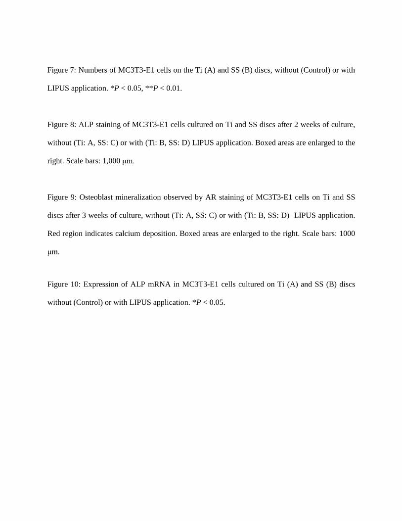
Figure 7: Numbers of MC3T3-E1 cells on the Ti (A) and SS (B) discs, without (Control) or with
LIPUS application. *P < 0.05, **P < 0.01.
Figure 8: ALP staining of MC3T3-E1 cells cultured on Ti and SS discs after 2 weeks of culture,
without (Ti: A, SS: C) or with (Ti: B, SS: D) LIPUS application. Boxed areas are enlarged to the
right. Scale bars: 1,000 μm.
Figure 9: Osteoblast mineralization observed by AR staining of MC3T3-E1 cells on Ti and SS
discs after 3 weeks of culture, without (Ti: A, SS: C) or with (Ti: B, SS: D) LIPUS application.
Red region indicates calcium deposition. Boxed areas are enlarged to the right. Scale bars: 1000
μm.
Figure 10: Expression of ALP mRNA in MC3T3-E1 cells cultured on Ti (A) and SS (B) discs
without (Control) or with LIPUS application. *P < 0.05.
Figure 1
Figure 2
Figure 3
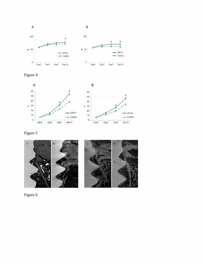
Figure 4
Figure 5
Figure 6
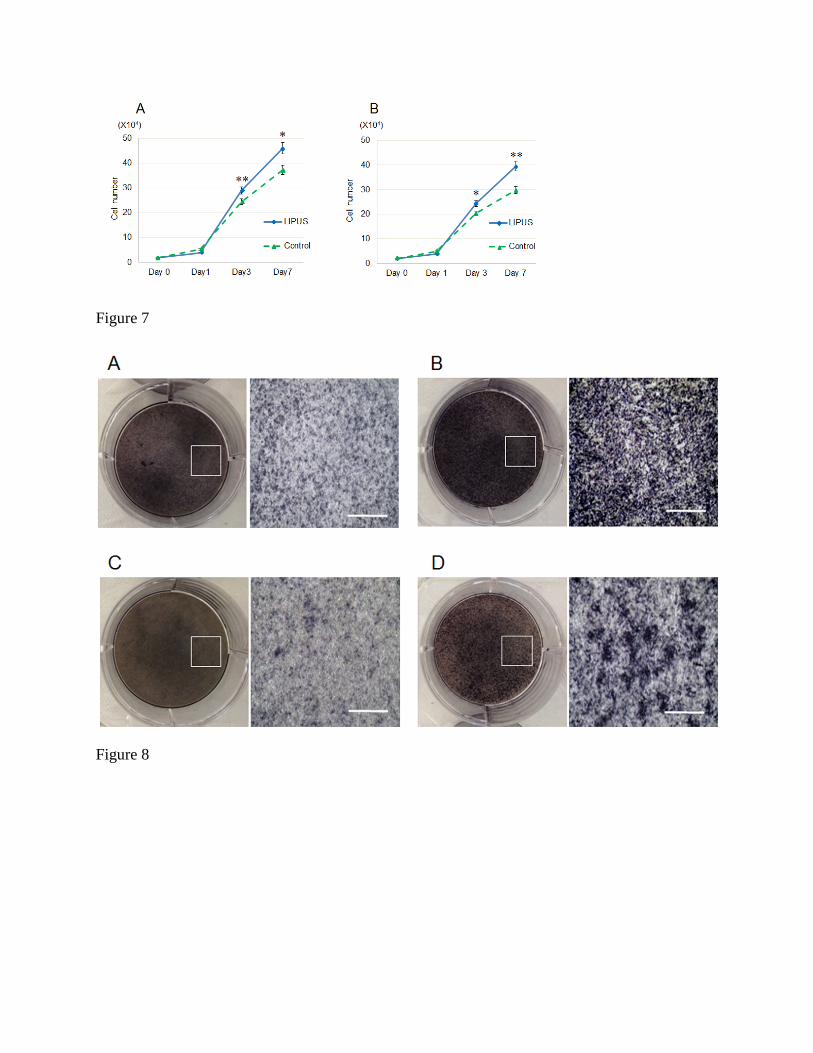
Figure 7
Figure 8
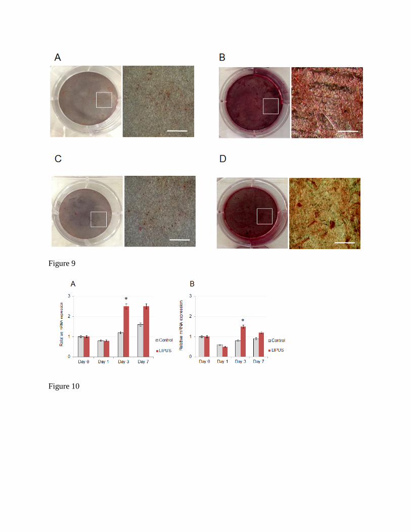
Figure 9
Figure 10
Related Documents


![Miniscrew Applications in Orthodontics · 2020. 12. 21. · ‘microscrews’, ‘miniscrew implants’. and ‘mini-implants’ [13,19-21]. In this chapter, we refer to them as miniscrews.](https://static.cupdf.com/doc/110x72/6148d5dc2918e2056c22f27f/miniscrew-applications-in-orthodontics-2020-12-21-amicroscrewsa-aminiscrew.jpg)