© 2016 Dental Press Journal of Orthodontics Dental Press J Orthod. 2016 July-Aug;21(4):17-23 17 Hideo Suzuki 1 , Won Moon 2 , Luiz Henrique Previdente 3 , Selly Sayuri Suzuki 4 , Aguinaldo Silva Garcez 5 , Alberto Consolaro 6 How to cite this article: Suzuki H, Moon W, Previdente LH, Suzuki SS, Garcez AS, Consolaro A. Miniscrew-assisted rapid palatal expander (MARPE): the quest for pure or- thopedic movement. Dental Press J Orthod. 2016 July-Aug;21(4):17-23. DOI: http://dx.doi.org/10.1590/2177-6709.21.4.017-023.oin Submitted: June 15, 2016 - Revised and accepted: June 30, 2016 » The authors report no commercial, proprietary or financial interest in the products or companies described in this article. Contact address: Alberto Consolaro E-mail: [email protected] Miniscrew-assisted rapid palatal expander (MARPE): the quest for pure orthopedic movement orthodontic insight 1 Professor, responsible for the Masters Program in Orthodontics, São Leopoldo Mandic, School of Dentistry, Campinas, São Paulo, Brazil. 2 Associate clinical professor and director of the Residency Program of Orthodontics, University of California, Los Angeles, USA. 3 Professor, São Leopoldo Mandic, School of Dentistry, Graduate Program, Campinas, São Paulo, Brazil. 4 Professor, São Leopoldo Mandic, School of Dentistry, Masters Program in Orthodontics, Campinas, São Paulo, Brazil. 5 Professor of Microbiology, São Leopoldo Mandic, School of Dentistry, Campinas, São Paulo, Brazil. 6 Full professor, Universidade de São Paulo (USP), School of Dentistry, Department of Orthodontics, Bauru, São Paulo, Brazil. Professor, Universidade de São Paulo (USP), School of Dentistry, Graduate Program, Ribeirão Preto, São Paulo, Brazil. The midpalatal suture has bone margins with thick connective tissue interposed between them, and it does not represent the fusion of maxillary palatal processes only, but also the fusion of palatal processes of the jaws and horizontal osseous laminae of palatal bones. Changing it implies affecting neighboring areas. It has got three segments that should be considered by all clinical analyses, whether therapeutic or experimental: the anterior segment (before the incisive foramen, or intermaxillary segment), the middle segment (from the incisive foramen to the suture transversal to the palatal bone ) and the posterior segment (aſter the suture transversal to the palatal bone ). Rapid palatal expansion might be recommended for patients at the final pubertal growth stage, in addition to adult patients with maxillary constriction. It represents a treatment solution that can potentially avoid surgical intervention. When performed in association with rapid palatal expanders, it might enhance the skeletal effects of the latter. Of the various designs of expansion appliances, MARPE (miniscrew-assisted rapid palatal expander) has been modified in order to allow its operational advantages and outcomes to become familiar in the clinical practice. Keywords: Midpalatal suture. Rapid palatal expansion. Palatal expansion. MARPE. DOI: http://dx.doi.org/10.1590/2177-6709.21.4.017-023.oin A sutura palatina mediana tem margens ósseas interpostas por tecido conjuntivo denso e não representa apenas a união dos processos palatinos da maxila, mas também a dos processos alveolares das maxilas e das lâminas horizontais dos ossos palati- nos. Modificá-la implica em influenciar outras áreas vizinhas. Ela tem três segmentos a serem considerados em todas as aná- lises clínicas, terapêuticas e experimentais: anterior (antes do forame incisivo, ou intermaxilar), médio (do forame incisivo até a sutura transversa com o osso palatino) e posterior (após a sutura transversa com o osso palatino). A expansão rápida da maxila pode ser indicada para pacientes que se encontram no final da fase de crescimento da puberdade, além de pacientes adultos com atresia maxilar, representando uma solução de tratamento, possivelmente evitando uma intervenção cirúrgica. Quando associada a aparelhos de protração maxilar, pode maximizar os efeitos esqueléticos desses. Entre os vários designs de aparelhos disjuntores, ultimamente encontra-se o MARPE (Miniscrew-Assisted Rapid Palatal Expander), que tem sido modificado para que ocorra, na rotina clínica, uma familiarização com suas vantagens operatórias e resultados obtidos. Palavras-chave: Sutura palatina mediana. Expansão rápida da maxila. Disjunção palatina. MARPE.

Welcome message from author
This document is posted to help you gain knowledge. Please leave a comment to let me know what you think about it! Share it to your friends and learn new things together.
Transcript

© 2016 Dental Press Journal of Orthodontics Dental Press J Orthod. 2016 July-Aug;21(4):17-2317
Hideo Suzuki1, Won Moon2, Luiz Henrique Previdente3, Selly Sayuri Suzuki4, Aguinaldo Silva Garcez5, Alberto Consolaro6
How to cite this article: Suzuki H, Moon W, Previdente LH, Suzuki SS, Garcez AS, Consolaro A. Miniscrew-assisted rapid palatal expander (MARPE): the quest for pure or-thopedic movement. Dental Press J Orthod. 2016 July-Aug;21(4):17-23. DOI: http://dx.doi.org/10.1590/2177-6709.21.4.017-023.oin
Submitted: June 15, 2016 - Revised and accepted: June 30, 2016
» The authors report no commercial, proprietary or financial interest in the products or companies described in this article.
Contact address: Alberto ConsolaroE-mail: [email protected]
Miniscrew-assisted rapid palatal expander (MARPE):
the quest for pure orthopedic movement
orthodontic insight
1 Professor, responsible for the Masters Program in Orthodontics, São Leopoldo Mandic, School of Dentistry, Campinas, São Paulo, Brazil.
2 Associate clinical professor and director of the Residency Program of Orthodontics, University of California, Los Angeles, USA.
3 Professor, São Leopoldo Mandic, School of Dentistry, Graduate Program, Campinas, São Paulo, Brazil.
4 Professor, São Leopoldo Mandic, School of Dentistry, Masters Program in Orthodontics, Campinas, São Paulo, Brazil.
5 Professor of Microbiology, São Leopoldo Mandic, School of Dentistry, Campinas, São Paulo, Brazil.
6 Full professor, Universidade de São Paulo (USP), School of Dentistry, Department of Orthodontics, Bauru, São Paulo, Brazil. Professor, Universidade de São Paulo (USP), School of Dentistry, Graduate Program, Ribeirão Preto, São Paulo, Brazil.
The midpalatal suture has bone margins with thick connective tissue interposed between them, and it does not represent the fusion of maxillary palatal processes only, but also the fusion of palatal processes of the jaws and horizontal osseous laminae of palatal bones. Changing it implies affecting neighboring areas. It has got three segments that should be considered by all clinical analyses, whether therapeutic or experimental: the anterior segment (before the incisive foramen, or intermaxillary segment), the middle segment (from the incisive foramen to the suture transversal to the palatal bone ) and the posterior segment (after the suture transversal to the palatal bone ). Rapid palatal expansion might be recommended for patients at the final pubertal growth stage, in addition to adult patients with maxillary constriction. It represents a treatment solution that can potentially avoid surgical intervention. When performed in association with rapid palatal expanders, it might enhance the skeletal effects of the latter. Of the various designs of expansion appliances, MARPE (miniscrew-assisted rapid palatal expander) has been modified in order to allow its operational advantages and outcomes to become familiar in the clinical practice.
Keywords: Midpalatal suture. Rapid palatal expansion. Palatal expansion. MARPE.
DOI: http://dx.doi.org/10.1590/2177-6709.21.4.017-023.oin
A sutura palatina mediana tem margens ósseas interpostas por tecido conjuntivo denso e não representa apenas a união dos processos palatinos da maxila, mas também a dos processos alveolares das maxilas e das lâminas horizontais dos ossos palati-nos. Modificá-la implica em influenciar outras áreas vizinhas. Ela tem três segmentos a serem considerados em todas as aná-lises clínicas, terapêuticas e experimentais: anterior (antes do forame incisivo, ou intermaxilar), médio (do forame incisivo até a sutura transversa com o osso palatino) e posterior (após a sutura transversa com o osso palatino). A expansão rápida da maxila pode ser indicada para pacientes que se encontram no final da fase de crescimento da puberdade, além de pacientes adultos com atresia maxilar, representando uma solução de tratamento, possivelmente evitando uma intervenção cirúrgica. Quando associada a aparelhos de protração maxilar, pode maximizar os efeitos esqueléticos desses. Entre os vários designs de aparelhos disjuntores, ultimamente encontra-se o MARPE (Miniscrew-Assisted Rapid Palatal Expander), que tem sido modificado para que ocorra, na rotina clínica, uma familiarização com suas vantagens operatórias e resultados obtidos.
Palavras-chave: Sutura palatina mediana. Expansão rápida da maxila. Disjunção palatina. MARPE.

© 2016 Dental Press Journal of Orthodontics Dental Press J Orthod. 2016 July-Aug;21(4):17-2318
Miniscrew-assisted rapid palatal expander (MARPE): the quest for pure orthopedic movementorthodontic insight
Both the macroscopic and microscopic mor-phologies of the midpalatal suture were described by Ennes,13 Ennes et al12 and Ennes and Consolaro14 in 2004 and 2004, including its morphological changes in terms of the chronological evolution of humans, primates, rabbits and rats.
Understanding the biological events implicated in orthodontic, orthopedic and surgical procedures carried out in the midface requires knowledge about the structures of the midpalatal suture vertically and horizontally at different age groups.
The midpalatal suture is wrinkled and arranged in an overlapping as well as sinuous pattern, with bone margins with thick connective tissue interposed be-tween them in three to five layers. It should be high-lighted that the midpalatal suture:
1) Does not represent the fusion of maxillary palatal processes only, but also the fusion of alveolar palatal processes of the jaws and horizontal osseous laminae of palatal bones. Changing it implies affect-ing neighboring areas (Fig 1).
2) It has got three segments (Fig 1) that should be considered by all clinical analyses, whether therapeutic or experimental:
» the anterior segment: before the incisive foramen, or intermaxillary segment;
» the middle segment: from the incisive foramen to the suture transversal to the palatal bone;
» the posterior segment: after the suture transversal to the palatal bone.
Morphological and clinical-therapeutic approaches often aim at the midpalatal suture, but do not include its anterior segment. Likewise, they occasionally aim at its posterior segment.
The influence exerted by rapid maxillary expan-sion (RME) over other structures relative to the mid-palatal suture is rarely carefully studied.31 To what ex-tent does the suture (Figs 1 and 2) transversal to the palatal bone lessen forces applied by maxillary expan-sion appliances, given that it represents an interrup-tion of a continuous solid structure?
The palate develops from the sixth to the 12th week of embryonic life. The primary palate is the secondary ossification center of the maxilla, arising from the medial nasal processes in the intermaxil-lary segment. During the eighth week of intrauter-ine development, ossification centers merge to form
the maxilla.19 The primary palate establishes a palatine view of the suture between the premaxilla and the maxilla with the secondary palate,27 without leaving any signs or marks in the adult individual. The same process does not apply to the palatal bone with which the suture is well established.
The osteogenic process in the midpalatal suture is similar to the growth and bone remodeling provided by the periosteum in other bone surfaces, and ful-fills the requirements of adjacent tissues by means of external stimuli also known as functional demands.9
The midpalatal suture is not a center of maxillary bone growth,11,20,25 but it responds to osteogenic stimuli.
MIDPALATAL SUTURE CHRONOLOGY AS PER AGE GROUP
The chronological developmental phases of ossifica-tion of the human midpalatal suture were microscopi-cally studied in 24 individuals, aged between 15 and 35 years old, by Persson and Thilander29 in 1977. Os-sification of the suture starts at the posterior region by means of mineralized bridges formed from posteriorly to anteriorly, varying according to the chronological age at which they form — in consonance with the end of the growth stage and facial development, under in-fluence exerted by bone maturation.
After analyzing the skeletal age of the palatal su-tures of 186 human skulls, Mann et al2 identified, in 1991, the following sequence of ossification:
1. It starts with the incisive suture. 2. Followed by the posterior segment of the mid-
palatal suture. 3. Followed by the transverse palatine suture. 4. Finally, it is followed by the middle segment of
the midpalatal suture.Ossification of the suture has been considered
as a limiting factor for rapid palatal expansion, and it seems to be a consensus that it starts at the posterior segment.32
Imaging diagnosis of closure or ossification of the midpalatal suture remains limited2,12,13,14 in occlusal ra-diographs or tomographic slices, especially if we con-sider that the small bridges formed are microscopically identified, but do not necessarily result in visible im-ages. Small bone length and/or thickness cannot be de-tected by imaging examination, especially if calibration and resolution of modern appliances are considered.

Suzuki H, Moon W, Previdente LH, Suzuki SS, Garcez AS, Consolaro A
© 2016 Dental Press Journal of Orthodontics Dental Press J Orthod. 2016 July-Aug;21(4):17-2319
orthodontic insight
Figure 1 -Midpalatal suture and its three segments (anterior, middle and posterior) which are relative to other structures, for instance, the
transverse palatine suture, and nasal bones.
Despite the advances made in the last ten years, the conclusions reached by Ennes,13 Ennes et al12
and Ennes and Consolaro,14 in 2002 and 2004, with studies conducted on the morphology of the midpalatal suture at different stages of develop-ment, remain appropriate, especially regarding the following:
1) By using modern diagnostic tools, it is impos-sible to precisely and safely perform clinical and ra-diographic analyses of the onset of ossification of the midpalatal suture and its degree of structural implica-tion. It is possible to claim that the older the patient, the higher the likelihood that the midpalatal suture and maxillary inter-relations become ossified.
© 2016 Dental Press Journal of Orthodontics Dental Press J Orthod. 2016 July-Aug;21(4):17-2320
Miniscrew-assisted rapid palatal expander (MARPE): the quest for pure orthopedic movementorthodontic insight
2) In human beings, ossification of the midpalatal suture occurs within the period from adulthood to the elderly stages in life; however, the primary fragil-ity of ossification bridges is likely not to allow us to render them responsible for the unsuccessful outcomes of some intermaxillary expansion procedures. Should that be the case, it is recommended that factors inher-ent to technique and ossification of other facial sutures be reassessed.
In other words, the quest for new means and appli-ances aimed at performing palatal expansion must be encouraged, so that we can increasingly make advances in this technique in favor of the best results for patients.
RAPID PALATAL EXPANSION AND MARPEThe first palatal expansion appliance was developed
by Angell3 in 1860. At that time, the procedure was not incorporated to the orthodontic practice, as it was
considered inappropriate by Angell’s colleagues who were influenced by rhinologists’ fears.
European orthodontists, Maxillary Orthope-dics enthusiasts, brought the technique back based on the works by Derischsweiler (1953) and Korkhaus18
(1960). American Orthodontics became interested in it when Haas,15 in 1961, carried out the procedure in pigs and proved the existence of the microscopic events implicated. The technique employed in patients with atrophic maxilla achieve positive outcomes and the procedure was considered safe and as an alternative for more complicated cases, such as Class II malocclusion associated with posterior crossbite.
From that time onwards, other appliance designs were proposed with the same purpose: as an alterna-tive to correct malocclusions associated with atrophic maxilla. If the patient performs 2/4 of a turn every day, expansion occurs within one to two weeks. The desired extension is achieved, including overcorrection and re-lapse. The latter often occurs as a result of lack of balance or tensegrity among the pieces that form the midface.7
The orthopedic expansion appliance — with or without a screw secured to a resin support adapted to the palatal mucosa — exerts force onto supporting teeth, thereby leading to a decrease of blood flow in the buccal periodontium while also forming extensive hyaline areas8 in which the conditions for the recov-ery of bone modeling units, essential for tooth move-ment to occur within the bone, are nonexistent. Bone resorption occurs at a distance and at a slower pace, keeping the supporting teeth in position until the su-ture yields to the resultant force.
Once expansion has been evinced, by means of dia-stema opening between maxillary central incisors, the next step is to achieve crossbite overcorrection, so as to prevent relapse. At this stage, the expansion appliance is inactivated for three months. After this period, a re-tention is installed and kept for six months. In cases in which maxillary expansion is not successfully achieved by means of orthodontic procedures, surgery might be recommended for rapid palatal expansion.
Undesirable effects include discomfort at the regions of incisors or nasal suture, and ulceration or necrosis of the palatal mucosa. There might be some swelling at the midpalatal suture, particularly soon after expansion. Ischemia and necrosis of the palatal mucosa might occur when the suture does not yield to forces applied by the
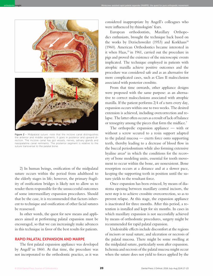
Figure 2 - Midpalatal suture: note that the incisive canal distinguishes the anterior and middle segments. It goes in posterior and upward di-rection. The incisive canal has got vessels, nerves, salivary glands and nasopalatine canal remnants. The posterior segment is relative to the suture transversal to the palatal bone.

Suzuki H, Moon W, Previdente LH, Suzuki SS, Garcez AS, Consolaro A
© 2016 Dental Press Journal of Orthodontics Dental Press J Orthod. 2016 July-Aug;21(4):17-2321
orthodontic insight
tooth-mucosa-supported appliance.30,34 The mucosal le-sion resulting from decreased blood flow – caused by the resultant force – make the procedure unfeasible in the event of occurring before expansion. Supporting teeth might undergo tooth resorption on the buccal surface of roots.8,28
The outcomes of palatal expansion vary from fail-ure to a horizontal gain of 4 mm. Failure has been at-tributed to: patient’s skeletal maturity;36,37 variation in the transverse measure, depending on the post-treat-ment moment when data were collected; and reten-tion,33 with relapse of horizontal measures and max-illary movement downward being identified — with the maxilla, later on, remaining unchanged or with its primary position restored.4,5,6,17,37
In order to increasingly enhance the procedure of palatal expansion, one seeks to improve or innovate the appliances used. In 2010, Lee et al21 treated a 20-year-old patient with severe transverse discrepancy and mandibular prognathism. Before orthognathic sur-gery, the patient used an expansion appliance secured to the palate by means of miniscrews (miniscrew-as-sisted rapid palatal expander, or MARPE). Expansion was achieved with minimal damage to teeth and peri-odontium, with stable outcomes confirmed by clinical and radiographic examination. The authors concluded that it is an effective treatment modality used for trans-verse correction and which might eliminate the need for a few surgical procedures in patients with cranio-facial discrepancies, thus taking advantage of the pos-sibilities offered by the sutures.
Recently, based on Lee’s studies, Park and Hwang,10,21
Moon26 and MacGinnis et al23 developed the maxillary skeletal expander (MSE, Biomaterial Korea, Seoul, South Korea) with four miniscrews installed into the
expansion screw body, parallel to the midpalatal suture and to itself. Even more recently, Suzuki et al35 changed the rapid maxillary expansion appliance, securing it by means of miniscrews (MARPE); however, with a dif-ferent design (Peclab, Belo Horizonte, Brazil) (Fig 3). MARPE’s new design has been used in a number of patients with atrophic maxilla, both young, growing pa-tients and adult ones.
In the appliance developed by Lee et al,21 the mini-screws are secured to the turn-key by means of exten-sions welded to the expansion screw, and joined with light-curing resin. With miniscrews kept away from the midpalatal suture, there is an increase in the risk of per-forating underlying structures (such as canals and nerves in both anterior and posterior regions), as well as on the sides, which is even more serious, as there would be four sites to be chosen individually. Alves et al1 mapped the areas of risk implied in securing miniscrews onto the human palate. In MSE26,35 and MARPE10,35 appli-ances, miniscrews are used as a support for the expan-sion screw (Fig 3) and would be secured in a more even manner parallel to the suture, with a view to aiming at a thicker bone area, so as to increase primary stability and provide a more efficient propagation of forces to the nasomaxillary complex.
The midpalatal suture is located right behind the incisive foramen, which represents the mouth of a ca-nal that goes up in posterior direction. It might have an opening at the nasal cavity, as high as the line tangent to the distal surfaces of both maxillary canines (Fig 2). The risk of screws affecting this structure is little, al-though this might occasionally happen.
Likewise, at canines height, in the lateral palatal re-gion, there is the nerve and blood vessel plexus, with anastomosis between vessels coming out from the
Figure 3 - MARPE appliance in which miniscrews are incorporated to the screw support design, with measures determined on the basis of morphology of the palatal region parallel to the midpalatal suture: A) MSE expansion appliance (maxillary skeletal expander, Biomaterial Korea, Seul, South Korea, developed by Moon et al26,35); B) MARPE appliance modified by Suzuki et al10,35 (Peclab, Belo Horizonte/MG, Brazil). C) computed tomography after expansion (in B).
A B C

© 2016 Dental Press Journal of Orthodontics Dental Press J Orthod. 2016 July-Aug;21(4):17-2322
Miniscrew-assisted rapid palatal expander (MARPE): the quest for pure orthopedic movementorthodontic insight
Figure 4 - Vessels and nerves in the palatal region, relative, directly or indirectly, to the midpalatal suture. In the region of canines, a plexus comprising those structures are formed on the submucosa.
incisive foramen and, in forward direction, from the palatine foramina (Fig 4).
Meanwhile, in the posterior hard palate, there is a transverse suture between maxillary palatine processes and the horizontal osseous laminae of the palatal bone. Miniscrews secured too posteriorly might be located at that structure. Although the latter does not under-go natural movement, it is a fibrous connective tissue under remodeling influence due to the expanding pro-cedure. In this context, adding the fixing presence of a miniscrew might not be convenient, although there have been no studies assessing potential biological in-convenience.
In continuity with the soft palate, the posterior hard palate has a number of small salivary glands. Minis-crews placed too posteriorly might affect such glands, thus provoking mucus-retention phenomena – simi-larly to what occurs with oral mucocele and/or necro-tizing sialometaplasia.30 At the posterior lateral portion of the hard palate, near the posterior alveolar process,
there are nerves and vessels which emerge from the palatine foramina (Fig 4).
Placing miniscrews away from the body of the ex-pansion screw allows a more effective use; however, with greater risks. Miniscrews might be contextualized and become part of orthodontic and orthopedic treat-ment carried out with elastics and wires, in addition to being useful as anchorage units. Whenever they are in-ternalized, as in the design by Suzuki et al10,35 (Fig 3B), those possibilities are not applicable.
The miniscrew-assisted rapid palatal expander (MARPE) is characterized by a decrease in the exces-sive load performed by conventional appliances onto the buccal periodontal ligament of teeth to which they are anchored, thus resulting in flat, multiple resorption on their roots. However, clinically speak-ing, it does not involve any risks to patients. There is also a considerable decrease in the accidental move-ment of anchoring teeth, given that, with the use of MARPE, the support for the palatal expansion is no longer dental, but osseous.
More recent studies have recommended MARPE to treat growing patients with transverse and antero-posterior maxillary deficiency and recommendation of maxillary protraction. MARPE miniscrews would enhance the skeletal effects produced by maxillary ad-vancement, as they are anchored at the basal bone of the maxilla, thus resulting in pure orthopedic movement while minimizing the effects produced on teeth.22
FINAL CONSIDERATIONSRapid palatal expansion might be recommended
for patients at the final pubertal growth stage, in ad-dition to adult patients with maxillary constriction. It represents a treatment solution that can potentially avoid surgical intervention. When performed in asso-ciation with rapid palatal expanders, it might enhance the skeletal effects of the latter.
The MARPE appliance modified by Suzuki et al10,35
aims at making its use appropriate to the clinical prac-tice, so that operational advantages and outcomes are rendered familiar to all.

Suzuki H, Moon W, Previdente LH, Suzuki SS, Garcez AS, Consolaro A
© 2016 Dental Press Journal of Orthodontics Dental Press J Orthod. 2016 July-Aug;21(4):17-2323
orthodontic insight
1. Alves M Jr, Baratieri C, Marquezan M, Nojima LI, Pacheco MCT,
Araújo MTS. Palato: o que saber previamente à instalação de mini-
implantes? Rev Clín Ortod Dental Press. 2012;11(1):108-14.
2. Angelieri F, Cevidanes LH, Franchi L, Gonçalves JR, Benavides E,
McNamara JA Jr. Midpalatal suture maturation: classification method
for individual assessment before rapid maxillary expansion. Am J Orthod
Dentofacial Orthop. 2013 Nov;144(5):759-69.
3. Angell EC. Treatment of irregularities of the permanent or adult teeth.
Part 1. Dent Cosmos. 1860;1(10):541-4.
4. Capelozza Filho L, Cardoso Neto J, Silva Filho OG, Ursi WJ.
Non-surgically assisted rapid maxillary expansion in adults. Int J Adult
Orthodon Orthognath Surg. 1996;11(1):57-66; discussion 67-70.
5. Capelozza Filho L, Silva Filho OG. Expansão rápida da maxila:
considerações e aplicações clínicas. In: Interlandi S. Ortodontia: bases
para a iniciação. 4a ed. São Paulo: Artes médicas; 1999. p. 287-328.
6. Carreira DG. Avaliação cefalométrica longitudinal das alterações
produzidas pela expansão rápida da maxila [dissertação]. Bauru (SP):
Universidade de São Paulo; 1999.
7. Consolaro A. As recidivas da expansão palatina: por que ocorrem? Rev Clín
Ortod Dental Press. 2003;2(1):100.
8. Consolaro A, Ortiz MFM, Ennes JP, Velloso TRG. O periósteo e a ortopedia
dos maxilares. Rev Dental Press Ortod Ortop Facial. 2001;6(4):77-89.
9. Consolaro A. Reabsorções dentárias nas especialidades clínicas. 3ª ed.
Maringá: Dental Press; 2012.
10. Curado MM, Suzuki SS, Suzuki H, Garcez AS. Uma nova alternativa para
a expansão rápida da maxila assistida por mini-implantes usada para a
correção ortopédica em paciente Classe III esquelética em crescimento.
In: Junqueira JLC, Napimoga MH, organizadores. Ciência e Odontologia
casos clínicos baseado em evidências científica. 1ª ed. Campinas: Mundi
Brasil; 2015. v. 1, cap. 25, p. 232-7.
11. Enlow DH, Hans MG. Complexo nasomaxilar. In: Noções básicas sobre o
crescimento facial. São Paulo: Ed. Santos; 1998. p. 79-96.
12. Ennes J, Neuvald LR, Consolaro A. Sutura palatina mediana: revelações
e sonegações da radiografia oclusal total da maxila. Anais do 15ª Jornada
Odontológica de Bauru. Bauru: Universidade de São Paulo; 2002. p. 40.
13. Ennes JP. Análise morfológica da sutura palatina mediana em ratos,
coelhos, macacos e homens em diferentes fases do desenvolvimento
cronológico [tese]. Bauru (SP): Universidade de São Paulo; 2002.
14. Ennes JP, Consolaro A. Sutura palatina mediana: avaliação do grau de
ossificação em crânios humanos. Rev Dental Press Ortod Ortop Facial.
2004;9(4):64-73.
15. Haas AJ. Rapid expansion of the maxillary dental arch and nasal cavity by
opening the midpalatal suture. Angle Orthod. 1961;31(2):73-90.
16. Haas AJ. Long-term posttreatment evaluation of rapid palatal expansion.
Angle Orthod. 1980 July;50(3):189-217.
17. Haas AJ. Palatal expansion: just the beginning of dentofacial orthopedics.
Am J Orthod. 1970 Mar;57(3):219-55.
18. Korkhaus G. Jaw widening with active appliances in cases of mouth
breathing. Am J Orthod. 1960 Mar;46(3):187-206.
19. Kraus BS. Prenatal growth and morphology of the human bony palate.
J Dent Res. 1960 Nov-Dec;39:1177-99.
REFERENCES
20. Latham RA, Burston WR. The postnatal pattern of growth at the sutures
of the human skull. An histological survey. Dent Pract Dent Rec. 1966
Oct;17(2):61-7.
21. Lee KJ, Park YC, Park JY, Hwang WS. Miniscrew-assisted nonsurgical
palatal expansion before orthognathic surgery for a patient with severe
mandibular prognathism. Am J Orthod Dentofacial Orthop. 2010
Jun;137(6):830-9.
22. Ludwig B, Baumgaertel S, Zorkun B, Bonitz L, Glasl B, Wilmes B, et al.
Application of a new viscoelastic finite element method model and
analysis of miniscrew-supported hybrid Hyrax treatment. Am J Orthod
Dentofacial Orthop. 2013 Mar;143(3):426-35.
23. MacGinnis M, Chu H, Youssef G, Wu KW, Machado AW, Moon W.
The effects of micro-implant assisted rapid palatal expansion (MARPE)
on the nasomaxillary complex–a finite element method (FEM) analysis.
Prog Orthod. 2014 Aug 29;15:52.
24. Mann RW, Jantz RL, Bass WM, Willey PS. Maxillary suture obliteration:
a visual method for estimating skeletal age. J Forensic Sci. 1991
May;36(3):781-91.
25. Melsen B. Palatal growth studied on human autopsy material. A histologic
microradiographic study. Am J Orthod. 1975 July;68(1):42-54.
26. Moon W, Machado A. Interview with Won Moon. Dental Press J Orthod.
2013;18(3):12-28.
27. Moore KL. O aparelho faríngeo. In: Moore KL, Persand TVN. Embriologia
básica. 5a ed. Rio de Janeiro: Guanabara Koogan; 2000. p. 171-89.
28. Odenrick L, Lilja E, Lindbäck KF. Root surface resorption in two cases of
rapid maxillary expansion. Br J Orthod. 1982 Jan;9(1):37-40.
29. Persson M, Thilander B. Palatal suture closure in man from 15 to 35 years
of age. Am J Orthod. 1977 July;72(1):42-52.
30. Rebellato Júnior V. Análise microscópica da sialometaplasia necrosante e
apresentação de uma hipótese sobre suas relações etiopatogênicas, em
especial com aparelhos disjuntores palatinos [dissertação]. Bauru (SP):
Universidade de São Paulo; 2003.
31. Remmelink HJ. Orientation of maxillary sutural surfaces. Eur J Orthod.
1988 Aug;10(3):223-6.
32. Revelo B, Fishman LS. Maturational evaluation of ossification of the
midpalatal suture. Am J Orthod Dentofacial Orthop. 1994;105(5):288-92.
33. Schiffman PH, Tuncay OC. Maxillary expansion: a meta analysis.
Clin Orthod Res. 2001 May;4(2):86-96.
34. Silva Filho OG, Valladares Neto J, Almeida RR. Early correction of posterior
crossbite: biomechanical characteristics of the appliances. J Pedod.
1989 Spring;13(3):195-221.
35. Suzuki H, Moon W, Previdente LH, Suzuki SS, Garcez AS, Consolaro A.
Expansão rápida da maxila assistida com mini-implantes ou MARPE: em
busca de um movimento ortopédico puro. Rev Clín Ortod Dental Press.
2016 Fev-Mar;15(1):110-25.
36. Wertz R, Dreskin M. Midpalatal suture opening: a normative study.
Am J Orthod. 1977 Apr;71(4):367-81.
37. Wertz RA. Skeletal and dental changes accompanying rapid midpalatal
suture opening. Am J Orthod. 1970 July;58(1):41-66.
Related Documents