N N N M Copyright © 2005 Society of Porphyrins & Phthalocyanines Long wavelength absorbing cationic Zn(II)-phthalocya- nines as fluorescent contrast agents for B16 pigmented melanoma Vanya Mantareva* a , Daniela Petrova a , Latchezar Avramov b , Ivan Angelov a , Ekaterina Borisova b , Margarita Peeva a and Dieter Wöhrle c a Institute of Organic Chemistry, Bulgarian Academy of Sciences, Acad. “G. Bonchev” str., Bl. 9, 1113 Sofia, Bulgaria b Institute of Electronics, Bulgarian Academy of Sciences, 1784 Sofia, Bulgaria c Institute of Organic and Macromolecular Chemistry, University of Bremen, P.O. Box 330 440, 28334 Bremen, Germany Received 27 May 2004 Accepted 16 August 2004 ABSTRACT: Three cationic zinc phthalocyanines (ZnPcs), tetrakis-(3-methylpyridyloxy)-, tetrakis- (3-hexyl-pyridyloxy)-, and tetrakis-(3-dodecylpyridyloxy)phthalocyaninezinc (ZnPcMe, ZnPcHe and ZnPcDo) have been studied as advanced fluorescent contrast agents for pigmented melanoma tumor. UV-vis spectroscopic properties of the monomers were investigated. Their photophysical behavior as a substantial part of dye-induced fluorescence was evaluated. The selective accumulation and labeling capacity towards B16F0 pigmented melanoma tumor were determined. Melanin containing cells were isolated and incubated with ZnPcs at several time intervals (1, 1.5 and 6 h) following the kinetics of cellular uptake. The highest accumulation was found for ZnPcHe. A lower uptake was detected for the more lipophilic ZnPcDo and more hydrophilic ZnPcMe. The fluorescence diagnostic potential of ZnPcs towards pigmented melanoma by using an argon-dye laser detection set-up was demonstrated. Copyright © 2005 Society of Porphyrins & Phthalocyanines. KEYWORDS: diagnostic agents, emission spectroscopy, fluorescence diagnosis, melanin, phthalocyanines. INTRODUCTION Phthalocyanines (Pcs) are well known extended porphyrin analogues with optical properties sugges- ting their promising application in several processes induced by visible light [1, 2]. Detailed research with regard to photophysics and photochemistry shows a remarkable potential of Pcs and structurally related compounds as advanced fluorescent contrast agents [3-5]. A very strong absorption in the red and near infrared spectral region, the high fluorescent quantum yield with the position of fluorescence emission not overlapping the fluorescence of the native biological fluorophores, as well as the significant accumulation and prolonged retention in several tissue malignances and relatively high selectivity, are the advantages of phthalocyanines compared with the existing drugs for fluorescence diagnosis [6]. The spreading of pigmented malignances is increasing during the last decade [7]. The first publication on fluorescence spectroscopy for dia- gnostic purposes was reported in 1988 by Lohmann and Paul [8]. However, recent studies on pigmented malignances did not confirm these results [9, 10]. The difference in light induced autofluorescence (excitation at 365 nm, emission at 475 nm) for malignant melanoma compared to the healthy skin in humans appears to be related to different tissueʼs *Correspondence to: Vanya Mantareva, email: [email protected] Journal of Porphyrins and Phthalocyanines Published at http://www.u-bourgogne.fr/jpp/ J. Porphyrins Phthalocyanines 2005; 9: 47-53 Published on web 03/17/2005

Welcome message from author
This document is posted to help you gain knowledge. Please leave a comment to let me know what you think about it! Share it to your friends and learn new things together.
Transcript

N
NN
M
Copyright © 2005 Society of Porphyrins & Phthalocyanines
Long wavelength absorbing cationic Zn(II)-phthalocya-nines as fluorescent contrast agents for B16 pigmented melanoma
Vanya Mantareva*a, Daniela Petrovaa, Latchezar Avramovb, Ivan Angelova, Ekaterina Borisovab, Margarita Peevaa and Dieter Wöhrlec
a Institute of Organic Chemistry, Bulgarian Academy of Sciences, Acad. “G. Bonchev” str., Bl. 9, 1113 Sofia, Bulgaria b Institute of Electronics, Bulgarian Academy of Sciences, 1784 Sofia, Bulgaria c Institute of Organic and Macromolecular Chemistry, University of Bremen, P.O. Box 330 440, 28334 Bremen, Germany
Received 27 May 2004Accepted 16 August 2004
ABSTRACT: Three cationic zinc phthalocyanines (ZnPcs), tetrakis-(3-methylpyridyloxy)-, tetrakis-(3-hexyl-pyridyloxy)-, and tetrakis-(3-dodecylpyridyloxy)phthalocyaninezinc (ZnPcMe, ZnPcHe and ZnPcDo) have been studied as advanced fluorescent contrast agents for pigmented melanoma tumor. UV-vis spectroscopic properties of the monomers were investigated. Their photophysical behavior as a substantial part of dye-induced fluorescence was evaluated. The selective accumulation and labeling capacity towards B16F0 pigmented melanoma tumor were determined. Melanin containing cells were isolated and incubated with ZnPcs at several time intervals (1, 1.5 and 6 h) following the kinetics of cellular uptake. The highest accumulation was found for ZnPcHe. A lower uptake was detected for the more lipophilic ZnPcDo and more hydrophilic ZnPcMe. The fluorescence diagnostic potential of ZnPcs towards pigmented melanoma by using an argon-dye laser detection set-up was demonstrated. Copyright © 2005 Society of Porphyrins & Phthalocyanines.
KEYWORDS: diagnostic agents, emission spectroscopy, fluorescence diagnosis, melanin, phthalocyanines.
INTRODUCTIONPhthalocyanines (Pcs) are well known extended
porphyrin analogues with optical properties sugges-ting their promising application in several processes induced by visible light [1, 2]. Detailed research with regard to photophysics and photochemistry shows a remarkable potential of Pcs and structurally related compounds as advanced fluorescent contrast agents [3-5]. A very strong absorption in the red and near infrared spectral region, the high fluorescent quantum yield with the position of fluorescence emission not overlapping the fluorescence of the native biological
fluorophores, as well as the significant accumulation and prolonged retention in several tissue malignances and relatively high selectivity, are the advantages of phthalocyanines compared with the existing drugs for fluorescence diagnosis [6].
The spreading of pigmented malignances is increasing during the last decade [7]. The first publication on fluorescence spectroscopy for dia-gnostic purposes was reported in 1988 by Lohmann and Paul [8]. However, recent studies on pigmented malignances did not confirm these results [9, 10]. The difference in light induced autofluorescence (excitation at 365 nm, emission at 475 nm) for malignant melanoma compared to the healthy skin in humans appears to be related to different tissueʼs
*Correspondence to: Vanya Mantareva, email: [email protected]
Journal of Porphyrins and Phthalocyanines Published at http://www.u-bourgogne.fr/jpp/
J. Porphyrins Phthalocyanines 2005; 9: 47-53
Published on web 03/17/2005

Copyright © 2005 Society of Porphyrins & Phthalocyanines J. Porphyrins Phthalocyanines 2005; 9: 47-53
V. MANTAREVA ET AL.48
oxygenation rather than to the specificity of the fluorophores of malignances. As is known, melanin has an unique light absorption behavior, completely different from the other organic fluorophores. It is characterized with exponential decrease of the absorption from the UV to the near infrared region [11]. On the other hand the extremely low fluores-cence quantum yield of melanin makes practically impossible any fluorescent detection. Recently, a new promising approach based on the femtosecond two-photon excitation of melanin has been offered [12]. Although it has potential, this new method is very expensive, and the conventional application of fluorescence contrast agents as positional markers for detection of malignances appears to be more applicable for diagnosis of pigmented melanoma tumors.
Presently δ-5-aminolaevulinic acid (ALA) is applied as a drug for fluorescence diagnosis [13-15]. The absorption spectrum of ALA has two sharp absorption maxima around 365 and 405 nm. Melanin has also a high absorption slope in the same wavelength region (300-400 nm). Phthalocyanines due to their long wavelength absorption (650-700 nm) have the advantage in comparison to much shorter wavelength absorption of ALA and especially for detection of pigmented malignances.
The fluorescence-based detection of abnormal tissues is due to the temporary differences in the kinetics of drug uptake for normal and malignant cells [16-18]. Studies on pharmacological properties of phthalocyanines indicate a relatively high selectivity and low doses needed for successful tumor treatment and suggest their potential use as fluorescent contrast agents [19, 20]. The further development and evaluation of advanced fluorescent contrast agents could lead to a successful realization of many fundamental clinical problems [21-23].
In the present study three cationic phthalocya-nines differing in their lipophilicity are evaluated as long wavelength absorbing fluorescent agents for pigmented melanoma tumors. In order to study the drug cellular uptake and selectivity, lipophilic and hydrophilic Pcs are prepared. Their fluorescence behavior is studied in solution and in turbid media. In vivo fluorescent diagnostic potential of the studied compounds towards pigmented melanoma tumor is demonstrated. A reliable detection equipment is presented.
MATERIALS AND METHODS
Synthesis and chemicals
Tetrakis-(3-alkylpyridyloxy)phthalocyanine-zinc(II) complexes with three different alkyl chains:
methyl- for ZnPcMe, hexyl- for ZnPcHe and dodecyl- for ZnPcDo (Scheme 1) were synthesized and purified according to a procedure already described by Wöhrle et al. [24]. The starting material for the synthesis was 4-nitrophthalonitrile (Toyo Ink. MFG, Japan). All reagents used were of analytical or spectroscopic grade (Riedel-de Haën, Germany). The organic solvents were additionally dried and distilled. The steady-state absorption and fluorescence studies were carried out with N,N-dimethylformamide (DMF) and tetrahydrofuran (THF) for spectroscopy (Fluka) and dimethylsulphoxide (DMSO) of spectroscopic grade (Merck, Uvasol). DL-alfa-dipalmitoyl-phosphatidylcholine (DPPC) over 98% pure was purchased from Sigma Chemicals Co. (Diesenhafen, Germany).
Absorption and fluorescence studies
UV-vis studies were performed using a Shimadzu UV-3000 spectrophotometer. Fluorescence spectra were recorded on a Perkin-Elmer LS5 fluorescence spectrometer (Perkin-Elmer, Beaconsfield, UK) equipped with a red sensitive R928 photomultiplier and a 3600 data station. The diluted ZnPc solutions in DMF and DMSO were freshly prepared in order to avoid spectral distortions due to the inner filter effect and emission reabsorption. Relative fluorescence quantum yields of ZnPcs were determined according to the equation: Φ = FAoΦo/FoA {A is absorbance, F is fluorescence intensity, and the subscript (o) refers to the standard compound Φo of ZnPc = 0.3 [25]}. The excitation wavelength was 610 nm, where the absorbance was kept under 0.05 for a 1 cm pathlength cell. Fluorescence spectra were recorded in the range 600-800 nm. The emission was monitored at the maximum for all samples.
Cell culture and treatment
Cultures of pigmented melanoma tumor cells (B16-F0, ATCC, CRL-6322, UK) were used. As supplied the tumor line was kept under liquid nitrogen. In our experiments, isolated melanocytes were used. The cells were thawed out and transplanted subcutaneously into the right leg of male, C57/Bl mice, 6-8 weeks old. The isolation was processed by following the conditions described previously [26, 27]. In brief, the whole tumor was taken out two weeks after transplantation. Only non-necrotic tumor tissue was removed, washed in saline and cut into small pieces. The incubation was carried out with trypsin solution (0.25% trypsin, 0.15M NaCl, 0.04 M KCI, 0.1% glucosa, pH 7.4) overnight at 4 °C. The pieces were gently mechanically agitated and the loose cells were cultured in Petri dishes at 37 °C in a CO2 atmosphere. The growth medium used consisted of Dulbeccoʼs modified Eagleʼs medium with 4 mM
Copyright © 2005 Society of Porphyrins & Phthalocyanines J. Porphyrins Phthalocyanines 2005; 9: 47-53
ZN(II)-PHTHALOCYANINES AS FLUORESCENT CONTRAST AGENTS 49
L-glutamine and 10% fetal bovine serum (ATCC 30-2002, UK). The cells were washed and collected by centrifugation. Approximately 106 cells per mL were treated with 0.5 mL NaOH (1N), ultrasound and 60 °C heating. After centrifugation the supernatants containing melanin were collected. The spectra was recorded on a spectrophotometer Shimadzu UV-3000 (Japan).
Dye-uptake studies
B16-F0 pigmented melanoma cells were isolated and cultured. The confluent cells were washed three times with phosphate-buffered saline (PBS) and detached with 2 mL of trypsin solution. The medium was removed and the cells were incubated with solutions of ZnPcMe (1.6 × 10⁻⁶ M), ZnPcHe (2.3 × 10⁻⁶ M) and ZnPcDo (2.3 × 10⁻⁶ M) in DMSO-PBS (0.5-99.5 vol) for 1, 1.5 and 6 h. Each experiment was repeated three times. Then cells were detached and washed in PBS. After centrifugation and removal of the medium, the cells were treated with 0.5 mL NaOH (1N), in ultrasound at 60 °C. The respective ZnPc was extracted with 1 mL THF and the organic phases were collected for fluorescence measurements on a Perkin-Elmer LS5 spectrometer (excitation at 610 nm, emission at 690 nm). Using the calibration curves, the calculations were performed .
In vivo treatments
C57/Bl mice were transplanted subcutaneously into the right flank with B16F0 pigmented melanoma [28]. Five mice per group were used for each excitation wavelength (613 nm, 630 nm and 660 nm). A week after transplantation the tumor reached an outer diameter between 0.3 and 0.5 cm. ZnPcHe
incorporated into DPPC liposomes was prepared by the injection method of Kremer and De Esker [29] as modified by Valduga et al. [30]. The concentration of ZnPcHe in liposomes dispersion was evaluated by extraction with DMF and measuring the absorbance at λmax (673 nm, ε = 0.53 × 10⁶ M-1.cm-1). The mice were injected i.p. at a dose of 0.3 mg/kg b.w., 24 hour before irradiation.
Fluorescence analysis
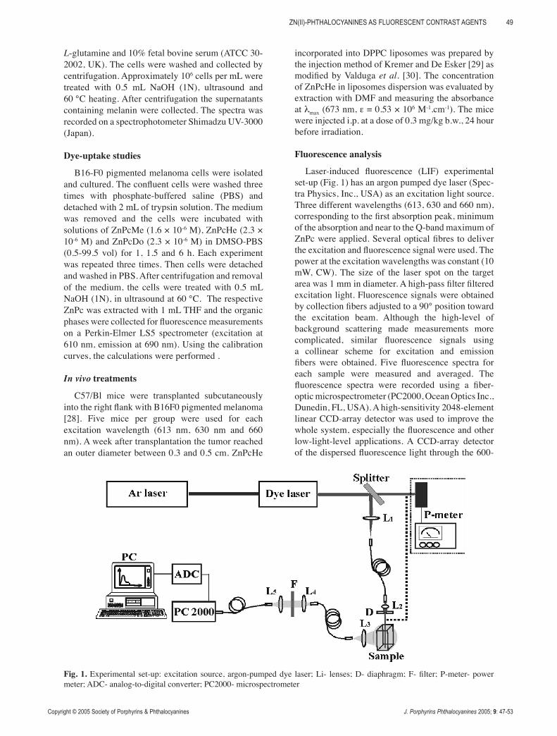
Laser-induced fluorescence (LIF) experimental set-up (Fig. 1) has an argon pumped dye laser (Spec-tra Physics, Inc., USA) as an excitation light source. Three different wavelengths (613, 630 and 660 nm), corresponding to the first absorption peak, minimum of the absorption and near to the Q-band maximum of ZnPc were applied. Several optical fibres to deliver the excitation and fluorescence signal were used. The power at the excitation wavelengths was constant (10 mW, CW). The size of the laser spot on the target area was 1 mm in diameter. A high-pass filter filtered excitation light. Fluorescence signals were obtained by collection fibers adjusted to a 90° position toward the excitation beam. Although the high-level of background scattering made measurements more complicated, similar fluorescence signals using a collinear scheme for excitation and emission fibers were obtained. Five fluorescence spectra for each sample were measured and averaged. The fluorescence spectra were recorded using a fiber-optic microspectrometer (PC2000, Ocean Optics Inc., Dunedin, FL, USA). A high-sensitivity 2048-element linear CCD-array detector was used to improve the whole system, especially the fluorescence and other low-light-level applications. A CCD-array detector of the dispersed fluorescence light through the 600-
Fig. 1. Experimental set-up: excitation source, argon-pumped dye laser; Li- lenses; D- diaphragm; F- filter; P-meter- power meter; ADC- analog-to-digital converter; PC2000- microspectrometer
Copyright © 2005 Society of Porphyrins & Phthalocyanines J. Porphyrins Phthalocyanines 2005; 9: 47-53
V. MANTAREVA ET AL.50
lines/mm grating was used. The spectral resolution of the microspectrometer was approximately 8.5 nm. A computer to control the system and to store and to display the data was connected. The spectra were recorded using the microspectrometer specialized software OOI Base (Ocean Optics, Inc., Dunedin, USA). The data were analyzed and graphically represented by means of an advanced computer program (Microcal Origin 5.0, Microcal Software, Inc., Northampton, MA, USA).
RESULTS AND DISCUSSION
Synthesis
Cationic phthalocyanines (ZnPcMe, ZnPcHe and ZnPcDo, Scheme 1) were synthesized starting from 4-nitrophthalonitrile following a three-step procedure [24]. The preparation included the reaction of nucleophilic displacement of the nitro group by 3-hydroxypyridine and then the cyclotetramerization in the presence of an organic base and zinc acetate. The alkylation reactions were carried out with an excess of alkyliodine and the products were obtained after lyophilization in good yields and high purity as was described in reference 24.
Absorption and fluorescence properties
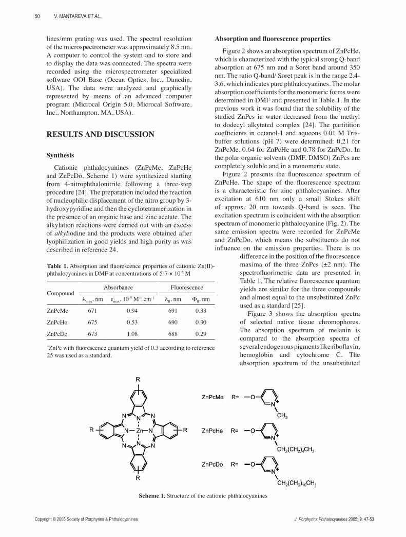
Figure 2 shows an absorption spectrum of ZnPcHe, which is characterized with the typical strong Q-band absorption at 675 nm and a Soret band around 350 nm. The ratio Q-band/ Soret peak is in the range 2.4-3.6, which indicates pure phthalocyanines. The molar absorption coefficients for the monomeric forms were determined in DMF and presented in Table 1. In the previous work it was found that the solubility of the studied ZnPcs in water decreased from the methyl to dodecyl alkytated complex [24]. The partitition coefficients in octanol-1 and aqueous 0.01 M Tris-buffer solutions (pH 7) were determined: 0.21 for ZnPcMe, 0.64 for ZnPcHe and 0.78 for ZnPcDo. In the polar organic solvents (DMF, DMSO) ZnPcs are completely soluble and in a monomeric state.
Figure 2 presents the fluorescence spectrum of ZnPcHe. The shape of the fluorescence spectrum is a characteristic for zinc phthalocyanines. After excitation at 610 nm only a small Stokes shift of approx. 20 nm towards Q-band is seen. The excitation spectrum is coincident with the absorption spectrum of monomeric phthalocyanine (Fig. 2). The same emission spectra were recorded for ZnPcMe and ZnPcDo, which means the substituents do not influence on the emission properties. There is no
difference in the position of the fluorescence maxima of the three ZnPcs (±2 nm). The spectrofluorimetric data are presented in Table 1. The relative fluorescence quantum yields are similar for the three compounds and almost equal to the unsubstituted ZnPc used as a standard [25].
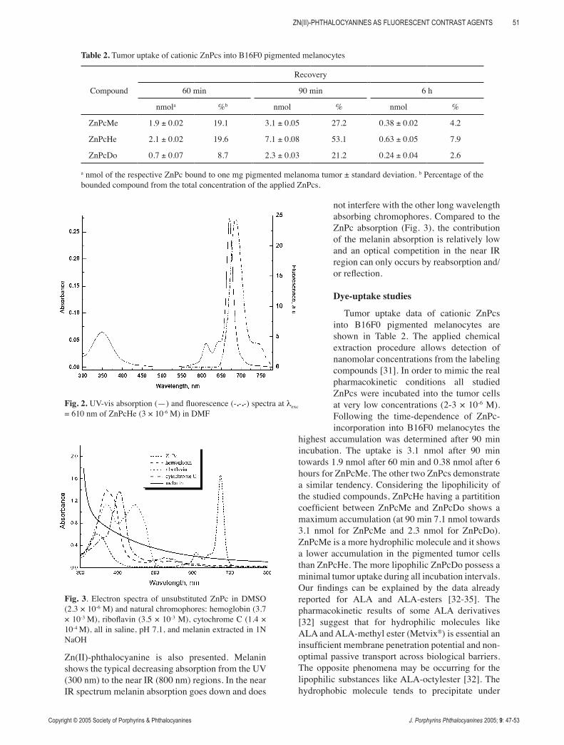
Figure 3 shows the absorption spectra of selected native tissue chromophores. The absorption spectrum of melanin is compared to the absorption spectra of several endogenous pigments like riboflavin, hemoglobin and cytochrome C. The absorption spectrum of the unsubstituted
Table 1. Absorption and fluorescence properties of cationic Zn(II)-phthalocyanines in DMF at concentrations of 5-7 × 10⁻⁶ M
CompoundAbsorbance Fluorescence
λmax, nm εmax, 10-5 M-1.cm-1 λfl, nm Φfl, nm
ZnPcMe 671 0.94 691 0.33
ZnPcHe 675 0.53 690 0.30
ZnPcDo 673 1.08 688 0.29
*ZnPc with fluorescence quantum yield of 0.3 according to reference 25 was used as a standard.
Scheme 1. Structure of the cationic phthalocyanines
Copyright © 2005 Society of Porphyrins & Phthalocyanines J. Porphyrins Phthalocyanines 2005; 9: 47-53
ZN(II)-PHTHALOCYANINES AS FLUORESCENT CONTRAST AGENTS 51
Zn(II)-phthalocyanine is also presented. Melanin shows the typical decreasing absorption from the UV (300 nm) to the near IR (800 nm) regions. In the near IR spectrum melanin absorption goes down and does
not interfere with the other long wavelength absorbing chromophores. Compared to the ZnPc absorption (Fig. 3), the contribution of the melanin absorption is relatively low and an optical competition in the near IR region can only occurs by reabsorption and/or reflection.
Dye-uptake studies
Tumor uptake data of cationic ZnPcs into B16F0 pigmented melanocytes are shown in Table 2. The applied chemical extraction procedure allows detection of nanomolar concentrations from the labeling compounds [31]. In order to mimic the real pharmacokinetic conditions all studied ZnPcs were incubated into the tumor cells at very low concentrations (2-3 × 10⁻⁶ M). Following the time-dependence of ZnPc-incorporation into B16F0 melanocytes the
highest accumulation was determined after 90 min incubation. The uptake is 3.1 nmol after 90 min towards 1.9 nmol after 60 min and 0.38 nmol after 6 hours for ZnPcMe. The other two ZnPcs demonstrate a similar tendency. Considering the lipophilicity of the studied compounds, ZnPcHe having a partitition coefficient between ZnPcMe and ZnPcDo shows a maximum accumulation (at 90 min 7.1 nmol towards 3.1 nmol for ZnPcMe and 2.3 nmol for ZnPcDo). ZnPcMe is a more hydrophilic molecule and it shows a lower accumulation in the pigmented tumor cells than ZnPcHe. The more lipophilic ZnPcDo possess a minimal tumor uptake during all incubation intervals. Our findings can be explained by the data already reported for ALA and ALA-esters [32-35]. The pharmacokinetic results of some ALA derivatives [32] suggest that for hydrophilic molecules like ALA and ALA-methyl ester (Metvix®) is essential an insufficient membrane penetration potential and non-optimal passive transport across biological barriers. The opposite phenomena may be occurring for the lipophilic substances like ALA-octylester [32]. The hydrophobic molecule tends to precipitate under
Table 2. Tumor uptake of cationic ZnPcs into B16F0 pigmented melanocytes
Compound
Recovery
60 min 90 min 6 h
nmola %b nmol % nmol %
ZnPcMe 1.9 ± 0.02 19.1 3.1 ± 0.05 27.2 0.38 ± 0.02 4.2
ZnPcHe 2.1 ± 0.02 19.6 7.1 ± 0.08 53.1 0.63 ± 0.05 7.9
ZnPcDo 0.7 ± 0.07 8.7 2.3 ± 0.03 21.2 0.24 ± 0.04 2.6
a nmol of the respective ZnPc bound to one mg pigmented melanoma tumor ± standard deviation. b Percentage of the bounded compound from the total concentration of the applied ZnPcs.
Fig. 2. UV-vis absorption (—) and fluorescence (-.-.-) spectra at λexc = 610 nm of ZnPcHe (3 × 10⁻⁶ M) in DMF
Fig. 3. Electron spectra of unsubstituted ZnPc in DMSO (2.3 × 10⁻⁶ M) and natural chromophores: hemoglobin (3.7 × 10⁻³ M), riboflavin (3.5 × 10⁻³ M), cytochrome C (1.4 × 10⁻⁴ M), all in saline, pH 7.1, and melanin extracted in 1N NaOH
Copyright © 2005 Society of Porphyrins & Phthalocyanines J. Porphyrins Phthalocyanines 2005; 9: 47-53
V. MANTAREVA ET AL.52
physiological conditions (pH~7) and an excessive lipophilicity may promote also a non-sufficient accumulation into the cell membranes [33-35]. The cationic ZnPcs showed similar tendency for tissue uptake as reported for ALA [10, 32-35]. Obviously the differences in the tumor uptake are due to the origin of the malignances and the hydrophilic/lipophilic nature of the fluorescent molecules.
In vivo fluorescence diagnosis
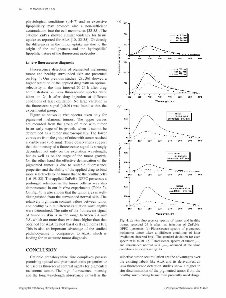
Fluorescence detection of pigmented melanoma tumor and healthy surrounded skin are presented on Fig. 4. Our previous studies [28, 36] showed a higher retention of the applied drug with an optimal selectivity in the time interval 20-24 h after drug administration. In vivo fluorescence spectra were taken on 24 h after drug injection at different conditions of laser excitation. No large variation in the fluorescent signal (±0.01) was found within the experimental group.
Figure 4a shows in vivo spectra taken only for pigmented melanoma tumors. The upper curves are recorded from the group of mice with tumor in an early stage of its growth, when it cannot be determined as a tumor macroscopically. The lower curves are from the group of mice with tumor reached a visible size (3-5 mm). These observations suggest that the intensity of a fluorescence signal is strongly dependent not only on the excitation wavelength, but as well as on the stage of the tumor growth. On the other hand the effective demarcation of the pigmented tumor is due to suitable fluorescence properties and the ability of the applied drug to bind more selectively to the tumor than to the healthy cells [16-19, 32]. The applied ZnPcHe-DPPC possesses a prolonged retention in the tumor cells as was also demonstrated in our in vitro experiments (Table 2). On Fig. 4b is also shown that the tumor area is well-distinguished from the surrounded normal skin. The relatively high mean contrast values between tumor and healthy skin at different excitation wavelengths were determined. The ratio of the fluorescent signal of tumor vs skin is in the range between 2.4 and 3.8, which are more than two times higher than that obtained for ALA treated basal cell carcinoma [10]. This is also an important advantage of the studied phthalocyanine in comparison to ALA, which is leading for an accurate tumor diagnosis.
CONCLUSIONCationic phthalocyanine zinc complexes possess
promising optical and pharmacokinetic properties to be used as fluorescent contrast agents for pigmented melanoma tumor. The high fluorescence intensity and the long wavelength absorbance as well as the
selective tumor accumulation are the advantages over the existing labels like ALA and its derivatives. In vivo fluorescence detection studies show a higher in situ discrimination of the pigmented tumor from the healthy surrounding tissue than presently used drugs.
Fig. 4. In vivo fluorescence spectra of tumor and healthy tissues recorded 24 h after i.p. injection of ZnPcHe-DPPC liposomes. (a) Fluorescence spectra of pigmented melanoma tumor taken at different conditions of laser irradiation (inserted box). The standard deviation for each spectrum is ±0.01. (b) Fluorescence spectra of tumor (—) and surrounded normal skin (----) obtained at the same conditions as spectra in Fig. 4a
(a)
(b)
Copyright © 2005 Society of Porphyrins & Phthalocyanines J. Porphyrins Phthalocyanines 2005; 9: 47-53
ZN(II)-PHTHALOCYANINES AS FLUORESCENT CONTRAST AGENTS 53
The limitations of pigmented melanoma detection when using ALA have a good solution by application of a cationic phthalocyanine complex with a suitable hydrophilic/lipophilic balance. The proposed expe-rimental fluorescence spectroscopy technique can be used for fluorescence detection of pigmented malignances in clinical practice.
Acknowledgements
This work was supported by Optella Ltd (Sofia, Bulgaria) as a part of the 5th EC project G5RD-CT-2000-00372 (IMPECABLE). V. M. is grateful to DAAD for a fellowship under the NATO Science Programme in Germany (A/99/02178).
REFERENCES 1. Rosenthal I and Ben-Hur E. Phthalocyanines.
Properties and Applications. Leznoff CC, Levers ABR. (eds). VCH: New York 1996; 393.
2. van Lier JE and Spikes JD. Photosensitizing Compounds: their Chemistry, Biology and Clinical Use, 146 Ciba Foundation Symp. 1989; 17.
3. Davila J and Harriman A. Photochem. Photobiol. 1990; 51: 9.
4. Lagorio MG, Dicelio LE and San Roman EA. J. Photochem. Photobiol. A: Chem. 1993; 72: 153.
5. Aveline BM, Hasan T and Redmond RW. J. Photochem. Photobiol. B: Biol. 1995, 30: 161.
6. Ball DJ, Wood SR, Vernon DI, Griffiths J, Dubbelman TMAR and Brown SB. J. Photochem. Photobiol. B: Biol. 1998; 45: 28.
7. English DR, Armstrong BK, Kricker A and Fleming C. Cancer Causes Control, 1997; 8: 271.
8. Lohmann W and Paul E. Naturwissenshaften 1988; 75: 201.
9. Golub AL, Dickinson GEF, Kennedy JC, Marcus SL, Park Y and Pottier R. Laser Med. Sci. 1999; 14: 112.
10. Ericson MB, Sandberg C, Gudmundson F, Rosen A, Larko O and Wennberg A-M. J. Photochem. Photobiol. B: Biol. 2003; 69: 121.
11. Kollias N, Sayre RM, Zeise L and Chedekel MR. J. Photochem. Photobiol. B: Biol. 1991; 9: 135.
12. Teuchner K, Mueller S, Freyer W, Leupold D, Altmeyer P, Stűcker M and Hoffmann K. SPIE 2003; 4797: 211.
13. Dougherty TJ, Gomer CJ, Henderson BW, Jori G, Kessel D, Korbelik M, Moan J and Peng Q. J. Natl. Cancer Inst. 1998; 90: 889.
14. Wagnieres GA, Star WM and Wilson BC.
Photochem. Photobiol. 1998; 68: 603.15. Abels C, Heil P, Dellian M, Kuhnle GE,
Baumgartner R and Goetz AE. Br. J. Cancer 1994; 70: 826.
16. Moan J, Ma LW and Iani V. Int. J. Cancer 2001; 92: 139.
17. Heyerdahl H, Wang I, Liu DL, Berg R, Anderson-Engels S, Peng Q, Moan J and Svanberg S. Cancer Lett. 1997; 112: 225.
18. Ackermann G, Abels C, Baumler W, Langer S, Landthaler M, Lang EW and Szeimies RM. J. Photochem. Photobiol. B: Biol. 1998; 47: 121.
19. Rűck A, Beck G, Bachor R, Akgűn N, Gschwend MH and Steiner R. J. Photochem. Photobiol., B: Biol. 1996; 36: 127.
20. Peng Q, Farrants WG, Madslien K, Bommer CJ, Moan J, Danielsen H and Nesland MJ. Int. J. Cancer 1991; 49: 290.
21. Takemura T, Nakajima S and Sakata I. Photochem. Photobiol. 1994; 59: 366.
22. Reddi E, Segalla A, Jori G, Kerrigan PK, Liddell PA, Moore TA and Gust D. Br. J. Cancer 1994; 69: 40.
23. Burstein EA and Emelyanko VI. Photochem. Photobiol. 1996; 64: 316.
24. Wöhrle D, Iskander N, Graschew G, Sinn H, Friedrich EA, Maier-Borst W, Stern J and Schlag P. Photochem. Photobiol. 1990; 51: 351.
25. Darwent JR, Douglas P, Harriman A, Porter G and Richoux M-C. Coord. Chem. Reviews 1982; 44: 83.
26. Decreau R, Richard MJ, Verrando P, Chanon M and Julliard M. J. Photochem. Photobiol., B: Biol. 1999; 48: 48.
27. De Leeuw MS, Smith MPN, Veldhoven M, Pennings ME, Pavel S, Simons MIVJ and Schothrst AA. J. Photochem. Photobiol. B: Biol. 2001; 61: 106.
28. Shopova M, Wöhrle D, Mantareva V and Mueller S. J. Biomed. Optics 1999; 4: 276.
29. Kremer HMJ and De Esker JVM. Biochemistry 1977; 16: 3932.
30. Valduga G, Reddi E and Jori G. J. Inorg. Biochem. 1987; 25: 59.
31. Lee CC, Pouge BW, Strawbridge RR, Moodie, KL, Bartholomew LR, Burke GC and Hoopes JP. Photochem. Photobiol. 2001; 74: 453.
32. van den Bergh H, Lange N and Jichlinski P. Photodynamics News 1999; 2: 4.
33. Marcus LS. Int. Photodynamics 1996; 1: 2.34. Robinson D. Photodynamics News 2000; 3: 4.35. Morton C, Wong G and Taylor D. Brit. J.
Dermatol. 2002; 146: 552.36. Peeva M, Shopova M, Michelsen U, Wöhrle
D, Petrov G and Diddens H. J. Porphyrins Phthalocyanines 2001; 5: 645.
Related Documents