Long noncoding RNA HOTAIR reprograms chromatin state to promote cancer metastasis Rajnish A. Gupta 1 , Nilay Shah 2 , Kevin C. Wang 1 , Jeewon Kim 3 , Hugo M. Horlings 4 , David J. Wong 1 , Miao-Chih Tsai 1 , Tiffany Hung 1 , Pedram Argani 5 , John L. Rinn 6 , Yulei Wang 7 , Pius Brzoska 7 , Benjamin Kong 7 , Rui Li 8 , Robert B. West 8 , Marc J. van de Vijver 4 , Saraswati Sukumar 2 , and Howard Y. Chang 1 1 Howard Hughes Medical Institute and Program in Epithelial Biology, Stanford University School of Medicine, Stanford, CA 94305, USA 2 Sidney Kimmel Comprehensive Cancer Center, Johns Hopkins University School of Medicine, Baltimore, MD 21231, USA 3 Stanford Comprehensive Cancer Center and Transgenic Mouse Research Center, Stanford University School of Medicine, Stanford, CA 94305, USA 4 Dept. of Pathology, Academic Medical Center, Meibergdreef 9, 1105AZ, Amsterdam, The Netherlands 5 Dept. of Pathology, Johns Hopkins University School of Medicine, Baltimore, MD 21231, USA 6 The Broad Institute of Harvard and Massachusetts Institute of Technology, Cambridge, MA 02142, USA 7 Applied Biosystems, Foster City, CA 94404, USA 8 Department of Pathology, Stanford University School of Medicine, Stanford, CA 94305, USA Abstract Large intervening noncoding RNAs (lincRNAs) are pervasively transcribed in the genome1 , 2 , 3 yet their potential involvement in human disease is not well understood4 , 5. Recent studies of dosage compensation, imprinting, and homeotic gene expression suggest that individual lincRNAs can function as the interface between DNA and specific chromatin remodeling activities6 , 7 , 8. Here we show that lincRNAs in the HOX loci become systematically dysregulated during breast cancer progression. The lincRNA termed HOTAIR is increased in expression in primary breast tumors and metastases, and HOTAIR expression level in primary tumors is a powerful predictor of eventual metastasis and death. Enforced expression of HOTAIR in epithelial cancer cells induced genome-wide re-targeting of Polycomb Repressive Complex 2 (PRC2) to an occupancy pattern more resembling embryonic fibroblasts, leading to altered histone H3 lysine 27 methylation, gene expression, and increased cancer invasiveness and metastasis in a manner dependent on PRC2. Conversely, loss of HOTAIR can inhibit cancer invasiveness, particularly in cells that possess excessive PRC2 activity. These findings suggest that lincRNAs play active roles in modulating the cancer epigenome and may be important targets for cancer diagnosis and therapy. Users may view, print, copy, download and text and data- mine the content in such documents, for the purposes of academic research, subject always to the full Conditions of use: http://www.nature.com/authors/editorial_policies/license.html#terms Correspondence and request for materials should beaddressed to H.Y.C. ([email protected]). AUTHOR CONTRIBUTIONS R.A.G. measured lincRNAs in cancer samples, performed all gene transfer and knockdown experiments. R.A.G. and N.S. performed cell growth, invasion, and in vivo xenograft assays. R.A.G., K.C.W., M.C.T., T.H. performed ChIP-chip studies and analyses. R.A.G., J.L.R., D.J.W. performed bioinformatic analyses. J.K. performed in vivo bioluminescence studies. H.M.H., P.A., M.J. vd V. procured and analyzed human tumor samples. Y.W., P.B., B.K. designed lincRNA Taqman probes and analyzed tumor RNAs by qRT-PCR. R.L. and R.B.W. performed in situ hybridization studies. R.A.G., N.S., S.S., and H.Y.C. designed the experiments and interpreted the results. R.A.G. and H.Y.C. wrote the paper. AUTHOR INFORMATION Microarray data are deposited in Gene Expression Omnibus (accession number GSE20435). Reprints and permission information is available at www.nature.com/reprints. The authors declare no competing financial interests. NIH Public Access Author Manuscript Nature. Author manuscript; available in PMC 2011 March 8. Published in final edited form as: Nature. 2010 April 15; 464(7291): 1071–1076. doi:10.1038/nature08975. NIH-PA Author Manuscript NIH-PA Author Manuscript NIH-PA Author Manuscript

Welcome message from author
This document is posted to help you gain knowledge. Please leave a comment to let me know what you think about it! Share it to your friends and learn new things together.
Transcript
Long noncoding RNA HOTAIR reprograms chromatin state topromote cancer metastasis
Rajnish A. Gupta1, Nilay Shah2, Kevin C. Wang1, Jeewon Kim3, Hugo M. Horlings4, David J.Wong1, Miao-Chih Tsai1, Tiffany Hung1, Pedram Argani5, John L. Rinn6, Yulei Wang7, PiusBrzoska7, Benjamin Kong7, Rui Li8, Robert B. West8, Marc J. van de Vijver4, SaraswatiSukumar2, and Howard Y. Chang1
1 Howard Hughes Medical Institute and Program in Epithelial Biology, Stanford University Schoolof Medicine, Stanford, CA 94305, USA 2 Sidney Kimmel Comprehensive Cancer Center, JohnsHopkins University School of Medicine, Baltimore, MD 21231, USA 3 Stanford ComprehensiveCancer Center and Transgenic Mouse Research Center, Stanford University School of Medicine,Stanford, CA 94305, USA 4 Dept. of Pathology, Academic Medical Center, Meibergdreef 9,1105AZ, Amsterdam, The Netherlands 5 Dept. of Pathology, Johns Hopkins University School ofMedicine, Baltimore, MD 21231, USA 6 The Broad Institute of Harvard and MassachusettsInstitute of Technology, Cambridge, MA 02142, USA 7 Applied Biosystems, Foster City, CA94404, USA 8 Department of Pathology, Stanford University School of Medicine, Stanford, CA94305, USA
AbstractLarge intervening noncoding RNAs (lincRNAs) are pervasively transcribed in the genome1, 2, 3yet their potential involvement in human disease is not well understood4,5. Recent studies ofdosage compensation, imprinting, and homeotic gene expression suggest that individual lincRNAscan function as the interface between DNA and specific chromatin remodeling activities6,7,8.Here we show that lincRNAs in the HOX loci become systematically dysregulated during breastcancer progression. The lincRNA termed HOTAIR is increased in expression in primary breasttumors and metastases, and HOTAIR expression level in primary tumors is a powerful predictor ofeventual metastasis and death. Enforced expression of HOTAIR in epithelial cancer cells inducedgenome-wide re-targeting of Polycomb Repressive Complex 2 (PRC2) to an occupancy patternmore resembling embryonic fibroblasts, leading to altered histone H3 lysine 27 methylation, geneexpression, and increased cancer invasiveness and metastasis in a manner dependent on PRC2.Conversely, loss of HOTAIR can inhibit cancer invasiveness, particularly in cells that possessexcessive PRC2 activity. These findings suggest that lincRNAs play active roles in modulating thecancer epigenome and may be important targets for cancer diagnosis and therapy.
Users may view, print, copy, download and text and data- mine the content in such documents, for the purposes of academic research,subject always to the full Conditions of use: http://www.nature.com/authors/editorial_policies/license.html#terms
Correspondence and request for materials should beaddressed to H.Y.C. ([email protected]).
AUTHOR CONTRIBUTIONSR.A.G. measured lincRNAs in cancer samples, performed all gene transfer and knockdown experiments. R.A.G. and N.S. performedcell growth, invasion, and in vivo xenograft assays. R.A.G., K.C.W., M.C.T., T.H. performed ChIP-chip studies and analyses. R.A.G.,J.L.R., D.J.W. performed bioinformatic analyses. J.K. performed in vivo bioluminescence studies. H.M.H., P.A., M.J. vd V. procuredand analyzed human tumor samples. Y.W., P.B., B.K. designed lincRNA Taqman probes and analyzed tumor RNAs by qRT-PCR.R.L. and R.B.W. performed in situ hybridization studies. R.A.G., N.S., S.S., and H.Y.C. designed the experiments and interpreted theresults. R.A.G. and H.Y.C. wrote the paper.
AUTHOR INFORMATIONMicroarray data are deposited in Gene Expression Omnibus (accession number GSE20435). Reprints and permission information isavailable at www.nature.com/reprints. The authors declare no competing financial interests.
NIH Public AccessAuthor ManuscriptNature. Author manuscript; available in PMC 2011 March 8.
Published in final edited form as:Nature. 2010 April 15; 464(7291): 1071–1076. doi:10.1038/nature08975.
NIH
-PA Author Manuscript
NIH
-PA Author Manuscript
NIH
-PA Author Manuscript
We hybridized RNA derived from normal human breast epithelia, primary breastcarcinomas, and distant metastases to ultra-dense HOX tiling arrays7 (Fig. 1a, b). We foundthat 233 transcribed regions in the HOX loci, comprising 170 ncRNAs and 63 HOX exons,were differentially expressed (Fig. 1a). Unsupervised hierarchical clustering showedsystematic variation in the expression of HOX lincRNAs among normal breast epithelia,primary tumor, and metastases. HOXA5, a known breast tumor suppressor11 along withdozens of HOX lincRNAs, are expressed in normal breast but with reduced expression in allcancer samples (Supplementary Fig. 1). A set of HOX lincRNAs and mRNAs, including theknown oncogene HOXB712, are frequently expressed in primary tumors but not inmetastases (Supplementary Fig. 1). A distinct set of HOX lincRNAs are sometimesoverexpressed in primary tumors, and very frequently overexpressed in metastases (Fig. 1b).Notably, one such metastasis-associated lincRNA is HOTAIR (Fig. 1b), which has a uniqueassociation with patient prognosis (Supplementary Figs. 1, 2, Supplementary Table 1).HOTAIR is a lincRNA in the mammalian HOXC locus that binds to and targets the PRC2complex to the HOXD locus, located on a different chromosome7. PRC2 is a histone H3lysine 27 methylase involved in developmental gene silencing and cancer progression9,10.We hypothesized that altered HOTAIR expression may be involved in human cancer bypromoting genomic relocalization of Polycomb complex and H3K27 trimethylation.
Quantitative PCR showed that HOTAIR is overexpressed from hundreds to nearly twothousand- fold in breast cancer metastases, and HOTAIR level is sometimes high butheterogeneous among primary tumors (Fig. 1c). We next measured HOTAIR level in anindependent panel of 132 primary breast tumors (stage I and II) with extensive clinicalfollow-up13. Indeed, nearly one third of primary breast tumors overexpress HOTAIR byover 125-fold over normal breast epithelia, the minimum level of HOTAIR overexpressionobserved in bona fide metastases (Fig. 1d), and high HOTAIR level is a significant predictorof subsequent metastasis and death (p=0.0004 and p=0.005 for metastasis and death,respectively, Fig. 1e, f). Multivariate analysis showed that prognostic stratification ofmetastasis and death by HOTAIR is independent of known clinical risk factors such astumor size, stage, and hormone receptor status (Supplementary Table 2).
We next examined the effects of manipulating HOTAIR level in several breast cancer celllines. HOTAIR levels in cell lines are significantly lower than those seen in primary ormetastatic breast tumors (Supplementary Fig. 3, 4). Retroviral transduction allowed stableoverexpression of HOTAIR to several hundred fold over vector-transduced cells, which arecomparable to levels observed in patients (Supplementary Fig. 4). HOTAIR overexpressionpromoted colony growth in soft agar (Supplementary Fig. 5). In addition, enforcedexpression of HOTAIR in four different breast cancer cell lines increased cancer cellinvasion through Matrigel, a basement-membrane like extracellular matrix, (Fig. 2a).Conversely, depletion of HOTAIR by small inferring RNAs (siRNAs) in MCF7, a cell linethat expresses endogenous HOTAIR, decreased its matrix invasiveness (Fig. 2b andSupplementary Fig. 6). To probe the effects of HOTAIR on cancer cell dynamics in vivo,we labeled control and HOTAIR-expressing cells with firefly luciferase, enabling in vivobioluminescence imaging. When MDA-MB-231 cells expressing vector or HOTAIR wereorthotopically grafted into mammary fat pads, serial imaging showed that HOTAIRexpression modestly increased the rate of primary tumor growth (Fig. 2C, left panel).Importantly, in the same animals, we observed significantly increased foci of luciferasesignal in the lung fields of mice bearing HOTAIR+ primary tumors (Fig. 2C, right panel),which suggests that HOTAIR promotes lung metastasis.
To further quantify metastatic potential in vivo, we performed tail vein xenografts andcompared the rates of lung colonization. Vector expression in the non-metastatic cell line
Gupta et al. Page 2
Nature. Author manuscript; available in PMC 2011 March 8.
NIH
-PA Author Manuscript
NIH
-PA Author Manuscript
NIH
-PA Author Manuscript
SK-BR3 never showed lung colonization after tail vein xenograft (0 of 15 mice), butHOTAIR expression allowed SK-BR3 cells to colonize the lung in 80% of animals (12 of 15mice, Fig. 2C). SK-BR3 cells apparently lack additional genetic elements required to persistin the lung, because HOTAIR-transduced SK-BR3 cells in the lung disappeared afterapproximately one week. In contrast, HOTAIR expression in MDA-MB-231 cells resultedin approximately eight-to ten- fold more cells to engraft the lung after tail vein xenograft(Fig. 2D). These differences persisted until the end of the experiment, resulting in ten-foldmore lung metastases as verified by histology (p= 0.00005, Fig. 2E). The tumors retainedHOTAIR expression for the length of the experiment (Supplementary Fig. 7).
We next tested if HOTAIR overexpression affected the pattern of PRC2 occupancy. Wemapped PRC2 occupancy genome-wide by chromatin immunoprecipitation followed byhybridization to tiling microarrays interrogating all human promoters (ChIP-chip, Fig. 3).Compared to vector expressing cells, HOTAIR overexpression induced localization ofH3K27me3 and PRC2 subunits SUZ12, EZH2, on 854 new genes while concomitantlylosing PRC2 occupancy and H3K27me3 on 37 genes (Fig. 3a). A significant fraction ofthese 854 genes also showed consequent changes in gene expression after HOTAIR over-expression (39% observed vs. 7.0% expected by chance alone, p = 2.5 ×10−209,hypergeometric distribution). The majority of PRC2 occupancy sites on promoters genome-wide showed little change (data not shown), and HOTAIR overexpression did not changethe levels of PRC2 subunits (Fig. 4a, lane 1 vs. lane 4). A number of the genes withHOTAIR-induced PRC2 occupancy are implicated in inhibiting breast cancer progression,including transcription factors HOXD1014 and PRG1, encoding progesterone receptor (aclassic favorable prognostic factor); cell adhesion molecules of the protocadherin (PCDH)gene family15 and JAM216; and EPHA117,18, encoding an ephrin receptor involved intumor angiogenesis. Gene Ontology19 analysis suggested a majority of the 854 genes areinvolved in pathways related to cell-cell signaling and development (Supplementary Fig. 8).HOTAIR-induced PRC2 occupancy tended to spread over promoters, and to a lesser extent,gene bodies (Fig. 3b). HOTAIR may also induce PRC2 localization to other intergenicregions not present on our tiling arrays. ChIP followed by quantitative PCR confirmed thatHOTAIR substantially increased PRC2 occupancy and H3K27me3 of all target genesexamined (Supplementary Fig. 9). Notably, like HOTAIR itself, the 854 HOTAIR-PRC2target genes are coordinately down regulated in aggressive breast tumors that tend to causedeath (p<0.0003, Supplementary Fig. 10).
We next compared the 854 genes with HOTAIR-induced PRC2 occupancy in MDA-MB-231 cells with a compendium of published PRC2 occupancy profiles in diverse celltypes (Fig. 3c). PRC2 occupancy patterns from different cancer, fibroblastic, and embryonicstem cell lines were annotated from existing databases (Supplementary Table 3). Using apattern matching algorithm20, we found that the HOTAIR-induced PRC2 occupancy patternin breast cancer cells most resembled the endogenous PRC2 occupancy pattern in embryonicand neonatal fibroblasts, especially fibroblasts derived from posterior and distal anatomicsites where endogenous HOTAIR is expressed7 (p<10−50 for each comparison, FDR≪0.05,Fig. 3c). These 854 genes are also significantly enriched for genes in primary fibroblasts thatare bound by PRC2 in a HOTAIR dependent manner (32% overlap observed vs. 9.9%expected by chance alone, p = 8.5 ×10−93, hypergeometric distribution, M.C.T., unpublisheddata). These results suggest that elevated HOTAIR expression in breast cancer cells appearsto reprogram the Polycomb binding profile of a breast epithelial cell to that of an embryonicfibroblast.
Finally, we addressed whether the ability of HOTAIR to induce breast cancer invasivenessrequired an intact PRC2 complex. We transduced vector- or HOTAIR-expressing MDA-MB-231 cells with short hairpin RNAs (shRNAs) targeting PRC2 subunits EZH2 or SUZ12.
Gupta et al. Page 3
Nature. Author manuscript; available in PMC 2011 March 8.
NIH
-PA Author Manuscript
NIH
-PA Author Manuscript
NIH
-PA Author Manuscript
Immunoblot analyses confirmed efficient depletion of the targeted proteins (Fig. 4a).Depletion of either SUZ12 or EZH2 had little impact on the invasiveness of control cells,but completely reversed the ability of HOTAIR to promote matrix invasion (Fig. 4b).Depletion of EZH2 also inhibited HOTAIR-driven lung colonization after tail vein xenograftby approximately 50% (p < 0.05). These results suggest that PRC2 is specifically requiredfor HOTAIR to promote cellular invasiveness. Global gene expression analysis revealedhundreds of genes that were induced or repressed as a consequence of HOTAIRoverexpression (Fig. 4c, left panel). Importantly, concomitant depletion of PRC2 in largepart reversed the global gene expression pattern to that of cells not overexpressing HOTAIR(Fig. 4c, right panel). Quantitative RT-PCR confirmed that HOTAIR-induced PRC2 targetgenes, such as JAM2, PCDH10, PCDHB5, were transcriptionally repressed upon HOTAIRexpression and de-repressed upon concomitant PRC2 depletion (Fig. 4d). HOTAIR-inducedgenes were also reversed upon PRC2 depletion (Fig. 4d). Of note, many of the genesinduced by HOTAIR are known positive regulators of cancer metastasis, including ABL221,SNAIL22, and laminins23. Conversely, overexpression of EZH2 in H16N2 breast cells isknown to promote matrix invasion10, but concomitant depletion of endogenous HOTAIR inlarge measure inhibited the ability of EZH2 to induce matrix invasion (Fig. 4e andSupplementary Figure 6). Together, these results demonstrate a functional inter-dependencybetween HOTAIR and PRC2 in promoting cancer invasiveness.
In summary, the cancer transcriptome is more complex than previously believed. In additionto protein coding genes and microRNAs, dysregulated expression of lincRNAs is likelypervasive in human cancers and can drive cancer development and progression. Notably, thelincRNA HOTAIR regulates metastatic progression. HOTAIR recruits PRC2 complex tospecific targets genes genome-wide, leading to H3K27 trimethylation and epigeneticsilencing of metastasis suppressor genes (Fig. 4f). The concept of epigenomicreprogramming by lincRNAs may also be applicable to many other human disease statescharacterized by aberrant lincRNA expression and chromatin states. HOTAIR is normallyinvolved in specifying the chromatin state associated with fibroblasts from anatomicallyposterior and distal sites. Within the context of cancer cells, ectopic expression of HOTAIRappear to re-impose that chromatin state, thereby enabling gene expression programs that areconducive to cell motility and matrix invasion.
The interdependence between HOTAIR and PRC2 has therapeutic implications. High levelsof HOTAIR may identify tumors that are sensitive to small molecules inhibitors of PRC224.Conversely, tumors that overexpress Polycomb proteins may be sensitive to therapeuticstrategies that target endogenous HOTAIR or inhibit HOTAIR-PRC2 interactions.Understanding the precise molecular mechanisms by which HOTAIR regulates PRC2 willbe a critical first step in exploring these potential new avenues in cancer therapy.
METHODS SUMMARYHuman materials were obtained from Johns Hopkins Hospital and the Netherlands CancerInstitute. Expression of HOX transcripts was determined using ultra-high density HOXtiling arrays7 and qRT-PCR. Kaplan-Meier analyses of breast cancer patients were asdescribed13. We used retroviral transduction to overexpress HOTAIR and luciferase, andused siRNA or shRNA to deplete the indicated transcripts. Matrix invasion was measured bythe transwell Matrigel assay. We implanted cells in the mammary fat pad of SCID mice, andmonitored primary tumor growth and lung metastasis by bioluminescence. Cells wereinjected into the tail vein of nude mice, and lungs were analyzed at 9 weeks to quantify lungcolonization in vivo. ChIP-chip was performed as described7 using human whole genomepromoter tiling arrays (Roche Nimblegen, Wisconsin). Module map and GO enrichmentanalyses were done using Genomica20.
Gupta et al. Page 4
Nature. Author manuscript; available in PMC 2011 March 8.
NIH
-PA Author Manuscript
NIH
-PA Author Manuscript
NIH
-PA Author Manuscript
METHODSReagents
The MDA-MB-231, SK-BR-3, MCF-10A, MCF-7, HCC1954, T47D, and MDA-MB-453cell lines were obtained from ATCC. The H16N2 cell line was a gift from V. Band(University of Nebraska medical center). pLZRS, pLZRS-luciferase and pSuper Retro –shGFP, -shSUZ12, and –shEZH225 were obtained from P. Khavari (Stanford University).pLZRS-HOTAIR and pLZRS-EZH2-Flag were constructed by subcloning the full-lengthhuman HOTAIR7 or Flag-EZH2-ER fusion protein [representing amino acids 1–751 ofEZH2 fused with the murine estrogen receptor (amino acids 281–599)] into pLZRS usingthe gateway cloning system (Invitrogen).
Human MaterialsNormal Breast organoid RNA was prepared as reported26. Briefly, tissues from reductionmammoplasties performed at Johns Hopkins Hospital were mechanically macerated thendigested overnight with hyaluronic acid and collagenase. The terminal ductal units areplaced into suspension by this method; they were then isolated by serial filtration. Sampleswere treated with TRIzol and RNA extracted.
Fresh frozen primary breast tumor specimens were obtained from the Department ofPathology breast tumor bank; specimens were all from patients 45–55 years of age, withestrogen receptor expression by immunohistochemistry as performed during routine tumorstaging at diagnosis, for uniformity of samples.
Metastatic breast carcinoma samples were obtained from the Rapid Autopsy Program atJohns Hopkins Hospital27. All specimens were snap frozen at time of autopsy and stored at−80 degrees. Twenty 20-micron sections were obtained from metastasis to the liver (foruniformity of samples) and embedded in OCT. These slices were macerated by use of theBioMasher centrifugal sample preparation device (Cartagen), with 350 uL of lysis bufferfrom the Qiagen RNEasy Mini Extraction kit. RNA extraction was completed with the flow-through from the BioMasher, as per the commercial protocol.
HOTAIR Expression and Survival/Metastasis Analysis of Primary Breast TumorsThe database of 295 breast cancer patients from the Netherlands Cancer Institute withdetailed clinical and gene expression data was used 13. Clinical data are available at http://microarray-pubs.stanford.edu/wound_NKI, www.rii.com/publications, or http://microarrays.nki.nl. RNA from 132 primary breast tumors from the NKI 295 cohort wasisolated along with RNA from normal breast organoid cultures (n=6). HOTAIR andGAPDH were measured by qRT-PCR. HOTAIR values were normalized to GAPDH andexpressed relative to pooled normal HOTAIR RNA levels. For both univariate andmultivariate analysis, the expression of HOTAIR was treated as a binary variable dividedinto “high” and “low” HOTAIR expression. To determine the criteria for “high” HOTAIRexpression, the minimum relative level of HOTAIR seen in six metastatic breast cancersamples (see Fig. 1c and accompanying methods) was determined (≥ 125 above normal). Bythis criteria, 44 of 132 primary breast tumors were categorized as “high” and 88 of 132tumors were labeled as “low”. For statistical analysis, overall survival was defined by deathfrom any cause. Distant metastasis-free probability was defined by a distant metastasis asthe first recurrence event. Kaplan-Meier survival curves were compared by the Cox-Mantellog-rank test in Winstat (R. Fitch Software). Multivariate analysis by the Cox proportionalhazard method was done using SPSS 15.0 (SPSS, Inc.)
Gupta et al. Page 5
Nature. Author manuscript; available in PMC 2011 March 8.
NIH
-PA Author Manuscript
NIH
-PA Author Manuscript
NIH
-PA Author Manuscript
RNA expression analysisqRT-PCR—Total RNA from cells was extracted using TRIzol and the RNeasy mini kit(Qiagen). RNA levels (starting with 50–100 ng per reaction) for a specific gene (primer setsequences listed in Supplementary Table S4) was measured using the Brilliant SYBR GreenII qRT-PCR kit (Strategene) according to manufacturer instructions. All samples werenormalized to GAPDH.
HOX tiling array—RNA samples (Primary or Metastatic breast carcinoma in channel inCy5 channel and normal breast organoid RNA representing a pool of six unique samples inCy3 channel) were labeled and hybridized to a custom human HOX tiling array with 50 basepair resolution (Roche Nimblegen) as described7. For each sample, RMA normalizedintensity values for previously defined peaks encoding HOX coding gene exons (as definedin version HG17) and HOX lincRNAs (as defined by Rinn et al.7) were determined relativeto normal. Unsupervised hierarchical clustering was performed by CLUSTER28.
Microarray—Total RNA from cells was extracted using TRIzol and the RNeasy mini kit(Qiagen) and hybridized to Stanford human oligonucleotide (HEEBO) arrays asdescribed29. Data analysis was done using CLUSTER28.
Gene Transfer ExperimentsRetrovirus was generated using amphotrophic phoenix cells and used to infect target cells asdescribed30. For LZRS vector, HOTAIR, and EZH2-ER, and firefly luciferase no furtherselection was done post-infection. For pRetro-Super -shGFP, -shSUZ12, and shEZH2, targetcells were selected using puromycin (0.5 μg/ml). Many of the epigenetic changes due toHOTAIR expression were only seen after several cell passages; thus all experiments post-HOTAIR transduction were done after passage 10.
Nonradioactive ISH of Paraffin SectionsDigoxigenin (DIG)-labeled sense and antisense RNA probes were generated by PCRamplification of T7 promoter incorporated into the primers. In vitro transcription wasperformed with DIG RNA labeling kit and T7 polymerase according to the manufacturer’sprotocol (Roche Diagnostics, Indianapolis, IN, USA). Sections (5 um thick) were cut fromthe paraffin blocks, deparaffinized in xylene, and hydrated in graded concentrations ofethanol for 5 min each. Sections were incubated with 1% hydrogen peroxide, followed bydigestion in 10 ug/ml of proteinase K at 37C for 30 min. Sections were hybridized overnightat 55C with either sense or antisense riboprobes at 200 ng/ml dilution in mRNAhybridization buffer (Chemicon). The following day, sections were washed in 2xSSC andincubated with 1:35 dilution of RNase A cocktail (Ambion, Austin, TX, USA) in 2xSSC for30 min at 37C. Next, sections were stringently washed in 2xSSC/50% formamide twice,followed by one wash at 0.08xSSC at 55C. Biotin-blocking reagents (Dako) were applied tothe section to block the endogenous biotin. For signal amplification, a horseradishperoxidase (HRP)-conjugated sheep anti-DIG antibody (Roche) was used to catalyze thedeposition of biotinyl-tyramide, followed by secondary streptavidin complex (GenPoint kit;Dako). The final signal was developed with DAB(GenPoint kit; Dako), and the tissues werecounterstained in hematoxylin for 30s.
RNA InterferenceRNA interference for HOTAIR was done as described7. Briefly, cells were transfected with50 nM of siRNAs targeting HOTAIR (siHOTAIR #1 GAACGGGAGUACAGAGAGAUU;#2 CCACAUGAACGCCCAGAGAUU; #3 UAACAAGACCAGAGAGCUGUU) or siGFP(CUACAACAGCCACAACGUCdTdT) using Lipofectamine 2000 (Invitrogen) per the
Gupta et al. Page 6
Nature. Author manuscript; available in PMC 2011 March 8.
NIH
-PA Author Manuscript
NIH
-PA Author Manuscript
NIH
-PA Author Manuscript
manufacturer’s direction. Total RNA was harvested for total RNA 72 hr later for qRT-PCRanalysis.
RNA interference of EZH2 and SUZ12 was done by infecting target cells with retrovirusexpressing shEZH2, shSUZ12, and shGFP as described25. To confirm knock-down, proteinlysates were resolved on 10% SDS-PAGE followed by immunoblot analysis as described30using SUZ12 (Abcam), anti-EZH2 (Upstate), and anti-tubulin (Santa Cruz).
Matrigel Invasion Assay and Cell Proliferation AssayThe matrigel invasion assay was done using the Biocoat Matrigel Invasion Chamber fromBecton Dickson according to manufacturer protocol. Briefly, 5 × 10⁴ cells were plated in theupper chamber in serum free media. The bottom chamber contained DMEM media with10% FBS. Following 24–48 hrs, the bottom of the chamber insert was fixed and stained withDiff-Quick stain. Cells on the stained membrane were counted under a dissectingmicroscope. Each membrane was divided into four quadrants and an average from all fourquadrants was calculated. Each matrigel invasion assay was at least done in biologicaltriplicates. For invasion assays in the H16N2 cell line using EZH2-ER, all experiments (bothvector and with EZH2-ER) were done in the presence of 500 nM estradiol.
For cell proliferation assays, 1×10³ cells were plated in quadruplicate in 96 well plates andcell number was calculated using the MTT assay (Roche).
Soft Agar Colony Formation AssaySoft Agar Assays were constructed in 6-well plates. The base layer of each well consisted of2mL with final concentrations of 1x media (RPMI (HCC1954), McCoy’s Media (SKBR3),or DMEM (MDA-MB-231) + 10% or 2% Heat-Inactivated FBS (Invitrogen)) and 0.6% lowmelting point agarose. Plates were chilled at 4°C until solid. Upon this, a 1 ml growth agarlayer was poured, consisting of 1 × 10⁴ cells (either infected with LZRS-HOTAIR or LZRSvector as described above) suspended in 1X media and 0.3% low melting point agarose.Plates were again chilled at 4°C until the growth layer congealed. An additional 1 mL of 1Xmedia without agarose was added on top of the growth layer on day 0 and again on day 14of growth. Cells were allowed to grow at 37°C for 1 month and total colonies counted (>200micron in diameter for MDA-MB-231; >50 micron in diameter for HCC1954 and SKBR3).Assays were repeated a total of 3 times. Results were statistically analyzed by paired T-testusing the PRISM Graphpad program.
Mammary Fat Pad XenograftsSix week old female SCID Beige mice were purchased from Charles River laboratories(Wilmington, MA), housed at the animal care facility at Stanford University Medical Center(Stanford, CA) and kept under standard temperature, humidity, and timed lightingconditions and provided mouse chow and water ad libitum. MDA-MB-231-Luc or MDA-MB-231-Luc tumor cells transduced with HOTAIR were injected directly into the mammaryfat pad of the mice semi-orthotopically (n=10 each) in 0.05mL of sterile DMEM (2,500,000cells/animal).
Mouse Tail-Vein AssayFemale athymic nude mice were used. 2.5 × 10⁶ MDA-MB-231 HOTAIR-luciferase orVECTOR-luciferase cells in 0.2 mL PBS were injected via the tail vein into individual mice(18 for each cell line). Mice were observed generally for signs of illness weekly for thelength of the experiment. The lungs were excised and weighed fresh, then bisected. Half wasfixed in formalin overnight then embedded in paraffin, from which sections were made andH&E stained by our pathology consultation service. These slides were examined for the
Gupta et al. Page 7
Nature. Author manuscript; available in PMC 2011 March 8.
NIH
-PA Author Manuscript
NIH
-PA Author Manuscript
NIH
-PA Author Manuscript
presence of micrometastases, which were counted in 3 low power (5x) fields per specimen.The other half of the tumor was fast-frozen into OCT and stored at -80°C. RNA wasextracted by the TRIzol protocol from 10 sections, 20 microns thick each, obtained from thefrozen sections. RT-PCR confirmed expression of HOTAIR RNA in lungs bearingmicrometastases of MDA-MB-231 HOTAIR cells at the end of the experiment.
Bioluminescence ImagingMice received luciferin (300 mg/kg, 10 minutes prior to imaging) and were anesthetized(3% isoflurane) and imaged in an IVIS spectrum imaging system (Xenogen, part of CaliperLife Sciences). Images were analyzed with Living Image software (Xenogen, part of CaliperLife Sciences). Bioluminescent flux (Photons/sec/sr/cm2) was determined for the primarytumors or lungs (upper abdomen region of interest).
ChIP-chipChIP-chip experiments were done as previously described7. Each experiment was done inbiological triplicate. The following antibodies were used: anti-H3K27me3 (Abcam), anti-SUZ12 (Abcam) and anti-EZH2 (Upstate). Immunoprecipitated DNA was amplified usingthe Whole Genome Amplification kit (Sigma) based on the manufacturers protocol.Amplified and labeled DNA was hybridized to the HG18 whole genome two array promoterset from Roche Nimblegen. Probe labeling, hybridization, and data extraction and analysiswas performed using Roche Nimblegen protocols. The relative ratio of HOTAIR over vectorwas calculated for each promoter peak by extracting the normalized (over input) intensityvalues for promoter peaks showing peaks with an FDR score ≤0.2 in either vector orHOTAIR cells. These values were weighted to determine the significance of the relativeratio: using Cluster 28, only those promoters with a consistent relative ratio (HOTAIR/vector) ≥1.5 fold or ≤0.5 fold in two out of the three ChIP were selected and displayed inTreeView. Selected ChIP –chip results were confirmed by PCR using the Lightcycler 480SYBR Green I kit (see Supplementary Table S5 for primer sequences).
TaqMan® real-time PCR assaysA panel of 96 TaqMan® real-time PCR HOX assays (Supplementary Table 6) wasdeveloped targeting 43 HOX lincRNAs and 39 HOX transcription factors across the fourHOX loci. Two housekeeping genes (ACTB and PPIA) were also included in this panel intriplicates as endogenous controls for normalization between samples. The transcriptspecificity and genome specificity of all TaqMan assays were verified using a positionspecific alignment matrix to predict potential cross-reactivity between designed assays andgenome-wide non-target transcripts or genomic sequences. Using this HOX assay panel weprofiled 88 total RNA samples from a cohort of five normal breast organoids, seventy-eightprimary breast tumors (from the NKI 295 cohort) and five metastatic breast tumors. cDNAswere generated from 30ng of total RNA using the High Capacity cDNA ReverseTranscription Kit (Life Technologies, Foster City, CA). The resulting cDNA was subjectedto a 14-cycle PCR amplification followed by real-time PCR reaction using themanufacturer’s TaqMan® PreAmp Master Mix Kit Protocol (Life Technologies, FosterCity, CA). Four replicates were run for each gene for each sample in a 384-well format plateon 7900HT Fast Real-Time PCR System (Life Technologies, Foster City, CA). Between thetwo measured endogenous control genes (PPIA and ACTB), we chose PPIA fornormalization across different samples based on the fact that this gene showed the mostrelatively constant expression in different breast carcinomas (data not shown).
Gupta et al. Page 8
Nature. Author manuscript; available in PMC 2011 March 8.
NIH
-PA Author Manuscript
NIH
-PA Author Manuscript
NIH
-PA Author Manuscript
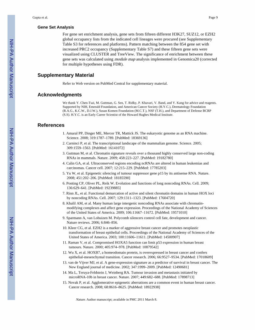
Gene Set AnalysisFor gene set enrichment analysis, gene sets from fifteen different H3K27, SUZ12, or EZH2global occupancy lists from the indicated cell lineages were procured (see SupplementaryTable S3 for references and platforms). Pattern matching between the 854 gene set withincreased PRC2 occupancy (Supplementary Table S7) and these fifteen gene sets werevisualized using CLUSTER and TreeView. The significance of enrichment between thesegene sets was calculated using module map analysis implemented in Genomica20 (correctedfor multiple hypotheses using FDR).
Supplementary MaterialRefer to Web version on PubMed Central for supplementary material.
AcknowledgmentsWe thank Y. Chen-Tsai, M. Guttman, G. Sen, T. Ridky, P. Khavari, V. Band, and Y. Kang for advice and reagents.Supported by NIH, Emerald Foundation, and American Cancer Society (H.Y.C.), Dermatology Foundation(R.A.G., K.C.W., D.J.W.), Susan Komen Foundation (M.C.T.), NSF (T.H.), and Department of Defense BCRP(S.S). H.Y.C. is an Early Career Scientist of the Howard Hughes Medical Institute.
References1. Amaral PP, Dinger ME, Mercer TR, Mattick JS. The eukaryotic genome as an RNA machine.
Science. 2008; 319:1787–1789. [PubMed: 18369136]
2. Carninci P, et al. The transcriptional landscape of the mammalian genome. Science. 2005;309:1559–1563. [PubMed: 16141072]
3. Guttman M, et al. Chromatin signature reveals over a thousand highly conserved large non-codingRNAs in mammals. Nature. 2009; 458:223–227. [PubMed: 19182780]
4. Calin GA, et al. Ultraconserved regions encoding ncRNAs are altered in human leukemias andcarcinomas. Cancer cell. 2007; 12:215–229. [PubMed: 17785203]
5. Yu W, et al. Epigenetic silencing of tumour suppressor gene p15 by its antisense RNA. Nature.2008; 451:202–206. [PubMed: 18185590]
6. Ponting CP, Oliver PL, Reik W. Evolution and functions of long noncoding RNAs. Cell. 2009;136:629–641. [PubMed: 19239885]
7. Rinn JL, et al. Functional demarcation of active and silent chromatin domains in human HOX lociby noncoding RNAs. Cell. 2007; 129:1311–1323. [PubMed: 17604720]
8. Khalil AM, et al. Many human large intergenic noncoding RNAs associate with chromatin-modifying complexes and affect gene expression. Proceedings of the National Academy of Sciencesof the United States of America. 2009; 106:11667–11672. [PubMed: 19571010]
9. Sparmann A, van Lohuizen M. Polycomb silencers control cell fate, development and cancer.Nature reviews. 2006; 6:846–856.
10. Kleer CG, et al. EZH2 is a marker of aggressive breast cancer and promotes neoplastictransformation of breast epithelial cells. Proceedings of the National Academy of Sciences of theUnited States of America. 2003; 100:11606–11611. [PubMed: 14500907]
11. Raman V, et al. Compromised HOXA5 function can limit p53 expression in human breasttumours. Nature. 2000; 405:974–978. [PubMed: 10879542]
12. Wu X, et al. HOXB7, a homeodomain protein, is overexpressed in breast cancer and confersepithelial-mesenchymal transition. Cancer research. 2006; 66:9527–9534. [PubMed: 17018609]
13. van de Vijver MJ, et al. A gene-expression signature as a predictor of survival in breast cancer. TheNew England journal of medicine. 2002; 347:1999–2009. [PubMed: 12490681]
14. Ma L, Teruya-Feldstein J, Weinberg RA. Tumour invasion and metastasis initiated bymicroRNA-10b in breast cancer. Nature. 2007; 449:682–688. [PubMed: 17898713]
15. Novak P, et al. Agglomerative epigenetic aberrations are a common event in human breast cancer.Cancer research. 2008; 68:8616–8625. [PubMed: 18922938]
Gupta et al. Page 9
Nature. Author manuscript; available in PMC 2011 March 8.
NIH
-PA Author Manuscript
NIH
-PA Author Manuscript
NIH
-PA Author Manuscript

16. Naik MU, Naik TU, Suckow AT, Duncan MK, Naik UP. Attenuation of junctional adhesionmolecule-A is a contributing factor for breast cancer cell invasion. Cancer research. 2008;68:2194–2203. [PubMed: 18381425]
17. Fox BP, Kandpal RP. Invasiveness of breast carcinoma cells and transcript profile: Eph receptorsand ephrin ligands as molecular markers of potential diagnostic and prognostic application.Biochemical and biophysical research communications. 2004; 318:882–892. [PubMed: 15147954]
18. Herath NI, Doecke J, Spanevello MD, Leggett BA, Boyd AW. Epigenetic silencing of EphA1expression in colorectal cancer is correlated with poor survival. British journal of cancer. 2009;100:1095–1102. [PubMed: 19277044]
19. Ashburner M, et al. Gene ontology: tool for the unification of biology. The Gene OntologyConsortium. Nature genetics. 2000; 25:25–29. [PubMed: 10802651]
20. Segal E, Friedman N, Koller D, Regev A. A module map showing conditional activity ofexpression modules in cancer. Nature genetics. 2004; 36:1090–1098. [PubMed: 15448693]
21. Srinivasan D, Plattner R. Activation of Abl tyrosine kinases promotes invasion of aggressive breastcancer cells. Cancer research. 2006; 66:5648–5655. [PubMed: 16740702]
22. Olmeda D, et al. SNAI1 is required for tumor growth and lymph node metastasis of human breastcarcinoma MDA-MB-231 cells. Cancer research. 2007; 67:11721–11731. [PubMed: 18089802]
23. Marinkovich MP. Tumour microenvironment: laminin 332 in squamous-cell carcinoma. Naturereviews. 2007; 7:370–380.
24. Tan J, et al. Pharmacologic disruption of Polycomb-repressive complex 2-mediated generepression selectively induces apoptosis in cancer cells. Genes & development. 2007; 21:1050–1063. [PubMed: 17437993]
ADDITIONAL REFERENCES (FOR FULL METHODS SECTION)25. Sen GL, Webster DE, Barragan DI, Chang HY, Khavari PA. Control of differentiation in a self-
renewing mammalian tissue by the histone demethylase JMJD3. Genes & development. 2008;22:1865–1870. [PubMed: 18628393]
26. Bergstraesser LM, Weitzman SA. Culture of normal and malignant primary human mammaryepithelial cells in a physiological manner simulates in vivo growth patterns and allowsdiscrimination of cell type. Cancer research. 1993; 53:2644–2654. [PubMed: 8495428]
27. Wu JM, et al. Heterogeneity of breast cancer metastases: comparison of therapeutic targetexpression and promoter methylation between primary tumors and their multifocal metastases.Clin Cancer Res. 2008; 14:1938–1946. [PubMed: 18381931]
28. Eisen MB, Spellman PT, Brown PO, Botstein D. Cluster analysis and display of genome-wideexpression patterns. Proceedings of the National Academy of Sciences of the United States ofAmerica. 1998; 95:14863–14868. [PubMed: 9843981]
29. Rinn JL, Bondre C, Gladstone HB, Brown PO, Chang HY. Anatomic demarcation by positionalvariation in fibroblast gene expression programs. PLoS genetics. 2006; 2:e119. [PubMed:16895450]
30. Adler AS, et al. Genetic regulators of large-scale transcriptional signatures in cancer. Naturegenetics. 2006; 38:421–430. [PubMed: 16518402]
Gupta et al. Page 10
Nature. Author manuscript; available in PMC 2011 March 8.
NIH
-PA Author Manuscript
NIH
-PA Author Manuscript
NIH
-PA Author Manuscript
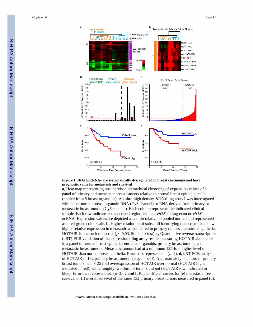
Figure 1. HOX lincRNAs are systematically dysregulated in breast carcinoma and haveprognostic value for metastasis and survivala, Heat map representing unsupervised hierarchical clustering of expression values of apanel of primary and metastatic breast cancers relative to normal breast epithelial cells(pooled from 5 breast organoids). An ultra-high density HOX tiling array7 was interrogatedwith either normal breast organoid RNA (Cy3 channel) or RNA derived from primary ormetastatic breast tumors (Cy5 channel). Each column represents the indicated clinicalsample. Each row indicates a transcribed region, either a HOX coding exon or HOXncRNA. Expression values are depicted as a ratio relative to pooled normal and representedas a red-green color scale. b, Higher resolution of subset iii identifying transcripts that showhigher relative expression in metastatic as compared to primary tumors and normal epithelia.HOTAIR is one such transcript (p= 0.03, Student t-test). c, Quantitative reverse transcription(qRT)-PCR validation of the expression tiling array results measuring HOTAIR abundancein a panel of normal breast epithelial-enriched organoids, primary breast tumors, andmetastatic breast tumors. Metastatic tumors had at a minimum 125-fold higher level ofHOTAIR than normal breast epithelia. Error bars represent s.d. (n=3). d, qRT-PCR analysisof HOTAIR in 132 primary breast tumors (stage I or II). Approximately one third of primarybreast tumors had >125 fold overexpression of HOTAIR over normal (HOTAIR high,indicated in red), while roughly two third of tumors did not (HOTAIR low, indicated inblue). Error bars represent s.d. (n=3). e and f, Kaplan-Meier curves for (e) metastasis freesurvival or (f) overall survival of the same 132 primary breast tumors measured in panel (d).
Gupta et al. Page 11
Nature. Author manuscript; available in PMC 2011 March 8.
NIH
-PA Author Manuscript
NIH
-PA Author Manuscript
NIH
-PA Author Manuscript
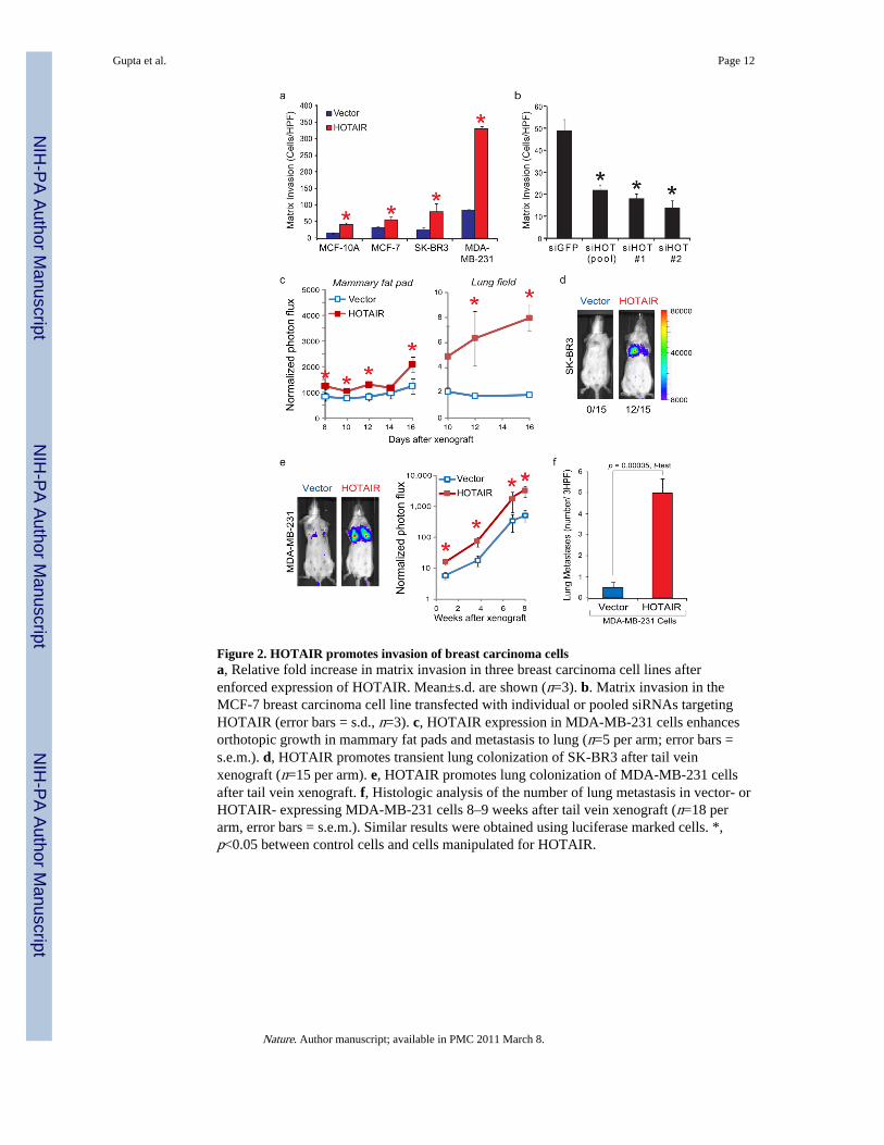
Figure 2. HOTAIR promotes invasion of breast carcinoma cellsa, Relative fold increase in matrix invasion in three breast carcinoma cell lines afterenforced expression of HOTAIR. Mean±s.d. are shown (n=3). b. Matrix invasion in theMCF-7 breast carcinoma cell line transfected with individual or pooled siRNAs targetingHOTAIR (error bars = s.d., n=3). c, HOTAIR expression in MDA-MB-231 cells enhancesorthotopic growth in mammary fat pads and metastasis to lung (n=5 per arm; error bars =s.e.m.). d, HOTAIR promotes transient lung colonization of SK-BR3 after tail veinxenograft (n=15 per arm). e, HOTAIR promotes lung colonization of MDA-MB-231 cellsafter tail vein xenograft. f, Histologic analysis of the number of lung metastasis in vector- orHOTAIR- expressing MDA-MB-231 cells 8–9 weeks after tail vein xenograft (n=18 perarm, error bars = s.e.m.). Similar results were obtained using luciferase marked cells. *,p<0.05 between control cells and cells manipulated for HOTAIR.
Gupta et al. Page 12
Nature. Author manuscript; available in PMC 2011 March 8.
NIH
-PA Author Manuscript
NIH
-PA Author Manuscript
NIH
-PA Author Manuscript
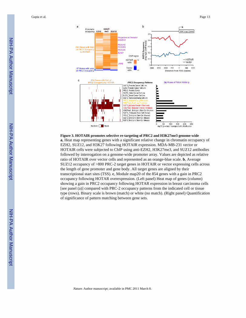
Figure 3. HOTAIR promotes selective re-targeting of PRC2 and H3K27me3 genome-widea, Heat map representing genes with a significant relative change in chromatin occupancy ofEZH2, SUZ12, and H3K27 following HOTAIR expression. MDA-MB-231 vector orHOTAIR cells were subjected to ChIP using anti-EZH2, H3K27me3, and SUZ12 antibodiesfollowed by interrogation on a genome-wide promoter array. Values are depicted as relativeratio of HOTAIR over vector cells and represented as an orange-blue scale. b, AverageSUZ12 occupancy of >800 PRC-2 target genes in HOTAIR or vector expressing cells acrossthe length of gene promoter and gene body. All target genes are aligned by theirtranscriptional start sites (TSS). c, Module map20 of the 854 genes with a gain in PRC2occupancy following HOTAR overexpression. (Left panel) Heat map of genes (column)showing a gain in PRC2 occupancy following HOTAR expression in breast carcinoma cells[see panel (a)] compared with PRC-2 occupancy patterns from the indicated cell or tissuetype (rows). Binary scale is brown (match) or white (no match). (Right panel) Quantificationof significance of pattern matching between gene sets.
Gupta et al. Page 13
Nature. Author manuscript; available in PMC 2011 March 8.
NIH
-PA Author Manuscript
NIH
-PA Author Manuscript
NIH
-PA Author Manuscript
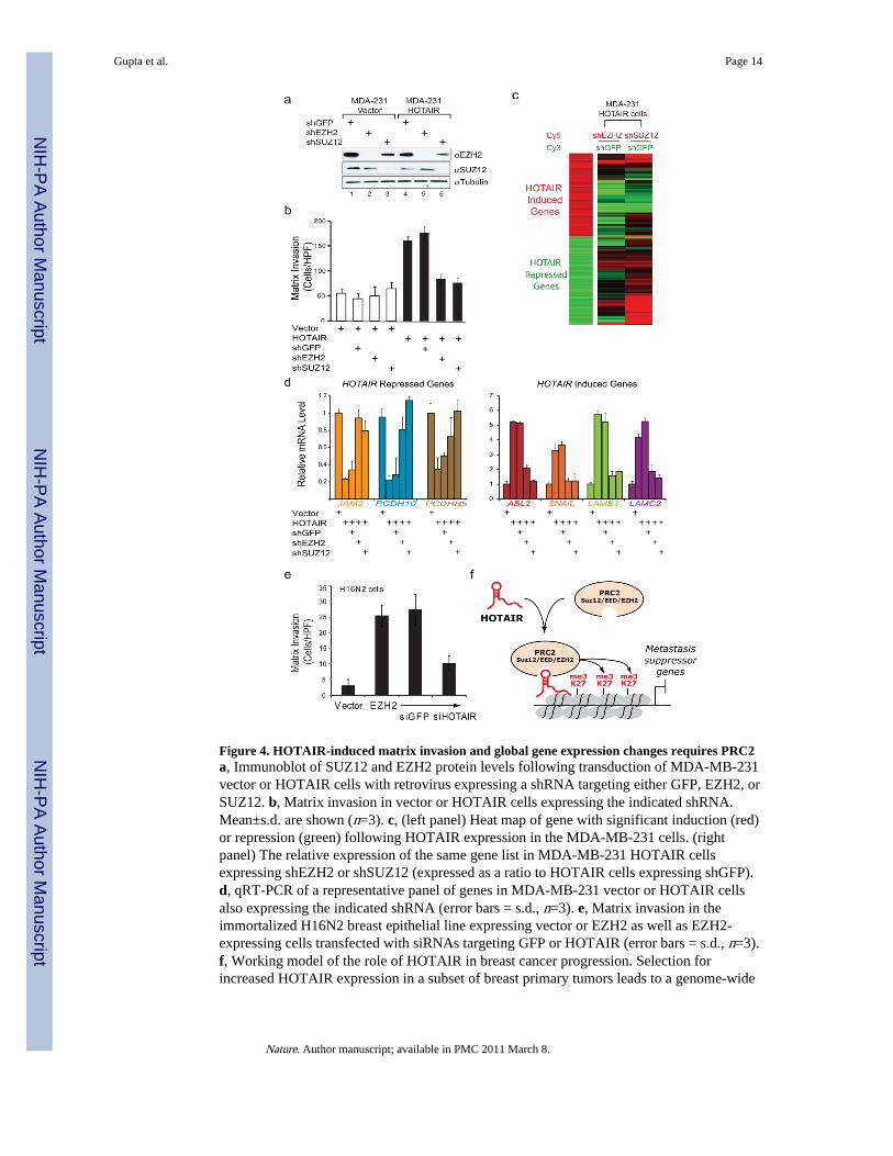
Figure 4. HOTAIR-induced matrix invasion and global gene expression changes requires PRC2a, Immunoblot of SUZ12 and EZH2 protein levels following transduction of MDA-MB-231vector or HOTAIR cells with retrovirus expressing a shRNA targeting either GFP, EZH2, orSUZ12. b, Matrix invasion in vector or HOTAIR cells expressing the indicated shRNA.Mean±s.d. are shown (n=3). c, (left panel) Heat map of gene with significant induction (red)or repression (green) following HOTAIR expression in the MDA-MB-231 cells. (rightpanel) The relative expression of the same gene list in MDA-MB-231 HOTAIR cellsexpressing shEZH2 or shSUZ12 (expressed as a ratio to HOTAIR cells expressing shGFP).d, qRT-PCR of a representative panel of genes in MDA-MB-231 vector or HOTAIR cellsalso expressing the indicated shRNA (error bars = s.d., n=3). e, Matrix invasion in theimmortalized H16N2 breast epithelial line expressing vector or EZH2 as well as EZH2-expressing cells transfected with siRNAs targeting GFP or HOTAIR (error bars = s.d., n=3).f, Working model of the role of HOTAIR in breast cancer progression. Selection forincreased HOTAIR expression in a subset of breast primary tumors leads to a genome-wide
Gupta et al. Page 14
Nature. Author manuscript; available in PMC 2011 March 8.
NIH
-PA Author Manuscript
NIH
-PA Author Manuscript
NIH
-PA Author Manuscript

retargeting of the PRC2 and H3K27me3 patterns, resulting in gene expression changes thatpromote tumor metastasis.
Gupta et al. Page 15
Nature. Author manuscript; available in PMC 2011 March 8.
NIH
-PA Author Manuscript
NIH
-PA Author Manuscript
NIH
-PA Author Manuscript
Related Documents