Locomotor training alters the behavior of flexor reflexes during walking in human spinal cord injury Andrew C. Smith, 2 Chaithanya K. Mummidisetty, 1 William Zev Rymer, 1,2,3 and Maria Knikou 1,3,4,5 1 Sensory Motor Performance Program, Rehabilitation Institute of Chicago, Chicago, Illinois; 2 Northwestern University Interdepartmental Neuroscience Program, Chicago, Illinios; 3 Department of Physical Medicine and Rehabilitation, Northwestern University Feinberg Medical School, Chicago, Illinois; 4 Graduate Center/The City University of New York, New York, New York; and 5 Department of Physical Therapy, College of Staten Island, Staten Island, New York Submitted 21 April 2014; accepted in final form 12 August 2014 Smith AC, Mummidisetty CK, Rymer WZ, Knikou M. Loco- motor training alters the behavior of flexor reflexes during walking in human spinal cord injury. J Neurophysiol 112: 2164 –2175, 2014. First published August 13, 2014; doi:10.1152/jn.00308.2014.—In humans, a chronic spinal cord injury (SCI) impairs the excitability of pathways mediating early flexor reflexes and increases the excitability of late, long-lasting flexor reflexes. We hypothesized that in individ- uals with SCI, locomotor training will alter the behavior of these spinally mediated reflexes. Nine individuals who had either chronic clinically motor complete or incomplete SCI received an average of 44 locomotor training sessions. Flexor reflexes, elicited via sural nerve stimulation of the right or left leg, were recorded from the ipsilateral tibialis anterior (TA) muscle before and after body weight support (BWS)-assisted treadmill training. The modulation pattern of the ipsilateral TA responses following innocuous stimulation of the right foot was also recorded in 10 healthy subjects while they stepped at 25% BWS to investigate whether body unloading during walking affects the behavior of these responses. Healthy subjects did not receive treadmill training. We observed a phase-dependent modula- tion of early TA flexor reflexes in healthy subjects with reduced body weight during walking. The early TA flexor reflexes were increased at heel contact, progressively decreased during the stance phase, and then increased throughout the swing phase. In individuals with SCI, locomotor training induced the reappearance of early TA flexor reflexes and changed the amplitude of late TA flexor reflexes during walking. Both early and late TA flexor reflexes were modulated in a phase-dependent pattern after training. These new findings support the adaptive capability of the injured nervous system to return to a prelesion excitability and integration state. locomotor training; neuroplasticity; SCI; reorganization; spinal cir- cuits TWO OF THE REFLEX CHANGES observed in the human spinal cord following injury are impaired excitability of pathways mediat- ing early flexor reflexes and increased excitability of pathways mediating late long-lasting flexor reflexes (Roby-Brami and Bussel 1987; Knikou and Conway 2005; Knikou 2007b; Con- way and Knikou 2008; Dietz et al. 2009). Specifically, the early flexor reflexes are reduced or can be completely absent at the chronic stage of spinal cord injury (SCI) in humans (Roby- Brami and Bussel 1987; Knikou and Conway 2005; Knikou 2007a), while their increased presence has been linked to improved mobility in individuals with SCI (Hubli et al. 2012). Late flexor reflexes in individuals with SCI have a similar interneuronal reorganization to that reported in acute spinal cats treated with L-dihydroxyphenylalanine (L-DOPA; norepi- nephrine precursor) or 5-hydroxytryptophan (serotonin precur- sor) (Anden et al. 1966a,b; Jankowska et al. 1967a,b; Fu et al. 1975). Accordingly, spinal interneurons [termed flexor reflex afferent (FRA) interneurons] mediating the late flexor reflexes are responsible for three major roles: 1) they evoke presynaptic inhibition of extensor group Ia afferent transmission to alpha motoneurons of the ipsilateral limb, 2) they inhibit actions of contralateral FRA interneurons, and 3) they facilitate reflex actions of contralateral extensors supporting step progression (Bussel et al. 1989; Roby-Brami and Bussel 1990, 1992). It should be noted that flexor reflex responses evoked at innoc- uous stimulation intensities in healthy subjects appear at early and not at long latencies and are modulated in a phase- dependent manner during walking (Duysens et al. 1993; Zehr et al. 1997, 1998; Baken et al. 2005). The phase-dependent modulation of flexor and extensor reflexes during walking in humans is an integral part of locomotion (Knikou 2010b), and its reappearance in neurological disorders can thus be viewed as functional reorganization. We have recently shown that in individuals with chronic SCI, the phase-dependent modulation pattern of the extensor reflexes (soleus H reflex) is reestablished, homosynaptic facil- itation is reversed to homosynaptic depression, and spinal inhibition exerted at premotoneuronal level is improved after locomotor training (Knikou 2013; Knikou and Mummidisetty 2014). Based on the discussed evidence and our recent find- ings, we hypothesized that the prevalence ratio between early and late tibialis anterior (TA) flexor reflexes will change after locomotor training and that these reflexes will be modulated in a phase-dependent manner during walking. Although repetitive step training on a motorized treadmill has shown to improve step progression, step coordination, kinematics of walking, and spasticity in both animals and humans (Lovely et al. 1986; Rossignol et al. 1996; Dietz et al. 1998; Behrman and Harkema 2000; Dobkin et al. 2007; Knikou 2013; Manella and Field- Fote 2013), the specific features of the reorganization of flexor reflexes during walking after locomotor training in individuals with SCI remain poorly understood. Collectively, in this study, we assessed whether locomotor training alters the behavior of the flexor reflexes during walk- ing in individuals with chronic SCI. The results provide unique evidence that following locomotor training, early flexor re- flexes reappear and late flexor reflexes can be altered in amplitude. These new findings support the adaptive capability Address for reprint requests and other correspondence: M. Knikou, Graduate Center of the City Univ. of New York, Dept. of Physical Therapy, College of Staten Island, 2800 Victory Blvd., Bldg. 5N-207 Staten Island, NY 10314 (e-mail: [email protected]; [email protected]). J Neurophysiol 112: 2164 –2175, 2014. First published August 13, 2014; doi:10.1152/jn.00308.2014. 2164 0022-3077/14 Copyright © 2014 the American Physiological Society www.jn.org on November 2, 2014 Downloaded from

Welcome message from author
This document is posted to help you gain knowledge. Please leave a comment to let me know what you think about it! Share it to your friends and learn new things together.
Transcript
Locomotor training alters the behavior of flexor reflexes during walking inhuman spinal cord injury
Andrew C. Smith,2 Chaithanya K. Mummidisetty,1 William Zev Rymer,1,2,3 and Maria Knikou1,3,4,5
1Sensory Motor Performance Program, Rehabilitation Institute of Chicago, Chicago, Illinois; 2Northwestern UniversityInterdepartmental Neuroscience Program, Chicago, Illinios; 3Department of Physical Medicine and Rehabilitation,Northwestern University Feinberg Medical School, Chicago, Illinois; 4Graduate Center/The City University of New York,New York, New York; and 5Department of Physical Therapy, College of Staten Island, Staten Island, New York
Submitted 21 April 2014; accepted in final form 12 August 2014
Smith AC, Mummidisetty CK, Rymer WZ, Knikou M. Loco-motor training alters the behavior of flexor reflexes during walking inhuman spinal cord injury. J Neurophysiol 112: 2164–2175, 2014.First published August 13, 2014; doi:10.1152/jn.00308.2014.—Inhumans, a chronic spinal cord injury (SCI) impairs the excitability ofpathways mediating early flexor reflexes and increases the excitabilityof late, long-lasting flexor reflexes. We hypothesized that in individ-uals with SCI, locomotor training will alter the behavior of thesespinally mediated reflexes. Nine individuals who had either chronicclinically motor complete or incomplete SCI received an average of44 locomotor training sessions. Flexor reflexes, elicited via suralnerve stimulation of the right or left leg, were recorded from theipsilateral tibialis anterior (TA) muscle before and after body weightsupport (BWS)-assisted treadmill training. The modulation pattern ofthe ipsilateral TA responses following innocuous stimulation of theright foot was also recorded in 10 healthy subjects while they steppedat 25% BWS to investigate whether body unloading during walkingaffects the behavior of these responses. Healthy subjects did notreceive treadmill training. We observed a phase-dependent modula-tion of early TA flexor reflexes in healthy subjects with reduced bodyweight during walking. The early TA flexor reflexes were increased atheel contact, progressively decreased during the stance phase, andthen increased throughout the swing phase. In individuals with SCI,locomotor training induced the reappearance of early TA flexorreflexes and changed the amplitude of late TA flexor reflexes duringwalking. Both early and late TA flexor reflexes were modulated in aphase-dependent pattern after training. These new findings support theadaptive capability of the injured nervous system to return to aprelesion excitability and integration state.
locomotor training; neuroplasticity; SCI; reorganization; spinal cir-cuits
TWO OF THE REFLEX CHANGES observed in the human spinal cordfollowing injury are impaired excitability of pathways mediat-ing early flexor reflexes and increased excitability of pathwaysmediating late long-lasting flexor reflexes (Roby-Brami andBussel 1987; Knikou and Conway 2005; Knikou 2007b; Con-way and Knikou 2008; Dietz et al. 2009). Specifically, theearly flexor reflexes are reduced or can be completely absent atthe chronic stage of spinal cord injury (SCI) in humans (Roby-Brami and Bussel 1987; Knikou and Conway 2005; Knikou2007a), while their increased presence has been linked toimproved mobility in individuals with SCI (Hubli et al. 2012).Late flexor reflexes in individuals with SCI have a similar
interneuronal reorganization to that reported in acute spinalcats treated with L-dihydroxyphenylalanine (L-DOPA; norepi-nephrine precursor) or 5-hydroxytryptophan (serotonin precur-sor) (Anden et al. 1966a,b; Jankowska et al. 1967a,b; Fu et al.1975). Accordingly, spinal interneurons [termed flexor reflexafferent (FRA) interneurons] mediating the late flexor reflexesare responsible for three major roles: 1) they evoke presynapticinhibition of extensor group Ia afferent transmission to alphamotoneurons of the ipsilateral limb, 2) they inhibit actions ofcontralateral FRA interneurons, and 3) they facilitate reflexactions of contralateral extensors supporting step progression(Bussel et al. 1989; Roby-Brami and Bussel 1990, 1992). Itshould be noted that flexor reflex responses evoked at innoc-uous stimulation intensities in healthy subjects appear at earlyand not at long latencies and are modulated in a phase-dependent manner during walking (Duysens et al. 1993; Zehret al. 1997, 1998; Baken et al. 2005). The phase-dependentmodulation of flexor and extensor reflexes during walking inhumans is an integral part of locomotion (Knikou 2010b), andits reappearance in neurological disorders can thus be viewedas functional reorganization.
We have recently shown that in individuals with chronicSCI, the phase-dependent modulation pattern of the extensorreflexes (soleus H reflex) is reestablished, homosynaptic facil-itation is reversed to homosynaptic depression, and spinalinhibition exerted at premotoneuronal level is improved afterlocomotor training (Knikou 2013; Knikou and Mummidisetty2014). Based on the discussed evidence and our recent find-ings, we hypothesized that the prevalence ratio between earlyand late tibialis anterior (TA) flexor reflexes will change afterlocomotor training and that these reflexes will be modulated ina phase-dependent manner during walking. Although repetitivestep training on a motorized treadmill has shown to improvestep progression, step coordination, kinematics of walking, andspasticity in both animals and humans (Lovely et al. 1986;Rossignol et al. 1996; Dietz et al. 1998; Behrman and Harkema2000; Dobkin et al. 2007; Knikou 2013; Manella and Field-Fote 2013), the specific features of the reorganization of flexorreflexes during walking after locomotor training in individualswith SCI remain poorly understood.
Collectively, in this study, we assessed whether locomotortraining alters the behavior of the flexor reflexes during walk-ing in individuals with chronic SCI. The results provide uniqueevidence that following locomotor training, early flexor re-flexes reappear and late flexor reflexes can be altered inamplitude. These new findings support the adaptive capability
Address for reprint requests and other correspondence: M. Knikou, GraduateCenter of the City Univ. of New York, Dept. of Physical Therapy, College ofStaten Island, 2800 Victory Blvd., Bldg. 5N-207 Staten Island, NY 10314(e-mail: [email protected]; [email protected]).
J Neurophysiol 112: 2164–2175, 2014.First published August 13, 2014; doi:10.1152/jn.00308.2014.
2164 0022-3077/14 Copyright © 2014 the American Physiological Society www.jn.org
on Novem
ber 2, 2014D
ownloaded from

of the injured nervous system to return to a prelesion excit-ability and integration state.
METHODS
Subjects. Nine individuals with chronic SCI and 10 healthy subjects(6 female, age range 20–45 yr) participated in the study. Studyparticipation for SCI individuals varied from 1 mo to 3½ mo depend-ing on the number of locomotor training sessions attended (Table 1).All subjects signed an informed consent form before participation inthe study for neurophysiological tests, clinical evaluation, and/orlocomotor training, which was approved by the Northwestern Univer-sity and the City University of New York Institutional Review Boards.Subjects’ consent was obtained according to the Declaration ofHelsinki. Individuals with SCI also participated in studies examiningthe reorganization of premotoneuronal control and soleus H-reflexexcitability after locomotor training (Knikou 2013; Knikou and Mum-midisetty 2014) and are identified here with the same code. During theduration of the study, none of the subjects received conventionalphysical therapy or participated in other research studies.
Locomotor training. Individuals with SCI received body weightsupport (BWS)-assisted locomotor training with a robotic exoskeletonsystem (Lokomat Pro, Hocoma, Switzerland) and were trained 5days/wk, 1 h/day. The protocol employed to train individuals withmotor complete and motor incomplete SCI has been previouslypublished in detail (see Fig. 1 in Knikou 2013). Briefly, training on thefirst session in AIS A started at 1.58 km/h treadmill speed with 60%BWS. At each subsequent session the targeted treadmill speed was setto increase by 0.07 km/h and BWS to decrease by 5%. In AIS C-Dsubjects, when quadriceps manual muscle test score was �3/5, train-ing started at 40% BWS at 1.98 km/h. The treadmill speed and BWSwere targeted to be adjusted by 0.07 km/h and 5% at each subsequenttraining session, respectively. When quadriceps and triceps suraestrength was increased by a full grade, then the BWS was decreasedby 10%. Based on the TA muscle strength, which was assessed everyfive training sessions, the position of the straps of the ankle braces wasadjusted. The ultimate goal in AIS C-D subjects was to reach atreadmill speed of 2.98 km/h at the lowest BWS possible without kneebuckling or toe dragging during the stance and swing phases, respec-
tively. On the first day of training, the average treadmill speed was1.84 � 0.12 km/h, while on the last day of training it was 2.68 � 0.16km/h across subjects. The BWS required before training was 47 �0.05% while after training it was 24 � 0.07% across subjects.
Experimental procedures. A pulse train of 30 ms (1-ms pulses at300 Hz) was delivered to the sural nerve (a purely sensory nerve) atthe lateral submalleolar region through a stainless-steel bipolar elec-trode to individuals with SCI while seated with both feet at rest. Inhealthy subjects, stimulation was delivered to the skin over the medialedge of the right foot innervated by the saphenous nerve and not to thesural nerve, because healthy subjects expressed a great discomfortupon sural nerve stimulation even at low intensities, while noxiousstimulation was required to evoke responses in the ipsilateral TAmuscle. Stimulation in both cases was delivered by a constant currentstimulator (DS7A; Digitimer, Hertfordshire, UK) triggered with cus-tomized LabVIEW scripts. With subjects seated, a bipolar electrodewas used to establish the site that elicited a response in the ipsilateralTA muscle, without limb movement, at the lowest stimulus intensitypossible. The bipolar electrode was then replaced by two monopolarelectrodes (SureTrace) that were secured via a surgical transparentfilm and maintained in place via prewrap and an athletic tape, whilethe reflex threshold was checked several times during the experiment.
Each subject was then transferred to the treadmill and wore anupper body harness that was connected to an overhead pulley system.While subjects were standing with equal distribution of body weightin both limbs and arms parallel to the trunk, BWS was applied. Thighand shank segments of the exoskeleton were adjusted based on eachsubject’s leg length and diameter, and both feet were secured into thefoot lifters (SCI individuals only). In all subjects, the reflex thresholdduring BWS standing was established and corresponded to the loweststimulation intensity that evoked an EMG response in the ipsilateralTA muscle without limb or ankle joint movement. In healthy subjects,this intensity ranged from 37 to 110 mA (70.1 � 8.85; means � SE)and was 3.15 times the sensory threshold (22.2 � 2.17 mA). In allhealthy subjects, 10 reflexes were recorded from the ipsilateral TAmuscle at 1.2 times the reflex threshold while standing with 25% BWSat 0.1 Hz. In individuals with SCI, sural nerve stimulation wasdelivered at 1.1 to 1.5 (1.3 � 0.04; 140 � 23 mA) times the reflexthreshold before locomotor training, and at 1.05 to 1.46 (1.24 � 0.03;
Table 1. Characteristics of SCI participants*
ID GenderAge,
yrPostinjury,
yr Level Cause of SCIAIS
Scale
Clonus ASIA(LightTouch)
ASIA(Pin
Prick)
ASIA(Motor)
Medication
No. ofTrainingSessionsLL RL LL RL
R03 M 24 3.0 T10 GSW A 1 1 72 72 0 0 None 60R10 F 52 11 T7 Fall D 1 0 78 78 16 24 Neurontin: 27 mg
Baclofen: 60 mg65
R13 F 39 7.0 T4 Transversemyelitis
C 3 3 112 74 9 2 Gabapentin: 0.3 gBaclofen: 20 mg
53
R14 M 25 0.5 C5-C6 Diving D 1 1 112 110 25 13 None during thestudy
44
R15 M 37 1.0 C1 Spinal Tumor C 3 2 64 64 12 5 Not known 36R16 M 49 2.5 C5 MVA C 0 0 64 34 17 12 Baclofen: 15 mg 41R17 M 21 3.0 T10 GSW D 3 3 105 105 13 15 Baclofen: 60 mg
Gabapentin: 50.9 g48
R18 M 29 2.0 C7 MVA D 1 1 86 86 25 21 None 26R20 M 55 3.0 T6-T7 Blood clot during
spinal surgeryC 2 3 82 82 8 9 None 21
*Level of spinal cord injury (SCI) corresponds to neurological injury level. For each subject, the American Spinal Injury Association (ASIA) standardneurological classification of SCI for sensation (sensory light touch and pin prick; out of 112 maximal points) is shown and evaluated as 0 � absent; 1 �impaired; and 2 � normal. ASIA motor score (out of 50 maximal points for each leg) is indicated for the left leg (LL) and right leg (RL) based on the manualmuscle test of key muscles and evaluated as 0 � no contraction, 1 � flicker or trace of contraction; 2 � active movement, with gravity eliminated; 3 � activemovement against gravity; 4 � active movement against gravity and resistance; 5 � normal power. Values for ASIA sensory and motor scores are indicatedfrom clinical evaluation tests conducted before training. Ankle clonus was assessed as follows: 0 � reaction, 1 � mild, clonus was maintained �3 s; 2 �moderate, clonus persisted between 3 and 10 s; 3 � severe, clonus persisted for �10 s. Medication for each subject is indicated as total mg taken per day. AIS,abbreviated injury scale; C, cervical; T, thoracic; MVA, motor vehicle accident; GSW, gunshot wound; M, male; F, female.
2165TREADMILL TRAINING ALTERS FRA PATHWAYS
J Neurophysiol • doi:10.1152/jn.00308.2014 • www.jn.org
on Novem
ber 2, 2014D
ownloaded from
137 � 19 mA) times the reflex threshold after locomotor training, butat equivalent multiples of reflex threshold for each individual withSCI before and after training. Furthermore, after training recordingswere conducted at similar BWS levels (47 � 0.05) and treadmillspeeds (1.84 � 0.12 km/h) to those utilized before training.
During stepping, stimulation was delivered at the same multiples ofreflex threshold utilized during standing to evoke ipsilateral TA flexorreflexes. The constant current stimulator was triggered based on theipsilateral foot switch signals (BIOPAC Systems, Goleta, CA) andwas delivered randomly across the different phases (n � 16) of a stepcycle every three to five steps. During stepping, a custom writtensoftware program (LabVIEW; National Instruments, Austin, TX,USA) divided the step cycle based on the foot switch signal into 16bins with bin 1 corresponding to heel contact and bins 8, 9, and 16corresponding approximately to stance-to-swing transition, swingphase initiation, and swing-to-stance transition, respectively (Knikouet al. 2009; Knikou 2010a). In all subjects, 10 reflexes or more wererecorded at each bin (except bin 16). TA EMG and foot switch signalswere sampled at 2,000 Hz and stored on a personal computer foroffline analysis.
Data analysis. Offline data analysis commenced with marking andvisual inspection of the foot switch signals for determination of thestep cycle phases, followed by marking of the first triggering pulse ofthe pulse train for determination of stimulation onset within a step.The full-wave rectified band-pass filtered (40–1,000 Hz) areas of theearly (50 ms poststimulus with 50-ms duration) and late (100-mspoststimulus with 400-ms duration) ipsilateral TA flexor reflexes(whichever present) were calculated, grouped for each bin, and aver-aged (Knikou 2010a). Then, the TA EMG from the nonstimulatedsteps corresponding to the same time windows and bins was sub-tracted from the reflex EMGs to remove background EMG activity.The subtracted reflex values were normalized to the maximum TAlocomotor EMG observed across all bins of the step cycle, yielding aresponse activation profile ranging from 0 to 1 (Zehr et al. 1997, 1998,2012; Knikou et al. 2009; Knikou 2010a). To establish task-dependentchanges (standing vs. walking), reflexes were measured as the areaof the TA rectified EMG and were normalized to the mean amplitudeof the associated reflex component recorded with the subject standing.The mean amplitude of the normalized TA reflex from each subjectwas grouped based on the bin number and/or time of testing.
The ipsilateral TA background EMG activity was calculated fromthe mean value of the rectified and band-pass filtered EMG forduration of 50 ms, beginning 100 ms before stimulation for each binof the step cycle, and was normalized to the maximum TA locomotorEMG observed across all bins of the step cycle. To quantify changesin muscle and reflex excitability during walking after training, amodulation index for SCI and healthy subjects for the overall changein the TA EMG over the step cycle was determined by subtractingthe minimum EMG from the maximum EMG and expressed to themaximum (Zehr and Loadman 2012). This was done separately forthe background TA EMG, control EMG, and late TA reflex EMGof the right and left legs during stepping.
Statistical analysis. The effect of locomotor training on the ampli-tude of the TA flexor reflexes during walking was analyzed with aWilcoxon rank sum test at each bin before and after training forreflexes normalized to those recorded during standing and withtwo-way repeated-measures ANOVA (between group factor: time;within-subject factor: bins of the step cycle) for reflexes representedas normalized subtracted reflex EMGs. The amplitude of the ipsilat-eral TA flexor reflexes in healthy subjects during walking was ana-lyzed with one-way repeated-measures ANOVA (within-subject fac-tor: bins of the step cycle) and with two-way repeated-measuresANOVA (between group factor: control/reflex EMG; within-subjectfactor: bins of the step cycle). For all statistical tests, when astatistically significant difference was found, post hoc Bonferroni testsfor multiple comparisons were applied to the data.
The effect of locomotor training on the TA background acti-vity during walking was analyzed with two-way repeated-measuresANOVA (between group factor: time; within-subject factor: binsof the step cycle). Differences of modulation indexes betweenhealthy subjects and individual with SCI on the mean averages ofbackground EMG activity were established with one-way Kruskal-Wallis ANOVA on ranks, while differences between modulationindexes of control EMG for each bin of the step cycle wereestablished with two-way Kruskal-Wallis ANOVA on ranks fol-lowed by pairwise multiple comparisons when a statistically sig-nificant difference was detected.
The mean amplitude of the ipsilateral early and late TA flexorreflexes was plotted on the y-axis vs. the associated TA backgroundactivity on the x-axis, and a linear least-square regression was fitted tothe data. This analysis was conducted on data obtained from eachsubject. Last, to establish the extent to which flexor reflex modulationafter training was related to leg motor scores (clinically assessedbased on American Spinal Injury Association guidelines), the normal-ized flexor reflexes before training were subtracted from those ob-tained after training, grouped for stance and swing phases of gait anda linear relationship to the percentage of change in the motor scoresobserved after training was completed. Effects were considered sig-nificant when P � 0.05.
RESULTS
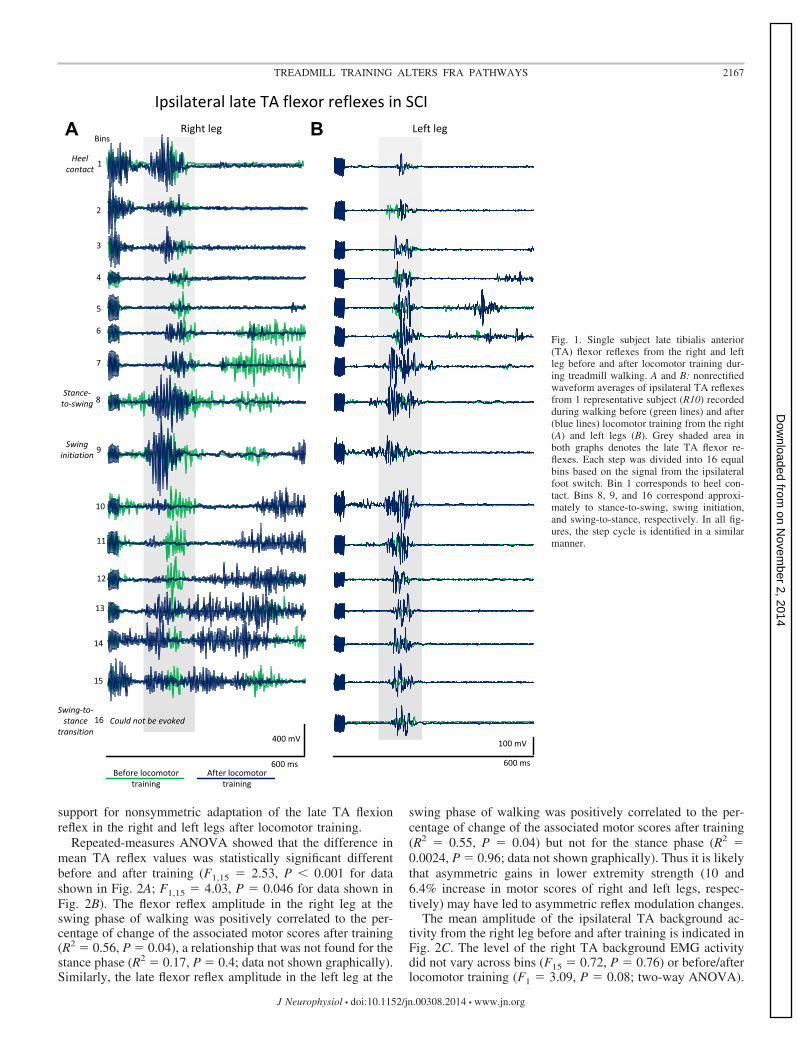
Flexor reflexes during walking after locomotor training inSCI. Locomotor training altered the behavior of TA flexorreflexes during walking in individuals with chronic SCI. Wave-form averages of the ipsilateral TA flexor reflexes of a repre-sentative subject (R10) while stepping during sural nervestimulation before and after locomotor training are presented inFig. 1. In the right leg, the late TA flexor reflexes weremodulated in a phase-dependent pattern after locomotor train-ing. Specifically, the late TA flexor reflexes were profoundlyincreased at stance-to-swing and swing initiation phases (bins8, 9) compared with the other phases of the step cycle.Furthermore, at heel contact (bin 1) and late swing (bins 13,14) phases, the onset of the TA reflex EMG was decreased by40 ms after locomotor training (Fig. 1A). In the left leg,however, the modulation pattern of the ipsilateral late TAflexor reflexes after training was different from that recorded inthe right leg. The late ipsilateral TA flexor reflexes in the leftleg in this subject were profoundly increased after locomotortraining regardless of the step cycle phase (Fig. 1B).
The average amplitude of the ipsilateral late TA flexorreflexes from all individuals with SCI during BWS-assistedstepping from the right and left legs is shown in Fig. 2. In Fig.2, A and D, the late TA flexor reflexes recorded from the rightand left legs are normalized to the associated flexor reflexesrecorded with subjects standing, while in Fig. 2, B and E, thereflexes are normalized to the maximum ipsilateral TA loco-motor EMG across all bins having subtracted the controlEMGs. Locomotor training significantly reduced the amplitudeof the ipsilateral late TA flexor reflexes of the right legthroughout the step cycle (bins 2–14; P � 0.05, Fig. 2A). In theleft leg, locomotor training reestablished a phase-dependentmodulation of the late TA flexor reflexes by increasing theiramplitude at late stance phase (bins 6, 7) and swing phaseinitiation (bin 9) (P � 0.05; Fig. 2D). A similar reflex adap-tation pattern in both legs was observed in the normalizedsubtracted late TA flexion reflexes (Fig. 2, B and E), providing
2166 TREADMILL TRAINING ALTERS FRA PATHWAYS
J Neurophysiol • doi:10.1152/jn.00308.2014 • www.jn.org
on Novem
ber 2, 2014D
ownloaded from
support for nonsymmetric adaptation of the late TA flexionreflex in the right and left legs after locomotor training.
Repeated-measures ANOVA showed that the difference inmean TA reflex values was statistically significant differentbefore and after training (F1,15 � 2.53, P � 0.001 for datashown in Fig. 2A; F1,15 � 4.03, P � 0.046 for data shown inFig. 2B). The flexor reflex amplitude in the right leg at theswing phase of walking was positively correlated to the per-centage of change of the associated motor scores after training(R2 � 0.56, P � 0.04), a relationship that was not found for thestance phase (R2 � 0.17, P � 0.4; data not shown graphically).Similarly, the late flexor reflex amplitude in the left leg at the
swing phase of walking was positively correlated to the per-centage of change of the associated motor scores after training(R2 � 0.55, P � 0.04) but not for the stance phase (R2 �0.0024, P � 0.96; data not shown graphically). Thus it is likelythat asymmetric gains in lower extremity strength (10 and6.4% increase in motor scores of right and left legs, respec-tively) may have led to asymmetric reflex modulation changes.
The mean amplitude of the ipsilateral TA background ac-tivity from the right leg before and after training is indicated inFig. 2C. The level of the right TA background EMG activitydid not vary across bins (F15 � 0.72, P � 0.76) or before/afterlocomotor training (F1 � 3.09, P � 0.08; two-way ANOVA).
Fig. 1. Single subject late tibialis anterior(TA) flexor reflexes from the right and leftleg before and after locomotor training dur-ing treadmill walking. A and B: nonrectifiedwaveform averages of ipsilateral TA reflexesfrom 1 representative subject (R10) recordedduring walking before (green lines) and after(blue lines) locomotor training from the right(A) and left legs (B). Grey shaded area inboth graphs denotes the late TA flexor re-flexes. Each step was divided into 16 equalbins based on the signal from the ipsilateralfoot switch. Bin 1 corresponds to heel con-tact. Bins 8, 9, and 16 correspond approxi-mately to stance-to-swing, swing initiation,and swing-to-stance, respectively. In all fig-ures, the step cycle is identified in a similarmanner.
2167TREADMILL TRAINING ALTERS FRA PATHWAYS
J Neurophysiol • doi:10.1152/jn.00308.2014 • www.jn.org
on Novem
ber 2, 2014D
ownloaded from
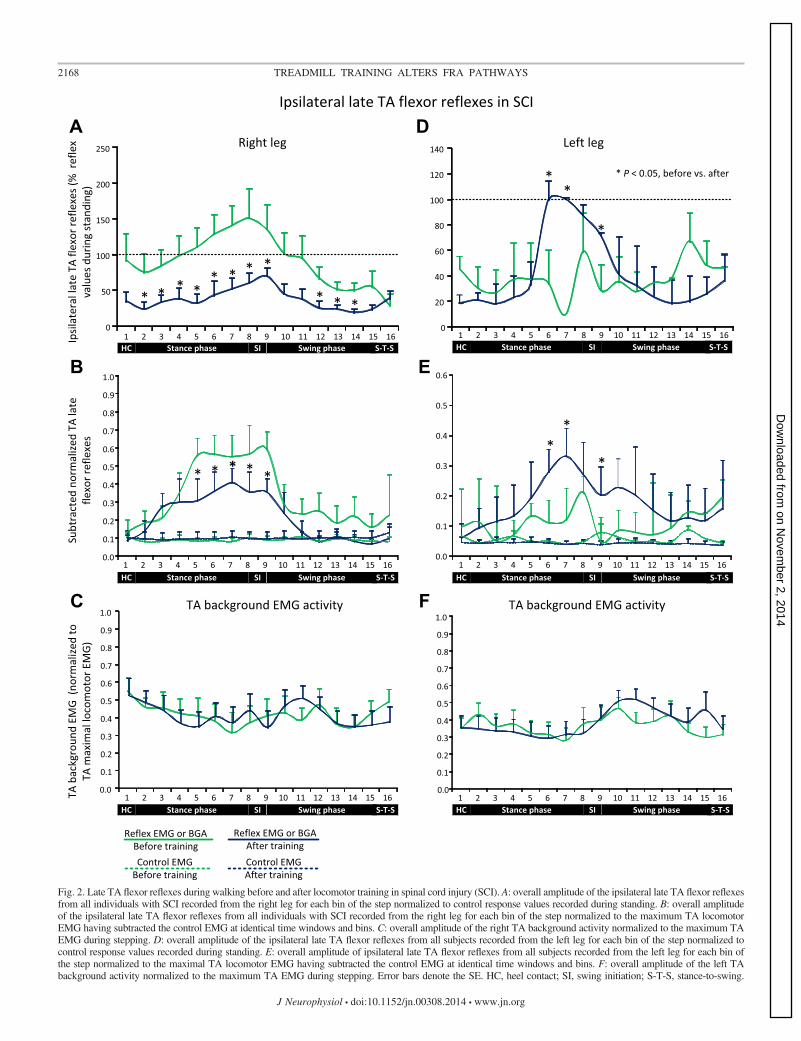
Fig. 2. Late TA flexor reflexes during walking before and after locomotor training in spinal cord injury (SCI). A: overall amplitude of the ipsilateral late TA flexor reflexesfrom all individuals with SCI recorded from the right leg for each bin of the step normalized to control response values recorded during standing. B: overall amplitudeof the ipsilateral late TA flexor reflexes from all individuals with SCI recorded from the right leg for each bin of the step normalized to the maximum TA locomotorEMG having subtracted the control EMG at identical time windows and bins. C: overall amplitude of the right TA background activity normalized to the maximum TAEMG during stepping. D: overall amplitude of the ipsilateral late TA flexor reflexes from all subjects recorded from the left leg for each bin of the step normalized tocontrol response values recorded during standing. E: overall amplitude of ipsilateral late TA flexor reflexes from all subjects recorded from the left leg for each bin ofthe step normalized to the maximal TA locomotor EMG having subtracted the control EMG at identical time windows and bins. F: overall amplitude of the left TAbackground activity normalized to the maximum TA EMG during stepping. Error bars denote the SE. HC, heel contact; SI, swing initiation; S-T-S, stance-to-swing.
2168 TREADMILL TRAINING ALTERS FRA PATHWAYS
J Neurophysiol • doi:10.1152/jn.00308.2014 • www.jn.org
on Novem
ber 2, 2014D
ownloaded from

The right late TA flexor reflexes were not linearly related to theright TA background activity before (R2 � 0.33, P � 0.04) orafter (R2 � 0.34, P � 0.35) locomotor training. The meanamplitude of the ipsilateral TA background activity from theleft leg before and after training is indicated in Fig. 2F. Thelevel of the left TA background EMG activity did not varyacross bins (F15 � 1.1, P � 0.35) or before/after locomotortraining (F1 � 0.51, P � 0.47). The left late TA flexor reflexeswas not linearly related to the left TA background activitybefore (R2 � 0.007, P � 0.87) or after (R2 � 0.04, P � 0.55)locomotor training.
Ipsilateral early TA flexor reflexes were present duringwalking after locomotor training in three out of nine individ-
uals with SCI (R03, R13, and R14) in the right leg, and in oneindividual with SCI (R14) in the left leg. Data from these threesubjects are shown in Fig. 3. Nonrectified waveform averagesof early TA flexor reflexes during BWS-assisted walkingrecorded from the right leg before and after locomotor trainingare indicated in Fig. 3A from subject R14. It is apparent that theshape, latency, and amplitude of these reflexes were alteredthroughout the step cycle after training.
In Fig. 3B, the subtracted normalized early TA flexor re-flexes recorded from the right leg before and after locomotortraining are presented. Locomotor training promoted reflexdepression in stance phase and reflex facilitation during theearly and mid-swing phases of the step cycle (P � 0.05 for bins
Fig. 3. Early TA flexor reflexes during walking before and after locomotor training in SCI. A: nonrectified waveform averages of TA flexor reflexes from 1representative subject (R014) recorded during stepping before (green lines) and after (blue lines) locomotor training. Grey shaded area denotes the early TA flexorreflex. B: amplitude of ipsilateral early TA flexor reflexes from all subjects recorded from the right leg for each bin of the step normalized to the maximal TAlocomotor EMG having subtracted the control EMG at identical time windows and bins. C: amplitude of ipsilateral early TA flexor reflexes recorded from theleft leg of subject R014 normalized to the maximum control EMG having subtracted the control EMG at identical time windows and bins.
2169TREADMILL TRAINING ALTERS FRA PATHWAYS
J Neurophysiol • doi:10.1152/jn.00308.2014 • www.jn.org
on Novem
ber 2, 2014D
ownloaded from
6 and 10–15 pre/post, Fig. 3B). The early TA flexor reflexesduring walking from the right leg were not linearly related tothe right TA background activity during stepping before (R2 �0.2, P � 0.22) or after (R2 � 0.12, P � 0.11) locomotortraining. In one subject (R14), early TA flexor reflexes were notpresent when recorded during seated before or after locomotortraining but reemerged during BWS-assisted stepping afterlocomotor training (Fig. 3C). The increased amplitude of theearly TA flexor reflexes at heel contact and early swing phase(bins 1–4, 10, 11; Fig. 3C) cannot be attributed to peripheralsources because the level of the left TA background activitydid not vary across the step cycle (Fig. 2F).
Flexor reflexes during walking with partial BWS in healthysubjects. To determine the modulation pattern of the TA flexorreflexes in healthy subjects under similar experimental condi-tions utilized for SCI subjects, 10 healthy subjects stepped at25% BWS with a treadmill speed set at 2.89 km/h. The averagelatency of the TA reflexes with subjects seated or standing was75 � 5 ms (including the pulse train duration), and only earlyreflexes in the ipsilateral TA muscle were present duringstanding or during walking in all tested subjects. Waveformaverages of nonrectified early TA reflexes from one represen-tative subject during standing and stepping are indicated in Fig.4A. It is apparent that the early TA flexor reflexes weremodulated in a phase-dependent pattern under conditions ofreduced body weight. Specifically, the early TA flexion reflexwas completely depressed during the stance phase (bins 3–8),and facilitated throughout the swing phase (Fig. 4A). A linearrelationship between the right TA early flexor reflex and theright TA background EMG activity was found (P � 0.0001;data not shown graphically).
The average amplitude of the ipsilateral early TA flexorreflexes recorded from the right leg from all healthy subjects isshown in Fig. 4B as percentages of the reflex values recordedduring standing and in Fig. 4C as subtracted reflex EMGsnormalized to the maximum TA locomotor EMG. One-wayANOVA showed that the early TA flexor reflexes were statis-tically significant different across bins (F15 � 10.6, P �0.001), being statistically significant different from controlreflex values at bins 10, 11, 12, 15, and 16 (Fig. 4B). Similarfindings were observed for the normalized subtracted TAreflexes (F15 � 59.2, P � 0.001), which were significantlyincreased at heel contact (bin 1) and then progressively de-creased reaching control EMG values at late stance phase,followed by a significant facilitation at swing-phase initiationand throughout the swing phase (bins 9–16, P � 0.05; Fig.4C). The level of the right TA background activity from allsubjects is indicated in Fig. 4D. The early TA flexor reflexesrecorded from the right leg were linearly related to the right TAbackground EMG activity (both normalized to the maximumTA locomotor EMG; R2 � 0.42, P � 0.001).
Modulation indexes of background and control EMG be-tween healthy and SCI subjects. To determine to what extentthe modulation pattern of EMG activity in individuals with SCIresembled that observed in healthy subjects, we compared themodulation indexes of TA background EMG (100 ms beforestimulation) and TA control EMG (at similar time windowsand bins of nonstimulated steps) in healthy and SCI subjectsbefore and after training. These are indicated in Fig. 5 as theoverall mean from all 16 bins of the step cycle. Significantdifferences between subject groups and within the SCI
group before and after training were found. These differ-ences were found for the modulation indexes of backgroundTA EMG activity (P � 0.007; Fig. 5A) and control TA EMG(P � 0.001; Fig. 5B) between healthy and SCI subjects forthe right leg. Differences were also found for the modulationindexes of TA control and reflex EMG within the SCI groupbefore and after training for the right leg (Fig. 5, B and C).Last, significant differences for the modulation indexes inthe left TA were found only for the control EMG before andafter training (P � 0.038; Fig. 5B) for the individuals withSCI.
DISCUSSION
We hypothesized that locomotor training alters the behaviorof flexor reflexes in individuals with chronic SCI. We demon-strate two novel findings on the reorganization of this spinallymediated reflex: 1) locomotor training contributed to the ap-pearance of early TA flexor reflexes during walking in bothlimbs; and 2) locomotor training reduced the amplitude of lateTA flexor reflexes in the right leg and increased their amplitudein the left leg and promoted a phase-dependent modulation ofthese reflexes.
Behavior of TA flexor reflexes after locomotor training inSCI. In individuals with chronic SCI, the late TA flexorreflexes in the right leg were significantly reduced duringwalking after training (Fig. 2A), and when depression wasreplaced by facilitation, the onset of the reflex EMG changed(Fig. 1A). The atypical late TA flexor reflex modulation patternin the left leg before training was replaced by increased reflexexcitability that was modulated in a phase-dependent patternafter training (Fig. 2D). These findings suggest that there is analtered behavior of interneurons mediating the late TA flexorreflexes and demonstrate that FRA pathways were not reorga-nized in a similar manner in both limbs. Specifically, twoasymmetries were noted between limbs. One, the onset ofincreased excitability in the late TA flexor reflexes occurred atdifferent phases of the step cycle. Two, the late TA flexorreflexes were modulated differently in the right and left legsbefore training (compare Fig. 1, A and E). The reorganizationof the late TA flexor reflexes we observed here may be relatedto the motor capacity of each individual with SCI beforetraining (Table 1), the amplitude and prevalence relationshipbetween early and late TA flexor reflexes, the asymmetryfound in the late TA flexor reflex modulation between legsbefore training (Fig. 1), the type of SCI (cervical vs. thoracic),and the number, localization, and amount of damaged andspared spinal and supraspinal pathways (Rossignol et al. 2009).
Early TA flexor reflexes deteriorate in amplitude as the SCIbecomes more chronic (Dietz et al. 2009), and in some casesthey might be completely absent (Roby-Brami and Bussel1987; Knikou 2007a; Conway and Knikou 2008). Our mostimportant finding was that the early TA flexor reflexes reap-peared in 1 individual with AIS D SCI after 44 locomotortraining sessions (Fig. 3C) and regained their phase-dependentmodulation excitability pattern during walking (Fig. 3B) in 3out of 9 individuals with SCI. These findings are consistentwith the return of the early TA flexor reflex in 1 individual withSCI while seated after �16 locomotor training sessions (Hubliet al. 2012). While early TA flexor reflexes are mostly absentduring the seated or supine position in individuals with SCI
2170 TREADMILL TRAINING ALTERS FRA PATHWAYS
J Neurophysiol • doi:10.1152/jn.00308.2014 • www.jn.org
on Novem
ber 2, 2014D
ownloaded from

Fig. 4. Ipsilateral early TA flexor reflexes during walking in healthy subjects. A: nonrectified waveform averages of the early TA flexor reflexes from 1 healthysubject (CT25A) during standing and walking at 25% body weight support (BWS). In this subject, the TA reflexes were completely depressed during the stancephase (bins 3–8) and facilitated throughout the swing phase. B: amplitude of ipsilateral early TA flexor reflexes from all healthy subjects for each bin of the stepnormalized to the reflexes recorded during standing. C: amplitude of ipsilateral early TA flexor reflexes from all subjects for each bin of the step normalized tothe maximum TA locomotor EMG having subtracted the control EMG at identical time windows and bins of the step cycle. D: mean TA background activitynormalized to the maximum TA EMG during stepping. Error bars denote the SE.
2171TREADMILL TRAINING ALTERS FRA PATHWAYS
J Neurophysiol • doi:10.1152/jn.00308.2014 • www.jn.org
on Novem
ber 2, 2014D
ownloaded from

(Knikou 2007a; Conway and Knikou 2008; Bolliger et al.2010), they were reemerged during walking in nine individualswith complete SCI, and the late TA flexor reflexes wereconcomitantly decreased in amplitude (Bolliger et al. 2010).This behavior was likely driven by appropriate locomotorproprioceptive inputs (Bolliger et al. 2010) that changed theexcitability state of FRA pathways, a mechanism also applica-ble to our findings.
It should be noted that the altered behavior of FRA interneu-rons coincided with improvements in locomotor ability asdemonstrated by decreased BWS, increased treadmill speed,and decreased step cycle duration (Knikou 2013) as well aswith improvements in locomotor EMG activity and decreasedcocontraction of antagonistic muscle pairs (Knikou and Mum-midisetty 2014).
Behavior of TA flexor reflexes during walking in healthysubjects. In healthy human subjects, only early TA flexorreflexes were present and were modulated in a phase-depen-dent manner (Fig. 4C) despite walking with reduced body
weight, consistent with the modulation pattern reported duringwalking without BWS (Duysens et al. 1990, 1992, 1993). Thephase-dependent modulation pattern of the early TA flexorreflexes can be attributed to amplitude modulation of presyn-aptic inhibition of cutaneous afferent volleys (Eccles et al.1962; Menard et al. 2002). These early TA flexor reflexes,elicited by innocuous stimulation of the skin, represent cuta-neous reflexes and should not be regarded similar to the earlyTA flexor reflexes elicited by stimulation of a sensory nerve inindividuals with SCI, since FRA pathways have a differentneuronal organization in individuals with SCI. However, whenthe modulation of the early TA flexor reflexes in healthysubjects and individuals with SCI is compared, a distinctivedifference observed is that in the SCI group neither the earlynor the late flexor reflexes were linearly related to the ipsilat-eral TA EMG background activity. Nonetheless, the depth ofmodulation of the background TA EMG activity was similar inboth SCI and healthy subjects (Fig. 5A). This is likely relatedto the fact that inhibitory and facilitatory responses indepen-dent of the background activity are also present upon stimula-tion of sensory nerves during human walking (Duysens et al.1992; Van Wezel et al. 1997), although spinal reflexes duringwalking may increase in parallel with the ongoing backgroundactivity. Based on the differences in the modulation indexes ofthe control EMG between subject groups and limbs of SCIsubjects (Fig. 5, B and C), and the nonlinear relationship ofreflex modulation and background EMG activity in neurolog-ically impaired patients (Knikou 2013; Zehr and Loadman2012), we propose that the relationship between TA back-ground activity and amplitude modulation of early and late TAflexor reflexes may be utilized to establish or predict recoveryof walking with locomotor training in individuals with SCI.
Mechanisms of reorganization of FRA pathways after loco-motor training in SCI. The changes in the behavior of early andlate TA flexor reflexes during walking in SCI may be driven byseparated or combined modifications in the strength of afferenttransmission, physiology and connectivity of spinal interneu-rons, and supraspinal inputs (Thomas and Gorassini 2005;Knikou 2007b; Barriere et al. 2008; Dietz et al. 2009; Kapitzaet al. 2012).
In complete SCI, training likely reinforced proprioceptivefeedback of receptors that can directly affect the locomotorrhythm such as the hip afferents responding to stretch andplantar cutaneous and group I afferents responding to load(Knikou 2010a, 2012). Changes of afferent feedback integra-tion may have contributed to the increased amplitude of re-flexes at swing phase initiation (Figs. 1A and 3A), which is inagreement with the postulated regulation of FRA interneuronsby signals transmitting hip position (Knikou et al. 2006), thenormalized function of late TA flexor reflexes during walk-ing upon stimulation of plantar cutaneous afferents (Knikou2010a), and locomotor Ib facilitation (Knikou 2012) in un-trained individuals with SCI. Because proprioceptive feedbackregulates but does not give genesis to rhythmic movement(Brown 1911), the altered behavior of early and late TA flexorreflexes after locomotor training must be related to changes inthe connectivity and physiological properties of spinal FRAinterneurons. Studies in animals have clearly shown that loco-motor training induces specific changes in spinal neurons,including but not limited to increasing the density of theglycinergic axonal terminals and decreasing the size of both
Fig. 5. Modulation indexes of background, control, and reflex EMG in SCI andhealthy subjects. A: modulation indexes for background EMG. B: modulationindexes for control EMG. C: modulation indexes for reflex EMG correspond-ing to the late TA flexor reflexes for individuals with SCI only. Modulationindexes are indicated as the overall mean from all bins of the step cycle forhealthy (gray bars) and SCI subjects before (white bars) and after (black bars)training for the right and/or left TA muscle. *Differences between healthy andSCI subjects or within the SCI group before and after training at P � 0.05.Error bars denote the SE.
2172 TREADMILL TRAINING ALTERS FRA PATHWAYS
J Neurophysiol • doi:10.1152/jn.00308.2014 • www.jn.org
on Novem
ber 2, 2014D
ownloaded from
glycinergic and GABAergic axon terminals (Bras et al. 2013),increasing the size of the soma of neurons (Stigger et al. 2011),reversing the proportion of inhibitory to excitatory boutons toalpha and gamma motoneurons (Ichiyama et al. 2011), andchanging the cellular properties of motoneurons and theirsynaptic input from the spinal white matter (Petruska et al.2007).
In individuals with motor complete SCI, FRA stimulationinduces contralateral soleus H-reflex facilitation and ipsilateralsoleus H-reflex depression, while contralateral FRA stimula-tion decreases the heteronymous Ia facilitation exerted fromthe quadriceps onto soleus alpha motoneurons thereby affect-ing transmission of Ia afferents before they synapse with alphamotoneurons (Roby-Brami and Bussel 1990, 1992). Based onthis evidence and our current findings, neuronal changes in thespinal stepping generator in humans after locomotor trainingmay explain the reflex adaptation we observed. These changeslikely involved FRA interneurons (based on the altered behav-ior of early and late TA flexor reflexes), alpha motoneurons(based on the improved locomotor EMG pattern of thesepatients; see Knikou 2013), and improvements in premotoneu-ronal control (see Knikou and Mummidisetty 2014).
The behavior of cutaneous reflex pathways during walking isinfluenced by corticospinal inputs in healthy humans (Pijnap-pels et al. 1998; Christensen et al. 1999), while FRA interneu-rons provide ascending information to the cerebellum (Os-carsson 1957). Furthermore, early TA flexor reflexes are ab-normally modulated during walking in patients with spasticparaparesis (Duysens et al. 2004), while corticospinal inhibi-tion of the late TA flexor reflex was potentiated after locomotortraining in one person with motor incomplete SCI (Hajela et al.2013). Thus reappearance of the early TA flexor reflexes in
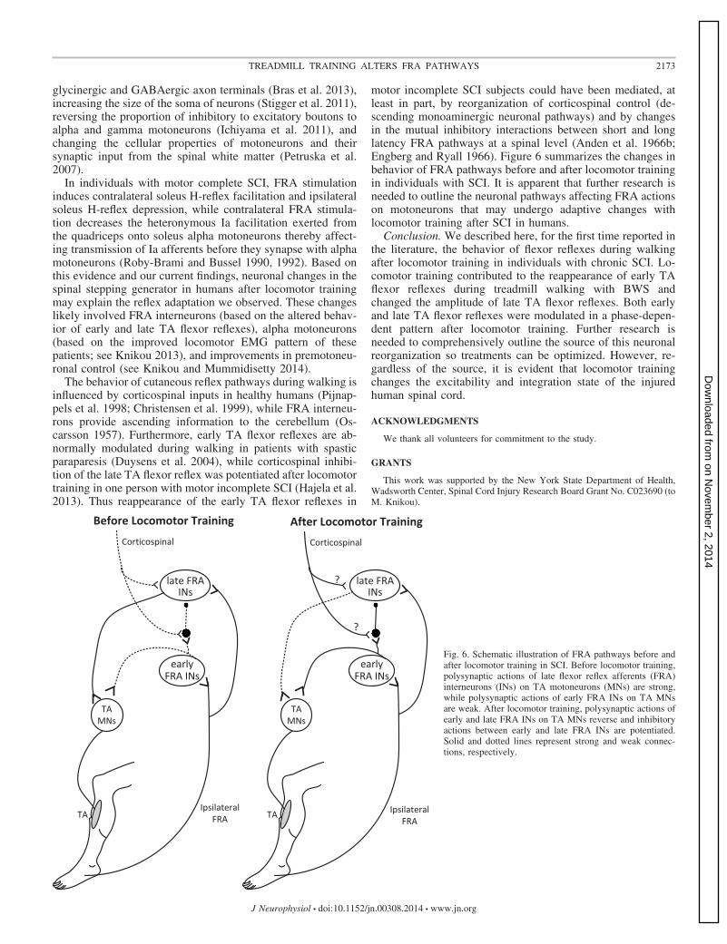
motor incomplete SCI subjects could have been mediated, atleast in part, by reorganization of corticospinal control (de-scending monoaminergic neuronal pathways) and by changesin the mutual inhibitory interactions between short and longlatency FRA pathways at a spinal level (Anden et al. 1966b;Engberg and Ryall 1966). Figure 6 summarizes the changes inbehavior of FRA pathways before and after locomotor trainingin individuals with SCI. It is apparent that further research isneeded to outline the neuronal pathways affecting FRA actionson motoneurons that may undergo adaptive changes withlocomotor training after SCI in humans.
Conclusion. We described here, for the first time reported inthe literature, the behavior of flexor reflexes during walkingafter locomotor training in individuals with chronic SCI. Lo-comotor training contributed to the reappearance of early TAflexor reflexes during treadmill walking with BWS andchanged the amplitude of late TA flexor reflexes. Both earlyand late TA flexor reflexes were modulated in a phase-depen-dent pattern after locomotor training. Further research isneeded to comprehensively outline the source of this neuronalreorganization so treatments can be optimized. However, re-gardless of the source, it is evident that locomotor trainingchanges the excitability and integration state of the injuredhuman spinal cord.
ACKNOWLEDGMENTS
We thank all volunteers for commitment to the study.
GRANTS
This work was supported by the New York State Department of Health,Wadsworth Center, Spinal Cord Injury Research Board Grant No. C023690 (toM. Knikou).
Fig. 6. Schematic illustration of FRA pathways before andafter locomotor training in SCI. Before locomotor training,polysynaptic actions of late flexor reflex afferents (FRA)interneurons (INs) on TA motoneurons (MNs) are strong,while polysynaptic actions of early FRA INs on TA MNsare weak. After locomotor training, polysynaptic actions ofearly and late FRA INs on TA MNs reverse and inhibitoryactions between early and late FRA INs are potentiated.Solid and dotted lines represent strong and weak connec-tions, respectively.
2173TREADMILL TRAINING ALTERS FRA PATHWAYS
J Neurophysiol • doi:10.1152/jn.00308.2014 • www.jn.org
on Novem
ber 2, 2014D
ownloaded from

DISCLOSURES
No conflicts of interest, financial or otherwise, are declared by the author(s).
AUTHOR CONTRIBUTIONS
Author contributions: A.C.S., C.K.M., and M.K. performed experiments;A.C.S., C.K.M., and M.K. analyzed data; A.C.S., W.Z.R., and M.K. inter-preted results of experiments; A.C.S., W.Z.R., and M.K. edited and revisedmanuscript; A.C.S., C.K.M., W.Z.R., and M.K. approved final version ofmanuscript; C.K.M. and M.K. prepared figures; M.K. conception and design ofresearch; M.K. drafted manuscript.
REFERENCES
Anden N, Jukes M, Lundberg A, Vyklicky L. The effect of DOPA on thespinal cord. 1. Influence on transmission from primary afferents. ActaPhysiol Scand 67: 373–386, 1966a.
Anden N, Jukes M, Lundberg A, Vyklicky L. The effect of DOPA on thespinal cord. 3. Depolarization evoked in the central terminals of ipsilateral1a afferents by volleys in the FRA. Acta Physiol Scand 68: 322–336, 1966b.
Baken BC, Dietz V, Duysens J. Phase-dependent modulation of short latencycutaneous reflexes during walking in man. Brain Res 1031: 268–275, 2005.
Barriere G, Leblond H, Provencher J, Rossignol S. Prominent role of thespinal central pattern generator in the recovery of locomotion after partialspinal cord injuries. J Neurosci 28: 3976–3987, 2008.
Behrman A, Harkema S. Locomotor training after human spinal cord injury:a series of case studies. Phys Ther 80: 688–700, 2000.
Bolliger M, Trepp A, Zörner B, Dietz V. Modulation of spinal reflex byassisted locomotion in humans with chronic complete spinal cord injury.Clin Neurophysiol 121: 2152–2158, 2010.
Bras H, Sadlaoud K, Khalhi L, Boulenguez P, Viallat-Lieutaud A, VinayL. Plasticity of the lumbar inhibitory synaptic transmission underlyingrecovery of locomotion in transacted rats. Society for Neuroscience, Pro-gram No. 466.23, 2013.
Brown TG. The intrinsic factors in the act of progression in the mammal. ProcR Soc Lond B 84: 308–319, 1911.
Bussel B, Roby-Brami A, Yakovleff A, Bennis N. Late flexion reflex inparaplegic patients. Evidence for a spinal stepping generator. Brain Res Bull22: 53–56, 1989.
Christensen LOD, Morita H, Petersen N, Nielsen J. Evidence suggestingthat a transcortical reflex pathway contributes to cutaneous reflexes in thetibialis anterior muscle during walking in man. Exp Brain Res 124: 59–68,1999.
Conway BA, Knikou M. The action of plantar pressure on flexion reflexpathways in the isolated human spinal cord. Clin Neurophysiol 119: 892–896, 2008.
Dietz V, Grillner S, Trepp A, Hubli M, Bolliger M. Changes in spinal reflexand locomotor activity after a complete spinal cord injury: a commonmechanism? Brain 132: 2196–2205, 2009.
Dietz V, Wirtz M, Curt A, Colombo G. Locomotor pattern in paraplegicpatients: training effects and recovery of spinal cord function. Spinal Cord36: 380–390, 1998.
Dobkin B, Barbeau H, Deforge D, Ditunno J, Elashoff R, Apple D, BassoM, Behrman A, Harkema S, Saulino M, Scott M; Spinal Cord InjuryLocomotor Trial Group. The evolution of walking-related outcomes overthe first 12 weeks of rehabilitation for incomplete traumatic spinal cordinjury: the multicenter randomized Spinal Cord Injury Locomotor Trial.Neurorehabil Neural Repair 21: 25–35, 2007.
Duysens J, Baken BC, Burgers L, Plat FM, den Otter AR, Kremer HP.Cutaneous reflexes from the foot during gait in hereditary spastic parapa-resis. Clin Neurophysiol 115: 1057–1062, 2004.
Duysens J, Tax AA, Trippel M, Dietz V. Phase-dependent reversal ofreflexly induced movements during human gait. Exp Brain Res 90: 404–414, 1992.
Duysens J, Tax AA, Trippel M, Dietz V. Increased amplitude of cutaneousreflexes during human running as compared to standing. Brain Res 613:230–238, 1993.
Duysens J, Trippel M, Horstmann GA, Dietz V. Gating and reversal ofreflexes in ankle muscles during human walking. Exp Brain Res 82:351–358, 1990.
Eccles JC, Kostyuk PG, Schmidt RF. Presynaptic inhibition of the centralactions of flexor reflex afferents. J Physiol 161: 258–281, 1962.
Engberg I, Ryall RW. The inhibitory action of noradrenaline and othermonoamines on spinal neurones. J Physiol 185: 298–322, 1966.
Fu T, Jankowska E, Lundberg A. Reciprocal Ia inhibition during the latereflexes evoked from the flexor reflex afferents after DOPA. Brain Res 85:99–102, 1975.
Hajela N, Mummidisetty CK, Smith AC, Knikou M. Corticospinal reorga-nization after locomotor training in a person with motor incomplete para-plegia. Biomed Res Int 2013: 516427, 2013.
Hubli M, Dietz V, Bolliger M. Spinal reflex activity: a marker for neuronalfunctionality after spinal cord injury. Neurorehabil Neural Repair 26:188–196, 2012.
Ichiyama RM, Broman J, Roy RR, Zhong H, Edgerton VR, Havton LA.Locomotor training maintains normal inhibitory influence on both alpha-and gamma-motoneurons after neonatal spinal cord transection. J Neurosci31: 26–33, 2011.
Jankowska E, Jukes M, Lund S, Lundberg A. The effect of DOPA on thespinal cord. 5. Reciprocal organization of pathways transmitting excitatoryaction to alpha motoneurons of flexors and extensors. Acta Physiol Scand70: 369–388, 1967a.
Jankowska E, Jukes M, Lund S, Lundberg A. The effect of DOPA on thespinal cord. 6. Half-centre organization of interneurones transmitting effectsfrom the flexor reflex afferents. Acta Physiol Scand 70: 389–402, 1967b.
Kapitza S, Zörner B, Weinmann O, Bolliger M, Filli L, Dietz V, SchwabME. Tail spasms in rat spinal cord injury: changes in interneuronal connec-tivity. Exp Neurol 236: 179–189, 2012.
Knikou M. Plantar cutaneous input modulates differently spinal reflexes insubjects with intact and injured spinal cord. Spinal Cord 45: 69–77, 2007a.
Knikou M. Hip-phase-dependent flexion reflex modulation and expression ofspasms in patients with spinal cord injury. Exp Neurol 204: 171–181, 2007b.
Knikou M. Plantar cutaneous afferents normalize the reflex modulationpatterns during stepping in chronic human spinal cord injury. J Neurophysiol103: 1304–1314, 2010a.
Knikou M. Neural control of locomotion and training-induced plasticity afterspinal and cerebral lesions. Clin Neurophysiol 121: 1655–1668, 2010b.
Knikou M. Function of group Ib inhibition during assisted stepping in humanspinal cord injury. J Clin Neurophysiol 29: 271–277, 2012.
Knikou M. Functional reorganization of soleus H-reflex modulation duringstepping after robotic-assisted step training in people with complete andincomplete spinal cord injury. Exp Brain Res 228: 279–296, 2013.
Knikou M, Conway BA. Effects of electrically induced muscle contraction onflexion reflex in human spinal cord injury. Spinal Cord 43: 640–648, 2005.
Knikou M, Angeli C, Ferreira CK, Harkema SJ. Flexion reflex modulationduring stepping in human spinal cord injury. Exp Brain Res 196: 341–351,2009.
Knikou M, Kay E, Rymer WZ. Modulation of flexion reflex induced by hipangle changes in human spinal cord injury. Exp Brain Res 168: 577–586,2006.
Knikou M, Mummidisetty CK. Locomotor training improves premotoneu-ronal control after chronic spinal cord injury. J Neurophysiol 111: 2264–2275, 2014.
Lovely RG, Gregor RJ, Roy RR, Edgerton VR. Effects of training on therecovery of full-weight-bearing stepping in the adult spinal cat. Exp Neurol92: 421–435, 1986.
Manella KJ, Field-Fote EC. Modulatory effects of locomotor training onextensor spasticity in individuals with motor-incomplete spinal cord injury.Restor Neurol Neurosci 31: 633–646, 2013.
Ménard A, Leblond H, Gossard JP. Sensory integration in presynapticinhibitory pathways during fictive locomotion in the cat. J Neurophysiol 88:163–171, 2002.
Oscarrson O. Functional organization of the ventral spino-cerebellar tract inthe cat. II. Connections with muscle, joint, and skin nerve afferents andeffects on adequate stimulation of various receptors. Acta Physiol Scand 42,Suppl 146: 1–107, 1957.
Petruska JC, Ichiyama RM, Jindrich DL, Crown ED, Tansey KE, RoyRR, Edgerton VR, Mendell LM. Changes in motoneuron properties andsynaptic inputs related to step training after spinal cord transection in rats.J Neurosci 27: 4460–4471, 2007.
Pijnappels M, Van Wezel BM, Colombo G, Dietz V, Duysens J. Corticalfacilitation of cutaneous reflexes in leg muscles during human gait. BrainRes 787: 149–153, 1998.
Roby-Brami A, Bussel B. Effects of flexor reflex afferent stimulation on thesoleus H-reflex in patients with a complete spinal cord lesion: evidence forpresynaptic inhibition of Ia transmission. Exp Brain Res 81: 593–601, 1990.
2174 TREADMILL TRAINING ALTERS FRA PATHWAYS
J Neurophysiol • doi:10.1152/jn.00308.2014 • www.jn.org
on Novem
ber 2, 2014D
ownloaded from
Roby-Brami A, Bussel B. Inhibitory effects on flexor reflexes in patients witha complete spinal cord lesion. Exp Brain Res 90: 201–208, 1992.
Roby-Brami A, Bussel B. Long-latency spinal reflex in man after flexor reflexafferent stimulation. Brain 110: 707–725, 1987.
Rossignol S, Barriere G, Alluin O, Frigon A. Re-expression of locomotorfunction after partial spinal cord injury. Physiology (Bethesda) 24: 127–139,2009.
Rossignol S, Chau C, Brustein E, Bélanger M, Barbeau H, Drew T.Locomotor capacities after complete and partial lesions of the spinal cord.Acta Neurobiol Exp (Warsz) 56: 449–463, 1996.
Stigger F, do Nascimento PS, Dutra MF, Couto GK, Ilha J, Achaval M,Marcuzzo S. Treadmill training induces plasticity in spinal motoneuronsand sciatic nerve after sensorimotor restriction during early postnatal period:new insights into the clinical approach for children with cerebral palsy. IntJ Dev Neurosci 29: 833–838, 2011.
Thomas SL, Gorassini MA. Increases in corticospinal tract function bytreadmill training after incomplete spinal cord injury. J Neurophysiol 94:2844–2855, 2005.
Van Wezel BM, Ottenhoff FS, Duysens J. Dynamic control of location-specific information in tactile cutaneous reflexes from the foot during humanwalking. J Neurosci 17: 3804–3814, 1997.
Zehr E, Komiyama T, Stein R. Cutaneous reflexes during human gait:electromyographic and kinematic responses to electrical stimulation. JNeurophysiol 77: 3311–3325, 1997.
Zehr EP, Loadman PM, Hundza SR. Neural control of rhythmic arm cyclingafter stroke. J Neurophysiol 108: 891–905, 2012.
Zehr EP, Loadman PM. Persistence of locomotor-related interlimb reflex net-works during walking after stroke. Clin Neurophysiol 123: 796–807, 2012.
Zehr EP, Stein RB, Komiyama T. Function of sural nerve reflexes duringhuman walking. J Physiol 507: 305–314, 1998.
2175TREADMILL TRAINING ALTERS FRA PATHWAYS
J Neurophysiol • doi:10.1152/jn.00308.2014 • www.jn.org
on Novem
ber 2, 2014D
ownloaded from
Related Documents