Short Article Localized Smooth Muscle Differentiation Is Essential for Epithelial Bifurcation during Branching Morphogenesis of the Mammalian Lung Graphical Abstract Highlights d Regions of epithelial shape change coincide with differentiating smooth muscle d Differentiating smooth muscle cells appear at lung bud bifurcation sites d Blocking differentiation or surgically removing smooth muscle disrupts bifurcation Authors Hye Young Kim, Mei-Fong Pang, Victor D. Varner, ..., Erin Miller, Derek C. Radisky, Celeste M. Nelson Correspondence [email protected] In Brief Epithelial morphogenesis is influenced by soluble signals from the surrounding mesenchyme, but the physical role of this tissue is unknown. Here, Kim, Pang et al. show that stereotyped differentiation of smooth muscle is required for branching morphogenesis of the airway epithelium in the mammalian lung. Kim et al., 2015, Developmental Cell 34, 719–726 September 28, 2015 ª2015 Elsevier Inc. http://dx.doi.org/10.1016/j.devcel.2015.08.012

Welcome message from author
This document is posted to help you gain knowledge. Please leave a comment to let me know what you think about it! Share it to your friends and learn new things together.
Transcript

Short Article
Localized SmoothMuscleDifferentiation Is Essential
for Epithelial Bifurcation during BranchingMorphogenesis of the Mammalian LungGraphical Abstract
Highlights
d Regions of epithelial shape change coincide with
differentiating smooth muscle
d Differentiating smooth muscle cells appear at lung bud
bifurcation sites
d Blocking differentiation or surgically removing smooth
muscle disrupts bifurcation
Kim et al., 2015, Developmental Cell 34, 719–726September 28, 2015 ª2015 Elsevier Inc.http://dx.doi.org/10.1016/j.devcel.2015.08.012
Authors
Hye Young Kim, Mei-Fong Pang,
Victor D. Varner, ..., Erin Miller,
Derek C. Radisky, Celeste M. Nelson
In Brief
Epithelial morphogenesis is influenced by
soluble signals from the surrounding
mesenchyme, but the physical role of this
tissue is unknown. Here, Kim, Pang et al.
show that stereotyped differentiation of
smooth muscle is required for branching
morphogenesis of the airway epithelium
in the mammalian lung.

Developmental Cell
Short Article
Localized Smooth Muscle DifferentiationIs Essential for Epithelial Bifurcation duringBranching Morphogenesis of the Mammalian LungHye Young Kim,1,4 Mei-Fong Pang,1,4 Victor D. Varner,1 Lisa Kojima,1 Erin Miller,3 Derek C. Radisky,3
and Celeste M. Nelson1,2,*1Department of Chemical and Biological Engineering, Princeton University, Princeton, NJ 08544, USA2Department of Molecular Biology, Princeton University, Princeton, NJ 08544, USA3Department of Cancer Biology, Mayo Clinic Cancer Center, Jacksonville, FL 32224, USA4Co-first author
*Correspondence: [email protected]
http://dx.doi.org/10.1016/j.devcel.2015.08.012
SUMMARY
The airway epithelium develops into a tree-likestructure via branching morphogenesis. Here, weshow a critical role for localized differentiation ofairway smooth muscle during epithelial bifurcationin the embryonic mouse lung. We found that dur-ing terminal bifurcation, changes in the geometryof nascent buds coincided with patterned smoothmuscle differentiation. Evaluating spatiotemporaldynamics of a-smooth muscle actin (aSMA) in re-porter mice revealed that aSMA-expressing cellsappear at the basal surface of the future epithe-lial cleft prior to bifurcation and then increase indensity as they wrap around the bifurcating bud.Disrupting this stereotyped pattern of smoothmuscle differentiation prevents terminal bifurca-tion. Our results reveal stereotyped differentiationof airway smooth muscle adjacent to nascentepithelial buds and suggest that localized smoothmuscle wrapping at the cleft site is required for ter-minal bifurcation during airway branching morpho-genesis.
INTRODUCTION
The developing lung begins as a simple epithelial tube sur-
rounded by thick mesenchyme. New buds emerge sequentially
along the length of the epithelial tube via domain branching,
while the tip of the elongated tube bifurcates to form two
daughter buds (Metzger et al., 2008). Repetition of these bifurca-
tions at defined angles (planar or orthogonal to the long axis of
the parent tube) generates the stereotyped, hierarchically orga-
nized three-dimensional (3D) branched architecture of the lung
(Metzger et al., 2008). Pioneering tissue grafting studies revealed
that the mesenchyme provides inductive cues for the branching
epithelium (Alescio and Cassini, 1962; Grobstein, 1953), and
several signaling molecules have since been identified including
fibroblast growth factor (FGF)-9, FGF10, bone morphogenetic
Developmen
protein (BMP)-4, and sonic hedgehog (SHH) (reviewed in
Metzger and Krasnow, 1999 and Morrisey and Hogan, 2010).
However, a possible connection between patterns of mesen-
chymal differentiation and airway epithelial branching has re-
mained largely unexplored.
During morphogenesis of the lung, the mesenchyme differ-
entiates into several cell types, including smooth muscle,
vasculature, and nerves that envelop the entire airway epithe-
lium as it branches (McCulley et al., 2015; Schachtner et al.,
2000; Sparrow and Lamb, 2003; Tollet et al., 2001). Among
these, the airway smooth muscle forms tightly packed bun-
dles around the circumference of the epithelium in a cranial
to caudal direction along the primary bronchus (Sparrow and
Lamb, 2003). After the smooth muscle forms, it contracts
spontaneously in a peristaltic wave, narrowing the airways
and pushing luminal fluid toward the terminal ends (Feather-
stone et al., 2005; Jesudason et al., 2005; Schittny et al.,
2000). Although the presence of airway smooth muscle and
its contractile behaviors have been observed in several spe-
cies (Featherstone et al., 2005; Lewis, 1924; McCray, 1993;
Schittny et al., 2000), its role in patterning airway branching
has not been defined (Jesudason et al., 2005; Unbekandt
et al., 2008).
Here, we identified a role for stereotyped differentiation
of airway smooth muscle in branching morphogenesis of the
embryonic mouse lung and found that terminal bifurcations
of the epithelium require the localized presence of smooth
muscle at the cleft site. Time-lapse imaging of embryonic
mouse lung explants revealed that changes in the shape
of the epithelial bud coincide with patterned differentiation of
smooth muscle. Remarkably, we found, using a transgenic
reporter mouse, that a-smooth muscle actin (aSMA)-express-
ing cells appear adjacent to the airway epithelium prior to
its bifurcation and then increase in density as they wrap
around the bifurcating cleft and neck of the bud. Disrupt-
ing patterns of smooth muscle differentiation abolishes ter-
minal bifurcation of the epithelium. Furthermore, surgically
removing the smooth muscle from the cleft causes the epithe-
lium to pop back into an un-bifurcated geometry. These
results reveal a major role for the spatial pattern of smooth
muscle differentiation during terminal bifurcation of the airway
epithelium.
tal Cell 34, 719–726, September 28, 2015 ª2015 Elsevier Inc. 719
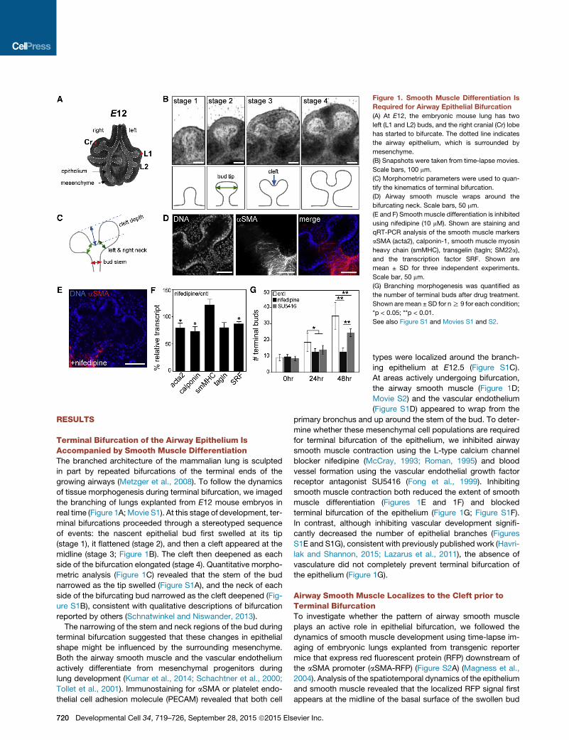
Figure 1. Smooth Muscle Differentiation Is
Required for Airway Epithelial Bifurcation
(A) At E12, the embryonic mouse lung has two
left (L1 and L2) buds, and the right cranial (Cr) lobe
has started to bifurcate. The dotted line indicates
the airway epithelium, which is surrounded by
mesenchyme.
(B) Snapshots were taken from time-lapse movies.
Scale bars, 100 mm.
(C) Morphometric parameters were used to quan-
tify the kinematics of terminal bifurcation.
(D) Airway smooth muscle wraps around the
bifurcating neck. Scale bars, 50 mm.
(E and F) Smooth muscle differentiation is inhibited
using nifedipine (10 mM). Shown are staining and
qRT-PCR analysis of the smooth muscle markers
aSMA (acta2), calponin-1, smooth muscle myosin
heavy chain (smMHC), transgelin (tagln; SM22a),
and the transcription factor SRF. Shown are
mean ± SD for three independent experiments.
Scale bar, 50 mm.
(G) Branching morphogenesis was quantified as
the number of terminal buds after drug treatment.
Shown are mean ± SD for nR 9 for each condition;
*p < 0.05; **p < 0.01.
See also Figure S1 and Movies S1 and S2.
RESULTS
Terminal Bifurcation of the Airway Epithelium IsAccompanied by Smooth Muscle DifferentiationThe branched architecture of the mammalian lung is sculpted
in part by repeated bifurcations of the terminal ends of the
growing airways (Metzger et al., 2008). To follow the dynamics
of tissue morphogenesis during terminal bifurcation, we imaged
the branching of lungs explanted from E12 mouse embryos in
real time (Figure 1A; Movie S1). At this stage of development, ter-
minal bifurcations proceeded through a stereotyped sequence
of events: the nascent epithelial bud first swelled at its tip
(stage 1), it flattened (stage 2), and then a cleft appeared at the
midline (stage 3; Figure 1B). The cleft then deepened as each
side of the bifurcation elongated (stage 4). Quantitative morpho-
metric analysis (Figure 1C) revealed that the stem of the bud
narrowed as the tip swelled (Figure S1A), and the neck of each
side of the bifurcating bud narrowed as the cleft deepened (Fig-
ure S1B), consistent with qualitative descriptions of bifurcation
reported by others (Schnatwinkel and Niswander, 2013).
The narrowing of the stem and neck regions of the bud during
terminal bifurcation suggested that these changes in epithelial
shape might be influenced by the surrounding mesenchyme.
Both the airway smooth muscle and the vascular endothelium
actively differentiate from mesenchymal progenitors during
lung development (Kumar et al., 2014; Schachtner et al., 2000;
Tollet et al., 2001). Immunostaining for aSMA or platelet endo-
thelial cell adhesion molecule (PECAM) revealed that both cell
720 Developmental Cell 34, 719–726, September 28, 2015 ª2015 Elsevier Inc.
types were localized around the branch-
ing epithelium at E12.5 (Figure S1C).
At areas actively undergoing bifurcation,
the airway smooth muscle (Figure 1D;
Movie S2) and the vascular endothelium
(Figure S1D) appeared to wrap from the
primary bronchus and up around the stem of the bud. To deter-
mine whether these mesenchymal cell populations are required
for terminal bifurcation of the epithelium, we inhibited airway
smooth muscle contraction using the L-type calcium channel
blocker nifedipine (McCray, 1993; Roman, 1995) and blood
vessel formation using the vascular endothelial growth factor
receptor antagonist SU5416 (Fong et al., 1999). Inhibiting
smooth muscle contraction both reduced the extent of smooth
muscle differentiation (Figures 1E and 1F) and blocked
terminal bifurcation of the epithelium (Figure 1G; Figure S1F).
In contrast, although inhibiting vascular development signifi-
cantly decreased the number of epithelial branches (Figures
S1E and S1G), consistent with previously published work (Havri-
lak and Shannon, 2015; Lazarus et al., 2011), the absence of
vasculature did not completely prevent terminal bifurcation of
the epithelium (Figure 1G).
Airway Smooth Muscle Localizes to the Cleft prior toTerminal BifurcationTo investigate whether the pattern of airway smooth muscle
plays an active role in epithelial bifurcation, we followed the
dynamics of smooth muscle development using time-lapse im-
aging of embryonic lungs explanted from transgenic reporter
mice that express red fluorescent protein (RFP) downstream of
the aSMA promoter (aSMA-RFP) (Figure S2A) (Magness et al.,
2004). Analysis of the spatiotemporal dynamics of the epithelium
and smooth muscle revealed that the localized RFP signal first
appears at the midline of the basal surface of the swollen bud
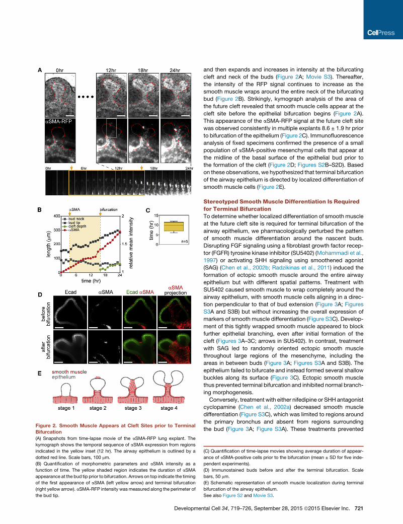
Figure 2. Smooth Muscle Appears at Cleft Sites prior to Terminal
Bifurcation
(A) Snapshots from time-lapse movie of the aSMA-RFP lung explant. The
kymograph shows the temporal sequence of aSMA expression from regions
indicated in the yellow inset (12 hr). The airway epithelium is outlined by a
dotted red line. Scale bars, 100 mm.
(B) Quantification of morphometric parameters and aSMA intensity as a
function of time. The yellow shaded region indicates the duration of aSMA
appearance at the bud tip prior to bifurcation. Arrows on top indicate the timing
of the first appearance of aSMA (left yellow arrow) and terminal bifurcation
(right yellow arrow). aSMA-RFP intensity wasmeasured along the perimeter of
the bud tip.
Developmen
and then expands and increases in intensity at the bifurcating
cleft and neck of the buds (Figure 2A; Movie S3). Thereafter,
the intensity of the RFP signal continues to increase as the
smooth muscle wraps around the entire neck of the bifurcating
bud (Figure 2B). Strikingly, kymograph analysis of the area of
the future cleft revealed that smooth muscle cells appear at the
cleft site before the epithelial bifurcation begins (Figure 2A).
This appearance of the aSMA-RFP signal at the future cleft site
was observed consistently in multiple explants 8.6 ± 1.9 hr prior
to bifurcation of the epithelium (Figure 2C). Immunofluorescence
analysis of fixed specimens confirmed the presence of a small
population of aSMA-positive mesenchymal cells that appear at
the midline of the basal surface of the epithelial bud prior to
the formation of the cleft (Figure 2D; Figures S2B–S2D). Based
on these observations, we hypothesized that terminal bifurcation
of the airway epithelium is directed by localized differentiation of
smooth muscle cells (Figure 2E).
Stereotyped Smooth Muscle Differentiation Is Requiredfor Terminal BifurcationTo determine whether localized differentiation of smooth muscle
at the future cleft site is required for terminal bifurcation of the
airway epithelium, we pharmacologically perturbed the pattern
of smooth muscle differentiation around the nascent buds.
Disrupting FGF signaling using a fibroblast growth factor recep-
tor (FGFR) tyrosine kinase inhibitor (SU5402) (Mohammadi et al.,
1997) or activating SHH signaling using smoothened agonist
(SAG) (Chen et al., 2002b; Radzikinas et al., 2011) induced the
formation of ectopic smooth muscle around the entire airway
epithelium but with different spatial patterns. Treatment with
SU5402 caused smooth muscle to wrap completely around the
airway epithelium, with smooth muscle cells aligning in a direc-
tion perpendicular to that of bud extension (Figure 3A; Figures
S3A and S3B) but without increasing the overall expression of
markers of smooth muscle differentiation (Figure S3C). Develop-
ment of this tightly wrapped smooth muscle appeared to block
further epithelial branching, even after initial formation of the
cleft (Figures 3A–3C; arrows in SU5402). In contrast, treatment
with SAG led to randomly oriented ectopic smooth muscle
throughout large regions of the mesenchyme, including the
areas in between buds (Figure 3A; Figures S3A and S3B). The
epithelium failed to bifurcate and instead formed several shallow
buckles along its surface (Figure 3C). Ectopic smooth muscle
thus prevented terminal bifurcation and inhibited normal branch-
ing morphogenesis.
Conversely, treatment with either nifedipine or SHH antagonist
cyclopamine (Chen et al., 2002a) decreased smooth muscle
differentiation (Figure S3C), which was limited to regions around
the primary bronchus and absent from regions surrounding
the bud (Figure 3A; Figure S3A). These treatments prevented
(C) Quantification of time-lapse movies showing average duration of appear-
ance of aSMA-positive cells prior to the bifurcation (mean ± SD for five inde-
pendent experiments).
(D) Immunostained buds before and after the terminal bifurcation. Scale
bars, 50 mm.
(E) Schematic representation of smooth muscle localization during terminal
bifurcation of the airway epithelium.
See also Figure S2 and Movie S3.
tal Cell 34, 719–726, September 28, 2015 ª2015 Elsevier Inc. 721

Figure 3. Pharmacologically Disrupting Patterned Smooth Muscle Differentiation Blocks Terminal Bifurcation
(A) Lung explants treatedwith SU5402 (5 mM), SAG (1 mg/ml), cyclopamine (1 mM), or nifedipine (10 mM). SU5402was added after 24 hr of treatment with nifedipine
for the ‘‘nifedipine + SU5402’’ condition. Fixed lungs were stained for E-cadherin and aSMA. Scale bars, 100 mm. Cntl, control.
(B and C) Disrupting the pattern of smooth muscle differentiation (B) disrupts terminal bifurcation and (C) induces epithelial buckling. (B) Shown are mean ± SD for
five independent experiments. (C) The box-and-whiskers plot shows median, interquartile range, maxima, and minima. **p < 0.01.
(D) Snapshots from time-lapse movies of aSMA-RFP lung explants treated with SU5402, SAG, or cyclopamine. Scale bars, 100 mm.
(E) Quantification of aSMA intensity and epithelial length around the perimeter of the bud from time-lapse movies in (D).
See also Figure S3 and Movie S4.
terminal bifurcation of the epithelium (Figure 3B), and surpris-
ingly led to the formation of shallow buckles along the surface
of the buds (Figure 3C). The morphology of the buckled epithe-
lium was distinct for each treatment despite the similar inhibition
of smooth muscle differentiation (Figure S3D). Furthermore,
the buckles that formed as a result of treatment with nifedipine
were blocked by simultaneously inducing ectopic smooth mus-
cle differentiation along the airway epithelium with SU5402
(Figures 3A–3C), suggesting that the presence of smooth mus-
cle, not its contractility, shapes the epithelial bud during terminal
bifurcation. Quantitative morphometric analysis of time-lapse
movies of explants from aSMA-RFP embryos (Figure 3D; Movie
S4) revealed that, as might be expected, drug treatments
affected both the rate of smooth muscle differentiation and the
rate of epithelial growth (Figure 3E). Inhibiting FGFR or activating
SHH accelerated smooth muscle differentiation while simulta-
722 Developmental Cell 34, 719–726, September 28, 2015 ª2015 Els
neously halting epithelial growth. Conversely, disrupting SHH
halted smoothmuscle differentiation while accelerating epithelial
growth.
To directly modulate smooth muscle differentiation without
altering signaling in the airway epithelium, we used an adenoviral
approach to augment or reduce the levels of serum response
factor (SRF) in the mesenchyme. Recombinant adenovirus
only transduced the mesenchyme (Figure S4A), consistent
with work by others showing that embryonic epithelium is refrac-
tory to adenovirus (Hsu et al., 2012). Ectopic expression of
SRF throughout the mesenchyme (Figures S4B and S4C) led
to disorganized differentiation of smooth muscle cells (Figure
4A) and elevated levels of smooth muscle markers (Figure 4B).
Conversely, short hairpin RNA (shRNA)-mediated depletion
of SRF (Figure S4B) blocked smooth muscle differentiation
around the growing epithelium (Figures 4A and 4B; Figure S4D).
evier Inc.

Figure 4. Specifically Targeting the Airway
Smooth Muscle Blocks Terminal Bifurcation
(A) Lung explants transduced with AdGFP, AdSRF,
or AdshSRF. Fixed lungs were stained for E-cad-
herin (Ecad) and aSMA. The red arrow indicates
bifurcation; gray arrowheads indicate buckling.
Scale bars, 100 mm.
(B) Relative transcript levels for markers of smooth
muscle differentiation in explants transduced with
AdSRF or AdshSRF. Shown are mean ± SD for
three independent experiments. ***p < 0.001.
(C and D) Disrupting SRF levels (C) disrupts
terminal bifurcation and (D) induces epithelial
buckling. (C) Shown are mean ± SEM for three
independent experiments. (D) The box-and-whis-
kers plot shows median, interquartile range,
maxima, and minima. **p < 0.01; ***p < 0.001.
(E) Bright-field images of lung explant before and
after dissecting off themesenchyme from a stage 3
bud. The graph indicates the depth of the cleft
before and after surgical removal of the smooth
muscle. Shown are mean ± SEM for five indepen-
dent experiments. *p < 0.05.
See also Figure S4.
Both manipulations completely prevented terminal bifurcation
(Figure 4C), with SRF depletion inducing epithelial buckling
(Figure 4D). Consistently, surgically removing the mesenchyme
from the epithelium at a stage immediately after cleft formation
(stage 3) caused the epithelium to pop back into a flattened
(stage 2) morphology (Figure 4E), suggesting that the initial cleft
Developmental Cell 34, 719–726, Se
is sculpted physically by the mesen-
chyme. Localized differentiation of airway
smooth muscle thus appears to be
required for terminal bifurcation during
branching morphogenesis of the epithe-
lium in the embryonic mouse lung.
DISCUSSION
As the lung branches, a subpopulation
of mesenchymal cells differentiate into
smooth muscle that tightly surrounds the
airway epithelium in a proximal to distal
direction (Kumar et al., 2014). Although
its existence in the embryonicmammalian
lung has been recognized for over a
decade (Tollet et al., 2001), the role of
airway smooth muscle in epithelial
branching has largely been ignored (Mitz-
ner, 2004). Similar patterns of smooth
muscle development have been noted in
other branching organs. In the neonatal
prostate of the rat, the smooth muscle
layer around the ducts is noticeably
thicker proximally and thins distally (Hay-
ward et al., 1996). In the pubertal murine
mammary gland, the myoepithelium sur-
rounds the extending ducts, as well as
the neck of the terminal end buds (War-
burton et al., 1982), and time-lapse analysis of mammary orga-
noids revealed that myoepithelial cells frequently localize to sites
of bifurcation (Ewald et al., 2008). Whether these localized
patterns of smooth muscle or myoepithelial cells direct epithe-
lial bifurcation in the prostate or mammary gland remains to be
determined, but our data show a clear role for localized smooth
ptember 28, 2015 ª2015 Elsevier Inc. 723
muscle differentiation in terminal bifurcation of the airway epithe-
lium. Our time-lapse analysis revealed remarkable dynamics of
aSMA-positive cells, which appear at the midline of epithelial
buds prior to the formation of any noticeable clefts. Importantly,
any perturbation in the stereotyped pattern of smooth muscle
differentiation prevented terminal bifurcation of the epithelium.
Because transgenic knockout of smooth muscle is embryonic
lethal (Hines et al., 2013), organ-specific genetic modulations
will be required for definitive understanding of the role for visceral
smooth muscle in epithelial morphogenesis.
Nonetheless, the spatiotemporal dynamics of airway smooth
muscle cells suggest that they behave as a girdle around the
bifurcating epithelial bud. As the airway epithelium grows, this
girdle-like smooth muscle may constrain the epithelial tube at
specific locations to direct the epithelial bifurcation. This conclu-
sion is supported by our dissection experiments, which show
that removing smooth muscle relaxes the epithelial cleft. The lo-
cations of smooth muscle around the bud neck and bifurcating
cleft coincide with dense regions of extracellular matrix in the
basement membrane of the lung (Liu et al., 2004; Mollard and
Dziadek, 1998; Moore et al., 2005). Moreover, the overlapping
patterns of smooth muscle and matrix are similar to the patterns
of fibronectin observed at cleft sites in the branching salivary
gland, suggesting a potential role for smooth muscle in regu-
lating epithelial cell-cell adhesions during terminal bifurcation
(Larsen et al., 2006; Sakai et al., 2003). In addition to these phys-
ical roles, the tightly packed smooth muscle could serve as a
barrier to the diffusion of molecules between the epithelium
and the mesenchyme, thus tuning bidirectional signaling, as in
the induction of prostate morphogenesis (Thomson et al.,
2002). Regardless, in the absence of the smooth muscle land-
mark at the future cleft site, stereotyped branching is blocked
and instead the growing epithelium buckles into the mesen-
chyme. This epithelial buckling could result from constrained
epithelial growth (Varner et al., 2015) or from a reduction in intra-
luminal pressure, which is normally maintained by the aligned
smooth muscle around the circumference of the epithelial tube
(Featherstone et al., 2005; Jesudason et al., 2005).
How airway smooth muscle differentiation is directed to the
midline of the expanding epithelial bud is unclear. Several
signaling molecules have been found to regulate the differentia-
tion of smooth muscle adjacent to the proximal airway epithe-
lium. FGF10-positive cells in the distal mesenchyme give rise
to the smooth muscle cell population (Mailleux et al., 2005),
whereas FGF9 secreted by the mesothelium simultaneously in-
hibits differentiation of smooth muscle to restrict its localization
to the proximal airway (Yi et al., 2009). In addition, SHH signaling
from the airway epithelium induces smooth muscle differentia-
tion within the neighboring mesenchyme (Weaver et al., 2003;
White et al., 2006). Whether and how these signals act as guid-
ance cues for precisely patterned smooth muscle differentia-
tion remains unclear. Further investigations are required to
unlock the relationship between this physical mediator of airway
branching and known genetic controls.
Airway epithelial branching morphogenesis is stereotyped in
the mouse (Metzger et al., 2008). Thousands of budding events
are thought to follow three simple genetically encoded subrou-
tines to build recursively the complex branching structure of
the lung, yet how the budding process is restricted to specific
724 Developmental Cell 34, 719–726, September 28, 2015 ª2015 Els
times and locations remains a mystery. The close spatiotem-
poral coincidence of smoothmuscle differentiation and epithelial
bifurcation suggests a physical or signaling role for stereo-
typed differentiation of the mesenchyme in the shaping of the
airway epithelium. It will be interesting to determine whether
stereotyped smooth muscle differentiation is controlled by the
same genetically encoded subroutines as epithelial branching
morphogenesis. Given the prevalence of smooth muscle around
branching epithelial tissues, spatiotemporally patterned smooth
muscle differentiation may represent a general physical strategy
to sculpt complex epithelial architectures.
EXPERIMENTAL PROCEDURES
Mice
Timed-pregnant CD1 mice were obtained from Charles River. Breeding of
aSMA-RFP transgenic reporter mice (Magness et al., 2004) and isolation of
embryos were carried out in accordance with institutional guidelines following
the NIHGuide for the Care andUse of Laboratory Animals and approved by the
Mayo Clinic’s Institutional Animal Care and Use Committee.
Organ Culture
Ex vivo culture of embryonic lungs was performed following previously
described protocols (Carraro et al., 2010). Briefly, lungs from E12 mouse
embryos were dissected in PBS supplemented with antibiotics (50 units/ml
of penicillin and streptomycin; Invitrogen) and cultured on porous membranes
(nucleopore polycarbonate track-etch membrane, 8 mm pore size, 25 mm
diameter; Whatman) in DMEM/F12 medium (without HEPES) supplemented
with 5% fetal bovine serum (FBS, heat inactivated; Atlanta Biologicals) and an-
tibiotics (50 units/ml of penicillin and streptomycin). Reagents used to perturb
the pattern of smooth muscle and vascular endothelial differentiation included
cyclopamine (1 or 2 mM; Tocris), nifedipine (10 mM; Sigma), SU5402 (5 or
10 mM; Santa Cruz), SU5416 (50 mM; Cayman Chemical), and SAG (1 or
2 mg/ml; Calbiochem). To disrupt smooth muscle differentiation specifically,
lung explants were transduced with custom recombinant adenoviruses
(Vector Biolabs)—adenovirus encoding GFP and SRF (AdSRF), GFP and
shRNA against SRF (AdshSRF), or GFP alone (AdGFP)—for 48 hr. To surgically
remove the mesenchyme, explants were incubated in 10 units/ml of dispase
(Invitrogen) on ice for 15 min. The reaction was quenched by submerging
the explants in FBS for 10 min. The explants were then transferred to a dissec-
tion dish containing PBS, and the mesenchyme was removed manually using
fine tungsten needles (Del Moral and Warburton, 2010).
Immunofluorescence Staining and Imaging
Lung explants were fixedwith 4%paraformaldehyde in PBS for 15min at room
temperature followed by washing with 0.3% Triton X-100 in PBS and blocking
with 10% goat serum. Samples were incubated with primary antibodies
against E-cadherin (Invitrogen), aSMA (Sigma), or PECAM (rat anti-mouse
CD31; BD Biosciences), followed by Alexa Fluor-conjugated secondary anti-
bodies and nuclear counter-staining with Hoechst 33342 (Invitrogen). Stained
lungs were dehydrated and cleared with Murray’s clear (1:2 ratio of benzyl
alcohol to benzyl benzoate; Sigma) and confocal images were collected using
a spinning disk confocal (Bio-Rad) fitted to an invertedmicroscope. For live im-
aging, explants were cultured over porous membranes within custom-made
glass bottom culture dishes within a stage-top incubator (Pathology Devices).
The morphology of the explants was monitored every 24 hr under bright field
on an inverted microscope (Nikon Ti).
Image Analysis and Statistics
Quantitative image analysis and image projections were completed using
ImageJ (Schneider et al., 2012). The orientation of smooth muscle cells in
the neck and bud regions of at least three different lungs for each treatment
were calculated using the OrientationJ plugin (Rezakhaniha et al., 2012). To
quantify differences in branching, the fold-change in the number of ter-
minal buds was determined by counting the terminal buds at the end of the
experiment and normalizing to the number at time zero for each explant. To
evier Inc.

characterize effects on terminal bifurcation per se, the total number of clefts (or
buckles) for each bud was counted at the end of the culture period. ANOVA
was conducted using IBMSPSSStatistics 19 for comparisons among different
treatments.
qRT-PCR Analysis
Total RNA from cultured lung explants was isolated using the QIAGEN RNeasy
fibrous tissuemini kit and reverse-transcribed using the Verso cDNA synthesis
kit (Thermo Scientific) according tomanufacturer’s instructions. qRT-PCRwas
performed using the Bio-Rad Mini Opticon instrument and iTaq Universal
SYBR Green Supermix (Bio-Rad). Amplification was followed bymelting curve
analysis to verify the presence of a single PCR product. The expression level of
each mRNA was normalized to that of 18S in the same sample. Primer se-
quences are listed in Supplemental Experimental Procedures.
SUPPLEMENTAL INFORMATION
Supplemental Information includes Supplemental Experimental Procedures,
four figures, and four movies and can be found with this article online at
http://dx.doi.org/10.1016/j.devcel.2015.08.012.
AUTHOR CONTRIBUTIONS
Conceptualization, H.Y.K. and C.M.N.; Methodology, H.Y.K., M.-F.P., and
V.D.V.; Investigation, H.Y.K., M.-F.P., V.D.V., L.K., and E.M.; Writing, H.Y.K.,
D.C.R., and C.M.N.; Resources, D.C.R.; Project Administration, C.M.N.
ACKNOWLEDGMENTS
This work was supported in part by grants from the NIH (GM083997,
HL110335, HL118532, and HL120142), the NSF (CMMI-1435853), the David
and Lucile Packard Foundation, the Alfred P. Sloan Foundation, the Camille
and Henry Dreyfus Foundation, and Susan G. Komen for the Cure. C.M.N.
holds a Career Award at the Scientific Interface from the Burroughs Wellcome
Fund. M.-F.P. was supported in part by a postdoctoral fellowship from the
Swedish Society for Medical Research (SSMF). L.K. was supported in part
by the Lidow Senior Thesis Fund.
Received: March 20, 2013
Revised: July 10, 2015
Accepted: August 14, 2015
Published: September 17, 2015
REFERENCES
Alescio, T., and Cassini, A. (1962). Induction in vitro of tracheal buds by pulmo-
nary mesenchyme grafted on tracheal epithelium. J. Exp. Zool. 150, 83–94.
Carraro, G., delMoral, P.-M., andWarburton, D. (2010). Mouse embryonic lung
culture, a system to evaluate the molecular mechanisms of branching. J. Vis.
Exp. 40, 2035.
Chen, J.K., Taipale, J., Cooper, M.K., and Beachy, P.A. (2002a). Inhibition of
Hedgehog signaling by direct binding of cyclopamine to Smoothened.
Genes Dev. 16, 2743–2748.
Chen, J.K., Taipale, J., Young, K.E., Maiti, T., and Beachy, P.A. (2002b). Small
molecule modulation of Smoothened activity. Proc. Natl. Acad. Sci. USA 99,
14071–14076.
Del Moral, P.M., and Warburton, D. (2010). Explant culture of mouse embry-
onic whole lung, isolated epithelium, or mesenchyme under chemically
defined conditions as a system to evaluate the molecular mechanism of
branching morphogenesis and cellular differentiation. Methods Mol. Biol.
633, 71–79.
Ewald, A.J., Brenot, A., Duong, M., Chan, B.S., andWerb, Z. (2008). Collective
epithelial migration and cell rearrangements drive mammary branching
morphogenesis. Dev. Cell 14, 570–581.
Featherstone, N.C., Jesudason, E.C., Connell, M.G., Fernig, D.G., Wray, S.,
Losty, P.D., and Burdyga, T.V. (2005). Spontaneous propagating calcium
Developmen
waves underpin airway peristalsis in embryonic rat lung. Am. J. Respir. Cell
Mol. Biol. 33, 153–160.
Fong, T.A.T., Shawver, L.K., Sun, L., Tang, C., App, H., Powell, T.J., Kim, Y.H.,
Schreck, R., Wang, X., Risau, W., et al. (1999). SU5416 is a potent and selec-
tive inhibitor of the vascular endothelial growth factor receptor (Flk-1/KDR) that
inhibits tyrosine kinase catalysis, tumor vascularization, and growth of multiple
tumor types. Cancer Res. 59, 99–106.
Grobstein, C. (1953). Inductive epitheliomesenchymal interaction in cultured
organ rudiments of the mouse. Science 118, 52–55.
Havrilak, J.A., and Shannon, J.M. (2015). Branching of lung epithelium in vitro
occurs in the absence of endothelial cells. Dev. Dyn. 244, 553–563.
Hayward, S.W., Baskin, L.S., Haughney, P.C., Foster, B.A., Cunha, A.R.,
Dahiya, R., Prins, G.S., and Cunha, G.R. (1996). Stromal development in the
ventral prostate, anterior prostate and seminal vesicle of the rat. Acta Anat.
(Basel) 155, 94–103.
Hines, E.A., Jones, M.K., Verheyden, J.M., Harvey, J.F., and Sun, X. (2013).
Establishment of smooth muscle and cartilage juxtaposition in the developing
mouse upper airways. Proc. Natl. Acad. Sci. USA 110, 19444–19449.
Hsu, J.C., Di Pasquale, G., Harunaga, J.S., Onodera, T., Hoffman, M.P.,
Chiorini, J.A., and Yamada, K.M. (2012). Viral gene transfer to developing
mouse salivary glands. J. Dent. Res. 91, 197–202.
Jesudason, E.C., Smith, N.P., Connell, M.G., Spiller, D.G., White, M.R.H.,
Fernig, D.G., and Losty, P.D. (2005). Developing rat lung has a sided pace-
maker region for morphogenesis-related airway peristalsis. Am. J. Respir.
Cell Mol. Biol. 32, 118–127.
Kumar, M.E., Bogard, P.E., Espinoza, F.H., Menke, D.B., Kingsley, D.M., and
Krasnow,M.A. (2014). Mesenchymal cells: defining amesenchymal progenitor
niche at single-cell resolution. Science 346, 1258810.
Larsen, M., Wei, C., and Yamada, K.M. (2006). Cell and fibronectin dynamics
during branching morphogenesis. J. Cell Sci. 119, 3376–3384.
Lazarus, A., Del-Moral, P.M., Ilovich, O., Mishani, E., Warburton, D., and
Keshet, E. (2011). A perfusion-independent role of blood vessels in deter-
mining branching stereotypy of lung airways. Development 138, 2359–2368.
Lewis, M.R. (1924). Spontaneous rhythmical contraction of the muscles of the
bronchial tubes and air sacs of the chick embryo. Am. J. Physiol. 68, 385–388.
Liu, Y., Stein, E., Oliver, T., Li, Y., Brunken, W.J., Koch, M., Tessier-Lavigne,
M., and Hogan, B.L.M. (2004). Novel role for Netrins in regulating epithelial
behavior during lung branching morphogenesis. Curr. Biol. 14, 897–905.
Magness, S.T., Bataller, R., Yang, L., and Brenner, D.A. (2004). A dual reporter
gene transgenic mouse demonstrates heterogeneity in hepatic fibrogenic cell
populations. Hepatology 40, 1151–1159.
Mailleux, A.A., Kelly, R., Veltmaat, J.M., De Langhe, S.P., Zaffran, S., Thiery,
J.P., and Bellusci, S. (2005). Fgf10 expression identifies parabronchial smooth
muscle cell progenitors and is required for their entry into the smooth muscle
cell lineage. Development 132, 2157–2166.
McCray, P.B., Jr. (1993). Spontaneous contractility of human fetal airway
smooth muscle. Am. J. Respir. Cell Mol. Biol. 8, 573–580.
McCulley, D., Wienhold, M., and Sun, X. (2015). The pulmonary mesenchyme
directs lung development. Curr. Opin. Genet. Dev. 32, 98–105.
Metzger, R.J., and Krasnow, M.A. (1999). Genetic control of branching
morphogenesis. Science 284, 1635–1639.
Metzger, R.J., Klein, O.D., Martin, G.R., and Krasnow, M.A. (2008). The
branching programme of mouse lung development. Nature 453, 745–750.
Mitzner, W. (2004). Airway smooth muscle: the appendix of the lung. Am. J.
Respir. Crit. Care Med. 169, 787–790.
Mohammadi, M., McMahon, G., Sun, L., Tang, C., Hirth, P., Yeh, B.K.,
Hubbard, S.R., and Schlessinger, J. (1997). Structures of the tyrosine kinase
domain of fibroblast growth factor receptor in complex with inhibitors.
Science 276, 955–960.
Mollard, R., and Dziadek, M. (1998). A correlation between epithelial prolif-
eration rates, basement membrane component localization patterns, and
morphogenetic potential in the embryonic mouse lung. Am. J. Respir. Cell
Mol. Biol. 19, 71–82.
tal Cell 34, 719–726, September 28, 2015 ª2015 Elsevier Inc. 725

Moore, K.A., Polte, T., Huang, S., Shi, B., Alsberg, E., Sunday, M.E., and
Ingber, D.E. (2005). Control of basement membrane remodeling and epithelial
branching morphogenesis in embryonic lung by Rho and cytoskeletal tension.
Dev. Dyn. 232, 268–281.
Morrisey, E.E., and Hogan, B.L.M. (2010). Preparing for the first breath: genetic
and cellular mechanisms in lung development. Dev. Cell 18, 8–23.
Radzikinas, K., Aven, L., Jiang, Z., Tran, T., Paez-Cortez, J., Boppidi, K., Lu, J.,
Fine, A., and Ai, X. (2011). A Shh/miR-206/BDNF cascade coordinates innerva-
tion and formation of airway smooth muscle. J. Neurosci. 31, 15407–15415.
Rezakhaniha, R., Agianniotis, A., Schrauwen, J.T., Griffa, A., Sage, D., Bouten,
C.V., van de Vosse, F.N., Unser, M., and Stergiopulos, N. (2012). Experimental
investigation of collagen waviness and orientation in the arterial adventitia
using confocal laser scanning microscopy. Biomech. Model. Mechanobiol.
11, 461–473.
Roman, J. (1995). Effects of calcium channel blockade on mammalian lung
branching morphogenesis. Exp. Lung Res. 21, 489–502.
Sakai, T., Larsen, M., and Yamada, K.M. (2003). Fibronectin requirement in
branching morphogenesis. Nature 423, 876–881.
Schachtner, S.K., Wang, Y., and Scott Baldwin, H. (2000). Qualitative and
quantitative analysis of embryonic pulmonary vessel formation. Am. J.
Respir. Cell Mol. Biol. 22, 157–165.
Schittny, J.C., Miserocchi, G., and Sparrow, M.P. (2000). Spontaneous peri-
staltic airway contractions propel lung liquid through the bronchial tree of
intact and fetal lung explants. Am. J. Respir. Cell Mol. Biol. 23, 11–18.
Schnatwinkel, C., and Niswander, L. (2013). Multiparametric image analysis of
lung-branching morphogenesis. Dev. Dyn. 242, 622–637.
Schneider, C.A., Rasband, W.S., and Eliceiri, K.W. (2012). NIH Image to
ImageJ: 25 years of image analysis. Nat. Methods 9, 671–675.
726 Developmental Cell 34, 719–726, September 28, 2015 ª2015 Els
Sparrow, M.P., and Lamb, J.P. (2003). Ontogeny of airway smooth muscle:
structure, innervation, myogenesis and function in the fetal lung. Respir.
Physiol. Neurobiol. 137, 361–372.
Thomson, A.A., Timms, B.G., Barton, L., Cunha, G.R., and Grace, O.C. (2002).
The role of smooth muscle in regulating prostatic induction. Development 129,
1905–1912.
Tollet, J., Everett, A.W., and Sparrow, M.P. (2001). Spatial and temporal distri-
bution of nerves, ganglia, and smooth muscle during the early pseudoglandu-
lar stage of fetal mouse lung development. Dev. Dyn. 221, 48–60.
Unbekandt, M., del Moral, P.-M., Sala, F.G., Bellusci, S., Warburton, D., and
Fleury, V. (2008). Tracheal occlusion increases the rate of epithelial branching
of embryonic mouse lung via the FGF10-FGFR2b-Sprouty2 pathway. Mech.
Dev. 125, 314–324.
Varner, V.D., Gleghorn, J.P., Miller, E., Radisky, D.C., and Nelson, C.M. (2015).
Mechanically patterning the embryonic airway epithelium. Proc. Natl. Acad.
Sci. USA 112, 9230–9235.
Warburton, M.J., Mitchell, D., Ormerod, E.J., and Rudland, P. (1982).
Distribution of myoepithelial cells and basement membrane proteins in the
resting, pregnant, lactating, and involuting rat mammary gland. J. Histochem.
Cytochem. 30, 667–676.
Weaver, M., Batts, L., and Hogan, B.L.M. (2003). Tissue interactions pattern
the mesenchyme of the embryonic mouse lung. Dev. Biol. 258, 169–184.
White, A.C., Xu, J., Yin, Y., Smith, C., Schmid, G., and Ornitz, D.M. (2006).
FGF9 and SHH signaling coordinate lung growth and development through
regulation of distinct mesenchymal domains. Development 133, 1507–1517.
Yi, L., Domyan, E.T., Lewandoski, M., and Sun, X. (2009). Fibroblast growth
factor 9 signaling inhibits airway smooth muscle differentiation in mouse
lung. Dev. Dyn. 238, 123–137.
evier Inc.
Related Documents


![Lipschitzian Piecewise Smooth Minimization [0.5ex] via ... EuroAd Workshop - Sabrina Fieg… · Lipschitzian Piecewise Smooth Minimization via Algorithmic Differentiation Sabrina](https://static.cupdf.com/doc/110x72/5fc48388ca73b406955dcfd3/lipschitzian-piecewise-smooth-minimization-05ex-via-euroad-workshop-sabrina.jpg)