Acta Astronautica 56 (2005) 357 – 366 www.elsevier.com/locate/actaastro Loading eects on rat craniomandibular morphology: a system for gravity studies Ranbir Singh a , Thais Carvalho b , Georey E. Gerstner a; c ; ∗ a Department of Biologic and Materials Sciences, School of Dentistry, University of Michigan, Ann Arbor, MI 48109-1078, USA b Department of Orthodontics, School of Dentistry, University of Michigan, Ann Arbor, MI 48109-1078, USA c Department of Psychology, University of Michigan, Ann Arbor, MI 48109-1078, USA Received 19 September 2002; received in revised form 11 September 2003; accepted 11 May 2004 Available online 30 July 2004 Abstract Gravity eects on muscle and bone are a major impediment to long-term space travel. We introduce a model for studying these eects, the craniomandibular system. Some advantages of this system include: (1) craniomandibular morphology is determined by epigenetic factors including gravity, (2) relatively light forces can signicantly alter its morphology, and (3) soft diet and tooth loss produce eects that are similar to those produced in lower limbs by weightlessness. In the study, implants made either of gold (experimental group) or lightweight acrylic (controls) were attached to adult rats’ mandibles. After 13 weeks, the animals’ skulls and mandibles were dissected. Pair-wise comparisons indicated that the experimental animals showed signicantly shortened and narrowed cranial bases, and signicant changes in the posterior zygomatic arch region. These results indicate that simulated macrogravity inuences bone remodeling in the adult craniomandibular system. c 2004 Elsevier Ltd. All rights reserved. Keywords: Craniofacial; Craniomandibular; Morphometrics; Cephalometrics; Craniometrics; Macrogravity 1. Introduction Eects of microgravity on muscle and bone are well documented and often severe. Numerous investiga- tions involving animal or human subjects have studied these eects so that countermeasures can be developed to make long-term space travel feasible. In this study, we evaluate the eects of simulated macrogravity on the craniomandibular system (CMS). ∗ Corresponding author. Department of Biological and Materials Sciences, School of Dentistry, University of Michigan, Ann Arbor, MI 48109-1078, USA. Tel.: +1-734-763-7717; fax: +1-734-647-2110. E-mail address: [email protected] (G.E. Gerstner). The CMS has several features that make it at- tractive for gravity studies. One feature is that the mandible is a lever system that is normal to the force of gravity during waking hours (Fig. 1). Tonically active fatigue-resistant masticatory muscle bers counter the eects of gravity. As a result, there is a constant anti-gravitational force on the CMS imposed by the jaw adductor or closer muscles. Examples of jaw closer muscles are the temporalis and masseter muscles schematically depicted in Fig. 1. Jaw closer muscles keep the jaw within 2–4 mm of maximum tooth intercuspation during routine awake periods. In an upright posture, if muscle tone diminishes (as when one falls asleep while sitting), the jaw opens much further. The gap that occurs with minimal jaw 0094-5765/$ - see front matter c 2004 Elsevier Ltd. All rights reserved. doi:10.1016/j.actaastro.2004.06.002

Welcome message from author
This document is posted to help you gain knowledge. Please leave a comment to let me know what you think about it! Share it to your friends and learn new things together.
Transcript
Acta Astronautica 56 (2005) 357–366www.elsevier.com/locate/actaastro
Loading e�ects on rat craniomandibularmorphology: a system for gravity studies
Ranbir Singha, Thais Carvalhob, Geo�rey E. Gerstnera;c;∗
aDepartment of Biologic and Materials Sciences, School of Dentistry, University of Michigan, Ann Arbor, MI 48109-1078, USAbDepartment of Orthodontics, School of Dentistry, University of Michigan, Ann Arbor, MI 48109-1078, USA
cDepartment of Psychology, University of Michigan, Ann Arbor, MI 48109-1078, USA
Received 19 September 2002; received in revised form 11 September 2003; accepted 11 May 2004
Available online 30 July 2004
Abstract
Gravity e�ects on muscle and bone are a major impediment to long-term space travel. We introduce a model for studyingthese e�ects, the craniomandibular system. Some advantages of this system include: (1) craniomandibular morphology isdetermined by epigenetic factors including gravity, (2) relatively light forces can signi�cantly alter its morphology, and (3)soft diet and tooth loss produce e�ects that are similar to those produced in lower limbs by weightlessness. In the study,implants made either of gold (experimental group) or lightweight acrylic (controls) were attached to adult rats’ mandibles.After 13 weeks, the animals’ skulls and mandibles were dissected. Pair-wise comparisons indicated that the experimentalanimals showed signi�cantly shortened and narrowed cranial bases, and signi�cant changes in the posterior zygomatic archregion. These results indicate that simulated macrogravity in�uences bone remodeling in the adult craniomandibular system.c© 2004 Elsevier Ltd. All rights reserved.
Keywords: Craniofacial; Craniomandibular; Morphometrics; Cephalometrics; Craniometrics; Macrogravity
1. Introduction
E�ects of microgravity on muscle and bone are welldocumented and often severe. Numerous investiga-tions involving animal or human subjects have studiedthese e�ects so that countermeasures can be developedto make long-term space travel feasible. In this study,we evaluate the e�ects of simulated macrogravity onthe craniomandibular system (CMS).
∗ Corresponding author. Department of Biological andMaterials Sciences, School of Dentistry, University of Michigan,Ann Arbor, MI 48109-1078, USA. Tel.: +1-734-763-7717;fax: +1-734-647-2110.
E-mail address: [email protected] (G.E. Gerstner).
The CMS has several features that make it at-tractive for gravity studies. One feature is that themandible is a lever system that is normal to the forceof gravity during waking hours (Fig. 1). Tonicallyactive fatigue-resistant masticatory muscle �berscounter the e�ects of gravity. As a result, there is aconstant anti-gravitational force on the CMS imposedby the jaw adductor or closer muscles. Examples ofjaw closer muscles are the temporalis and massetermuscles schematically depicted in Fig. 1. Jaw closermuscles keep the jaw within 2–4 mm of maximumtooth intercuspation during routine awake periods.In an upright posture, if muscle tone diminishes (aswhen one falls asleep while sitting), the jaw opensmuch further. The gap that occurs with minimal jaw
0094-5765/$ - see front matter c© 2004 Elsevier Ltd. All rights reserved.doi:10.1016/j.actaastro.2004.06.002
358 R. Singh et al. / Acta Astronautica 56 (2005) 357–366
G
T
TMJ
anteriorposterior
Mandible
SkullM
G
leftright
Mandible
Skullz
TM
z
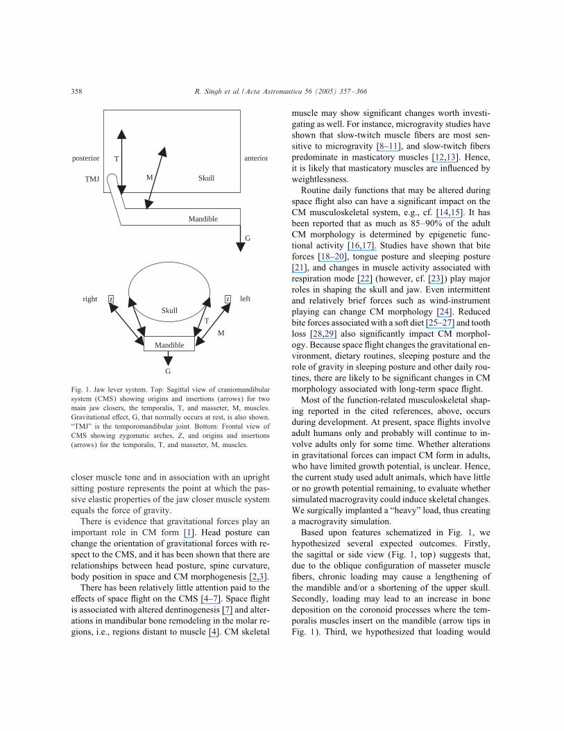
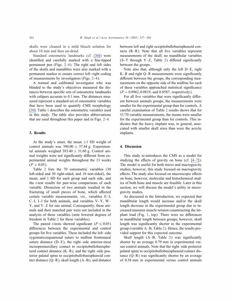
Fig. 1. Jaw lever system. Top: Sagittal view of craniomandibularsystem (CMS) showing origins and insertions (arrows) for twomain jaw closers, the temporalis, T, and masseter, M, muscles.Gravitational e�ect, G, that normally occurs at rest, is also shown.“TMJ” is the temporomandibular joint. Bottom: Frontal view ofCMS showing zygomatic arches, Z, and origins and insertions(arrows) for the temporalis, T, and masseter, M, muscles.
closer muscle tone and in association with an uprightsitting posture represents the point at which the pas-sive elastic properties of the jaw closer muscle systemequals the force of gravity.There is evidence that gravitational forces play an
important role in CM form [1]. Head posture canchange the orientation of gravitational forces with re-spect to the CMS, and it has been shown that there arerelationships between head posture, spine curvature,body position in space and CM morphogenesis [2,3].There has been relatively little attention paid to the
e�ects of space �ight on the CMS [4–7]. Space �ightis associated with altered dentinogenesis [7] and alter-ations in mandibular bone remodeling in the molar re-gions, i.e., regions distant to muscle [4]. CM skeletal
muscle may show signi�cant changes worth investi-gating as well. For instance, microgravity studies haveshown that slow-twitch muscle �bers are most sen-sitive to microgravity [8–11], and slow-twitch �berspredominate in masticatory muscles [12,13]. Hence,it is likely that masticatory muscles are in�uenced byweightlessness.Routine daily functions that may be altered during
space �ight also can have a signi�cant impact on theCM musculoskeletal system, e.g., cf. [14,15]. It hasbeen reported that as much as 85–90% of the adultCM morphology is determined by epigenetic func-tional activity [16,17]. Studies have shown that biteforces [18–20], tongue posture and sleeping posture[21], and changes in muscle activity associated withrespiration mode [22] (however, cf. [23]) play majorroles in shaping the skull and jaw. Even intermittentand relatively brief forces such as wind-instrumentplaying can change CM morphology [24]. Reducedbite forces associated with a soft diet [25–27] and toothloss [28,29] also signi�cantly impact CM morphol-ogy. Because space �ight changes the gravitational en-vironment, dietary routines, sleeping posture and therole of gravity in sleeping posture and other daily rou-tines, there are likely to be signi�cant changes in CMmorphology associated with long-term space �ight.Most of the function-related musculoskeletal shap-
ing reported in the cited references, above, occursduring development. At present, space �ights involveadult humans only and probably will continue to in-volve adults only for some time. Whether alterationsin gravitational forces can impact CM form in adults,who have limited growth potential, is unclear. Hence,the current study used adult animals, which have littleor no growth potential remaining, to evaluate whethersimulated macrogravity could induce skeletal changes.We surgically implanted a “heavy” load, thus creatinga macrogravity simulation.Based upon features schematized in Fig. 1, we
hypothesized several expected outcomes. Firstly,the sagittal or side view (Fig. 1, top) suggests that,due to the oblique con�guration of masseter muscle�bers, chronic loading may cause a lengthening ofthe mandible and/or a shortening of the upper skull.Secondly, loading may lead to an increase in bonedeposition on the coronoid processes where the tem-poralis muscles insert on the mandible (arrow tips inFig. 1). Third, we hypothesized that loading would
R. Singh et al. / Acta Astronautica 56 (2005) 357–366 359
widen the mandible, and/or narrow the skull (Fig. 1,bottom). We hypothesized that the narrowing wouldbe greatest between the zygomatic arches (boxesmarked “z”), which are relatively thin bones that formfree-standing arches to which the masseter musclesattach. Finally, since CMS growth and responses toforces are complex, we studied many other standardCMS variables to determine whether loading woulda�ect them.
2. Materials and Methods
2.1. Animals
Twenty-four-month old male Sprague Dawley ratswere used in this study. The animals were housed asmatched pairs in standard small-animal cages and fedrat chow and tap water ad libitum. The animals wererandomly divided into two groups, a control group andan experimental group. Cage-mate pairs consisted ofone control and one experimental groupmember. Cagenumbering and ear notching were used to identify in-dividual animals and group membership. All animalswere kept under controlled environmental conditionson a 12-h light–dark cycle.
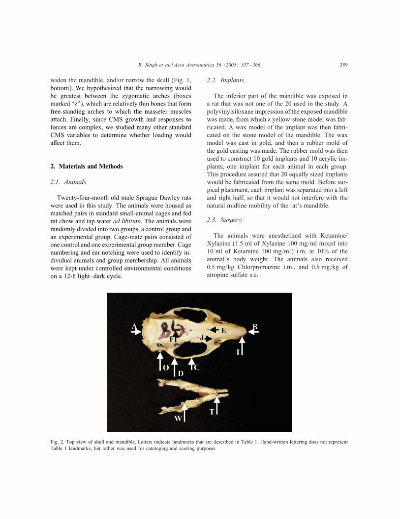
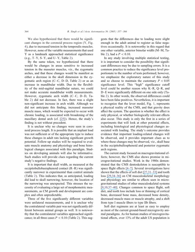
Fig. 2. Top view of skull and mandible. Letters indicate landmarks that are described in Table 1. Hand-written lettering does not representTable 1 landmarks, but rather was used for cataloging and scoring purposes.
2.2. Implants
The inferior part of the mandible was exposed ina rat that was not one of the 20 used in the study. Apolyvinylsiloxane impression of the exposed mandiblewas made, from which a yellow-stone model was fab-ricated. A wax model of the implant was then fabri-cated on the stone model of the mandible. The waxmodel was cast in gold, and then a rubber mold ofthe gold casting was made. The rubber mold was thenused to construct 10 gold implants and 10 acrylic im-plants, one implant for each animal in each group.This procedure assured that 20 equally sized implantswould be fabricated from the same mold. Before sur-gical placement, each implant was separated into a leftand right half, so that it would not interfere with thenatural midline mobility of the rat’s mandible.
2.3. Surgery
The animals were anesthetized with Ketamine/Xylazine (1:5 ml of Xylazine 100 mg=ml mixed into10 ml of Ketamine 100 mg=ml) i.m. at 10% of theanimal’s body weight. The animals also received0:5 mg=kg Chlorpromazine i.m., and 0:5 mg=kg ofatropine sulfate s.c.
360 R. Singh et al. / Acta Astronautica 56 (2005) 357–366
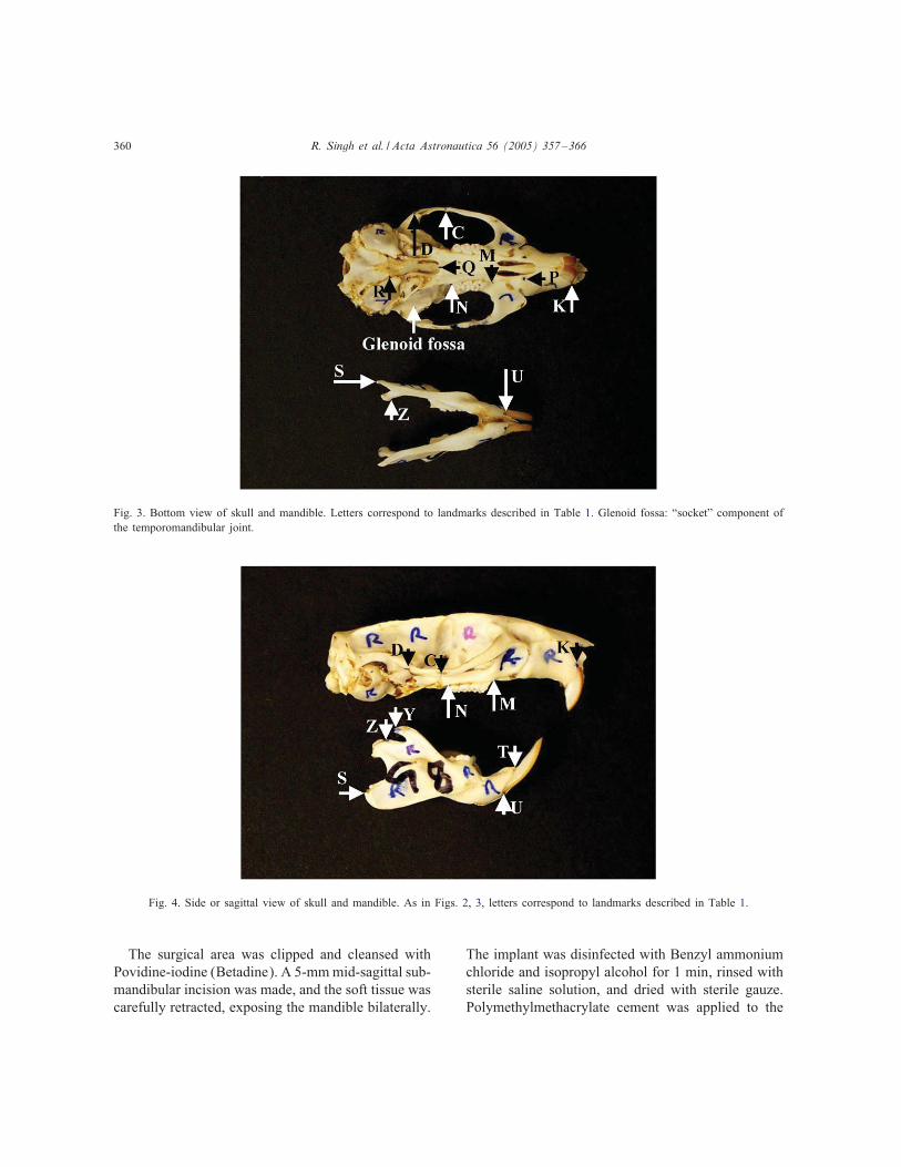
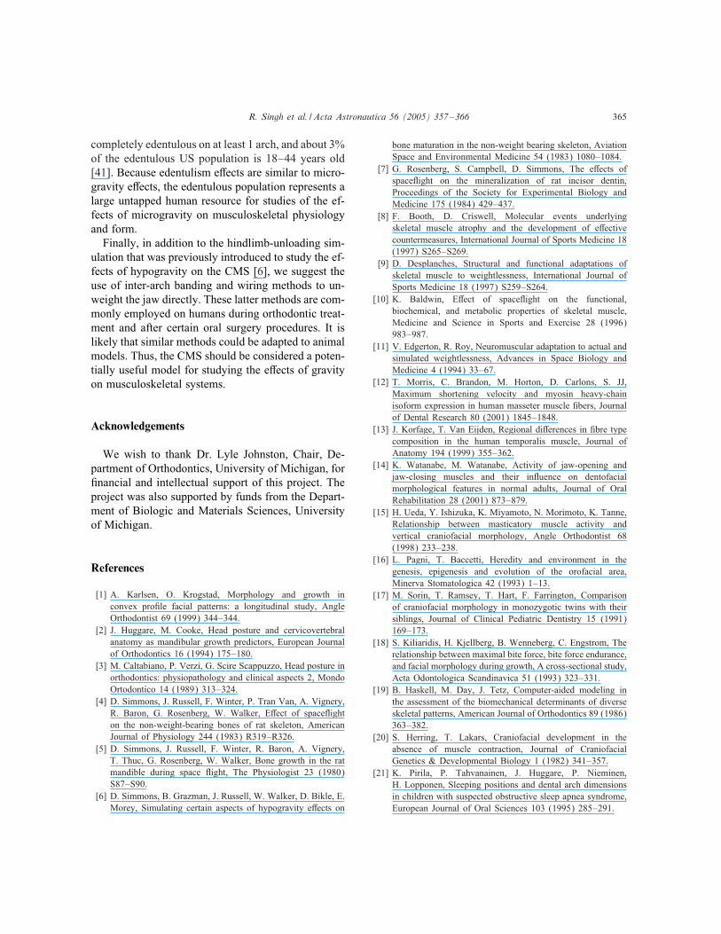
Fig. 3. Bottom view of skull and mandible. Letters correspond to landmarks described in Table 1. Glenoid fossa: “socket” component ofthe temporomandibular joint.
Fig. 4. Side or sagittal view of skull and mandible. As in Figs. 2, 3, letters correspond to landmarks described in Table 1.
The surgical area was clipped and cleansed withPovidine-iodine (Betadine). A 5-mmmid-sagittal sub-mandibular incision was made, and the soft tissue wascarefully retracted, exposing the mandible bilaterally.
The implant was disinfected with Benzyl ammoniumchloride and isopropyl alcohol for 1 min, rinsed withsterile saline solution, and dried with sterile gauze.Polymethylmethacrylate cement was applied to the
R. Singh et al. / Acta Astronautica 56 (2005) 357–366 361
Table 1Variable descriptions and abbreviations
Cranial measurementsE–I Midline frontonasal suture to premaxillary sutureC–I Maxillozygomatic suture to premaxillonasal sutureC–F Maxillozygomatic suture to bregmaD–J Zygomaticosquamosal suture to premaxillofrontomaxillary sutureD–E Zygomaticosquamosal suture to midline frontonasal sutureK–M Anterior-most incisopremaxillary contact to anterior �rst molarK–R Anterior-most incisopremaxillary contact to occipitobullobasisphenoid contactP–M Premaxillomaxillary suture to anterior �rst molarP–C Premaxillomaxillary suture to maxillozygomatic sutureM–C Anterior �rst molar to maxillozygomatic sutureM–D Anterior �rst molar to zygomaticosquamosal sutureN–C Posterior third molar to maxillaozygomatic sutureN–D Posterior third molar to zygomaticosquamosal sutureN–R Posterior third molar to occipitobullobasisphenoid contactQ–C Posterior palatal spine to maxillozygomatic sutureQ–R Posterior palatal spine to occipitobullobasisphenoid contactR–C Occipitobullobasisphenoid contact to maxillozygomatic sutureA–B Skull lengthC–C Anterior zygomatic arch widthD–D Posterior zygomatic arch widthI–I Binasal widthJ–J Bifacial widthP–P Bipremaxillary widthM–M Anterior bimolar widthN–N Posterior bimolar widthO–O Bitympanic widthR–R Occipitobasisphenoid width
Mandibular measurementsS–T Gonial angle to superiormost incisorcorpus contactS–W Gonial angle to posterior third molarS–Z Gonial angle to anteriormost condylar articular surfaceT–W Superiormost incisorcorpus contact to posterior third molarT–Y Superiormost incisorcorpus contact to superiormost coronoid processT–Z Superiormost incisorcorpus contact to anteriormost condylar articular surfaceU–Y Inferiormost incisorcorpus contact to superiormost coronoid processU–Z Inferiormost incisorcorpus contact to anteriormost condylar articular surfaceW–Z Posterior third molar to anteriormost condylar articular surfaceY–Z Superiormost coronoid process to anteriormost condylar articular surface
inner aspect of the implant, and the implant was thencarefully bonded to the mandibular bone.The control animals received light cured acrylic
(Triad) implants weighing 0:179 ± 0:009 g. The ex-perimental animals received gold implants (Type IIgold-Harmony Medium) weighing 1:783± 0:076 g.The surgical area was rinsed with a sterile saline so-
lution and the skin closed with interrupted 5–0 braidedsilk sutures (Ethicon). The surgical area was cleansedwith sterile saline solution, and Betadine was reap-
plied to prevent infection. One week post-operatively,the sutures were removed.On the 13th week post-operatively, the rat pairs
were killed by iso�urane overdose followed by CO2gas. The animals were then decapitated.
2.4. Data collection
Skulls were cleansed using a combination of dis-section and maceration techniques. Thereafter, the
362 R. Singh et al. / Acta Astronautica 56 (2005) 357–366
skulls were cleaned in a mild bleach solution forabout 10 min and then air-dried.Standard osteometric landmarks (cf. [30]) were
identi�ed and carefully marked with a �ne-tippedpermanent pen (Figs. 2–4). The right and left sidesof the skulls and mandibles were also marked with apermanent marker to ensure correct left–right codingof measurements by investigators (Figs. 2–4).A trained and calibrated investigator who was
blinded to the study’s objectives measured the dis-tances between speci�c sets of osteometric landmarkswith calipers accurate to 0:1 mm. The distances mea-sured represent a standard set of osteometric variablesthat have been used to quantify CMS morphology[30]. Table 1 describes the osteometric variables usedin this study. The table also provides abbreviationsthat are used throughout this paper and in Figs. 2–4.
3. Results
At the study’s onset, the mean ±1 SD weight ofcontrol animals was 390:00 ± 37:44 g. Experimen-tal animals weighed 393:40 ± 31:60 g. Control ani-mal weights were not signi�cantly di�erent from ex-perimental animal weights throughout the 13 weeks(P¿ 0:05).Table 2 lists the 70 osteometric variables (30
left-sided and 30 right-sided, and 10 non-sided), themean, and 1 SD for each group and each side, andthe t-test results for pair-wise comparisons of eachvariable. Dissection of two animals resulted in thefracturing of small pieces of bone, which a�ectedcertain variable measurements, viz., variables E–I,C–I, I–I for both animals, and variables V–Y, W–Y, and Y–Z for one animal. Consequently, these ani-mals and their matched pair were not included in theanalysis of these variables (note lowered degrees offreedom in Table 2 for these variables).The paired t-tests showed signi�cant (P6 0:05)
di�erences between the experimental and controlgroups for �ve variables. These included the left–sidezygomaticosquamosal suture to midline frontonasalsuture distance (D–E), the right–side anterior-mostincisopremaxillary contact to occipitobullobasisphe-noid contact distance (K–R), and the right–side pos-terior palatal spine to occipitobullobasisphenoid con-tact distance (Q–R), skull length (A–B), and distance
between left and right occipitobullobasisphenoid con-tacts (R–R). Note that all �ve variables representmeasurements of the skull; no mandibular variables(S–T through Y–Z, Table 2) di�ered signi�cantlybetween the groups.Note also that, although only the left D–E, right
K–R and right Q–R measurements were signi�cantlydi�erent between the groups, the corresponding mea-surements on the opposite side of the midline for eachof these variables approached statistical signi�cance(P = 0:0962; 0:0819, and 0.0587, respectively).For all �ve variables that were signi�cantly di�er-
ent between animals groups, the measurements weresmaller for the experimental group than for controls. Acareful examination of Table 2 results shows that for51/70 variable measurements, the means were smallerfor the experimental group than for controls. This in-dicates that the heavy implant was, in general, asso-ciated with smaller skull sizes than were the acrylicimplants.
4. Discussion
This study re-introduces the CMS as a model forstudying the e�ects of gravity on bone (cf. [4–7]).The model is useful for both micro and macrogravitystudies; however, this study focused on macrogravitye�ects. The study also focused on macroscopic e�ectson bone; however, molecular and histochemical stud-ies of both bone and muscle are feasible. Later in thissection, we will discuss the model’s utility in micro-gravity studies.As discussed in the Introduction, we expected that
mandibular length would increase and/or the skulllength decrease in the experimental group due to in-creased masseter muscle tension counteracting the im-plant load (Fig. 1, top). There were no di�erencesin mandibular length between groups; however, skulllength was signi�cantly shorter in the experimentalgroup (variable A–B, Table 2). Hence, the results pro-vided support for this expected outcome.Skull length (A–B, Table 2) was signi�cantly
shorter by an average 0:79 mm in experimental ver-sus control animals. Note that the right–side posteriorpalatal spine to occipitobullobasisphenoid contact dis-tance (Q–R) was signi�cantly shorter by an averageof 0:38 mm in experimental versus control animals
R. Singh et al. / Acta Astronautica 56 (2005) 357–366 363
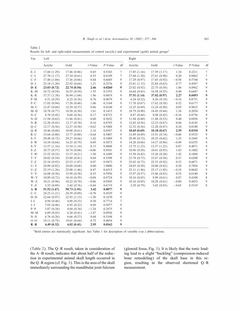
Table 2Results for left- and right-sided measurements of control (acrylic) and experimental (gold) animal groups1
Var. Left Right
Acrylic Gold t-Value P-Value df Acrylic Gold t-Value P-Value df
E–I 17.60 (1.29) 17.46 (0.86) 0.65 0.5343 7 17.85 (1.16) 17.59 (1.17) 1.34 0.2211 7C–I 27.70 (1.17) 27.54 (0.61) 0.53 0.6149 7 27.68 (1.28) 27.61 (0.98) 0.20 0.8462 7C–F 17.48 (1.04) 17.36 (0.86) 0.44 0.6685 9 17.29 (0.97) 17.45 (0.92) −0:58 0.5744 9D–J 23.18 (1.20) 22.92 (0.64) 1.21 0.2576 9 23.01 (1.13) 22.84 (0.63) 0.77 0.4587 9D–E 23.03 (0.72) 22.74 (0.54) 2.66 0.0260 9 23.02 (0.92) 22.75 (0.58) 1.86 0.0962 9K–M 16.72 (0.54) 16.55 (0.54) 1.55 0.1553 9 16.64 (0.43) 16.58 (0.55) 0.48 0.6447 9K–R 37.37 (1.38) 36.94 (1.04) 1.96 0.0819 9 37.51 (1.14) 37.02 (0.97) 2.27 0.0493 9P–M 6.31 (0.29) 6.22 (0.16) 0.76 0.4679 9 6.24 (0.32) 6.26 (0.18) −0:16 0.8751 9P–C 17.85 (0.94) 17.58 (0.48) 1.06 0.3184 9 17.70 (0.67) 17.62 (0.58) 0.52 0.6177 9M–C 12.47 (0.68) 12.29 (0.37) 0.86 0.4140 9 12.25 (0.69) 12.24 (0.50) 0.05 0.9623 9M–D 18.78 (0.77) 18.50 (0.30) 1.61 0.1415 9 18.79 (0.90) 18.45 (0.44) 1.36 0.2054 9N–C 8.70 (0.45) 8.68 (0.36) 0.17 0.8723 9 8.87 (0.44) 8.90 (0.45) −0:16 0.8756 9N–D 11.94 (0.62) 11.86 (0.41) 0.60 0.5652 9 11.94 (0.60) 11.88 (0.53) 0.40 0.6956 9N–R 12.20 (0.43) 12.17 (0.70) 0.16 0.8793 9 12.43 (0.56) 12.23 (0.67) 0.86 0.4145 9Q–C 12.17 (0.56) 12.27 (0.50) −0:62 0.5488 9 12.32 (0.56) 12.28 (0.47) 0.24 0.8180 9Q–R 10.46 (0.60) 10.08 (0.61) 2.16 0.0587 9 10.69 (0.69) 10.18 (0.67) 2.55 0.0310 9R–C 15.64 (0.80) 15.77 (0.80) −0:64 0.5407 9 15.89 (0.89) 15.91 (0.74) −0:06 0.9535 9S–T 29.49 (0.72) 29.27 (0.66) 1.42 0.1889 9 29.49 (0.72) 29.25 (0.62) 1.25 0.2445 9S–W 14.34 (0.64) 14.25 (0.79) 0.69 0.5073 9 14.28 (0.66) 14.37 (0.96) −0:49 0.6355 9S–Y 13.57 (1.16) 13.54 (1.14) 0.15 0.8808 9 13.75 (1.23) 13.57 (1.23) 0.87 0.4071 9S–Z 10.73 (0.57) 10.74 (0.88) −0:06 0.9541 9 10.96 (0.58) 10.61 (0.93) 1.83 0.1002 9T–W 15.52 (0.33) 15.37 (0.21) 1.50 0.1689 9 15.50 (0.42) 15.26 (0.28) 1.42 0.1888 9T–Y 24.02 (0.54) 23.88 (0.41) 0.64 0.5398 9 23.78 (0.73) 23.67 (0.58) 0.51 0.6208 9T–Z 25.54 (0.93) 25.53 (1.07) 0.07 0.9473 9 25.62 (0.73) 25.53 (0.92) 0.53 0.6071 9U–Y 24.09 (0.82) 24.09 (0.68) 0.00 1.0000 9 24.05 (0.58) 24.00 (0.82) 0.28 0.7856 9U–Z 25.19 (1.26) 25.08 (1.46) 0.47 0.6515 9 25.11 (1.46) 25.17 (1.05) −0:18 0.8642 9V–Y 16.08 (0.56) 15.99 (0.58) 0.55 0.5956 9 15.97 (0.57) 15.86 (0.63) 0.52 0.6140 8W–Y 10.09 (0.72) 10.10 (0.59) −0:04 0.9724 9 10.16 (0.83) 9.89 (0.61) 0.87 0.4108 8W–Z 10.21 (0.96) 10.22 (0.70) −0:06 0.9505 9 10.16 (0.85) 10.28 (0.61) −0:80 0.4433 9Y–Z 3.35 (0.89) 3.42 (0.76) −0:44 0.6719 9 3.29 (0.79) 3.42 (0.83) −0:65 0.5319 8A–B 51.52 (1.67) 50.73 (1.91) 3.42 0.0077 9C–C 24.23 (1.21) 24.39 (0.89) −0:78 0.4529 9D–D 22.64 (0.97) 22.93 (1.13) −1:50 0.1670 9I–I 4.94 (0.46) 4.88 (0.33) 0.30 0.7714 7J–J 7.03 (0.46) 6.92 (0.22) 0.69 0.5077 9P–P 3.87 (0.34) 4.04 (0.34) −1:24 0.2475 9M–M 6.09 (0.43) 6.30 (0.41) −1:87 0.0936 9N–N 6.76 (0.26) 6.66 (0.37) 0.68 0.5108 9O–O 19.11 (0.75) 19.01 (0.64) 0.73 0.4854 9R–R 4.49 (0.32) 4.02 (0.41) 2.95 0.0162 9
1Bold entries are statistically signi�cant. See Table 1 for description of variable (var.) abbreviations.
(Table 2). The Q–R result, taken in consideration ofthe A–B result, indicates that about half of the reduc-tion in experimental animal skull length occurred inthe Q–R region (cf. Fig. 3). This is the area of the skullimmediately surrounding the mandibular joint fulcrum
(glenoid fossa, Fig. 3). It is likely that the tonic load-ing lead to a slight “buckling” (compression-inducedbone remodeling) of the skull base in this re-gion, resulting in the observed shortened Q–Rmeasurement.
364 R. Singh et al. / Acta Astronautica 56 (2005) 357–366
We also hypothesized that there would be signi�-cant changes in the coronoid process region (Y, Fig.4), due to increased tension in the temporalis muscles.However, none of the variable measurements that usedY as a landmark approached statistical signi�cance(e.g., T–Y, S–Y, and U–Y, Table 2).By the same token, we hypothesized that there
would be changes in areas sensitive to increasedtension in the masseter muscles, viz., the zygomaticarches, and that these changes would be manifest aseither a decrease in the skull dimension in the zy-gomatic arch region (C–C, D–D, Table 2) or as anincrease in mandibular width. Due to the �exibil-ity of the mid-sagittal mandibular suture, we couldnot make accurate mandibular width measurements.However, zygomatic arch width (C–C, D–D, Ta-ble 2) did not decrease. In fact, there was a slightnon-signi�cant increase in arch width. Although wedid not anticipate this �nding, increased massetermuscle mass, which would be expected to occur withchronic loading, is associated with broadening of themaxillary dental arch (cf. [25]). Hence, the study’s�nding is not without precedent.It is unclear why no changes were seen in coro-
noid process length. It is possible that an implant loadwas not su�cient or of the appropriate type to inducethese changes in adult rats lacking signi�cant growthpotential. Follow-up studies will be required to eval-uate muscle anatomy and physiology and bone histo-logical changes associated with this paradigm. Stud-ies on developing animals will also be informative.Such studies will provide clues regarding the currentstudy’s negative �ndings.It is important that skull width, as measured at the
occipitobullobasisphenoid contact (R–R) was signi�-cantly narrower in experimental than control animals(Table 2). This indicates that, as anticipated, loadingdid lead to skull narrowing; however, the location ofthe narrowing was unexpected. This stresses the ne-cessity of evaluating a large set of morphometric mea-surements, as CM growth and development are com-plex and often unpredictable.Three of the �ve signi�cantly di�erent variables
were unilateral measurements, and it is unclear whythe contralateral variable pair was not signi�cantly dif-ferent between animal groups as well. It is notewor-thy that the contralateral variables approached signi�-cance; in all three cases P¡ 0:10 (Table 2). This sug-
gests that the di�erences due to loading were slightenough in the adult animal to register as false nega-tives occasionally. It is noteworthy in this regard thatone other variable, anterior bimolar width (M–M, Ta-ble 2), had a P¡ 0:10.In any study involving multiple statistical testing,
it is important to consider the possibility that signi�-cant di�erences may be due to sampling errors. It is acommon practice to reduce the signi�cance level pro-portionate to the number of tests performed; however,we emphasize the exploratory nature of this studyand so choose to maintain the customary P¡ 0:05signi�cance level. This “high” signi�cance cuto�level could be another reason why K–R, Q–R, andD–E were signi�cantly di�erent on one side only (Ta-ble 2). In other words, the observed di�erences couldhave been false positives. Nevertheless, it is importantto recognize that the lever model, Fig. 1, representsa physical reality of the CMS, and that gravity doesa�ect the system. The question is whether the e�ect isonly physical, or whether biologically relevant e�ectsalso occur. This study is only the �rst in a series ofexperiments that will look at other potential e�ects,including histological and physiological changes as-sociated with loading. The study’s outcome providesevidence that important loading-related changes willbe observed, and it provides important clues as towhere these changes may be observed, viz., skull basein the occipitobullosphenoid and posterior zygomaticarch regions.The current study focused on macrogravitational ef-
fects; however, the CMS also shows promise in mi-crogravitational studies. Work in the 1980s demon-strated that the CMS dentoskeleton is susceptible tospace �ight e�ects [4–7]. Several investigations haveshown that the e�ects of soft diet [27,31–33] and toothloss [29,34–36] on CM musculoskeletal morphologyand physiology are similar to e�ects seen in micro-gravitational studies of other musculoskeletal systems[9,10,37–40]. Changes common to space �ight, softdiet, and tooth loss include loss or thinning of corticalbone, decreased bone mass, decreased bone density,decreased muscle mass or muscle atrophy, and a shiftfrom type I muscle �bers to type IIb �bers.Soft diet regimens are at least as easy to employ
as most of the current ground-based microgravity ani-mal paradigms. As for human studies of microgravita-tional e�ects, over 13% of the adult US population is
R. Singh et al. / Acta Astronautica 56 (2005) 357–366 365
completely edentulous on at least 1 arch, and about 3%of the edentulous US population is 18–44 years old[41]. Because edentulism e�ects are similar to micro-gravity e�ects, the edentulous population represents alarge untapped human resource for studies of the ef-fects of microgravity on musculoskeletal physiologyand form.Finally, in addition to the hindlimb-unloading sim-
ulation that was previously introduced to study the ef-fects of hypogravity on the CMS [6], we suggest theuse of inter-arch banding and wiring methods to un-weight the jaw directly. These latter methods are com-monly employed on humans during orthodontic treat-ment and after certain oral surgery procedures. It islikely that similar methods could be adapted to animalmodels. Thus, the CMS should be considered a poten-tially useful model for studying the e�ects of gravityon musculoskeletal systems.
Acknowledgements
We wish to thank Dr. Lyle Johnston, Chair, De-partment of Orthodontics, University of Michigan, for�nancial and intellectual support of this project. Theproject was also supported by funds from the Depart-ment of Biologic and Materials Sciences, Universityof Michigan.
References
[1] A. Karlsen, O. Krogstad, Morphology and growth inconvex pro�le facial patterns: a longitudinal study, AngleOrthodontist 69 (1999) 344–344.
[2] J. Huggare, M. Cooke, Head posture and cervicovertebralanatomy as mandibular growth predictors, European Journalof Orthodontics 16 (1994) 175–180.
[3] M. Caltabiano, P. Verzi, G. Scire Scappuzzo, Head posture inorthodontics: physiopathology and clinical aspects 2, MondoOrtodontico 14 (1989) 313–324.
[4] D. Simmons, J. Russell, F. Winter, P. Tran Van, A. Vignery,R. Baron, G. Rosenberg, W. Walker, E�ect of space�ighton the non-weight-bearing bones of rat skeleton, AmericanJournal of Physiology 244 (1983) R319–R326.
[5] D. Simmons, J. Russell, F. Winter, R. Baron, A. Vignery,T. Thuc, G. Rosenberg, W. Walker, Bone growth in the ratmandible during space �ight, The Physiologist 23 (1980)S87–S90.
[6] D. Simmons, B. Grazman, J. Russell, W. Walker, D. Bikle, E.Morey, Simulating certain aspects of hypogravity e�ects on
bone maturation in the non-weight bearing skeleton, AviationSpace and Environmental Medicine 54 (1983) 1080–1084.
[7] G. Rosenberg, S. Campbell, D. Simmons, The e�ects ofspace�ight on the mineralization of rat incisor dentin,Proceedings of the Society for Experimental Biology andMedicine 175 (1984) 429–437.
[8] F. Booth, D. Criswell, Molecular events underlyingskeletal muscle atrophy and the development of e�ectivecountermeasures, International Journal of Sports Medicine 18(1997) S265–S269.
[9] D. Desplanches, Structural and functional adaptations ofskeletal muscle to weightlessness, International Journal ofSports Medicine 18 (1997) S259–S264.
[10] K. Baldwin, E�ect of space�ight on the functional,biochemical, and metabolic properties of skeletal muscle,Medicine and Science in Sports and Exercise 28 (1996)983–987.
[11] V. Edgerton, R. Roy, Neuromuscular adaptation to actual andsimulated weightlessness, Advances in Space Biology andMedicine 4 (1994) 33–67.
[12] T. Morris, C. Brandon, M. Horton, D. Carlons, S. JJ,Maximum shortening velocity and myosin heavy-chainisoform expression in human masseter muscle �bers, Journalof Dental Research 80 (2001) 1845–1848.
[13] J. Korfage, T. Van Eijden, Regional di�erences in �bre typecomposition in the human temporalis muscle, Journal ofAnatomy 194 (1999) 355–362.
[14] K. Watanabe, M. Watanabe, Activity of jaw-opening andjaw-closing muscles and their in�uence on dentofacialmorphological features in normal adults, Journal of OralRehabilitation 28 (2001) 873–879.
[15] H. Ueda, Y. Ishizuka, K. Miyamoto, N. Morimoto, K. Tanne,Relationship between masticatory muscle activity andvertical craniofacial morphology, Angle Orthodontist 68(1998) 233–238.
[16] L. Pagni, T. Baccetti, Heredity and environment in thegenesis, epigenesis and evolution of the orofacial area,Minerva Stomatologica 42 (1993) 1–13.
[17] M. Sorin, T. Ramsey, T. Hart, F. Farrington, Comparisonof craniofacial morphology in monozygotic twins with theirsiblings, Journal of Clinical Pediatric Dentistry 15 (1991)169–173.
[18] S. Kiliaridis, H. Kjellberg, B. Wenneberg, C. Engstrom, Therelationship between maximal bite force, bite force endurance,and facial morphology during growth, A cross-sectional study,Acta Odontologica Scandinavica 51 (1993) 323–331.
[19] B. Haskell, M. Day, J. Tetz, Computer-aided modeling inthe assessment of the biomechanical determinants of diverseskeletal patterns, American Journal of Orthodontics 89 (1986)363–382.
[20] S. Herring, T. Lakars, Craniofacial development in theabsence of muscle contraction, Journal of CraniofacialGenetics & Developmental Biology 1 (1982) 341–357.
[21] K. Pirila, P. Tahvanainen, J. Huggare, P. Nieminen,H. Lopponen, Sleeping positions and dental arch dimensionsin children with suspected obstructive sleep apnea syndrome,European Journal of Oral Sciences 103 (1995) 285–291.
366 R. Singh et al. / Acta Astronautica 56 (2005) 357–366
[22] I. Baumann, P. Plinkert, E�ect of breathing mode and noseventilation on growth of the facial bones, HNO. 44 (1996)229–234.
[23] G. Kluemper, P. Vig, K. Vig, Nasorespiratory characteristicsand craniofacial morphology, European Journal ofOrthodontics 17 (1995) 491–495.
[24] V. Brattstrom, L. Odenrick, E. Kvam, Dentofacialmorphology in children playing musical wind instruments:a longitudinal study, European Journal of Orthodontics 11(1989) 179–185.
[25] C. Katsaros, Masticatory muscle function and transversedentofacial growth, Swedish Dental Journal 151 (2001)1–47.
[26] S. Chuang, The relationship between masticatory functionand craniofacial morphology, Kao-Hsiung i Hsueh Ko HsuehTsa Chih 11 (1995) 458–469.
[27] S. Kiliaridis, Masticatory muscle function and craniofacialmorphology, An experimental study in the growing rat fed asoft diet, Swedish Dental Journal 36 (1986) 1–55.
[28] M. Nodal, I. Kjaer, B. Solow, Craniofacial morphology inpatients with multiple congenitally missing permanent teeth,European Journal of Orthodontics 16 (1994) 104–109.
[29] S. Percac, V. Nikolic, In�uence of teeth loss on morphometriccharacteristics of the maxilla, Acta Stomatologica Croatica25 (1991) 199–205.
[30] K. Byrd, Craniofacial sequelae of lesions to facial andtrigeminal motor nuclei in growing rats, American Journal ofPhysical Anthropology 76 (1988) 87–103.
[31] T. Saito, Y. Ohnuki, A. Yamane, Y. Saeki, E�ects ofdiet consistency on the myosin heavy chain mRNAs of ratmasseter muscle during postnatal development, Archives ofOral Biology 47 (2002) 109–115.
[32] A. Bresin, S. Kiliaridis, K. Strid, E�ect of masticatoryfunction on the internal bone structure in the mandible of the
growing rat, European Journal of Oral Sciences 107 (1999)35–44.
[33] A. Bresin, E�ects of masticatory muscle function andbite-raising on mandibular morphology in the growing rat,Swedish Dental Journal (Suppl) (2001) 1–49.
[34] A. Raustia, M. Salonen, J. Pyhtinen, Evaluation of masticatorymuscles of edentulous patients by computed tomography andelectromyography, Journal of Oral Rehabilitation 23 (1996)11–16.
[35] B. Miehe, J. Fanghandel, D. Kubein-Meesenburg, H. Nagerl,R. Schwestka-Polly, Masticatory musculature under alteredocclusal relationships—a model study with experimentalanimals, Anatomischer Anzeiger 181 (1999) 37–40.
[36] J. Fanghanel, B. Otto, G. Schumacher, Animal experimentstudies on the e�ect of unilateral tooth extraction on thegrowth of the skull and masticatory muscles. III. Themasticatory muscles, Anatomischer Anzeiger 160 (1985)305–313.
[37] R. Roy, K. Baldwin, Response of the neuromuscular unitto space�ight: what has been learned from the rat model,Exercise and Sport Sciences Reviews 24 (1996) 399–425.
[38] S. Arnaud, J. Harper, M. Navidi, Mineral distribution in ratskeletons after exposure to a microgravity model, Journal ofGravitational Physiology 2 (1995) P115–P116.
[39] R. Johnson, The bearable lightness of being: bones, muscles,and space�ight, Anatomical Record 251 (1998) 24–27.
[40] E. Morey-Holton, R. Globus, Hindlimb unloading of growingrats: a model for predicting skeletal changes during space�ight, Bone 22 (1998) 83S–88S.
[41] D. Blackwell, J. Collins, R. Coles, Summary health statisticsfor U.S. adults: National Health Interview Survey, 1997.National Center for Health Statistics, Vital Health Statistics10 (2002) 205.
Related Documents
![exposure impairs germ cell developmentin human fetal ... · drug during fetal, neonatal and adult age induces altered morphology and apoptosis in mouse and rat ovaries [44-46]. Petrik](https://static.cupdf.com/doc/110x72/5f0737db7e708231d41be59c/exposure-impairs-germ-cell-developmentin-human-fetal-drug-during-fetal-neonatal.jpg)