ARTICLE MOLECULAR THERAPY Vol. 2, No. 6, December 2000 Copyright The American Society of Gene Therapy 1525-0016/00 $35.00 545 doi:10.1006/mthe.2000.0210, available online at http://www.idealibrary.com on IDEAL Liver cirrhosis represents a worldwide health problem and is a major cause of mortality. Cirrhosis is the result of extensive hepatocyte death and fibrosis induced by chronic alcohol abuse and hep- atitis B and C viruses. Successful gene therapy approaches to this disease may require both rever- sal of fibrosis and stimulation of hepatocyte growth. Urokinase-type plasminogen activator (uPA) may serve this function, as it is an initiator of the matrix proteolysis cascade and induces hepa- tocyte growth factor expression. In a rat cirrhosis model, a single iv administration of a replica- tion-deficient adenoviral vector encoding a nonsecreted form of human uPA resulted in high pro- duction of functional uPA protein in the liver. This led to induction of collagenase expression and reversal of fibrosis with concomitant hepatocyte and improved liver function. Thus, uPA gene therapy may be an effective strategy for treating cirrhosis in humans. Key Words: experimental cirrhosis; hepatic regeneration; adenovirus; vector; gene therapy; met- alloproteinases. Liver Cirrhosis Is Reverted by Urokinase-Type Plasminogen Activator Gene Therapy Silvia Salgado, * Jesus Garcia, * Jose Vera, * Fernando Siller, * Miriam Bueno, * Alejandra Miranda, * Aida Segura, * Guillermo Grijalva, * Jorge Segura, * Hector Orozco, † Rogelio Hernandez-Pando, † Mary Fafutis, ‡ Laura K. Aguilar, § Estuardo Aguilar-Cordova, § and Juan Armendariz-Borunda *,1 *Institute Molecular Biology in Medicine and Gene Therapy, CUCS, University of Guadalajara, Apdo. Postal 2-123, Guadalajara, Jalisco, Mexico 44281 §Harvard Gene Therapy Initiative, Boston, Massachusetts 02115 †Instituto Nacional de la Nutricion “Salvador Zubiran,” Guadalajara, Jalisco, Mexico ‡Instituto Dermatologico de Guadalajara, Guadalajara, Jalisco, Mexico Received for publication August 10, 2000; accepted in revised form October 5, 2000; published online November 16, 2000 1 To whom correspondence and reprint requests should be addressed. Fax: (52) (3) 617-4159. E-mail:armendbo@ cucs.udg.mx, [email protected]. INTRODUCTION Liver cirrhosis represents a worldwide health problem and is a major cause of mortality. Cirrhosis is the com- mon end for chronic alcohol abuse and hepatitis C and B virus infections (1) and no definitive cure is available so far. Alcoholism alone affects around 14 million people in the United States (2). The potential application of gene therapy protocols to human hepatic cirrhosis will depend on the successful and tissue-specific delivery of therapeutic genes to livers affected with extensive fibro- sis. We reason that gene therapy applications to reverse the exacerbated fibrosis, typical major hallmark of cir- rhotic livers, might be developed. Likewise, targeted delivery of genes promoting liver cell proliferation could favor the rapid reestablishment of functional hepatic mass. An adenoviral vector bearing a modified cDNA coding for a nonsecreted form of human urokinase plas- minogen activator (Ad-∆huPA) was used to deliver the gene in vivo (kindly provided by Dr. Mark Kay) (3). The nonsecreted uPA was chosen to diminish the risk of bleeding, which would be particularly problematic in cir- rhotic animals that may have preexisting coagulopathy. A huPA cDNA was used because it is known as a potent activator of latent hepatic collagenases (4) which in turn would promote the degradation of extracellular matrix deposited in the Disse’s space (perisinusoidal space). Excess collagenous proteins hamper the flow exchange of nutrients between circulating blood and hepatocytes (4–6) and provoke liver dysfunction. Furthermore, non- secreted uPA leads to rates of brisk hepatic liver cell regeneration in normal noncirrhotic animals similar to the unmodified or wild-type uPA, without the side effects related to bleeding tendency (3). The adenovector approach was chosen to quickly analyze the effects of potentially therapeutic genes, as adenovirus has been shown to be an efficient vector in cirrhotic liver models (7). In this paper we report that one single injection of the adenoviral Ad-∆huPA through the iliac vein of severely cirrhotic rats can target liver tissue in a specific way and

Welcome message from author
This document is posted to help you gain knowledge. Please leave a comment to let me know what you think about it! Share it to your friends and learn new things together.
Transcript

ARTICLE
MOLECULAR THERAPY Vol. 2, No. 6, December 2000Copyright � The American Society of Gene Therapy1525-0016/00 $35.00
545
doi:10.1006/mthe.2000.0210, available online at http://www.idealibrary.com on IDEAL
Liver cirrhosis represents a worldwide health problem and is a major cause of mortality. Cirrhosisis the result of extensive hepatocyte death and fibrosis induced by chronic alcohol abuse and hep-atitis B and C viruses. Successful gene therapy approaches to this disease may require both rever-sal of fibrosis and stimulation of hepatocyte growth. Urokinase-type plasminogen activator (uPA)may serve this function, as it is an initiator of the matrix proteolysis cascade and induces hepa-tocyte growth factor expression. In a rat cirrhosis model, a single iv administration of a replica-tion-deficient adenoviral vector encoding a nonsecreted form of human uPA resulted in high pro-duction of functional uPA protein in the liver. This led to induction of collagenase expression andreversal of fibrosis with concomitant hepatocyte and improved liver function. Thus, uPA genetherapy may be an effective strategy for treating cirrhosis in humans.
Key Words: experimental cirrhosis; hepatic regeneration; adenovirus; vector; gene therapy; met-alloproteinases.
Liver Cirrhosis Is Reverted by Urokinase-TypePlasminogen Activator Gene TherapySilvia Salgado,* Jesus Garcia,* Jose Vera,* Fernando Siller,* Miriam Bueno,*Alejandra Miranda,* Aida Segura,* Guillermo Grijalva,* Jorge Segura,* Hector Orozco,†Rogelio Hernandez-Pando,† Mary Fafutis,‡ Laura K. Aguilar,§ Estuardo Aguilar-Cordova,§and Juan Armendariz-Borunda*,1
*Institute Molecular Biology in Medicine and Gene Therapy, CUCS, University of Guadalajara, Apdo. Postal 2-123, Guadalajara, Jalisco, Mexico 44281§Harvard Gene Therapy Initiative, Boston, Massachusetts 02115†Instituto Nacional de la Nutricion “Salvador Zubiran,” Guadalajara, Jalisco, Mexico‡Instituto Dermatologico de Guadalajara, Guadalajara, Jalisco, Mexico
Received for publication August 10, 2000; accepted in revised form October 5, 2000; published online November 16, 2000
1To whom correspondence and reprint requests should be addressed.Fax: (52) (3) 617-4159. E-mail:armendbo@ cucs.udg.mx,[email protected].
INTRODUCTION
Liver cirrhosis represents a worldwide health problemand is a major cause of mortality. Cirrhosis is the com-mon end for chronic alcohol abuse and hepatitis C andB virus infections (1) and no definitive cure is availableso far. Alcoholism alone affects around 14 million peoplein the United States (2). The potential application ofgene therapy protocols to human hepatic cirrhosis willdepend on the successful and tissue-specific delivery oftherapeutic genes to livers affected with extensive fibro-sis. We reason that gene therapy applications to reversethe exacerbated fibrosis, typical major hallmark of cir-rhotic livers, might be developed. Likewise, targeteddelivery of genes promoting liver cell proliferation couldfavor the rapid reestablishment of functional hepaticmass. An adenoviral vector bearing a modified cDNAcoding for a nonsecreted form of human urokinase plas-
minogen activator (Ad-∆huPA) was used to deliver thegene in vivo (kindly provided by Dr. Mark Kay) (3). Thenonsecreted uPA was chosen to diminish the risk ofbleeding, which would be particularly problematic in cir-rhotic animals that may have preexisting coagulopathy.A huPA cDNA was used because it is known as a potentactivator of latent hepatic collagenases (4) which in turnwould promote the degradation of extracellular matrixdeposited in the Disse’s space (perisinusoidal space).Excess collagenous proteins hamper the flow exchangeof nutrients between circulating blood and hepatocytes(4–6) and provoke liver dysfunction. Furthermore, non-secreted uPA leads to rates of brisk hepatic liver cellregeneration in normal noncirrhotic animals similar tothe unmodified or wild-type uPA, without the side effectsrelated to bleeding tendency (3). The adenovectorapproach was chosen to quickly analyze the effects ofpotentially therapeutic genes, as adenovirus has beenshown to be an efficient vector in cirrhotic liver models(7).
In this paper we report that one single injection of theadenoviral Ad-∆huPA through the iliac vein of severelycirrhotic rats can target liver tissue in a specific way and
ARTICLE
MOLECULAR THERAPY Vol. 2, No. 6, December 2000Copyright � The American Society of Gene Therapy
546
induce profound beneficial changes. We have been ableto induce a significant reversion in CCl4-induced hepat-ic fibrosis and at the same time stimulate liver cells toregenerate. Furthermore, an improvement in functionalhepatic tests was evident.
MATERIAL AND METHODS
Animals. The experimental model consisted of Wistar rats undergoingchronic administration of CCl4 (8), an animal model that closely resem-bles human hepatic cirrhosis induced by alcohol abuse or chronic infec-tion with hepatitis C virus. Briefly, animals weighing 80 g received threedoses a week via ip of a mixture 1:6 of CCl4–mineral oil for the first week,a ratio of 1:5 the second week, 1:4 the third week, and 1:3 the fourththrough eighth week. Control rats were pair-fed and injected with vehi-cle only. All animal studies were performed on male Wistar rats in accor-dance with the University of Guadalajara’s animal guidelines. All experi-mental methods have been described previously (7). Adenovirus infu-sions were performed through the iliac vein.
Adenovirus vectors. Adenoviral plasmid pAd.PGK-∆N∆C-huPA (pAd-∆huPA) cloning has been previously described (3) and is essentially afirst-generation adenoviral vector bearing a modified cDNA coding fornonsecreted human urokinase plasminogen activator (Ad-∆uPA). Thepreparations of this Ad vector were monitored for endotoxin andmycoplasm contaminants and were titered as previously described (9).The rationale for using this vector resided in the advantage of the non-secreted uPA that does not cause hypocoagulation and spontaneousbleeding, which represents a major drawback in cirrhotic animals.
The Ad-GFP vector used here (irrelevant adenovirus) is an E1- and E3-deleted replication-defective adenovirus vector previously described (9).The vectors were produced at the Baylor College of Medicine Gene VectorLaboratory under Good Laboratory Practice conditions. The vectors weretitered and characterized as described (9) and had a vector particles (vp)to infection units (IU) ratio of ≤30.
Preparation of liver homogenates. Rats were sacrificed at indicated timesin Fig. 1a and liver homogenates were prepared from 150 mg of tissue asdescribed (10) and kept at −70°C. Briefly, for uPA, in the presence of pro-tease inhibitors, 150 mg of liver was homogenized in 400 µl of homoge-nization buffer (0.05 M Tris–HCl, 0.15 M NaCl, 0.01 M Hepes, 2 mMCaCl2, 0.01% Tween 80, 1 mM phenylmethylsulfonyl fluoride, pH 8.5).One hundred microliters of such homogenates was used for ELISA. At thesame time, serum samples (100 µl) were obtained and kept at −20°C untilused for ELISA. Total protein levels in serum were determined usingBradford assay of protein quantification (11). For metalloproteinase-2(MMP-2) assay, samples were homogenized using a high-speed mixerhomogenizer (Politron PT 3000, Kinematica AG, Brinkmann,Switzerland) for 5 min at 8000g in 4 ml of 0.15 M NaCl at 4°C. After threefreeze–thaw cycles, the homogenate of each sample was sonicated twiceat 21 kilocyles per second for 1 min at 4°C and centrifuged at 8000g for10 min at 4°C, aliquoted, and kept at −70°C until further use.
uPA and MMP-2 ELISA determinations. ELISAs were performed using acommercially available kit (Biopool TintElize uPA, Sweden) which is anenzyme immunoassay specific for quantitative determination of humanuPA, with a detection limit of 0–4 ng/ml.
The Biotrak MMP-2 activity assay system from Amersham PharmaciaBiotech provides quantitative determination of MMP-2 in tissuehomogenates and others (detection limit 0.75–12 ng/ml).
Biochemical assays. Blood was drawn from animals at specified times,and serum transaminases ALT, AST, alkaline phosphatase, and bilirubinswere determined in an automated Sincron-7 analyzer at Hospital Civil deGuadalajara.
Histological examination and immunohistochemistry of liver sections.Rats were sacrificed at 2, 4, 6, 8, and 10 days after Ad-∆huPA administra-tion (Fig. 1a). A group of cirrhotic animals injected with Ad-GFP (irrele-vant adenovirus) and vehicle only as well as a group of normal animalswere used as controls. Five rats were included for each group. For the his-
tologic study, the liver was immediately removed and fixed by immer-sion in 10% paraformaldehyde diluted in phosphate–saline buffer (PBS),dehydrated in graded ethylic alcohol, and embedded in paraffin. Sections5 µm thick were stained with hematoxylin/eosin and Masson trichrome.In these latter slides the percentage of liver tissue affected by fibrosis wasdetermined using a computer-assisted automated image analyzer (QwinLeica) by analyzing 10 random fields per slide and calculating the ratioof connective tissue to the whole area of the liver. For immunohisto-chemistry, liver sections were mounted in silane-covered slides anddeparaffinized, and the endogenous activity of peroxidase was quenchedwith 0.03% H2O2 in absolute methanol. Liver sections were incubatedovernight at room temperature with mouse monoclonal antibodiesagainst PCNA and α-smooth muscle actin (Boehringer Mannheim,Germany) diluted 1/20 and 1/50, respectively, in PBS and with goat poly-clonal antibodies against human uPA (Chemicon International, U.S.A.)diluted 1/400 in PBS. Bound antibodies were detected with peroxidase-labeled rabbit polyclonal antibodies against mouse or goat immunoglob-ulins and diaminobenzidine and counterstained with hematoxylin. Forquantification, 10 random fields of intralobular and periportal areas wereevaluated at ×400 magnification. Immunohistochemical positive andnegative cells were counted by an automated image analyzer (Qwin,Leica) and the data expressed as percentages of positive cells.Histopathology was interpreted by two independent board-certifiedpathologists who were blinded to the study. There was a 5% margin ofdifference in their analyses.
Semiquantitative RT-PCR of cDNA. Total RNA was prepared immedi-ately after liver was extracted at the time of sacrifice and RT-PCR was car-ried out essentially as described (12) with some modifications byDelgado-Rizo et al. (13). The following specific primers were used: 5′-ATGCTCATGGACCCTGGT-3′ (HGF sense primer), 5′GCCTG-GCAGCTTCATTA-3′ (HGF antisense primer), 5′-CAGTGATGATCT-CAATGGGCAAT-3′ (c-met sense primer), and 5′AATGCCCTCTTCCTAT-GACTTC-3′ (c-met antisense primer). Thirty cycles of PCR were per-formed, with the following conditions: denaturation for 5 min at 94°C,annealing for 1 min at 60°C, and elongation for 1.5 min at 72°C.Standardization of constitutive gene expression was accomplished withthe HPRT gene.
RESULTS AND DISCUSSION
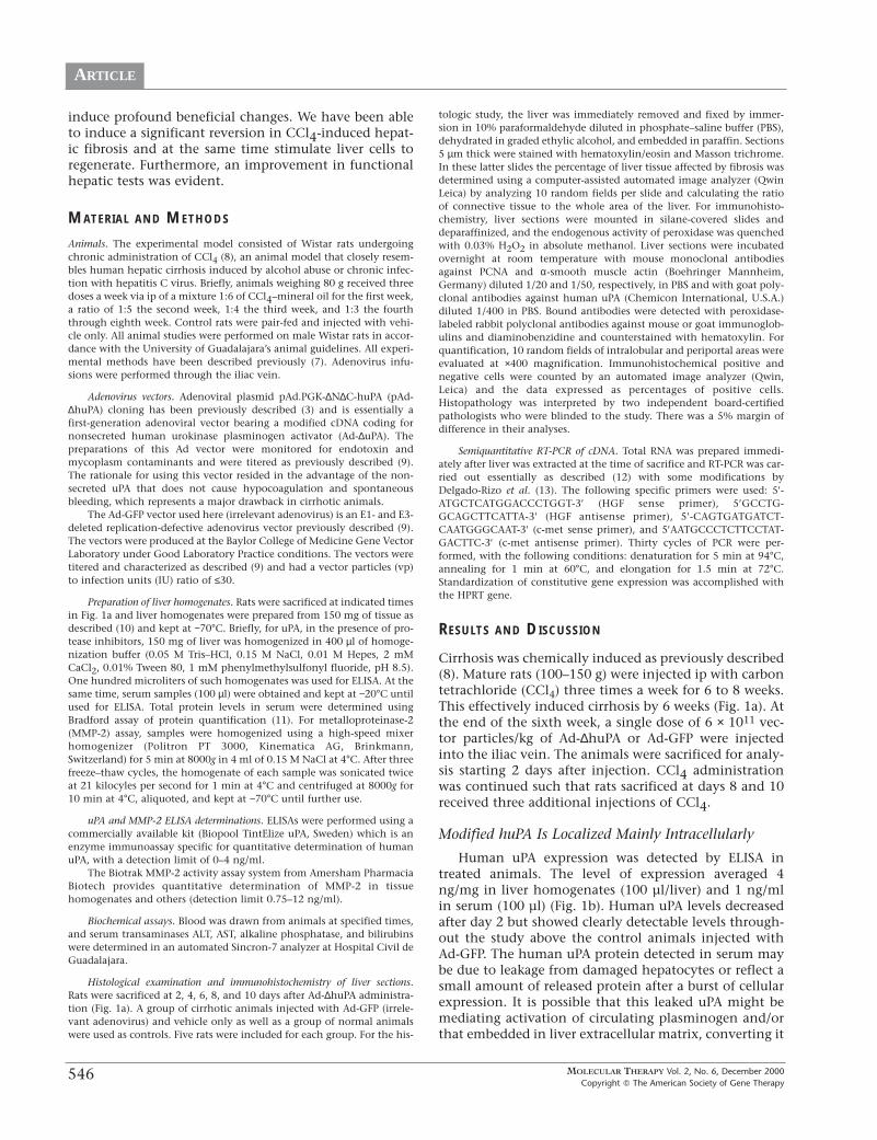
Cirrhosis was chemically induced as previously described(8). Mature rats (100–150 g) were injected ip with carbontetrachloride (CCl4) three times a week for 6 to 8 weeks.This effectively induced cirrhosis by 6 weeks (Fig. 1a). Atthe end of the sixth week, a single dose of 6 × 10¹¹ vec-tor particles/kg of Ad-∆huPA or Ad-GFP were injectedinto the iliac vein. The animals were sacrificed for analy-sis starting 2 days after injection. CCl4 administrationwas continued such that rats sacrificed at days 8 and 10received three additional injections of CCl4.
Modified huPA Is Localized Mainly Intracellularly
Human uPA expression was detected by ELISA intreated animals. The level of expression averaged 4ng/mg in liver homogenates (100 µl/liver) and 1 ng/mlin serum (100 µl) (Fig. 1b). Human uPA levels decreasedafter day 2 but showed clearly detectable levels through-out the study above the control animals injected withAd-GFP. The human uPA protein detected in serum maybe due to leakage from damaged hepatocytes or reflect asmall amount of released protein after a burst of cellularexpression. It is possible that this leaked uPA might bemediating activation of circulating plasminogen and/orthat embedded in liver extracellular matrix, converting it
ARTICLE
MOLECULAR THERAPY Vol. 2, No. 6, December 2000Copyright � The American Society of Gene Therapy
547
to plasmin, and thus activating latent MMPs (14, 15),which in turn digest collagenous matrix. Human uPAexpression was detected in 46–80% of hepatocytes inimmunostained liver sections from Ad-∆huPA-injectedrats starting on day 2 and peaking on day 6 (Fig. 1c).Kupffer cells and biliary epithelium also showedimmunoreactivity.
Overexpression of Hepatic huPA Correlates with Induction of Metalloproteinases and Reversal of Fibrosis
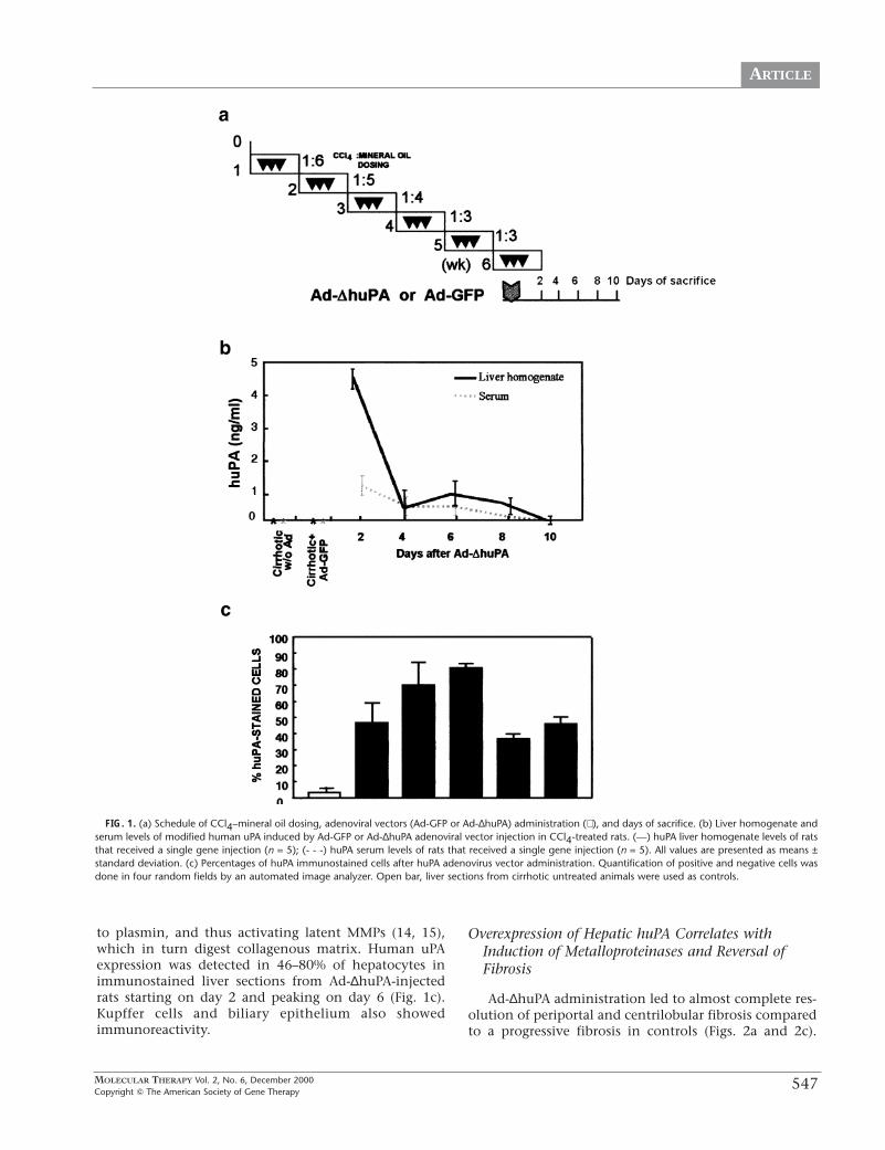
Ad-∆huPA administration led to almost complete res-olution of periportal and centrilobular fibrosis comparedto a progressive fibrosis in controls (Figs. 2a and 2c).
FIG. 1. (a) Schedule of CCl4–mineral oil dosing, adenoviral vectors (Ad-GFP or Ad-∆huPA) administration (⇓ ), and days of sacrifice. (b) Liver homogenate andserum levels of modified human uPA induced by Ad-GFP or Ad-∆huPA adenoviral vector injection in CCl4-treated rats. (—) huPA liver homogenate levels of ratsthat received a single gene injection (n = 5); (- - -) huPA serum levels of rats that received a single gene injection (n = 5). All values are presented as means ±standard deviation. (c) Percentages of huPA immunostained cells after huPA adenovirus vector administration. Quantification of positive and negative cells wasdone in four random fields by an automated image analyzer. Open bar, liver sections from cirrhotic untreated animals were used as controls.

ARTICLE
MOLECULAR THERAPY Vol. 2, No. 6, December 2000Copyright � The American Society of Gene Therapy
548
FIG. 2. (a and b) Liver sections of cirrhotic rats after CCl4 administration and Ad-∆huPA or Ad-GFP adenoviral vector injection. (Upper panels) Massontrichromic staining (×50), (lower panels) Sirius red staining (×50). (a) Cirrhotic nodules are surrounded by thick fibrotic bands after 2, 4, 6, 8, and 10 days ofAd-GFP adenoviral vector injection showing progressive fibrosis. (b) In contrast, after 10 days of huPA adenoviral vector injection there are only thin fibrousbands extending from portal areas (green arrow) and mild central vein sclerosis (yellow arrow). (c) Percentages of fibrous tissue deposition after Ad-GFP or Ad-∆huPA adenoviral vector infusion and normal rats. Determinations of fibrous tissue were done in 10 random fields by an automated image analyzer. Open bar,normal rats; gray bars, rats treated with CCl4 for 6 weeks with Ad-GFP adenoviral vector injection; black bars, rats treated with CCl4 and injected with huPAadenoviral vector. N represents normal noncirrhotic rats. Values are presented as means ± standard deviation. (d) Determination of matrix metalloproteinase-2 (MMP-2) activity in liver homogenates by a specific ELISA of normal and cirrhotic rats treated with adenoviral vectors (Ad-GFP or Ad-∆huPA). Open bar, nor-mal rats; crossed bar, liver sections from cirrhotic untreated animals; gray bar, cirrhotic animals treated with Ad-GFP adenoviral vector (after 4 days); black bars,cirrhotic animals treated with Ad-∆huPA adenoviral vector. N represents normal noncirrhotic rats. (e) Percentages of αSMA-immunostained cells after Ad-GFPor Ad-∆huPA adenoviral vector infusion. Quantification of positive and negative cells was done in four random fields by an automated image analyzer. Crossedbar, cirrhotic untreated rats; black bars, rats treated with CCl4 and injected with huPA adenoviral vector. Values are presented as means ± standard deviation.
FIG. 3. (a) Hepatocyte regeneration as measured by PCNA staining. Mitosis in hepatocytes was assessed by immunohistochemistry using anti-PCNA antibodyafter Ad-GFP or Ad-∆huPA adenovirus vector injection. (Left panel) Hepatocytes with nuclear PCNA immunostaining after huPA adenoviral vector injection.Cirrhotic rats after 2 days have abundant PCNA-positive hepatocytes (red arrow) in coexistence with frequent mitotic figures (black arrows) and binucleatedcells (blue arrows) (×320). Moreover, positive cells were detected at 4, 6, 8, and 10 days (×200). In comparison (right panel), few hepatocytes with PCNAimmunostaining (×200) at 2, 4, 6, 8, and 10 days after Ad-GFP adenovirus vector injection are shown. (b and c) Percentages of PCNA-immunostained cells inperiportal and lobular areas, respectively, after Ad-∆huPA- or Ad-GFP adenovirus vector injection. Quantification of positive and negative cells was done ran-dom field by an automated image analyzer. Liver sections from cirrhotic untreated animals were used as controls. Open bars, normal rats; gray bars, rats treat-ed with CCl4 for 6 weeks with Ad-GFP adenoviral vector injection; black bars, rats treated with CCl4 and injected with huPA adenoviral vector. Values are pre-sented as means ± standard deviation. (d) Determination of HGF and c-met gene expression by semiquantitative RT-PCR. Total RNA was extracted from liversat indicated times and cDNA was obtained. Standardization of constitutive gene expression was accomplished with the HPRT gene. (e) Prothrombin time ofcirrhotic rats sacrificed at different days after Ad-∆huPA- or Ad-GFP adenoviral vector injection. Open bar, normal rats; gray bars, rats treated with CCl4 for 6weeks with Ad-GFP adenoviral vector injection; black bars, rats treated with CCl4 for 6 weeks with Ad-∆huPA adenoviral vector injection. Dotted line representsnormal prothrombin time. Values are presented as means ± standard deviation.

ARTICLE
MOLECULAR THERAPY Vol. 2, No. 6, December 2000Copyright � The American Society of Gene Therapy
549
3

ARTICLE
MOLECULAR THERAPY Vol. 2, No. 6, December 2000Copyright � The American Society of Gene Therapy
550
Computer-assisted analyses showed that Ad-∆huPA-treated rats had an 85% reduction in hepatic fibrosis atday 10 (Figs. 2b and 2c). In addition, in cirrhotic liverswith severe fibrosis, hepatic stellate cells (HSC) areincreased in fibrotic areas, many of which have an acti-vated phenotype identified by expression of α-smoothmuscle actin (SMA) (1). Cirrhotic livers showed numer-ous α-SMA-positive cells embedded in the perinodularfibrous tissue which increased 2 days after Ad-∆huPAadministration but decreased to 50% of the control cir-rhotic livers by day 10 (Fig. 2e).
A potential mechanism for the degradation of fibrot-ic tissue observed with Ad-∆huPA is through the activa-tion of latent metalloproteinases complexed with TIMPs.These MMPs are effector proteins downstream of uPA inthe matrix proteolysis cascade. We found an increasedexpression of MMP-2 in liver homogenates of Ad-∆huPA-treated animals (Fig. 2d). This assay is a very sensitiveELISA, which detects minimal changes in levels of totalMMP-2 (active and pro-MMP-2) (16), which specificallydegrades collagen type IV and other collagens to a lesserdegree (17, 18). We believe that most MMP-2 in Ad-∆huPA animals is in the form of active MMP-2. However,we need to further elucidate quantitative amounts ofactive MMP-2 and MMP-2 species complexed with itsspecific inhibitor, TIMP-1.
HuPA Induces a Brisk Liver Cell Regeneration
In addition to resolution of fibrosis, liver regenera-tion was observed in cirrhotic rats treated with Ad-∆huPA. Two days after Ad-∆huPA administration, liversections contained a substantial increase in the numberof mitotic figures, binucleated hepatocytes, and cellsexpressing proliferative cell nuclear antigen (PCNA)(Figs. 3a–3c). The percentage of PCNA-positive cells wasstill as high as 40% on day 8 and included periportal andlobular areas (Figs. 3b and 3c). The hepatocyte growthobserved correlated with increased expression of hepato-cyte growth factor (HGF) and its cognate receptor c-metmeasured by semiquantitative RT-PCR (Fig. 3d). It hasbeen reported that HGF is produced by hepatic stellatecells. However, there is also evidence that Kupffer cells,
biliary epithelial cells, and hepatocytes produce HGF (19,20). We believe that this HGF mRNA upregulation isaccounted for by these latter cells. HGF and c-met RNAwere not detectable in control Ad-GFP-treated or untreat-ed cirrhotic animals.
Ad-∆huPA gene therapy also led to improvement inhepatic function in cirrhotic animals (Fig. 3e). Althoughtransduction with adenovirus led to an increase intransaminases at day 2 (Table 1), Ad-∆huPA-treated cir-rhotic animals had a subsequent decline in liver enzymesreaching normal values by day 8. These importantchanges were accompanied by normalization of pro-thrombin time which was not observed in animals treat-ed with Ad-GFP (Fig. 3e).
Thus, expression of uPA in cirrhotic livers led to reso-lution of fibrosis and regeneration of functional hepato-cytes. Multiple mechanisms may account for the induc-tion of hepatocyte proliferation in this system. Matrixdegradation by activation of the metalloproteinases cas-cade may lead to remodeling of the distorted architec-ture and angiogenesis and may free up space for hepato-cyte expansion. In addition, metalloproteinases mayprime hepatocytes to be more responsive to growth fac-tors (4). Increased HGF mRNA expression was observed,and uPA has been reported to activate single-chain HGF(scHGF, inactive form) to two-chain HGF (tc HGF, activeform) (21–23). We believe that, in these experiments,HGF bound to matrix is released and then activated byuPA because of its structural similarity with plasminogenand related serine proteases as has been previouslyreported (24). In addition, expression of the c-met recep-tor for HGF was increased. Thus, degradation of fibrosistogether with upregulation of c-met and activation ofHGF may be the trigger for liver function recuperation inthis model of cirrhosis.
Ueki and colleagues recently reported the use of anHGF-based gene therapy approach for liver cirrhosis (25).However, uPA gene therapy has several advantages overtheir approach. HGF infusion into normal rat liver hasbeen reported to stimulate hepatocyte proliferation onlyin the periportal areas (26, 27). In contrast, pretreatmentwith collagenase led to increased hepatocyte–DNA syn-thesis in all lobular areas (27). In uPA-treated animals
TABLE 1Liver Enzymesa
Normal Ad-�huPAday2
Ad-GFPday2
Ad-GFPday8
Ad-�huPAday8
ALT(IU.L)
AST (IU/L)
Alkalinephosphatase(IU/L)
73.6 � 10.9
101.3 � 30.9
159.7 � 34.7
1563 � 948.8
1590 � 957.09
525.7 � 11.3
890 � 260.2
737 � 156.98
382.5 � 40.3
88.5 � 24.7
137 � 29.7
205 � 54.5
410 � 45
2250 � 55.6
454 � 46.5
a Measurements were performed at stipulated times in serum of cirrhotic rats after vector infusion.

ARTICLE
MOLECULAR THERAPY Vol. 2, No. 6, December 2000Copyright � The American Society of Gene Therapy
551
reported here, liver cell proliferation was inducedthroughout the entire parenchyma as well as in peripor-tal areas (Fig. 3). Induction of hepatocyte growth with-out establishment of normal architecture is not likely torestore liver function. Thus, we also observed arearrangement of the hepatic architecture in the livers ofAd-∆huPA-treated cirrhotic rats. Then, the results pre-sented here provide a potential new therapeuticapproach for liver cirrhosis.
ACKNOWLEDGMENTS
This work was supported in part by a grant from CONACyT (28832-M) to JuanArmendariz-Borunda. The authors are indebted with Dr. Mario Cardenas andQ.F.B. Rosa Lina Torres Rodriguez for invaluable help in liver functional tests.
REFERENCES
1Olaso, E., and Friedman, S. L. (1998). Molecular regulation of hepatic fibrogenesis. J.Hepatol. 29: 836–847.
2Mariani, S., Birmingham, K., and Novak, K. (1999). Knocking out alcohol damage.Nat. Med. 5: 1243.
3Lieber, A., Vrancken, P. M-J., Gown, A., Perkins, J., and Kay, M. A. (1995). A modifiedurokinase plasminogen activator induces liver regeneration without bleeding. Hum. GeneTher. 6: 1029–1037.
4Kim, T. H., Mars, W. M., Stolz, D. B., Petersen, B. E., and Michalopoulos, G. K. (1997).Extracellular matrix remodeling at the early stages of liver regeneration in the rat.Hepatology 26: 896–904.
5Arthur, M. J. P. (1995). Collagenases and liver fibrosis. J. Hepatol. 22: 43–48.6Friedman, S. L. (1993). The cellular basis of hepatic fibrosis. N. Engl. J. Med. 24:
1828–1835.7García-Bañuelos, J., Siller-López, F., Segura, J., Aguilar, L. K., Aguilar-Córdova, E., and
Armendáriz-Borunda, J. (2000). Preclinical study to monitor for safety of adenoviral-induced gene transduction to cirrhotic rat livers. J. Hepatol., in press.
8Armendáriz-Borunda, J., Seyer, J. M., Kang, A. H., and Raghow, R. (1990). Regulationof TGF gene expression in rat liver intoxicated with carbon tetrachloride. FASEB J. 4:215–221.
9Nyberg-Hoffman, C., Shabram, P., Li, W., Giroux, D., and Aguilar-Cordova, E. (1997).Sensitivity and reproducibility in adenoviral infectious titer determination. Nat. Med. 3:808–811.
10Gao, C., Jokerst, R., Gondipalli, P., Cai, S.-H., Kennedy, S., and Ponder, K. P. (1999).Intramuscular of an hepatic transduction with a retroviral vector in mice. Hum. Gene Ther.
10: 911–922.11Bradford, M. M. (1976). A rapid and sensitive method for the quantitation of micro-
gram quantities of protein utilizing the principle of protein-dye binding. Anal. Biochem. 72:248–254.
12Chomczynski, P., and Sacchi, N. (1987). Single-step method of RNA isolation by acidguanidinium thiocyanate–phenol chloroform extraction. Anal. Biochem. 162: 156–159.
13Delgado-Rizo, V., Salazar, A., Panduro, A., and Armendáriz-Borunda, J. (1998).Treatment with anti-transforming growth factor β antibodies influences an altered patternof cytokines gene expression in injured rat liver. Biochim. Biophys. Acta 1442: 20–27.
14Werb, Z., Banda, M. J., and Jones, P. A. (1980). Degradation of connective tissuematrices by macrophages. I. Proteolysis of elastin, glycoproteins and collagen by pro-teinases isolated from macrophages. J. Exp. Med. 152: 1340–1357.
15Murphy, G., Atkinson, S., Ward, R., Gavrilovic, J., and Reynolds, J. (1992). The roleof plasminogen activators in the regulation of connective tissue metalloproteinases. Ann.N.Y. Acad. Sci. 667: 1–12.
16Verheijen, J. H., et al. (1997). Modified proenzymes as artificial substrates for prote-olytic enzymes: Colorimetric assay of bacterial collagenase and matrix metalloproteinasesactivity using modified pro-urokinase. Biochem. J. 323: 603–609.
17Corcoran, M. L., Hewitt, R. E., Kleiner, D. E., Jr., and Stetler-Stevenson, W. G. (1996).MMP-2: Expression, activation and inhibition. Enzyme Protein 49: 7–19.
18Nagase, H., Ogata, Y., Suzuki, K., Enghild, J. J., and Salvensen, G. (1991). Substratespecific and activation mechanisms of matrix metalloproteinases. Biochem. Soc. Trans. 19:715–718.
19Okano, J.-I., Shiota, G., and Kawasaki, H. (1999). Expression of hepatocyte growthfactor (HGF) and HGF receptor (c-met) proteins in liver diseases: An immunohistochemi-cal study. Liver 19: 151–159.
20Kinoshita, T., Tashiro, K., and Nakamura, T. (1989). Marked increase of HGF mRNAin non-parenchymal liver cells of rats treated with hepatotoxins. Biochem. Biophys. Res.Commun. 165: 1229–1234.
21Schirmacher, P., et al. (1993). The role of Ito cells in the biosynthesis of HGF-SF in theliver. EXS 65: 285–299.
22Mars, W. M., et al. (1995). Immediate early detection of urokinase receptor afterhepatectomy and its implications for initiation of liver regeneration. Hepatology 21:1695–1701.
23Wickman, T. J., Mathias, P., Cheresh, D. A., and Nemerow, G. R. (1993). Integrinsalpha v beta 3 and alpha v beta 5 promote adenovirus internalization but not virus attach-ment. Cell 73: 309–319.
24Bottaro, D. P., et. al. (1991). Identification of the hepatocyte growth factor receptoras the c-met proto-oncogene product. Science 151: 802–804.
25Ueki, T., et al. (1999). Hepatocyte growth factor gene therapy of liver cirrhosis in rats.Nat. Med. 5: 226–230.
26Liu, M. L., Mars, W. M., Zarnegar, R., and Michalopoulos, G. K. (1994). Uptake anddistribution of hepatocyte growth factor in normal and regenerating adult rat liver. Am. J.Pathol. 144: 129–140.
27Liu, M. L., Mars, W. M., Zarnegar, R., and Michalopoulos, G. K. (1994). Collagenasepretreatment and the mitogenic effects of hepatocyte growth factor and transforminggrowth factor-alpha in adult rat liver. Hepatology 19: 1521–1527.
Related Documents


![Arecombinantchimeric plasminogenactivatorwithhighaffinity for … › content › pnas › 88 › 22 › 10337.full.pdf · urokinase-type plasminogen activator [scuPA(32kDa)], afi-brin-selective](https://static.cupdf.com/doc/110x72/5f1cd2e4e4e08d6801761b19/arecombinantchimeric-plasminogenactivatorwithhighaffinity-for-a-content-a-pnas.jpg)
