Activation of the Urokinase Plasminogen Activator/ Urokinase Plasminogen Activator Receptor System and Redistribution of E-Cadherin Are Associated with Hepatocyte Growth Factor-Induced Motility of Pancreas Tumor Cells Overexpressing Met Rosanna Paciucci,* Maya R. Vila ´ ,* Teresa Adell,* Vı ´ctor M. Dı ´az,* Montserrat Tora ` ,* Toshikazu Nakamura, ² and Francisco X. Real* From the Unitat de Biologia Cel.lular i Molecular,* Institut Municipal d’Investigacio ´ Me `dica, Barcelona, Spain, and the Division of Biochemistry, Biomedical Research Center, Osaka Medical School, Osaka, Japan Because hepatocyte growth factor (HGF) is a potent mitogen for normal human exocrine pancreas cells (NPCs) in vitro , we have analyzed the expression of HGF and its receptor, Met, in NPC and pancreas can- cer cells and studied its effects in vitro. Using immu- nohistochemistry , Northern blotting , and reverse transcription-polymerase chain reaction , we exam- ined the expression of HGF and Met in normal pan- creas and pancreas cancer. Scatter assays , wound- healing assays , and migration through transwell filters were used to study HGF-stimulated motility of IMIM-PC-2 cancer cells. In tumors, HGF is mainly de- tected in stromal cells , whereas Met is overexpressed in cancer cells with an unpolarized distribution. In vitro , HGF stimulates motogenesis but not prolifera- tion in cancer cells. Cell motility is accompanied by a rapid decrease in the cytoskeleton-bound E-cadherin , an acceleration of cellular adhesion to the substrate , an up-regulation of urokinase plasminogen activator (u-PA) RNA and protein , and a change in the solubility and proteolysis of the u-PA receptor. Cell motility is significantly reduced by inhibitors of u-PA proteolytic activity such as antibodies neutralizing u-PA activity , plasminogen activator inhibitor 1 , and amiloride. These results show that a paracrine loop of HGF acti- vation may participate in the development or pro- gression of pancreas cancer. In vitro , the HGF-stimu- lated motogenesis of pancreas cancer cells involves the activation of the u-PA/u-PA receptor proteolytic sys- tem , suggesting its role in the invasive stages of tumor progression. (Am J Pathol 1998, 153:201–212) Hepatocyte growth factor (HGF), identified also as scat- ter factor, 1 is a potent mitogen for primary hepatocyte cultures and a variety of other epithelial cell types, includ- ing normal exocrine pancreas, an inducer of tubular net- works and a scatter factor for epithelial cells. 2–7 There- fore, HGF has mitogenic, morphogenetic, and motogenic activity. The receptor for HGF, Met, is a heterodimeric membrane tyrosine kinase synthesized as a 170-kd pre- cursor that is proteolytically cleaved to 50-kd and 145-kd subunits, and it was initially identified because of its transforming activity as part of a translocation in HOS cells treated with N-methyl-N-nitro-N-nitrosoguanidine. 8 Experimental evidence has led to the proposal that HGF and Met are involved in development, tissue regen- eration, and tumor progression through autocrine, para- crine, and endocrine mechanisms. First, HGF is mainly produced by mesenchymal cells, whereas Met is mainly expressed in epithelial cells. 4,8 During mouse develop- ment, HGF is detected in mesenchymal cells in the vicin- ity of Met-expressing epithelia. 9 In addition, mice in which the HGF gene has been inactivated by homologous re- combination fail to develop a normal liver and placen- ta. 10,11 Second, after partial hepatectomy, HGF produc- tion is induced in sinusoidal endothelial cells in the remnant liver as well as in other organs. 5 Third, numerous reports have described the overexpression of Met in a variety of tumor types. 12,13 Transfection of NIH 3T3 cells with the murine met cDNA leads to increased invasive- ness in vitro and metastatic capacity in vivo 14 ; similarly, the establishment of an autocrine HGF/Met loop by trans- fection of HGF cDNA in human leiomyosarcoma cells RP and MRV contributed equally to this paper. Supported in part by grants from Comisio ´ n Interministerial de Ciencia y Tecnologia (SAF94-0971 and SAF97-0084), Comissio ´ Interdepartamental de Recerca i Innovacio ´ Tecnolo ` gica (CIRIT) (Generalitat de Catalunya) (GRQ93-9301), and Marato ´-TV3. MRV is a recipient of a Beca de Forma- cio ´ n del Personal Investigador (Ministerio de Educacio ´n, Madrid). TA is a recipient of a Beca Predoctoral from CIRIT. Accepted for publication April 16, 1998. Address reprint requests to Francisco X. Real, Institut Municipal d’Investigacio ´ Me ` dica, Dr. Aiguader, 80, 08003-Barcelona, Spain. E-mail: [email protected]. American Journal of Pathology, Vol. 153, No. 1, July 1998 Copyright © American Society for Investigative Pathology 201

Welcome message from author
This document is posted to help you gain knowledge. Please leave a comment to let me know what you think about it! Share it to your friends and learn new things together.
Transcript
Activation of the Urokinase Plasminogen Activator/Urokinase Plasminogen Activator Receptor Systemand Redistribution of E-Cadherin Are Associatedwith Hepatocyte Growth Factor-Induced Motility ofPancreas Tumor Cells Overexpressing Met
Rosanna Paciucci,* Maya R. Vila,* Teresa Adell,*Vıctor M. Dıaz,* Montserrat Tora,* ToshikazuNakamura,† and Francisco X. Real*From the Unitat de Biologia Cel.lular i Molecular,* Institut
Municipal d’Investigacio Medica, Barcelona, Spain, and the
Division of Biochemistry,† Biomedical Research Center, Osaka
Medical School, Osaka, Japan
Because hepatocyte growth factor (HGF) is a potentmitogen for normal human exocrine pancreas cells(NPCs) in vitro , we have analyzed the expression ofHGF and its receptor, Met, in NPC and pancreas can-cer cells and studied its effects in vitro. Using immu-nohistochemistry, Northern blotting, and reversetranscription-polymerase chain reaction, we exam-ined the expression of HGF and Met in normal pan-creas and pancreas cancer. Scatter assays, wound-healing assays, and migration through transwellfilters were used to study HGF-stimulated motility ofIMIM-PC-2 cancer cells. In tumors, HGF is mainly de-tected in stromal cells, whereas Met is overexpressedin cancer cells with an unpolarized distribution. Invitro , HGF stimulates motogenesis but not prolifera-tion in cancer cells. Cell motility is accompanied by arapid decrease in the cytoskeleton-bound E-cadherin,an acceleration of cellular adhesion to the substrate,an up-regulation of urokinase plasminogen activator(u-PA) RNA and protein, and a change in the solubilityand proteolysis of the u-PA receptor. Cell motility issignificantly reduced by inhibitors of u-PA proteolyticactivity such as antibodies neutralizing u-PA activity,plasminogen activator inhibitor 1, and amiloride.These results show that a paracrine loop of HGF acti-vation may participate in the development or pro-gression of pancreas cancer. In vitro , the HGF-stimu-lated motogenesis of pancreas cancer cells involvesthe activation of the u-PA/u-PA receptor proteolytic sys-tem, suggesting its role in the invasive stages of tumorprogression. (Am J Pathol 1998, 153:201–212)
Hepatocyte growth factor (HGF), identified also as scat-ter factor,1 is a potent mitogen for primary hepatocytecultures and a variety of other epithelial cell types, includ-ing normal exocrine pancreas, an inducer of tubular net-works and a scatter factor for epithelial cells.2–7 There-fore, HGF has mitogenic, morphogenetic, and motogenicactivity. The receptor for HGF, Met, is a heterodimericmembrane tyrosine kinase synthesized as a 170-kd pre-cursor that is proteolytically cleaved to 50-kd and 145-kdsubunits, and it was initially identified because of itstransforming activity as part of a translocation in HOScells treated with N-methyl-N-nitro-N-nitrosoguanidine.8
Experimental evidence has led to the proposal thatHGF and Met are involved in development, tissue regen-eration, and tumor progression through autocrine, para-crine, and endocrine mechanisms. First, HGF is mainlyproduced by mesenchymal cells, whereas Met is mainlyexpressed in epithelial cells.4,8 During mouse develop-ment, HGF is detected in mesenchymal cells in the vicin-ity of Met-expressing epithelia.9 In addition, mice in whichthe HGF gene has been inactivated by homologous re-combination fail to develop a normal liver and placen-ta.10,11 Second, after partial hepatectomy, HGF produc-tion is induced in sinusoidal endothelial cells in theremnant liver as well as in other organs.5 Third, numerousreports have described the overexpression of Met in avariety of tumor types.12,13 Transfection of NIH 3T3 cellswith the murine met cDNA leads to increased invasive-ness in vitro and metastatic capacity in vivo14; similarly,the establishment of an autocrine HGF/Met loop by trans-fection of HGF cDNA in human leiomyosarcoma cells
RP and MRV contributed equally to this paper.
Supported in part by grants from Comision Interministerial de Ciencia yTecnologia (SAF94-0971 and SAF97-0084), Comissio Interdepartamentalde Recerca i Innovacio Tecnologica (CIRIT) (Generalitat de Catalunya)(GRQ93-9301), and Marato-TV3. MRV is a recipient of a Beca de Forma-cion del Personal Investigador (Ministerio de Educacion, Madrid). TA is arecipient of a Beca Predoctoral from CIRIT.
Accepted for publication April 16, 1998.
Address reprint requests to Francisco X. Real, Institut Municipald’Investigacio Medica, Dr. Aiguader, 80, 08003-Barcelona, Spain. E-mail:[email protected].
American Journal of Pathology, Vol. 153, No. 1, July 1998
Copyright © American Society for Investigative Pathology
201
increases tumorigenesis and confers metastatic capaci-ty.15 In addition, high HGF levels have been associatedwith more aggressive bladder cancers.16 Altogether,these findings suggest a role for HGF/Met in tumor pro-gression.
The mechanisms through which HGF exerts its pleio-tropic effects have been only partially elucidated. TheHGF-induced scatter response is dependent on phos-phatidylinositol-3-OH kinase and Rac activation,17,18 themitogenic effect requires stimulation of the Ras-mitogen-activated protein kinase cascade,19 while tubulogenesisis dependent on stimulation of the signal transducers andactivators of transcription pathway.20 Downstream ofthese effects, a role for the plasmin proteolytic systemhas been proposed. In vitro, HGF increases the expres-sion of urokinase plasminogen activator (u-PA) and itsreceptor (u-PAR),15,21,22 and pro-HGF, secreted as asingle-chain peptide, can be activated by u-PA23 and bytissue plasminogen activator (t-PA).24
We have described that HGF is a potent mitogen fornormal human exocrine pancreas cells (NPCs).6 Be-cause a strong desmoplastic reaction is a hallmark ofpancreas cancer,25 we and others have proposed thatthe HGF/Met autocrine/paracrine loop might be involvedin pancreas cancer development or progression.6,26–28
The availability of cultures of normal and neoplastic pan-creatic epithelial cells provides a unique opportunity tostudy in a systematic way the differential effects of HGFon the proliferation, migration, and cell-cell interactions ofboth normal and neoplastic epithelia.
In this work, we have first compared the expressionand distribution of Met and HGF in normal and neoplasticpancreas and show that the receptor is overexpressed intumor cells and HGF is present mainly in mesenchymalcells. Second, we have examined the effects of HGF invitro on proliferation and motility of NPCs and pancreascancer cells. NPCs display mainly a proliferative re-sponse, whereas pancreas cancers respond predomi-nantly with scattering. Because the plasmin proteolyticsystem, activated by t-PA and u-PA, is involved in normaland pathological forms of cell invasiveness,29,30 we in-vestigated its role in the HGF-stimulated motility of IMIM-PC-2 using a variety of motility assays and found thatu-PA plays a major role in migration. In addition, we havefound that the HGF-stimulated motility is accompanied bychanges in cell-cell and cell-substrate interactions.
Materials and Methods
Cell Culture and Reagents
The phenotypical properties of NPC and IMIM-PC-1,IMIM-PC-2, SK-PC-1, and SK-PC-3 pancreas cancer celllines have been reported.31,32 AsPC-1, RWP 1, and RWP2 pancreas cancer cell lines were obtained from theAmerican Type Culture Collection (Manassas, VA). MZ-PC-2, MZ-PC-3, and MZ-PC-4 were obtained from A.Knuth (Nordwest Krankenhaus, Frankfurt, Germany).MKN-45, a gastric carcinoma cell line in which met isamplified, was provided by J. Sakamoto (Aichi Cancer
Center, Nagoya, Japan). Tumor cell cultures were main-tained in 10% Dulbecco’s modified Eagle’s medium(DMEM) supplemented with fetal bovine serum (FBS), asdescribed.32 Collagen type IV and laminin were obtainedfrom Collaborative Biomedical Products (Bedford, MA).Recombinant human HGF was purified from transformedChinese hamster ovary cells. The HGF preparation usedwas greater than 90% pure, and its biological activity wasdetermined in a hepatocyte proliferation assay. Unlessstated otherwise, HGF was used at 10 ng/ml. Amilorideand e-amino caproic acid (EACA) were purchased fromSigma Chemical Co. (St. Louis, MO). Plasminogen acti-vator inhibitor 1 (PAI-1) was kindly provided by Dr. N.Booth (University of Aberdeen, Aberdeen, UK). Plasmin-ogen was purchased from Boehringer-Mannheim (Mann-heim, Germany).
Antibodies
Mouse monoclonal antibody (mAb) 19S, raised againstthe bacterially expressed p50 form of human Met, andC28 rabbit polyclonal serum, raised against a 28-aminoacid synthetic peptide corresponding to the C-terminaldomain of human Met, were kindly provided by Dr. G. F.Vande Woude (National Cancer Institute-Frederick Can-cer Research and Development Center, Frederick, MD).met-3, met-6, and met-7 polyclonal antisera were gener-ated by immunizing rabbits with synthetic peptides en-compassing residues 468 to 485, 1449 to 1467, and 1308to 1324 of human Met deduced sequence, respectively.6
Mouse mAb 10C11, detecting human HGF, was kindlyprovided by Dr. E. M. Rosen (Long Island Jewish MedicalCenter, New York, NY),33 and anti-E-cadherin antibodywas obtained from Dr. A Cano (Instituto de Investigacio-nes Biomedicas, Madrid, Spain). Polyclonal rabbit anti-cytokeratin antiserum was a kind gift of Dr. S. Vilaro(Universitat de Barcelona, Barcelona, Spain). Neutraliz-ing goat antibodies to u-PA (reference 398) and to t-PA(reference 387) and or rabbit anti u-PA antibodies (refer-ence 389) were purchased from American Diagnostica(Greenwich, CT). Anti-u-PAR antibody was a kind gift ofDr. D Talarico (Ospedale San Raffaele, Milano, Italy).Peroxidase-coupled anti-rabbit immunoglobulin was pur-chased from Dakopatts (Glostrup, Denmark). Preimmunesera from rabbits immunized with C28 and met-7 andisotype-matched irrelevant mAbs were used as negativecontrols.
Cell Adhesion Assays
Assays were performed by plating 35S-labeled cells oncollagen (10 mg/ml), laminin (10 mg/ml), or bovine serumalbumin as described.34
Scatter, Wound-Healing, and CellMigration Assays
IMIM-PC-2 cells were seeded at approximately 2.5 3103/cm2 in complete medium and cultured for 24 to 48 h.
202 Paciucci et alAJP July 1998, Vol. 153, No. 1
Cells were washed twice with serum-free medium andserum starved for 24 h, and HGF (10 ng/ml) was added inmedium without FBS. Scattering was evaluated 24 h later.
Confluent monolayers were serum starved for 24 h andwashed with phosphate-buffered saline, and woundswere made with a pipette tip. After washing to remove celldebris, cultures were incubated in DMEM alone or in thepresence of HGF. Healing was evaluated 24 h later.
In some experiments, cells cultured for four passagesin medium containing plasminogen-depleted FBS wereused. FBS was depleted of plasminogen by two consec-utive passages on lysine-Sepharose columns and elutionwith 50 mmol/L benzamidine. Soluble u-PA and t-PA pro-teins were subsequently removed by chromatography onimmobilized p-aminobenzamidine (Pierce, oud-Beijer-land, the Netherlands).
Cell migration was assessed using 8 mm-pore Trans-well culture chambers (Costar, Cambridge, MA). Quanti-tative determinations were obtained using overnight[3H]thymidine-labeled cells (1 mCi/2.5 3 105 cells/ml)seeded in medium with or without HGF placed in thebottom chamber. Forty-eight hours later, cells in the up-per part of the filters were removed with a cotton swab,and filters were extensively washed with phosphate-buff-ered saline and cut, and radioactivity was quantitated ina b-scintillation counter. All experiments were performedin triplicate and repeated twice.
[ 3H]Thymidine Uptake Assays
Cells (2 3 104) were seeded in 24-well plates (Nunc) incomplete medium supplemented with 1% FBS. Forty-eight hours later, HGF was added (10 ng/ml) for 12 h, andcultures were then labeled with 1 mCi/ml [3H]thymidinefor an additional 18 hours. Cells were processed as de-scribed by Hiraki et al.35 All measurements were carriedout in triplicate, and all experiments were performed atleast twice independently.
Western Blotting
Membrane fractions were prepared from preconfluentand 10-day postconfluent cells. Briefly, cells were soni-cated in scraping buffer (0.1 mol/L sodium phosphate,pH 7.4, 5 mmol/L EDTA, 0.25 mol/L sucrose, 50 mmol/LNaF, 1 mmol/L phenylmethylsulfonyl fluoride, aprotinin(10 mg/ml), and leupeptin (50 mg/ml)) and centrifuged at650 3 g for 10 minutes. Membranes were isolated byultracentrifugation at 100,000 3 g for 1 h at 4°C andresuspended in lysis buffer (5 mmol/L sodium phosphate,pH 7.4, 1 mmol/L EDTA, and 1% Triton X-100) supple-mented with phenylmethylsulfonyl fluoride and aprotinin.Membrane protein samples (50 mg) were fractionated bysodium dodecyl sulfate-polyacrylamide gel electrophore-sis and transferred to nitrocellulose filters. An even load-ing of all lanes was demonstrated by Western blottingwith biotinylated wheat germ agglutinin (Vector Labora-tories, Burlingame, CA) (10 mg/ml).
Triton X-100-soluble and -insoluble cell protein frac-tionation and immunoblotting were performed as de-
scribed elsewhere.36 Reacting antigens were visualizedusing a peroxidase-labeled secondary antibody and en-hanced chemoluminiscence detection reagents (Amer-sham). Rabbit sera were used at a 1/200 dilution.
Zymography
The conditioned medium of cells cultured in the absenceof FBS was centrifuged at 13,000 3 g for 15 minutes at4°C. Sample volumes were adjusted on the basis ofprotein concentration in the cell lysates, and proteinswere separated by 10% sodium dodecyl sulfate-polyac-rylamide gel electrophoresis in plasminogen- and gela-tin-containing gels, as described elsewhere.37 The pro-teolytic activity was evidenced by incubating the gels in2.5% Triton X-100 and transferring them to 0.1 mol/Lglycine, pH 8.3, overnight at 37°C. After fixation, gelswere stained with 0.1% amido black.
Domain-Selective Biotinylation Assays
Selective biotinylation of IMIM-PC-2 and SK-PC-1 cellswas performed as described.38
Northern Blotting
Total RNA was isolated using guanidine thiocyanate asdescribed elsewhere.39 Samples containing 15 mg oftotal cellular RNA were denatured, size fractionated using1% formaldehyde agarose gel electrophoresis, trans-ferred to nitrocellulose filters, and hybridized with thepMT2 human met cDNA probe (provided by Dr. PM Co-moglio, University of Torino, Torino, Italy) labeled by therandom priming method.40
Reverse Transcription-PolymeraseChain Reaction
To detect HGF transcripts, RNA was isolated as de-scribed above. The following oligonucleotides were usedfor amplification of cDNA: forward, ATCAGACACCACACCGGCACAAAT; reverse, GAAATAGGGCAATAATCCCAAGGAA. A total of 35 cycles of amplification wereperformed: 1 minute at 94°C, 30 seconds at 55°C, and1.5 minutes at 72°C. The reverse transcription-polymer-ase chain reaction products were analyzed by ethidiumbromide staining after electrophoresis in 1% agarosegels.
Immunohistochemical andImmunocytochemical Methods
Normal pancreas tissue was obtained from organ donors,and tumor samples were obtained from surgical speci-mens. The indirect immunoperoxidase method was used,as described elsewhere.31 Reactions were developedwith 3,39-diaminobenzidine and nitroblue tetrazolium/5-bromo-4-chloro-3-indolyl phosphate, respectively. Reac-tions were scored as “111” when reactions were clearly
HGF Effects on Pancreatic Tumor Cells in Vitro 203AJP July 1998, Vol. 153, No. 1
seen at the 3100 magnification, “11” when they wereclearly seen at the 3200 magnification, and “1” whendefinite reactions could only be seen at the 3400 mag-nification. In all assays, control preimmune antiserum orirrelevant mouse mAb was used and found to be unre-active. Inhibition assays with synthetic peptides or withHGF were performed to establish the specificity of reac-tions of anti-Met rabbit sera and anti-HGF mAb 10C11,respectively.
Confocal immunofluorescence analysis was per-formed on cells cultured on sterile SonicSeal plasticslides (Nunc). Cells were fixed with 4% paraformalde-hyde for 20 minutes at 4°C, incubated with anti E-cad-
herin antibody, washed, and incubated with rhodamine-coupled rabbit anti-mouse immunoglobulin (Pierce).Reactions were visualized using a Leica TCS 4D confocalmicroscope.
Results
Met and HGF Expression in PancreasTumor Tissues
As previously described,6 antibodies detecting Met wereweakly reactive with the apical membrane of ductal cells
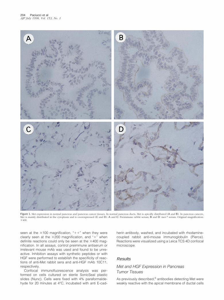
Figure 1. Met expression in normal pancreas and pancreas cancer tissues. In normal pancreas ducts, Met is apically distributed (A and B). In pancreas cancers,Met is mainly distributed in the cytoplasm and is overexpressed (C and D). A and C: Preimmune rabbit serum; B and D: met-7 serum. Original magnification:3400.
204 Paciucci et alAJP July 1998, Vol. 153, No. 1
in normal pancreas (Figure 1, A and B). In contrast,stronger reactivity with the cytoplasm of neoplastic cellsin 20 of 20 pancreas adenocarcinomas was observed(Figure 1, C and D), although there were slight differ-ences in the proportion and intensity of reactive cellsdepending on the antiserum used. Met expression wassimilar in primary and metastatic tumors.
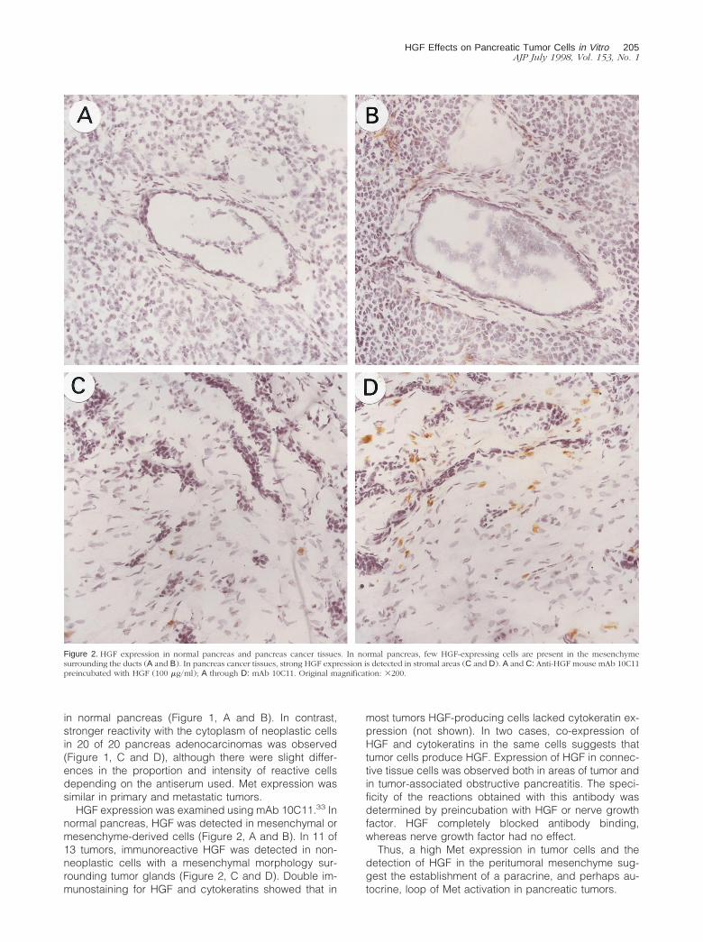
HGF expression was examined using mAb 10C11.33 Innormal pancreas, HGF was detected in mesenchymal ormesenchyme-derived cells (Figure 2, A and B). In 11 of13 tumors, immunoreactive HGF was detected in non-neoplastic cells with a mesenchymal morphology sur-rounding tumor glands (Figure 2, C and D). Double im-munostaining for HGF and cytokeratins showed that in
most tumors HGF-producing cells lacked cytokeratin ex-pression (not shown). In two cases, co-expression ofHGF and cytokeratins in the same cells suggests thattumor cells produce HGF. Expression of HGF in connec-tive tissue cells was observed both in areas of tumor andin tumor-associated obstructive pancreatitis. The speci-ficity of the reactions obtained with this antibody wasdetermined by preincubation with HGF or nerve growthfactor. HGF completely blocked antibody binding,whereas nerve growth factor had no effect.
Thus, a high Met expression in tumor cells and thedetection of HGF in the peritumoral mesenchyme sug-gest the establishment of a paracrine, and perhaps au-tocrine, loop of Met activation in pancreatic tumors.
Figure 2. HGF expression in normal pancreas and pancreas cancer tissues. In normal pancreas, few HGF-expressing cells are present in the mesenchymesurrounding the ducts (A and B). In pancreas cancer tissues, strong HGF expression is detected in stromal areas (C and D). A and C: Anti-HGF mouse mAb 10C11preincubated with HGF (100 mg/ml); A through D: mAb 10C11. Original magnification: 3200.
HGF Effects on Pancreatic Tumor Cells in Vitro 205AJP July 1998, Vol. 153, No. 1
Met and HGF Expression in Normal andNeoplastic Exocrine Pancreas Cultures
To study the effects of HGF, we used a series of well-characterized normal and neoplastic cells and examinedMet expression in vitro. When the normal exocrine fractionof the pancreas is cultured in vitro, NPCs rapidly loseacinar features and acquire phenotypic and functionalproperties characteristic of ductal cells.31 The pancreascancer cell lines selected for this study display a widespectrum of ductal differentiated properties: postconflu-ent IMIM-PC-2 and SK-PC-1 cells form a polarized mono-layer with domes and develop transmonolayer resis-tance; in contrast, IMIM-PC-1, SK-PC-3, and AsPC-1 cellsare not polarized and display a less differentiated phe-notype.32
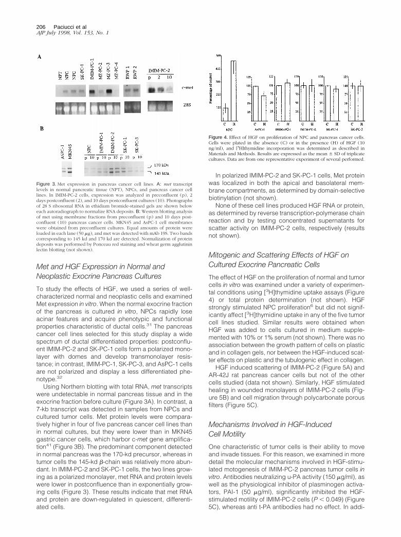
Using Northern blotting with total RNA, met transcriptswere undetectable in normal pancreas tissue and in theexocrine fraction before culture (Figure 3A). In contrast, a7-kb transcript was detected in samples from NPCs andcultured tumor cells. Met protein levels were compara-tively higher in four of five pancreas cancer cell lines thanin normal cultures, but they were lower than in MKN45gastric cancer cells, which harbor c-met gene amplifica-tion41 (Figure 3B). The predominant component detectedin normal pancreas was the 170-kd precursor, whereas intumor cells the 145-kd b-chain was relatively more abun-dant. In IMIM-PC-2 and SK-PC-1 cells, the two lines grow-ing as a polarized monolayer, met RNA and protein levelswere lower in postconfluence than in exponentially grow-ing cells (Figure 3). These results indicate that met RNAand protein are down-regulated in quiescent, differenti-ated cells.
In polarized IMIM-PC-2 and SK-PC-1 cells, Met proteinwas localized in both the apical and basolateral mem-brane compartments, as determined by domain-selectivebiotinylation (not shown).
None of these cell lines produced HGF RNA or protein,as determined by reverse transcription-polymerase chainreaction and by testing concentrated supernatants forscatter activity on IMIM-PC-2 cells, respectively (resultsnot shown).
Mitogenic and Scattering Effects of HGF onCultured Exocrine Pancreatic Cells
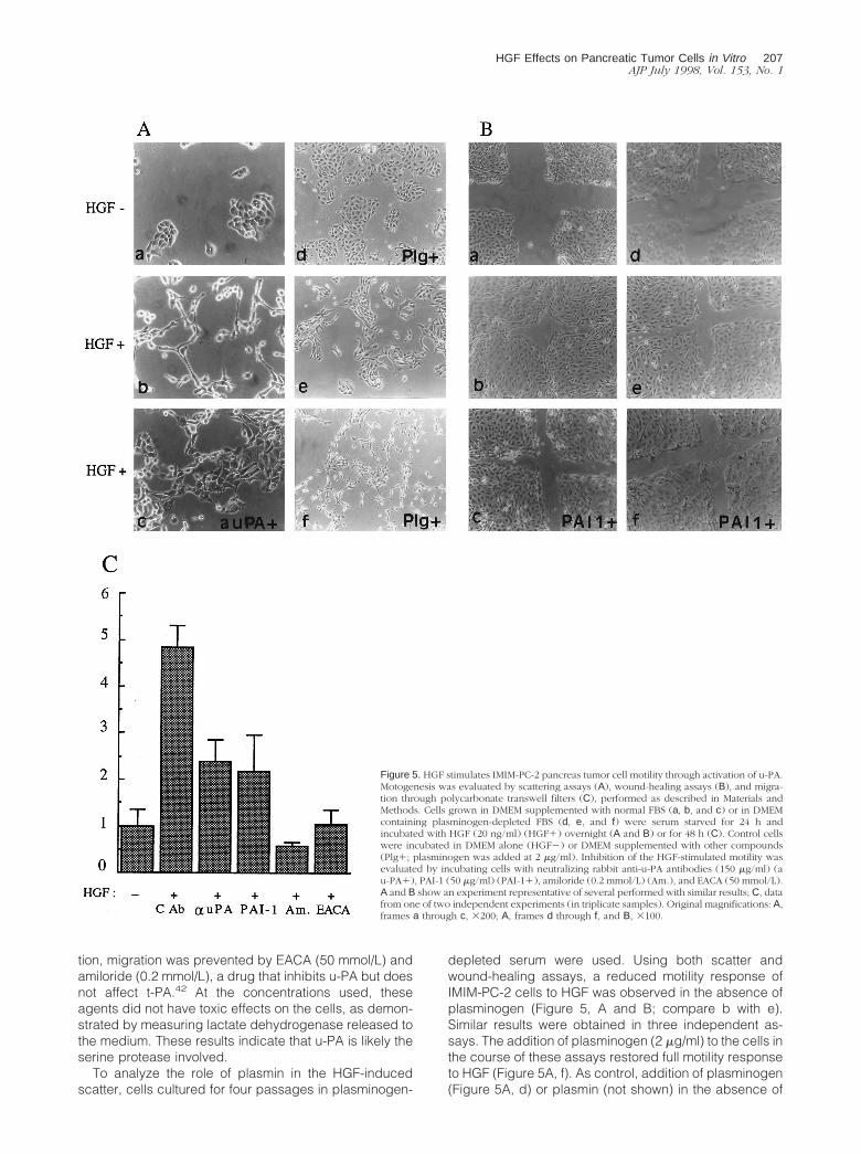
The effect of HGF on the proliferation of normal and tumorcells in vitro was examined under a variety of experimen-tal conditions using [3H]thymidine uptake assays (Figure4) or total protein determination (not shown). HGFstrongly stimulated NPC proliferation6 but did not signif-icantly affect [3H]thymidine uptake in any of the five tumorcell lines studied. Similar results were obtained whenHGF was added to cells cultured in medium supple-mented with 10% or 1% serum (not shown). There was noassociation between the growth pattern of cells on plasticand in collagen gels, nor between the HGF-induced scat-ter effects on plastic and the tubulogenic effect in collagen.
HGF induced scattering of IMIM-PC-2 (Figure 5A) andAR-42J rat pancreas cancer cells but not of the othercells studied (data not shown). Similarly, HGF stimulatedhealing in wounded monolayers of IMIM-PC-2 cells (Fig-ure 5B) and cell migration through polycarbonate porousfilters (Figure 5C).
Mechanisms Involved in HGF-InducedCell Motility
One characteristic of tumor cells is their ability to moveand invade tissues. For this reason, we examined in moredetail the molecular mechanisms involved in HGF-stimu-lated motogenesis of IMIM-PC-2 pancreas tumor cells invitro. Antibodies neutralizing u-PA activity (150 mg/ml), aswell as the physiological inhibitor of plasminogen activa-tors, PAI-1 (50 mg/ml), significantly inhibited the HGF-stimulated motility of IMIM-PC-2 cells (P , 0.049) (Figure5C), whereas anti t-PA antibodies had no effect. In addi-
Figure 3. Met expression in pancreas cancer cell lines. A: met transcriptlevels in normal pancreatic tissue (NPT), NPCs, and pancreas cancer celllines. In IMIM-PC-2 cells, expression was analyzed in preconfluent (p), 2days postconfluent (2), and 10 days postconfluent cultures (10). Photographsof 28 S ribosomal RNA in ethidium bromide-stained gels are shown beloweach autoradiograph to normalize RNA deposits. B: Western blotting analysisof met using membrane fractions from preconfluent (p) and 10 days post-confluent (10) pancreas cancer cells. MKN45 and AsPC-1 cell membraneswere obtained from preconfluent cultures. Equal amounts of protein wereloaded in each lane (50 mg), and met was detected with mAb 19S. Two bandscorresponding to 145 kd and 170 kd are detected. Normalization of proteindeposits was performed by Ponceau red staining and wheat germ agglutininlectin blotting (not shown).
Figure 4. Effect of HGF on proliferation of NPC and pancreas cancer cells.Cells were plated in the absence (C) or in the presence (H) of HGF (10ng/ml), and [3H]thymidine incorporation was determined as described inMaterials and Methods. Results are expressed as the mean 6 SD of triplicatecultures. Data are from one representative experiment of several performed.
206 Paciucci et alAJP July 1998, Vol. 153, No. 1
tion, migration was prevented by EACA (50 mmol/L) andamiloride (0.2 mmol/L), a drug that inhibits u-PA but doesnot affect t-PA.42 At the concentrations used, theseagents did not have toxic effects on the cells, as demon-strated by measuring lactate dehydrogenase released tothe medium. These results indicate that u-PA is likely theserine protease involved.
To analyze the role of plasmin in the HGF-inducedscatter, cells cultured for four passages in plasminogen-
depleted serum were used. Using both scatter andwound-healing assays, a reduced motility response ofIMIM-PC-2 cells to HGF was observed in the absence ofplasminogen (Figure 5, A and B; compare b with e).Similar results were obtained in three independent as-says. The addition of plasminogen (2 mg/ml) to the cells inthe course of these assays restored full motility responseto HGF (Figure 5A, f). As control, addition of plasminogen(Figure 5A, d) or plasmin (not shown) in the absence of
Figure 5. HGF stimulates IMIM-PC-2 pancreas tumor cell motility through activation of u-PA.Motogenesis was evaluated by scattering assays (A), wound-healing assays (B), and migra-tion through polycarbonate transwell filters (C), performed as described in Materials andMethods. Cells grown in DMEM supplemented with normal FBS (a, b, and c) or in DMEMcontaining plasminogen-depleted FBS (d, e, and f) were serum starved for 24 h andincubated with HGF (20 ng/ml) (HGF1) overnight (A and B) or for 48 h (C). Control cellswere incubated in DMEM alone (HGF2) or DMEM supplemented with other compounds(Plg1; plasminogen was added at 2 mg/ml). Inhibition of the HGF-stimulated motility wasevaluated by incubating cells with neutralizing rabbit anti-u-PA antibodies (150 mg/ml) (au-PA1), PAI-1 (50 mg/ml) (PAI-11), amiloride (0.2 mmol/L) (Am.), and EACA (50 mmol/L).A and B show an experiment representative of several performed with similar results; C, datafrom one of two independent experiments (in triplicate samples). Original magnifications: A,frames a through c, 3200; A, frames d through f, and B, 3100.
HGF Effects on Pancreatic Tumor Cells in Vitro 207AJP July 1998, Vol. 153, No. 1
HGF failed to induce motility, suggesting that the pro-tease by itself is not able to trigger motility in these cells.These results support that both active u-PA and plasminparticipate in the motogenic response to HGF.
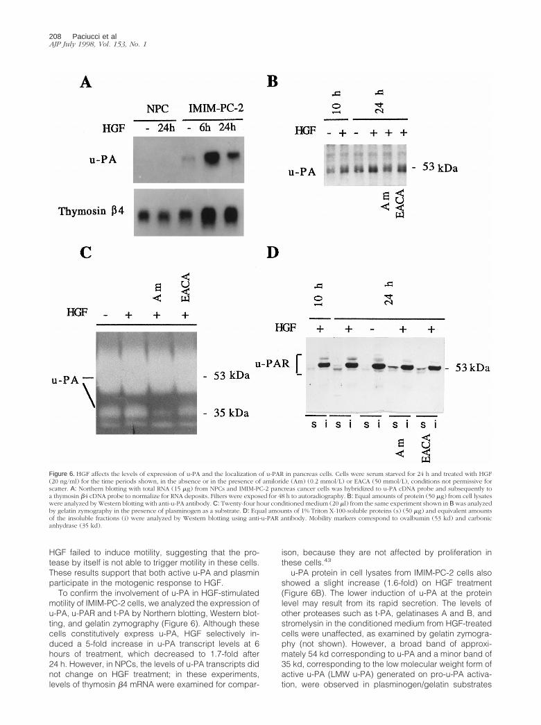
To confirm the involvement of u-PA in HGF-stimulatedmotility of IMIM-PC-2 cells, we analyzed the expression ofu-PA, u-PAR and t-PA by Northern blotting, Western blot-ting, and gelatin zymography (Figure 6). Although thesecells constitutively express u-PA, HGF selectively in-duced a 5-fold increase in u-PA transcript levels at 6hours of treatment, which decreased to 1.7-fold after24 h. However, in NPCs, the levels of u-PA transcripts didnot change on HGF treatment; in these experiments,levels of thymosin b4 mRNA were examined for compar-
ison, because they are not affected by proliferation inthese cells.43
u-PA protein in cell lysates from IMIM-PC-2 cells alsoshowed a slight increase (1.6-fold) on HGF treatment(Figure 6B). The lower induction of u-PA at the proteinlevel may result from its rapid secretion. The levels ofother proteases such as t-PA, gelatinases A and B, andstromelysin in the conditioned medium from HGF-treatedcells were unaffected, as examined by gelatin zymogra-phy (not shown). However, a broad band of approxi-mately 54 kd corresponding to u-PA and a minor band of35 kd, corresponding to the low molecular weight form ofactive u-PA (LMW u-PA) generated on pro-u-PA activa-tion, were observed in plasminogen/gelatin substrates
Figure 6. HGF affects the levels of expression of u-PA and the localization of u-PAR in pancreas cells. Cells were serum starved for 24 h and treated with HGF(20 ng/ml) for the time periods shown, in the absence or in the presence of amiloride (Am) (0.2 mmol/L) or EACA (50 mmol/L), conditions not permissive forscatter. A: Northern blotting with total RNA (15 mg) from NPCs and IMIM-PC-2 pancreas cancer cells was hybridized to u-PA cDNA probe and subsequently toa thymosin b4 cDNA probe to normalize for RNA deposits. Filters were exposed for 48 h to autoradiography. B: Equal amounts of protein (50 mg) from cell lysateswere analyzed by Western blotting with anti-u-PA antibody. C: Twenty-four hour conditioned medium (20 ml) from the same experiment shown in B was analyzedby gelatin zymography in the presence of plasminogen as a substrate. D: Equal amounts of 1% Triton X-100-soluble proteins (s) (50 mg) and equivalent amountsof the insoluble fractions (i) were analyzed by Western blotting using anti-u-PAR antibody. Mobility markers correspond to ovalbumin (53 kd) and carbonicanhydrase (35 kd).
208 Paciucci et alAJP July 1998, Vol. 153, No. 1
(Figure 6C). Cells treated with HGF and amiloride, con-ditions not permissive for scatter, showed a decrease inthe level of the LMW u-PA, suggesting that inhibition ofu-PA prevented plasmin generation and further proteo-lytic activation of pro-u-PA. However, in cells treated withHGF and EACA, the LMW u-PA is produced, althoughscatter is still inhibited. This was expected, becauseEACA acts by competing for plasminogen/plasmin-bind-ing sites at the cell membrane but does not inhibit plas-min proteolytic activation of soluble pro-u-PA. These re-sults indirectly suggest that it is the presence of activeu-PA and/or plasmin at the cell membrane that is impor-tant for the HGF-induced scattering.
Because u-PAR is essential to focus u-PA activity at thecell surface, we analyzed HGF effects on this protein.This receptor is anchored to the membrane through aglycosylphosphatidylinositol (GPI) bond, and it has beenlocalized in caveolae.44 Because GPI-linked proteins incaveolae distribute to the 1% Triton X-100-insolublephase,45 we used Triton X-100 fractionation to examinereceptor levels and distribution. Both in untreated and inHGF-treated IMIM-PC-2 cells, u-PAR was predominantlydetected in the insoluble fraction (Figure 6D). However,HGF induced an increase of u-PAR in the soluble phase,which was unaffected by the presence of amiloride orEACA. In this fraction, u-PAR was detected as a doublet:in the presence of plasminogen activator inhibitors, thelower mobility form was predominant; in contrast, in theabsence of inhibitors, the higher mobility form was moreabundant (Figure 6D), indicating that the inhibition ofu-PA activity affects the electrophoretic mobility of u-PAR.The two forms of u-PAR observed may correspond to theconformational receptor variants resulting from proteoly-sis already described.46,47
These results suggest that, in pancreatic tumor cells,an active u-PA/u-PAR complex is necessary, although notsufficient, to mediate the effects of HGF on cellular mo-tility.
The Scatter Effect Induced by HGF IsAssociated with Changes in Cell-Cell Contactsand Cell-Substrate Adhesion
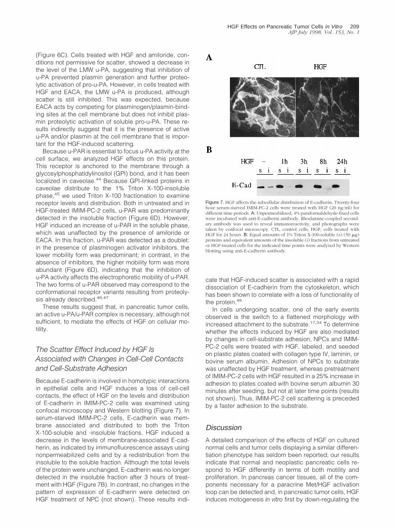
Because E-cadherin is involved in homotypic interactionsin epithelial cells and HGF induces a loss of cell-cellcontacts, the effect of HGF on the levels and distributionof E-cadherin in IMIM-PC-2 cells was examined usingconfocal microscopy and Western blotting (Figure 7). Inserum-starved IMIM-PC-2 cells, E-cadherin was mem-brane associated and distributed to both the TritonX-100-soluble and -insoluble fractions. HGF induced adecrease in the levels of membrane-associated E-cad-herin, as indicated by immunofluorescence assays usingnonpermeabilized cells and by a redistribution from theinsoluble to the soluble fraction. Although the total levelsof the protein were unchanged, E-cadherin was no longerdetected in the insoluble fraction after 3 hours of treat-ment with HGF (Figure 7B). In contrast, no changes in thepattern of expression of E-cadherin were detected onHGF treatment of NPC (not shown). These results indi-
cate that HGF-induced scatter is associated with a rapiddissociation of E-cadherin from the cytoskeleton, whichhas been shown to correlate with a loss of functionality ofthe protein.48
In cells undergoing scatter, one of the early eventsobserved is the switch to a flattened morphology withincreased attachment to the substrate.17,34 To determinewhether the effects induced by HGF are also mediatedby changes in cell-substrate adhesion, NPCs and IMIM-PC-2 cells were treated with HGF, labeled, and seededon plastic plates coated with collagen type IV, laminin, orbovine serum albumin. Adhesion of NPCs to substratewas unaffected by HGF treatment, whereas pretreatmentof IMIM-PC-2 cells with HGF resulted in a 25% increase inadhesion to plates coated with bovine serum albumin 30minutes after seeding, but not at later time points (resultsnot shown). Thus, IMIM-PC-2 cell scattering is precededby a faster adhesion to the substrate.
Discussion
A detailed comparison of the effects of HGF on culturednormal cells and tumor cells displaying a similar differen-tiation phenotype has seldom been reported; our resultsindicate that normal and neoplastic pancreatic cells re-spond to HGF differently in terms of both motility andproliferation. In pancreas cancer tissues, all of the com-ponents necessary for a paracrine Met/HGF activationloop can be detected and, in pancreatic tumor cells, HGFinduces motogenesis in vitro first by down-regulating the
Figure 7. HGF affects the subcellular distribution of E-cadherin. Twenty-fourhour serum-starved IMIM-PC-2 cells were treated with HGF (20 ng/ml) fordifferent time periods. A: Unpermeabilized, 4% paraformaldehyde-fixed cellswere incubated with anti-E-cadherin antibody. Rhodamine-coupled second-ary antibody was used to reveal immunoreactivity, and photographs weretaken by confocal microscopy. CTL, control cells; HGF, cells treated withHGF for 24 hours. B: Equal amounts of 1% Triton X-100-soluble (s) (50 mg)proteins and equivalent amounts of the insoluble (i) fractions from untreatedor HGF-treated cells for the indicated time points were analyzed by Westernblotting using anti-E-cadherin antibody.
HGF Effects on Pancreatic Tumor Cells in Vitro 209AJP July 1998, Vol. 153, No. 1
functional E-cadherin and later by activating the plasminproteolytic system.
In agreement with prior studies,26–28 we find that Met isoverexpressed in ductal-type adenocarcinomas of thepancreas; however, we also observe that its subcellulardistribution in normal ducts and in tumor cells is different.In the former, Met is mainly detected in the apical mem-brane using a panel of antibodies raised against threedifferent synthetic peptides of the Met sequence (thiswork and Ref. 6). Similarly, Tsarfaty et al. have reportedapical expression in the fetal gastrointestinal epitheliumin mice.49 In contrast, in pancreatic tumor tissues, Metprotein is mainly found in the cytoplasm of cancer cellsand, in polarized cultured tumor cells, it is present both inthe apical and basolateral membranes. A basolateraldistribution has been reported for Met in T84 and MDCKcells.50,51 The abnormal subcellular distribution of Met intumor with respect to normal pancreatic cells may in-crease the availability of mesenchyme-derived HGF totumor cells. Clearly, more work is necessary to establishthe mechanisms leading to the variable membrane dis-tribution of Met as well as the functional implicationsderived from it.
HGF was detected in mesenchymal cells of pancreascancer tissues and, in a few cases, in tumor cells as well,although not in cultured tumor cell lines. Recently, theproduction of HGF by some neoplastic epithelial cells hasbeen reported.52 Because HGF and c-met mRNA expres-sion can be regulated by inflammatory cytokines (ie,interleukin 1 and tumor necrosis factor),53,54 productionof HGF by fibroblasts, macrophages, or neoplastic cellsin the tumor may require complex molecular interactionsthat are lost on selective culture of pancreas cancer cells.
In vitro, HGF induced different effects on NPC andpancreas cancer cells; while the former respond withproliferation, the latter respond mainly with increasedmotility. The distinct mitogenic and motogenic responsesobserved in normal and neoplastic cells may be a reflec-tion of the constitutive K-ras activation resulting fromcodon 12 mutations present in the pancreas cancercells.32 This observation may support previous findingsindicating that when Ras is constitutively activated, fur-ther stimulation of mitogenic signal transduction path-ways is ineffective.19,55 In HPAF cells, a modest increaseof cell proliferation induced by HGF has been reported.27
We have shown that the full motogenic response ofIMIM-PC-2 cells to HGF is dependent on the activation ofthe u-PA/u-PAR system and on plasmin generation. Cellmigration was reduced in the absence of plasminogenand significantly decreased by specific inhibitors of u-PAproteolytic activity, including neutralizing anti-u-PA anti-bodies, PAI-1, and amiloride. In agreement with thesefindings, plasminogen-deficient mice show severe im-pairment of keratinocyte migration in wounded areas.56
Similarly, HGF-induced tubulogenesis of MDCK cells infibrin gels is reduced in the presence of inhibitors ofserine proteases, suggesting a role for plasminogen ac-tivators in the matrix remodeling required for tubulogen-esis.57 However, to our knowledge the present study isthe first description of the inhibition of the HGF-stimulatedcell scatter by inhibitors of urokinase activity. These ob-
servations suggest that protease activation is not onlyimportant for extracellular matrix degradation but also forthe degradation of other cellular components, eg, cellsurface adhesion molecules. Recently, the proteolyticdegradation of the ectodomain of membrane moleculeshas been implicated in the response to phorbol estersand growth factors.58
Relevant to the effect of HGF on motogenesis may alsobe the redistribution and cleavage of the u-PAR. HGFincreased the levels of u-PAR present in the Triton X-100-soluble fraction, whereas no major changes were ob-served in the insoluble phase enriched in GPI-linkedproteins. Caveolin and GPI-linked proteins are particu-larly abundant in caveolae, membrane invaginations im-plicated in potocytosis and transcytosis of macromole-cules,45 and u-PAR has been localized in caveolae.44
Thus, in IMIM-PC-2 cells u-PAR is in the fraction of pro-teins corresponding to caveolae, whereas, in HGF-stim-ulated cells, it also localizes in another compartment,soluble in Triton X-100. This situation is reminiscent of thereceptor for the protease tissue factor, which is active atthe cell surface in a compartment different from caveolae.Redistribution to caveolae is concomitant with the down-regulation of its function by formation of a complex withthe inhibitor.59 HGF treatment of IMIM-PC-2 cells resultedin a decrease in the electrophoretic mobility of the u-PAR,and this change was inhibited by amiloride and EACA,supporting the role of u-PA in the proteolytic processingof the receptor.60 Recently, it has been shown that onlythe proteolysed soluble form of u-PAR is active in signaltransduction during the chemotactic response.61 There-fore, we propose that the active u-PAR complexed tou-PA that participates in the motility response is repre-sented by the proteolysed Triton X-100-soluble form ofthe receptor.
In agreement with recent reports,62,63 the inhibition ofthe HGF-mediated IMIM-PC-2 cell motility by PAI-1 wouldpoint to a role for PAI-1 in blocking cell-substrate adhe-sion and migration mediated by u-PAR and/or integrincomplex. In those reports, it is proposed that u-PA re-leases this inhibition by physically sequestering PAI-1,thus uncovering vitronectin attachment sites that becomeavailable for interaction with integrin/u-PAR complex onthe cell membrane. Our data support those findings and,in addition, indicate that proteolytic activity of u-PA is alsodeterminant for cell movement, conceivably affecting theconformation of the u-PAR, which increases its affinity forthe substrate (vitronectin).63
HGF-induced scattering was also accompanied by adecrease in the Triton X-100-insoluble, cytoskeleton-as-sociated E-cadherin, which has been defined as thefunctional form of the protein. This novel finding would bein agreement with the observation that epithelial cells thatdisplay a disperse growth pattern do not express func-tional E-cadherin, unlike epithelial cells growing as com-pact colonies,36,64 and with more recent observationsshowing that E-cadherin-mediated cell-cell adhesion de-pends on Tiam and Rac signaling.65 Ectopic expressionof these proteins increases cell adhesion mediated byE-cadherin and blocks the HGF-induced scatter ofMDCK cells. Thus, HGF acts by reducing the functionality
210 Paciucci et alAJP July 1998, Vol. 153, No. 1
of E-cadherin, although expression levels are notchanged, as also described by other investiga-tors.50,64,66 The change in solubilization of E-cadherin isparalleled by a modest but rapid and transient increasein cell adhesion to the substrate. In addition, we observedthat the chronic treatment of IMIM-PC-2 cells with HGF isaccompanied by a flattened, scattered morphology andincreased cell-substrate adhesion (unpublished data).These observations are common to other scatter-induc-ing factors: scattering induced by TPA on HT-29 M6 cellsis accompanied by a decreased homotypic aggregation,enhanced attachment to the substrate, and changes inthe functionality of E-cadherin.34 In contrast to the effectsobserved in pancreas tumor cells, HGF stimulates mito-genesis but does not affect cell motility, E-cadherin func-tionality, and the induction of u-PA in NPCs. These ob-servations support the notion that proliferative andmotogenic signals go through different pathways that, asmentioned above, may be mutually exclusive.
The redistribution of E-cadherin to a nonfunctionalcompartment, the activation of the u-PA proteolytic sys-tem,21,67 and the increased expression of u-PA and u-PAR in invasive cells15 are mechanisms through whichHGF is able to confer cells with properties that lead totumor progression and invasion.
Acknowledgments
We thank M. Garrido and M. C. Torns for excellent tech-nical assistance; the investigators mentioned in the textfor providing reagents; and D. Andreu, A. Garcıa deHerreros, A. Skoudy, and T. Thomson for valuable con-tributions.
References
1. Weidner KM, Behrens J, Vandekerckhove J, Birchmeier W: Scatterfactor: molecular characteristics and effect on the invasiveness. J CellBiol 1990, 111:2097–2108
2. Gherardi E, Stoker M: Hepatocytes and scatter factor. Nature 1991,346:228
3. Weidner KM, Arakaki N, Hartmann G, Vandekerckhove J, Weingart S,Rieder H, Fonatsch C, Tsubouchi H, Hishida T, Daikuhara Y, Birch-meier W: Evidence for the identity of human scatter factor and humanhepatocyte growth factor. Proc Natl Acad Sci USA 1991, 88:7002–7005
4. Rosen EM, Nigam SK, Goldberg ID: Scatter factor and the c-metreceptor: a paradigm for mesenchymal/epithelial interaction. J CellBiol 1994, 127:1783–1787
5. Zarnegar R, Michalopoulos GK: The many faces of hepatocyte growthfactor: from hepatopoiesis to hematopoiesis. J Cell Biol 1995, 129:1177–1180
6. Vila MR, Nakamura T, Real FX: Hepatocyte growth factor is a potentmitogen for normal human pancreas cells in vitro. Lab Invest 1995,73:409–418
7. Montesano R, Matsumoto K, Nakamura T, Orci L: Identification of afibroblast-derived epithelial morphogen as hepatocyte growth factor.Cell 1991, 67:901–908
8. Cooper CS: The met oncogene: from detection by transfection totransmembrane receptor for hepatocyte growth factor. Oncogene1992, 7:3–7
9. Sonnenberg E, Meyer D, Weidner KM, Birchmeier C: Scatter factor/hepatocyte growth factor and its receptor, the c-met tyrosine kinase,
can mediate a signal exchange between mesenchyme and epitheliaduring mouse development. J Cell Biol 1993, 123:223–235
10. Schmidt C, Bladt F, Goedecke S, Brinkmann V, Zschlesche W,Sharpe M, Gherardi E, Birchmeier C: Scatter factor/hepatocytegrowth factor is essential for liver development. Nature 1995, 373:699–702
11. Uehara Y, Minowa O, Mori C, Shiota K, Kuno J, Noda T, Kitamura N:Placental defect and embryonic lethality in mice lacking hepatocytegrowth factor/scatter factor. Nature 1995, 373:702–705
12. Rong S, Jeffers M, Resau JH, Tsarfaty I, Oskarsson M, Vande WoudeGF: Met expression and sarcoma tumorigenicity. Cancer Res 1993,53:5355–5360
13. Ferracini R, Di Renzo MF, Scotland K, Baldini N, Olivero M, Lollini PL,Cremona O, Campanacci M, Comoglio PM: The Met/HGF receptor isover-expressed in human osteosarcomas and is activated by either aparacrine or an autocrine circuit. Oncogene 1995, 10:739–749
14. Rong S, Segal S, Anver M, Resau JH, Vande Woude GF: Invasivenessand metastasis of NIH 3T3 cells induced by Met-hepatocyte growthfactor/scatter factor autocrine stimulation. Proc Natl Acad Sci USA1994, 91:4731–4735
15. Jeffers M, Rong S, Vande Woude GF: Enhanced tumorigenicity andinvasion-metastasis by hepatocyte growth factor/scatter factor-metsignalling in human cells concomitant with induction of the urokinaseproteolysis network. Mol Cell Biol 1996, 16:1115–1125
16. Joseph A, Weiss GH, Jin L, Fuchs A, Chowdhury S, O’ShaughnessyP, Goldberg ID, Rosen EM: Expression of scatter factor in humanbladder carcinoma. J Natl Cancer Inst 1995, 87:372–376
17. Ridley AJ, Comoglio PM, Hall A: Regulation of scatter factor/hepato-cyte growth factor responses by Ras, Rac, and Rho in MDCK cells.J Cell Biol 1995, 15:1110–1122
18. Royal I, Park M: Hepatocyte growth factor-induced scatter of Madin-Darby canine kidney cells requires phosphatidylinositol 3-kinase.J Biol Chem 1995, 270:27780–27787
19. Ponzetto C, Zhen Z, Audero E, Maina F, Bardelli A, Basile ML,Giordano S, Narsimhan R, Comoglio P: Specific uncoupling of GRB2from the Met receptor. J Biol Chem 1996, 271:14119–14123
20. Boccaccio C, Ando M, Tamagnone L, Bardelli A, Michieli P, BattistiniC, Comoglio PM: Induction of epithelial tubules by growth factor HGFdepends on the STAT pathway. Nature 1998, 391:285–288
21. Pepper MS, Matsumoto K, Nakamura T, Orci L, Montesano R: Hepa-tocyte growth factor increases urokinase-type plasminogen activator(u-PA) and u-PA receptor expression in Madin-Darby canine kidneyepithelial cells. J Biol Chem 1992, 267:20493–20496
22. Boccaccio C, Gaudino G, Gambarotta G, Galimi F, Comoglio PM:Hepatocyte growth factor (HGF) receptor expression is inducible andis part of the delayed-early response to HGF. J Biol Chem 1994,269:12846–12851
23. Naldini L, Tamagnone L, Vigna E, Sachs M, Hartmann G, BirchmeierW, Daikuhara Y, Tsubouchi H, Blasi F, Comoglio PM: Extracellularproteolytic cleavage by urokinase is required for activation of hepa-tocyte growth factor/scatter factor. EMBO J 1992, 11:4825–4833
24. Mars WM, Zarnegar R, Michalopoulos GK: Activation of hepatocytegrowth factor by the plasminogen activators uPA and tPA. Am JPathol 1993, 143:949–958
25. Kern HF, Elsasser H-P: Fine structure of human pancreatic adeno-carcinoma. The Pancreas: Biology, Pathobiology, and Disease. Ed-ited by VLW Go, EP DiMagno, JD Gardner, E Lebenthal, HA Reber,GA Scheele. New York, Raven Press, 1993, pp 857–869
26. Ebert M, Yokoyama M, Friess H, Buchler MW, Korc M: Coexpressionof the c-met proto-oncogene and hepatocyte growth factor in humanpancreatic cancer. Cancer Res 1994, 54:5775–5778
27. Di Renzo MF, Poulsom R, Olivero M, Comoglio PM, Lemoine NR:Expression of the Met/hepatocyte growth factor receptor in humanpancreatic cancer. Cancer Res 1995, 55:1129–1138
28. Furukawa T, Duguid WP, Kobari M, Matsuno S, Tsao M-T: Hepatocytegrowth factor and Met receptor expression in human pancreaticcarcinogenesis. Am J Pathol 1995, 147:889–895
29. Dano K, Andreasen PA, Grondahl-Hansen J, Kristensen P, NielsenLS, Skriver L: Plasminogen activators, tissue degradation and cancer.Adv Cancer Res 1985, 44:139–266
30. Kwaan HC: The plasminogen-plasmin system in malignancy. CancerMetastasis Rev 1992, 11:291–311
31. Vila MR, Lloreta J, Real FX: Normal human pancreas cultures displayfunctional ductal characteristics. Lab Invest 1994, 71:423–431
HGF Effects on Pancreatic Tumor Cells in Vitro 211AJP July 1998, Vol. 153, No. 1
32. Vila MR, Lloreta J, Schussler MH, Berrozpe G, Welt S, Real FX: Newpancreas cancer cell lines which represent distinct stages of ductaldifferentiation. Lab Invest 1995, 72:395–404
33. Bhargava M, Joseph A, Knesel J, Halaban R, Li Y, Pang S, GoldbergI, Setter E, Donovan MA, Zarnegar R, Michalopoulos GA, NakamuraT, Faletto D, Rosen EM: Scatter factor and hepatocyte growth factor:activities, properties, and mechanisms. Cell Growth Differ 1992,3:11–20
34. Fabre M, Garcıa de Herreros A: Phorbol ester-induced scattering ofHT-29 human intestinal cancer cells is associated with down-modu-lation of E-cadherin. J Cell Sci 1993, 106:513–522
35. Hiraki Y, Rosen OM, Birnbaum MJ: Growth factors rapidly induceexpression of the glucose transporter gene. J Biol Chem 1988, 263:13655–13662
36. Skoudy A, Llosas MM, Garcıa de Herreros A: Intestinal HT-29 cellswith dysfunction of E-cadherin show increased pp60src activity, andtyrosine phosphorylation of p120-catenin. Biochem J 1996, 317:279–284
37. Heussen C, Dowdle EB: Electrophoretic analysis of plasminogenactivators in polyacrylamide gels containing sodium dodecyl sulfateand copolymerized substrate. Anal Biochem 1990, 102:196–202
38. Lisanti MP, Le Bivic A, Saltiel AR, Rodrıguez-Boulan E: Preferredapical distribution of glycosyl phosphatidylinositol (GPI)-anchoredproteins: a highly conserved feature of the polarized epithelial cellphenotype. J Membr Biol 1990, 113:155–167
39. Chomczynski P, Sacchi N: Single-step method for RNA isolation byacid guanidinium thiocyanate-phenol-chloroform extraction. Anal Bio-chem 1987, 162:156–159
40. Feinberg AP, Volgelstein B: A technique for radiolabeling DNA re-striction endonuclease fragments to high specific activity. Anal Bio-chem 1983, 132:6–13
41. Giordano S, Ponzetto C, Di Renzo MF, Cooper CS, Comoglio PM:Tyrosine kinase receptor indistinguishable from the c-Met protein.Nature 1989, 339:155–156
42. Vassalli JD, Belin D: Amiloride selectively inhibits the urokinase-typeplasminogen activator. FEBS Lett 1987, 214:187–191
43. Paciucci R, Berrozpe G, Tora M, Navarro E, Garcıa de Herreros A,Real FX: Isolation of tissue-type plasminogen activator, cathepsin H,and non-specific cross-reacting antigen from SK-PC-1 pancreas can-cer cells using subtractive hybridization. FEBS Lett 1996, 385:72–76
44. Stahl A, Mueller BM: The urokinase-type plasminogen activator re-ceptor, a GPI-linked protein, is localized in caveolae. J Cell Biol 1995,129:335–344
45. Lisanti MP, Scherer PE, Vidgiriene J, Tang ZL, Hermanowski-VosatkaA, Tu Y-H, Cook RF, Sargiacomo M: Characterization of caveolin-richmembrane domains isolated from an endothelial-rich source: impli-cations for human disease. J Cell Biol 1994, 126:111–126
46. Behrendt N, Ronne E, Ploug M, Petri T, Løbert D, Nielsen LS, Schleun-ing W-D, Blasi F, Apella E, Dano K: The human receptor for urokinaseplasminogen activator: NH2-terminal amino acid sequence and gly-cosylation variants. J Biol Chem 1990, 265:6453–6460
47. Ploug M, Ellis V, Dano K: Ligand interaction between urokinase-typeplasminogen activator and its receptor probed with 8-anilino-1-naphthalenesulfonate: evidence for a hydrophobic binding site ex-posed on the intact receptor. Biochemistry 1994, 33:8991–8997
48. Hinck L, Nelson WJ, Papkoff J: Wnt-1 modulates cell-cell adhesion inmammalian cells by stabilizing b-catenin binding to the cell adhesionprotein cadherin. J Cell Biol 1994, 124:729–741
49. Tsarfaty I, Resau JH, Rulong S, Keydar I, Faletto DL, Vande WoudeGF: The met proto-oncogene receptor, and lumen formation. Science1992, 257:1258–1261
50. Nusrat A, Parkos CA, Bacarra AE, Godowski PJ, Delp-Archer C,Rosen EM, Madara JL: Hepatocyte growth factor/scatter factor ef-fects on epithelia: regulation of intercellular junctions in transformed
and nontransformed cell lines, basolateral polarization of c-met intransformed and natural intestinal epithelia, and induction of rapidwound repair in a transformed model epithelium. J Clin Invest 1994,93:2056–2065
51. Crepaldi T, Pollack AL, Prat M, Zborek A, Mostov K, Comoglio PM:Targeting of the SF/HGF receptor to the basolateral domain of polar-ized epithelial cells. J Cell Biol 1994, 125:313–320
52. Jin L, Fuchs A, Schnitt SJ, Yao Y, Joseph A, Lamszus K, Park M,Goldberg ID, Rosen EM: Expression of scatter and c-met receptor inbenign and malignant breast tissue. Cancer 1996, 79:749–760
53. Tamura M, Arakaki N, Tsubouchi H, Takada H, Daikuhara Y: En-hancement of human hepatocyte growth factor production by inter-leukin-1a and 1b and tumor necrosis factor-a by fibroblasts in culture.J Biol Chem 1993, 268:8140–8145
54. Moghul A, Lin L, Beedle A, Kanbour-Shakir A, DeFrances MC, Liu Y,Zarnegar R: Modulation of c-met proto-oncogene (HGF receptor)mRNA abundance by cytokines and hormones: evidence for rapiddecay of the 8 kb c-met transcript. Oncogene 1994, 9:2045–2052
55. Thomson TM, Green SH, Trotta RJ, Burstein DE, Pellicer A: OncogeneN-ras mediates selective inhibition of c-fos induction by nerve growthfactor, and basic fibroblast growth factor in a PC12 cell line. Mol CellBiol 1990, 10:1556–1563
56. Romer J, Bugge TH, Pyke C, Lund LR, Flick MJ, Degen JL, Dano K:Impaired wound healing in mice with a disrupted plasminogen gene.Nat Med 1996, 2:287–292
57. Montesano R, Orci L: Tumor-promoting phorbol esters induce angio-genesis in vitro. Cell 1985, 42:469–477
58. Arribas J, Coodly L, Vollmer P, Kishimoto TK, Rose-John S, MassagueJ: Diverse cell surface protein ectodomains are shed by a systemsensitive to metalloprotease inhibitors. J Biol Chem 1996, 271:11376–11382
59. Sevinsky JR, Rao LVM, Ruf W: Ligand-induced protease receptortranslocation into caveolae: a mechanism for regulating cell surfaceproteolysis of the tissue factor-dependent coagulation pathway.J Cell Biol 1996, 133:293–304
60. Hoyer-Hansen G, Rone E, Solberg H, Behrendt N, Ploug M, Lund LR,Ellis V, Dano K: Urokinase plasminogen activator cleaves its cellsurface receptor releasing the ligand-binding domain. J Biol Chem1992, 267:18224–18229
61. Resnati M, Guttinger M, Valcamonica F, Sidenius N, Blasi F, Fazioli F:Proteolytic cleavage of the urokinase receptor substitutes for theagonist-induced chemotactic effect. EMBO J 1996, 15:1572–1582
62. Stefansson S, Lawrence DA: The serpin PAI-1 inhibits cell migrationby blocking integrin aVb3 binding to vitronectin. Nature 1996, 383:441–443
63. Deng G, Curriden SA, Wang S, Rosenberg S, Loskutoff DJ: Is plas-minogen activator inhibitor-1 the molecular switch that governs uroki-nase receptor-mediated cell adhesion and release? J Cell Biol 1996,134:1563–1571
64. Birchmeier W, Weidner KM, Behrens J: Molecular mechanisms lead-ing to loss of differentiation and gain of invasiveness in epithelial cells.J Cell Sci 1993, 17:159–164
65. Hordijk P, Ten Klooster JP, Van der Kammen RA, Michiels F, OomenL, Collar JG: Inhibition of invasion of epithelial cells by Tiam 1-Racsignaling. Science 1997, 278:1464–1466
66. Shibamoto S, Hayakawa M, Takeuchi K, Hori T, Oku N, Miyazawa K,Kitamura N, Takeichi M, Ito F: Tyrosine phosphorylation of b-cateninand plakoglobin enhanced by hepatocyte growth factor and epider-mal growth factor in human carcinoma cells. Cell Adhes Commun1994, 1:295–305
67. Paciucci R, Tora M, Dıaz VM, Real FX: The plasminogen activatorsystem in pancreas cancer: role of t-PA in the invasive potential invitro. Oncogene 1998, 16:625–634
212 Paciucci et alAJP July 1998, Vol. 153, No. 1
Related Documents


![Arecombinantchimeric plasminogenactivatorwithhighaffinity for … › content › pnas › 88 › 22 › 10337.full.pdf · urokinase-type plasminogen activator [scuPA(32kDa)], afi-brin-selective](https://static.cupdf.com/doc/110x72/5f1cd2e4e4e08d6801761b19/arecombinantchimeric-plasminogenactivatorwithhighaffinity-for-a-content-a-pnas.jpg)