LIQUID NITROGEN CRYOTHERAPY IN MANAGEMENT OF ORAL LESIONS A Dissertation submitted in partial fulfillment of the requirements for the degree of MASTER OF DENTAL SURGERY BRANCH – III ORAL AND MAXILLOFACIAL SURGERY THE TAMILNADU DR.M.G.R MEDICAL UNIERSITY CHENNAI -600032 2016 - 2019

Welcome message from author
This document is posted to help you gain knowledge. Please leave a comment to let me know what you think about it! Share it to your friends and learn new things together.
Transcript
LIQUID NITROGEN CRYOTHERAPY IN
MANAGEMENT OF ORAL LESIONS
A Dissertation submitted
in partial fulfillment of the requirements
for the degree of
MASTER OF DENTAL SURGERY
BRANCH – III
ORAL AND MAXILLOFACIAL SURGERY
THE TAMILNADU DR.M.G.R MEDICAL UNIERSITY
CHENNAI -600032
2016 - 2019
ADHIPARASAKTHI DENTAL COLLEGE & HOSPITAL
MELMARUVATHUR- 603319
DEPARTMENT OF ORAL &MAXILLOFACIAL SURGERY
CERTIFICATE
This is to certify that Dr.S.DURAIMURUGAN , Post graduate student (2016-
2019) in the Department of Oral & Maxillofacial Surgery (Branch III),
Adhiparasakthi Dental College and Hospital , Melmaruvathur – 603319, has
done this dissertation titled “LIQUID NITROGEN CRYOTHERAPY IN
MANAGEMENT OF ORAL LESIONS” under our direct guidance and
supervision in partial fulfilment of the regulations laid down by The
Tamilnadu Dr. M.G.R Medical University, Chennai – 600032, for MDS.,
(Branch III) Oral & Maxillofacial Surgery degree examination .
Co-guide: Guide:
Dr.G. SURESH KUMAR. M.D.S.,
Reader,
DR.D.DURAIRAJ.M.D.S.,
Professor & HOD,
Dr. S. THILLAINAYAGAM, M.D.S.
Principal,
ACKNOWLEDGEMENT
I thank ALMIGHTY GOD for answering my prayers and making me
what I am today.
Iam extremely indebted to Dr. T.Ramesh , M.D . , Correspondent
,Adhiparasakthi Dental College and Hospital , Melmaruvathur , for providing
infrastructure & Resources to perform the main dissertation .
My sincere thanks to Dr.S. Thillainayagam M.D.S . ,our beloved
Principal, Adhiparasakthi Dental College and Hospital , Melmaruvathur for
providing me with the opportunity to utilize the facilities of the college.
I would like to express my heartfelt thanks to my revered teacher
Dr.GokkulaKrishnan.S , for his guidance and encouragement during my
study. His encouragement was of great support in facing challenges that
occurred during my study.
I avail this opportunity to express m y gratitude and reverence to my
Guide &beloved teacherDr.D.Durairaj MDS . , Professor and Head,
Department of Oral & Maxillofacial Surgery , Adhiparasakthi Dental College
and Hospital, Melmaruvathur. His pursuit for perfection and immense support
were a source of constant inspiration to me and without which such an
endeavour would never have materialized.
At the very outset, I would like to express my sincere gratitude to my
Teacher Dr.M.Karthikeyan , Professor, Department of Oral & Maxillofacial
Surgery for al l the encouragement, motivation and valuable suggestions that
he offered in helping me to complete my course without any hurdles. Without
his support it would not have been possible to reach my goals.
It is my duty to express my thanks to my Co -Guide Dr. G.Suresh
Kumar MDS . , Reader, for his expert guidance and moral support dur ing the
completion of this study. I consider myself privileged, to have studied,
worked and completed my dissertation under him in the department.
I am extremely thankful to my teachers Dr.James Antony Bagat
M.D.S Reader Dr.Abishek R.Balaji , Dr.Rajprakash M.D.S Senior lecturer,
Dr.P.Srinivasulu M.D.S., Senior lecturer, Dr.Nathiya M.D.S., Senior
lecturer, for their constant support.
I thank Mr. Maveeran Librarian and library staff Mr.Selvakumar,
AdhiParasakthi Dental College and Hospital Melmaruvathur for favours
rendered.
I also wish to thank my co-pg Late Dr. Barathvikraman and my seniors
Dr.VinodKrishna, Dr.Mahalakshmi, Dr.N.PrithiviShankar,
Dr.R.Muralidharan and my juniors Dr. R.Chinnaiah, Dr.M.Veeramuthu,
Dr.S.Mariam, Dr. Naneshwari.
I thank Mrs.Mahalakshmi staff nurse, Miss Soundriya, Mrs .kanaga
non teaching staff Department of Oral & Maxillofacial surgery
AdhiParasakthi Dental College and Hospital Melmaruvathur for favours
rendered.
A special mention of thanks to all my patients for their consent, co-
operation and participation in this study.
I owe my gratitude to my parents T.Sivanantham & Mrs.S.Sumathi my
brother S.Pravinkumar and all my family members who stood beside me
during my tough times and sacrificed so much to make me what I am today.
.
Dr. S.DURAIMURUGAN
Post graduate student
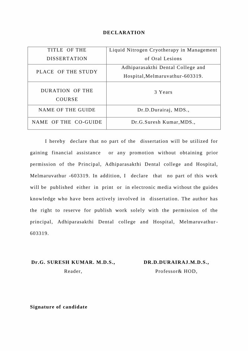
DECLARATION
TITLE OF THE
DISSERTATION
Liquid Nitrogen Cryotherapy in Management
of Oral Lesions
PLACE OF THE STUDY Adhiparasakthi Dental College and
Hospital,Melmaruvathur-603319.
DURATION OF THE
COURSE
3 Years
NAME OF THE GUIDE Dr.D.Durairaj , MDS.,
NAME OF THE CO-GUIDE Dr.G.Suresh Kumar,MDS.,
I hereby declare that no part of the dissertation will be utilized for
gaining financial assistance or any promotion without obtaining prior
permission of the Principal, Adhiparasakthi Dental college and Hospital ,
Melmaruvathur -603319. In addition, I declare that no part of this work
will be published ei ther in print or in electronic media wi thout the guides
knowledge who have been actively involved in dissertation. The author has
the right to reserve for publish work solely with the permission of the
principal, Adhiparasakthi Dental college and Hospital , Melmaruvathur -
603319.
Dr.G. SURESH KUMAR. M.D.S.,
Reader,
DR.D.DURAIRAJ.M.D.S.,
Professor& HOD,
Signature of candidate
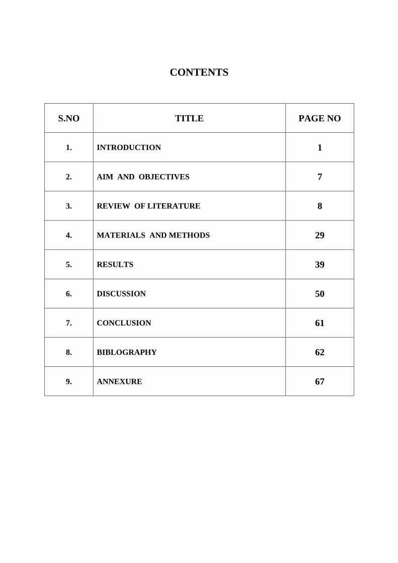
CONTENTS
S.NO TITLE PAGE NO
1. INTRODUCTION 1
2. AIM AND OBJECTIVES 7
3. REVIEW OF LITERATURE 8
4. MATERIALS AND METHODS 29
5. RESULTS 39
6. DISCUSSION 50
7. CONCLUSION 61
8. BIBLOGRAPHY 62
9. ANNEXURE 67
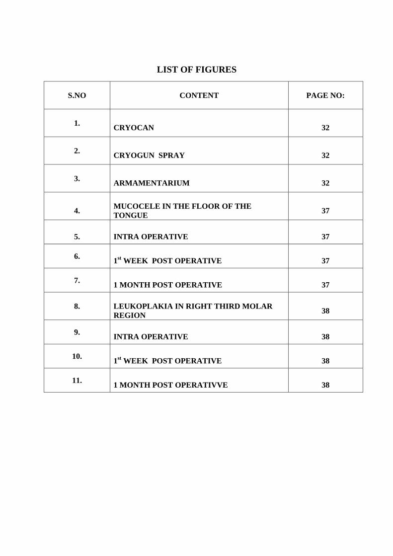
LIST OF FIGURES
S.NO
CONTENT
PAGE NO:
1.
CRYOCAN
32
2.
CRYOGUN SPRAY
32
3.
ARMAMENTARIUM
32
4.
MUCOCELE IN THE FLOOR OF THE
TONGUE
37
5.
INTRA OPERATIVE
37
6.
1st WEEK POST OPERATIVE
37
7.
1 MONTH POST OPERATIVE
37
8.
LEUKOPLAKIA IN RIGHT THIRD MOLAR
REGION
38
9.
INTRA OPERATIVE
38
10.
1st WEEK POST OPERATIVE
38
11.
1 MONTH POST OPERATIVVE
38
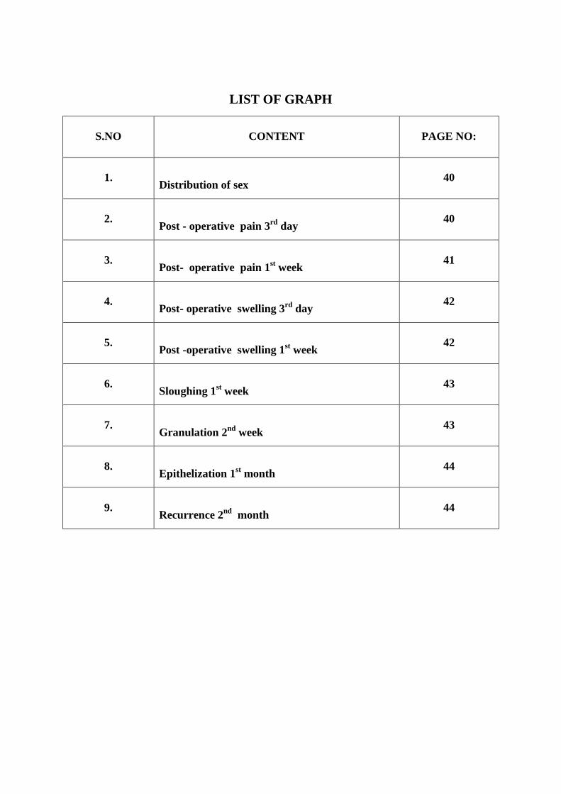
LIST OF GRAPH
S.NO
CONTENT
PAGE NO:
1.
Distribution of sex
40
2.
Post - operative pain 3rd
day
40
3.
Post- operative pain 1st week
41
4.
Post- operative swelling 3rd
day
42
5.
Post -operative swelling 1st week
42
6.
Sloughing 1st week
43
7.
Granulation 2nd
week
43
8.
Epithelization 1st month
44
9.
Recurrence 2nd
month
44
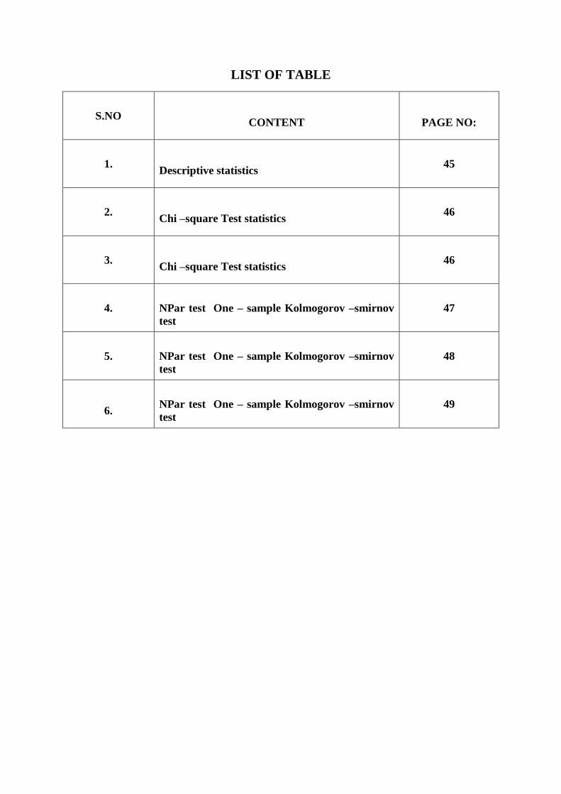
LIST OF TABLE
S.NO
CONTENT
PAGE NO:
1.
Descriptive statistics 45
2.
Chi –square Test statistics 46
3.
Chi –square Test statistics 46
4.
NPar test One – sample Kolmogorov –smirnov
test
47
5.
NPar test One – sample Kolmogorov –smirnov
test
48
6.
NPar test One – sample Kolmogorov –smirnov
test
49
Introduction
1
INTRODUCTION
Cryotherapy is derived from the Greek word “kryos”, meaning
frost hence cryosurgery is local destruction of tissue by freezing.
Cryotherapy has long been noted as a good technique that, when
used correctly, can reduce pain and swelling and destroy lesions
with litt le scarring. Local application of low temperature was first
used by Egyptians for pain relief, then during Franco -Prussian war
for amputated limbs. Hippocrates recommended the use of cold to
reduce swelling, hemorrhage and pain, while John Hunter in 1777
stated that “the local tissue response to freezing includes local
tissue necrosis, vascular stasis and excellent healing.” James Arnott
(1851) was the first to report and demonstrate this freezing therapy
by using a mixture of salt and ice in malignant breast neoplasm. In
1899, White was the first person to use extremely cold refrigerants
for medical conditions. He used liquefied a ir to treat warts and
other dermatologic conditions.(1 )
In 1908 A.W. Pusey used the term “Cryotherapy” to describe
the treatment of skin lesions with very low temperatures. Currently
Cryotherapy treatments invoved by lowering the body surface
temperature without tissue destruction, whereas in cryosurgery
diseased tissues are destroyed through freezing. The world's first
cryogenic temperature chamber was set up in Japan, in 1978 by
Yamauchi and his team.(1 ,2 )
Introduction
2
Contemporary cryogenics has been developed at the end of
the 19th century , l iquefaction of oxygen, nitrogen, carbon dioxide
and hydrogen, and also the industrial production and storage of
liquid coolants, enabled the development of cryobiology, used
extremely at low temperature. Virtually all bio logical tissues
subjected to a temperature of −20°C or below for a minute or more
undergo cryogenic coagulation or necrosis.(3 )
Principles of cryotherapy - Technique of cryotherapy stresses
rapid cooling, slow thawing and repetition of the freezing process
to maximize tissue destruction. The two methods recognized are a
closed system with use of probes and nitrous oxide, or an open
system with use of a liquid nitrogen spray or a cotton tip. Spray
techniques are useful in widespread dermatologi cal lesions, small
skin cancers and intra-bony cavities after curettage to prevent
recurrence. The nitrous oxide technique is useful for treatment of
various benign and malignant lesions of the oral cavity where more
predictable necrosis is necessary and depth of necrosis is also a
factor. Current protocols suggest that for most benign mucosal
lesions 1–2 minute freeze/thaw cycle using a cryoprobe is
sufficient. Premalignant/malignant lesions are recommended to
undergo three freeze/thaw for cycles for 2 mins. For smaller
lesions, shorter freeze (20–30 seconds) are adequate. In cases
where hyperplastic tissue exists, freezing of the mass and then
Introduction
3
removing the bulk of tissue, followed by further freezing of the
tissue base results in higher success rates.
Mechanism of Tissue damage by cryotherapy involves
several mechanisms. It has been determined that most t issues freeze
at -2.2ºC and that the temperature must fall below -20ºC for cell
death to occur. The treatment of more aggressive cancers in th e oral
cavity may require repetitive freeze cycles at temperatures of at
least -50ºC or more for tissue necrosis to occur. During the freeze
cycle as the temperature drops, it is believed that extracellular
water undergoes crystallization. In addition, mem brane lipids
harden take place at low temperatures decreasing cell resistance to
shrinkage. Extracellular stores of water diminish, as the electrolyte
concentration increases. In order to counteract this concentration
gradient, intracellular water moves o ut of the cell, and this water
becomes involved in the crystallization proces s. As intracellular ice
formed, remains trapped within the cellular membrane. As a result
of these processes, intracellular electrolytes reach toxic levels,
which become lethal to the cell . During a slow thaw cycle, cells at
the periphery of the cryolesion will take up excess electrolytes. To
equalize this gradient, water enters the cell and lead to swelling
and lysis. Further re -crystallization may contribute to cellular
damage, however, this phenomenon may be avoided if cells are
thawed rapidly.(2 )
Introduction
4
For the treatment of oral lesions, it is extremely easy to
attack oral lesions repeatedly with the cryoprobe, with only little
preliminary preparation of either patient or operative field. Tissues
close to the probe freeze quickly, but ice is an efficient insulator, so
advance freezing proceeds only slowly. As ice delays the spread of
freezing, it reduces the chance of accidental damage to the
underlying tissues. Because of the gradi ent of heat loss,
neighbouring tissues are unharmed. In cryosurgery procedure ,
nothing is excised rather, the lesion is frozen and the resultant
necrotic tissue is al lowed to slough spontaneously. Tissue death
results from a combination of direct cellular effects, such as
formation of ice crystals, cellular dehydration, protein denaturation
and disruption of cell membranes and from ischemic infarction
resulting from failure of microcirculation. Vascular stasis enhances
the direct lethal effect.(4 ,5 )
Oral mucosa, being both warm and moist is ideally suited to
this technical procedure. Init ial ly, the use of cryotherapy was
limited to the treatment of cancer of the lip and oral cavity. At
present its applications in the head and neck region are broad and
include treatment of various benign skin growths as well as
malignant lesions. In fact , over 40 different dermatological
conditions of the head and neck have been described as being
amenable to cryotherapy. (4 )
Introduction
5
Over the decades, cryosurgery has been used in many clinical
conditions of oro-facial region. Cryosurgery could be used to
produce an extended, but reversible, nerve block in the management
of intractable facial pain, neurogenic pain in the temporomandibular
joint (TMJ) and in the treatment of localize d intraoral tumors
overlying bone.(6 ,7 ,8 )
The main advantages of cryosurgery include
absence of bleeding, low incidence of secondary infection, minimal
scarring and pain and low treatment cost .(9 )
White lesion is a non specific term used to describe any
abnormal area of oral mucosa that on clinical examination appears
whiter than the normal tissue. It is usually slightly raised,
roughened or of different texture from the adjacent normal mucosa
(eg.frictional keratosis, leukoplakia, chronic hyperplastic
candidiasis, Linea alba buccalis,). This normal colour of mucosa
may turn into white due to increased thickness of the epithelium
with increased production of keratin ( hyperkeratosis) and
production of abnormal keratin and imbibition of fluid by upper
layers of mucosa. (1 0 )
Red lesion refers to an area of reddened mucosa that may
appear smooth and atrophic or exhibits a granular, velvety texture
(eg. Erythroplakia, Median rhomboid Glossitis, Erythematous
Candidiasis). These lesions may occur alone or in co mbination with
white lesions. Healthy masticatory mucosa appears light pink in
Introduction
6
colour, however, the lining mucosa (mucosa over the cheeks,
vestibule, lips, floor of the mouth and ventral surface of the tongue)
is reddish pink in colour.(1 0 )
In our study we describe the use of cryosurgery in
management of the oral lesions , followed by post operative healing
and recurrence of the lesion.
Aim & Objectives
7
AIM & OBJECTIVES
Evaluation of the efficacy of cryotherapy as a treatment
modality for Oral lesions
OBJECTIVES
To evaluate the post -operative clinical outcome of oral
lesions after treatment with cryotherapy, with respect to
Post-operative wound healing.
Recurrence of the lesion
Review of literature
8
REVIEW OF LITERATURE
Andrew A Gage(1965)1 6
, conducted a study on cryotherapy for oral
cancers of the lip and oral cavity with modern apparatus utilizing
liquid nitrogen in 5 patients. The reasons for the choice of therapy
included resistance to radiotherapy, lesions in areas not amenable to
excision without disabling bone sacrifice and severe heart disease
which made the risk of operation prohibitive. Local or general
anesthesia was used depending on patients overall condition. 1
patient died of arterial MI 4 Months post treatment and at autopsy
no residual tumor was found in the treated area. In other patients
the lesions completely healed and other was no sign of local
recurrence. It was concluded that the use of cryotherapy was an
effective way to destroy a lesion locally yet pressure structural
continuity of the area. Cryotherapy requires more extensive trials
and should be limited to carefully selected patients.
Poswillo DE (1971)3 done A comparative study to evaluate the
effect of electro surgery and cryosurgery in management of benign
oral lesions . In this study, 6 mature macca irus monkeys were
selected. Routine tissue resections were done in two monkeys and
the wound was closed using sutures. In the remaining animals one
side of the hyperplastic tissue of the mouth was treated by electro
surgery where as other side by cryosurgery. The treated sites were
observed 3 days, post -operatively and at weekly intervals until 5
Review of literature
9
weeks. At this time biopsy was done and histological examination
was carried out, specifically to demonstrate the amounts of mature
and immature college in the healing wounds. Results demonstrated
that in the clean excised and sutured wounds mature college bundles
were arranged in dermis parallel to epithelial surface. In the
electrosurgery wound, histologically, both mature and immature
collagen were present in approximately equal amounts. However, in
the cryosurgery wounds, there were slightly more mature collagen
formed but there arrangement was loose and irregular. The ove rall
impression was again about the retarded repair, of both electro and
cryosurgery treated case as compared to excised wound with rather
less scar formation after cryosurgery than in either the excised or
electro coagulated wounds. The author was conclud ed that the
cryosurgery appears to have advantages over excision and electro
surgery for small lesions and superficial lesions.
Sako et al (1972)2 2
done study with Sixty patients with oral
leukoplakia were treated by cryotherapy . The areas involved were
buccal mucosa, hard palate, soft palate and the floor of mouth. The
lesions ranged from discrete single lesions to involvement of
multiple areas. A cryosurgical unit with probe tip diameter of
9.5mm was used. Liquid nitrogen was the cooling agent. For
eradication of the lesions, 35 patients required 1 treatment, 14
required 2 treatments, 2 required 4 treatments and 2 required 5
treatments. Recurrence developed in 12 patients during the follow
Review of literature
10
up period ranging from 21/2 to 4 years.The authors have described
several advantages of cryotherapy over excisional surgery.
However, the major disadvantage appears to be the unavailability of
the complete specimen for microscopic study. It has been suggested
that 1 must lean towards overtreatment, to ensure adequate depth
over the entire area. The incidence of malignant transformation of
leukoplakia in 6.6% cases has led the authors to question
cryotherapy as a routine treatment for leukoplakia.
Bekke JPH (1979)1 4
used cryosurgery in 90 selected cases of intra
oral benign and malignant lesions. This clinical study was carried
out during a 6 year period to investigate the value of cryosurgery as
supplemental or substitute therapy. Cryosurgery was used in the
treatment of 22 cases of hemangioma, 5 cases of lymphangioma, 6
cases of inflammatory papillary hyperplasia, 24 cases of leukoplakia
and 33 cases of malignant tumors of oral cavity. They had obtained
good results in the treatment of small to moderate, superficially
situated angiomas. Good results also obtained in oral leukoplakia
without severe scare formation or impairment to function.
Cryosurgery is also useful in symptomatic treatment of painful
condition like inflammatory papillary hyperplasia of palate.
David Barnard, John Lloyd and James Evans (1981)6 evaluated
the use of cryoanalgesia to block peripheral branches of the
trigeminal nerve in the management of patients with chronic facial
Review of literature
11
pain. 54 patients wi th chronic facial pain were treated by cryogenic
blockade. The nerve was isolated and frozen with 2 one minute
freeze-thaw cycles with a fine cryoprobe, and the wound was
closed. The results showed that duration of pain relief exceeded the
period of sensory loss in 67 % of patients with non -herpetic
neuralgia (Tic doloureux 83 %;post surgicaly.
Tal H et al (1982)2 3
, Conducted a study in which, the effects of
cryotherapy on widespread leukoplakia of the buccal and vestibular
mucosa were observed clinically and studied histologically. The
treated areas were clinically normal after the treatment for 2
month, and discomfort and inconvenience of treatment were
minimal. The epithelium, which was orthokeratinized, with mild
dysplasia, and which was almost entire ly lacking in glycogen,
reverted to the parakeratinized or non-keratinized form, with normal
distribution of glycogen in the stratum spinosum.
Richard K Gonglof (1983)1 9
treated total 14 patients of oral
lesions to study the effect of cryosurgical treat ment on these
lesions. The lesions ranged from papillary hyperplasia to
superficially invasive squamous cell carcinoma. A CS -76
Cryosurgery System was used for the treatment. Lesions were frozen
to a minimum temperature of - 2 0 C to - 40 C at the basal margin,
depending on the histopathologic diagnosis of the lesion. Results
showed that while lesions of papillary hyperplasia showed complete
Review of literature
12
regression and no recurrence, lesions of carcinomas showed
regression initially but showed recurrence later. They con cluded
that when properly applied, cryosurgery is an effective, predictable,
relatively self limiting, and conservative treatment method for all
types of oral disease. Because of the necrosis and sloughing of the
treatment area that must occur with proper therapy, delayed healing
is an inherent problem with this surgical technique. Otherwise, it is
free of complications such as pain, hemorrhage, infection,
inadvertent damage to adjacent structures, or scar formation that are
seen with other modes of therap y.
Whittaker DK (1984)5 explained the mechanism of tissue
destruction following cryosurgery. In his review the author has
described the medical use of cold temperature & its application has
been documented as early as 3500 BC. The first application of
cryosurgery in and around the mouth appears to have been not
complete , some trial on the treatment of cancer of lip and oral
cavity. Studies indicate that the healing in case of cryosurgery is
slower than following infusion but there is eventually less scar
formation. Cryosurgery is a particularly suitable technique for the
treatment of benign oral neoplasms l ike vascular or angiomatous
lesions,lesions involving bone and for the treatment of poor risk
patients.The mechanism of cell death involves formation of ice
crystals, ei ther intra or extracellularly. In the cryoprobed t issues
variation in the size of ice crystal space (ICS) and their distribution
Review of literature
13
depends upon the proximity of the probe, the type of tissue and the
blood supply. Ice crystal pattern in the e pithelium and tissues at the
periphery differ from that in the area of the probe. Some workers
postulates that the initial freeze produces are increase in thermal
conductivity which results in a more effective second freeze. The
use of repeat freezes followed by a 5-30 mins thawing period has
been considered more effective. Intracellular ice is more lethal than
extracellular ice. Repeat freeze results in rather large intra cellular
ice crystals and it is this increase in size, which appears to be more
lethal following this technique. It is assumed that cryosurgery is a
painless procedure because of immediate blockage of nerve
transmission in the area. Although, cryosurgery caused ischemia in
the localized tissue but the initial cause of cell death of the
cryosurgery is due to the direct effects on ice crystals within the
cell. Authors have concluded that repeat freezing caused large
extracellular ice crystals and is more lethal . The effect of thawing
on epithelium and muscles indicate osmotic damage rather t han
physical disruption.
Greg A Loitz(1986)1 7
presented a case of erosive lichen planus of
the tongue was treated successfully with cryosurgery. A 57 -year-old
white man had biopsy-proved erosive licen planus of the tongue. He
complained of constant modera te pain in the tongue with
intermittent exacerbations of severe pain. The ulcer was
erythematous with a slightly raised margin, and measured 3 x 5 cm.
Review of literature
14
The remaining dorsal surface of the tongue appeared atrophic.
Under general anesthesia, the entire lesion was frozen with a
portable nitrous oxide cryosurgery unit and standard techniques.
The patient did well after surgery, requiring only mild oral
analgesics. Complete resolution of the symptoms was seen on day 6,
and the lesion was healed with mild scarring by day 16.A biopsy
performed 18 months after surgery showed moderate fibrosis with
mild chronic inflammation. The patient was asymptomatic 20
months after treatment. The author concluded that extensive l ingual
erosive lichen planus can be successfully tre ated with cryosurgery.
Cryoneurotomy to the temporomandibular joint capsule and/or the
great auricular nerve, for six consecutive patients with intractable
neurogenic pain in the preauricular region, was done by Goss
AN(1988)7. All patients had severe pain complicated by failed
previous treatment, analgesic abuse or psychiatric problems. All
patients had excellent pain relief for 1 year following
cryoneurotomy but with recurrence in four patients. Repeated
cryoneurotomy was performed but with decreasin g effectiveness. In
conclusion, the technique is a useful addition to the armamentarium
of the oral and maxillofacial surgeon who works in association with
a multidisciplinary pain clinic on patients with intractable facial
pain.
Review of literature
15
Kardos TB, Ferguson MM(1991)1 2
use portable carbon dioxide
laser may provide an alternative form of treatment. The
effectiveness and the healing response following use of the two
techniques was compared by producing lesions on the lateral border
of sheep tongues . The authors conc luded that Cryosurgery produced
more extensive lesions with a marked inflammatory reaction but no
differences in the time course of healing were evident. Laser
surgery was so as effective as cryosurgery in the removal of
superficial t issues but caused les s swelling and, therefore, may be
advantageous in some clinical situations.
M.Anthony Pogrel(1993)2 4
treated locally aggressive bone lesions
by using a combination of enucleation and cryosurgery to devitalize
the surrounding bone, thereby minimizing the n eed for segmental
mandibular resection. Thirty seven patients with locally aggressive
bone lesions like ameloblastoma , OKC, giant cell lesions etc. were
treated with liquid nitrogen cryotherapy over a 7 year period. For
osseous lesions, cryosurgery offers some unique advantages over
other treatment modalities because it will kill cells within the bone
but will leave the inorganic osseous framework untouched, so that it
can remain as a matrix for new bone formation. Based on the results
obtained in this study and those obtained by other investigators,
liquid nitrogen helps in new bone formation by a phenomenon
‘creeping substitution ‘ .
Review of literature
16
McCreary CE and McCartan BE (1999)1 1
reviewed the clinical
management of oral lichen planus. There is an array of treatments,
they are palliative rather than curative. Corticosteroids in various
forms remains the main stay of treatment, but newer
immunomodulatory agents have an increasing role. The authors have
also described the use and advantages of surgical treatme nts like
cryosurgery, CO2 lasers and conventional surgical excision.
Cryosurgery appears to be more advantageous than lasers and
excision due to its virtue of less scarring and better patient
acceptance.
Chin-Jyh Yeh (2000)1 5
conducted a study on the effec tiveness of
simple cryosurgery on 102 oral benign lesions on an outpatient
basis. Among these lesions, based upon the histopathology reports,
there were 36 mucoceles, 25 leukoplakias, 20 hemangiomas, 16
verrucous hyperplasia, 3 labial fibromas and 12 erosi ve lichen
planus. Topical anesthetic, 4% xylocaine jelly, was applied on the
lesion. Cotton swab was dipped into liquid nitrogen for 1 –2 seconds
and applying it on the lesion with pressure to form an ice -ball . Two
consecutive freeze-thaw cycles were used. Results showed that
Hyperemia and edema of the treated area began to appear
immediately after treatment. Bullous formation appeared in 10 cases
within 30 minutes after treatment. Swelling increased for 1 –2 days
and remained for 2–3 days. The lesion and overlying mucosa
became necrotic and sloughed in 3–5 days. There was none or very
Review of literature
17
little scar formation and bleeding and infection did not occur. Pain
in most patients, if present, was usually mild and easily controlled
with non-narcotic pain medication. Heal ing was uneventful in all
the patients and acceptance of the treatment procedure was
excellent. Primary recurrence developed in 8 cases of leukoplakia
(32%), 2 cases (5.6%) of mucocele, and 4 cases (25%) of verrucous
hyperplasia. All were successfully tr eated by additional
cryosurgery.
A.Darbandi, N.Amel Shahbaz(2004)1 0
did a study on the effects of
cryotherapy on physiological pigmentation of the oral mucosa in ten
patients. The location and extent of every lesion was determined
and local anaesthesia was obtained by supra periosteal injection.
Depending on the size of the lesion a proper probe was selected and
the pigmented area was frozen with nitrous oxide gas for 20 -30
seconds. Due to the treatment method a white line caused by the
necrosis of the mucosa appeared round the probe. In the second day
after the treatment the lesions in all patientsshowed satisfactory
appearance and 60% recovered by 7t h
day after treatment. In all the
patients the procedure was successful and the results were
satisfactory. This study concluded that because of the smooth
surface and presence of saliva, oral cavity is an ideal environment
for cryotherapy and can be used as an effective method of treating
oral pigmentations and other oral lesions.
Review of literature
18
Farah CS and Savage NW (2006)2 in their review explained that
Cryotherapy is the deliberate destruction of tissue by application of
extreme cold. It is well received by patients due to relative lack of
discomfort, absence of bleeding and minimal to no scarring after
healing. It has many applications in oral medicine and oral
pathology, and is extremely useful in patients for whom surgery is
contra-indicated due to either age or medical history. The authors
have also described the principles, mechanisms of action, and
current applications of cryotherapy in the treatment of oral lesions.
I Phill ip J Ameerally and Graham B Clover (2007)4, emphasized
on the, use, biology and clinical application of cryotherapy in
maxillo facial region. The rate of heat exchange depends on several
factors including water context, blood supply thermal conductively
of the tissue, rate of freeze and temperature of the refrigerant. There
are two principle methods of application, through closed probes or
by spraying liquid nitrogen directly over the tissues. The contour of
the cryolesion is approximately dome shaped down to a depth of
6mm and the lateral spread of the ice is approximately equal to the
depth of freeze. The cell death occurs due to extra and intra cellular
ice crystal formation. Ice crystal formatio n reduces extracellular
water causing fluid shift and disrupts the cell membrane
intracellular ice damages mitochondria and endoplasmic reticulum.
The advantages of cryotherapy includes that it can be used in all age
groups and even in those with poor oral health. Cryotherapy can be
Review of literature
19
used at sites like shoulder and anterior chest was which are prone to
scarring. Patients on anti -coagulants can be treated safely. Authors
have also described the application of cryotherapy in various
cutaneous lesions like benign lesions, pre malignant lesions,
Bowen’s disease, solar keratosis, Actinicheilitis and skin cancers.
Complication and side effects of cryotherapy include edema or
blister formation with in 24-72 hours. There can also be hemorrhage
and ulceration. Nerve conduction may be affected, pigmentary
changes are most common long term complication of cryotherapy.
Manu Prasad et al (2009)2 0
have discussed that out of other
cryogens liquid nitrogen cryotherapy is more effective. Most tissue
freezes at 2.2c and t issue death occurs at a temperature of 20c. The
amount of mature collagen found in a cryosurgery is less than in
scars produced by a knife or electrosurgery. However, due to lack of
precision in this procedure, judgement of final volume of tissue
necrosis is di fficult . The authors have concluded that the initial
results are promising and liquid nitrogen cryotherapy has certain
advantages over other treatment modalities.
Chuan-Hang Yu et al(2009)2 5
, studied the effect of cotton-swab
cryotherapy (CSC) technique on oral leukoplakia lesions, which is
performed by directly applying l iquid nitrogen to the lesion with a
cotton swab. Two kinds of cotton swab with diameters of 4 and 7
mm were used for the therapy depending on the size of the lesion.
Review of literature
20
The site of the lesion was air-dried before treatment to prevent the
cotton swab from sticking to the oral mucosa. The cotton swab was
dipped in liquid nitrogen for at least 5 seconds and applied to the
lesion with pressure for 20 seconds to form an ice ball and then
allowed to thaw for another 20 seconds. Four consecutive freeze -
thaw cycles were performed on the same area of the lesion. All 60
OL lesions showed complete regression without scar formation after
an average of 6.3 (range, 1–17) cryotherapy treatments.
Sunita J (2010)2 6
in her review the various aspects of cryotherapy
has been discussed . The commonly used cryogens are liquid
nitrogen, nitrous oxide, solidified CO2, Chlorodiflomehtane,
dimethyl ether and proposed cryotherapy is used for the treatment of
keratotic, hyperplastic, granulomatous, vascular, pigmented lesions
salivary grand lesions. The major disadvantage of using liquid
nitrogen is lack of control ones the temperature with in the cells and
area of freezing. Also rapid evaporation of liquid nitrogen re quires
numerous applications on the lesion. Current protocol suggests that
most begin mucosal lesions a 1 -2 min Freeze / thaw cycle using
lesionsare recommended to undergo thrice 2 min freeze / thaw
cycles. Cryotherapy does not convey cold to the tissues be cause
cold is not transferable, in contrast, the tissue looses heat because
they warm the cold agent. The author has also described the effects
of clod therapy in cases of inflammation, trauma and post -surgery.
Review of literature
21
It can be concluded that cryotherapy is an ef fective treatment
method for intraoral surgeries.
Leonardo tonietto et al (2011)2 7
did a study on keratocystic
odontogenic tumors (KOT’s) of the jaws by following a technique of
lesion enucleation without capsule disruption combined with liquid
nitrogen cryotherapy. Eight patients were induced in the study.
After enucleation, liquid nitrogen was applied twice for 1 minute,
with 5 minute intervals between applications to allow defrosting
between applications. No patients had any pathological fracture
during follow up period .One patient had loss of sensation in the
left lower lip region but gradually returned to normal within 12
months. In the conclusion, liquid nitrogen which has cell
necrotising properties and preserves inorganic structures, in contrast
to carnoy solution, which destroys osteogenic and osteoconductive
properties. Thus, cryotherapy technique in KOT’s preserves the
bone framework and results in better repair.
Sidebottom AJ, Carey EC and Madahar AK (2011)8, 17 done a
retrospective study for 5 years patients who had severe pain of
temporomandibular joint that had failed to respond to all forms of
conventional conservative treatment, were treated by cryotherapy.
Patients were given preliminary diagnostic injections of intra -
art icular bupivacaine to relieve the pain. Patients were treated under
general anaesthesia. A preauricular incision was made and the area
Review of literature
22
dissected unti l the capsule was reached. Using a cryoprobe, three
freeze-thaw cycles of 90 seconds duration were applied in each case
in an inverted L fashion to the posterior and lateral portion of the
capsule. The patients were followed up routinely at 6 weeks and up
to one year. Results suggested that Cryoanalgesia provides short -
term relief of intractable neurogenic pain in the TMJ, with some
chance of long-term relief. The authors have concluded that
Cryoanalgesia can be a useful adjunct to the management of
intractable pain localised to the TMJ.
Hung-Pin Lin et al (2011)2 8
conducted a study to evaluate the
efficacy of cryogun to treat 60 oral leukoplakia lesions in 54
patients, with an aim to access whether oral leukoplakia lesions
treated by the cryogun cryotherapy needed significantly fewer
treatments to achieve complete regression than those treated by the
cotton-swab cryotherapy. Fif ty-four patients (48 men and 6 women;
mean age, 54 _ 11 years; range, 33 –80 years) with a total of 60 oral
leukoplakia lesions were recruited. All lesions were biopsied for
conformation and treated with cryogen cryotherapy.The lesion was
air-dried and sprayed with liquid nitrogen for 7 to 10 seconds onto
the lesional surface to form an ice ball or field that extended 2 to 3
mm beyond the visible pathologic border of the lesion. The frozen
field was then allowed to thaw for at least 20 seconds. Four or 5
consecutive freeze–thaw cycles were performed on the same lesion.
All 60 oral leukoplakia lesions showed complete regression, with
Review of literature
23
little or no scar formation after an average of cryogun cryotherapy
treatments.
Ashok Bansal et al (2012)1, described the applications of
cryosurgery in treatment of oro-facial lesions. Earlier authors have
documented that low temperatures could be used to destroy
cancerous growths. All biological tissues subjected to temperatures
of -20c or below for a minute or more undergo cryogenic
congelation or necrosis. Oral lesions being both warm and moist are
ideally suited to this technical procedure. In cryosurgery, the lesion
is not excised rather, the lesion is frozen and the resultant necrotic
tissue is allowed to slough spontaneously. Healing is usually
excellent and the mucosa largely returns to normal by 6 days after
treatment. There are various distinct mechanisms of tissue damage
depending on apparatus used, type of tissue, distance from
cryoprobe, rate and degree of cooling. An understanding of these
mechanisms enables one to vary technique according to nature, site,
size and depth of lesion. The factors associated with tissue
destruction have been explained as direct & indirect effect.The
direct effects being ice crystal formation, cellular dehydration &
electrolyte disruption, thermal shock, inhibition of enzymes, protein
changes and effects of thawing. In direct effects are vascular effects
& immunologic effects. The available apparatus for cryotherpy can
be classified as open system & closed system. Cryosurgery can be
useful and effective treatment for various oro -facial lesions. Like
Review of literature
24
vascular malformation, hyperkeratosis and leukoplakia,
granulomatous and hyperplastic conditions, mucus cysts, polyps and
lichen planus. Other applications of cryosurgery can be in cases of
intractable facial pain, TMJ pain, oral cancers and herpetic or
aphthus ulcers. Contraindications for cryosurgery are cold
intolerance, cold urt icaria, cryoglobulinemia, agammaglobulinemia,
Raynaud’s and collagen diseases, patients undergoing hemodialysis
or immune suppressive therapy patients with platelet alterations or
with multiple myeloma. The authors have concluded that
cryosurgery is a very safe easy to perform & inexpensive and
atraumatic for treating various oral lesions in an out -patient clinic.
Ravi Narula and Bhavna Malik (2012)9,studied in 34 patients
which includes (l ichen planus, mucocele, leukoplakia, pyogenic
granuloma) .He used freeze thaw cycles each of one and a half
minute freeze and 3 minute thaw at overlapping sites for all the
cases of leukoplakia. Epulis fissuratum required double freeze thaw
cycles each of 2 minutes freeze and four minutes thaw. All the
lesions of mucocele were treated in single session and each le sion
required double freeze-thaw cycle of one minute freeze and two -
minutes thaw. All cases of ranula required only a single session.
Two cases of lichen planus and one case of mucocele showed
recurrence. Two cases of lichen planus and one case of mucocele
showed recurrence. All cases showed normal healing between
second to fourth week postoperatively after last cryo -application
Review of literature
25
except one case of epulis fissuratum showed reduction in size but
incomplete healing. The results of treatment of various lesions
managed by cryosurgery support the clinical contention that
cryosurgery has earned a place on the armamentarium of
maxillofacial surgery.
Daveinthiran Thanabalan (2012)3 0
has described a t ime spot freeze
technique for lesions upto size of 2 cms in diameter . This method
utilizes a small spray gun holding liquid nitrogen. The spray gun is
positioned at a distance of 1 to 1.5 cm from the skin and aimed at
the center of lesion. After the init ial ice ball formation the spray is
kept on for atleast 30 seconds to allow adequate freezing of the
lesion. The applications of cryosurgery in various oral lesions like
hyperkeratotic and oral leukoplakia, lymphangioma, hemangiomas,
accelerations, hyperplastic condition, oral cancers and oral lichen
planus have also been described. The author have further described
the advantages, disadvantages and complications of cryosurgery and
concluded that i t is an effective treatment option for variety of
lesion of head and neck region.
Syed Nayeema and Subha M (2013)1 3
, in their review described
cryotherapy as a novel treatment modality in oral lesions. The
physical principle behind cryotherapy is based on Joule Thompson
expansion which enables substances to undergo a drop is
temperature when moved from a high pressure to a lower pressure
Review of literature
26
area. The biophysical changes in the tissue due to cooling is
vasoconstriction, however, when the temperature is reduced and
maintained low for more than 15 minutes it causes cold induced
vasodilation. The cycle keeps repeating continuo usly and is known
as hunting response. The tissue death occurs either due to direct
response to cold, like ice crystal formation, thermal shock, cellular
dehydration or by indirect effects like, ischemic neurosis and
immunological effects. The factors infl uencing cryotherapy are,
type of apparatus, coolant used, temperature achieved, duration,
number of cycles, volume of tissue and type of tissue. Cryotherapy
has various applications in lesions of oral mucosa like, vascular
malformations, leukoplakia, hyperplastic lesions, mucous clyster,
facial pain, TMJ pain and oral cancers. The authors have concluded
cryotherapy is advantageous over surgery and is well accepted in
plates, right from infants to elderly.
Aarti Garg et al (2014)2 1
treated a mucocle case in a 6 year old
male patient, with a painless swelling in the floor of mouth using
cryoprobe. After local anesthesia was administered the lesion was
directly exposed to 4 rounds of freeze and thaw cycles using a
cryoprobe, attached to the liquid nitrogen eq uipment. Each cycle
lasted 5 to 10 seconds and moved from the center of the lesion to
the borders until the lesion appeared white and frozen, resembling
and ice ball . No recurrence at the 1st , 3rd and 6th months follow up
was reported. The authors have fur ther discussed that the main
Review of literature
27
disadvantage of this technique is the lack of specimen to be
examined microscopically to confirm diagnosis. A biopsy prior to
cryotherapy may compromise the final result for clinically
diagnosable lesion such as mucocele. Other disadvantages include
unpredictable degree of swelling and lack of precision electron
depth and area of freezing. The authors have concluded that liquid
nitrogen cryosurgery is a useful and effective therapeutic alternative
for treating mucocele in children
A series of 5 cases were treated by Karla Myra Rezende et al
(2014)2 9
, to demonstrate the clinical efficiency of cryosurgery as an
alternative to invasive surgical treatments of the most common oral
lesions in children. The cases were randomly selected and consisted
of mucocels, Ranula, Verruca Valgaris, Molescum Coatagiosum and
pyogenic granuloma. Liquid nitrogen was used as a cryo -agent in all
the cases four quick freeze and thow cycles were used. Post -
operative period was uneventful in all the cases. Healing occurred
with out any pain, bleeding, discomfort, infection and with
minimum or scar formation. The author suggest that whenever
cryosurgery is possible, it should be the first option to treat a wide
variety of skin and oral mucous disorders inste ad of other surgical
techniques. It was concluded that cryosurgery is an effective and
painless treatment method of oral lesions in children.
Review of literature
28
Hsin-Ming Chen, Shih-Jung Cheng, Hung-Pin Lin, Chuan-Hang
Yu, Yang-Che Wu , Chun-pin Chiang(2015)1 8
did a study to
determine the effectiveness of cryogun cryotherapy for Oral
Leukoplakia and adjacent melanotic lesions. In this study cryogun
cryotherapy was used to treat 72 oral leukoplakia and adjacent
smoking induced melanosis (OLM) lesions on the buccal mucosa.
Complete regression was achieved in all 72 OLM lesions after a
mean of 3.3±1.3 cryogun cryotherapy treatments. We found that
OLM lesions in patients without smoking habit, with the greatest
diameter <2.8 cm, with epithelial dysplasia, or with su rface keratin
thickness ≤ 50 μm needed significantly fewer number of cryogun
cryotherapy treatment to achieve complete regression than those
OLM lesions in patients with smoking habit with greater diameter ≥
2.8 cm, without epithelial dysplasia or with a s urface keratin
thickness of > 50μm respectively. The study concluded that cryogun
cryotherapy is a good and effective treatment modality for oral
leukoplakia.
Materials and Methods
29
MATERIALS AND METHODS
Source of data
This study was undertaken up for the outpatient who reported
to Adhiparasakthi Dental College & Hospital . Patients were
included in the study were those affected with oral lesions, either
benign or premalignant lesions. 15 patients who were randomly
selected from the outpatient, to evaluate the effectiveness of
cryosurgical method of treating or al lesions with l iquid nitrogen
after obtaining ethical clearance.
Method of collection of data
This study was conducted in the Department of Oral and
Maxillofacial surgery, Adhiparasakthi Dental College & Hospital
with a sample size of 15 lesions.
• Informed/written consent was taken from the subjects/care takers.
• Routine, pre -surgical Haematologic investigations were done.
Inclusion criteria:
Patients who diagnosed with benign or premalignant oral
lesion
Lesion size more than 1cm
Patient willing for cryotherapy
Patients under ASA I / ASA II category
Materials and Methods
30
Exclusion criteria:
Patients diagnosed with oral malignant lesions.
Patients not willing for cryotherapy.
Medically compromised patients
STUDY DESIGN:
SAMPLE SIZE:-A sample size of 15 oral lesions.
All the subjects, selected on the basis of inclusion criteria
were treated with cryogun spray cryotherapy, using liquid
nitrogen . Clinical photographs of the lesions were taken prior
to cryotherapy and the procedure is performred
Materials:
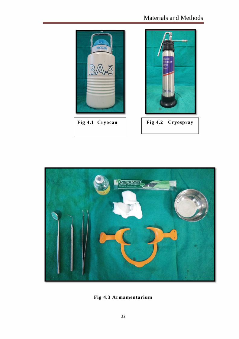
Liquid nitrogen is a l iquefied atmospheric gas produced
industrially in large quantities by performing fractional disti llation
of liquid air. It is colorless pure liquid at a very low temperature
(-196°C). It is stored and transported in cryocan or liquid n itrogen
low volume container available in wide range of capacities.
Materials and Methods
31
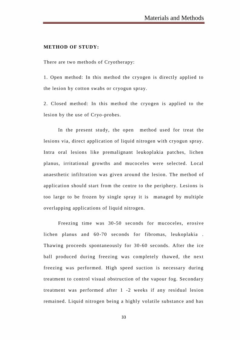
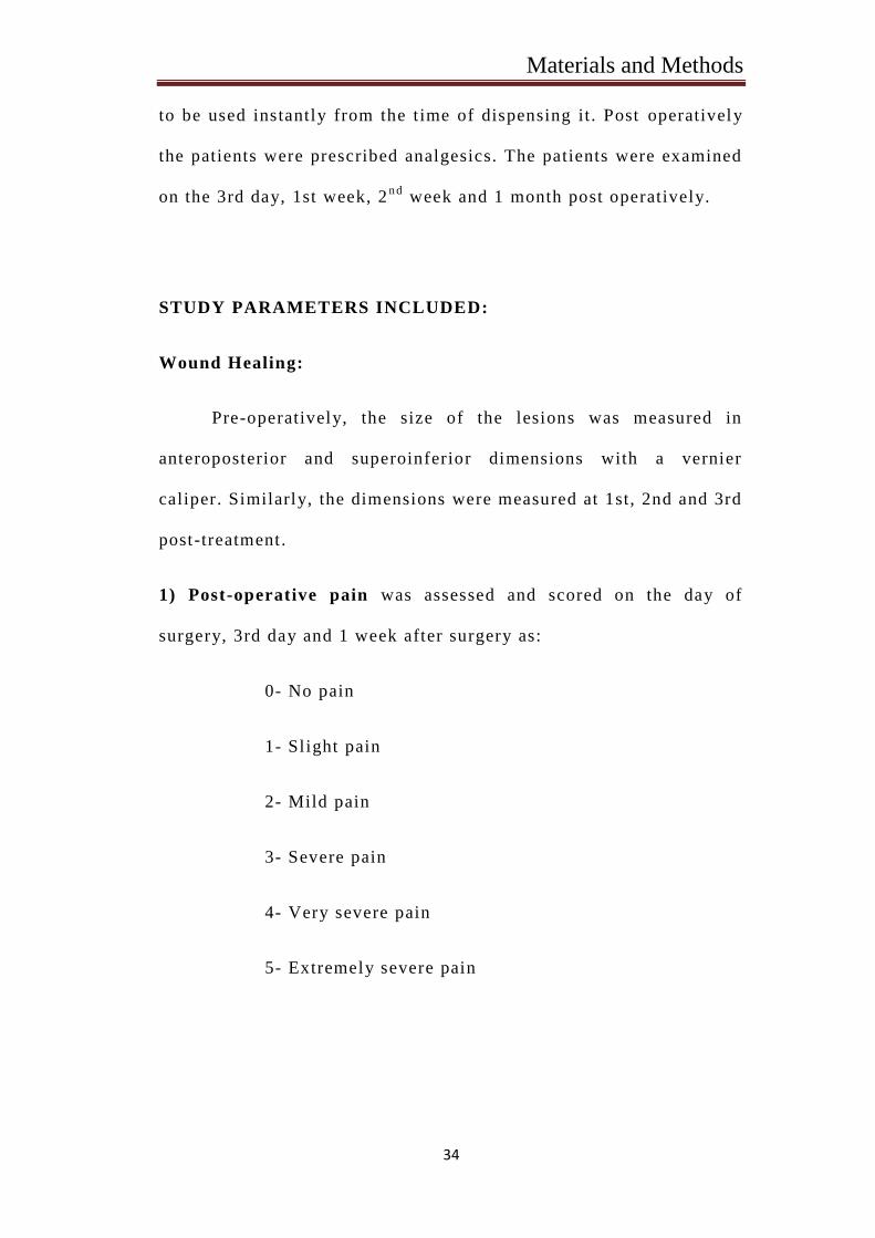
Armamentarium used:
1. Mouth mirror
2. Straight probe
3. Tweezer
4. Stainless steel bowl
5. Cheek retractor
6. Gauze
7. Cryospray
8. Liquid nitrogen
9. Cryocan for storage of l iquid nitrogen
10.Local Anasthesia
Materials and Methods
32
Fig 4.3 Armamentarium
Fig 4.1 Cryocan Fig 4.2 Cryospray
Materials and Methods
33
METHOD OF STUDY:
There are two methods of Cryotherapy:
1. Open method: In this method the cryogen is directly applied to
the lesion by cotton swabs or cryogun spray.
2. Closed method: In this method the cryogen is applied to the
lesion by the use of Cryo-probes.
In the present study, the open method used for treat the
lesions via, direct application of liquid nitrogen with cryogun spray.
Intra oral lesions l ike premalignant leukoplakia patches, lichen
planus, irritational growths and mucoceles were selected. Local
anaesthetic infiltration was given around the lesion. The method of
application should start from the centre to the periphery. Lesions is
too large to be frozen by single spray i t is managed by multiple
overlapping applications of liquid nitrogen.
Freezing time was 30-50 seconds for mucoceles, erosive
lichen planus and 60-70 seconds for fibromas, leukoplakia .
Thawing proceeds spontaneously for 30 -60 seconds. After the ice
ball produced during freezing was completely thawed, the next
freezing was performed. High speed suction is necessary during
treatment to control visual obstruction of the vapour fog. Secondary
treatment was performed after 1 -2 weeks if any residual lesion
remained. Liquid nitrogen being a highly volatile substance and has
Materials and Methods
34
to be used instantly from the t ime of dispensing it. Post operatively
the patients were prescribed analgesics. The patients were examined
on the 3rd day, 1st week, 2n d
week and 1 month post operatively.
STUDY PARAMETERS INCLUDED:
Wound Healing:
Pre-operatively, the size of the lesions was measured in
anteroposterior and superoinferior dimensions with a vernier
caliper. Similarly, the dimensions were measured at 1st, 2nd and 3rd
post-treatment.
1) Post-operative pain was assessed and scored on the day of
surgery, 3rd day and 1 week after surgery as:
0- No pain
1- Slight pain
2- Mild pain
3- Severe pain
4- Very severe pain
5- Extremely severe pain
Materials and Methods
35
2) Presence of post-operative swelling was evaluated on 3rd and 1
week surgery:
0- No swelling
1- Slight swelling
2- Mild swelling
3- Severe swelling
4- Very severe swelling
5- Extremely severe swelling
3) Presence of sloughing was noted at the end of 1 week as:
Present
Absent
4) Presence of granulation tissue was noted at the end of 2 weeks
as:
2-Good-(entire wound)
1-Fair-(nearly entire wound)
0-Poor - (inadequate)
5) Epithelization was noted at the end of the month as:
2-Good – (entire wound)
1-Fair- (nearly entire wound)
0-Poor-(inadequate)
Materials and Methods
36
Recurrence :
Recurrence of the lesion was evaluated clinically at the 2
month post op. The results of the study should be evaluated
statistically, with the help of CHI SQ TEST and N par TEST .
Materials and Methods
37
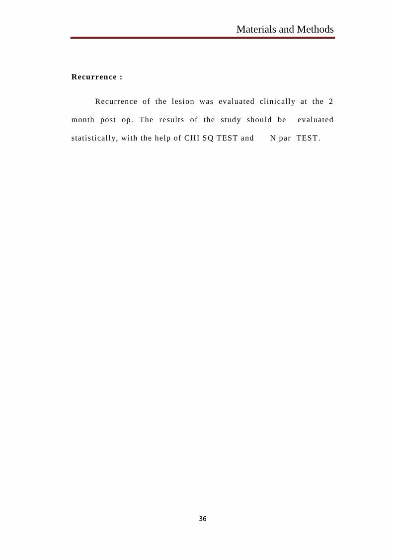
CASE 1
Pre operative mucocele seen
in the ventral tongue region
Intra operative
1st week post operative
showing sloughing
formation of the lesionion
1st
month post operative
showing complete
healing of the lesion
Materials and Methods
38
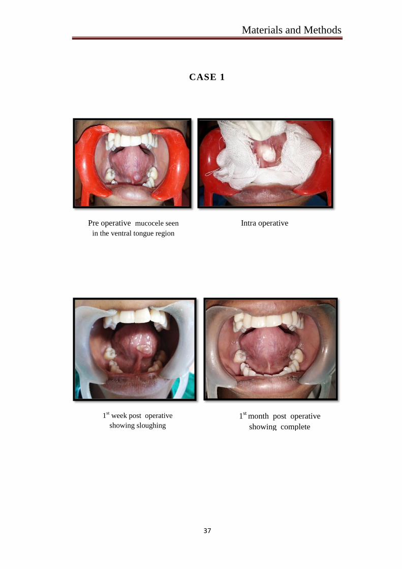
CASE 2
Pre operative Leukoplakia ,
seen in 48 region Intra operative
1s t
week post operative 1s t
month post operative
Results
39
RESULTS
The present studywas conducted to evaluate the efficacy of
cryotherapy as treatment modality for oral lesion, to evaluate the
post operative clinical outcome of oral lesion after treatment with
cryotherapy with respect to post - operative wound healing and
recurrence of the lesion in the department of Oral and Maxillofacial
Surgery, at Adhiparasakthi dental college and hospital. 15 patients
who was affected with oral lesion either benign or pre -malignant
lesion of size more than 1cm ,were randomly selected from out -
patient to evaluate the effectiveness of the cryosurgery method of
treating oral lesion with l iquid nitrogen after obtaining ethical
clearance. The age of patients ranged between 22yrs -52yrs with
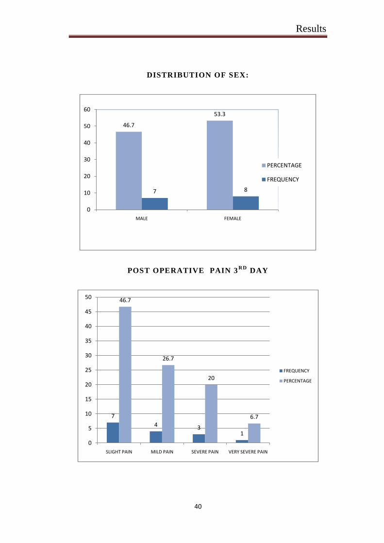
46.7% of male and 53.3% of female. The overall mean age of the
study population 33.6. In this study the open method was used to
treat the lesion by direct application of liquid nitrogen with cryogun
spray, all subjects selected on the basis of inclusion cri teria. Among
15 patients, there were 5 mucocele lesions, 2 traumatic fibroma, 1
leukoplakia, 2 erythroplakia, 3 lichen planus, 2 Apthous ulcers were
seen. The site of occurrence of the lesion varied in different
subjects the lesions were present on the buc cal mucosa, floor of the
tongue, upper vestibule, lower vestibule. Post -treatment follow up
was done at 3rd
day, 1s t
week and 1s t
month and 2n d
The results
were evaluated statically with the help of Descriptive statistics and
NPar tests: chi -square test , kolmogorov-smirnov test .
Results
40
DISTRIBUTION OF SEX:
POST OPERATIVE PAIN 3RD
DAY
46.7
53.3
7 8
0
10
20
30
40
50
60
MALE FEMALE
PERCENTAGE
FREQUENCY
7
4 31
46.7
26.7
20
6.7
0
5
10
15
20
25
30
35
40
45
50
SLIGHT PAIN MILD PAIN SEVERE PAIN VERY SEVERE PAIN
FREQUENCY
PERCENTAGE
Results
41
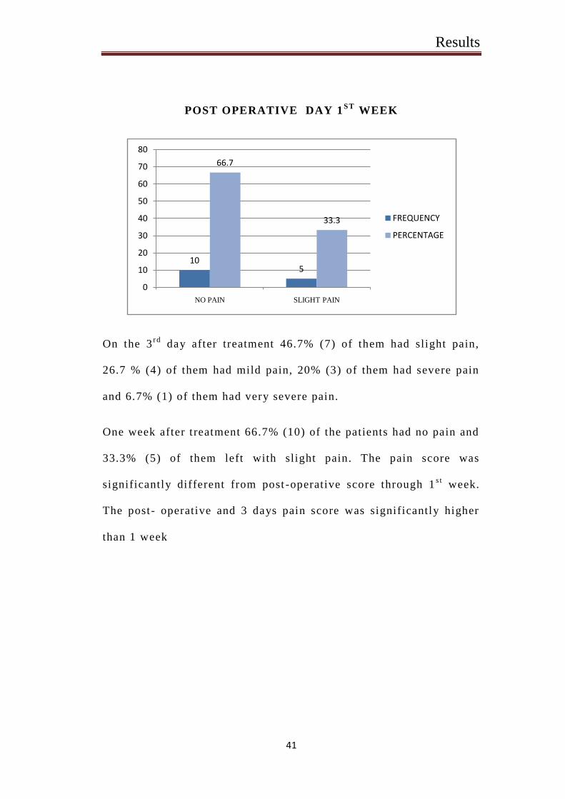
POST OPERATIVE DAY 1ST
WEEK
On the 3rd
day after treatment 46.7% (7) of them had slight pain,
26.7 % (4) of them had mild pain, 20% (3) of them had severe pain
and 6.7% (1) of them had very severe pain.
One week after treatment 66.7% (10) of the patients had no pain and
33.3% (5) of them left with slight pain. The pain score was
significantly different from post -operative score through 1s t
week.
The post- operative and 3 days pain score was significantly higher
than 1 week
105
66.7
33.3
0
10
20
30
40
50
60
70
80
NO PAIN SLIGHT PAIN
FREQUENCY
PERCENTAGE
Results
42
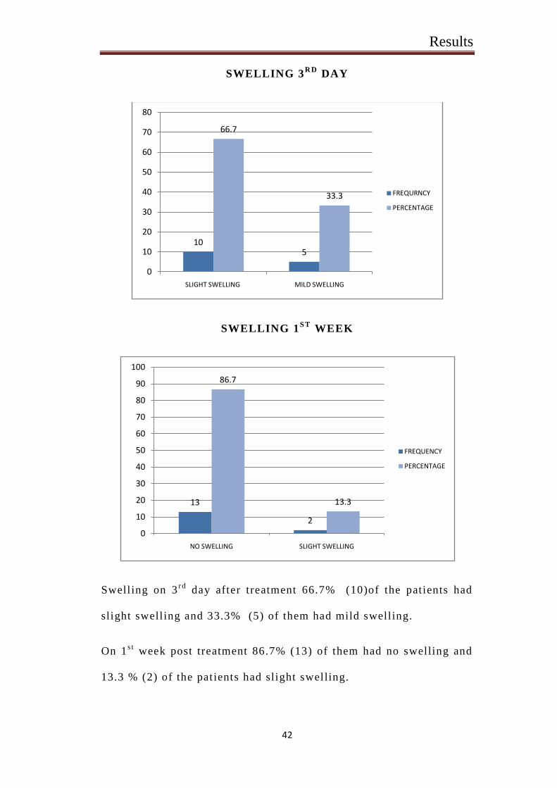
SWELLING 3R D
DAY
SWELLING 1ST
WEEK
Swelling on 3rd
day after treatment 66.7% (10)of the patients had
slight swelling and 33.3% (5) of them had mild swelling.
On 1s t
week post treatment 86.7% (13) of them had no swelling and
13.3 % (2) of the patients had slight swelling.
105
66.7
33.3
0
10
20
30
40
50
60
70
80
SLIGHT SWELLING MILD SWELLING
FREQURNCY
PERCENTAGE
13
2
86.7
13.3
0
10
20
30
40
50
60
70
80
90
100
NO SWELLING SLIGHT SWELLING
FREQUENCY
PERCENTAGE
Results
43
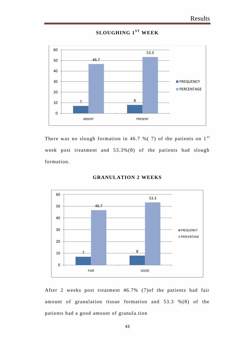
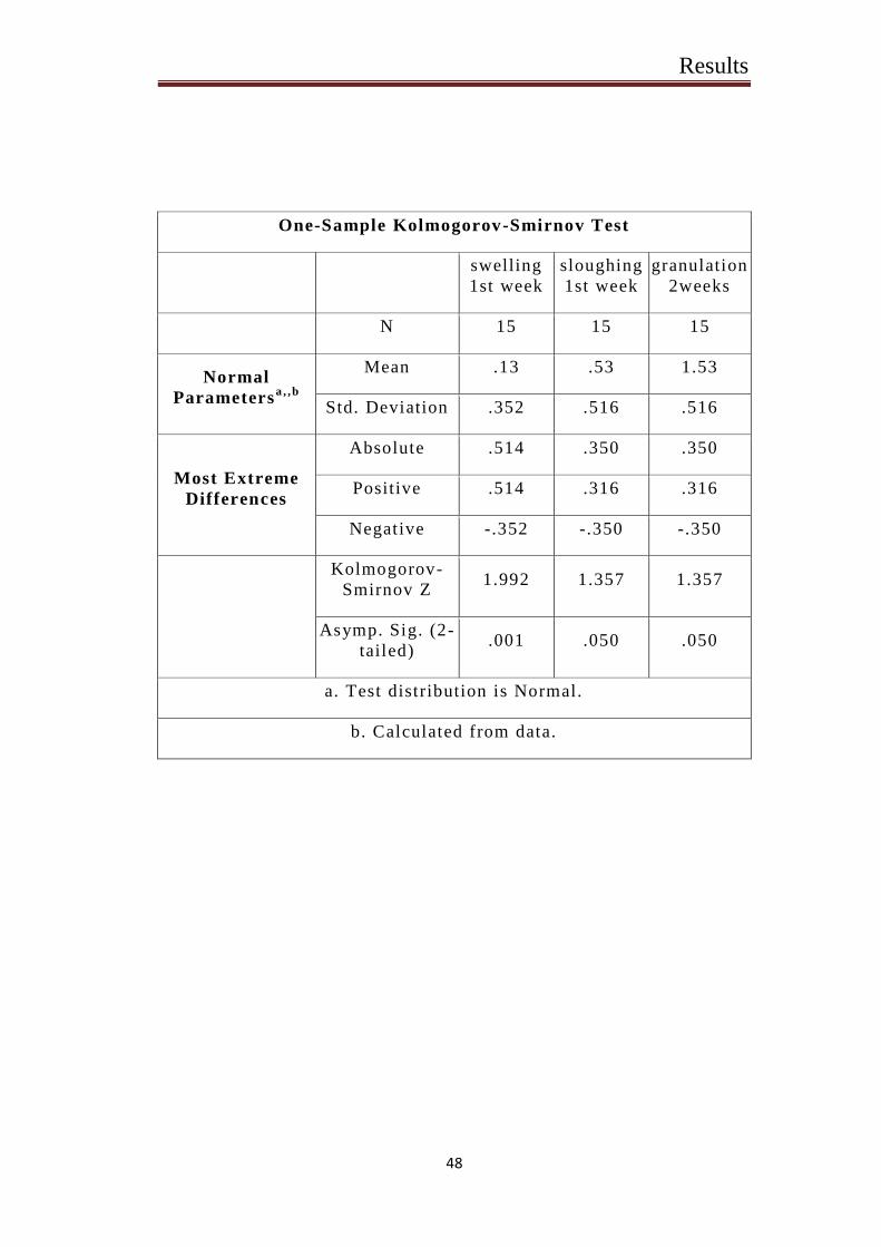
SLOUGHING 1ST
WEEK
There was no slough formation in 46.7 %( 7) of the patients on 1s t
week post treatment and 53.3%(8) of the patients had slough
formation.
GRANULATION 2 WEEKS
After 2 weeks post treatment 46.7% (7)of the patients had fair
amount of granulation tissue formation and 53.3 %(8) of the
patients had a good amount of granula.tion
7 8
46.7
53.3
0
10
20
30
40
50
60
ABSENT PRESENT
FREQUENCY
PERCENTAGE
7 8
46.7
53.3
0
10
20
30
40
50
60
FAIR GOOD
FREQUENCY
PERCENTAGE
Results
44
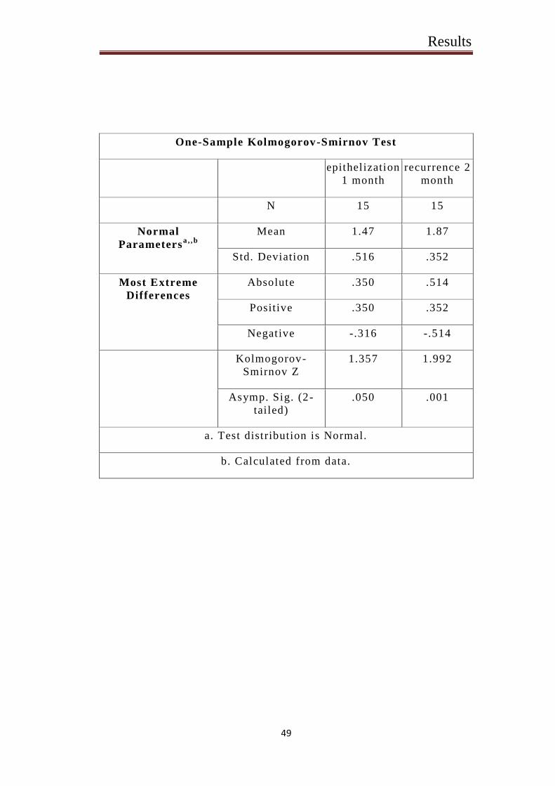
EPITHELIZATION 1 MONTH
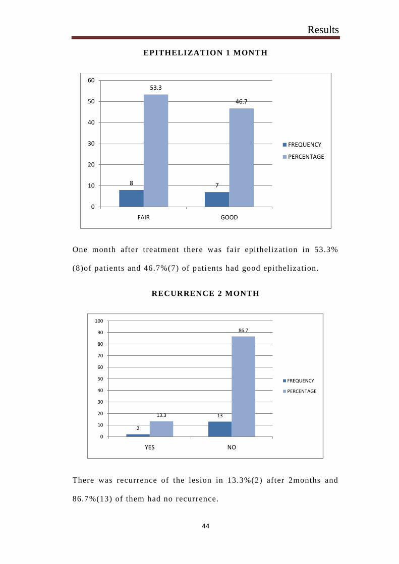
One month after treatment there was fair epithelization in 53.3%
(8)of patients and 46.7%(7) of patients had good epithelization.
RECURRENCE 2 MONTH
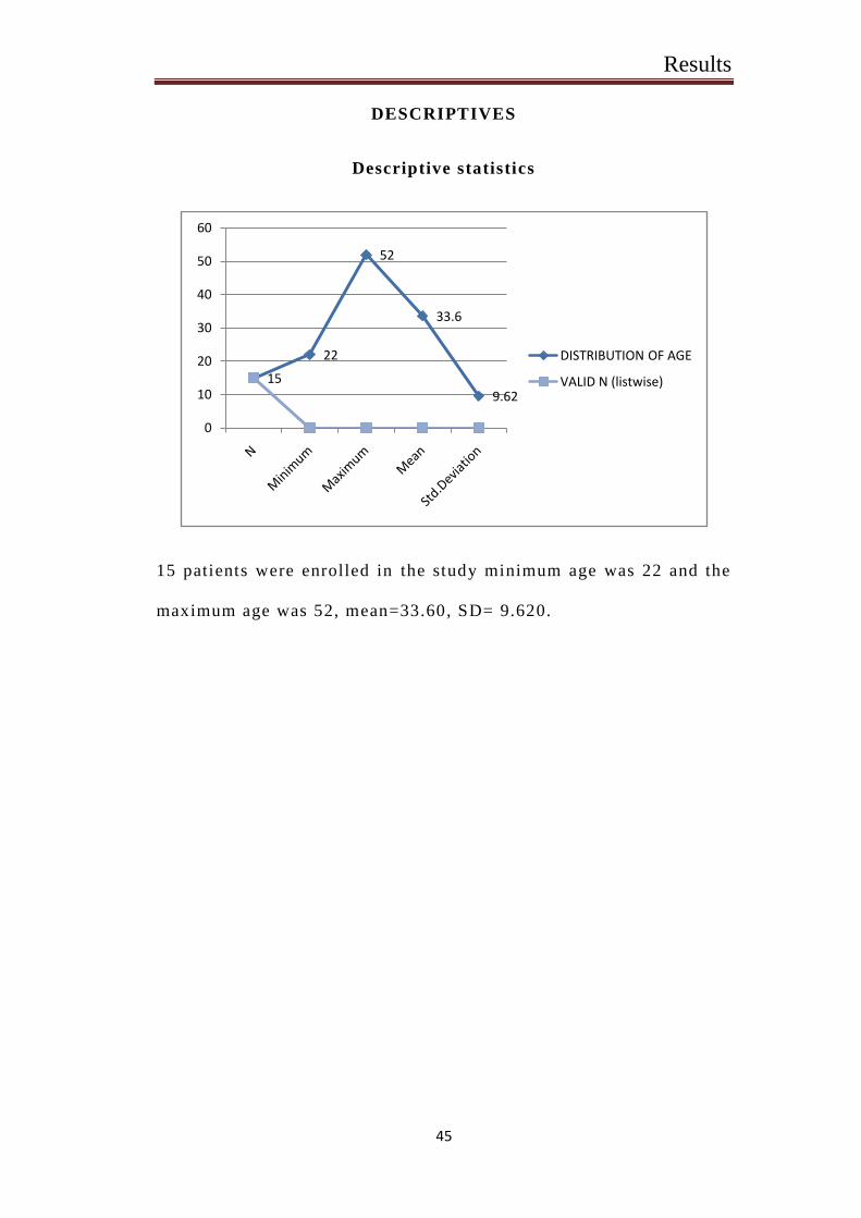
There was recurrence of the lesion in 13.3%(2) after 2months and
86.7%(13) of them had no recurrence.
8 7
53.3
46.7
0
10
20
30
40
50
60
FAIR GOOD
FREQUENCY
PERCENTAGE
2
1313.3
86.7
0
10
20
30
40
50
60
70
80
90
100
YES NO
FREQUENCY
PERCENTAGE
Results
45
DESCRIPTIVES
Descriptive statistics
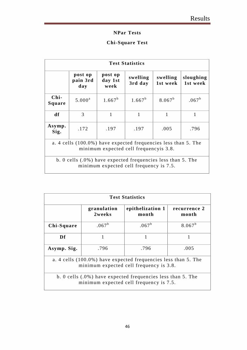
15 patients were enrolled in the study minimum age was 22 and the
maximum age was 52, mean=33.60, SD= 9.620.
15
22
52
33.6
9.62
0
10
20
30
40
50
60
DISTRIBUTION OF AGE
VALID N (listwise)
Results
46
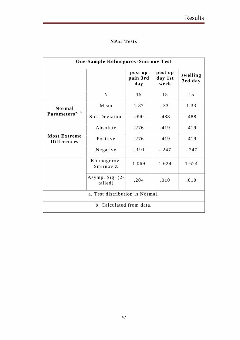
NPar Tests
Chi-Square Test
Test Statistics
post op
pain 3rd
day
post op
day 1st
week
swelling
3rd day
swelling
1st week
sloughing
1st week
Chi-
Square 5.000
a 1.667
b 1.667
b 8.067
b .067
b
df 3 1 1 1 1
Asymp.
Sig. .172 .197 .197 .005 .796
a. 4 cells (100.0%) have expected frequencies less than 5. The
minimum expected cell frequencyis 3.8.
b. 0 cells (.0%) have expected frequencies less than 5. The
minimum expected cell frequency is 7.5.
Test Statistics
granulation
2weeks
epithelization 1
month
recurrence 2
month
Chi-Square .067b .067
b 8.067
b
Df 1 1 1
Asymp. Sig. .796 .796 .005
a. 4 cells (100.0%) have expected frequencies less than 5. The
minimum expected cell frequency is 3.8.
b. 0 cells (.0%) have expected frequencies less than 5. The
minimum expected cell frequency is 7.5.
Results
47
NPar Tests
One-Sample Kolmogorov-Smirnov Test
post op
pain 3rd
day
post op
day 1st
week
swelling
3rd day
N 15 15 15
Normal
Parametersa , ,b
Mean 1.87 .33 1.33
Std. Deviation .990 .488 .488
Most Extreme
Differences
Absolute .276 .419 .419
Positive .276 .419 .419
Negative -.191 -.247 -.247
Kolmogorov-
Smirnov Z 1.069 1.624 1.624
Asymp. Sig. (2-
tailed) .204 .010 .010
a. Test distribution is Normal.
b. Calculated from data.
Results
48
One-Sample Kolmogorov-Smirnov Test
swelling
1st week
sloughing
1st week
granulation
2weeks
N 15 15 15
Normal
Parametersa , ,b
Mean .13 .53 1.53
Std. Deviation .352 .516 .516
Most Extreme
Differences
Absolute .514 .350 .350
Positive .514 .316 .316
Negative -.352 -.350 -.350
Kolmogorov-
Smirnov Z 1.992 1.357 1.357
Asymp. Sig. (2-
tailed) .001 .050 .050
a. Test distribution is Normal.
b. Calculated from data.
Results
49
One-Sample Kolmogorov-Smirnov Test
epithelization
1 month
recurrence 2
month
N 15 15
Normal
Parametersa , ,b
Mean 1.47 1.87
Std. Deviation .516 .352
Most Extreme
Differences
Absolute .350 .514
Positive .350 .352
Negative -.316 -.514
Kolmogorov-
Smirnov Z
1.357 1.992
Asymp. Sig. (2-
tailed)
.050 .001
a. Test distribution is Normal.
b. Calculated from data.
Discussion
50
DISCUSSION
This Critique was undertaken to evaluate the efficacy of
cryosurgery, as a treatment modali ty in management of benign and
premalignant oral lesion which is of size greater than 1 cm and to
evaluate the post operative clinical outcome of oral lesion using
liquid nitrogen with cryogun spray through open method. Our
clinical study was done on 15 out - patients with the respective
lesion. a regular follow up was done at 3rd
day,1s t
week,2n d
week, 1s t
month and 2n d
month to evaluate the post operative pain,
swelling,granulation tissue formation, epithelization and recurrence.
This study presents a series of cases that demonstrate the clinical
efficacy of cryosurgery as an alternative to conventional surgical
technique to treat most common oral lesion, this techniq ue is well
tolerated by patients due to the rapid healing and minimal bleeding,
can be performed in absence of anesthesia and is extremely an
useful alternative in patients to whom minor surgery is
contraindicated due to age or medical history. It is the simple
procedure to perform, minimally invasive, low cost and very
effective.
Destruction of the diseased t issue in the appropriate posit ion
by means of freezing is well established in many branches of
surgery. The tissues are apparently unaltered at thaw but
progressive necrosis happens. There is controversy as to whether
tissue death is principally due to the direct effects of freezing or to
subsequent ischemia. Studies at the ultra structural level show that
Discussion
51
ice crystals are formed within the cells duri ng cryosurgery in which
resultant cell damage is osmotic rather than mechanical and that
microcirculatory changes are secondary in terms of the
chronological development of tissue necrosis. The experiments
carried out on intact tissues under conditions sim ilar to those used
in clinical cryosurgery have clarified some of the mechanisms
involved in tissue destruction. It has been shown, using freeze
substi tution and electron microscopy, which ice crystals form
intracellularly during cryosurgery although evide nce from freezing
isolated cells would suggest that the freezing rates used clinically
would be more l ikely to result in extracellular ice. It appears that
cells in close contact in living tissues behave differently to those
suspended in l iquids. Repeat fr eeze results in rather large
intracellular ice crystals and it is this increase in size which appears
to be more lethal following this technique. The changes occurring in
the living tissues immediately after thaw have been described both
in epithelium and muscle(5 )
.
The application of cryotherapy to oral surgery has been
facili tated by the development of keeler arul probe. preliminary
experiments with these applicators indicated that the probe could be
used most effectively in oral surgery, especially for the eradication
of superficial lesion by necrosis, with discomfort both during and
tissue reaction, eliminating bleeding, and reduced discomfort both
during and after the operation. To test the effectiveness of the
cryoprobe in this field and compare the r esults with excision and
Discussion
52
suture, and electro surgery. Cryosurgery would appear to have
advantage over excision and suture for small lesion, or superficial
lesion close to vital structures which are to be left intact if possible.
Eventhough healing is delayed, it is not associated with prolonged
discomfort, hemorrhage and resultant scar formation and wound
contraction is minimal. Electrosurgery would appear to have no
peculiar advantage over cryosurgery, except, in the treatment of
gingival lesions and few advantages over excision and suture except
in the preservation of sulcus depth. If cryosurgery is to become the
valuable tool in oral surgery that is formally suggest by this
investigation, there are two important factors that should govern its
uses- it should not be used for lesion more than 3 cm in diameter, or
lesions partly impossible to reach to the probe, all lesions should be
treated by freezing for two minutes and thawing and re -freezing to
be certain of achieving a satisfactory tissue kill . Improve d
instrumentations and the increase in freezing by local physical
means, or other, simultaneous applications, will further enhance the
value of the cryoprobe in outpatient oral surgery(3 )
Cryosurgery has been recommended for the removal of
superficial mucosal lesions in the oral cavity . More recently,
carbon dioxide lasers, which emit far infra -red radiation, have been
used as an alternative form of treatment . Comparisons of the
effectiveness of cryosurgery, laser surgery and electro - cautery have
been reported in the treatment of cervical mucosal lesions. In these
studies the quality of healing following use of the CO2 laser have
Discussion
53
been subjectively assessed to be "excellent", with high cure rates
being reported the effectiveness and the healing response f ollowing
use of the 2 techniques was compared by producing lesions on the
lateral border of sheep tongues. Cryosurgery produced more
extensive lesions with a marked inflammatory reaction but no
differences in the time course of healing were evident. Laser
surgery was as effective as cryosurgery in the removal of superficial
tissues but caused less swelling and, therefore, may be
advantageous in some clinical situations. Cryosurgery and CO 2
laser surgery has an advantage that are readily apparent, both
techniques are easily used in treating mucosal lesions, hemorrhage
is controlled during the procedure and they are relatively painless.
Cryosurgery has become established as an effective means of
treatment for leukoplakia, whereas carbon dioxide laser surgery is a
more recent development and has been recommended for clinical
use. This latter technique is confined to hospital treatment due to
the size of lasers presently available. The use of the Nd -YAG laser
on oral mucosa has been evaluated. This instrument di ffers from the
CO2 laser in that emits radiation in the near infra -red, with bands at
1064 nm and 1318 nm. As such, there is a greater extinction length
in the mucosa and the Nd-YAG laser causes deeper tissue
coagulation rather than surface vaporization. T he miniature CO2
laser may be useful in the treatment of oral lesions, comparable to
that of cryosurgery. A serious disadvantage with both techniques in
the management of any mucosal disease is the lack of an
Discussion
54
opportunity to sample the lesion and examine it microscopically in
order to determine the nature of the lesion being treated(1 2 )
.
Cryosurgery is a successful mode of therapy for the
conditions which have traditionally presented problems in the
management of leukoplakia,vascular malformation and cert ain
extensive surface lesions like lichen planus. The technique of
freezing has been used to treat hemangiomas , Applications of
selected freezing have been employed to treat leukoplakia and
hyperkeratosis, With the advancement in cryosurgical technique an d
equipment,it was util ized in pyogenic granuloma, angioma, fibroma,
keratoacanthoma. Lichen planus was treated with cryotherapy with
good results .cryosurgery is also used to treat salivary gland
neoplasm cryosurgery is an effective, simple, predictable,
relatively self limiting and safe method for almost all types of oral
lesions. As it causes necrosis and sloughing as part of treatment
,delayed healing is an intensive problem with this technique else, it
is free from complications such as pain, hemorrha ge ,infection,
unconditional damage to adjacent structures ,or scar formation that
are seen with knife excision or electro surgery(9 )
. In our study 15
patients were enrolled with benign and premalignant lesion among
which , there were 5 mucocele lesions, 2 traumatic fibroma, 1
leukoplakia, 2 erythroplakia, 3 lichen planus, 2 Apthous ulcers were
seen. The site of occurrence of the lesion varied in different
subjects the lesions were present on the buccal mucosa, floor of the
tongue, upper vestibule, lower vestibule. Post-treatment follow up
Discussion
55
was done at 3rd
day, 1s t
week and 1s t
month and 2n d
month to
evaluate the intensity of pain, swelling, sloughing, granulation,
epithilization and recurrence. There were good response after
treatment and comparing the 3rd
and 1s t
week there were a
reasonable amount of reduction in pain and swelling and sloughing
was present , on the 2n d
week granulation flesh was healthy, after a
month reepithelialization phase was good. In 15 cases recurrence
was seen in 2 cases one in l ichen planus other in erythroplakia. The
other lesions had no recurrence and responded well under open
method cryosurgery treatment.
Cryosurgery supports in treating orofacial lesions is a great
extent some vascular malformations like Cavernous hemangiom a
when treated with cryosurgery produces shows complete regression
of these malformations, whether of skin or of mucosa, with minimal
scarring. Cryosurgery is very effective in cases of strawberry nevi
where it reduces the excessive bleeding and ulceratio n or
overwhelming parental demands. Capillary nevi also responds to
cryosurgery. Lymphangiomas with fibrous element are usually less
responsive to cryosurgery. It is a satisfactory alternative to excision
or cautery for treating the Granulomatous and hype rplastic
conditions like papillary hyperplasia of the palate, fibrous epulis,
fibroepithelial polyps, myeloid epulides and viral warts. In case of
Denture hyperplasia for a say if lesion is present in the lower labial
sulcus, hyperplastic folds may be mul tiple and relatively broad
based. Simple excision in may leads to loss of sulcus depth, which
Discussion
56
may result in need of further management such as vestibuloplasty in
the elderly and debilitated, in such issue cryosurgery may be used.
Necrosis of the hyperplast ic tissue may be achieved with minimal
alteration of sulcus depth. Mucus retention cysts and antral polyps
respond to cryosurgery without recurrence and detectable scarring.
Cryosurgery for oral cancer benefits localized tissue destruction in
superficial accessible lesions. It is the treatment of choice in
recurrent nasopharyngeal carcinoma, It should not be the primary
treatment of oral cancer except in very early lesions of anterior part
of palate and in patients who are considered unfit for other forms of
treatment. In the management of basal cell carcinoma the ulcers at
the inner canthus or in cases where there are multiple lesions,
cryosurgery is a sound alternative to surgery and radiotherapy
provided the tumor has not invaded deeper structures. Cryo surgery
in herpetic or aphthous ulcers used for their eradication, gave very
satisfactory results. There was no intra or postoperative bleeding,
no surgical defects, minimal scarring, and no infection following
treatment(1 )
.
Chronic facial pain can be treated by cryogenic block , The
duration of the block is related to the distance in which the nerve
must regenerate from the point of freezing to the innervated area
and in this series in which peripheral branches of the trigeminal
nerve were frozen, The extended nerve block which follows
cryoanalgesia has been shown to be associated with Antrograde
degeneration. The management of chronic pain is often expremental,
Discussion
57
and employs techniques which interrupt or modify pain path - ways
but which do not incapacitate the whole patient. As the response to
treatment of patients with chronic pain is often unpredictable, it is
desirable to select techniques which do not produce irreversible
damage to the nerve or trigger secondary neuralgia. Above all
cryoanalgesia appears to offer advantages over other methods of
long term nerve block or neurectomy, and may result in prolonged
relief in some patients(6 )
.
Pain in tempromandibular joints is a common clinical
problems, cryoanalgia provides a short term relief of intractable
neurogenic pain in the TMJ with some long term relief. The main
advantages of the procedure are that the intra -atricular structures
are not damaged and case procedure with minimal morbidity.
Disadvantage include it have only a temporary relief under certa in
circumstances, with unpredictable outcomes, there is a good chance
of pain recurrence. The procedure may be repeated but there may a
decreasing in response. Cryoanalgesia is useful adjunct to the
management of extremely unmanageable pain localized to th e TMJ.
A suggested management protocol would involve as initial
conservative treatment followed by arthrocentesis and arthroscopy.
If these techniques fail and the patient responds to intra - articlar
bupivicaine, then cryoanalgesia should be considered(8 )
.
Cryotherapy in management of lesion in bone ,on comparing
the histological response of normal bone to freezing gives hope that
cryosurgery may have a significant role in the treatment of
Discussion
58
neoplastic and infiltrat ive lesions involving the calcified tissues of
the jaws. Relatively only a little is known, however, of the
response of abnormal cells within the bone and this will be
obviously governing the ult imate outlook for this form of therapy.
In assessing the freezing procedures in soft tissue one can usu ally
see a well defined frozen zone and palpate the size of the ice ball
formed. The zone of freezing is much harder to define in bone, and
the ice ball cannot be palpated. The insert ion of thermocouples to
the register tissue temperature is more difficult in bone than in soft
tissue and it is necessary to drill access holes. To help in
overcoming these problems in evaluation, it was decided to attempt
to use thermography. The use of this method, which monitors infra
red emission and does not appear to have been previously described
in relationship to cryosurgery. The clinical information available
indicates that sound repair of the bony cavities can occur after
curettage of a lesion and cryosurgery of the surrounding region.
Primary union of overlying soft tissue incisions can take place. The
factors which contribute to the inhibition of osteogenesis and
inadequate repair of the bone after freezing in certain cases require
careful study( 3 )
.
Though cryosurgery have many advantages such as minimal
general disturbance to the patient, particularly well accepted by
children and elder patients, Low complication rate, minimal volume
of tissue destruction, Particularly suited to extensive superficial
lesions ,Treatment may be repeated as often as necessary without
Discussion
59
increase in scarring, offers quality management of wide areas of
premalignant change, May be used as an alternate to surgery and/or
radiotherapy in palliative tumor control , Cryosurgery is a very safe,
easy to perform, and relatively inexpensive techniqu e for treating
various oral lesions in an out -patient clinic, it have its own
disadvantages and limitations as follows:
Difficulty in judging the extent of the lesion can lead to
involvement of an inadequate amount of tissue, use is minimally
confined to surface lesions of no more than a certain depth, surgical
access is required for deeper lesions. The depth of destruction is
limited although the base of the lesion is refrozen after excision of
frozen tissue. Certain tissue seems to have a greater resista nce to
freezing than others, cryosurgery is non -specific in its destructive
effects. In lesion of tongue the procedure can l imit its functions.
Healing is slow, Extensive cryosurgery procedures may produce
considerable scarring. Following healing by second ary intention,
loss of normal anatomy can lead to limitation of mouth opening,
speech disturbances and prosthetic problems, as in the case of
widespread keratoses, after extensive cryosurgery severe pain can
be troublesome and narcotic analgesics may be re quired. If a biopsy
is not taken prior to cryosurgery, the true nature of the lesion may
not be found.
Complication categorized as immediate complication
includes bleeding, blistering, edema, pain, vascular head ache,
vasovagal syncope. Delayed complicat ions includes excessive
Discussion
60
granulation, infection is rare but possible with delayed healing
tendon repture due to deeper freezing on extensor surface of fingers
, ulceration, temporary complications include altered sensation may
occur when damage in particular nerve, More profound freezing
causes Wallerian degeneration which is followed by regeneration, as
the nerve sheath architecture remains intact. This is also the reason
for reduced sensation following cryosurgery, caution is required
with lesion over nerve trunks. Peripheral nerve fibers may be
painful following moderate freezing of adjacent structures, possibly
due to the action of cellular breakdown products ,
hyperpigmentations may occur, increased risk with aggressive
freezing hypertrophic scaring may occur , Scarring of facial skin if
freezing is done for longer than 20–30 seconds. Healing occurs with
reduction in pigmentation in such cases. Pyogenic granuloma rarely
occurs. Permanent complications may include alopecia, atrophy,
cartiladge necrosis, hypopigmentations.
There are some more contraindications such as cold
intolerance, cold urticaria, cryoglobulinemia, dysfibrinogenemia,
agammaglobulinemia, Raynaud’s and collagen diseases, pyoderma
gangrenosum, patients undergoing hemodialysis or
immunosuppressive therapy, patients with platelet al terations or
with multiple myeloma(1 )
. After all cryosurgery in treating oral
lesions overweighs with its advantages.
Conclusion
61
CONCLUSION
Cryosurgery is the use liquid nitrogen were extreme cold is
produced to destroy abnormal tissue. Cryosurgery is very safe, easy
to perform and relatively inexpensive technique for treating various
oral lesions in out -patient clinic it is very useful for oral lesion as
the oral mucosa is humid which makes it an ideal site for freezing.
With recent development of better equipment this mode of therapy
appears to have a promising role in the management of benign
mucosal lesion, more reasonable with adequate success, the
procedure is relatively more acceptable and has shown to have ,
faster recovery. Though cryosurgery have some difficult ies like
portability of the cryogenic agent and storage of highly volatile
material , it require special storage container for short -term storage
and canister for dispensing the liquid from the container during its
use, its advantages are numerous , this technique provide a
bloodless field , less pain when compared to conventional surgery,
anesthesia is optional, excellent cosmetic results , low cost, low risk
of infection, minimal wound care, no need of suture removal, no
work or sports restrictions, short preparation time, useful in
pregnancy. In this study the open method used to treat lesion with
cryogen spray using liquid nitrogen has shown significantly good
results in achieving complete regression of the benign and
premalignant lesion of oral cavity.
Biblography
62
BIBLOGRAPHY
1. Bansal A, Jain S, Gupta S. Cryosurgery in the treatment of
oro-facial lesions. Indian Journal of Dental Research. 2012
Mar 1;23(2):297.
2. Farah CS, Savage NW. Cryotherapy for treatment of oral
lesions. Australian dental journal. 2006 Mar;51(1):2-5.
3. Bradley PF, Fisher AD. The cryosurgery of bone. An
experimental and clinical assessment. British Journal of Oral
Surgery. 1975 Nov 1;13(2):111-27.
4. Ameerally PJ, Colver GB. Cutaneous cryotherapy in
maxillofacial surgery. Journal of Oral and Maxillofacial
Surgery. 2007 Sep 1;65(9):1785-92.
5. Whittaker DK. Mechanisms of t issue destruction following
cryosurgery. Annals of the Royal College of Surgeons of
England. 1984 Sep;66(5):313.
6. Barnard D, Lloyd J, Evans J. Cryoanalgesia in the
management of chronic facial pain. Journal of maxillofacial
surgery. 1981 Jan 1;9:101-2.
7. Goss AN. Cryoneurotomy for intractable temporomandibular
joint pain. British Journal of Oral and Maxillofacial Surgery.
1988 Feb 1;26(1):26-31.
Biblography
63
8. Sidebottom AJ, Carey EC, Madahar AK. Cryoanalgesia in the
management of intractable pain in the temporomandibular
joint: a five-year retrospective review. British Journal of Oral
and Maxillofacial Surgery. 2011 Dec 1;49(8):653 -6.
9. Narula R, Malik B. Role Of Cryosurgery In The Management
Of Benign And Premalignant Lesions Of The Maxillofacial
Region. Indian Journal of Dental Sciences. 2012 Jun 1;4(2).
10. Darbandi A, Shahbaz NA. Effect of cryotherapy on
physiologic pigmentation of oral mucosa: a preliminary study.
Journal of Dentistry of Tehran Unive rsity of Medical
Sciences. 2004;1(2):49-52.
11. McCreary CE, McCartan BE. Clinical management of oral
lichen planus. Brit ish Journal of Oral and Maxillofacial
Surgery. 1999 Oct 1;37(5):338-43.
12. Kardos TB, Ferguson MM. Comparison of cryosurgery and the
carbon dioxide laser in mucosal healing. International journal
of oral and maxillofacial surgery. 1991 Apr 1;20(2):108 -11.
13. Syed Nayeema, Dr.M.Subha: Cryotherapy- a Novel treatment
modality in oral lesions, International Journal of Pharmacy
and Pharmaceutical Sciences 2013 (5) 4-5
14. Bekke JP, Baart JA. Six years ' experience with cryosurgery in
the oral cavity. International journal of oral surgery. 1979
Aug 1;8(4):251-70.
Biblography
64
15. Yeh CJ. Simple cryosurgical treatment for oral lesions.
International Journal of Oral & Maxillof acial Surgery. 2000
Jun;29(3):212-6.
16. Gage AA, Koepf S, Wehrle D, Emmings F. Cryotherapy for
cancer of the lip and oral cavity. Cancer. 1965
Dec;18(12):1646-51.
17. Loitz GA, O'Leary JP. Erosive l ichen planus of the tongue
treated by cryosurgery. Journal of Ora l and Maxillofacial
Surgery. 1986 Jul 1;44(7):580-2.
18. Chen HM, Cheng SJ, Lin HP, Yu CH, Wu YC, Chiang CP.
Cryogun cryotherapy for oral leukoplakia and adjacent
melanosis lesions. Journal of Oral Pathology & Medicine.
2015 Sep;44(8):607-13.
19. Gongloff RK. Treatment of intraoral hemangiomas with
nitrous oxide cryosurgery. Oral Surgery, Oral Medicine, Oral
Pathology. 1983 Jul 1;56(1):20-4.
20. Prasad M, Kale TP, Halli R, Kotrashetti SM, Baliga SD.
Liquid nitrogen cryotherapy in the management of oral
lesions: a retrospective clinical study. Journal of
maxillofacial and oral surgery. 2009 Mar 1;8(1):40 -2.
21. Garg A, Tripathi A, Chowdhry S, Sharma A, Biswas G.
Cryosurgery: Painless and fearless management of mucocele
in young patient. Journal of clinical and diagnostic res earch:
JCDR. 2014 Aug;8(8):ZD04.
Biblography
65
22. Sako K, Marchetta FC, Hayes RL. Cryotherapy of intraoral
leukoplakia. The American Journal of Surgery. 1972 Oct
1;124(4):482-4.
23. Tal H, Cohen MA, Lemmer J. Clinical and histological
changes following cryotherapy in a case of widespread oral
leukoplakia. International Journal of Oral and Maxillofacial
Surgery. 1982 Feb 1;11(1):64 -8.
24. Toida M, Ishimaru JI, Hobo N. A simple cryosurgical method
for treatment of oral mucous cysts. International journal of
oral and maxillofacial surgery. 1993 Dec 1;22(6):353-5.
25. Yu CH, Chen HM, Chang CC, Hung HY, Hsiao CK, Chiang
CP. Cotton‐swab cryotherapy for oral leukoplakia. Head &
Neck: Journal for the Sciences and Specialties of the Head
and Neck. 2009 Aug;31(8):983-8.
26. J sunitha ,Cryotherapy –A review ,Journal of clinical and
Diagnostic research , January 2010 4(2) 2325 -2329
27. Tonietto L, Borges HO, Martins CA, Silva DN, Sant 'Ana
Filho M. Enucleation and liquid nitrogen cryotherapy in the
treatment of keratocystic odontogenic tumors: a case s eries.
Journal of Oral and Maxillofacial Surgery. 2011 Jun
1;69(6):e112-7.
28. Lin HP, Chen HM, Cheng SJ, Yu CH, Chiang CP. Cryogun
cryotherapy for oral leukoplakia. Head & neck. 2012
Sep;34(9):1306-11.
Biblography
66
29. Rezende KM, Moraes PD, Oliveira LB, Thomaz LA, Junqueira
JL, Bönecker M. Cryosurgery as an effective alternative for
treatment of oral lesions in children. Brazilian dental journal.
2014;25(4):352-6.
30. Daveinthiran Thanabalan, the Use Of Cryosurgery In The
Management Of Oral Lesions, International Journal of
Scientific Research 3(5):362-371 · June 2012
Annexure
67
CASE SHEET
Patient Name: O.P. No:
Age/Sex: Date :
Occupation:
Address:
Phone No:
Chief Complaint:
History Of Present Illness:
Past Medical History:
Past Dental History:
Personal History:
Diet:
Habits:
Oral Hygiene:
General Physical Examination:
Vital Signs:
Blood Pressure:
Pulse:
Respiratory Rate:
Temperature:
Extra Oral Examination:
Facial Symmetry:
Annexure
68
Jaws:
Tmj:
Lips:
Cheeks:
Mouth Opening:
Lymph Nodes:
Intra Oral Examination:
Teeth:
Soft Tissue:
Examination Of The Lesion:
Inspection:
Site:
Size:
Shape:
Surface Over The Swelling:
Surrounding Area:
Colour:
Palpation:
Site:
Site & Extent :
Shape:
Surface:
Fixity To Underlying & Overlying Structures:
Consistency:
Raise In Temperature:
Any Other Findings:
Provisional Diagnosis:
Differential Diagnosis:
Annexure
69
Investigations:
Treatment Planned:
Final Diagnosis:
Surgical Notes:
Surgeon:
Assistant Surgeon:
Signature Of Staff
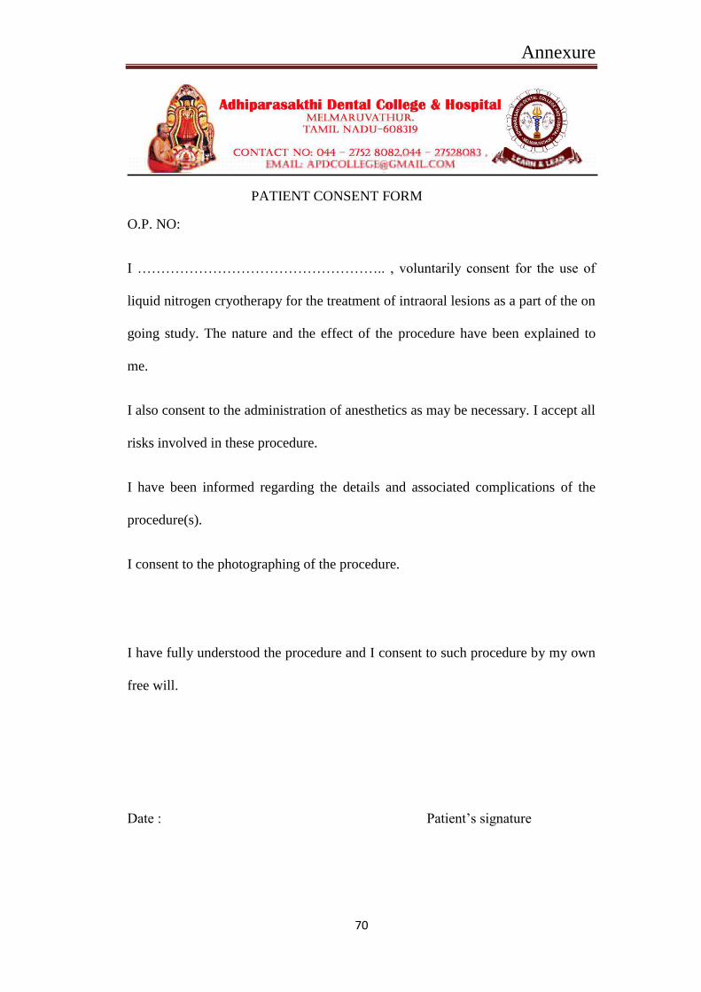
Annexure
70
PATIENT CONSENT FORM
O.P. NO:
I …………………………………………….. , voluntarily consent for the use of
liquid nitrogen cryotherapy for the treatment of intraoral lesions as a part of the on
going study. The nature and the effect of the procedure have been explained to
me.
I also consent to the administration of anesthetics as may be necessary. I accept all
risks involved in these procedure.
I have been informed regarding the details and associated complications of the
procedure(s).
I consent to the photographing of the procedure.
I have fully understood the procedure and I consent to such procedure by my own
free will.
Date : Patient’s signature
Annexure
71
xg;Gjy; gbtk;
GwNehahsp ml;il vz; : Njjp :
jpU/jpUkjp ...................................................... Mfpa ehd;>
vdf;F thapy; cl;Gwkhf cs;s fl;bia jput iel;bhp[d;
gad;gLj;jp rhpnra;J nfhs;s KOkdJld; rk;kjpf;fpNwd;.
NkYk; gpd; tpisTfisAk; mwpe;J nfhz;Nld;. vdJ Gif
glj;ij Nkw;$hpa Ma;tpw;F gad;gLj;jyhk; vd;gjw;Fk; KO
rk;kjk;.
vdNt ,e;j Ma;tpid gw;wp KOikahf mwpe;J
nfhz;Nld; vd;gjidAk; NkYk; ,e;j Ma;tpid elj;j
vdf;F KO rk;kjk; vd;gjidAk; ,jd; %yk; njhptpj;Jf;
nfhs;fpNwd;.
kUj;JtH ifnahg;gk; gq;Nfw;ghsH ifnahg;gk;
Annexure
72
Annexure
73
Related Documents