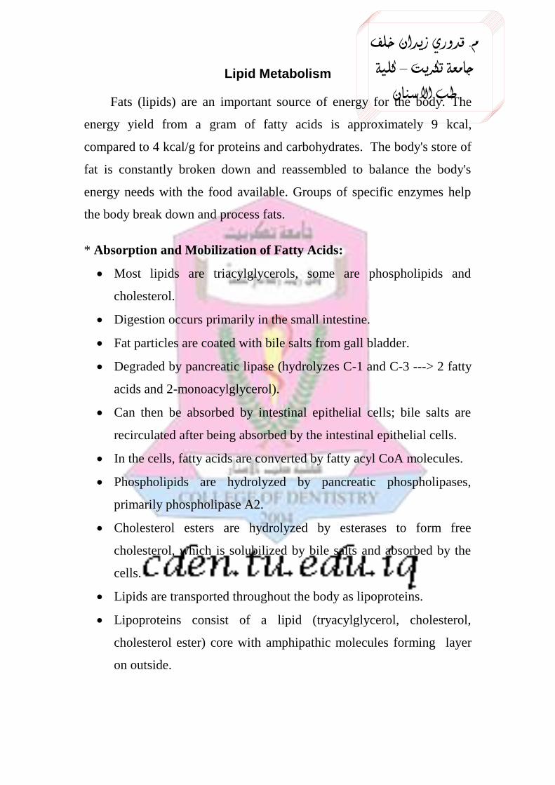
م. قدوري زيدان خلف جامعةكريت ت– كلية طبانسن اLipid Metabolism Fats (lipids) are an important source of energy for the body. The energy yield from a gram of fatty acids is approximately 9 kcal, compared to 4 kcal/g for proteins and carbohydrates. The body's store of fat is constantly broken down and reassembled to balance the body's energy needs with the food available. Groups of specific enzymes help the body break down and process fats. * Absorption and Mobilization of Fatty Acids: Most lipids are triacylglycerols, some are phospholipids and cholesterol. Digestion occurs primarily in the small intestine. Fat particles are coated with bile salts from gall bladder. Degraded by pancreatic lipase (hydrolyzes C-1 and C-3 ---> 2 fatty acids and 2-monoacylglycerol). Can then be absorbed by intestinal epithelial cells; bile salts are recirculated after being absorbed by the intestinal epithelial cells. In the cells, fatty acids are converted by fatty acyl CoA molecules. Phospholipids are hydrolyzed by pancreatic phospholipases, primarily phospholipase A2. Cholesterol esters are hydrolyzed by esterases to form free cholesterol, which is solubilized by bile salts and absorbed by the cells. Lipids are transported throughout the body as lipoproteins. Lipoproteins consist of a lipid (tryacylglycerol, cholesterol, cholesterol ester) core with amphipathic molecules forming layer on outside.

Welcome message from author
This document is posted to help you gain knowledge. Please leave a comment to let me know what you think about it! Share it to your friends and learn new things together.
Transcript
خلف زيدان قدوري. م
كلية – تكريت جامعة
االسنان طب
Lipid Metabolism
Fats (lipids) are an important source of energy for the body. The
energy yield from a gram of fatty acids is approximately 9 kcal,
compared to 4 kcal/g for proteins and carbohydrates. The body's store of
fat is constantly broken down and reassembled to balance the body's
energy needs with the food available. Groups of specific enzymes help
the body break down and process fats.
* Absorption and Mobilization of Fatty Acids:
Most lipids are triacylglycerols, some are phospholipids and
cholesterol.
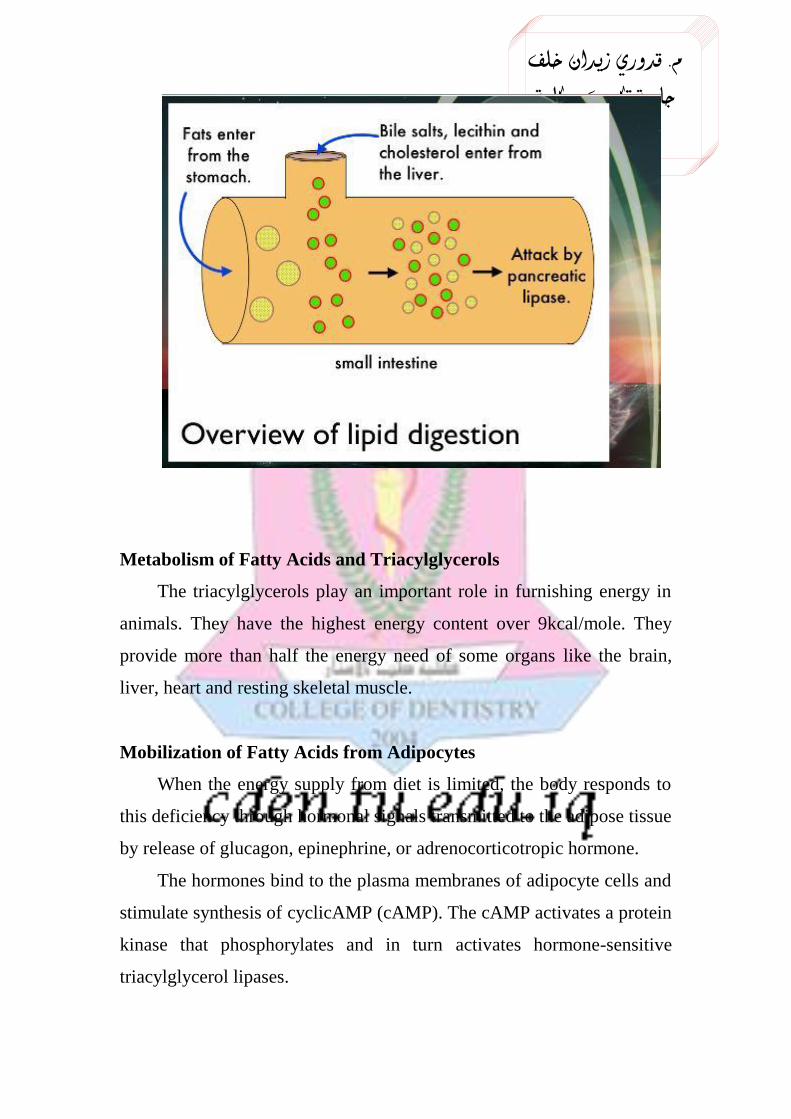
Digestion occurs primarily in the small intestine.
Fat particles are coated with bile salts from gall bladder.
Degraded by pancreatic lipase (hydrolyzes C-1 and C-3 ---> 2 fatty
acids and 2-monoacylglycerol).
Can then be absorbed by intestinal epithelial cells; bile salts are
recirculated after being absorbed by the intestinal epithelial cells.
In the cells, fatty acids are converted by fatty acyl CoA molecules.
Phospholipids are hydrolyzed by pancreatic phospholipases,
primarily phospholipase A2.
Cholesterol esters are hydrolyzed by esterases to form free
cholesterol, which is solubilized by bile salts and absorbed by the
cells.
Lipids are transported throughout the body as lipoproteins.
Lipoproteins consist of a lipid (tryacylglycerol, cholesterol,
cholesterol ester) core with amphipathic molecules forming layer
on outside.
خلف زيدان قدوري. م
كلية – تكريت جامعة
االسنان طب
Metabolism of Fatty Acids and Triacylglycerols
The triacylglycerols play an important role in furnishing energy in
animals. They have the highest energy content over 9kcal/mole. They
provide more than half the energy need of some organs like the brain,
liver, heart and resting skeletal muscle.
Mobilization of Fatty Acids from Adipocytes
When the energy supply from diet is limited, the body responds to
this deficiency through hormonal signals transmitted to the adipose tissue
by release of glucagon, epinephrine, or adrenocorticotropic hormone.
The hormones bind to the plasma membranes of adipocyte cells and
stimulate synthesis of cyclicAMP (cAMP). The cAMP activates a protein
kinase that phosphorylates and in turn activates hormone-sensitive
triacylglycerol lipases.
خلف زيدان قدوري. م
كلية – تكريت جامعة
االسنان طب
These lipases hydrolyze the triacylglycerols at position 1 or 3 to
produce diacylglycerols (DAG) and fatty acid, which is the rate limiting
step in the hydrolysis. The diacylglycerol lipases hydrolyze the DAG to
monoacylglycerols (MAG) and a fatty acid. Finally MAG lipases
hydrolyze MAG to fatty acid and glycerol.
The free fatty acids (FFA) produced by lipolysis move through the
plasma membranes of the adipose cells and endothelial cells of blood
capillaries by simple diffusion and bind to albumin in the blood plasma,
which are transported to peripheral tissues. The glycerol produced is
taken up by liver, phosphorylated and oxidized to dihydroxyacetone
phosphate, which is isomerised to glyceraldehydes-3-phosphate, an
intermediate of both glycolysis and gluconeogenesis.
Therefore, the glycerol is either converted to glucose
(gluconeogenesis) or to pyruvate (glycolysis).
Transport of Fatty Acids to the Mitochondria
The fatty acids transported to the different tissue cells must first be
activated or primed by reaction with Coenzyme A at the expense of ATP.
The reaction is catalyzed by Acyl CoA synthetase or also called
thiokinase, found in the cytosol and mitochondria of cells. The
pyrophosphate generated from ATP favors more Acyl CoA formation by
further hydrolysis. In order to undergo β-oxidation, the fatty acids must
enter the mitochondria. But they cannot easily cross it as such by passive
diffusion.
There are two fatty acid sources, those coming from absorption of
FFA and those from hydrolysis of triacylglycerols from adipose tissue.
The transport of acyl derivatives across the mitochondrial membrane
needs three acyltransferases (shuttles).
1. Specific for short chain acyl groups, does not require carnitine
خلف زيدان قدوري. م
كلية – تكريت جامعة
االسنان طب
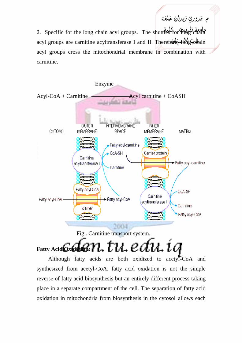
2. Specific for the long chain acyl groups. The shuttles for long chain
acyl groups are carnitine acyltransferase I and II. Therefore, long chain
acyl groups cross the mitochondrial membrane in combination with
carnitine.
Enzyme
Acyl-CoA + Carnitine Acyl carnitine + CoASH
Fig . Carnitine transport system.
Fatty Acid Oxidation:
Although fatty acids are both oxidized to acetyl-CoA and
synthesized from acetyl-CoA, fatty acid oxidation is not the simple
reverse of fatty acid biosynthesis but an entirely different process taking
place in a separate compartment of the cell. The separation of fatty acid
oxidation in mitochondria from biosynthesis in the cytosol allows each
خلف زيدان قدوري. م
كلية – تكريت جامعة
االسنان طب
process to be individually controlled and integrated with tissue
requirements.
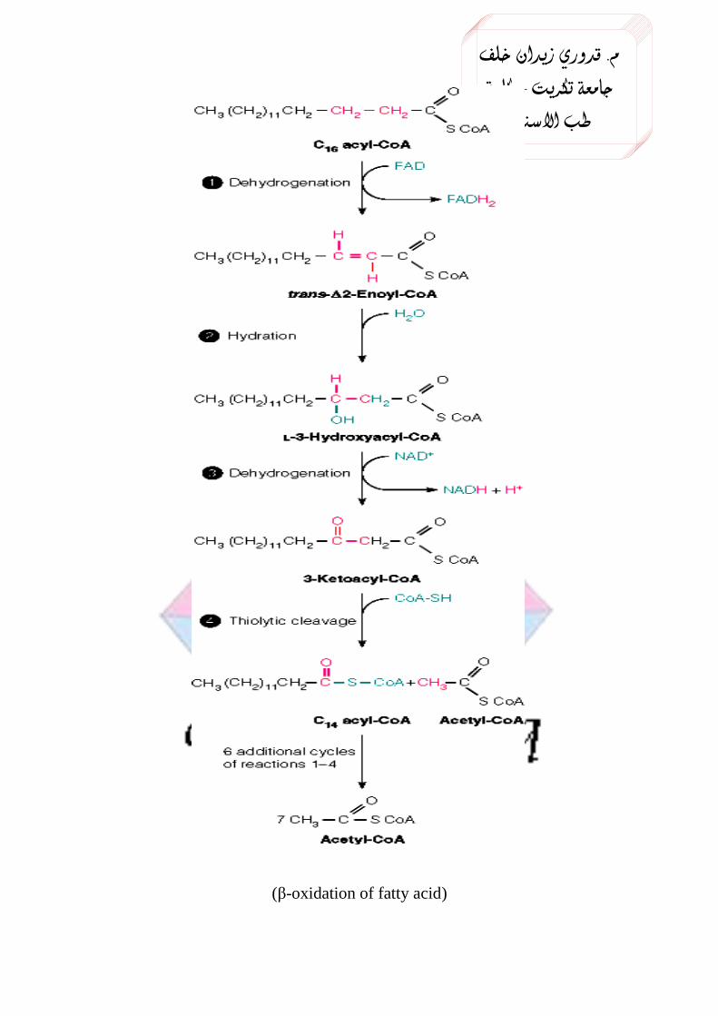
The successive oxidative removal of two carbons in the form of
acetyl–CoA beginning from the carboxyl end is called β-oxidation.
Each step in fatty acid oxidation involves acyl-CoA derivatives
catalyzed by separate enzymes, utilizes NAD+ and FAD as coenzymes,
and generates ATP. It is an aerobic process, requiring the presence of
oxygen.
Overall activation of fatty acid requires hydrolysis of two
phosphodiester bonds.
1. Acyl CoA dehydrogenase converts acyl CoA to acyl trans enoyl CoA
2. Hydratase converts it to 3-hydroxy acyl CoA.
3. Hydroxy acyl CoA dehydrogenase converts it to 3keto acyl CoA.
4. It is further converted to acyl CoA and acetyl CoA.by Thiolase.
The cycle is repeated 7 times for palmitic acid for complete oxidation.
خلف زيدان قدوري. م
كلية – تكريت جامعة
االسنان طب
(β-oxidation of fatty acid)
خلف زيدان قدوري. م
كلية – تكريت جامعة
االسنان طب
Complete oxidation of fatty acid can be divided in to two stages.
A. Formation of acetyl CoA.
B. Oxidation of acetyl CoA to CO2, water via TCA cycle.
Stochiometry of the reaction:
Palmitoyl CoA + 7FAD + 7 NAD +7CoA = 8 Acetyl CoA+7FADH2 +7
NADHH.
Energetics of palmitate oxidation:
Reduced equivalents enter ETC and produce energy rich phosphate
bonds. Acetyl CoA release energy through TCA cycle.
7 FADH2 → 7 x 2 = 14 ATPs
7NADHH → 7 x 3 = 21 ATPs
8 Acetyl CoA → 8 x 12 = 96 ATPs
Total ATP produced from one molecule of palmitic acid is 131. Two
ATPs (Two energy rich bonds) are utilized, during activation of fatty
acid. Therefore total gain of ATPs is 129.
Oxidation of Unsaturated Fatty Acids
The oxidation of unsaturated fatty acids requires two additional
enzymes called isomerase and reductase. Most naturally occurring
unsaturated fatty acids are in cis- configuration, which are not suitable for
the action of enoyl-CoA hydratases and hence they must be changed to
their trans isomer by an isomerase. The rest of the enzymes are needed
for the oxidation in addition to these two for the oxidation are the same.
Oxidation of Fatty Acids with Odd Number of Carbons
Ruminant animals can oxidize them by B- oxidation producing
acetylCoAs until a three carbon propionylCoA residue is left.The
acetylCoAs produced are funneled to the Krebs cycle but the
propionylCoA produced is converted to succinylCoA by three enzymatic
خلف زيدان قدوري. م
كلية – تكريت جامعة
االسنان طب
steps. SuccinoyCoA is an intermediate in the Kreb’s cycle and it can be
metabolized.
The fates of acetyl-CoA formed by b-oxidation of fatty acids are:
1. Oxidation to CO2 and H2O by citric acid cycle.
2. Synthesis of lipids like cholesterol, fatty acids and other steroids.
3. Formation of ketone bodies in the liver.
Regulation of Oxidation of Fatty Acids
• Hormones regulate lipolysis, in adipose tissue.More free fatty acids are
available for the β- oxidation.
• Insulin inhibits lipolysis.
• Acylcarnitine transferase-1 is inhibited by malonyl CoA , one of the
intermediates of fattyacid synthesis.
• High level of NADHH inhibits acyl CoA dehydrogenase.
• Increased concentration of acetyl CoA inhibits Thiolase.
• When the animal is well fed by carbohydrate, fatty acid oxidation is
lowered.
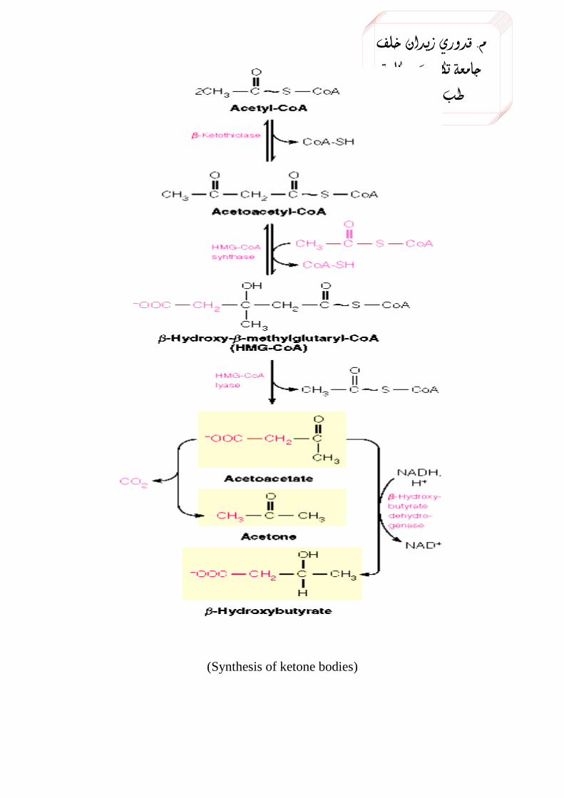
The metabolism of Ketone Bodies
When the level of acetyl CoA from β-oxidation increases in excess
of that required for entry into the citric acid cycle, It undergoes
ketogenesis in the mitochondria of liver (ketone body synthesis).
The three compounds, acetoacetate, β-hydroxybutyrate, and acetone
are collectively known as ketone bodies. The synthesis of ketone bodies
takes place during severe starvation or severe diabetes mellitus. During
such conditions, the body totally depends on the metabolism of stored
triacylglycerols to fulfill its energy demand.
In the synthesis, two molecules of acetyl CoA condense together to
form acetoacetyl CoA, a reaction catalyzed by thoilase. Another molecule
خلف زيدان قدوري. م
كلية – تكريت جامعة
االسنان طب
of acetyl CoA reacts with the acetoacetyl CoA to form 3-Hydroxy-3-
methyl glutaryl CoA (HMGCoA). This step is the rate limiting step and
the reaction is catalyzed by HMGCoA synthase enzyme. Note that this
compound is also an intermediate in the synthesis of cholesterol in the
liver cell cytosol but the mitochondrial HMGCoA goes to ketone body
synthesis.
The HMGCoA formed in the hepatocytes mitochondria by the action
of the enzyme HMGCoA lyase is changed to acetoacetate.
The acetoacetate, when its concentration is very high in blood is
spontaneously decarboxylated to acetone.
Acteoacetate can be converted to β-hydroxy butyrate by a
dehydrogenase enzyme. It is a reversible reaction.
The odor of acetone may be detected in the breath of a person who
has a high level of acetoacetate, like diabetic patients. During starvation
and severe diabetes mellitus peripheral tissues fully depend on ketone
bodies. Even tissues like the heart and brain depend mainly on ketone
bodies during such conditions to meet their energy demand.
خلف زيدان قدوري. م
كلية – تكريت جامعة
االسنان طب
(Synthesis of ketone bodies)
خلف زيدان قدوري. م
كلية – تكريت جامعة
االسنان طب
Utilization of Ketone Bodies
Ketone bodies are produced in the Liver and they are utilized in
extrahepatic tissues. Liver does not contain the enzyme required for
activation of ketone bodies.
Aceto acetate is activated and converted to aceto acetyl CoA for its
utilization. Aceto acetyl CoA is broken down to two molecules of acetyl
CoA, which enters TCA cycle for the production of energy.
Aceto acetate and β-hydroxy butyrate are the normal substrates for
respiration and important sources of energy .Renal cortex and heart
muscle use acetoacetate in preference to glucose .Brain switches over to
utilization of ketone bodies for energy during starvation and in
uncontrolled diabetes.
Acetone is exhaled out .It does not produce energy. Normal level of
ketone bodies in blood is 1mg %.In ketonemia, the level increases.
Excretion of ketone bodies increases in urine, called ketonuria. If the
patient suffers from both the signs, it is called ketosis.
Causes of Ketosis
1. Prolonged starvation, depletion of carbohydrate stores results in
increased fatty acid oxidation and ketosis.
2. Lactating mothers develop ketosis, if the carbohydrate demands are not
met with.
3. Diabetic patients with uncontrolled blood glucose, invariably suffer
from ketosis, ketoacidosis. Ketosis usually associated with sustained high
levels of free fatty acids in blood. Lipolysis and ketogenesis are regulated
by hormones.
خلف زيدان قدوري. م
كلية – تكريت جامعة
االسنان طب
Fatty Acid Synthesis
There are three basic sources of fatty acids in animals that can be
used for energy conversion processes, 1) fatty acids present in
triacylglycerols obtained from the diet, 2) fatty acids stored as
triacylglycerols in adipose tissue that are released by hydrolysis following
hormone stimulation (glucagon or epinephrine signaling), and 3) fatty
acids synthesized in the liver from excess carbohydrates and exported as
triacylglycerols.
For F.A. synthesis which occur in endoplasmic reticulum membrane,
acetyl-CoA is transported out of the mitochondria as citrate, Which is
cleaved in the cytosol to regenerate acetyl-CoA and oxaloacetate.
Prior to its utilization for F.A. synthesis, acetyl-CoA is converted by
carboxylation reaction into malonyl-CoA, this reaction is catalyzed by the
enzyme acetyl-CoA carboxylase(ACC).
The synthesis of F.A. from acetyl-CoA and malonyl-CoA is carried
out by fatty acid synthase(FAS).
Fatty acid synthesis involves four enzymatic activities, these are β-
ketoacyl-ACP synthase, β-ketoacyl-ACP reductase, 3-OH acyl-ACP
dehydratase, and enoyl-CoA reductase; where ACP is the acyl carrier
protein (a carrier portion in synthetic complex).
One might predict that the pathway for the synthesis of fatty acids
would be the reversal of the oxidation pathway. However, this would not
allow distinct regulation of the two pathways to occur even given the fact
that the pathways are separated within different cellular compartments.
The pathway for fatty acid synthesis occurs in the cytoplasm,
whereas, oxidation occurs in the mitochondria. The other major
difference is the use of nucleotide cofactors. Oxidation of fats involves
خلف زيدان قدوري. م
كلية – تكريت جامعة
االسنان طب
the reduction of FAD+ and NAD
+. Synthesis of fats involves the
oxidation of NADPH. However, the essential chemistry of the two
processes are reversals of each other. Both oxidation and synthesis of fats
utilize an activated two carbon intermediate, acetyl-CoA. However, the
acetyl-CoA in fat synthesis exists temporarily bound to the enzyme
complex as malonyl-CoA.
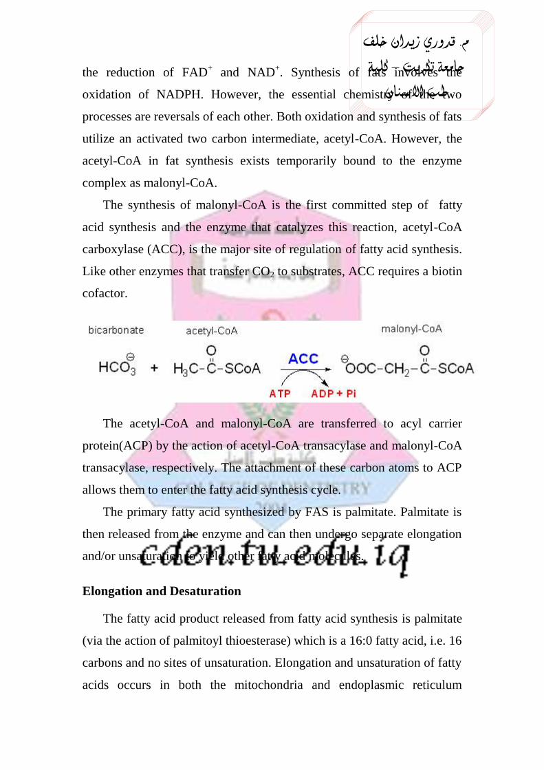
The synthesis of malonyl-CoA is the first committed step of fatty
acid synthesis and the enzyme that catalyzes this reaction, acetyl-CoA
carboxylase (ACC), is the major site of regulation of fatty acid synthesis.
Like other enzymes that transfer CO2 to substrates, ACC requires a biotin
cofactor.
The acetyl-CoA and malonyl-CoA are transferred to acyl carrier
protein(ACP) by the action of acetyl-CoA transacylase and malonyl-CoA
transacylase, respectively. The attachment of these carbon atoms to ACP
allows them to enter the fatty acid synthesis cycle.
The primary fatty acid synthesized by FAS is palmitate. Palmitate is
then released from the enzyme and can then undergo separate elongation
and/or unsaturation to yield other fatty acid molecules.
Elongation and Desaturation
The fatty acid product released from fatty acid synthesis is palmitate
(via the action of palmitoyl thioesterase) which is a 16:0 fatty acid, i.e. 16
carbons and no sites of unsaturation. Elongation and unsaturation of fatty
acids occurs in both the mitochondria and endoplasmic reticulum
خلف زيدان قدوري. م
كلية – تكريت جامعة
االسنان طب
(microsomal membranes). The predominant site of these processes is in
the endoplasmic reticulum (ER) membranes. Elongation involves
condensation of acyl-CoA groups with malonyl-CoA. The resultant
product is two carbons longer (CO2 is released from malonyl-CoA as in
the FAS reaction) which undergoes reduction, dehydration and reduction
yielding a saturated fatty acid. The reduction reactions of elongation
require NADPH as cofactor just as for the similar reactions catalyzed by
FAS. Mitochondrial elongation involves acetyl-CoA units and is a
reversal of oxidation except that the final reduction utilizes NADPH
instead of FADH2 as cofactor.
Desaturation occurs in the ER membranes as well and in mammalian
cells involves 4 broad specificity fatty acyl-CoA desaturases (non-heme
iron containing enzymes). These enzymes introduce unsaturation at C4,
C5, C6 or C9. Since these enzymes cannot introduce sites of unsaturation
beyond C9 they cannot synthesize either linoleate (18:2Δ9,12
) or linolenate
(18:3Δ9,12,15
). These fatty acids must be acquired from the diet and are,
therefore, referred to as essential fatty acids. Linoleic is especially
important in that it required for the synthesis of arachidonic acid. As we
shall encounter later, arachindonate is a precursor for the eicosanoids (the
prostaglandins and thromboxanes). It is this role of fatty acids in
eicosanoid synthesis that leads to poor growth, wound healing and
dermatitis in persons on fat free diets. Also, linoleic acid is a constituent
of epidermal cell sphingolipids that function as the skins water
permeability barrier.
* Biosynthesis of cholesterol:
Slightly less than half of the cholesterol in the body derives from
biosynthesis. Biosynthesis in the liver accounts for approximately 10%,
and in the intestines approximately 15%, of the amount produced each
خلف زيدان قدوري. م
كلية – تكريت جامعة
االسنان طب
day. Cholesterol synthesis occurs in the cytoplasm and in the microsomes
from the two-carbon acetate group of acetyl-CoA.
The process has five major steps:
1- Acetyl-CoAs are converted to 3-hydroxy-3-methylglutaryl-CoA
(HMG-CoA).
2- HMG-CoA is converted to mevalonate.
3- Mevalonate is converted to the isoprene based molecule,
isopentenyl pyrophosphate(IPP), with the concomitant loss of
CO2.
4- IPP is converted to squalene.
5- Squalene is converted to cholesterol.
Synthesis of Triglycerides
Fatty acids are stored for future use as triacylglycerols (TAGs) in all
cells, but primarily in adipocytes of adipose tissue. TAGs constitute
molecules of glycerol to which three fatty acids have been esterified. The
fatty acids present in TAGs are predominantly saturated. The major
building block for the synthesis of TAGs, in tissues other than adipose
tissue, is glycerol. Adipocytes lack glycerol kinase, therefore,
dihydroxyacetone phosphate (DHAP), produced during glycolysis, is the
precursor for TAG synthesis in adipose tissue. This means that adipoctes
must have glucose to oxidize in order to store fatty acids in the form of
TAGs. DHAP can also serve as a backbone precursor for TAG synthesis
in tissues other than adipose, but does so to a much lesser extent than
glycerol.
The glycerol backbone of TAGs is activated by phosphorylation at
the C-3 position by glycerol kinase. The fatty acids incorporated into
TAGs are activated to acyl-CoAs through the action of acyl-CoA
خلف زيدان قدوري. م
كلية – تكريت جامعة
االسنان طب
synthetases. Two molecules of acyl-CoA are esterified to glycerol-3-
phosphate to yield 1,2-diacylglycerol phosphate (commonly identified as
phosphatidic acid). The phosphate is then removed, by phosphatidic acid
phosphatase (PAP1), to yield 1,2-diacylglycerol, the substrate for
addition of the third fatty acid. Intestinal monoacylglycerols, derived
from the hydrolysis of dietary fats, can also serve as substrates for the
synthesis of 1,2-diacylglycerols.
* Formation of chylomicrons(CM):
Chylomicrons are formed from dietary fat (principally triglyceride; but
also cholesterol) in enterocytes; they enter the lymphatics and reach the
systemic circulation via the thoracic duct. Chylomicrons are the major
transport form of exogenous (dietary) fat. Triglyceride constitutes about
ninety percent of the lipid. Triglyceride is removed from chylomicrons by
the action of the enzyme lipoprotein lipase(LPL), located on the luminal
surface of the capillary endothelium of adipose tissue, skeletal and
cardiac muscle and lactating breast, so that free fatty acids are delivered
to these tissues either to be used as energy substrates or, after re-
esterification to triglyceride, for energy storage.
* Essential features of lipoprotein metabolism:
• Dietary triglyceride is transported in chylomicrons to tissues where it
may be used as an energy source or stored.
• Endogenous triglyceride, synthesized in the liver, is transported in
VLDL and is also available to tissues as an energy source or for storage.
• Cholesterol synthesized in the liver is transported to tissues in LDL,
derived from VLDL; dietary cholesterol reaches the liver in chylomicron
remnants.
خلف زيدان قدوري. م
كلية – تكريت جامعة
االسنان طب
• HDL acquire cholesterol from peripheral cells and other lipoproteins
and this is esterified by lecithin-cholesterol acyltransferase(LCAT).
Cholesterol ester is transferred to remnant particles which are taken up by
the liver, whence the cholesterol is excreted.
* Lipolysis
Lipolysis is the breakdown of fat stored in fat cells. During this
process, free fatty acids are released into the bloodstream and circulate
throughout the body. Ketones are produced, and are found in large
quantities in ketosis (an adaptive metabolic state that occurs when
insufficient carbohydrates are present in the diet). The following
hormones induce lipolysis: epinephrine, norepinephrine, glucagon and
adrenocorticotropic hormone.
Triglycerides undergo lipolysis (hydrolysis by lipases) and are broken
down into glycerol and fatty acids. Once released into the blood, the
relatively hydrophobic free fatty acids bind to serum albumin for
transport to tissues that require energy. The glycerol also enters the
bloodstream and is absorbed by the liver or kidney where it is converted
to glycerol 3-phosphate by the enzyme glycerol kinase. Hepatic glycerol
3-phosphate is mostly converted into dihydroxyacetone (DHAP) and then
glyceraldehyde 3-phosphate (G3P) to rejoin the glycolysis and
gluconeogenesis pathway.
* Hyperlipidemia hyperlipoproteinemia or dyslipidemia: is the
presence of raised or abnormal levels of lipids and/or lipoproteins in the
blood.
Lipid and lipoprotein abnormalities are extremely common in the
general population, and are regarded as a highly modifiable risk factor for
خلف زيدان قدوري. م
كلية – تكريت جامعة
االسنان طب
cardiovascular disease due to the influence of cholesterol, one of the most
clinically relevant lipid substances, on atherosclerosis. In addition, some
forms may predispose to acute pancreatitis.
* Hypercholesterolemia: Hypercholesterolemia is the presence of high
levels of cholesterol in the blood. It is not a disease but a metabolic
derangement that can be secondary to many diseases and can contribute
to many forms of disease, most notably cardiovascular disease. It is
closely related to the terms. Familial hypercholesterolemia is a rare
genetic disorder that can occur in families, where sufferers cannot
properly metabolise cholesterol.
* Hypocholesterolemia: Abnormally low levels of cholesterol,some
studies suggest a link with depression, cancer and cerebral hemorrhage.
* After digestion of lipids, some changes are happened in intestine for
absorption, these are:
1- Hydrolysis of triglycerides(TG) to free fatty acids(FFA) and
mono- acylglycerols.
2- Solubilization of FFA and monoacylglycerols by detergents (bile
acids) and transportation from the intestinal lumen toward the cell
surface.
3- Uptake of FFA and monoacylglycerols into the cell and resynthesis
to triglycerides.
4- Packaging of newly synthesized TG into special lipid- rich
globules called chylomicrons.
5- Exocytosis of chylomicrons from cells and release into lymph.

خلف زيدان قدوري. م
كلية – تكريت جامعة
االسنان طب
Related Documents