Lipid dependence of diadinoxanthin solubilization and de-epoxidation in artificial membrane systems resembling the lipid composition of the natural thylakoid membrane Reimund Goss a, ⁎ , Dariusz Latowski b,c , Joanna Grzyb b , Astrid Vieler a , Martin Lohr d , Christian Wilhelm a , Kazimierz Strzalka b a Institute of Biology I, Plant Physiology, University of Leipzig, Johannisallee 21-23, 04103 Leipzig, Germany b Faculty of Biotechnology, Jagiellonian University, ul. Gronostajowa 7, 30-387 Krakow, Poland c Department of Chemistry, Pedagogical University, ul. Podchorazych 2, 30-084 Krakow, Poland d Institute of General Botany, Johannes Gutenberg-University Mainz, Saarstrasse 21, 55099 Mainz, Germany Received 18 April 2006; received in revised form 2 June 2006; accepted 2 June 2006 Available online 7 June 2006 Abstract In the present study, the solubility and enzymatic de-epoxidation of diadinoxanthin (Ddx) was investigated in three different artificial membrane systems: (1) Unilamellar liposomes composed of different concentrations of the bilayer forming lipid phosphatidylcholine (PC) and the inverted hexagonal phase (H II phase) forming lipid monogalactosyldiacylglycerol (MGDG), (2) liposomes composed of PC and the H II phase forming lipid phosphatidylethanolamine (PE), and (3) an artificial membrane system composed of digalactosyldiacylglycerol (DGDG) and MGDG, which resembles the lipid composition of the natural thylakoid membrane. Our results show that Ddx de-epoxidation strongly depends on the concentration of the inverted hexagonal phase forming lipids MGDG or PE in the liposomes composed of PC or DGDG, thus indicating that the presence of inverted hexagonal structures is essential for Ddx de-epoxidation. The difference observed for the solubilization of Ddx in H II phase forming lipids compared with bilayer forming lipids indicates that Ddx is not equally distributed in the liposomes composed of different concentrations of bilayer versus non-bilayer lipids. In artificial membranes with a high percentage of bilayer lipids, a large part of Ddx is located in the membrane bilayer. In membranes composed of equal proportions of bilayer and H II phase forming lipids, the majority of the Ddx molecules is located in the inverted hexagonal structures. The significance of the pigment distribution and the three-dimensional structure of the H II phase for the de-epoxidation reaction is discussed, and a possible scenario for the lipid dependence of Ddx (and violaxanthin) de-epoxidation in the native thylakoid membrane is proposed. © 2006 Elsevier B.V. All rights reserved. Keywords: Bilayer lipid; Non-bilayer lipid; Inverted hexagonal phase; Thylakoid membrane; Diadinoxanthin; Xanthophyll cycle 1. Introduction The xanthophyll cycles of vascular plants and algae are photo- protective mechanisms located in the chloroplast. At present, three different cycles are known: the violaxanthin (Vx) cycle of vascular plants and green and brown algae [1,2], the lutein epoxide cycle that has been detected in a limited set of vascular plants [3,4], and the diadinoxanthin (Ddx) cycle of the algal classes Bacillariophy- ceae, Chrysophyceae, Xanthophyceae, Haptophyceae and Dino- phyceae [5]. The de-epoxidation reaction of the Ddx cycle comprises one step from the mono-epoxy xanthophyll Ddx to the epoxy-free diatoxanthin (Dtx), whereas the Vx cycle consists of a forward reaction with two de-epoxidation steps, and which trans- forms the di-epoxy-xanthophyll Vx into the epoxy-free zeaxanthin (Zx) via the intermediate antheraxanthin (Ax). Both de-epoxida- tion sequences take place when vascular plants or algae are illu- minated with high actinic light intensities. Low light or darkness Biochimica et Biophysica Acta 1768 (2007) 67 – 75 www.elsevier.com.locate/bbamem Abbreviations: LHC/FCP, light-harvesting complexes of vascular plants or diatoms, respectively; DDE, diadinoxanthin de-epoxidase; VDE, violaxanthin de-epoxidase; Ddx, diadinoxanthin; Dtx, diatoxanthin; Vx, violaxanthin; Zx, zeaxanthin; MGDG, monogalactosyldiacylgycerol; DGDG, digalactosyldiacyl- glycerol; PC, phosphatidylcholine; PE, phosphatidylethanolamine ⁎ Corresponding author. Tel.: +49 341 9736873; fax: +49 341 9736899. E-mail address: [email protected] (R. Goss). 0005-2736/$ - see front matter © 2006 Elsevier B.V. All rights reserved. doi:10.1016/j.bbamem.2006.06.006

Welcome message from author
This document is posted to help you gain knowledge. Please leave a comment to let me know what you think about it! Share it to your friends and learn new things together.
Transcript
ta 1768 (2007) 67–75www.elsevier.com.locate/bbamem
Biochimica et Biophysica Ac
Lipid dependence of diadinoxanthin solubilization and de-epoxidation inartificial membrane systems resembling the lipid composition of
the natural thylakoid membrane
Reimund Goss a,⁎, Dariusz Latowski b,c, Joanna Grzyb b, Astrid Vieler a,Martin Lohr d, Christian Wilhelm a, Kazimierz Strzalka b
a Institute of Biology I, Plant Physiology, University of Leipzig, Johannisallee 21-23, 04103 Leipzig, Germanyb Faculty of Biotechnology, Jagiellonian University, ul. Gronostajowa 7, 30-387 Krakow, Polandc Department of Chemistry, Pedagogical University, ul. Podchorazych 2, 30-084 Krakow, Poland
d Institute of General Botany, Johannes Gutenberg-University Mainz, Saarstrasse 21, 55099 Mainz, Germany
Received 18 April 2006; received in revised form 2 June 2006; accepted 2 June 2006Available online 7 June 2006
Abstract
In the present study, the solubility and enzymatic de-epoxidation of diadinoxanthin (Ddx) was investigated in three different artificialmembrane systems: (1) Unilamellar liposomes composed of different concentrations of the bilayer forming lipid phosphatidylcholine (PC) and theinverted hexagonal phase (HII phase) forming lipid monogalactosyldiacylglycerol (MGDG), (2) liposomes composed of PC and the HII phaseforming lipid phosphatidylethanolamine (PE), and (3) an artificial membrane system composed of digalactosyldiacylglycerol (DGDG) andMGDG, which resembles the lipid composition of the natural thylakoid membrane. Our results show that Ddx de-epoxidation strongly depends onthe concentration of the inverted hexagonal phase forming lipids MGDG or PE in the liposomes composed of PC or DGDG, thus indicating thatthe presence of inverted hexagonal structures is essential for Ddx de-epoxidation. The difference observed for the solubilization of Ddx in HII
phase forming lipids compared with bilayer forming lipids indicates that Ddx is not equally distributed in the liposomes composed of differentconcentrations of bilayer versus non-bilayer lipids. In artificial membranes with a high percentage of bilayer lipids, a large part of Ddx is located inthe membrane bilayer. In membranes composed of equal proportions of bilayer and HII phase forming lipids, the majority of the Ddx molecules islocated in the inverted hexagonal structures. The significance of the pigment distribution and the three-dimensional structure of the HII phase forthe de-epoxidation reaction is discussed, and a possible scenario for the lipid dependence of Ddx (and violaxanthin) de-epoxidation in the nativethylakoid membrane is proposed.© 2006 Elsevier B.V. All rights reserved.
Keywords: Bilayer lipid; Non-bilayer lipid; Inverted hexagonal phase; Thylakoid membrane; Diadinoxanthin; Xanthophyll cycle
1. Introduction
The xanthophyll cycles of vascular plants and algae are photo-protective mechanisms located in the chloroplast. At present, threedifferent cycles are known: the violaxanthin (Vx) cycle of vascular
Abbreviations: LHC/FCP, light-harvesting complexes of vascular plants ordiatoms, respectively; DDE, diadinoxanthin de-epoxidase; VDE, violaxanthinde-epoxidase; Ddx, diadinoxanthin; Dtx, diatoxanthin; Vx, violaxanthin; Zx,zeaxanthin; MGDG, monogalactosyldiacylgycerol; DGDG, digalactosyldiacyl-glycerol; PC, phosphatidylcholine; PE, phosphatidylethanolamine⁎ Corresponding author. Tel.: +49 341 9736873; fax: +49 341 9736899.E-mail address: [email protected] (R. Goss).
0005-2736/$ - see front matter © 2006 Elsevier B.V. All rights reserved.doi:10.1016/j.bbamem.2006.06.006
plants and green and brown algae [1,2], the lutein epoxide cyclethat has been detected in a limited set of vascular plants [3,4], andthe diadinoxanthin (Ddx) cycle of the algal classes Bacillariophy-ceae, Chrysophyceae, Xanthophyceae, Haptophyceae and Dino-phyceae [5]. The de-epoxidation reaction of the Ddx cyclecomprises one step from the mono-epoxy xanthophyll Ddx to theepoxy-free diatoxanthin (Dtx), whereas the Vx cycle consists of aforward reaction with two de-epoxidation steps, and which trans-forms the di-epoxy-xanthophyll Vx into the epoxy-free zeaxanthin(Zx) via the intermediate antheraxanthin (Ax). Both de-epoxida-tion sequences take place when vascular plants or algae are illu-minated with high actinic light intensities. Low light or darkness

68 R. Goss et al. / Biochimica et Biophysica Acta 1768 (2007) 67–75
stimulates the back reaction of the cycles, in which the de-epox-idized xanthophylls are reverted to the respective epoxidized forms[for a review see 6].
The de-epoxidation step in the Ddx cycle is catalyzed by theenzyme Ddx de-epoxidase (DDE), which is located in thethylakoid lumen and becomes activated by a decreasing lumenalpH due to light-driven photosynthetic electron transport. DDE isoptimized in various ways compared with the xanthophyll cycleenzyme of vascular plants, the Vx de-epoxidase (VDE). DDEactivation can be observed at almost neutral pH values [7,8],whereas for VDE activationmore acidic pHvalues are needed [9].Further, DDE exhibits a significantly lower KM value for its co-substrate ascorbate comparedwith theVDE of vascular plants [8].
Like VDE, DDE requires the presence of the major thylakoidmembrane lipid MGDG for activity [10–13]. MGDG comprisesabout half of the total thylakoid membrane lipid, the secondmost abundant thylakoid lipid is the bilayer-forming DGDGwhich amounts to about 30% of the thylakoid lipids [14]. Theoverall lipid composition of thylakoid membranes of vascularplants and diatoms is comparable and MGDG is also the mainthylakoid lipid of diatoms. MGDG and DGDG from diatomsmay, however, contain long chain fatty acids with up to 22carbon atoms and as many as six double bonds [for a review ondiatom lipids see 15]. Due to a strongly increased solubility ofDdx in MGDG compared with Vx, the MGDG concentrationsfor optimal DDE activity are much lower compared to thoseneeded for full activation of VDE [13]. In addition to its role inpigment solubilization, MGDG provides another essential fea-ture needed for xanthophyll de-epoxidation. MGDG belongs tothe so-called non-bilayer lipids which, due to their small head-group area and critical packing parameter value higher than one,form inverted hexagonal structures (HII phases) in an aqueousmedium [16,17]. These three-dimensional structures have beenshown to be required for efficient Vx de-epoxidation in artificialliposomes composed of the lipids phosphatidylcholine (PC) andMGDG [11,12,18]. Latowski et al. [11] proposed that the HII
phases enable the binding of VDE to the thylakoid membraneafter the pH-dependent activation of the enzyme has takenplace. Inverted hexagonal structures which may also be formedby the phospholipid phosphatidylethanolamine (PE) and whichsupport Vx de-epoxidation [12], seem to be essential for Ddxde-epoxidation, as well [13]. However, the data of Goss et al.[13] were derived from undefined lipid/water systems. More-over, experiments showing the influence of HII phase forminglipids on Ddx de-epoxidation in liposome systems resemblingthe natural thylakoid membrane are still lacking.
Although the existence of HII phases in thylakoid membraneshas been indicated by various methods [19,20], their precisearrangement in artificial lipid/water systems or in the naturalthylakoid membrane is still unclear. It has been suggested thatthe non-bilayer lipids are forced into a bilayer structure in thenative thylakoid membrane due to interactions with the mainlight-harvesting complex of photosystem II, the LHC II [21,22].Garab et al. [22] also proposed, that under certain conditions,such as structural rearrangements of proteins or even proteindegradation, non-bilayer lipids can be sequestered from themembrane and form HII phases which are attached to the surface
of the thylakoid bilayer. These inverted hexagonal phases stay indirect contact with the bilayer, so that rapid and direct inter-actions between the two lipid phases are possible.
In the present study, we have investigated, in more detail, thelipid dependence of Ddx de-epoxidation. To overcome thelimitations of the undefined MGDG/water system we chose touse artificial membrane systems that more closely resemble thenatural thylakoid membrane. The influence of inverted hexag-onal structures on Ddx de-epoxidation was studied in PC lipo-somes that were supplemented with increasing concentrations ofeither of the HII phase forming lipids MGDG or PE. In a thirdmembrane system, a combination of the bilayer lipid DGDG andthe non-bilayer lipid MGDG was used. In addition to the kineticanalysis of Ddx de-epoxidation, solubilization of Ddx in lipo-somes or bilayer/non-bilayer lipids was measured to obtaininformation about the distribution of xanthophyll cycle pigmentsin bilayer versus inverted hexagonal phases. The data of thepresent study are discussed with respect to their relevance for thelipid dependence of de-epoxidation in artificial membrane sys-tems and in the natural thylakoid membrane.
2. Materials and methods
2.1. Plant material
The diatom Cyclotella meneghiniana (strain 1020-1a) was obtained from theCulture Collection of Algae, Göttingen (SAG, FRG) and was grown as batchculture in silica-enriched ASP medium according to Lohr andWilhelm [23]. ThePPFD during cultivation was set to 50 μmol m−2 s−1 with a light/dark regime of16/8 h. The temperature of the growth chamber was adjusted to 20 °C. For thepreparation of DDE, Ddx and Dtx, algal cultures with a chlorophyll concen-tration of 10 mg Chl a/c L−1 were used.
2.2. Preparation of DDE
DDE was prepared from dark-adapted cells of C. meneghiniana according toGoss et al. [13]. After the preparation, thylakoid lumen extracts enriched in DDEwere frozen in liquid nitrogen and stored at −80 °C until use in the in vitro de-epoxidation assays.
2.3. Preparation of pigments
Ddx and Dtx were extracted from illuminated cells of C. meneghiniana andpurified by HPLC, essentially as described by Goss et al. [13] and Grounevaet al. [8]. Ddx and Dtx from several HPLC separations were pooled, dried andstored at −80 °C until further use in the DDE enzyme assays.
2.4. Preparation of liposomes
Liposomes were prepared from three different combinations of lipids: thebilayer forming lipid phosphatidylcholine (PC) was combined with (1) theinverted hexagonal structure forming lipid monogalactosyldiacylglycerol(MGDG) or (2) another HII forming lipid, phosphatidylethanolamine (PE). In athird combination of lipids, the bilayer forming lipid digalactosyldiacylglycerol(DGDG)was combinedwithMGDG. For all different combinations, ratios of 85/15, 70/30 and 50/50 mol% of bilayer versus HII forming lipids were prepared.This corresponds to final lipid concentrations of 32.9/5.8, 27.1/11.6 and 19.35/19.35 μM, respectively. The mixture of lipids in chloroform was complementedwith Ddx, subsequently evaporated under a stream of nitrogen, and further driedunder vacuum for 1 h. The dried lipid/pigment mixture was dissolved in ethanoland injected slowly with a Hamilton syringe into the reaction medium of the de-epoxidation assay (10 mM KCl, 5 mM MgCl2 and 40 mM MES, pH 5.2) undercontinuous bubbling with nitrogen. The final concentration of ethanol in the
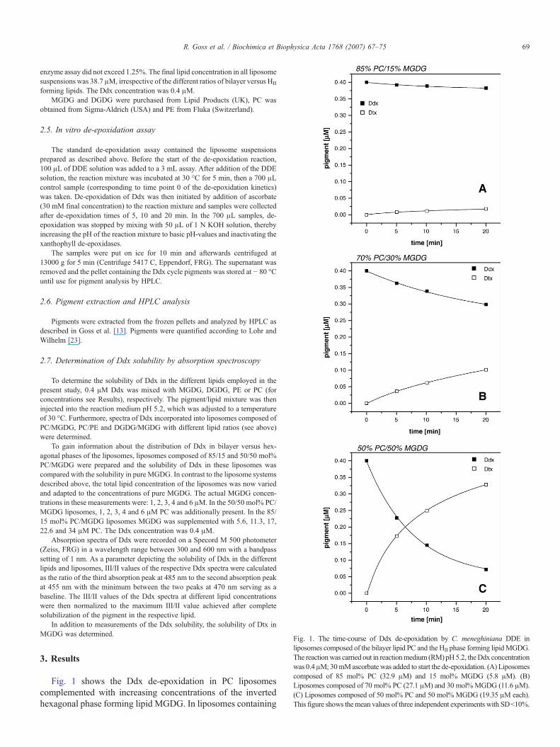
Fig. 1. The time-course of Ddx de-epoxidation by C. meneghiniana DDE inliposomes composed of the bilayer lipid PC and the HII phase forming lipidMGDG.The reactionwas carried out in reactionmedium (RM)pH5.2, theDdx concentrationwas 0.4μM; 30mMascorbate was added to start the de-epoxidation. (A) Liposomescomposed of 85 mol% PC (32.9 μM) and 15 mol% MGDG (5.8 μM). (B)Liposomes composed of 70 mol% PC (27.1 μM) and 30 mol%MGDG (11.6 μM).(C) Liposomes composed of 50 mol% PC and 50 mol% MGDG (19.35 μM each).This figure shows themean values of three independent experiments with SD<10%.
69R. Goss et al. / Biochimica et Biophysica Acta 1768 (2007) 67–75
enzyme assay did not exceed 1.25%. The final lipid concentration in all liposomesuspensions was 38.7 μM, irrespective of the different ratios of bilayer versus HII
forming lipids. The Ddx concentration was 0.4 μM.MGDG and DGDG were purchased from Lipid Products (UK), PC was
obtained from Sigma-Aldrich (USA) and PE from Fluka (Switzerland).
2.5. In vitro de-epoxidation assay
The standard de-epoxidation assay contained the liposome suspensionsprepared as described above. Before the start of the de-epoxidation reaction,100 μL of DDE solution was added to a 3 mL assay. After addition of the DDEsolution, the reaction mixture was incubated at 30 °C for 5 min, then a 700 μLcontrol sample (corresponding to time point 0 of the de-epoxidation kinetics)was taken. De-epoxidation of Ddx was then initiated by addition of ascorbate(30 mM final concentration) to the reaction mixture and samples were collectedafter de-epoxidation times of 5, 10 and 20 min. In the 700 μL samples, de-epoxidation was stopped by mixing with 50 μL of 1 N KOH solution, therebyincreasing the pH of the reaction mixture to basic pH-values and inactivating thexanthophyll de-epoxidases.
The samples were put on ice for 10 min and afterwards centrifuged at13000 g for 5 min (Centrifuge 5417 C, Eppendorf, FRG). The supernatant wasremoved and the pellet containing the Ddx cycle pigments was stored at − 80 °Cuntil use for pigment analysis by HPLC.
2.6. Pigment extraction and HPLC analysis
Pigments were extracted from the frozen pellets and analyzed by HPLC asdescribed in Goss et al. [13]. Pigments were quantified according to Lohr andWilhelm [23].
2.7. Determination of Ddx solubility by absorption spectroscopy
To determine the solubility of Ddx in the different lipids employed in thepresent study, 0.4 μM Ddx was mixed with MGDG, DGDG, PE or PC (forconcentrations see Results), respectively. The pigment/lipid mixture was theninjected into the reaction medium pH 5.2, which was adjusted to a temperatureof 30 °C. Furthermore, spectra of Ddx incorporated into liposomes composed ofPC/MGDG, PC/PE and DGDG/MGDG with different lipid ratios (see above)were determined.
To gain information about the distribution of Ddx in bilayer versus hex-agonal phases of the liposomes, liposomes composed of 85/15 and 50/50 mol%PC/MGDG were prepared and the solubility of Ddx in these liposomes wascompared with the solubility in pure MGDG. In contrast to the liposome systemsdescribed above, the total lipid concentration of the liposomes was now variedand adapted to the concentrations of pure MGDG. The actual MGDG concen-trations in these measurements were: 1, 2, 3, 4 and 6 μM. In the 50/50 mol% PC/MGDG liposomes, 1, 2, 3, 4 and 6 μM PC was additionally present. In the 85/15 mol% PC/MGDG liposomes MGDG was supplemented with 5.6, 11.3, 17,22.6 and 34 μM PC. The Ddx concentration was 0.4 μM.
Absorption spectra of Ddx were recorded on a Specord M 500 photometer(Zeiss, FRG) in a wavelength range between 300 and 600 nm with a bandpasssetting of 1 nm. As a parameter depicting the solubility of Ddx in the differentlipids and liposomes, III/II values of the respective Ddx spectra were calculatedas the ratio of the third absorption peak at 485 nm to the second absorption peakat 455 nm with the minimum between the two peaks at 470 nm serving as abaseline. The III/II values of the Ddx spectra at different lipid concentrationswere then normalized to the maximum III/II value achieved after completesolubilization of the pigment in the respective lipid.
In addition to measurements of the Ddx solubility, the solubility of Dtx inMGDG was determined.
3. Results
Fig. 1 shows the Ddx de-epoxidation in PC liposomescomplemented with increasing concentrations of the invertedhexagonal phase forming lipid MGDG. In liposomes containing

Fig. 2. Absorption spectra of Ddx in RM pH 5.2 and in liposomes with differentconcentrations of PC and MGDG. The mol percentages of the lipids in theliposomes were: 85/15, 70/30 and 50/50 mol% PC/MGDG, respectively (foractual lipid concentrations see legend of Fig. 1). The Ddx concentration was0.4 μM.
Table 1Ddx conversion rate in the artificial membrane systems used in the present study
Bilayer/non-bilayer lipid(mol%)
Ddx conversion rate (nM Ddx min−1) in differentartificial membrane systems
PC/MGDG DGDG/MGDG PC/PE
100/0 0.4 0.3 0.485/15 1.7 1 0.870/30 7.3 5 350/50 34.8 25.9 16
Ddx de-epoxidation rates of the C. meneghiniana DDE in the three differentartificial membrane systems with different ratios of bilayer (PC, DGDG) versusnon-bilayer lipids (PE, MGDG), for actual lipid concentrations see Fig. 1. Thede-epoxidation rate is depicted as nM Ddx min−1 and was calculated 5 min afterthe start of the in vitro de-epoxidation assay. The total lipid concentration was38.7 μM in all membrane systems. The Ddx concentration at the beginning ofthe measurement was 0.4 μM; the de-epoxidation reaction was started with theaddition of 30 mM ascorbate. This table shows the mean values of threeindependent experiments with SD<10%.
70 R. Goss et al. / Biochimica et Biophysica Acta 1768 (2007) 67–75
15 mol% of MGDG (Fig. 1A), a slow de-epoxidation of Ddxwas observed, typically leading to the conversion of only 5% ofthe total Ddx to Dtx within a timespan of 20 min. The de-epoxidation rate (Table 1) was consequently low with a value ofonly 1.7 nM Ddx de-epoxidized per minute. In the liposomesystem composed of 70 mol% PC and 30 mol% MGDG(Fig. 1B) a more pronounced de-epoxidation of Ddx to Dtx wasdetected, and after 20 min of the in vitro enzyme assay about25% of the Ddx pool had been converted to Dtx. The de-epoxidation rate was also significantly increased, and showed avalue of 7.3 nM Ddx min− 1. A fast Ddx de-epoxidation ac-companied by an almost complete conversion of Ddx to Dtxwas found in liposomes which were composed of equalamounts of both the bilayer forming lipid PC and the HII
inducing lipid MGDG (Fig. 1C). In this liposome system Ddxde-epoxidation proceeded with a high rate of 34.8 nM Ddxmin− 1 (Table 1), a value that is more than 20 times higher thanthe de-epoxidation rate in liposomes complemented with 15 mol%MGDG. The fast de-epoxidation reaction led to the conversion ofhalf of the Ddx pool to Dtx within 5–6 min.
The dependence of the enzymatic reaction on the concen-tration of MGDG in the liposomes was not caused by a limitedsolubility of Ddx in the liposomes with lower amounts ofMGDG. From the absorbance spectra in Fig. 2, it is clear that inall different PC liposome systems containing 85/15, 70/30 and50/50 mol% PC/MGDG, respectively, Ddx was completelysolubilized. In all liposomes, a clear Ddx spectrum, indicatingcomplete solubilization, was visible. Aggregation of Ddx mole-cules in the aqueous reaction medium in the absence of lipidsled to a loss of the typical xanthophyll absorption maxima in thewavelength range 400–500 nm accompanied by the rise of abroad absorption peak at 385 nm (Fig. 2).
Although all different combinations of PC and MGDG witha total lipid concentration of 38.7 μM led to complete solu-bilization of Ddx, Fig. 3 shows that Ddx exhibited a much bettersolubility in the inverted hexagonal phase forming lipid MGDGcompared with the bilayer inducing lipid PC. For a completesolubilization of 0.4 μM Ddx, MGDG concentrations of only2.9 to 5.8 μM were needed (Fig. 3A), corresponding to a lipid
per pigment ratio of about 7. This is illustrated by Fig. 3C,where the lipid dependence of Ddx solubility (depicted asnormalized III/II values) is shown. Normalized III/II values of 1,which indicate complete solubilization of Ddx, were achievedwhen Ddx was solubilized in 5.8 μM MGDG. However, muchhigher concentrations of PC were necessary to completelysolubilize Ddx (Fig. 3B). At PC concentrations of 5.8 and11.6 μM the Ddx spectrum was distorted, indicating that asignificant amount of pigment was still in an aggregated state.The incomplete solubilization of Ddx at these PC concentra-tions is also visible as normalized III/II values which are sig-nificantly lower than 1 (Fig. 3C). A further increase of PCenhanced the Ddx solubility until at a final PC concentration of38.7 μM complete pigment solubilization was accomplished.The lipid per pigment ratio for the complete solubilization ofDdx in PC was found to be around 90 and was more than tentimes higher than the ratio for the solubilization of Ddx inMGDG. However, since all PC/MGDG liposome systemsemployed in the present study contain a total lipid concentrationof 38.7 μM complete solubilization of Ddx was ensured, ir-respective of the different percentages of PC and MGDG in theliposome (see Fig. 2).
However, the distribution of Ddx between the PC andMGDGphases in the different liposome types can be expected to differsignificantly. It is reasonable to assume that in the liposomescomposed of 85 mol% PC and 15 mol% MGDG a significantpart of Ddx will be solubilized in the PC phase, whereas withincreasing MGDG concentration more and more Ddx will beconcentrated in the MGDG phases. Due to the higher solubilityof Ddx in the HII forming lipid MGDG, liposomes composed of50mol% PC and 50mol%MGDGwill finally contain the largestpart of the total Ddx in the MGDG phases. This assumption iscorroborated by the results depicted in Fig. 4 where the de-pendence of Ddx solubility on the MGDG concentration inliposomes composed of 85/15 and 50/50 mol% PC/MGDG is

Fig. 4. Dependence of Ddx solubility (depicted as normalized III/II values) onthe MGDG concentration in pure MGDG or in liposomes composed of 85/15 or50/50 mol% PC/MGDG, for actual lipid concentrations see Materials andmethods. The Ddx concentration was 0.4 μM.
Fig. 3. Absorption spectra of Ddx in RM pH 5.2 in the presence of differentconcentrations of the inverted hexagonal phase forming lipid MGDG (A) or thebilayer forming lipid PC (B). The Ddx concentration was 0.4 μM. In (C) thedependence of the normalized III/II value of theDdx spectrum on the concentrationof the respective bilayer or inverted hexagonal phase forming lipids is shown.
71R. Goss et al. / Biochimica et Biophysica Acta 1768 (2007) 67–75
compared to the solubility in pure MGDG. In contrast to theother liposome systems used in the present study, the total lipidconcentration was not held constant in this case, and the differentMGDG concentrations used for Ddx solubilization were sup-plemented with the respective PC concentrations (see Materialsand methods). Fig. 4 shows that the MGDG dependence of Ddxsolubility in liposomes composed of 50 mol% PC and MGDG
was similar to the Ddx solubility in pure MGDG. This indicatesthat in these liposomes, Ddx was almost completely located inthe MGDG phases. In the liposomes containing high amounts ofPC, i.e., the 85/15 mol% PC/MGDG liposomes, Ddx was com-pletely solubilized at significantly lower MGDG concentrationsthan in pure MGDG. The shift of complete Ddx solubilizationtowards lower MGDG concentrations can only be explainedtaking into account that now a significant part of Ddx is presentin the bilayer phases of the liposomes formed by PC.
In addition to the liposome system composed of PC andMGDG, two additional artificialmembrane systemswere studied,namelymembranes composed of the bilayer forming lipid DGDGin combination withMGDG, and liposomes composed of PC andanother hexagonal phase forming lipid PE. The results of all threeliposome systems are summarized in Fig. 5 and Table 1. FromFig. 5 it becomes visible that efficient Ddx de-epoxidation in thePC/MGDG liposomes was dependent on the concentration ofMGDG in the artificial membranes. Increasing concentrations ofMGDG led to significant increases in the de-epoxidation rate. It isimportant to note that the increase in the MGDG concentrationfrom 30 to 50 mol% (from 11.6 to 19.3 μM in Fig. 5) induced aparticularly drastic acceleration of the de-epoxidation. Similarobservations were made for the DGDG/MGDG system, wherede-epoxidation rates comparable to the PC/MGDG system werenoted. Again, the de-epoxidation rate correlated with the amountof MGDG in the artificial membranes, and the strongestenhancement of Ddx conversion was observed when theMGDG concentration was changed from 30 mol% to 50 mol%(see also Table 1). The same result was obtained for Ddx de-epoxidation in liposomes composed of PC and PE. Increases inthe concentration of theHII forming lipid PE led to increases in thede-epoxidation rate. The overall rates of Ddx de-epoxidation inthe PC/PE system were, however, lower than those in the PC/MGDG and DGDG/MGDG systems (Table 1). This is due in partto the less efficient Ddx solubilization in PE compared with

Fig. 6. Comparison of Ddx and Dtx solubility (depicted as normalized III/IIvalues) in MGDG. The pigment concentration was 0.4 μM.
72 R. Goss et al. / Biochimica et Biophysica Acta 1768 (2007) 67–75
MGDG (Fig. 3C and see below) but may also be influenced by asmaller size of the inverted hexagonal phases formed in thepresence of PE (see Discussion).
Complete solubilization of Ddx was observed in all lipo-somes with different ratios of DGDG/MGDG and PC/PE at atotal lipid concentration of 38.7 μM, and resulted in Ddx ab-sorption spectra similar to those depicted in Fig. 2 (data notshown). We also tested the solubility of Ddx in DGDG and PE(Fig. 3C) and found that Ddx had a much better solubility in PEthan in DGDG. The PE concentrations needed for Ddx solu-bilization were comparable to those of the other HII phaseforming lipid MGDG, although slightly higher PE concentra-tions were necessary to achieve complete pigment solubiliza-tion. DGDG, like the other bilayer forming lipid PC, was muchless suited for Ddx solubilization and a similarly high lipid/pigment ratio as observed for PC was found for DGDG. Takinginto account that Ddx exhibits a higher solubility in all HII
forming lipids, one can assume that the situation outlined abovefor the distribution of Ddx in the different phases of PC/MGDGliposomes also holds true for the DGDG/MGDG and the PC/PEsystem.
In addition to the determination of Ddx solubility in MGDGwe also analyzed the solubility of Dtx in the HII forming lipid(Fig. 6). Our data show that the de-epoxidized xanthophyllcycle pigment Dtx exhibited a significantly decreased solubilityin MGDG compared with the epoxidized Ddx (Fig. 6). In ourpresent experiments, 11.6 μM of MGDG were needed for thecomplete solubilization of Dtx whereas Ddx solubilization wasalready achieved at MGDG concentrations between 2.9 and5.8 μM as indicated by normalized III/II values of around 1. The
Fig. 5. Dependence of the Ddx conversion rate of the C. meneghiniana DDE onthe concentration of HII phase forming lipids in three artificial membranesystems: (1) liposomes composed of PC and MGDG, (2) membranes composedof DGDG and MGDG and (3) liposomes composed of PC and PE. For actualconcentrations of lipids and corresponding mol percentages see Materials andmethods. The Ddx concentration at the beginning of the de-epoxidation reactionwas 0.4 μM; the reaction was started with 30 mM ascorbate. Ddx de-epoxidationrates were calculated from the values 5 min after the start of the enzyme assay.This figure shows the mean values of three independent experiments withSD<10%.
lipid/pigment ratio for complete Dtx solubilization was found tobe around 30 and was four times higher than the ratio for thecomplete solubilization of Ddx.
4. Discussion
4.1. Dependence of Ddx de-epoxidation on the concentration ofHII forming lipids
The results of the present study show that Ddx de-epoxidationby the xanthophyll cycle enzyme DDE depends on the con-centration of inverted hexagonal structure forming lipids inartificial membrane systems. The nature of the lipid providingthe inverted hexagonal phases, however, is not important. Inother words, both the HII forming galactolipid MGDG and thephospholipid PE are capable of strongly stimulating Ddx de-epoxidation. These results are in agreement with data from theliterature showing that Vx de-epoxidation by the VDE of vas-cular plants depends on the concentration of MGDG in artificialmembrane systems [11]. Our present data provide the additionalinformation that PE can play the same role as MGDG in lipo-somes composed of PC, most probably by providing similarthree-dimensional structures necessary for DDE activity. Wealso show that in a membrane system composed of DGDG andMGDG, which closely resembles the lipid composition of thenatural thylakoid membrane, efficient Ddx de-epoxidationlikewise depends on the concentration of the HII formingMGDG. In liposomes composed solely of the bilayer forminglipids PC or DGDG, i.e. in the absence of inverted hexagonalphases, Ddx de-epoxidation is almost completely inhibited(Table 1). The extremely low de-epoxidation rates observed inthese liposomes were most probably due to very low concentra-tions of residual MGDG molecules in the DDE enzyme prepa-rations. Our present results indicate that low amounts of HII
forming lipids are not able to significantly stimulate Ddx de-epoxidation, and that a certain threshold concentration of these

73R. Goss et al. / Biochimica et Biophysica Acta 1768 (2007) 67–75
lipids must be reached to achieve efficient pigment conversion.This ismost likely due to the size of the inverted hexagonal phasesin the liposome system, which is suggested to be much smallercompared to the aggregate size when injecting similar amounts ofHII lipids in an aqueous medium [11]. This assumption iscorroborated by structural data derived from a binary mixture ofDGDG and MGDG which show that at high DGDG/MGDGratios MGDG is not able to form HII phases of significant size[24,25]. Under these conditions MGDG is even forced into abilayer structure by the surplus of DGDG. With increasingamounts ofMGDG, it is then sequestered from the DGDGbilayerand forms inverted spherical or hexagonal phases attached to thebilayer. The localization of the HII phases in artificial and naturalmembrane systems is still a matter of debate. Garab et al. [22]proposed that the inverted hexagonal phases are located outsidethe lipid bilayer, but stay in close contact with the membranebilayer and act as reservoirs formembrane lipids and as the sites ofspecific physiological reactions. We propose that in the artificialmembrane systems used in the present study, the HII phasescreated by MGDG or PE are either located outside of the bilayeror are laterally separated from the bilayer composed of PC orDGDG. However, in both cases the HII phases would form acontinuum with the remaining membrane bilayer. Based on ourfinding that the inverted hexagonal phases are essential forefficient Ddx de-epoxidation, we propose that both possible lo-calizations of the HII phases facilitate the fast binding of activatedDDE to the inverted hexagonal phases. In this scenario, thenumber of DDE molecules which is able to bind to the HII phaseswould be limited by the size and number of the inverted hex-agonal structures.With respect to the mode of DDE binding to theHII phases there is, in our opinion, a plausible explanation. Due tothe small headgroup size of the MGDG molecule, the packingdensity at the membrane–water interface in the HII phases will bestrongly reduced compared to the rest of the membrane consistingof bilayer-lipids where the headgroups cover a significantly largerarea. MGDG and other non-bilayer lipids are therefore able tocreate hydrophobic insertion sites for membrane associated pro-teins [for a review see 26]. The pH-dependent activation of DDE/VDE probably leads to the protonation of charged amino acidresidues of the enzyme followed by a conformational change[27,28]. Especially the protonation of four histidine residueswhich are located within the lipocalin region of the enzyme [28]may lead to an exposure of hydrophobic protein regions, inclu-ding the catalytic site of the enzyme. We propose that activatedDDE/VDE is then able to bind to the hydrophobic insertion sitesof the HII phases, thereby gaining access to the hydrophobicmembrane domains where the xanthophyll cycle pigments arelocated. Furthermore, we suggest that due to the dense packing atthe membrane–water interface in membranes or membrane re-gions composed solely of bilayer lipids, penetration of the en-zymes catalytic site into the membrane is impossible andxanthophyll de-epoxidation is inhibited. According to ourassumptions, the efficient binding and insertion of a high numberof DDE molecules to the HII phases is likely one of the keycomponents regulating Ddx de-epoxidation. The second keyfactormay be differences in the solubility of the xanthophyll cyclepigments in bilayer and HII forming lipids.
4.2. Dependence of Ddx de-epoxidation on pigmentsolubilization in the artificial membrane systems
Our data show that, in all different liposome systems containinga total lipid concentration of 38.7 μM, complete solubilization of0.4 μM Ddx was achieved. However, the drastic difference inpigment solubilization in either non-bilayer or bilayer forminglipids, as well as the data for the MGDG dependence of Ddxsolubility in liposomes and pure MGDG, indicate that the Ddxdistribution in the liposomes composed of different percentages ofbilayer and non-bilayer lipids is not uniform. In liposomes withhigh concentrations of bilayer forming lipids, a significant part ofthe total Ddx must be located in the lipid bilayer, especially whentaking into account that the HII phases are relatively small in theseartificial membranes. This means that, apart from restrictions inDDE binding to the inverted hexagonal phases, Ddx de-epoxidation will be limited by the necessity of a large amount ofDdx molecules to diffuse from the bilayer to the HII phases. Thisdiffusion is supposed to be slow and has also been described byLatowski et al. [11] as the time-limiting step of Vx de-epoxidationin PC/MGDG liposomes containing high amounts of PC. Withincreasing concentrations of HII lipids in the artificial membranes,increasing Ddx is situated in the inverted hexagonal phases due tothe significantly better solubilization of this pigment in non-bilayer lipids until in liposomes composed of equal amounts ofbilayer and non-bilayer lipids the majority of Ddx molecules islocated in the inverted hexagonal phases. This enrichment of Ddxmolecules, together with an efficient binding of a high number ofDDEmolecules to theHII phases, leads to the strong increase in theDdx de-epoxidation rate observed upon increasing the ratio ofnon-bilayer forming lipids from 30 to 50 mol% in the liposomesuspensions.We conclude that the number and size of the invertedhexagonal phases must pass a certain threshold level, such thatboth a high concentration of Ddx molecules can be present insidethe HII phases and a high amount of DDE can attach to the outsideof the HII phases.
4.3. Lipid dependence of Ddx/Vx de-epoxiation in the naturalthylakoid membrane
We believe that the data of the present study provide valuableinformations for the lipid dependence of xanthophyll de-epox-idation in vivo and propose the following scenario for thesituation in the native thylakoid membrane (please note that thisworking model is intended to provide ideas for future measure-ments and that significant aspects addressed below still awaitexperimental verification).
In the thylakoids of vascular plants and diatoms light-harvestingcomplexes (LHC, FCP) enriched in xanthophyll cycle pigments[29–31] are incorporated into a lipid bilayer. In vascular plantsthese LHC proteins were shown to be surrounded by a highconcentration of the non-bilayer lipidMGDG [32], which is forcedinto a bilayer structure by the respective antenna proteins [21]. Inthe dark-adapted plant or alga, the LHC/FCP is in a non-aggre-gated state and Ddx or Vx are bound to the antenna complexes.Upon high light illumination, the light-harvesting proteins becomeprotonated by the increase of the proton concentration in the

74 R. Goss et al. / Biochimica et Biophysica Acta 1768 (2007) 67–75
thylakoid lumen and aggregate [for a review see 33]. After theaggregation a part of theMGDG,whichwas stabilized by the light-harvesting proteins, may be segregated from the bilayer to form alipid phase resembling the HII phases in the artificial liposomesystem used in the present study. Although, at present, there existno direct experimental data showing a light-induced MGDG-segregation, the experiments of Simidjiev et al. [21,34] provideevidence that the amount of MGDG which can be incorporatedinto a bilayer structure depends on the conformation of the LHCproteins and that additional MGDG molecules form HII phaseswhich remain closely associated with the LHC/MGDG bilayer.Based on these experimental findings and the proposals made byGarab et al. [22] the assumption of anMGDG segregation from thebilayer upon the light-induced LHC/FCP aggregation seems to bejustified. According to our opinion this segregation could eithertake place into the thylakoid lumen or as a lateral segregationwithin the plane of the thylakoidmembrane. After the formation ofthe MGDG phases Ddx/Vx might disconnect from the LHC andmigrate preferentially into theseMGDG enriched regions which inboth cases still form a continuous space with the rest of themembrane bilayer. The proposed release of xanthophyll cyclepigments from the LHC/FCP proteins is supported by variousstudies which show that during high-light illumination a dynamicequilibrium exists between protein-associated and lipid matrix-localized xanthophylls [35–37]. The MGDG phases would alsorepresent the docking site for the xanthophyll cycle enzymes DDEor VDE, which have been activated by the pH-drop in thethylakoid lumen caused by photosynthetic electron transport [35].In our view, segregation of the non-bilayer lipids is required tocreate hydrophobic insertion sites for the xanthophyll de-epox-idases. Without segregation of MGDG from the bilayer phase ofthe thylakoid membrane, the MGDGmolecules would not be ableto reduce the packing density at the membrane–water interface,due to their strong interactions with the membrane proteins. In thiscase, binding ofVDE/DDE toMGDGmolecules incorporated intothe membrane bilayer would not be possible. After the binding ofDDE or VDE to the segregatedMGDG phases, Ddx or Vx are de-epoxidized to Dtx or Zx which then would rebind to the LHC/FCPto induce the thermal dissipation of excess excitation energy[38,39]. The rebinding of Dtx and Zx to the light-harvestingproteins may be facilitated by the different chemical properties ofthe de-epoxidized xanthophyll cycle pigments which in the case ofDtx lead to a decreased solubility in MGDG compared with Ddx.Although the solubility of both pigments in the bilayer forminglipid PC is comparable (data not shown), the difference in Dtxsolubility between the MGDG and the PC phase is much lesspronounced than that of Ddx solubility, thereby supporting the re-entrance of Dtx into the bilayer phase of the membrane incor-porating the light-harvesting proteins. The differences in solubilitybetween epoxidized and de-epoxidized xanthophyll cycle pig-ments may also be responsible for the increase in membranerigidity observed upon the light-induced conversion of Vx to Zx inhigher plants [40]. It should be emphasized that the hydrophobicarea of the segregated MGDG phases should form a continuitywith the hydrophobic area of themembrane bilayer and the integralmembrane proteins. Such an arrangement would minimize thediffusion times of the xanthophyll cycle pigments between their
protein binding sites and the sites of de-epoxidation, leading to thefast rebinding of de-epoxidized xanthophylls necessary for ef-ficient photoprotection.
We conclude that the role of MGDG in the de-epoxidationsequence is twofold: (1) MGDG provides the special lipid phasesneeded for pigment solubilization and enzyme attachment, and (2)MGDG, due to its association with the light-harvesting proteins,specifically targets the xanthophyll de-epoxidases to the sites ofthe thylakoid membrane enriched in xanthophyll cycle pigments.
Acknowledgements
This work was financed from sources of the Polish Ministerof Science for the years 2004–2007 as Ordered Research projectPBZ-KBN-110/P04/19 and a grant from the German AcademicExchange Service (DAAD, D/03/44638). The authors wouldlike to thank Dr. Gyözö Garab for fruitful discussions about theinteraction between non-bilayer and bilayer lipids.
References
[1] H.Y. Yamamoto, T.O.M. Nakayama, C.O. Chichester, Studies on the lightinterconversions of the leaf xanthophylls, Arch. Biochem. Biophys. 97(1962) 168–173.
[2] A. Hager, Die Zusammenhänge zwischen lichtinduzierten Xanthophyll-Umwandlungen und Hill-Reaktion, Ber. Dtsch. Bot. Ges. 79 (1966)94–107.
[3] J.I. García-Plazaola, A. Hernández, E. Errasti, J.M. Becerril, Occurrenceand operation of the lutein epoxide cycle in Quercus species, Funct. PlantBiol. 29 (2002) 1075–1080.
[4] S. Matsubara, T. Morosinotto, R. Bassi, A.-L. Christian, E. Fischer-Schliebs, U. Lüttge, B. Orthen, A.C. Franco, F.R. Scarano, B. Förster, B.J.Pogson, C.B. Osmond, Occurrence of the lutein-epoxide cycle inmistletoes of the Loranthaceae and Viscaceae, Planta 217 (2003) 868–879.
[5] H. Stransky, A. Hager, Das Carotinoidmuster und die Verbreitung deslichtinduzierten Xanthophyllzyklus in verschiedenen Algenklassen, Arch.Mikrobiol. 73 (1970) 315–323.
[6] D. Latowski, J. Grzyb, K. Strzalka, The xanthophyll cycle—Molecularmechanism and physiological significance, Acta Physiol. Plant. 26 (2004)197–212.
[7] T. Jakob, R. Goss, C. Wilhelm, Unusual pH-dependence of diadinoxanthinde-epoxidase activation causes chlororespiratory induced accumulation ofdiatoxanthin in the diatom Phaeodactylum tricornutum, J. Plant Physiol.158 (2001) 383–390.
[8] I. Grouneva, T. Jakob, C. Wilhelm, R. Goss, Influence of ascorbate and pHon the activity of the diatom xanthophyll cycle-enzyme diadinoxanthin de-epoxidase, Physiol. Plant. 126 (2006) 205–211.
[9] E. Pfündel, M. Renganathan, A.M. Gilmore, H.Y. Yamamoto, R.A. Dilley,Intrathylakoid pH in isolated pea chloroplasts as probed by violaxanthinde-epoxidation, Plant Physiol. 106 (1994) 1647–1658.
[10] H.Y. Yamamoto, R.M. Higashi, Violaxanthin de-epoxidase. Lipidcomposition and substrate specificity, Arch. Biochem. Biophys. 190(1978) 514–522.
[11] D. Latowski, J. Kruk, K. Burda, M. Skrzynecka-Jaskier, A. Kostecka-Gugala, K. Strzalka, Kinetics of violaxanthin de-epoxidation byviolaxanthin de-epoxidase, a xanthophyll cycle enzyme, is regulated bymembrane fluidity in model lipid bilayers, Eur. J. Biochem. 269 (2002)4656–4665.
[12] D. Latowski, H.-E. Akerlund, K. Strzalka, Violaxanthin de-epoxidase, thexanthophyll cycle enzyme, requires lipid hexagonal structures for itsactivity, Biochemistry 43 (2004) 4417–4420.
[13] R. Goss, M. Lohr, D. Latowski, J. Grzyb, A. Vieler, C. Wilhelm, K. Strzalka,Role of hexagonal structure forming lipids in diadinoxanthin and violaxanthinsolubilization and de-epoxidation, Biochemistry 44 (2005) 4028–4036.

75R. Goss et al. / Biochimica et Biophysica Acta 1768 (2007) 67–75
[14] J. Joyard, E. Marechal, C. Miege, M.A. Block, A.-J. Dorne, R. Douce,Structure, distribution and biosynthesis of glycerolipids from higher plantchloroplasts, in: N. Murata, P.-A. Siegenthaler (Eds.), Lipids inPhotosynthesis, Kluwer Academic Publishers, The Netherlands, 1998,pp. 21–52.
[15] J.L. Harwood, Membrane lipids in algae, in: N. Murata, P.-A. Siegenthaler(Eds.), Lipids in Photosynthesis, Kluwer Academic Publishers, TheNetherlands, 1998, pp. 53–64.
[16] G.G. Shipley, J.P. Green, B.W. Nichols, The phase behaviour ofmonogalactosyl, digalactosyl and sulphoquinovosyl diglycerides, Bio-chim. Biophys. Acta 311 (1973) 531–544.
[17] J.N. Israelachvili, D.J. Mitchell, A model for the packing of lipids inbilayer membranes, Biochim. Biophys. Acta 389 (1975) 13–19.
[18] D. Latowski, A. Kostecka, K. Strzalka, Effect of monogalactosyldiacyl-glycerol and other thylakoid lipids on violaxanthin de-epoxidation inliposomes, Biochem. Soc. Trans. 28 (2000) 810–812.
[19] K. Gounaris, A. Sen, A.P.R. Brain, P. Quinn, W.P. Williams, The formationof non-bilayer structures in total polar lipid extracts of chloroplastmembranes, Biochim. Biophys. Acta 728 (1983) 129–139.
[20] H. Haranczyk, K. Strzalka, W. Dietrich, J.S. Blicharski, 31P-NMRobservation of the temperature and glycerol induced non-lamellar phaseformation in wheat thylakoid membranes, J. Biol. Phys. 21 (1995)125–139.
[21] I. Simidjiev, S. Stoylova, H. Amenitsch, T. Javorfi, L. Mustardy, P.Laggner, A. Holzenburg, G. Garab, Self-assembly of large, orderedlamellae from non-bilayer lipids and integral membrane proteins in vitro,Proc. Natl. Acad. Sci. U. S. A. 97 (2000) 1473–1476.
[22] G. Garab, K. Lohner, P. Laggner, T. Farkas, Self-regulation of the lipidcontent of membranes by non-bilayer lipids, Trends Plant Sci. 5 (2000)489–494.
[23] M. Lohr, C. Wilhelm, Xanthophyll synthesis in diatoms: quantification ofputative intermediates and comparison of pigment conversion kinetics withrate constants derived from a model, Planta 212 (2001) 382–391.
[24] A. Sen, W.P. Williams, A.P.R. Brain, M.J. Dickens, P.J. Quinn, Formationof inverted micelles in dispersions of mixed galactolipids, Nature 293(1981) 488–490.
[25] A. Sen, W.P. Williams, A.P.R. Brain, P.J. Quinn, Bilayer and non-bilayertransformations in aqueous dispersions of mixed sn-3-galactosyldiacyl-glycerols isolated from chloroplasts, Biochim. Biophys. Acta 685 (1982)297–306.
[26] E. van den Brink-van der Laan, J.A. Killian, B. de Kruijff, Non-bilayerlipids affect peripheral and integral membrane proteins via changes in thelateral pressure profile, Biochim. Biophys. Acta 1666 (2004) 275–288.
[27] R.C. Bugos, A.D. Hieber, H.Y. Yamamoto, Xanthophyll cycle enzymes aremembers of the lipocalin family, the first identified from plants, J. Biol.Chem. 273 (1998) 15321–15324.
[28] A. Emanuelsson, M. Eskling, H.-E. Akerlund, Chemical and mutationalmodification of histidines in violaxanthin de-epoxidase from Spinaciaoleracea, Physiol. Plant. 119 (2003) 97–104.
[29] R. Bassi, B. Pineau, P. Dainese, J. Marquardt, Carotenoid-binding proteinsof photosystem II, Eur. J. Biochem. 212 (1993) 297–303.
[30] R. Goss, M. Richter, A. Wild, Pigment composition of PS II pigmentprotein complexes purified by anion exchange chromatography. Identifi-cation of xanthophyll cycle pigment binding proteins, J. Plant Physiol. 151(1997) 115–119.
[31] J. Lavaud, B. Rousseau, A.-L. Etienne, Enrichment of the light-harvestingcomplex in diadinoxanthin and implications for the nonphotochemicalfluorescence quenching in diatoms, Biochemistry 42 (2003) 5802–5808.
[32] A. Tremolieres, P. Dainese, R. Bassi, Heterogeneous lipid distributionamong chlorophyll-binding proteins of photosystem II in maize mesophyllchloroplasts, Eur. J. Biochem. 221 (2004) 721–730.
[33] P. Horton, A.V. Ruban, R.G. Walters, Regulation of light harvesting in greenplants, Annu. Rev. Plant Physiol. Plant Mol. Biol. 47 (1996) 655–684.
[34] I. Simidjiev, V. Barzda, L. Mustardy, G. Garab, Role of thylakoid lipids inthe structural flexibility of lamellar aggregates of the isolated light-harvesting chlorophyll a/b complex of photosystem II, Biochemistry 37(1998) 4169–4173.
[35] A. Hager, K. Holocher, Localization of the xanthophyll cycle enzymeviolaxanthin de-epoxidase within the thylakoid lumen and abolition of itsmobility by a (light-dependent) pH decrease, Planta 192 (1994) 581–589.
[36] F. Tardy, M. Havaux, Thylakoid membrane fluidity and thermostabilityduring the operation of the xanthophyll cycle in higher plant chloroplasts,Biochim. Biophys. Acta 1330 (1997) 179–193.
[37] W.I. Gruszecki, W. Grudzinski, A. Banaszek-Glos, M. Matula, P. Kernen,Z. Krupa, J. Sielewiesiuk, Xanthophyll pigments in light-harvestingcomplex II in monomolecular layers: localization, energy transfer andorientation, Biochim. Biophys. Acta 1412 (1999) 173–183.
[38] K.K. Niyogi, Photoprotection revisited: genetic and molecular approaches,Annu. Rev. Plant Physiol. Plant Mol. Biol. 50 (1999) 333–359.
[39] R. Goss, E.A. Pinto, C. Wilhelm, M. Richter, The importance of a highlyactive andΔpH-dependent diatoxanthin epoxidase for the regulation of thePS II antenna function in diadinoxanthin cycle containing algae, J. PlantPhysiol. (in press) doi:10.1016/j.jplph.2005.09.008.
[40] W. Gruszecki, K. Strzalka, Does the xanthophyll cycle take part in theregulation of the fluidity of the thylakoid membrane? Biochim. Biophys.Acta 1060 (1991) 310–314.
Related Documents