47 From: Membrane Microdomain Signaling: Lipid Rafts in Biology and Medicine Edited by: M. P. Mattson © Humana Press Inc., Totowa, NJ 3 Lipid Raft Membrane Skeletons Elizabeth J. Luna, Thomas Nebl, Norio Takizawa, and Jessica L. Crowley 1. INTRODUCTION Most cellular plasma membranes are organized into specialized domains that are defined by extracellular cues. For instance, contact with specific extracellular matrix components triggers the formation of focal adhesions and hemidesmosomes at basal cell surfaces (Hahn and Labouesse, 2001; Wehrle-Haller and Imhof, 2002). Similarly, homophilic interactions between cell–cell adhesion molecules create junctional complexes that integrate tis- sues (Green and Gaudry, 2000; Vasioukhin and Fuchs, 2001; Braga, 2002; Jamora and Fuchs, 2002). Adaptor proteins—which bind simultaneously to cytoplasmic sequences in adhesion molecules, ion channels, and cytoskeletal and signaling proteins—then create protein scaffolds that constitute sites of localized signal transduction. Although the components of these membrane domains can be dynamic, the domains themselves are intra-membrane “land- marks,” some lasting for the duration of the cell cycle. Stabilized by mul- tiple strong protein–protein interactions, these “membrane skeletons” resist extraction with nonionic detergents and can, in many cases, be purified to homogeneity (Yu et al., 1973; Skerrow and Matoltsy, 1974; Tsukita, 1989; Slusarewicz et al., 1994). But, how do highly motile cells—cells that lack stable associations with the substrate or with other cells—create order out of chaotic, or at least tran- sient, signals from the external world? In the fluid mosaic model of mem- brane structure (Singer and Nicolson, 1972), most of the lipids and proteins

Welcome message from author
This document is posted to help you gain knowledge. Please leave a comment to let me know what you think about it! Share it to your friends and learn new things together.
Transcript
Lipid Raft Membrane Skeletons 47
47
From: Membrane Microdomain Signaling: Lipid Rafts in Biology and MedicineEdited by: M. P. Mattson © Humana Press Inc., Totowa, NJ
3Lipid Raft Membrane Skeletons
Elizabeth J. Luna, Thomas Nebl, Norio Takizawa,and Jessica L. Crowley
1. INTRODUCTION
Most cellular plasma membranes are organized into specialized domainsthat are defined by extracellular cues. For instance, contact with specificextracellular matrix components triggers the formation of focal adhesionsand hemidesmosomes at basal cell surfaces (Hahn and Labouesse, 2001;Wehrle-Haller and Imhof, 2002). Similarly, homophilic interactions betweencell–cell adhesion molecules create junctional complexes that integrate tis-sues (Green and Gaudry, 2000; Vasioukhin and Fuchs, 2001; Braga, 2002;Jamora and Fuchs, 2002). Adaptor proteins—which bind simultaneously tocytoplasmic sequences in adhesion molecules, ion channels, and cytoskeletaland signaling proteins—then create protein scaffolds that constitute sites oflocalized signal transduction. Although the components of these membranedomains can be dynamic, the domains themselves are intra-membrane “land-marks,” some lasting for the duration of the cell cycle. Stabilized by mul-tiple strong protein–protein interactions, these “membrane skeletons” resistextraction with nonionic detergents and can, in many cases, be purified tohomogeneity (Yu et al., 1973; Skerrow and Matoltsy, 1974; Tsukita, 1989;Slusarewicz et al., 1994).
But, how do highly motile cells—cells that lack stable associations withthe substrate or with other cells—create order out of chaotic, or at least tran-sient, signals from the external world? In the fluid mosaic model of mem-brane structure (Singer and Nicolson, 1972), most of the lipids and proteins
48 Luna et al.
in the membranes of fast-moving cells (e.g., lymphocytes and neutrophils)would be expected to float free in the membrane “sea.” In this model, theprimary constraints on lipid and protein distributions are intermittently dis-tributed transmembrane proteins with strong attachments to underlyingcytoskeletal proteins. In fact, measurements of lateral diffusion coefficientsdo support the existence of zones in migrating cells in which free diffusionis restricted by the presence of transmembrane protein “fence posts” associ-ated with underlying membrane skeleton “corrals” (Jacobson et al., 1981;Edidin, 1993; Ritchie et al., 2003).
One approach to establishing organization in the absence of stable exter-nal stimuli is the assembly of relatively stable membrane domains throughprotein–protein interactions that assemble in response to transient or sto-chastic interactions. For instance, activation of G protein-coupled chemo-tactic receptors leads to the directional localization of signaling proteins atthe plasma membrane (Devreotes and Janetopoulos, 2003). After about 30minutes of a unidirectional chemotactic signal, these localizations result indifferential amplification of subsequent chemoattractant signaling at the“front” vs the “rear” of the migrating cell. Clathrin-coated pits and the mem-branes of cell surface extensions, such as microvilli and filopodia, provideother examples of dynamic, specialized membrane domains (Bretscher,1991; Brodsky et al., 2001; Vasioukhin and Fuchs, 2001; Adams, 2002). Ad-ditional identifiable, transient domains include those associated with focal con-tacts and podosomes, actin-containing structures at basal cell surfaces that areimplicated in rapid cycles of cell-substrate attachment and detachment(Wehrle-Haller and Imhof, 2002; Destaing et al., 2003; Evans et al., 2003).
Another way to generate transient membrane microdomains is throughlocalized lateral phase partitioning of membrane lipids. As is describedextensively elsewhere in this volume (Fielding, 2004; Matkó and Szöll,2004; Yavin and Brand, 2004), membranes that are enriched in cholesterol,(glyco)sphingolipids, and saturated long-chain glycerolipids have beenshown by biophysical methods to contain small areas with a gel-like, liquid-ordered character (Edidin, 2003). Such membrane domains are often called“lipid rafts.” Although liquid-ordered bilayers can be assembled in vitrofrom appropriate mixtures of these lipids (Ahmed et al., 1997; London andBrown, 2000; Dietrich et al., 2001), the organization of such domains invivo are likely to be controlled by cholesterol-binding proteins (Galbiati etal., 2001; Anderson and Jacobson, 2002; Pike, 2003).
Many families of cholesterol-binding, raft-organizing proteins are known.For instance, the human genome is predicted to encode three caveolins, fourstomatins, two flotillins, and podocin. Stomatin is probably the evolution-arily most ancient of these four protein families, with representatives in flies
Lipid Raft Membrane Skeletons 49
(D. melanogaster), worms (C. elegans), yeast (S. pombe), fungi (N. crassa),and plants (Z. mays, A. thaliana). The prohibitins, molecular chaperones inmitochondria and prokaryotes, may have been the ancestral members of allthese proteins (Nijtmans et al., 2002). Although different raft-organizingproteins can co-oligomerize or associate closely within the same lipidmicrodomain (Volonte et al., 1999), cup-shaped, caveolin-enriched caveolaeand planar, flotillin-enriched rafts can localize to separate, nonoverlappingregions of the plasma membrane (Ilangumaran et al., 1998).
No one knows how many types of lipid rafts exist. On the one hand, ifeach human raft-organizing protein assembles a characteristic type of raft,there could be ten different types of rafts. Add to that the possibilities ofdiversity as a result of alternative splicing and combinatorial effects in raftscontaining multiple types of raft-organizing proteins (Galbiati et al., 2001),and at least theoretically, there may be hundreds of distinct types of mem-brane microdomains. On the other hand, many of the raft-organizing pro-teins exhibit specific tissue distributions, suggesting a level of functionalredundancy. Even mice that lack detectable expression of any of the threecaveolins are viable and fertile, although they develop a severe cardiomy-opathy (Park et al., 2002).
In vivo, liquid-ordered membrane domains are thought to be quite small(25–75 nm) and consequently below the level of resolution of the light micro-scope (Jacobson and Dietrich, 1999). Thus, they are optimally investigatedwith biophysical techniques that can detect short-range interactions. Lesstechnically sophisticated approaches include the induction of clustering ofraft components within the plane of the membrane (e.g., with polyclonalantibodies or by receptor activation) and the visualization of membrane com-ponents that are resistant to extraction with nonionic detergents, especiallyTriton X-100 (Simons and Toomre, 2000; Maxfield, 2002; Pike, 2003).
Although alternative strategies have been described (Schnitzer et al.,1995; Smart et al., 1995; Song et al., 1996), lipid raft components are com-monly isolated by extracting cells or isolated cell fractions at 0–4°C with1% Triton X-100, followed by flotation of Triton-insoluble lipoprotein com-plexes into sucrose gradients and recovery of fractions with buoyant densi-ties of approximately 1.09–1.12 g/mL, which corresponds to about 10–30%(w/v) sucrose (Brown and Rose, 1992; Hope and Pike, 1996). The resultingdetergent-resistant membranes (DRMs) are depleted in transmembrane pro-teins with hydrophobic alpha-helices and enriched in cholesterol-bindingraft-organizing proteins and in proteins modified with long-chain fatty acids,e.g., with two or more palmitate groups. DRM-associated proteins anchoredto the external surface of the membrane include those containingglycosylphosphatidylinositol (GPI) linkages with long-chain fatty acids.
50 Luna et al.
Many signaling proteins, e.g., Src family kinases and heterotrimeric G pro-teins, co-isolate both with DRMs and with low buoyant density membranedomains purified in the absence of detergent (Sargiacomo, et al, 1993; Smartet al., 1995; Song et al., 1996; Li et al., 2001). Thus DRMs and, by exten-sion, liquid-ordered domains in biological membranes are thought to be sitesof dynamic signal transduction.
The relative contributions of lipid self-assembly and protein-mediatedlipid organization to lipid raft structure are still vigorously debated. Ander-son and Jacobson propose that detergents potentiate the coalescence of small“condensed complexes” of proteins that contain large numbers of tightly-associated lipids (Anderson and Jacobson, 2002). Linda Pike (Pike, 2003)extends this idea into an “induced-fit model of raft heterogeneity” that isakin to the “hundreds of rafts” concept outlined above. Finally and of spe-cial interest for the topic of this chapter, Sean Munro suggests that a greatdeal of the diversity observed among “lipid rafts” in vivo may be explainedby compartmentalization of raft-associated proteins by the underlying cyto-skeleton (Munro, 2003).
2. ACTIN AND LIPID RAFTS
The actin cytoskeleton has long been implicated in dynamic events involv-ing liquid-ordered domains. Clustering of membrane skeleton proteins withlipid raft components occurs during the compartmentalized signaling asso-ciated with cell activation (Bourguignon and Bourguignon, 1984; Holowkaet al., 2000; Simons and Toomre, 2000; Ayala-Sanmartin et al., 2001; Ander-son and Jacobson, 2002; Montoya et al., 2002), and actin filament organiza-tion plays a role in internalization of raft-associated receptors (Deckert etal., 1996; Ayala-Sanmartin et al., 2001; Stuart et al., 2002). During theredistribution of activated or crosslinked receptors, the receptors co-local-ize with F-actin, nonmuscle myosin II, fodrin/spectrin, and associatedcytoskeletal proteins (Bourguignon and Bourguignon, 1984). Furthermore,disruption of actin filament integrity interferes with lipid raft-associated pro-cesses, such as the capping of EGF and insulin receptors (Bourguignon etal., 1988; Khrebtukova et al., 1991), T lymphocyte polarization and activa-tion (Moran and Miceli, 1998; Gomez-Mouton et al., 2001), and down-regu-lation of Fc RI-mediated signaling in mast cells (Holowka et al., 2000).Defects in myosin I, spectrin, or fodrin function can inhibit redistribution ofraft-associated proteins (Dahl et al., 1994; Kwiatkowska and Sobota, 1999;Durrbach et al., 2000; Bose et al., 2002; Pradhan and Morrow, 2002). Re-ceptor redistributions also are inhibited in cells that either lack myosin II(Pasternak et al., 1989; Aguado-Velasco and Bretscher, 1997) or express a
Lipid Raft Membrane Skeletons 51
dominant-negative myosin II mutant (Fukui et al., 1990; Burns et al., 1995;Arhets et al., 1998). Taken together, there is a wealth of evidence implicat-ing actin filaments in both the coalescence of lipid rafts and subsequentdownstream events during lipid raft-mediated signaling.
While little is known about the biochemical bases for cytoskeletal par-ticipation in lipid raft-mediated signaling processes, the involvement ofmyosins I and II suggest ATP-based movements along tracks of actin fila-ments. Especially intriguing in this context is the observation that Lynkinase down-regulation during mast cell signaling is accompanied by thesegregation of Lyn along linear actin tracks emanating from centralosmophilic patches of aggregated Fc RI receptors (Wilson et al., 2000). Theraft-associated GLUT4 glucose transporter also is transported along actinfilament tracks (Patki et al., 2001), and insulin-stimulated translocation ofGLUT4 to the cell surface depends on the unconventional myosin 1c (Boseet al., 2002). Thus, regulated engagement of myosin motors may functionboth in moving lipid raft components within the plane of the plasma mem-brane and in recruitment of signaling proteins to and from internal mem-brane stores.
3. “HEAVY” DETERGENT-RESISTANTMEMBRANES (DRM-H)
As part of our ongoing studies of actin-based membrane skeletons inhighly motile cells, our laboratory recently identified a group of cytoskeletal,signaling, and raft-organizing proteins that co-isolate after extraction of neu-trophil plasma membranes with Triton X-100 (Nebl et al., 2002). We usedantibodies against fodrin (nonerythrocyte spectrin) and the actin- and myo-sin II-binding protein, supervillin, to follow this subset of the neutrophilmembrane skeleton during fractionation on Optiprep™ and sucrose gradi-ents. We found that fodrin and supervillin exhibited a buoyant density ofapproximately 1.15–1.18 g/mL during both flotation and sedimentation.Because this membrane skeleton fraction is significantly “heavier” than the“light” detergent-resistant membrane fraction with a buoyant density ofapproximately 1.09–1.12 that is resolved on the sucrose gradient, we referto these fractions as “DRM-H” and “DRM-L,” respectively.
The DRM-H and DRM-L fractions are both enriched in cholesterol,heterotrimeric G i-2 proteins, flotillin 2, and Lyn tyrosine kinase—all ofwhich are characteristic lipid raft components (Nebl et al., 2002). Othermajor raft-associated proteins abundant in the DRM-H fraction includeflotillin 1, stomatin, the GPI-anchored membrane type 6 matrix metallo-proteinase (MT-6-MMP, leukolysin), and Golgi-associated plant pathogen-
52 Luna et al.
esis-related protein-1 (Salzer and Prohaska, 2001). Fc receptors (Grewal etal., 1978; Worku et al., 1994) also are inferred to be present, based on thetight binding of surface immunoglobulin (Ig) M. While these proteins arealso found at DRM-L densities, only DRM-H contains large amounts ofcytoskeletal proteins. In addition to actin, fodrin, and supervillin, majorcytoskeletal DRM-H proteins are -actinins 1 and 4, vimentin, nonmusclemyosin II, and myosin 1G, which is a class I unconventional myosin struc-turally similar to Myo1c. Thus, to a first approximation, the DRM-H frac-tion can be thought of as a DRM-L fraction attached to a membrane skeleton,or as a “lipid raft membrane skeleton.”
The buoyant density of the DRM-H fraction (approximately 1.16 g/mL)is intermediate between that of typical DRM fractions (1.09–1.12; �30%sucrose) and the densities (1.25–1.3; 50–64% sucrose) of the well-charac-terized Triton-insoluble membrane skeletons from human erythrocytes(Sheetz, 1979) and Dictyostelium discoideum amoebae (Goodloe-Hollandand Luna, 1987). This intermediate density is reasonable, given the largeamounts of tightly bound cytoskeletal proteins, which increase the DRM-Hbuoyant density to that observed for membrane domains with comparableprotein-to-lipid ratios.
The intermediate buoyant density of the DRM-H fraction precludes itsrecovery using standard protocols for purification of DRMs (Brown andRose, 1992; Schnitzer et al., 1995; Smart et al., 1995; Hope and Pike, 1996;Song et al., 1996). DRMs are usually obtained by floating Triton extractsfrom cells or purified organelles up through a sucrose step of 30–35%sucrose and collecting the material that bands on top of this sucrose solu-tion, between it and an overlying layer of 5–10% sucrose (Hope and Pike,1996). Under these conditions, DRMs with buoyant densities of approximately1.16 g/mL would be either concentrated near the bottom of the centrifugetube (if using 30% sucrose) or spread throughout a 35% sucrose layer. Thus,gradients of approximately 20% to 45% sucrose solutions are probablyrequired for the identification and recovery of DRMs containing appre-ciable amounts of associated membrane skeleton proteins. Alternative pro-cedures for generating light membrane fractions involve cell sonication and/or carbonate extraction before flotation into sucrose gradients (Smart et al.,1995). These treatments might release DRM-H lipoprotein components aslow buoyant density membranes, but most endogenously associated mem-brane skeleton proteins would be released by the sonication and/or carbon-ate pretreatments.
Lipid Raft Membrane Skeletons 53
4. DRM-H MEMBRANE SKELETON ARCHITECTUREAs a first step toward understanding the nature of the interactions among
DRM-H proteins, we have extracted isolated DRM-H membrane fragmentswith octylglucoside (Nebl et al., 2002), a nonionic detergent that efficientlysolubilizes most liquid-ordered membrane domains (Melkonian et al., 1995;Smart et al., 1995). Except for a small percentage of Lyn kinase, none of themajor DRM-H polypeptides are significantly extracted under these condi-tions. These results indicate that the DRM-H membrane skeleton is stabi-lized by strong protein–protein interactions in addition to the hydrophobicinteractions that presumably also exist.
To determine which proteins are the most proximal to the DRM-H bilayer,we have extracted purified DRM-H membranes with 0.1 M sodium carbon-ate, pH 11.5 (Nebl et al., 2002). This solution disrupts electrostatic interac-tions and effectively removes most proteins that are peripherally bound tomembranes (Hubbard and Ma, 1983). Carbonate extraction removes essen-tially all actin and -actinin from DRM-H membranes, approximately 50%of the associated fodrin, and approximately 20–25% of the myosin II andmyosin 1G.
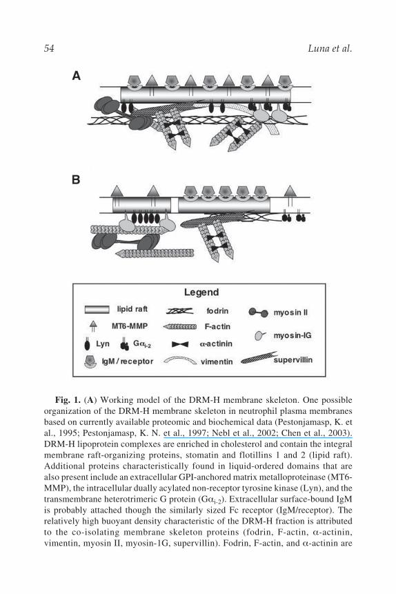
Virtually no supervillin or integral membrane DRM-H proteins can beextracted with carbonate. These results suggest that supervillin is the mostproximal to the bilayer of the major peripheral DRM-H membrane proteinsand that both myosin isoforms can associate with membranes independentof any indirect binding through membrane-associated actin (Fig. 1A).
Although it is not known how supervillin or either of the myosins bindsto the DRM-H membrane, it is possible that supervillin is a linker proteinfor the attachment of both myosin II and F-actin. The amino-terminal 174amino acids of supervillin bind directly to the regulatory S2 domain of myo-sin II, and supervillin residues 171–830 contain at least three binding sitesfor F-actin (Chen et al., 2003). Supervillin sequences promote actin fila-ment bundling and crosslinking, suggesting that supervillin may promotethe assembly and/or organization of actin and myosin filaments at liquid-ordered membrane domains. Other regions of supervillin exhibit high degreesof sequence similarity to polyphosphoinositide-binding sequences in thegelsolin/villin family of actin-binding proteins (Pestonjamasp, K. N. et al.,1997; Kwiatkowski, 1999), sequences that mediate gelsolin association withbilayer lipids (Hartwig et al., 1989; Ahmed et al., 1997; He et al., 1997).Regardless of the membrane attachment mechanism(s), all the major DRM-H proteins co-immunoprecipitate with supervillin from neutrophil plasma
54 Luna et al.
Fig. 1. (A) Working model of the DRM-H membrane skeleton. One possibleorganization of the DRM-H membrane skeleton in neutrophil plasma membranesbased on currently available proteomic and biochemical data (Pestonjamasp, K. etal., 1995; Pestonjamasp, K. N. et al., 1997; Nebl et al., 2002; Chen et al., 2003).DRM-H lipoprotein complexes are enriched in cholesterol and contain the integralmembrane raft-organizing proteins, stomatin and flotillins 1 and 2 (lipid raft).Additional proteins characteristically found in liquid-ordered domains that arealso present include an extracellular GPI-anchored matrix metalloproteinase (MT6-MMP), the intracellular dually acylated non-receptor tyrosine kinase (Lyn), and thetransmembrane heterotrimeric G protein (G i-2). Extracellular surface-bound IgMis probably attached though the similarly sized Fc receptor (IgM/receptor). Therelatively high buoyant density characteristic of the DRM-H fraction is attributedto the co-isolating membrane skeleton proteins (fodrin, F-actin, -actinin,vimentin, myosin II, myosin-1G, supervillin). Fodrin, F-actin, and -actinin are
Lipid Raft Membrane Skeletons 55
membranes solubilized with either Triton X-100 or octylglucoside (Nebl etal., 2002). Thus, supervillin is an excellent candidate marker protein for theDRM-H membrane skeleton, because it apparently binds, directly or indi-rectly, to the other major DRM-H polypeptides through protein–proteininteractions. These interactions may be representative of more than onetype of DRM-H complex and may be a mixture of stable and transient asso-ciations that are regulated during cell signaling.
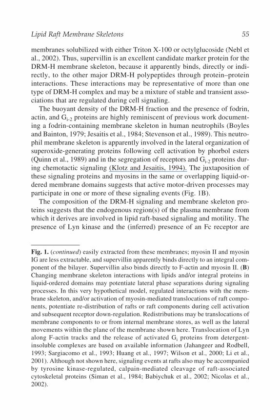
The buoyant density of the DRM-H fraction and the presence of fodrin,actin, and Gi-2 proteins are highly reminiscent of previous work document-ing a fodrin-containing membrane skeleton in human neutrophils (Boylesand Bainton, 1979; Jesaitis et al., 1984; Stevenson et al., 1989). This neutro-phil membrane skeleton is apparently involved in the lateral organization ofsuperoxide-generating proteins following cell activation by phorbol esters(Quinn et al., 1989) and in the segregation of receptors and Gi-2 proteins dur-ing chemotactic signaling (Klotz and Jesaitis, 1994). The juxtaposition ofthese signaling proteins and myosins in the same or overlapping liquid-or-dered membrane domains suggests that active motor-driven processes mayparticipate in one or more of these signaling events (Fig. 1B).
The composition of the DRM-H signaling and membrane skeleton pro-teins suggests that the endogenous region(s) of the plasma membrane fromwhich it derives are involved in lipid raft-based signaling and motility. Thepresence of Lyn kinase and the (inferred) presence of an Fc receptor are
Fig. 1. (continued) easily extracted from these membranes; myosin II and myosinIG are less extractable, and supervillin apparently binds directly to an integral com-ponent of the bilayer. Supervillin also binds directly to F-actin and myosin II. (B)Changing membrane skeleton interactions with lipids and/or integral proteins inliquid-ordered domains may potentiate lateral phase separations during signalingprocesses. In this very hypothetical model, regulated interactions with the mem-brane skeleton, and/or activation of myosin-mediated translocations of raft compo-nents, potentiate re-distribution of rafts or raft components during cell activationand subsequent receptor down-regulation. Redistributions may be translocations ofmembrane components to or from internal membrane stores, as well as the lateralmovements within the plane of the membrane shown here. Translocation of Lynalong F-actin tracks and the release of activated Gi proteins from detergent-insoluble complexes are based on available information (Jahangeer and Rodbell,1993; Sargiacomo et al., 1993; Huang et al., 1997; Wilson et al., 2000; Li et al.,2001). Although not shown here, signaling events at rafts also may be accompaniedby tyrosine kinase-regulated, calpain-mediated cleavage of raft-associatedcytoskeletal proteins (Siman et al., 1984; Babiychuk et al., 2002; Nicolas et al.,2002).
56 Luna et al.
reminiscent of the role of similar proteins in IgE signaling during mast-cellactivation (Sheets et al., 1999; Draber and Draberova, 2002). Becausesecreted IgM can bind simultaneously to as many as five Fc receptors(Feinstein and Munn, 1969), additional crosslinking may not be required forinitial activation. Signaling downstream of IgM binding to bovine neutro-phils is unexplored, but it may resemble signaling cascades present in mastcells and in T (and B) lymphocytes (Cheng et al., 2001; Babiychuk et al.,2002; Sedwick and Altman, 2002; Werlen and Palmer, 2002). As currentlyunderstood, these cascades are domino-like sequences of events in which anonreceptor tyrosine kinase such as Lyn (or Lck or Fyn) is recruited to theraft, which then mediates the tyrosine phosphorylation of receptor subunitsand induces the recruitment of Syk/ZAP-70 family tyrosine kinases, whichphosphorylate additional raft components and lead to the activation of raft-associated adaptor proteins, phospholipase C, and protein kinase C. T-cellsignaling requires actin filament polymerization and integrity for optimalactivity (Valensin et al., 2002; Tskvitaria-Fuller et al., 2003), whereas actinfilaments promote down-regulation in mast-cell signaling, presumably bypromoting the lateral separation of Lyn from the Fc receptor substrate(Holowka et al., 2000; Wilson et al., 2000). Down-regulation of signaling inT cells is thought to involve the binding of Csk kinase to raft-associated Cbp(Csk-binding protein), followed by Csk-mediated inactivation of Lck or Fyn(Kawabuchi et al., 2000).
5. “DRM-H-LIKE” MEMBRANE SKELETONSIN OTHER CELL TYPES
As discussed above, the composition of the neutrophil DRM-H fractionstrongly suggests similarities with lipid raft-mediated signaling processesin mast cells and T lymphocytes. This hypothesis is supported byproteomic analyses of total DRMs from Jurkat T lymphoma cells (vonHaller et al., 2001), monocytes (Burkart et al., 2003), and Raji B lym-phoma cells (Saeki et al., 2003). Fodrin, supervillin, myosin II, vimentin,and actin co-isolate with T-cell DRMs (von Haller et al., 2001); -actinin4, vimentin, and actin with monocyte DRMs (Burkart et al., 2003); andmyosin II, myosin 1G, myosin 1 , and actin with B-cell DRMs (Saeki et al.,2003). The absence of various DRM-H proteins from these DRM fractionsis not surprising, because the isolation procedures were optimized for recov-ery of DRMs with lower buoyant densities. Thus, available evidence is con-sistent with a general role for components of the neutrophil DRM-Hmembrane skeleton in the structure and/or functioning of hematopoieticlipid rafts.
Lipid Raft Membrane Skeletons 57
Lipid raft-associated membrane skeletons containing neutrophil DRM-Hproteins also may function in nonhematopoietic cells. Myosin II, vimentin,and actin are recovered as components of light DRM fractions from HeLacells (Foster et al., 2003); erythrocyte DRMs contain spectrin and actin inaddition to stomatin, flotillin 1, and flotillin 2 (Salzer and Prohaska, 2001);and low-density membrane fractions from skeletal muscle contain archvillin,a muscle-specific isoform of supervillin (Chen et al., 2003). In fact, if thedistribution of supervillin/archvillin can be used as an indicator of the pres-ence of a “DRM-H like” membrane skeleton, then such domains should beabundant in muscle tissues, carcinoma cell lines, and secretory epithelia(Pestonjamasp, K. N. et al., 1997; Pope et al., 1998; Anderson and Jacobson,2002; Chen et al., 2003).
6. OTHER DYNAMIC LIPID RAFT-ASSOCIATEDMEMBRANE SKELETONS
Consistent with the multitude of lipid raft organizing proteins, many typesof “lipid raft membrane skeletons” may exist. It is currently unclear whethersome membrane skeleton proteins preferentially associate with particulartypes of lipid rafts, or whether tissue-specific differences in protein expres-sion have led to the diversity of known interactions. For instance, membersof the band 4.1/ezrin/radixin/moesin family of actin-binding membrane-skeleton proteins co-purify with DRMs from erythrocytes (Salzer andProhaska, 2001), B cells (Saeki et al., 2003), T cells (Babiychuk et al., 2002;Tomas et al., 2002), monocytes (Burkart et al., 2003), and HeLa cells (Fos-ter et al., 2003). A potential mechanism for this interaction is through bind-ing of proteins in this family to the NHERF/EBP50 adaptor protein (Na+/H+
exchanger regulatory factor/ezrin-binding phosphoprotein of 50 kDa), whichin turn binds to raft-associated Cbp (Brdickova et al., 2001; Itoh et al., 2002).The observation that neutrophil ezrin and moesin segregate preferentiallywith DRM-L, rather than with DRM-H (Pestonjamasp, K. N. et al., 1997),suggests that at least two types of raft-associated membrane skeletons canco-exist in neutrophils.
Annexin II/calpactin 1 may be a marker protein for a third type of lipidraft-associated membrane skeleton, or this protein may be a currently unap-preciated component of the DRM-L or DRM-H membrane skeleton.Annexin II, which has been reported in DRM-L fractions (Burkart et al.,2003; Foster et al., 2003), binds directly to F-actin, fodrin, and anionic phos-pholipids (Gerke and Weber, 1984; Hubaishy et al., 1995; Filipenko andWaisman, 2001). Most annexin binding interactions are potentiated by high[Ca2+] and inhibited by tyrosine phosphorylation, but binding to cholesterol-
58 Luna et al.
rich membranes can occur in the absence of Ca2+ (Harder et al., 1997; Ayala-Sanmartin et al., 2001). Treatment of low-density BHK cell membranes withcholesterol-sequestering agents in the absence of Ca2+ releases a co-immu-noprecipitating complex of annexin II, -actinin, ezrin, moesin, and actin(Harder et al., 1997). Annexin II also is reported to co-immunoprecipitatewith caveolin (Uittenbogaard et al., 2002), the small GTPase Rac1 (Hansenet al., 2002), the tyrosine phosphatase SHP-2 (Burkart et al., 2003), andCD44/H-CAM, the major cell surface transmembrane receptor for hyalu-ronic acid (Oliferenko et al., 1999). CD44 partitions into low-density mem-brane fractions (Ilangumaran et al., 1998) and binds directly to members ofthe ezrin-radixin-moesin family (Hirao et al., 1996; Legg and Isacke, 1998).Annexin II is required for regulated vesicle budding from early endosomes(Mayran et al., 2003), and both annexin II and splice forms of CD44 havebeen implicated in the control of cell growth and migration (Chiang et al.,1999; Orian-Rousseau et al., 2002; Burkart et al., 2003). Thus, annexin IIand CD44 may function in a type of lipid raft that mediates regulated cyclesof cell-substrate attachment and plasma membrane recycling during dynamiccell attachment and migration.
A fourth type of raft-associated membrane skeleton may potentiate cellu-lar responses to mechanosensory forces, including changes in extracellulartonicity. GAP-43, a membrane-bound neuronal protein that modulates actinassembly (Meiri and Gordon-Weeks, 1990; Aguado-Velasco and Bretscher,1997), enhances increases in intracellular calcium ion concentrations([Ca2+]i) triggered by hypotonic media, at least after overexpression inHEK293 cells (Caprini et al., 2003). Mutation of the palmitoylation sites onGAP-43 that anchor it to lipid rafts abrogates the observed increases in [Ca2+]i.The mechanism apparently includes GAP-43 binding and activation of PLC-
1, which leads to increased concentrations of diacylglycerol and intracellu-lar inositol-1,4,5-trisphosphate, which in turn causes release of Ca2+ fromthe endoplasmic reticulum. Although it is unclear whether endogenous lev-els of GAP-43 perform similar functions in neuronal cells, this protein haslong been known to be important for axonal growth and synaptic plasticity(Skene, 1989).
Membrane skeletons associated with lipid raft components also may playa role in dynamic cell–cell interactions. In a process that appears to be simi-lar to early stages in the formation of at least some vertebrate adhesion com-plexes (Vasioukhin et al., 2000), Dictyostelium discoideum amoebae initiatecell attachment through what may be a fifth type of lipid raft membraneskeleton. An 80-kDa GPI-linked adhesion protein (gp80), which is seques-tered in sterol-rich membrane domains at the tips of cell surface projections,binds through homophilic interactions to an adjacent cell (Harris and Siu,
Lipid Raft Membrane Skeletons 59
2002). Binding induces a reorganization of the cell surface, with a flatteningof surface extensions into large areas of cell-cell contact (Choi and Siu,1987; Vasioukhin et al., 2000). In Dictyostelium, these mature cell-cell con-tact regions then recruit the approximately 17-kDa actin-binding and nucle-ating protein, ponticulin (Wuestehube and Luna, 1987; Chia et al., 1991;Hitt et al., 1994; Harris et al., 2003), which is attached to the membranethrough both transmembrane domains and a GPI-anchor (Hitt et al., 1994).Because any direct interaction between gp80 and ponticulin is too weak todetect (Harris et al., 2003), ponticulin may be targeted to rafts through theaffinity of its GPI-anchor for sterol-rich membrane domains. In this theory,ponticulin’s transmembrane sequences, which are redundant from the per-spective of membrane attachment, are required to juxtaposition the F-actinbinding site on the cytoplasmic surface of the membrane (Hitt et al., 1994).The high-affinity ponticulin–actin interaction thus constitutes a key linkagebetween the regions of cell-cell contact and the underlying membrane skel-eton, which is rich in myosin II as well as in actin and actin-bundling pro-teins (Ingalls et al., 1986; Harris et al., 2001).
The Dictyostelium contact regions fractionate on sucrose gradients aslipoprotein complexes with densities of 1.16–1.18 g/mL, i.e., approxi-mately that of the neutrophil DRM-H fraction. Similar fractionation behav-ior is observed regardless of whether the cells are extracted with cold TritonX-100 (Ingalls et al., 1986), or whether low buoyant density membranes aregenerated by sonication in the absence of detergent (Harris et al., 2001).Although no vertebrate ponticulin sequence is known, a glycosylated pro-tein similar in size to Dictyostelium ponticulin has been detected in neutro-phil membranes by both immunological approaches (Wuestehube et al.,1989) and direct binding to F-actin (Keresztes and Lajtos, 1997). Becausemany GPI-anchored proteins participate in lipid raft-based adhesive pro-cesses requiring the cytoskeleton (Harris and Siu, 2002), vertebrate mem-brane skeleton proteins containing both GPI anchors and cytoskeletalattachment sites may yet be discovered.
7. LIPID RAFT MEMBRANE SKELETONSASSOCIATED WITH “STABLE” MEMBRANE DOMAINS
Many of the morphologically recognizable, presumably more stableplasma membrane domains also may be regulated by lipid raft signalingpathways. For instance, apical surfaces of intestinal epithelial cells containtwo types of detergent-resistant lipid rafts (Danielsen and Hansen, 2003).One type localizes to microvilli, where the lectin galectin-4 crosslinks manyglycolipids and GPI-anchored extracellular hydrolytic enzymes into stable
60 Luna et al.
“super-rafts.” The second type of apically localized lipid raft, which is con-centrated in deep apical tubules, is thought to be dynamic and may be asso-ciated with a membrane skeleton containing caveolin 1, annexin A2,
-actinin, ezrin, and/or moesin (Danielsen and Hansen, 2003). Thus, thetubule-associated lipid raft may be similar to the annexin II and/or ezrin-associated dynamic rafts discussed above in Section 6.
Cell–cell adhesion and paracellular permeability also may be regulatedby lipid raft components. ZO-1 and occludin, two of the major structuralproteins in the sealing strands of the tight junctions in polarized epithelialcells, partition in a DRM-L fraction when hyperphosphoryated (Nusrat etal., 2000). Occludin also co-immunoprecipitates with caveolin-1 (Nusrat etal., 2000). Both of these observations are consistent with a role for caveolaeand/or rafts in the assembly or function of tight junctions.
Cell–substrate interactions and locomotion in fibroblasts (Shima et al.,2003), as well as in hematopoietic cells (Hogg et al., 2003), are regulated byraft-associated signaling proteins. Furthermore, integrin function at fibroblastfocal adhesions is influenced by cholesterol and glycosphingolipid levels(Pande, 2000). Given that GFP-tagged focal adhesion proteins at the rearends of migrating fibroblasts “slide” along the substrate (Wehrle-Haller andImhof, 2002), it may be that the major difference between the highly dynamicfocal contacts of immune cells and the longer-lived focal adhesions in fibro-blasts is the rate at which similar processes occur.
8. CONCLUDING REMARKS
The term “lipid raft membrane skeleton” may at first appear to be para-doxical. “Lipid raft” connotes a dynamic cholesterol-rich membrane domainof low buoyant density, whereas “membrane skeleton” conjures up imagesof static structures composed of lots of proteins that each bind to multipleother proteins through mostly high-affinity interactions. Yet, the demon-strated association of lipid raft components with cytoskeletal proteins, espe-cially with myosin motors, resonates with the dynamic natures of lipid rafts.Both myosin I and myosin II may participate in lateral movements of lipidraft components within the plane of the plasma membrane. Alternatively,either may function during internalization or trafficking of raft-associatedvesicles through the cytoplasm. The binding of myosin II and actin to sepa-rable sites on supervillin—which is bound to the DRM bilayer through other,currently unknown sequences—raises the further possibility that supervillinmay assemble actin and myosin II at lipid rafts and/or regulate contractilityat these membrane domains. Such regulation is presumably controlled byraft-associated signaling events that remain to be investigated. The study of
Lipid Raft Membrane Skeletons 61
these signaling processes is inherently complicated by the temporal natureof the interactions, as well as by the potential inherent diversity of raft types.Furthermore, additional types of lipid raft-associated membrane skeletonsprobably await discovery. These discoveries will be accelerated, though, byappreciating that the attachment of “heavy” cytoskeletal proteins to “light”membranes shifts the buoyant density of the resulting lipid raft membraneskeleton to “heavier” regions of sucrose gradients.
REFERENCESAdams J. C. (2002) Regulation of protrusive and contractile cell-matrix contacts.
J. Cell Sci. 115, 257–265.Aguado-Velasco C. and Bretscher M. (1997) Dictyostelium myosin II null mutant
can still cap Con A receptors. Proc. Natl. Acad. Sci. USA, 94, 9684–9686.Ahmed S. N., Brown D. A., and London E. (1997) On the origin of sphingolipid/
cholesterol-rich detergent-insoluble cell membranes: physiological concentra-tions of cholesterol and sphingolipid induce formation of a detergent-insoluble,liquid-ordered lipid phase in model membranes. Biochemistry 36, 10,944–10,953.
Anderson R. G. and Jacobson K. (2002) A role for lipid shells in targeting proteinsto caveolae, rafts, and other lipid domains. Science 296, 1821–1825.
Arhets P., Olivo J.-C., Gounon P., Sansonetti P., Guillén N. (1998) Virulence andfunctions of myosin II are inhibited by overexpression of light meromyosin inEntamoeba histolytica. Mol. Biol. Cell 8, 1537–1547.
Ayala-Sanmartin J., Henry J. P., and Pradel L. A. (2001) Cholesterol regulatesmembrane binding and aggregation by annexin 2 at submicromolar Ca2+ con-centration. Biochim. Biophys. Acta 1510, 18–28.
Babiychuk E. B., Monastyrskaya K., Burkhard F. C., Wray S., and Draeger A.(2002) Modulating signaling events in smooth muscle: cleavage of annexin 2abolishes its binding to lipid rafts. FASEB J. 16, 1177–1184.
Bose A., Guilherme A., Robida S. I., Nicoloro S. M., Zhou Q. L., Jiang Z. Y., et al.(2002) Glucose transporter recycling in response to insulin is facilitated bymyosin Myo1c. Nature 420, 821–824.
Bourguignon L. Y. and Bourguignon G. J. (1984) Capping and the cytoskeleton.Int. Rev. Cytol. 87, 195–224.
Bourguignon L. Y., Jy W., Majercik M. H., and Bourguignon G. J. (1988) Lympho-cyte activation and capping of hormone receptors. J. Cell Biochem. 37, 131–150.
Boyles J. and Bainton D. F. (1979) Changing patterns of plasma membrane-associ-ated filaments during the initial phases of polymorphonuclear leukocyte adher-ence. J. Cell Biol. 82, 347–368.
Braga V. M. (2002) Cell-cell adhesion and signalling. Curr. Opin. Cell Biol. 14(5),546–556.
Brdickova N., Brdicka T., Andera L., Spicka J., Angelisova P., Milgram S. L., et al.(2001) Interaction between two adapter proteins, PAG and EBP50: a possiblelink between membrane rafts and actin cytoskeleton. FEBS Lett. 507, 133–136.
62 Luna et al.
Bretscher A. (1991) Microfilament structure and function in the cortical cytoskel-eton. Annu. Rev. Cell Biol. 7, 337–374.
Brodsky F. M., Chen C. Y., Knuehl C., Towler M. C., Wakeham D. E. (2001)Biological basket weaving: formation and function of clathrin-coated vesicles.Annu. Rev. Cell Dev. Biol. 17, 517–568.
Brown D. A. and Rose J. K. (1992) Sorting of GPI-anchored proteins to glycolipid-enriched membrane subdomains during transport to the apical cell surface. Cell68, 533–544.
Burkart A., Samii B., Corvera S., and Shpetner H. S. (2003) Regulation of the SHP-2 tyrosine phosphatase by a novel cholesterol- and cell confluence-dependentmechanism. J. Biol. Chem. 278, 18,360–18,367.
Burns C. G., Reedy M., Heuser J., and De Lozanne A. (1995) Expression of lightmeromyosin in Dictyostelium blocks normal myosin II function. J. Cell Biol.130, 605–612.
Caprini M., Gomis A., Cabedo H., Planells-Cases R., Belmonte C., Viana F., et al.(2003) GAP43 stimulates inositol trisphosphate-mediated calcium release inresponse to hypotonicity. EMBO J. 22, 3004–3014.
Chen Y., Takizawa N., Crowley J. L., Oh S. W., Gatto C. L., Kambara T., et al.(2003) F-actin and myosin II binding domains in supervillin. J. Biol. Chem.278, 46,094–46,106.
Cheng P. C., Brown B. K., Song W., and Pierce S. K. (2001) Translocation of the Bcell antigen receptor into lipid rafts reveals a novel step in signaling. J.Immunol. 166, 3693–3701.
Chia C. P., Hitt A. L., and Luna E. J. (1991) Direct binding of F-actin to ponticulin, anintegral plasma membrane glycoprotein. Cell Motil. Cytoskeleton 18, 164–179.
Chiang Y., Rizzino A., Sibenaller Z. A., Wold M. S., and Vishwanatha J. K. (1999)Specific down-regulation of annexin II expression in human cells interfereswith cell proliferation. Mol. Cell Biochem. 199, 139–147.
Choi A. H. C. and Siu C.-H. (1987) Filopodia are enriched in a cell cohesion mol-ecule of Mr 80,000 and participate in cell-cell contact formation inDictyostelium discoideum. J. Cell Biol. 104, 1375–1387.
Dahl S. C., Geib R. W., Fox M. T., Edidin M., and Branton D. (1994) Rapid cap-ping in -spectrin-deficient MEL cells from mice afflicted with hereditaryhemolytic anemia. J. Cell Biol. 125, 1057–1065.
Danielsen E. M. and Hansen G. H. (2003) Lipid rafts in epithelial brush borders:atypical membrane microdomains with specialized functions. Biochim.Biophys. Acta 1617, 1–9.
Deckert M., Ticchioni M., and Bernard A. (1996) Endocytosis of GPI-anchored pro-teins in human lymphocytes: Role of glycolipid-based domains, actin cytoskel-eton, and protein kinases. J. Cell Biol. 133, 791–799.
Destaing O., Saltel F., Geminard J. C., Jurdic P., and Bard F. (2003) Podosomesdisplay actin turnover and dynamic self-organization in osteoclasts expressingactin-green fluorescent protein. Mol. Biol. Cell 14, 407–416.
Devreotes P. and Janetopoulos C. (2003) Eukaryotic chemotaxis: distinctions betweendirectional sensing and polarization. J. Biol. Chem. 278, 20,445–20,448.
Lipid Raft Membrane Skeletons 63
Dietrich C., Bagatolli L. A., Volovyk Z. N., Thompson N. L., Levi M., JacobsonK., et al. (2001) Lipid rafts reconstituted in model membranes. Biophys. J. 80,1417–1428.
Draber P. and Draberova L. (2002) Lipid rafts in mast cell signaling. Mol. Immunol.38, 1247–1252.
Durrbach A., Raposo G., Tenza D., Louvard D., and Coudrier E. (2000) Truncatedbrush border myosin I affects membrane traffic in polarized epithelial cells.Traffic 1, 411–424.
Edidin M. (1993) Patches and fences: Probing for plasma membrane domains. J.Cell Sci. Suppl. 17, 165–169.
Edidin M. (2003) The state of lipid rafts: from model membranes to cells. Annu.Rev. Biophys. Biomol. Struct. 32, 257–283.
Evans J. G., Correia I., Krasavina O., Watson N., and Matsudaira P. (2003) Mac-rophage podosomes assemble at the leading lamella by growth and fragmenta-tion. J. Cell Biol. 161, 697–705.
Feinstein A. and Munn E. A. (1969) Conformation of the free and antigen-boundIgM antibody molecules. Nature 224, 1307–1309.
Fielding C. J. (2004) Role of cholesterol in membrane microdomain signaling, inMembrane Microdomain Signaling: Lipid Rafts in Biology and Medicine,Mattson M., ed., Humana, Totowa, NJ.
Filipenko N. R. and Waisman D. M. (2001) The C terminus of annexin II mediatesbinding to F-actin. J. Biol. Chem. 276, 5310–5315.
Foster L. J., De Hoog C. L., and Mann M. (2003) Unbiased quantitative proteomicsof lipid rafts reveals high specificity for signaling factors. Proc. Natl. Acad.Sci. USA 100, 5813–5818.
Fukui Y., De Lozanne A., and Spudich J. A. (1990) Structure and function of thecytoskeleton of a Dictyostelium myosin-defective mutant. J. Cell Biol. 110,367–378.
Galbiati F., Razani B., and Lisanti M. P. (2001) Emerging themes in lipid rafts andcaveolae. Cell 106, 403–411.
Gerke V. and Weber K. (1984) Identity of p36K phosphorylated upon Rous sar-coma virus transformation with a protein purified from brush borders; calcium-dependent binding to non-erythroid spectrin and F-actin. EMBO J. 3, 227–233.
Gomez-Mouton C., Abad J. L., Mira E., Lacalle R. A., Gallardo E., Jimenez-Baranda S., et al. (2001) Segregation of leading-edge and uropod componentsinto specific lipid rafts during T cell polarization. Proc. Natl. Acad. Sci. USA98, 9642–9647.
Goodloe-Holland C. M. and Luna E. J. (1987) Purification and characterization ofDictyostelium discoideum plasma membranes. Methods Cell Biol. 28, 103–128.
Green K. J. and Gaudry C. A. (2000) Are desmosomes more than tethers for inter-mediate filaments? Nat. Rev. Mol. Cell Biol. 1, 208–216.
Grewal A. S., Rouse B. T., and Babiuk L. A. (1978) Characterization of surface recep-tors on bovine leukocytes. Int. Arch. Allergy Appl. Immunol. 56, 289–300.
Hahn B. S. and Labouesse M. (2001) Tissue integrity: hemidesmosomes and resis-tance to stress. Curr. Biol. 11, R858–861.
64 Luna et al.
Hansen M. D., Ehrlich J. S., and Nelson W. J. (2002) Molecular mechanism fororienting membrane and actin dynamics to nascent cell-cell contacts in epithe-lial cells. J. Biol. Chem. 277, 45,371–45,376.
Harder T., Kellner R., Parton R. G., and Gruenberg J. (1997) Specific release ofmembrane-bound annexin II and cortical cytoskeletal elements by sequestra-tion of membrane cholesterol. Mol. Biol. Cell 8, 533–545.
Harris T. J., Awrey D. E., Cox B. J., Ravandi A., Tsang A., and Siu C. H. (2001)Involvement of a triton-insoluble floating fraction in Dictyostelium cell-celladhesion. J. Biol. Chem. 276, 18,640–18,648.
Harris T. J., Ravandi A., Awrey D. E., and Siu C. H. (2003) Cytoskeleton interac-tions involved in the assembly and function of glycoprotein-80 adhesion com-plexes in dictyostelium. J. Biol. Chem. 278, 2614–2623.
Harris T. J.and Siu C. H. (2002) Reciprocal raft–receptor interactions and the assem-bly of adhesion complexes. Bioessays, 24, 996–1003.
Hartwig J. H., Chambers K. A. and Stossel T. P. (1989) Association of gelsolinwith actin filaments and cell membranes of macrophages and platelets. J. CellBiol. 108, 467–479.
He Q., Dent E. W., and Meiri K. F. (1997) Modulation of actin filament behaviorby GAP-43 (neuromodulin) is dependent on the phosphorylation status ofserine 41, the protein kinase C site. J. Neurosci 17, 3515–3524.
Hirao M., Sato N., Kondo T., Yonemura S., Monden M., Sasaki T., et al. (1996)Regulation mechanism of ERM (ezrin/radixin/moesin) protein/plasma mem-brane association: possible involvement of phosphatidylinositol turnover andRho-dependent signaling pathway. J. Cell Biol. 135, 37–51.
Hitt A. L., Hartwig J. H., Luna E. J. (1994) Ponticulin is the major high affinity linkbetween the plasma membrane and the cortical actin network in Dictyostelium.J. Cell Biol. 126, 1433–1444.
Hitt A. L., Lu T. H., and Luna E. J. (1994) Ponticulin is an atypical membraneprotein. J. Cell Biol. 126, 1421–1431.
Hogg N., Laschinger M., Giles K., and McDowall A. (2003) T-cell integrins: morethan just sticking points. J. Cell Sci. 116, 4695–4705.
Holowka D., Sheets E. D., and Baird B. (2000) Interactions between Fc RI andlipid raft components are regulated by the actin cytoskeleton. J. Cell Sci. 113,1009–1019.
Hope H. R. and Pike L. J. (1996) Phosphoinositides and phosphoinositide-utilizingenzymes in detergent-insoluble lipid domains. Mol. Biol. Cell 7, 843–851.
Huang C., Hepler J. R., Chen L. T., Gilman A. G., Anderson R. G., and Mumby S.M. (1997) Organization of G proteins and adenylyl cyclase at the plasma mem-brane. Mol. Biol. Cell 8, 2365–2378.
Hubaishy I., Jones P. G., Bjorge J., Bellagamba C., Fitzpatrick S., Fujita D. J., et al.(1995) Modulation of annexin II tetramer by tyrosine phosphorylation. Bio-chemistry 34, 14,527–14,534.
Hubbard A. L. and Ma A. (1983) Isolation of rat hepatocyte plasma membranes. II.Identification of membrane-associated cytoskeletal proteins. J. Cell Biol. 96,230–239.
Lipid Raft Membrane Skeletons 65
Ilangumaran S., Briol A., and Hoessli D. C. (1998) CD44 selectively associateswith active Src family protein tyrosine kinases Lck and Fyn in glycosphin-golipid-rich plasma membrane domains of human peripheral blood lympho-cytes. Blood 91, 3901–3908.
Ingalls H. M., Goodloe-Holland C. M. and Luna E. J. (1986) Junctional plasmamembrane domains isolated from aggregating Dictyostelium discoideum ame-bae. Proc. Natl. Acad. Sci. USA 83, 4779–4783.
Itoh K., Sakakibara M., Yamasaki S., Takeuchi A., Arase H., Miyazaki M., et al.(2002) Cutting edge: Negative regulation of immune synapse formation byanchoring lipid raft to cytoskeleton through Cbp-EBP50-ERM assembly. J.Immunol. 168, 541–544.
Jacobson K. and Dietrich C. (1999) Looking at lipid rafts? Trends Cell Biol. 9, 87–91.Jacobson K., Hou Y., Derzko Z., Wojcieszyn J., and Organisciak D. (1981) Lipid
lateral diffusion in the surface membrane of cells and in multibilayers formedfrom plasma membrane lipids. Biochemistry 20, 5268–5275.
Jahangeer S. and Rodbell M. (1993) The disaggregation theory of signal transduc-tion revisited: further evidence that G proteins are multimeric and disaggregateto monomers when activated. Proc. Natl. Acad. Sci. USA 90, 8782–8786.
Jamora C. and Fuchs E. (2002) Intercellular adhesion, signalling and the cytoskel-eton. Nat. Cell Biol. 4, E101–108.
Jesaitis A. J., Naemura J. R., Sklar L. A., Cochrane C. G., and Painter R. G. (1984)Rapid modulation of N-formyl chemotactic peptide receptors on the surface ofhuman granulocytes: Formation of high-affinity ligand-receptor complexes intransient association with cytoskeleton. J. Cell Biol. 98, 1378–1387.
Kawabuchi M., Satomi Y., Takao T., Shimonishi Y., Nada S., Nagai K., et al.(2000) Transmembrane phosphoprotein Cbp regulates the activities of Src-family tyrosine kinases. Nature 404, 999–1003.
Keresztes M. and Lajtos Z. (1997) Major laminin-binding and F-actin-linked gly-coproteins of neutrophils. Cell Biol. Int. 21, 543–550.
Khrebtukova I. A., Kwiatkowska K., Gudkova D. A., Sorokin A. B., and Pinaev G.P. (1991) The role of microfilaments in the capping of epidermal growth factorreceptor in A431 cells. Exp. Cell Res. 194, 48–55.
Klotz K.-N. and Jesaitis A. J. (1994) Neutrophil chemoattractant receptors and themembrane skeleton. BioEssays 16, 193–198.
Kwiatkowska K. and Sobota A. (1999) Engagement of spectrin and actin in cap-ping of FcgammaRII revealed by studies on permeabilized U937 cells.Biochem. Biophys. Res. Commun. 259, 287–293.
Kwiatkowski D. J. (1999) Functions of gelsolin: motility, signaling, apoptosis, can-cer. Curr. Opin. Cell Biol. 11, 103–108.
Legg J. W. and Isacke C. M. (1998) Identification and functional analysis of theezrin-binding site in the hyaluronan receptor, CD44. Curr. Biol. 8, 705–708.
Li X., Galli T., Leu S., Wade J. B., Weinman E. J., Leung G., et al. (2001) Na+-H+
exchanger 3 (NHE3) is present in lipid rafts in the rabbit ileal brush border: Arole for rafts in trafficking and rapid stimulation of NHE3. J. Physiol. 537,537–552.
66 Luna et al.
London E. and Brown D. A. (2000) Insolubility of lipids in Triton X-100: physicalorigin and relationship to sphingolipid/cholesterol membrane domains (rafts).Biochim. Biophys. Acta 1508, 182–195.
Matkó J. and Szöll J. (2004) Regulatory aspects of membrane microdomain (raft)dynamics in live cells: A biophysical approach, in Membrane MicrodomainSignaling: Lipid Rafts in Biology and Medicine, Mattson M., ed., Humana,Totowa, NJ.
Maxfield F. R. (2002) Plasma membrane microdomains. Curr. Opin. Cell Biol. 14,483–487.
Mayran N., Parton R. G., and Gruenberg J. (2003) Annexin II regulatesmultivesicular endosome biogenesis in the degradation pathway of animalcells. EMBO J. 22, 3242–3253.
Meiri K. F. and Gordon-Weeks P. R. (1990) GAP-43 in growth cones is associatedwith areas of membrane that are tightly bound to substrate and is a componentof a membrane skeleton subcellular fraction. J. Neurosci. 10, 256–266.
Melkonian K. A., Chu T., Tortorella L. B., and Brown D. A. (1995) Characteriza-tion of proteins in detergent-resistant membrane complexes from Madin-Darbycanine kidney epithelial cells. Biochemistry 34, 16,161–16,170.
Montoya M. C., Sancho D., Vicente-Manzanares M., and Sanchez-Madrid F. (2002)Cell adhesion and polarity during immune interactions. Immunol. Rev. 186,68–82.
Moran M. and Miceli M. C. (1998) Engagement of GPI-linked CD48 contributes toTCR signals and cytoskeletal reorganization: a role for lipid rafts in T cellactivation. Immunity 9, 787–796.
Munro S. (2003) Lipid rafts: elusive or illusive? Cell 115, 377–388.Nebl T., Pestonjamasp K. N., Leszyk J. D., Crowley J. L., Oh S. W., and Luna E. J.
(2002) Proteomic analysis of a detergent-resistant membrane skeleton fromneutrophil plasma membranes. J. Biol. Chem. 277, 43399–43409.
Nicolas G., Fournier C. M., Galand C., Malbert-Colas L., Bournier O., KroviarskiY., et al. (2002) Tyrosine phosphorylation regulates alpha II spectrin cleavageby calpain. Mol. Cell Biol. 22, 3527–3536.
Nijtmans L. G., Artal S. M., Grivell L. A., and Coates P. J. (2002) The mitochon-drial PHB complex: Roles in mitochondrial respiratory complex assembly,ageing and degenerative disease. Cell. Mol. Life Sci. 59, 143–155.
Nusrat A., Parkos C. A., Verkade P., Foley C. S., Liang T. W., Innis-WhitehouseW., et al. (2000) Tight junctions are membrane microdomains. J. Cell Sci. 113,1771–1781.
Oliferenko S., Paiha K., Harder T., Gerke V., Schwärzler C., Schwarz H., et al.(1999) Analysis of CD44-containing lipid rafts: Recruitment of annexin II andstabilization by the actin cytoskeleton. J. Cell Biol. 146, 843–854.
Orian-Rousseau V., Chen L., Sleeman J. P., Herrlich P., and Ponta H. (2002) CD44is required for two consecutive steps in HGF/c-Met signaling. Genes Dev. 16,3074–3086.
Lipid Raft Membrane Skeletons 67
Pande G. (2000) The role of membrane lipids in regulation of integrin functions.Curr. Opin. Cell Biol. 12, 569–574.
Park D. S., Woodman S. E., Schubert W., Cohen A. W., Frank P. G., Chandra M.,et al. (2002) Caveolin-1/3 double-knockout mice are viable, but lack bothmuscle and non-muscle caveolae, and develop a severe cardiomyopathic phe-notype. Am. J. Pathol. 160, 2207–2217.
Pasternak C., Flicker P. F., Ravid S., and Spudich J. A. (1989) Intermolecular ver-sus intramolecular interactions of Dictyostelium myosin: Possible regulationby heavy chain phosphorylation. J. Cell Biol. 109, 203–210.
Patki V., Buxton J., Chawla A., Lifshitz L., Fogarty K., Carrington W., et al. (2001)Insulin action on GLUT4 traffic visualized in single 3T3-l1 adipocytes by usingultra-fast microscopy. Mol. Biol. Cell 12, 129–141.
Pestonjamasp K., Amieva M. R., Strassel C. P., Nauseef W. M., Furthmayr H., andLuna E. J. (1995) Moesin, ezrin, and p205 are actin-binding proteins associ-ated with neutrophil plasma membranes. Mol. Biol. Cell 6, 247–259.
Pestonjamasp K. N., Pope R. K., Wulfkuhle J. D., and Luna E. J. (1997) Supervillin(p205): A novel membrane-associated, F-actin-binding protein in the villin/gelsolin superfamily. J. Cell Biol. 139, 1255–1269.
Pike L. J. (2004) Lipid rafts: Heterogeneity on the high seas. Biochem. J. 378, 281–292.Pope R. K., Pestonjamasp K. N., Smith K. P., Wulfkuhle J. D., Strassel C. P.,
Lawrence J. B., et al. (1998) Cloning, characterization, and chromosomallocalization of human supervillin (SVIL) Genomics 52, 342–351.
Pradhan D. and Morrow J. (2002) The spectrin-ankyrin skeleton controls CD45surface display and interleukin-2 production. Immunity 17, 303–315.
Quinn M. T., Parkos C. A., and Jesaitis A. J. (1989) The lateral organization of com-ponents of the membrane skeleton and superoxide generation in the plasma mem-brane of stimulated human neutrophils. Biochim. Biophys. Acta 987, 83–94.
Ritchie K., Iino R., Fujiwara T., Murase K., and Kusumi A. (2003) The fence andpicket structure of the plasma membrane of live cells as revealed by singlemolecule techniques (Review). Mol. Membr. Biol. 20, 13–18.
Saeki K., Miura Y., Aki D., Kurosaki T., and Yoshimura A. (2003) The B cell-specific major raft protein, Raftlin, is necessary for the integrity of lipid raftand BCR signal transduction. EMBO J. 22, 3015–3026.
Salzer U. and Prohaska R. (2001) Stomatin, flotillin-1, and flotillin-2 are majorintegral proteins of erythrocyte lipid rafts. Blood 97, 1141–1143.
Sargiacomo M., Sudol M., Tang Z., and Lisanti M. P. (1993) Signal transductingmolecules and glycosylphosphatidylinositol-linked proteins form a caveolin-rich insoluble complex in MDCK cells. J. Cell Biol. 122, 789–807.
Schnitzer J. E., McIntosh D. P., Dvorak A. M., Liu J., and Oh P. (1995) Separationof caveolae from associated microdomains of GPI-anchored proteins. Science269, 1435–1439.
Sedwick C. E. and Altman A. (2002) Ordered just so: Lipid rafts and lymphocytefunction. Sci. STKE 2002 re2.
68 Luna et al.
Sheets E. D., Holowka D., and Baird B. (1999) Membrane organization in immu-noglobulin E receptor signaling. Curr. Opin. Chem. Biol. 3, 95–99.
Sheetz M. P. (1979) Integral membrane protein interaction with Tritoncytoskeletons of erythrocytes. Biochim. Biophys. Acta 557, 122–134.
Shima T., Nada S., and Okada M. (2003) Transmembrane phosphoprotein Cbpsenses cell adhesion signaling mediated by Src family kinase in lipid rafts.Proc. Natl. Acad. Sci. USA 100, 14,897–14,902.
Siman R., Baudry M., and Lynch G. (1984) Brain fodrin: substrate for calpain I,an endogenous calcium-activated protease. Proc. Natl. Acad. Sci. USA 81,3572–3576.
Simons K. and Toomre D. (2000) Lipid rafts and signal transduction. Nat. Rev.Mol. Cell Biol. 1, 31–39.
Singer S. J. and Nicolson G. L. (1972) The fluid mosaic model of the structure ofcell membranes. Science 175, 720–731.
Skene J. H. P. (1989) Axonal growth-associated proteins. Ann. Rev. Neurosci. 12,127–156.
Skerrow C. J. and Matoltsy A. G. (1974) Isolation of epidermal desmosomes. J.Cell Biol. 63, 515–523.
Slusarewicz P., Nilsson T., Hui N., Watson R., and Warren G. (1994) Isolation of amatrix that binds medial Golgi enzymes. J. Cell Biol. 124, 405–413.
Smart E. J., Ying Y.-S., Mineo C., and Anderson R. G. W. (1995) A detergent-freemethod for purifying caveolae membrane from tissue culture cells. Proc. Natl.Acad. Sci. USA 92, 10,104–10,108.
Song K. S., Li S., Okamoto T., Quilliam L. A., Sargiacomo M., and Lisanti M. P.(1996) Co-purification and direct interaction of Ras with caveolin, an integralmembrane protein of caveolae microdomains. Detergent-free purification ofcaveolae microdomains. J. Biol. Chem. 271, 9690–9697.
Stevenson K. B., Clark R. A., and Nauseef W. M. (1989) Fodrin and band 4.1 ina plasma membrane-associated fraction of human neutrophils. Blood 74,2136–2143.
Stuart A. D., Eustace H. E., McKee T. A., and Brown T. D. (2002) A novel cellentry pathway for a DAF-using human enterovirus is dependent on lipid rafts.J. Virol. 76, 9307–9322.
Tomas E. M., Chau T. A., and Madrenas J. (2002) Clustering of a lipid-raft associ-ated pool of ERM proteins at the immunological synapse upon T cell receptoror CD28 ligation. Immunol. Lett. 83, 143–147.
Tskvitaria-Fuller I., Rozelle A. L., Yin H. L., and Wulfing C. (2003) Regulation ofsustained actin dynamics by the TCR and costimulation as a mechanism ofreceptor localization. J. Immunol. 171, 2287–2295.
Tsukita S. (1989) Isolation of cell-to-cell adherens junctions from rat liver. J. CellBiol. 108, 31–41.
Uittenbogaard A., Everson W. V., Matveev S. V., and Smart E. J. (2002)Cholesteryl ester is transported from caveolae to internal membranes as part ofa caveolin-annexin II lipid-protein complex. J. Biol. Chem. 277, 4925–4931.
Lipid Raft Membrane Skeletons 69
Valensin S., Paccani S. R., Ulivieri C., Mercati D., Pacini S., Patrussi L., et al.(2002) F-actin dynamics control segregation of the TCR signaling cascade toclustered lipid rafts. Eur. J. Immunol. 32, 435–446.
Vasioukhin V., Bauer C., Yin M., and Fuchs E. (2000) Directed actin polymeriza-tion is the driving force for epithelial cell-cell adhesion. Cell 100, 209–219.
Vasioukhin V. and Fuchs E. (2001) Actin dynamics and cell-cell adhesion in epi-thelia. Curr. Opin. Cell Biol 13, 76–84.
Volonte D., Galbiati F., Li S., Nishiyama K., Okamoto T., and Lisanti M. P. (1999)Flotillins/cavatellins are differentially expressed in cells and tissues and form ahetero-oligomeric complex with caveolins in vivo. Characterization andepitope-mapping of a novel flotillin-1 monoclonal antibody probe. J. Biol.Chem. 274, 12,702–12,709.
von Haller P. D., Donohoe S., Goodlett D. R., Aebersold R., and Watts J. D. (2001)Mass spectrometric characterization of proteins extracted from Jurkat T celldetergent-resistant membrane domains. Proteomics 1, 1010–1021.
Wehrle-Haller B. and Imhof B. (2002) The inner lives of focal adhesions. TrendsCell Biol. 12, 382–389.
Werlen G. and Palmer E. (2002) The T-cell receptor signalosome: A dynamic struc-ture with expanding complexity. Curr. Opin. Immunol. 14, 299–305.
Wilson B. S., Pfeiffer J. R., and Oliver J. M. (2000) Observing Fc RI signalingfrom the inside of the mast cell membrane. J. Cell Biol. 149, 1131–1142.
Worku M., Paape M. J., Filep R., and Miller R. H. (1994) Effect of in vitro and invivo migration of bovine neutrophils on binding and expression of Fc recep-tors for IgG2 and IgM. Am. J. Vet. Res. 55, 221–226.
Wuestehube L. J., Chia C. P., and Luna E. J. (1989) Indirect immunofluorescencelocalization of ponticulin in motile cells. Cell Motil. Cytoskeleton 13, 245–263.
Wuestehube L. J. and Luna E. J. (1987) F-actin binds to the cytoplasmic surface ofponticulin, a 17kD integral glycoprotein from Dictyostelium discoideumplasma membranes. J. Cell Biol. 105, 1741–1751.
Yavin E. and Brand A. (2004) From intramolecular asymmetries to raft assemblies:A short guide for the puzzled in lipidomics, in Membrane Microdomain Sig-naling: Lipid Rafts in Biology and Medicine, Mattson M., ed., Humana,Totowa, NJ.
Yu J., Fischman D. A., and Steck T. L. (1973) Selective solubilization of proteinsand phospholipids from red blood cell membranes by non-ionic detergents. J.Supramol. Struct. 1, 233–248.

Related Documents