University of the Incarnate Word University of the Incarnate Word The Athenaeum The Athenaeum Theses & Dissertations 12-2005 Limits of Temperature Tolerance of Leishmania Enriettii and Limits of Temperature Tolerance of Leishmania Enriettii and Leishmania Hertigi in a Real-Time PCR Assay Leishmania Hertigi in a Real-Time PCR Assay Christina Salinas University of the Incarnate Word Follow this and additional works at: https://athenaeum.uiw.edu/uiw_etds Part of the Biology Commons Recommended Citation Recommended Citation Salinas, Christina, "Limits of Temperature Tolerance of Leishmania Enriettii and Leishmania Hertigi in a Real-Time PCR Assay" (2005). Theses & Dissertations. 53. https://athenaeum.uiw.edu/uiw_etds/53 This Thesis is brought to you for free and open access by The Athenaeum. It has been accepted for inclusion in Theses & Dissertations by an authorized administrator of The Athenaeum. For more information, please contact [email protected].

Welcome message from author
This document is posted to help you gain knowledge. Please leave a comment to let me know what you think about it! Share it to your friends and learn new things together.
Transcript
University of the Incarnate Word University of the Incarnate Word
The Athenaeum The Athenaeum
Theses & Dissertations
12-2005
Limits of Temperature Tolerance of Leishmania Enriettii and Limits of Temperature Tolerance of Leishmania Enriettii and
Leishmania Hertigi in a Real-Time PCR Assay Leishmania Hertigi in a Real-Time PCR Assay
Christina Salinas University of the Incarnate Word
Follow this and additional works at: https://athenaeum.uiw.edu/uiw_etds
Part of the Biology Commons
Recommended Citation Recommended Citation Salinas, Christina, "Limits of Temperature Tolerance of Leishmania Enriettii and Leishmania Hertigi in a Real-Time PCR Assay" (2005). Theses & Dissertations. 53. https://athenaeum.uiw.edu/uiw_etds/53
This Thesis is brought to you for free and open access by The Athenaeum. It has been accepted for inclusion in Theses & Dissertations by an authorized administrator of The Athenaeum. For more information, please contact [email protected].
LIMITS OF TEMPERATURE TOLERANCE OF
Leishmania enriettii and Leishmania hertigi
IN A REAL-TIME PGR ASSAY
A Thesis
by
CHRISTINA SALINAS
Presented to the Graduate Faculty of the
University of the Incarnate Word
In Partial Fulfillment
of the Requirements
for the Degree of
MASTER OF SCIENCE
UNIVERSITY OF THE INCARNATE WORD
DECEMBER, 2005
LIMITS OF TEMPERATURE TOLERANCE OF
Leishmania enriettii and Leishmania hertigi
IN A REAL-TIME PGR ASSAY
A Thesis
by
CHRISTINA SALINAS
APPROVED:
Sara F. Kerr, Chairman of Committee
Committee Member
Russell W. Raymond, Committee Member

COPYRIGHT
The text and figures contained in this document are protected in accordance with
copyright laws of the United States of America. All rights reserved.

ACKNOWLEDGEMENTS
I would first like to express my appreciation to Dr. Jean L. Patterson for
allowing me to conduct my research at the Southwest Foundation for Biomedical
Research. I especially want to thank Dr. Ricardo Carrion at the SFBR for all the time
and interest he has given to me and my research. I am appreciative of his suggestion
of incorporating real-time PGR to my experiment. My experience at SFBR has been
beneficial in that I was able to acquaint myself to the research setting. Not only did I
have access to lab equipment and facilities, but also I had the cooperation and help of
many of the employees at SFBR. I want to thank Russell W. Raymond for his
involvement with my thesis. His encouragement and humor has helped ease some of
the stress. Last, but most importantly I would like to thank Dr. Sara Kerr for
providing me the opportunity to receive my M.S. in Biology at the University of the
Incarnate Word. I am also very grateful for her decision to allow me the experience
of traveling to Nicaragua to participate in field studies. My exposure to Nicaragua
will always be remembered and will have an impact on my appreciation for the
diversity of different cultures. This research was funded by National Institutes of
Health grants GM 55337 and GM 50080.
SUMMARY
LIMITS OF TEMPERATURE TOLERANCE OF
Leishmania enriettii and Leishmania hertigi
IN A REAL-TIME PGR ASSAY
Christina Salinas, B.S., University of North Texas
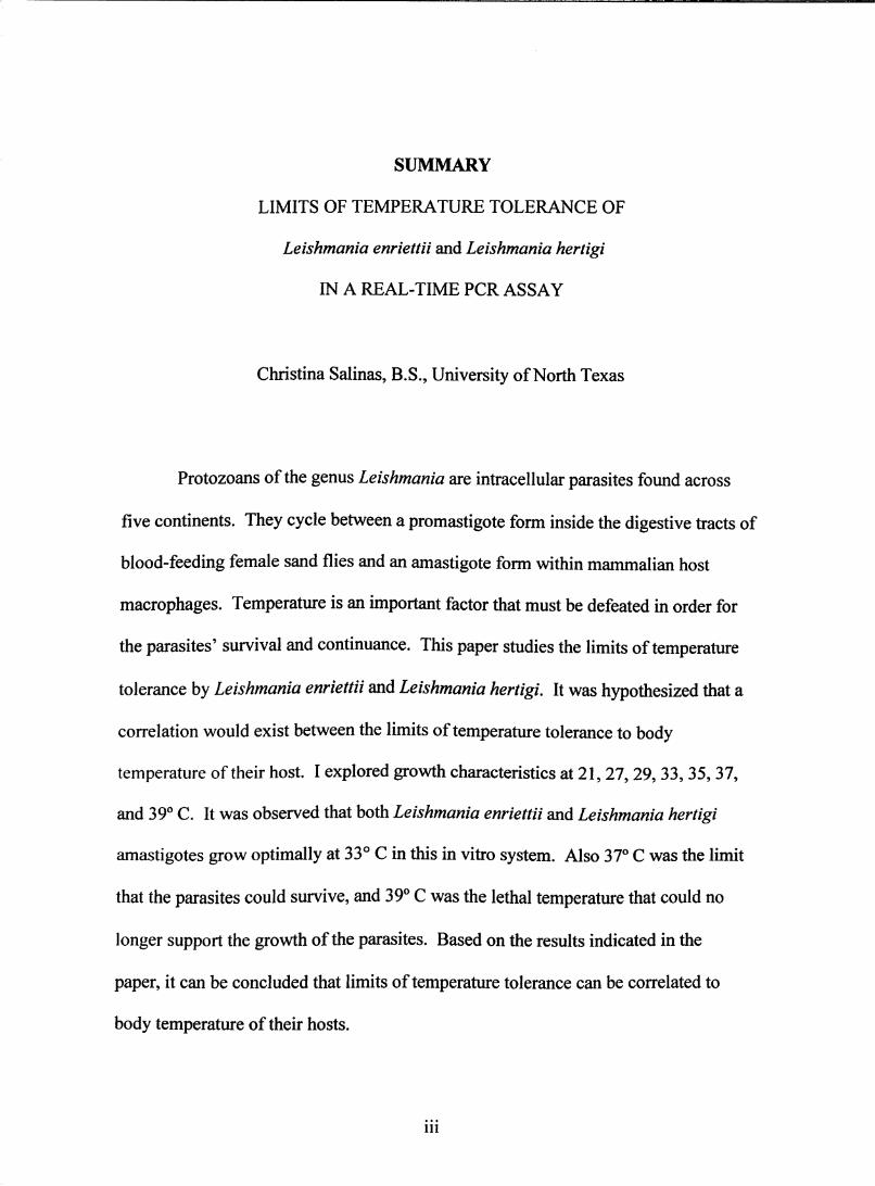
Protozoans of the genus Leishmania are intracellular parasites found across
five continents. They cycle between a promastigote form inside the digestive tracts of
blood-feeding female sand flies and an amastigote form within mammalian host
macrophages. Temperature is an important factor that must be defeated in order for
the parasites' survival and continuance. This paper studies the limits of temperature
tolerance by Leishmania enriettii and Leishmania hertigi. It was hypothesized that a
correlation would exist between the limits of temperature tolerance to body
temperature of their host. I explored growth characteristics at 21,27,29, 33, 35, 37,
and 39° C. It was observed that both Leishmania enriettii and Leishmania hertigi
amastigotes grow optimally at 33° C in this in vitro system. Also 37° C was the limit
that the parasites could survive, and 39° C was the lethal temperature that could no
longer support the growth of the parasites. Based on the results indicated in the
paper, it can be concluded that limits of temperature tolerance can be correlated to
body temperature of their hosts.
Ill

TABLE OF CONTENTS
Summary
List of Figures
Introduction. 1
Materials and Methods 11
Results 1
Discussion 47
References 52
Appendix 59
Vita 64
IV

LIST OF FIGURES
Figure 1: Alignment of Leishmania enriettii and Leishmania hertigi
DNAPolymerase 15
Figure 2A: Amplification plot of standards 20
Figure 2B: Standard Curve 21
Figure 3 A: Growth of Leishmania enriettii at 2 T C 25
Figure 3B: Growth of Leishmania enriettii at 27° C 26
Figure 3C: Growth of Leishmania enriettii at 29° C 27
Figure 3D: Growth of Leishmania enriettii at 33° C 28
Figure 3E: Growth of Leishmania enriettii at 35° C 29
Figure 3F: Growth of Leishmania enriettii at 37° C 30
Figure 3G: Growth of Leishmania enriettii at 39° C 31
Figure 4: Maximum growth of Leishmania enriettii at different temperatures
studied 32
Figure 5: Maximum number of parasite divisions of Leishmania enriettii
at different temperatures studied 33
Figure 6 A: Growth of Leishmania hertigi at2rc 38
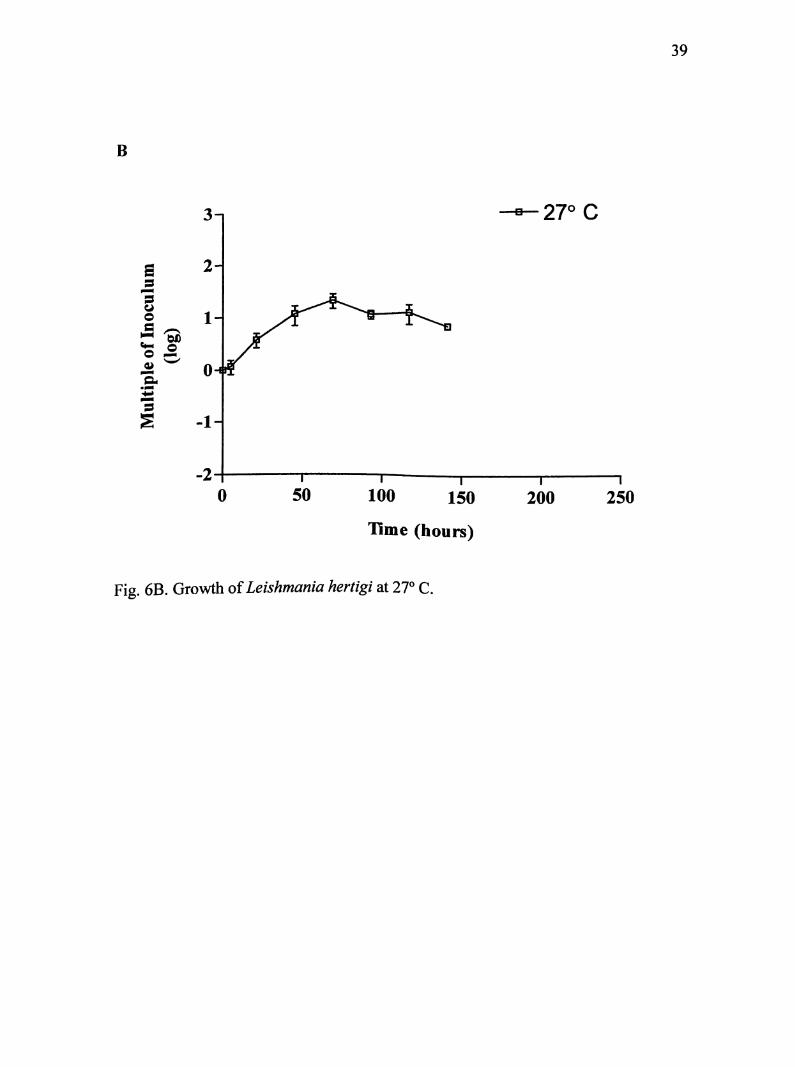
Figure 6B; Growth of Leishmania hertigi at 27° C 39
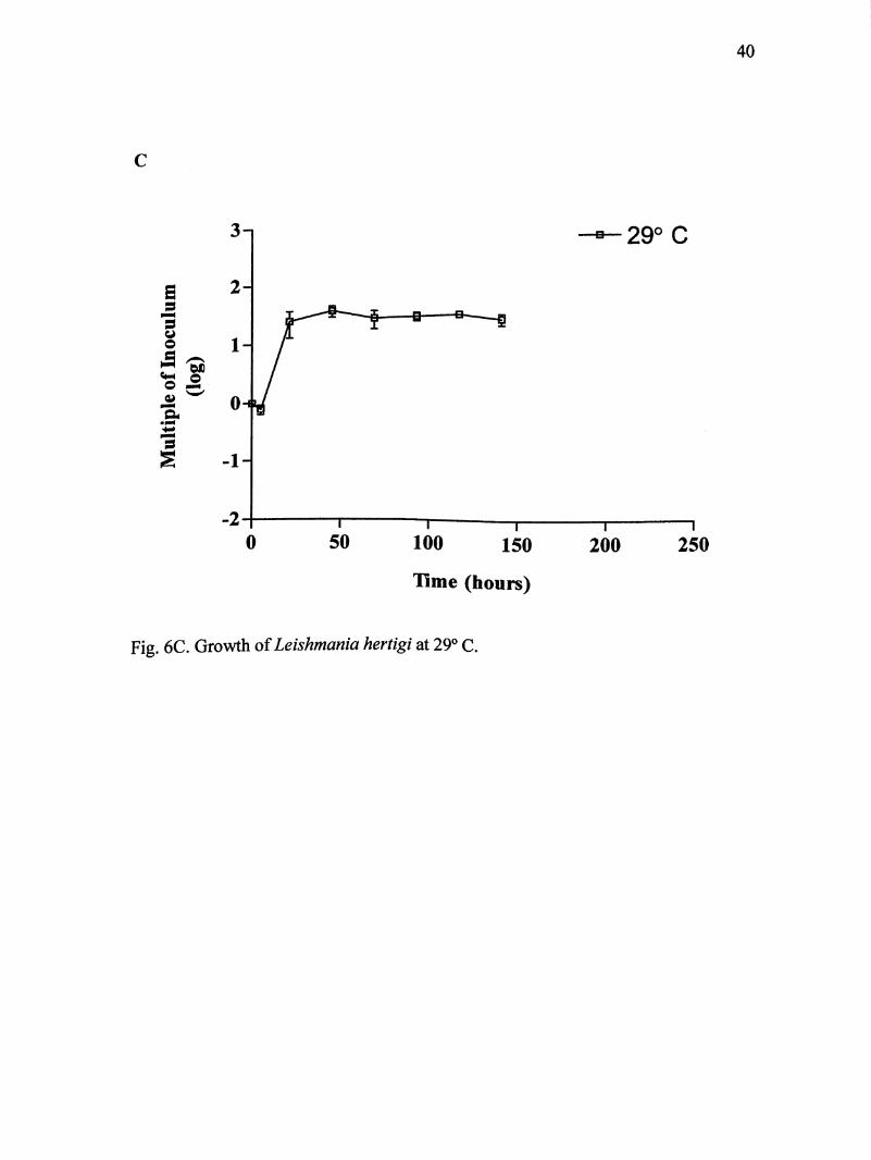
Figure 6C: Growth of Leishmania hertigi at 29° C 40
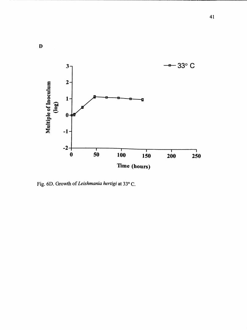
Figure 6D: Growth of Leishmania hertigi at 33° C 41
Figure 6E: Growth of Leishmania hertigi at 35° C 42
Figure 6F: Growth of Leishmania hertigi at 37° C 43

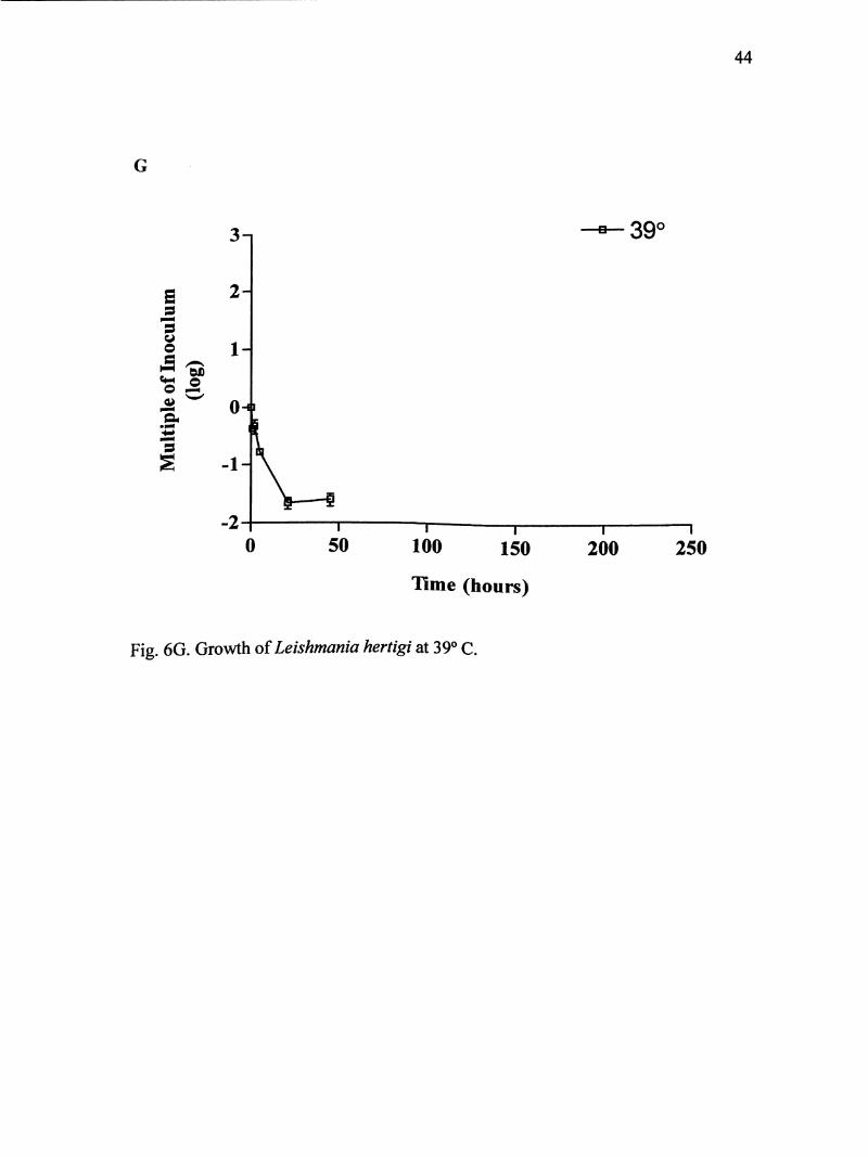
Figure 6G: Growth of Leishmania hertigi at 39° C 44
Figure 7; Maximum growth of Leishmania hertigi at different temperatures
studied 45
Figure 8: Maximum number of parasite divisions of Leishmania hertigi
at different temperatures studied 46
VI
INTRODUCTION
Infections caused by Leishmania spp. are considerable health problems in several
countries across the continents of Africa, Asia, Europe, North America, and South
America (World Health Organization, 1990). Every year nearly 350 million people
are at risk, and there are about 12 million infections per year (Magill, 1995). Three
hiunan infection syndromes have been characterized depending on the type of
infecting species and location of infection: 1) localized cutaneous lesions 2)
mucocutaneous lesions of the nasopharjmgeal region, and 3) severe visceral forms
(Magill, 1995). Each year, 1.5 million people will develop one of the two forms of
cutaneous infections, and another 500,000 people will acquire visceral leishmaniases
(Ogg et al. 2003).
Parasites of the genus Leishmania possess a biphasic life cycle that consists of
a promastigote form inside the digestive tracts of blood-feeding sand fly vectors and
an amastigote form within mammalian host macrophages (Pral et al. 2003). Of the
six genera of sand flies only Lutzomyia spp. (New World) and Phlebotomus spp. (Old
World) support the parasite's life cycle (World Health Organization, 1990).
Infections by parasites of Leishmania spp. are transmitted when infected female sand
flies take a blood meal from a vertebrate host (Lewis, 1974; Souza et al. 2005).
Before 1940, Leishmania spp. were identified only in infections of humans,
dogs, and rodents. However, it was not known that other species of Leishmania
existed that could result in disease in other mammals (Lainson, 1997). At least six
strains have been identified from both the Old and New World that do not cause
disease to humans, but in other mammals. Leishmania enriettii Muniz and Medina
(Kinetoplastida: Trypanosomatidae) and Leishmania hertigi Herrer (Kinetoplastida:
Trypanosomatidae) are two species found in neotropical regions that are non-
infectious to humans (Lainson, 1997).
The purpose of this project was to study how L enriettii and L. hertigi tolerate
a range of elevated temperatures. I investigated how these parasites responded to a
shift of cool to warm temperatures. The main aims of the project were to characterize
optimal temperature for this in vitro system, by comparing parasite population
densities and parasite divisions, and to determine the upper limits of temperature
tolerance. I hypothesize that tolerance of high temperatures by L. enriettii and L.
hertigi corresponds to body temperatures of their host.
LITERATURE REVIEW
Parasites
Leishmania enriettii was first isolated from domestic guinea pigs, Cavia porcellus, in
1946 by Medina in the State of Parana, Brazil (Lainson, 1997). The parasite was
named in 1948 by Muniz and Medina. The two observed unusually large amastigotes
found in tumor-like skin lesions on the ears and testicles of laboratory guinea pigs.
Infections in guinea pigs have displayed a range of symptoms, from self-curing
lesions to chronic metastatic lesions (El-On, Witztum and Schnur, 1986; Lainson,
1997). Immediately after the discovery, rhesus monkeys, dogs, white mice, golden
hamsters, puppies, rats, hares, and human volunteers were inoculated by
subcutaneous or intraperitoneal injections of promastigotes or amastigotes. Similar
lesions did not develop in any of the cases (Belehu and Turk, 1976; Machado et al.
1994). Since its first appearance, there have been at least three publications of its
recurrence in domestic guinea pigs, all occurring within the geographical region of
Brazil (Adler and Halff, 1955; Machado et al. 1994; Thomaz-Soccol et al 1996).
Leishmania enriettii has been a suitable model to study in the laboratory
because the parasite grows easily and large identifiable amastigotes can be obtained
in considerable numbers from infected sites. Also, the infection in guinea pigs
closely resembles human cutaneous leishmaniases (El-On et al 1986; Lainson, 1997)
Despite its uses in the laboratory, much is unknown about its epidemiology.
Currently no natural infections have been reported in humans or mammals other than
the guinea pig. After the second reappearance of L. enriettii in guinea pigs,
Lutzomyia monticola sand flies were captured near the area of its appearance. The
sand flies were found resting on tree trunks and feeding on humans. A study was
done in 1967 demonstrating the ability of Lu. monticola to support the growth of the
parasite; however the sand fly was unable to transmit the parasite naive guinea pigs.
Therefore the natural sand fly vector is still unknown (Machado et al. 1994; Thomaz-
Soccol et al 1996).
Leishmania hertigi was first reported in the Panamanian porcupine, Coendou
rothschildi in 1971. It was described to cause infection of the skin and sometimes
viscera of porcupines (Zeledon, Ponce and de Ponce, 1977). Herrer (1971) reported
the parasite was harmless to porcupines, and could be found in small numbers
throughout the skin and viscera. In 1975, Herrer and Christensen proposed that the
association of L hertigi with porcupines was of an ancient origin because no host cell
reaction has been observed, and they concluded by stating L. hertigi has developed a
commensal relationship with porcupines rather than a parasitic one.
Between April 1965 and September 1974, a study was accomplished that
demonstrated the infrequency of gross skin lesions among 13 Panamanian forest
mammals with cutaneous leishmaniases. It was reported that 89% of the mammals
infected with Leishmania parasites showed no skin lesions. Leishmania hertigi
accounted for 50% of the cryptic infections observed. Results in the article suggested
that cryptic leishmanial infections are a common phenomenon found among
Panamanian forest mammals (Herrer and Christensen, 1975). The parasite has also
been detected in porcupines in Brazil and Costa Rica (Zeledon et al. 1977). In 1978,
publications proclaimed that the Brazilian strains were significantly different to the
Panamanian strains based on morphological, biochemical, and serological
characteristics (Croft, Schnur and Chance, 1978). Brazilian strains are now given the
name Leishmania deanei (Lainson, 1997).
Taxonomy of Leishmania enriettii and Leishmania hertigi
Leishmania species originally were categorized based on symptoms, geographic
occurrences, and location of promastigote development within the sand fly (Cupolillo,
Momen and Grimaldi, 1998; Grimaldi and Schottelius, 2001). The genus Leishmania
was subdivided into two subgenera, the Leishmania and the Viannia. Within the
subgenera, complexes were grouped according to isoenzymatic profiling (World
Health Organization, 1990). However taxonomy of Leishmania is much more
complex and often disputed among scientists. Cupolillo et al. (1998) state that part of
the difficulty is due to the enormous diversity within the genus. One difference is the
selection of hosts. Some species are pathogenic to humans, while others are limited
to lower orders of mammals.
Leishmania enriettii and L hertigi are among the many species where
classification is still under debate. WHO (1990) lists the two species under the
subgenera Leishmania, but not under any of the five complexes. Cupolillo et al.
(2000) have proposed a revised classification of Leishmania and Endotrpanum
parasites based on a number of recent molecular studies. Based on these studies, it
was suggested that various species of Leishmania are more closely related to the
genus Endotrypanum than to other species of Leishmania. According to the results,
two phylogenetic lineages were proposed separating the species that demonstrated
more similarities with one another than to those that were most divergent. The two
lineages are named "section" Euleishmania and "section" Paraleishmania.
Euleishmania is comprised of both Old and New World species, which cluster
together based on biochemical and molecular analyses, including numerical
zymotaxonomy, multilocus enzyme electrophoresis, measurement of the sialidase
activity, PCR amplification and restriction enzyme digestion of rRNA, cloning and
sequencing of the conserved region of minicircle kDNA (Cupolillo et al. 1998).
Leishmania enriettii is among this lineage. Paraleishmania then was termed for
species that clustered with Endotrypamm, including Leishmania herreri, Leishmania
deanei, Leishmania equatorensis, Leishmania colombiensis, and L. hertigi, all of
which are New World species. (Cupolillo et al. 2001; Grimaldi and Schottelius,
2001;Noyes etal. 2000).
Temperature and pH effects
All species of Leishmania undergo major stress upon transmission from a
poikilothermic (non-temperature regulated) sand fly vector to the homeothermic
(temperature regulated) mammalian host. The two main stresses are temperature and
pH (Van der Ploeg, Giannini and Cantor, 1985; Zilberstein and Shapira, 1994).
Within the sand flies, promastigotes can experience temperatures in the range of 22-
28°C, while the temperature conditions of mammalian tissue can range from 31-39°C
depending on species, but never exceeds 42°C (Clos and Krobitsch, 1999; Zilberstein
and Shapira, 1994). Arends and McNab (2001) listed the energetics of eleven species
of New World hystricognath ('caviomorph') rodents. Species of Caviidae, which
include guinea pigs, had body temperatures in the range of 37.3-39.0° C, while

species of Erethizontidae, the New World porcupines, had a body temperature of
36.7° C.
In addition to temperature variability, parasites will experience drastic
changes in pH. Although the pH value in the digestive tract of the sand fly is
unknown, it has been estimated that the pH is similar to that of the mosquito (> 8.5)
(Zilberstein and Shapira, 1994). In contrast, pH values inside phagolysosomes can
range from 4.5-6.0 (Lukacs, Rotstein and Grinstein, 1991).
Questions have been raised as to what factors govern parasite tropism and
clinical manifestations (Callahan et al. 1996; Clos and Krobitsch, 1999). Clos and
Krobitsch (1999) proposed that temperature tolerance of Leishmania species offer
clues to their distribution within their mammalian host. For example, Leishmania
mexicana, Leishmania major, and Leishmania braziliensis are species commonly
associated with human cutaneous lesions. These species have a temperature tolerance
up to 35° C in vitro, which correlates with the maximum temperature of human skin.
Viseralizing species such as Leishmania donovanL Leishmania infantum, Leishmania
chagasi, and Leishmania tropica can withstand higher temperatures from 35-39° C,
which compares to temperatures in the human abdominal cavity.
Previous studies have shown the effects of temperature on several Leishmania
species (Berman and Neva, 1981; Greenblatt and Glaser, 1965; Krassner, 1965; Leon,
Soares and Temporal, 1995). Callahan et al. (1996) studied promastigotes of visceral
and cutaneous Leishmania spp. from both Old and New World. Eight different
species were used at three elevated temperatures (30, 32, and 34° C), and compared to
growth at the control temperature (25°). Results indicated that visceral L donovani
amastigotes were able to grow in higher temperatures than the cutaneous species L.
major, L. tropica, and L. mexicana. In addition. Old World cutaneous species had
significantly better growth potential in higher temperatures than New World species.
Real-time quantitative PCR
Real-time PCR is an increasingly popular technique applied in Parasitology, and is
currently being utilized for the detection and quantification of Leishmania spp. within
sand fly vectors and vertebrate blood and tissue (Bell and Ranford-Cartwright, 2002;
Bretagne et al. 2001; Gomez-Saladin, Doud and Maroli, 2005; Mary et al. 2004;
Rolao et al. 2004; Schulz et al. 2003; Vitale et al. 2004). The real-time PCR system
is designed using a 3 quencher dye and a 5 reporter dye. When the probe anneals to
its target sequence, Taq DNA Polymerase will cleave the probe due to its 5' nuclease
activity. This results in separation of the reporter dye from the quencher dye,
following an increase of fluorescence from the reporter dye. Primer extension will
then continue down the template strand. Fluorescence intensity is directly
proportional to the accumulation of PCR product produced (Wortmann et al. 2001).
Initial concentration of target DNA then can be determined by obtaining the
number of cycles necessary to reach a threshold concentration. This cycle number is
called the critical threshold (Ct) value. Quantification of parasites of unknown
concentration can ultimately be determined by the generation of a standard curve,
plotting the log concentrations of known standards against their Ct values. The initial
starting concentration of target DNA from samples can be determined with respect to
their Ct values along the standard curve (Mehra and Hu, 2005).
Conventional PGR is limited in this aspect because only the end-stage product
can be measured where primer template, enzyme, and dNTP components may be
exhausted (Bell and Ranford-Cartwright, 2002). Quantification of parasites by
microscopy is another method often used by researchers, but results often can be
inconsistent and the method is time consuming (Rolao et al. 2004).
One of the first studies using real-time PGR for the diagnosis of leishmaniases
was performed by Wortmann et al. (2001). The purpose of the study was to develop
an assay that could detect all Leishmania species. The small subunit rRNA gene was
the ideal choice for target primer and probe selection because it is conserved among
all Leishmania. Alignment of 16S rRNA gene sequences from Leishmania
aethiopica, Leishmania guyanensis, L. chagasi, L. donovani, L. mexicana, L. major,
L. tropica, and L. Viannia complex were used to design primers and probe targets.
Both cultured promastigotes and tissue biopsies were used to test the assay. Results
showed that the assay could detect all strains tested and could distinguish Leishmania
from the closely related Trypanosomes. With the use of this technology, health care
providers are able to give rapid diagnostic results at the genus level.
Schulz et al. (2003) expanded on the idea of detection of Leishmania with
real-time PGR by producing an assay that could differentiate between the three
relevant Leishmania groups (the L. donovani complex, the L. braziliensis complex,
and others). Leishmanial 18S rDNA sequences were targeted for the selection of

10
primers and probe binding sites. The sequence for 18S rDNA belongs to the
multipcopy group, in that there is more than one copy per cell (Guilaume et al. 1992).
The selection of rDNA for target primer and probe sites provide increased sensitivity
and specificity. Discrimination was achieved by using different melting
temperatures. Results indicated that the three clinically relevant groups could reliably
be distinguished from one another. It is now possible to allow health care
administrators to provide more effective courses of treatment directed towards
distinct clinical manifestations. It is also possible to do direct examination on blood,
bone marrow, and skin or liver biopsy specimens (Schulz et al. 2003).
11
MATERIALS AND METHODS
Parasites and culture conditions
Parasites of L. enriettii and L hertigi were obtained from the American Type Culture
Collection (ATCC number: 30035 for L. enriettii and 30286 for L. hertigi). Cultures
were maintained at 22° C in IX M199 media with Earle's salts, L-glutamine
(Invitrogen). Sodium Bicarbonate (Fisher) was added and supplemented with 5%
(v/v) heat-inactivated fetal bovine serum (GIBCO Life Technologies), IM hepes
buffer (pH 7.4) (Sigma Chemical Co., St. Louis, MO), lOOX penicillin/streptomycin
(GIBCO), 10 mM hypoxanthin (Sigma), 0.25% (v/v) bovine hemin (Sigma), and
sterile filtered human urine. The pH was adjusted to 7.4 by IM sodium hydroxide
(Fisher), and then sterilized by passage through a 0.22 pm Coming IL filter (Coming
Inc.). Parasites were cultured in tissue culture flasks (25 cm^) for a total of 5 mL and
subpassaged every four to five days as necessary.
Parasite temperature sensitivity was assessed in vitro by comparing growth of
both species at 21, 27, 29, 33, 35, 37, and 39°C.
Seeding Culture Flasks
Two culture flasks were initiated each with one species of Leishmania. Cultures were
allowed to cultivate to a density of approximately 2.0 X lO parasites/mL. Parasite
quantification was determined using a Neubauer hemacytometer (Hausser Scientific).
Parasites were first syringed 7 to 8 times using a 23 gauge 3/4 syringe needle to disrupt
12
any clumps of parasites. The hemacytometer slide was loaded with 10 pL of diluted
sample (10-fold or 100-fold dilutions were used depending on parasite
concentrations). Once concentrations were determined, a master flask was prepared.
Media was added for a total volume of 40 mL. Next, 10 mL of culture was
distributed to four flasks, each seeded with approximately 5X10 parasites/mL.
Sample Collection
After the initial seeding of each flask, 100 pL was immediately collected. Another
sample was extracted from each flask approximately 5 hours after incubation in the
given temperature. Each day thereafter, samples were extracted once each in the
morning at the same time until the end of the experiment. Collected samples were
kept at -80°C in polyprolene tubes until the final time point at which DNA could be
extracted as described below.
DNA extraction
To extract the DNA from the samples, parasites were homogenized with 1.0 mL of
DNA Stat-60 (Tel-Test Inc.) by passing several times through a sterile pipette tip.
Next 200 pL of chloroform (Sigma) was added to each sample, and incubated at room
temperature for 2-3 minutes. Samples were centrifuged at 14000 RPMs for 15
minutes at room temperature. After centrifugation, the aqueous layer containing the
DNA was transferred to a fresh tube. Ten pL of 3M sodium acetate (Ambion), 1 pL
of pellet paint (Novagen) was mixed with the DNA followed by the addition of 750
13
uL of isopropyl alcohol. Samples were stored at -20°C for at least 30 minutes, and
then centrifuged at 10000 RPMs for 10 minutes. Pellet was resuspended in 1 mL of
75% nuclease free ethanol and centrifuged at 10000 RPMs for 5 minutes. Ethanol
was decanted, followed by a brief spin, and residual ethanol was removed using a
pipette. Finally the pellet was resuspended in 50 pL of nuclease free water (Ambion).
Selection of Primers and Probe
Gene sequences were searched by the NCBI database. Primers and probe binding
sites were selected to target a region that is conserved for both strands of L. enriettii
and L. hertigi. Fig. 1 shows the alignment of Leishmanial DNA poljonerase from
both species generated by the BLAST algorithm (www.ncbi.nlm.nih.gov/BLASTl
(Altschul et ah, 1990). Nucleotides 535 to 839 from the L. enriettii DNA polymerase
sequence and nucleotides 543 to 847 from the L. hertigi DNA polymerase sequence
contains the region that housed the primers and probe for detection of Leishmania.
Total amplicon length is 304 bp. Both forward and reverse primers have a melting
temperature of 60° C, while the probe has a melting temperature of 70° C. The
following primers and probe were used: forward primer sequence: 5'-GGATGG
CAA GCG GAA GGC-3 '; reverse primer sequence: 5'-TCA GCC GCT TCA CCT
CGC-3 and DNA Pol probe sequence: 5'CCT CTA CCC GTC GCT GAT TCA G-
3'. The probe used in this procedure uses the reporter dye FAM
14
(6-carboxyfluorescein) at the 5' end and the quencher dye TAMRA (6-carbody-
N,N,N,N tetramethylrhodamine) at the 3' end. The primers were tested using a
standard PCR containing both L enriettii and L. hertigi DNA.

15
FWD—
E: 481 tacccctgatcgacatgtgcaagacttcaagcggggccgcgacgacgag gaggagga 537j h i I i I I 1 1 I I I I I I I I I I I I I I I I I I I I I I I I I I I I I I I I I I I I I I I
H: 486 tactcctgatcgttacgttcaagacttcaagcgcagccgcgacgatgagggagaggagga 545
E: 538 tggcaagcggaaggccaagtaccaaggtgggatggtgctcgaccccaagtgcggcctcta 597I i I I I I I I 1 I I I I I I I I I I I I I I I I I I I I I I I I I I I I I I I I I I I I I I I I I I I I I
H: 546 tggcaagcggaaggccaaataccagggtggtatggtgctcgatcctaagtgtggcctcta 605
PRO-
E: 598 ttcggattacattctacttctcgacttcaactccctctacccgtcgctgattcaggagtt 657I i I I I I I I I I I I I I I I I I I I I I I I I I I I I I I I I I I I I I I I I I I I I I I I I I I I I I I I
H: 606 ctcagattacattctgcttctagacttcaactccctctacccgtcgctgattcaggagtt 665
E: 658 caacatttgcttcaccactgtcgatcgcggaaatcagagtgagattgatgtaccgccgcc 717I I I I I I I I I I I I I I I I I M I I I I I I I I I I I I I I I I I I I I I I I I I I I I
H: 666 caacatttgcttcacgaccgttgaccgcaaggatcaaagtgtgatcgatgtgccgccacc 725
E: 718 ggaaaacctcatctgtgcttcgtgcgccgccgcgagtctctccgcaccgtgcctgcacaa 777I I 1 1 1 1 1 1 1 1 1 1 1 1 1 1 1 1 1 1 1 1 I I I I 1 1 1 1 1 1 1 1 1 1 1 1 1 1 1 1 1 1
H: 726 agagaaccttatctgtgcctcgtgtgctggggcaggcctttccgccccttgtttgcacaa 785
E: 778 gtgcgtgctgccgaaggtgatcaagagtcttgtcgacagccgtcgcgaggtgaagcggct 837I I I I I I I I 1 1 I I I I I I I I I I I I I I I I I I I I I I I I I I I I I I I I I I I I I I I I I I I I I
H: 786 gtgcatcctgccgaaggtgatcaagagtctcgtagacagccgccgcgaggtgaagoggot 845
-REV
E: 838 gatgaaagctgagaaggacgtgaacagcctggcactgctggagattcgccagaaggcgct 897I I I I I I I I I I I I I I I I I I I I I I I I I I I I I I I I I I I I I I I I I I I I I I I I I I I I I
H: 846 gatgaagacagagaaggatgcgaacagcttggcactgctggagattcgccagagggcgct 905
Fig. 1. DNA polymerase from Leishmania enriettii and Leishmania hertigi were
aligned by the NCBI database using the BLAST algorithm (Altschul et al., 1990).
Numbers on the left and right of the sequence indicate the nucleotide number for that
sequence. The vertical lines indicate homology between the two sequences. The
sequence from Leishmania enriettii correspond to the top strand and the sequence
from Leishmania hertigi is the bottom strand, labeled on the left with the letter E and
H respectively. Primer and probe oligonucleotide descriptions as well as their
designations are in bold and given above the alignment.
16
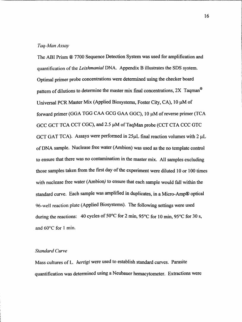
Taq-Man Assay
The ABI Prism ® 7700 Sequence Detection System was used for amplification and
quantification of the Leishmanial DNA. Appendix B illustrates the SDS system.
Optimal primer probe concentrations were determined using the checker board
pattern of dilutions to determine the master mix final concentrations, 2X Taqman®
Universal PGR Master Mix (Applied Biosystems, Foster City, CA), 10 pM of
forward primer (GGA TGG CAA GCG GAA GGC), 10 pM of reverse primer (TCA
GCC GCT TCA CCT CGC), and 2.5 pM of TaqMan probe (CCT CTA CCC GTC
GCT GAT TCA). Assays were performed in 25pL final reaction volumes with 2 pL
of DNA sample. Nuclease free water (Ambion) was used as the no template control
to ensure that there was no contamination in the master mix. All samples excluding
those samples taken from the first day of the experiment were diluted 10 or 100 times
with nuclease free water (Ambion) to ensure that each sample would fall within the
standard curve. Each sample was amplified in duplicates, in a Micro-Amp® optical
96-well reaction plate (Applied Biosystems). The following settings were used
during the reactions: 40 cycles of 50°C for 2 min, 95°C for 10 min, 95°C for 30 s,
and 60°C for 1 min.
Standard Curve
Mass cultures of L. hertigi were used to establish standard curves. Parasite
quantification was determined using a Neubauer hemacydtometer. Extractions were

17
initiated from 2.0 X 10 parasites/mL. The culture was then diluted in 10-fold
dilutions, ranging from 2.0 X 10 to 2000 parasites.
18
RESULTS
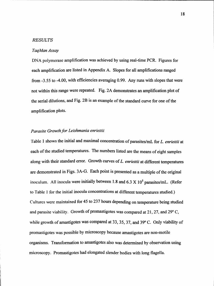
TaqMan Assay
DNA polymerase amplification was achieved by using real-time PGR. Figures for
each amplification are listed in Appendix A. Slopes for all amplifications ranged
from -3.55 to -4.00, with efficiencies averaging 0.99. Any runs with slopes that were
not within this range were repeated. Fig. 2A demonstrates an amplification plot of
the serial dilutions, and Fig. 2B is an example of the standard curve for one of the
amplification plots.
Parasite Growth for Leishmania enriettii
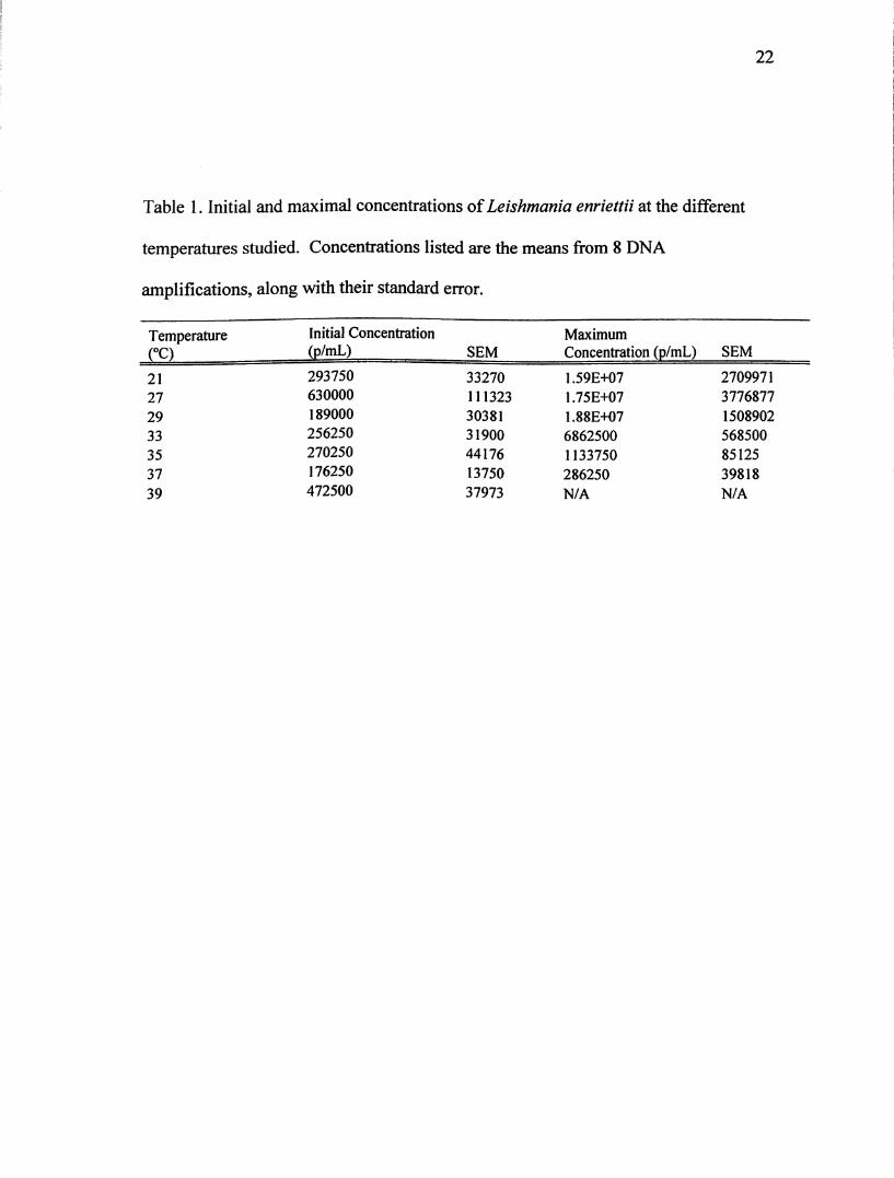
Table 1 shows the initial and maximal concentration of parasites/mL for L. enriettii at
each of the studied temperatures. The numbers listed are the means of eight samples
along with their standard error. Growth curves of L. enriettii at different temperatures
are demonstrated in Figs. 3A-G. Each point is presented as a multiple of the original
inoculum. All inocula were initially between 1.8 and 6.3 X 10 parasites/mL. (Refer
to Table 1 for the initial inocula concentrations at different temperatures studied.)
Cultures were maintained for 45 to 237 hours depending on temperature being studied
and parasite viability. Growth of promastigotes was compared at 21,27, and 29° C,
while growth of amastigotes was compared at 33,35, 37, and 39° C. Only viability of
promastigotes was possible by microscopy because amastigotes are non-mobile
organisms. Transformation to amastigotes also was determined by observation using
microscopy. Promastigotes had elongated slender bodies with long flagella.

19
Amastigotes were able to undergo complete transformation, their bodies were small
and rounded, and no flagellum was present.
20
10AO
10A-1 :
10-2 :
Amplification - novlsaiinas
1OA.3 =
—Samples
FAM-A1 A.
0 FAM - A2 ■ £0 FAM - A3 □0 FAM - A4 ■0 FAM - A5 B0 FAM - A6 ■0 FAM - A7 M0 FAM - A8 m •
0 FAM - A9 PI
0 2 4 6 8 10 12 14 16 18 20 22 24 26 28 30 32 34 36 38 40Cycle
Viewer. |
Reporter: | j
—Threshold Cycle Calculation—Threshold
Use Threshold:! .022^ f Suggest 1Mult, ♦ Stddev:! 1o!o~]*l .002 IOmit Threshold:! 2.0 IBaseline
art:! 3 I Stop:! 15 I
f Update Calculations
Ct Std DevFAM - A1 23.376 0.002FAM - A2 23.118 0.002 iiFAM - A3 27.283 0,001FAM - A4 27.008 0.002FAM - A5 30.090 0.001 ■w
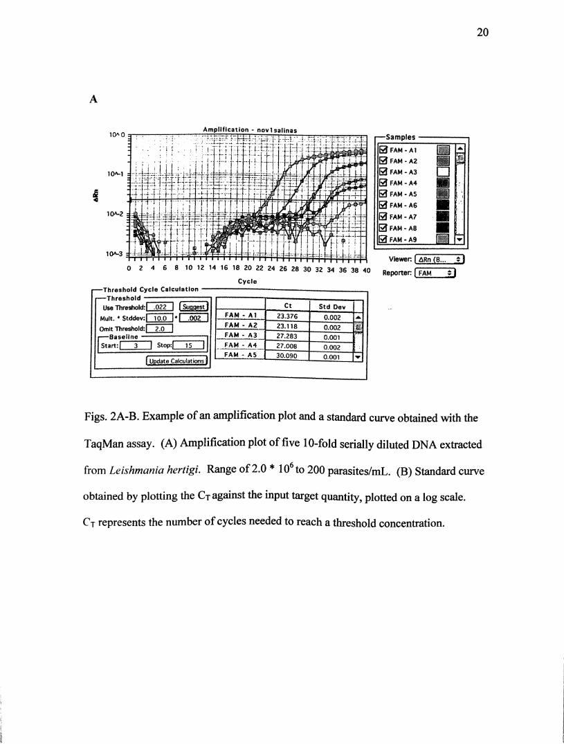
Figs. 2A-B. Example of an amplification plot and a standard curve obtained with the
TaqMan assay. (A) Amplification plot of five 10-fold serially diluted DNA extracted
from Leishmania hertigi. Range of 2.0* lO to 200 parasites/mL. (B) Standard curve
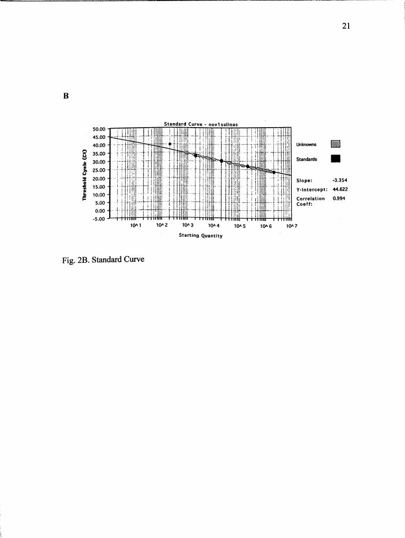
obtained by plotting the Ct against the input target quantity, plotted on a log scale.
Ct represents the number of cycles needed to reach a threshold concentration.
B
21
Standard Curve - novlsalinas
£o
u
50.00
45.00
Unknowns40.00
35.00
Standards30.00 -
25.00
20.00 -
15.00
10.00 -
5.00 -
0.00 -
-5.00
Slope:
Y-lntercept:
Correlation
Coeff:
-3.354
44.622
0.994
10^3 10A4
Starting Quantity
10A5 10^6 10A7
Fig. 2B. Standard Curve
22
Table 1. Initial and maximal concentrations of Leishmania enriettii at the different
temperatures studied. Concentrations listed are the means from 8 DNA
amplifications, along with their standard error.
TemperatureCO
Initial Concentration
(p/mL) SEM
Maximum
Concentration (p/mL) SEM
21 293750 33270 1.590+07 2709971
27 630000 111323 1.75E+07 3776877
29 189000 30381 1.88E+07 1508902
33 256250 31900 6862500 568500
35 270250 44176 1133750 85125
37 176250 13750 286250 39818
39 472500 37973 N/A N/A

23
Cultures of L. enriettii at 21° C were maintained for 237 hours (Fig. 3A).
Maximum concentration was 1.59 X lO' parasites/mL. Population increased about 60
times the original inoculum at around 237 hours. No initial lag phase was evident at
this temperature, but growth was continuous throughout the experiment. Cultures
were not maintained long enough to see parasites stabilize in stationary phase.
Promastigote growth at 27° C was maintained for 141 hours (Fig. 3B).
Parasites immediately went into log phase until growth was slowed after 45 hours.
Population density reached maximum growth of 1.75 X 10^ parasites/mL at 117
hours. Parasites multiplied 33 times from the original inoculum. Parasites remained
as promastigotes at this temperature.
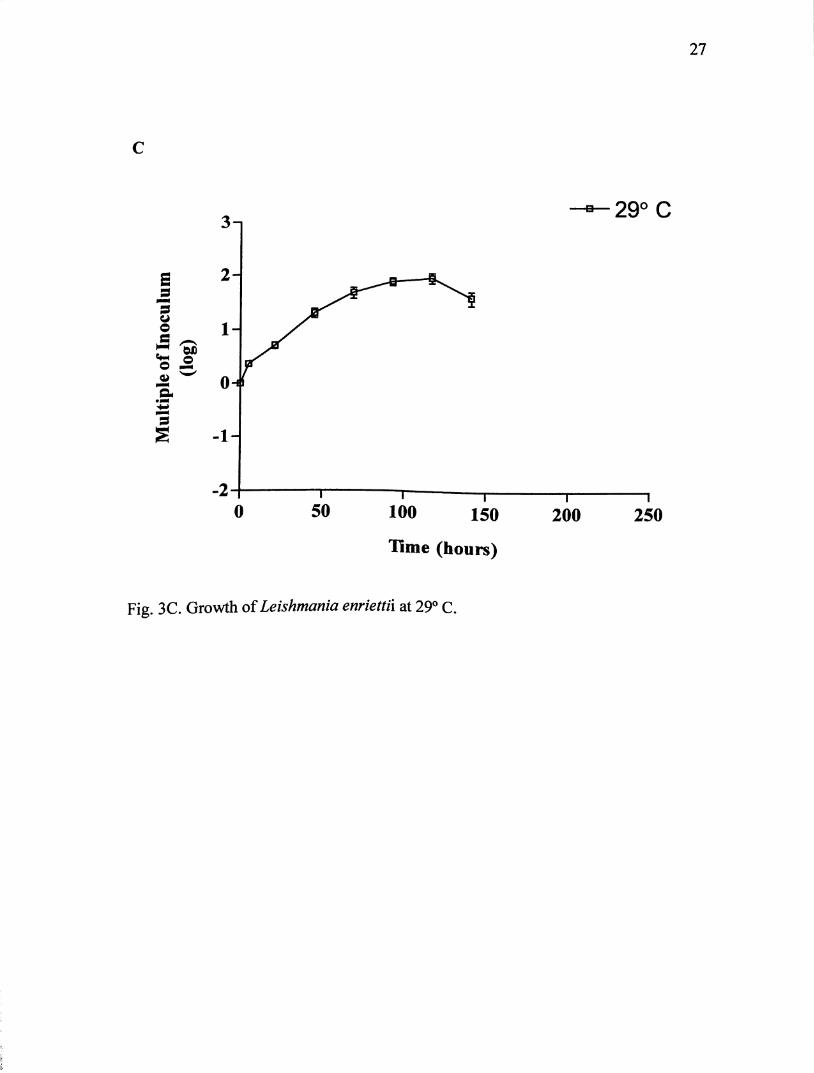
Growth at 29° C (Fig. 3C) was maintained for 141 hours. Parasites went
immediately into log phase until 117 hours, were they reached maximum growth of
about 1.88 X 10 parasites/mL. Population density increased 88 times the original
inoculum. While there was some transformation to amastigote-like organisms, most
of the parasites remained in promastigote form.
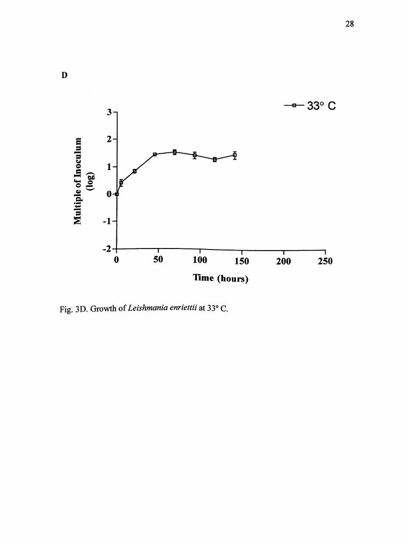
Parasites of I. enriettii were maintained for 141 hours at 33° C (Fig. 3D).
Again no initial lag phase was evident. Parasites went into immediate log phase until
about 45 hours and reached maximum growth of about 6.8 X 10 parasites/mL.
Population density increased 32 times the original inoculum size. After about 45
hours, promastigotes were completely transformed into amastigote-like organisms.

24
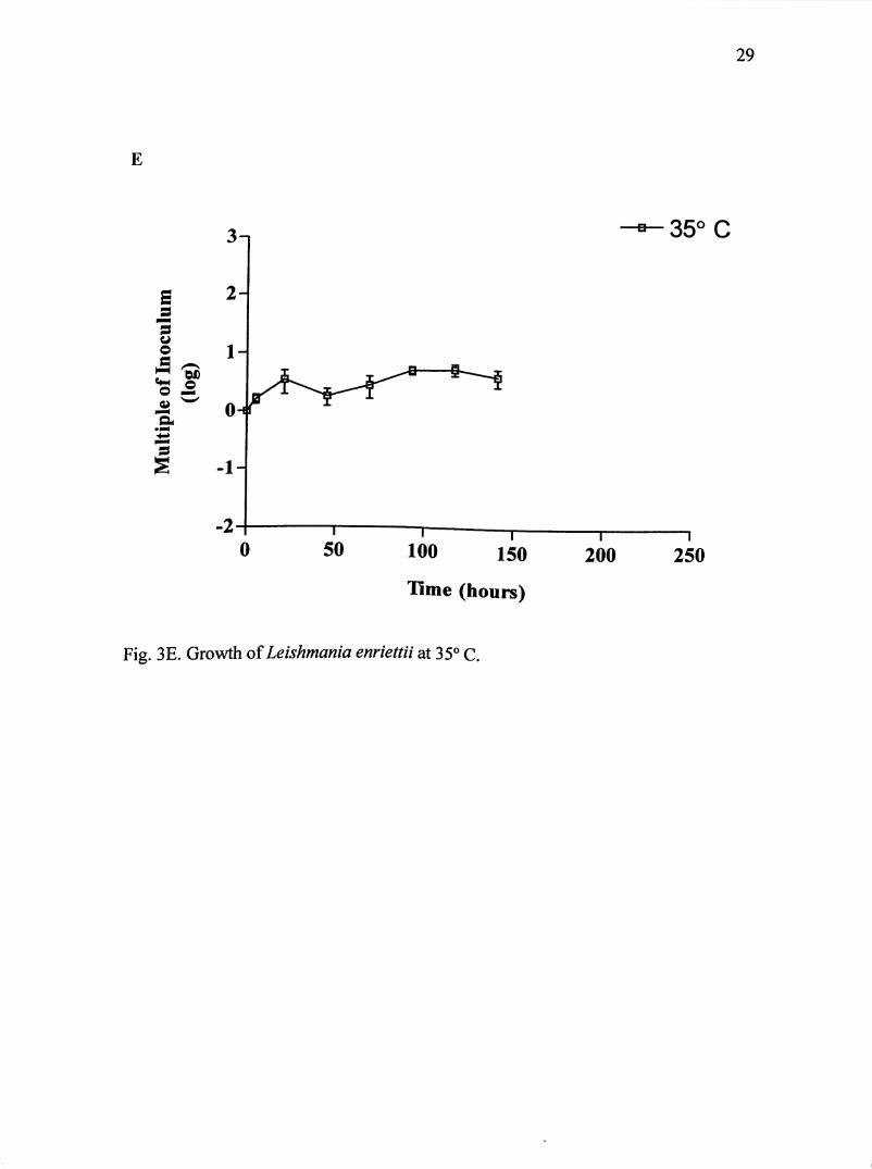
promastigotes began to completely transform into amastigote-like organisms. After
45 hours, complete transformation took place. Additional division occurred for about
48 hours, where amastigotes reach a maximum population of about 1.1 X 10^
parasites/mL within 58 hours. Population size increased 5 times the original
inoculum.
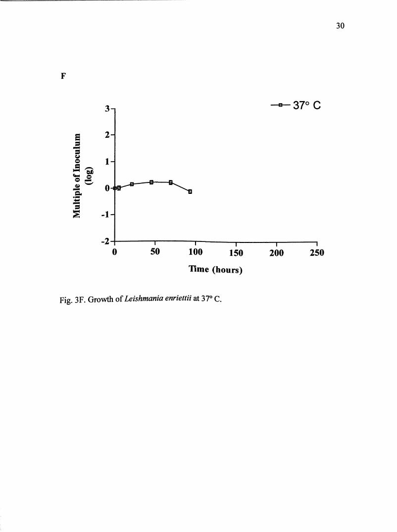
For growth at 37° C, cultures were maintained for 93 hours (Fig. 3F).
Parasites never entered log phase but remained in a lag phase until 46 hours. Parasite
population almost doubled the original inoculum. Maximum population reached
about 2.9 X 10^ parasites/mL at 46 hours. Parasites did transform completely into
amastigote-like orgamsms within the first 22 hours.
At 39° C, L. enriettii were allowed to cultivate for 45 hours (Fig. 3G).
Parasite death was immediate within the first hour and lysis of parasites occurred
rapidly after two hours.
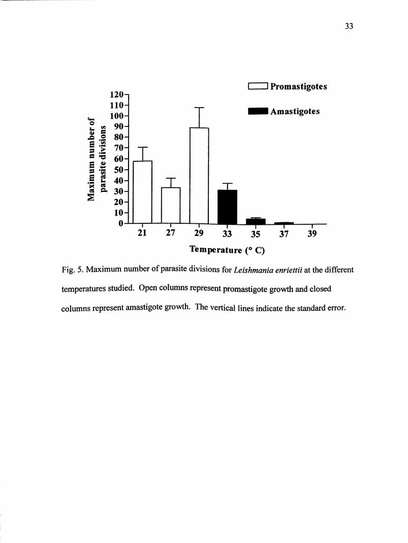
Maximum growth of L. enriettii (parasites/mL) at all temperatures studied is
illustrated in Fig. 4. Open vertical columns signify promastigote growth, while closed
bars represent amastigote growth. Also, maximum number of parasite divisions is
shown in Fig. 5. Parasite division occurred at all temperatures except at 39° C.

25
swo
a
100 150
Hme (hours)
-b-21°C
250
Figs. 3A-G. Growth curves of Leishmania enriettii at different temperatures studied.
Each point is presented as a multiple of the original inoculum. All inocula were
initially between 1.8 and 6.3 X 10 parasites/mL. Cultures were maintained for 45 to
237 hours depending on temperature studied and parasite viability. The vertical bars
indicate the standard error.

B
26
Ss
0OO
e.
3-1
2-
1-
0-
-1-
-2-
0
--27° C
—r-
50 100 150
Time (hours)
200 250
Fig. 3B. Growth of Leishmania enriettii at 27 C.
27
-29° C
s
uo
s
100 150
Time (hours)
200 250
Fig. 3C. Growth of Leishmania enriettii at 29° C.
D
28
-33°C
100 150
Time (hours)
200 250
Fig. 3D. Growth of Leishmania enriettii at 33° C.
E
29
-35°C
100 150
Time(hours)
200 250
Fig. 3E. Growth of Leishmania enriettii at 35° C.
30
9
wo
S
3n
2-
1-
0
-1-
-2-
0
--37°C
—1—
50 100 150
Time (hours)
200 250
Fig. 3F. Growth of Leishmania enriettii at 37° C.

31
o
S
3n
2-
1-
100 150
Time (hours)
39
200 250
Fig. 3G. Growth of Leishmania enriettii at 39° C.

32
lO
10'-s
5
lO'H
10
i Promastigotes
Amastigotes
21 27 29 33 35
Temperature (° C)
Fig. 4. Maximum growth (parasites/mL) of Leishmania enriettii at different
temperatures studied. Open columns represent promastigote growth and closed
columns represent amastigote growth. The vertical bars indicate the standard error.
33
a
^
2A
120-1
110-
100
90
80
70
60
50
40
30
20
10-
0-
I
-T"
21
T
' i Promastigotes
I Amastigotes
T"
27 29 33 35 37
Temperature (® C)
Fig. 5. Maximum number of parasite divisions for Leishmania enriettii at the different
temperatures studied. Open columns represent promastigote growth and closed
columns represent amastigote growth. The vertical lines indicate the standard error.
34
Parasite Growth for Leishmania hertigi
Initial and maximal parasite concentrations are given on Table 2 along with their
standard error. Growth curves for L. hertigi at different temperatures are
demonstrated in Figs. 6A-G. Each point is presented as a multiple of the original
inoculum. All inocula were initially between 2.6 to 9.0 X 10 parasites/mL. (Refer to
Table 2 for the initial inoculum concentrations at different temperatures studied.)
Cultures were maintained for 45 to 189 hours depending on temperature studied and
parasite viability. Again only promastigote viability was possible to estimate.
Growth of promastigotes was compared at 21,27, and 29° C, and growth of
amastigotes was compared at 33, 35,37, and 39° C. Unlike L. enriettii, L hertigi
promastigotes had different morphological shapes. Further morphological studies
need to be evaluated. Amastigote transformation was also difficult to determine due
to the variety of shapes observed. Complete transformation could not be evaluated.
However estimations were given as to when the majority of amastigotes in culture
were in a rounded form.
Leishmania hertigi cultures were maintained for 189 hours at 21° C (Fig. 6A).
Parasites immediately entered log phase for the first 21 hours, then slowed to a
gradual increase for another 72 hours. At 93 hours, maximum growth of 5.31 X 10
parasites/mL was achieved. On average, there was an increase of 85 times the
original inoculum.
At 27° C, cultures were maintained for 141 hours (Fig. 6B). Parasites entered
log phase at about 5 hours into incubation, and lasted another 64 hours. Maximum

35
growth of 4.21 X 10 parasites/mL was achieved near 69 hours. On average, this was
21 times the original inoculum size.
Leishmania hertigi cultures at 29°C were maintained for 141 hours (Fig. 6C).
Parasites entered log phase after 5 hours and lasted for 16 hours. Maximum growth
of 2.42 X lO' parasites/mL was achieved at 21 hours. On average, parasites
multiplied 36 times their original inoculum size.
The growth curve for L. enriettii at 33° C is demonstrated in Fig. 6D. Cultures
were maintained for 141 hours. Parasites entered log phase after 5 hours and
continued another 40 hours. Maximum growth of 1.17 X 10 parasites/mL was
achieved after 45 hours. On average, parasites multiplied 13 times their original
inoculum size. It is estimated that transformation occurred after 45 hours.
Cultures in 35° C were cultivated for 141 hours (Fig. 6E). Parasites did not
enter a log phase, but did achieve maximum growth of about 1.4 X 10 parasites/mL
at 21 hours. On average, parasites multiplied 3 times the original inoculum size. It is
estimated that transformation took place after 21 hours.
At 37° C parasites were incubated for 93 hours (Fig. 6F). Parasites did not
enter log phase, but after 5 hours reached its maximum concentration of about 8.3 X
10" parasites/mL. On average, parasites multiplied 1.3 times the original inoculum
size. It is estimated that transformation took place after 21 hours.
Fig. 6G shows the growth curve of L. hertigi at 39° C for 45 hours. Parasites
did not enter log phase, but after 5 hours reached its maximum concentration of about

36
2.2 X 10 parasites/mL. On average, parasites multiplied almost twice the original
inoculum size.
Maximum growth of L. hertigi (parasites/mL) at all temperatures studied is
illustrated in Fig. 7. Open vertical columns signify promastigote growth, while closed
bars represent amastigote growth. Also, maximum number of parasite divisions is
shown in Fig. 8. Parasite division occurred at all temperatures studied.
37
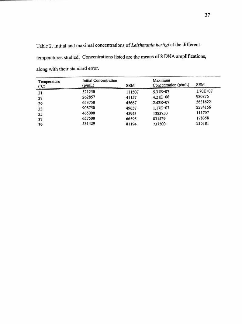
Table 2. Initial and maximal concentrations of Leishmania hertigi at the different
temperatures studied. Concentrations listed are the means of 8 DNA amplifications,
along with their standard error.
TemperatureCO
Initial Concentration
(p/mL) SEM
Maximum
Concentration (p/mL) SEM
21 521250 111507 5.31 £+07 1.70E+07
27 262857 41157 4.21E+06 980876
29 653750 45667 2.42E+07 5631622
33 908750 49657 1.17E+07 2274156
35 465000 45943 1383750 111707
37 657500 66595 831429 178358
39 531429 81194 737500 215181

38
oo
100 150
Time (hours)
200 250
Figs. 6A.-G. Growth curves of Leishmania hertigi at different temperatures studied.
Each point is presented as a multiple of the original inoculum. All inocula were
initially between 2.6 and 9.0 X 10 parasites/mL. Cultures were maintained for 45 to
189 hours depending on temperature studied and parasite viability. The vertical bars
indicate the standard error.
B
39
27° C
100 150
Time (hours)
200 250
Fig. 6B. Growth of Leishmania hertigi at 27° C.
40
29° C
100 150
Time (hours)
200 250
Fig. 6C. Growth of Leishmania hertigi at 29° C.
D
41
a
u
s
3-1
2-
1-
0
-1-
-2-
0
—T"
50 100 150
Time(hours)
33° C
200 250
Fig. 6D. Growth of Leishmania hertigi at 33° C.

E
42
o
5
3n
2-
-1-
-2-
0
C
—r-
50 100 150
Time (hours)
200 250
Fig. 6E. Growth of Leishmania hertigi at 35° C.

43
o
o
a
3n
2-
1-
0
-1-
0
—T"
50 100 150
Time (hours)
37° C
200 250
Fig. 6F. Growth of Leishmania hertigi at 37° C.
44
a
swo
2«
ft
3n
2-
1-
100 150
Time (hours)
39^
200 250
Fig. 6G. Growth of Leishmania hertigi at 39° C.

45
8_10
10-
0
10*-w
10' -T"
21
Promastigotes
I Amastigotes
27 29 33 35 37 39
Temperature (° C)
Fig. 7. Maximum growth (parasites/mL) of Leishmania hertigi at different
temperatures studied. Open columns represent promastigote growth and closed
columns represent amastigote growth. The vertical bars indicate the standard error.

46
o
110^10090
8070
60504030
20I0^0 -T"
21
I I Promastigotes
Amastigotes
X
T-
27 29 33 35 37
Temperature (° C)
Fig. 8. Maximum number of parasite divisions for Leishmania hertigi at the different
temperatures studied. Open columns represent promastigote growth and closed
columns represent amastigote growth. The vertical bars indicate the standard error.

47
Discussion
Leishmania parasites face hostile environmental conditions such as dramatic
temperature fluctuation, drastic pH changes, nutritional depletion in the sand fly, and
host lytic and oxidative product formation (Sacks and Kamhawi, 2001; Zilberstein
and Shapira, 1994). I have attempted to study one of the stresses faced by the
parasites, which is increase of temperature from the sand fly host to the mammalian
host. Temperature has been shown to induce the morphological transformation from
the promastigote to amastigote form in axenic conditions (Ismaeel et al. 1998;
Krassner, 1965; Leon et al. 1995). It also has been suggested that a correlation exists
between skin/body temperature and the extent of the lesions caused by these parasites
(Callahan et al. 1996). The purpose of this thesis project was to determine the upper
limits of temperature tolerance by L. enriettii and L. hertigi, and to test whether or
not body temperature of their mammalian host plays a role in their tolerance to high
temperatures. With the use of a range of incubation temperatures and the
incorporation of real-time PCR, growth curves were constructed to characterize
temperature effects.
Based on the results, it was observed that parasites of L. enriettii were able to
completely transform into amastigote-like organisms with temperature change alone.
By phase contrast microcopy, inconclusive results were yielded if parasites of L.
hertigi were able to undergo complete transformation with temperature change alone.
All other variables in this in vitro system remained unchanged (pH, control
temperature, and media). While the warmer temperatures were of more interest in

48
this study, growth studies also were done on a few cooler temperatures to demonstrate
any differences between promastigote and amastigote growth. The American Type
Culture Collection recommended that 25° C was the optimal temperature for growth
of L. enriettii and L. hertigi promastigotes. Since 25° C was omitted from the study,
the optimal temperature for promastigote growth can not be concluded for either L.
enriettii or L. hertigi. It can be said that from Figs. 3A-C, 21° C had continuous but
slow growth for a longer period of time, while at 29° C, growth was more rapid and
reached the highest maximum growth. Also at 29° C, some transformation to
amastigote-like organisms occurred. One reason this could have occurred is because
of pH fluctuations in the media with increase of temperature. The pH of the media
was never determined during the experiment.
Amastigote growth for L enriettii was compared at 33,35,37, and 39° C
(Figs. 3C-G). Complete transformation to amastigotes was evident after 45 hours for
both 33 and 35° C. At 37° C, transformation was much more rapid and took place
after 22 hours. Figures 4 and 5 show that of the elevated temperatures, 33° C had the
most growth and number of divisions. However, only at 35° C, once promastigotes
transformed to amastigotes, did additional division take place (Fig. 3E). At 33 and
37° C, amastigotes remained in a lag phase (Figs. 3D and 3F). Leishmania enriettii
was able to withstand 37° C, but only underwent one division (Fig. 3G). One reason
amastigotes were not able to enter exponential growth in the other temperatures
besides 35° C could be that 35° C was their optimal temperature. Also it could be that
other variables need to be changed in order to mimic in vivo conditions (Pan et al.
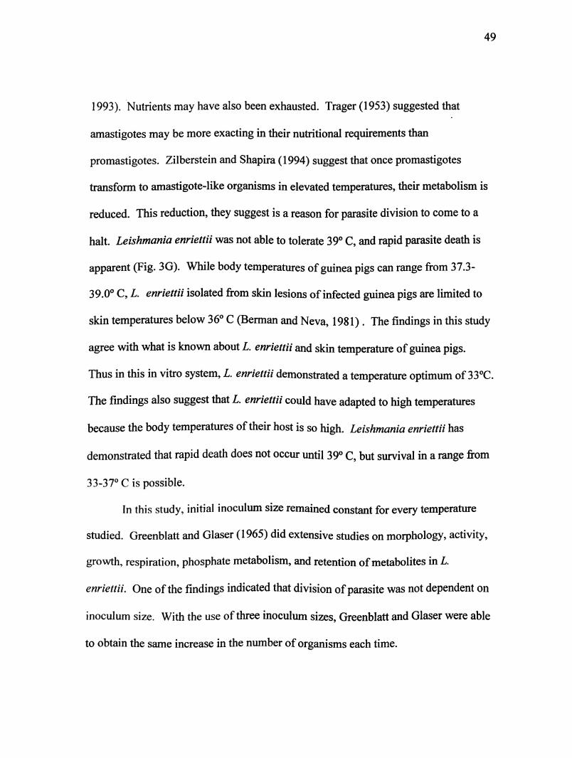
49
1993). Nutrients may have also been exhausted. Trager (1953) suggested that
amastigotes may be more exacting in their nutritional requirements than
promastigotes. Zilberstein and Shapira (1994) suggest that once promastigotes
transform to amastigote-like organisms in elevated temperatures, their metabolism is
reduced. This reduction, they suggest is a reason for parasite division to come to a
halt. Leishmania enriettii was not able to tolerate 39° C, and rapid parasite death is
apparent (Fig. 3G). While body temperatures of guinea pigs can range from 37.3-
39.0° C, L. enriettii isolated from skin lesions of infected guinea pigs are limited to
skin temperatures below 36° C (Berman and Neva, 1981). The findings in this study
agree with what is known about L. enriettii and skin temperature of guinea pigs.
Thus in this in vitro system, L. enriettii demonstrated a temperature optimum of 33°C.
The findings also suggest that L. enriettii could have adapted to high temperatures
because the body temperatures of their host is so high. Leishmania enriettii has
demonstrated that rapid death does not occur until 39° C, but survival in a range from
33-37° C is possible.
In this study, initial inoculum size remained constant for every temperature
studied. Greenblatt and Glaser (1965) did extensive studies on morphology, activity,
growth, respiration, phosphate metabolism, and retention of metabolites in L.
enriettii. One of the findings indicated that division of parasite was not dependent on
inoculum size. With the use of three inoculum sizes, Greenblatt and Glaser were able
to obtain the same increase in the number of organisms each time.

50
Optimal temperature for promastigote growth cannot be inferred for L. hertigi
because 25° C was not studied. However growth at 21° C produces maximum growth
and number of divisions (Fig. 7 and Fig. 8).
Amastigote growth also was compared at 33,35, 37, and 39° C. Leishmania
hertigi growth curves (Figs. 6C-G) demonstrate that once transformation took place,
amastigotes remained in a lag phase. Parasites might have been viable, but not
capable of further divisions. The main difference between 33, 35, and 37° C was the
time required for promastigotes to transform to amastigotes. At 35 and 37° C,
promastigotes transformed more rapidly. Growth at 37° C (Fig. 6F), shows that after
about 24 hours, there was slight growth, but amastigotes remained in a lag phase
thereafter. At 39 C (Fig. 6G) promastigotes multiplied almost twice the original size
after 5 hours, but quickly lysed due to the high temperature. This multiplication may
not indicate new growth but rather completion of divisions initiated at time zero. Fig.
7 illustrates that maximum parasites/mL were about the same for 33,35, and 37° C,
but Fig. 8 demonstrates there was more growth at 33° C. Thus in this in vitro system,
L hertigi had a temperature optimum of 33° C. These results are consistent to what is
known about New World species of Leishmcinici. In general, amastigotes of New
World species have an optimal temperature range of 28 to 33° C, while Old World
species prefer elevated temperatures of 37 to 39° C (Pan et al. 1993)
The findings also support the hypothesis in that the body temperature of the
host corresponds to the limits of temperature tolerance by L hertigi. Body
temperature of porcupines was reported at 36.7° C in Arends and McNab (2001)
51
study. Leishmania hertigi amastigotes in this in vitro system were able to tolerate a
range of 33-37° C. While death was evident at 39° C, it was not as rapid as the rate
observed with L. enriettii. Parasites were able to complete one division. These
findings are not surprising because L. hertigi has sometimes been found in the viscera
of porcupines (Zeledon e/a/. 1997). Although the temperature of the viscera is
unknown for porcupines, it appears L. hertigi has developed a thermotolerance to
extreme temperatures.
There is no question that parasites of Leishmania species have developed a
tolerance to extreme temperature change. Its survival and continuance is dependent
on making the necessary adaptations to extreme environmental stresses. Each species
could have developed their own limits of temperature tolerance depending on the
body temperatures of their mammalian hosts. By this host characteristic, parasite
coevolution with their host will persist. Additional studies using the in vitro system
described in this project need to be demonstrated with other species of Leishmania to
further develop this hypothesis.

52
REFERENCES
Adler, S. and Halff, L. (1955). Observations on Leishmania enriettii Muniz and
Medina, 1948. Annals of Tropical Medicine and Hygiene 49,37-41.
Altschul, S. F., Gish, W., Miller, W., Myers, E. W. and Lipman, D. J. (1990).
Basic local alignment search tool. Journal of Molecular Biology 215,403-
410.
Arends, A. and McNab, B. K. (2001). The comparative energetics of'caviomorph'
rodents. Comparative Biochemistry and Physiology. Part A, Molecular and
Integrative Physiology 130,105-122.
Belehu, A. and Turk, J. L. (1976). Establishment of cutaneous Leishmania enriettii
infection in hamsters. Infection and Immunity 13, 1235-1241.
Bell, A. and Randford-Cartwright, L. (2002). Real-time quantitative PGR in
parasitology. Trends in Parasitology 18,795-802.
Berman, J. D. and Neva, F. A. (1981). Effect of temperature on multiplication of
Leishmania amastigotes within human monoc)4e-derived macrophages in
vitro. The American Journal of Tropical Medicine and Hygiene 30,318-321.
Bretagne, S., Durand, R., Olivi, M., Garin, J. F., Sulahian, A., Rivollet, D.,
Vidaud, M. and Deniau, M. (2001). Real-time PGR as a new tool for
quantifying Leishmania infantum in liver in infected mice. Clinical and
Diagnostic Laboratory Immunology 8, 828-831.
53
Callahan, H. L., Portal, I. F., Bensinger, S. J. and Grogl, M. (1996). Leishmania
spp: temperature sensitivity of promastigotes in vitro as a model for tropism in
vivo. Experimental Parasitology 84,400-409.
Clos, J. and Krobitsch, S. (1999). Heat shock as a regular feature of the life cycle of
Leishmania parasites. American Zoologist 39, 848-856.
Croft, S. L., Schnur, L. F. and Chance, M. L. (1978). The morphological,
biochemical and serological characterization of strains of Leishmania hertigi
from Panama and Brazil and their differentiation. Annals of Tropical Medicine
and Hygiene 72,93-94.
Cupolillo, E., Aguiar Alves, F., Brahim, L. R., Naiff, M. F., Pereira, L. O.,
Oliveira-Neto, M. P., Falqueto, A. and Grimaldi, G., Jr. (2001). Recent
advances in the taxonomy of the New World leishmanial parasites. Medical
Microbiology and Immunology 190,57-60.
Cupolillo, E., Momen, H. amd Grimaldi, G., Jr. (1998). Genetic diversity in natural
populations of New World Leishmania. Memorias do Instituto Oswaldo Cruz
93, 663-668.
El-On, J., Witztum, A. and Schnur, L. F. (1986). Protection of guinea pigs against
cutaneous leishmaniasis by combined infection and chemotherapy. Infection
and Immunology 51, 704-706.
Gomez-Saladim, E., Doud, C. W, and Maroli, M. (2005). Short report: surveillance
of Leishmania spp. among sand flies in Sicily (Italy) using a fluorogenic real-
54
time polymerase chain reaction. The American Journal of Tropical Medicine
and Hygiene 12y 138-141.
Greenblatt, C. L. and Glaser, P. (1965). Temperature effect on Leishmania enriettii
in vitro. Experimental Parasitology 16,36-52.
Grimaldi, G. and Schottelius, J. (2001). Leishmaniases-their relationships to
monoxenous and dixenous trypanosomatids. Medical Microbiology and
Immunology (Berl) 190,3-8.
Guillaume, J. V. E., Schoone, G. J., Kroon, N. C. M., and Ebeling, S. B. (1992).
Sequence analysis of subunit ribosomal RNA genes and its use for detection
and identification of Leishmania parasites. Molecular and Biochemical
Parasitology 51, 133-142.
Herrer, A. and Christensen, H. A. (1975). Infrequency of gross skin lesions among
Panamaman forest mammals with cutaneous leishmaniasis. Parasitology 71,
87-92.
Ismaeel, A. Y., Garmson, J. C., Moyneux, D. H. and Bates, P. A. (1998).
Transformation, development, and transmission of axenically cultured
amastigotes of Leishmania mexicana in vitro and in Lutzomyia longipalpis.
The American Journal of Tropical Medicine and Hygiene 59,421-425.
Krassner, S. M. (1965). Effect of temperature on growth and nutritional
requirements of Leishmania tarentolae in a defined medium. Journal of
Protozoology 12,73-78.
55
Lainson, R. (1997). On Leishmania enriettii and other enigmatic Leishmania species
of the Neotropics. Memorias do Institute Oswaldo Cruz 92,377-387.
Leon, L. L., Scares, M. J. and Temporal, R. M. (1995). Effects of temperature on
promastigotes of several species of Leishmania. The Journal of Eukaryotic
Microbiology 42,219-223.
Lewis, D. J. (1974). The biology of Phlebotomidae in relation to leishmaniasis.
Annual Review ofEntomology 19, 363-384.
Lukacs, G. L., Rotstein, O. D. and Grinstein, S. (1991). Determinants of the
phagosomal pH in macrophages. In situ assessment of vacuolar H(+)-ATPase
activity, counterion conductance, and H+ "leak". The Journal ofBiological
Chemistry 266,24540-24548.
Machado, M. I., Milder, R V., Paeheco, R. S., Silva, M., Braga, R. R. and
Lainson, R. (1994). Naturally acquired infections with Leishmania enriettii
Muniz and Medina 1948 in guinea-pigs from Sao Paulo, Brazil. Parasitology
109, 135-138.
Ao J. (1995). Epidemiology of the leishmaniases. Dermatologic Clinics 13,
505-523.
U C., Faraiiat, F., Lascomlbe, L. and Dumon, H. (2004). Quantification of
Leishmania infantum DNA by a real-time PGR assay with high sensitivity.
Journal of Clinical Microbiology 42,5249-5255.
Mehra, S. and Hu, W. S. (2005). A kinetic model of quantitative real-time
polymerase chain reaction. Biotechnology and Bioengineering 91, 848-860.

56
Noyes, H. A., Morrison, D. A., Chance, M. L. and Ellis, J. T. (2000). Evidence for
a Neotropical origin of Leishmania. Memorias do Institute Oswaldo Cruz 95,
575-578.
Ogg, M. M., Carrion, R., Jr., Botelho, A. C., May rink, W., Correa-Oliveira, R.
and Patterson, J. L. (2003). Short report: quantification of leishmaniavirus
RNA in clinical samples and its possible role in pathogenesis. The American
Journal of Tropical Medicine and Hygiene 69,309-313.
Pan, A. A., Duboise, S. M., Eperon, S., Rivas, L., Hodgkinson, V., Traub-Cseko,
Y. and McMahon-Pratt, D. (1993). Developmental life cycle of Leishmania-
-cultivation and characterization of cultured extracellular amastigotes. Journal
ofEukaryotic Microbiology 40,213-223.
Pral, E. M., M. Da., Moitinho, M. L., Balanco, J. M., Teixeira, V. R., Milder, R.
V. and Alfieri, S. C. (2003). Growth phase and medium pH modulate the
expression of proteinase activities and the development of megasomes in
axenically cultivated Leishmania {Leishmania) amazonensis amastigote-like
organisms. The Journal of Parasitology 89,35-43.
Rolao, N., Cortes, S., Rodrigues, O. R. and Campino, L. (2004). Quantification of
Leishmania infantum parasites in tissue biopsies by real-time polymerase
chain reaction and polymerase chain reaction-enzyme-linked immunosorbent
assay. The Journal of Parasitology 90,1150-1154.
57
Sacks, D. and Kamawi, S. (2001). Molecular aspects of parasite-vector and vector-
host interactions in leishmaniasis. Annual Review of Microbiology 55,453-
483.
Schulz, A., Mellenthin, K., Schonian, G., Fleishcer, B. and Drosten, C. (2003).
Detection, differentiation, and quantitation of pathogenic Leishmania
organisms by a fluorescence resonance energy transfer-based real-time PGR
assay. Journal of Clinical Microbiology 41, 1529-1535.
Souza, N. A., Andrade-Coelho, C. A., Silva, V. C., Peixoto, A. A. and Rangel, E.
F. (2005). Moonlight and blood-feeding behaviour of Lutzomyia intermedia
and Lutzomyia whitmani (Diptera;Psychodidae:Phlebotominae), vectors of
American cutaneous leishmamasis in Brazil. Memorias do Instituto Oswaldo
Cruz 100,39-42.
Thomaz-Soccol, V., Pratlong, F., Langue, R., Castro, E., Luz, E. and Dedet, J. P.
(1996). New isolation of Leishmania enriettii Muniz and Medina, 1948 in
Parana State, Brazil, 50 years after the first description, and isoenzymatic
polymorphism of the L. enriettii taxon. Annals of Tropical Medicine and
Parasitology 90,491-495.
T rager, W. (1953). The development of Leishmania donovani in vitro at 37°C. The
effects of the kind of serum. The Journal of Experimental Medicine 97,101-
115.

58
Van Der Ploeg, L. H., Giannini, S. H. and Cantor, C. R. (1985). Heat shock genes:
regulatory role for differentiation in parasitic protozoa. Science 228,1443-
1446.
Vitale, F., Reale, S., Vitale, M., Petrotta, E., Torina, A. and Caracappa, S. (2004).
TaqMan-based detection of Leishmania infantum DNA using canine samples.
Annals of the New York Academy ofSciences 1026, 139-143.
World Health Organization (1990). Control of the leishmaniases. WHO Technical
Report Series 793. WHO, Geneva.
Wortmann, G., Sweeney, C., Houng, H. S., Aronson, N., Stiteler, J., Jackson, J.
and Ockenhouse, C. (2001). Rapid diagnosis of leishmaniasis by fluorogenic
polymerase chain reaction. The American Journal of Tropical Medicine and
Hygiene 65,583-587.
Zeledon, R., Ponce, C. and de Ponce, E. (1977). Finding of Leishmania hertigi in
the Costa Rican porcupine. Journal of Parasitology 63,924-925.
Zilberstein, D. and Shapira, M. (1994). The role of pH and temperature in the
development of Leishmania parasites. Annual Review of Microbiology 48,
449-470.

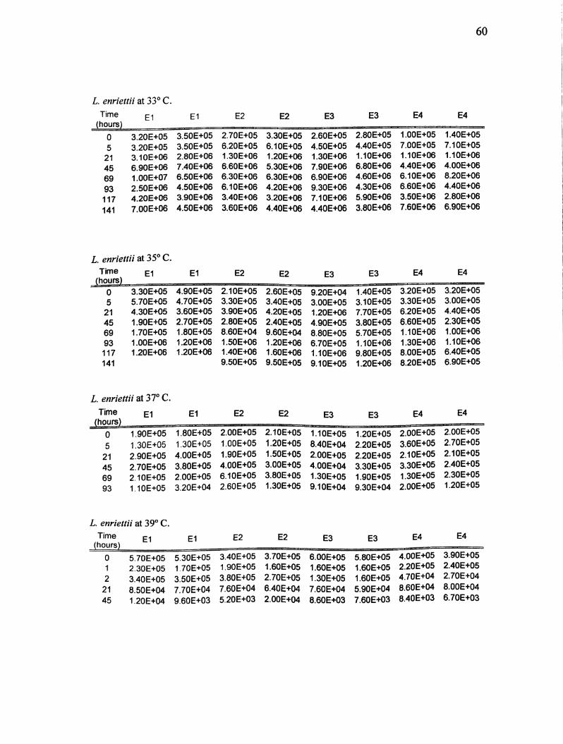
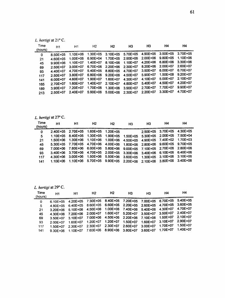
59
Appendix A. Real-time PCR results for L. enriettii and L. hertigi at 21,27,29, 33,
35, 37, and 39° C. Numbers reflect parasites/mL. Any gaps in data reflect that no
signal was detected in that well. Samples were run in duplicates. E1-E4 and H1-H4
represent each trial for that experiment.
L. enriettii at 21° C.
Time El El E2 E2 E3 E3 E4 E4(hours)
0 2.70E+05 2.80E+05 4.00E+05 4.40E+05 1.70E+05 1.80E+05 3.00E+05 3.10E+05
21 2.70E+05 4.40E+05 6.60E+05 4.30E+05 4.90E+05 3.70E+05 1.40E+05 2.30E+05
45 8.70E+05 8.40E+05 8.10E+05 7.80E+05 6.70E+05 5.90E+05 8.10E+05 7.50E+05
69 1.60E+06 2.00E+06 1.90E+06 1.40E+06 2.10E+06 2.60E+06 1.20E+06 1.50E+06
93 4.40E+06 2.40E+06 4.20E+06 4.30E+06 6.80E+06 4.10E+06 3.90E+06 4.30E+06
117 3.40E+06 5.00E+06 7.40E+06 6.60E+06 4.60E+06 4.40E+06 9.20E+06 9.20E+06
141 3.70E+06 3.60E+06 1.40E+07 1.40E+07 7.70E+06 1.30E+07 1.90E+06 1.30E+06
165 6.10E+06 3.60E+06 8.50E+06 1.30E+07 9.30E+06 9.20E+06 1.10E+07 1.10E+07
189 1.10E+07 1.10E+07 1.60E+07 2.20E+07 7.60E+06 5.50E+06 3.10E+06 3.80E+07
213 9.20E+05 1.10E+07 1.90E+07 1.90E+07 2.20E+07 150E+07 1.40E+07 2.60E+07
L. enriettii at 27° C.
Time E1 E1 E2 E2 E3 E3 E4 E4
0 5.30E+05 6.20E+05 5.50E+05 1.40E+06 4.80E+05 4.70E+05 4.90E+05 5.00E+05
5 4.20E+05 7.30E+05 8.60E+05 9,10E+05 6.30E+05 9.90E+05 6.50E+05 8.40E+05
21 1.30E+06 2.10E+06 1.60E+06 2.30E+06 1.30E+06 1.20E+06 1 .OOE+06 1.40E+06
45 1.40E+07 9.20E+06 2.20E+07 2.40E+07 9.90E+06 1.00E+07 7.00E+06 1.10E+07
69 1.10E+07 1.50E+07 1.40E+07 1.10E+07 9.30E+06 1.20E+07 1.30E+07 1.20E+07
93 2.90E+07 1.80E+07 1.80E+07 1.30E+07 6.60E+06 1.40E+07 9.70E+06 1.10E+07
117 8.90E+06 1.50E+07 8.60E+06 9.00E+06 9.60E+06 2.70E+07 3.60E+07 2.60E+07
141 1.30E+07 1.20E+07 1.70E+07 1.50E+07 5.50E+06 1.20E+07 2.10E+07 1.40E+07
L. enriettii at 29° C.
TimeEl El E2 E2 E3 E3 E4 E4
(hours)
0
5
21
45
69
93
117
141
1 70E+05
3.70E+05
1.10E+06
5.50E+06
6.60E+06
2.00E+07
1.10E+07
1.10E+07
1.70E+05
3.50E+05
1.50E+06
4.30E+06
9.20E+06
200E+07
1 90E+07
7.40E+06
2.80E+05
1.40E+05
9.40E+05
4.30E+06
1.20E+07
1.50E+07
2.10E+07
1.20E+07
3.60E+05
1.40E+05
1.10E+06
4.20E+06
9.30E+06
1.80E+072.30E+07
1.10E+07
1.20E+05
1.30E+05
1.80E+06
4.20E+06
1.00E+07
1.40E+07
1.60E+07
9.70E+06
1.20E+05
1.30E+05
1.60E+06
3.00E+06
1.20E+07
1.10E+07
1.60E+07
7.90E+0e
1.50E+05
1.20E+05
7.80E+05
5.10E+06
7.80E+06
1.70E+07
2.40E+07
5.50E+06
1.40E+05
1.40E+05
8.20E+05
3.60E+06
9.80E+06
1.80E+07
2.00E+07
2.00E+06
60
L enriettii at 33° C.
Time E1 E2 E2 E3 E3 E4 E4
0 3.20E+05 3.50E+05 2.70E+05 3.30E+05 2.60E+05 2.80E+05 1.00E+05 1.40E+05
5 3.20E+05 3.50E+05 6.20E+05 6.10E+05 4.50E+05 4.40E+05 7.00E+05 7.10E+05
21 3.10E+06 2.80E+06 1.30E+06 1.20E+06 1.30E+06 1.10E+06 1.10E+06 1.10E+06
45 6.90E+06 7.40E+06 6.60E+06 5.30E+06 7.90E+06 6.80E+06 4.40E+06 4.00E+06
69 1.00E+07 6.50E+06 6.30E+06 6.30E+06 6.90E+06 4.60E+06 6.10E+06 8.20E+06
93 2.50E+06 4.50E+06 6.10E+06 4.20E+06 9.30E+06 4.30E+06 6.60E+06 4.40E+06
117 4.20E+06 3.90E+06 3.40E+06 3.20E+06 7.10E+06 5.90E+06 3.50E+06 2.80E+06
141 7.00E+06 4.50E+06 3.60E+06 4.40E+06 4.40E+06 3.80E+06 7.60E+06 6.90E+06
L enriettii at 35° C.Time El El E2 E2 E3 E3 E4 E4
(hours)
0 3.30E+05 4.90E+05 2.10E+05 2.60E+05 9.20E+04 1.40E+05 3.20E+05 3.20E+05
5 5.70E+05 4.70E+05 3.30E+05 3.40E+05 3.00E+05 3.10E+05 3.30E+05 3.00E+05
21 4.30E+05 3.60E+05 3.90E+05 4.20E+05 1.20E+06 7.70E+05 6.20E+05 4.40E+05
45 1.90E+05 2.70E+05 2.80E+05 2.40E+05 4.90E+05 3.80E+05 6.60E+05 2.30E+05
69 1.70E+05 1.80E+05 8.60E+04 9.60E+04 8.80E+05 5.70E+05 1.10E+06 1.00E+06
93 1.00E+06 1.20E+06 1.50E+06 1.20E+06 6.70E+05 1.10E+06 1.30E+06 1.10E+06
117 1.20E+06 1.20E+06 1.40E+06 1.60E+06 1.10E+06 9.80E+05 8.00E+05 6.40E+05
141 9.50E+05 9.50E+05 9.10E+05 1.20E+06 8.20E+05 6.90E+05
L enriettii at 37° C.
Time El E1 E2 E2 E3 E3 E4 E4
(hours)
0 1.90E+05 1.80E+05 2.00E+05 2.10E+05 1.10E+05 1.20E+05 2.00E+05 2.00E+05
5 1.30E+05 1.30E+05 1.00E+05 1.20E+05 8.40E+04 2.20E+05 3.60E+05 2.70E+05
21 2.90E+05 4.00E+05 1.90E+05 1.50E+05 2.00E+05 2.20E+05 2.10E+05 2.10E+05
45 2.70E+05 3.80E+05 4.00E+05 3.00E+05 4,00E+04 3.30E+05 3.30E+05 2.40E+05
69 2.10E+05 2.00E+05 6.10E+05 3.80E+05 1.30E+05 1.90E+05 1.30E+05 2.30E+05
93 1.10E+05 3.20E+04 2.60E+05 1.30E+05 9.10E+04 9.30E+04 2.00E+05 1.20E+05
L. enriettii at 39° C.
TimeEl El E2 E2 E3 E3 E4 E4
(hours)
0 5.70E+05 5.30E+05 3.40E+05 3.70E+05 6.00E+05 5.80E+05 4.00E+05 3.90E+05
1 2.30E+05 1.70E+05 1.90E+05 1.60E+05 1.60E+05 1.60E+05 2.20E+05 2.40E+05
2 3.40E+05 3.50E+05 3.80E+05 2.70E+05 1.30E+05 1.60E+05 4.70E+04 2.70E+04
21 8.50E+04 7.70E+04 7.60E+04 6.40E+04 7.60E+04 5.90E+04 8.60E+04 8.00E+04
45 1.20E+04 9.60E+03 5.20E+03 2.00E+04 8.60E+03 7.60E+03 8.40E+03 6.70E+03
61
L hertigi at 21°C.
Time H1 HI H2 H2 H3 H3 H4 H4
(hours)
0 8.50E+05 1.10E+06 1.30E+05 3.10E+05 5.70E+05 4.90E+05 3.50E+05 3.70E+05
21 4.60E+05 1.00E+06 6.90E+04 1.70E+05 2.90E+06 2.00E+06 9.80E+05 1.1QE+G6
45 9.90E+06 1.10E+07 1.40E+07 8.10E+06 1.10E+07 4.20E+06 6.80E+06 3.3GE+G6
69 2.50E+07 3.00E+07 6.70E+06 2.20E+06 2.30E+07 8.20E+06 2.00E+07 2.6GE+G7
93 4.40 E+07 4.70E+07 5.40E+05 8.80E+05 4.70E+07 3.50E+Q7 6.00E+07 6.7GE+G7
117 2.50E+07 3.80E+07 6.80E+06 9.20E+G6 4.50E+07 5.90E+07 1.50E+08 9.2GE+G7
141 6.00E+07 4.60E+07 1.90E+07 1.60E+07 4.30E+07 4.10E+07 5.00E+07 2.1GE+G7
165 2.70E+07 1.60E+07 1.40E+07 2.10E+07 4.80E+07 5.40E+07 4.50E+07 4.2GE+G7
189 3.90E+07 7.20E+07 1.70E+06 1.30E+06 3.90E+07 2.70E+07 7.70E+07 9.9GE+G7
213 2.50E+07 2.40E+07 5.90E+06 5.00E+06 2.30E+07 2.20E+07 3.30E+07 4.7GE+G7
L hertigi at 2T C.Time HI HI H2 H2 H3 H3 H4 H4
(hours)
0 2.40E+05 2.7QE+05 1.60E+05 1.20E+05 2.50E+05 3.70E+05 4.3GE+G5
5 1.10E+05 8.40E+05 1.30E+05 1.90E+05 1.50E+05 5.30E+05 2.00E+05 7.5GE+G4
21 1.50E+06 1.30E+06 1.10E+06 1.0QE+06 4.50E+05 4.90E+05 7.40E+02 1.7GE+G3
45 5.30E+05 7.70E+05 4.70E+06 4.00E+06 1.80E+06 2.80E+06 9.60E+05 9.7GE+G5
69 7.00E+06 7.80E+06 6.00E+06 5.80E+06 9.00E+05 7.10E+05 2.70E+06 2.8GE+G6
93 3.40E+06 3.70E+G6 4.70E+05 2.00E+05 3.30E+06 3.40E+06 6.10E+06 6.4GE+G6
117 4.30E+06 3.00E+06 1.50E+06 5.50E+06 3.60E+05 1.30E+05 3.10E+06 3.1GE+G6
141 1.10E+06 1.10E+06 5.70E+05 9.90E+05 2.20E+06 2.10E+06 3.80E+06 3.4GE+G6
L. hertigi at29°C.
Time HI HI H2 H2 H3 H3 H4 H4
(hours)
0
5
21
45
69
93
117
141
6.10E+05
4.80E+05
3.20E+06
4.30E+06
3.50E+07
2.00E+07
1.50E+07
9.30E+06
4.20E+05
6.40E+05
5.10E+06
7.20E+06
3.10E+07
1.60E+07
2.30E+07
1.10E+07
7.30E+05
8.60E+05
4.50E+06
2.00E+07
7.00E+06
1.20E+07
2.30E+07
7.60E+06
8.40E+05
6.60E+05
1.00E+06
1.60E+07
4.50E+06
1.20E+07
2.30E+07
8.90E+06
7.20E+05
2.20E+05
7.40E+06
5.20E+07
2.20E+06
1.50E+07
2.60E+07
3.80E+07
7.00E+05
2.60E+05
5.40E+06
3.60E+07
7.10E+06
1,60E+07
3.00E+07
3.60E+07
6.70E+05
4.70E+05
4.30E+07
3.50E+07
1.50E+07
3.10E+07
1.70E+07
1.70E+07
5.40E+05
3.60E+05
4.70E+07
2.40E+07
2.10E+07
2.90E+07
1.50E+07
1.40E+07

62
L hertigi at 33° C.
TimeH1 HI H2 H2 H3 H3 H4 H4
(hours)
0 1.00E+06 1.10E+06 1.00E+06 1.00E+06 8.90E+05 7.30E+05 7.10E+05 8.40E+05
5 6.80E+05 8.70E+05 9.60E+05 1.00E+06 6.70E+05 4.70E+05 1.30E+06 1.90E+06
21 2.30E+06 1.50E+06 1.90E+06 2.50E+06 5.00E+06 2.90E+06 2.70E+06 2.50E+06
45 6.50E+06 9.80E+06 1,30E+07 2.60E+07 1.20E+07 1.30E+07 6.60E+06 6.70E+06
69 1.70E+07 1.40E+07 7.80E+06 9.00E+06 1.30E+07 9.60E+06 9.20E+06 6.60E+06
93 1.00E+07 1.40E+07 6.80E+06 7.70E+06 1.10E+07 1.20E+07 1.10E+07 1.00E+07
117 1.00E+07 6.40E+06 1.00E+07 8.60E+06 1.10E+07 9.80E+06 1.00E+07 9.60E+06
141 1.00E+07 7.70E+06 5.80E+06 8.00E+06 1.50E+07 1.30E+07 5.20E+06 5.40E+06
L. hertigi at 35° C.Tirn6 H1 H2 H2 H3
0
5
21
45
69
93
117
141
3.30E+05
2.90E+05
1.40E+06
1.60E+06
1.10E+08
1.50E+06
7.40E+05
6.60E+05
3.10E+05
3.00E+05
1.50E+06
1.50E+06
1.10E+06
1.40E+06
9.30E+05
7.90E+05
4.40E+05
8.50E+05
1.90E+06
1.30E+06
1.90E+06
1.30E+06
7.80E+05
6.00E+05
5.20E+05
8.70E+05
1.70E+06
1.30E+06
1.80E+06
1.00E+06
7.10E+05
1.10E+06
6.40E+05
3.50E+05
8.90E+05
1.10E+06
1.30E+06
9.70E+05
1.10E+06
1.20E+06
H3
6.50E+05
3.50E+05
8.00E+05
8.70E+05
1.20E+06
9.30E+05
1.10E+06
1.20E+06
H4 H4
4.50E+05
5.80E+05
1.20E+08
1.50E+06
1.30E+06
1.60E+06
1.50E+06
2.50E+06
3.80E+05
4.30E+05
1.10E+06
1.90E+06
1.00E+06
1.40E+06
1.20E+06
L. hertigi at 37° C.Time
(hours)HI HI H2 H2 H3 H3 H4 H4
0 5.20E+05 5.50E+05 6.40E+05 6.90E+05 8.70E+05 1.00E+06 5.20E+05 4.70E+05
5 1.10E+06 1.80E+06 5.80E+05 6.60E+05 5.10E+05 6.50E+05 5.20E+0521 3.00E+05 2.40E+05 2.70E+05 3.50E+05 1.80E+05 1.20E+05 1.10E+05 8.60E+04
45 5.30E+05 6.30E+05 7.00E+05 5.70E+05 2.70E+05 2.70E+05 1.90E+05 1.90E+05
69 4.30E+05 3.70E+05 2.40E+05 2.10E+05 2.20E+05 1.50E+05 2.90E+05 3.30E+05
93 2.60E+05 3.00E+05 5.00E+05 2.20E+05 3.60E+05 3.70E+05 1.30E+05 1.00E+05
L hertgi at 39° C.
Time
(hours)HI HI H2 H2 H3 H3 H4 H4
0 5.10E+05
1 5.30E+05 5.20E+05
2 2.30E+05 2.50E+05
21 1.40E+05 1.30E+05
45 2.70E+04 2.70E+04
3.70E+05 3.90E+052.70E+05 2.50E+055.90E+05 4.90E+058.60E+04
7.70E+038.00E+04
7.10E+03
7.80E+05
1.10E+06
5.90E+05
9.00E+04
1.70E+04
8.90E+05
9.30E+05
4.50E+05
9.90E+04
2.00E+04
3.90E+05
7.20E+05
1.30E+06
8.60E+04
9.40E+03
3.90E+05
6.90E+05
2.00E+06
8.50E+04
1.60E+04
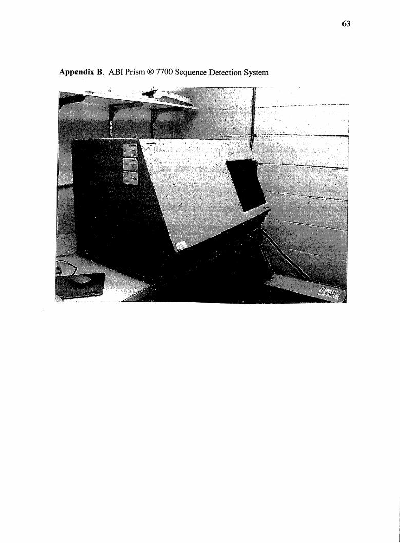
Appendix B. ABI Prism ® 7700 Sequence Detection System
63
■Mm
sill!?'-!)'
64
VITA
Christina Salinas, daughter of Jose Oscar and Thelma Salinas, was born on
February21,1980 in Corpus Christi, Texas. She is an honors graduate from Mary
Carroll High School. Her interest in science took her to the University of North
Texas, in Denton, Texas. In May 2003, Christina graduated with a B.S. in Biology
and a minor in Chemistry. She was awarded the honors of cum laude. After a year,
Christina moved to San Antonio where she was offered a Research Assistantship with
Dr. Sara F. Kerr. While working on her masters, she partook in extensive fieldwork
studies on the ecology of Leishmania mexicana. For her thesis, she conducted her
research at the Southwest Foundation for Biomedical Research under the supervision
of Dr. Ricardo Carrion. Her thesis involved the study of temperature tolerance by
Leishmania enriettii and Leishmania hertigi using a real-time PCR assay.
Related Documents


![2. Materials and Methods - Oswaldo Cruz Foundation · Echinacea purpurea showed activity against Leishmania enriettii [] and the presence of these ... activity for two anthraquinones](https://static.cupdf.com/doc/110x72/5c4620cd93f3c34c50618a11/2-materials-and-methods-oswaldo-cruz-foundation-echinacea-purpurea-showed.jpg)


