R ^ q 0 yx k%\/3^ LSI 14986 UC-48 annual report 1981-1982 V X * * ^ lf»,e ,o« v.eO-= of ^ wmva cr TC JIKKLST >x inuiKra

Welcome message from author
This document is posted to help you gain knowledge. Please leave a comment to let me know what you think about it! Share it to your friends and learn new things together.
Transcript

R ^
q0yx k%\/3^ LSI 14986 UC-48
annual report 1981-1982
V X * * ^ lf»,e
,o« v.eO-=
o f ^
wmva cr TC JIKKLST >x inuiKra

I.KI.VI \ O T K I
Tl! i \ Omik ' . i f . prLpp.>rci| . i - . .m . . L J U I I ; U\ mirk ipnn
v i i c i l in .in .if.-t.TiO • •! I'-K I nti:t! Nr .co fm-.L-::
I -.i-:i • H v n . , a

LBL—14986
DE83 011011
BIOLOGY AND MEDICINE DIVISION ANNUAL REPORT 1981-1982
Lawrence Berkeley Laboratory University of California
Berkeley, California 94720
DISCLAIMER
This report was prepared as an account of work sponsored by an agency of the United States Government. Neither the United States Government nor any agency thereof, nor any of their employees, makes any warranty, express or implied, or assumes any legal liability or response bility for the accuracy, completeness, or usefulness of any information, apparatus, product, or process disclosed, a represents that its use would not infringe privately owned rights. Reference herein to any specific commercial product, process, or service by trade name, trademark, manufacturer, or otherwise docs not necessarily constitute or imply its endorsement, recommendation, or favoring by the United States Government or any agency thereof. The views and opinions of authors expressed herein do not necessarily state or reflect those of the United States Government or any agency thereof.
This work was supported by the Office of Health and Environmental Research of the United States Department of Energy under Contract DE-AC03-765F00098. Portions of this work were also supported by the National Institutes of Health, Department of Health and Human Services; the Environmental Protection Agency; the National Aeronautics and Space Administration; the Nuclear Regulatory Commission; the United States Army; the Veterans Administration; the Electric Power Research Institute; the American Heart Association, and the American Lung Association. Contracts and grants are listed in Appendix A.
NOTICE PORTIONS OP THIS BEPMT ME H l M W i r ' It has bean reproduced from Ike best availafci<: copy to permit the broadest DiSTTtteUTfOH Df possible availability.

CONTENTS
INTRODUCTION Thomas L. Hayes, Acting Division Head 1
1. RESEARCH MEDICINE
INTRODUCTION 3
POSITRON IMAGING STUDIES IN ALZHEIMER-TYPE DEMENTIA Robert P. Friedland, Thomas F. Budinger, Yukio Yano, Ronald H. Huesman, Chester A. Mathis, Brian R. Moyer, Betty Koss, and Beth Ober 4
RADIONUCLIDE GENERATORS AND CYCLOTRON PRODUCED POSITRON EMITTERS FOR SYNTHESIS OF BIOCHEMICAL SUBSTRATES
Yukio Yano, Thomas F. Budinger, and Chester A. Mathis 5
A NEW BLOOD FLOW RADIOPHARMACEUTICAL Thornton Sargent I I I , Alexander T. Shulgin, Chester A. Mathis, and Thomas F. Budinger 9
IMPROVhD SAMPLING IN POSITRON EMISSION TOMOGRAPHY Ronald H. Huesman, Stephen E. Derenzo, and Thomas A. Budinger 10
NEW INSTRUMENTATION FOR HIGH-RESOLUTION, DYNAMIC, THREE-DIMENSIONAL TOMOGRAPHY OF POSITRON-LABELED COMPOUNDS IN THE HUMAN BODY
Stephen E. Derenzo, John L. Cahoon, Ronald H. Huesman, Tony Vuletich, and Thomas F. Budinger 12
DONNER CLINIC: KINETICS OF MEGAKARYOCYTE AND PLATELET TURNOVER
Shirley Ebbe, Clara Adrados, Cathryne Allan, Violet Barghe-Sharghi, Dorothy Carpenter, Ruth Cohen, Patricia Garbutt, Helen Londe, Carol Lum, and Elizabeth Phalen. Attending Physicians: Hunter Cutting, Lester; 'ollander, and Henry Stauffer , 14
REGULATION OF RED BLOOD CELL PRODUCTION IN HUMAN BEINGS BY ERYTHROPOIETIN
Rukmani Pennathur-Das, Edward L. Alpen, Elliott Vichinsky, Joseph F. Garcia, and Bertram H. Lubin 16
MARROW TRANSFUSIONS INTO NORMAL RECIPIENTS George Brecher 19

NUCLEAR MAGNETIC RESONANCE PROGRAMS AT LAWRENCE BERKELEY LABORATORY
Todd Richards, Thomas F Budinger, and Rudi Nunlist 20
2. DONNER PAVILION
INTRODUCTION 23
STEREOTACTIC HEAVY-ION IRRATIATION OF INTRACRANIAL VASCULAR DISORDERS
Jacob I. Fabrikant, Yoshio Hosobuchi, and John T. Lyman 23
RADIATION EPIDEMIOLOGY Jacob I. Fabrikant, John T. Lyman, and Edward L. Alpen 25
3. ENVIRONMENTAL PHYSIOLOGY
INTRODUCTION 27
THYROID HORMONE STUDIES AFTER OZONE EXPOSURE Gisela K. demons and Joseph F. Garcia 28
IDENTIFICATION OF ANDROGEN RECEPTORS IN THE MALE MOUSE LUNG
Gerald M. Connell and Betsy Carr 29
EVIDENCE FOR A SUPPRESSOR CELL IN SHORT-TERM BONE MARROW CULTURES
Joan Wright Goodman, Sarah Gardner Shinpock, and Elizabeth A. Hall 31
ERYTHROPOIETIN STUDIES: PURIFICATION FROM URINE AND PRODUCTION OF MONOCLONAL ANTIBODIES
Robert J. Webber, Gisela K. demons, and Joseph F. Garcia 33
NEW SEQUESTERING AGENTS FOR THE ACTINIDES Patricia W. Durbin, Nyian Jeung, Steven J. Rodgers, David >.. White, and Kenneth N. Raymond 34
4. RADIATION BIOPHYSICS
INTRODUCTION C. A. Tobias 39
Bevalac Studies
PHYSICAL CHARACTERIZATION OF ENERGETIC HEAVY-ION BEAMS Walter Schimmerling, T. S. Subramanian, W. John McDonald, Selig N. Kaplan, Ahren Sadoff, and George Gabor 41

INCLUSIVE NEUTRON PRODUCTION BY ENERGETIC HEAVY IONS Walter Schimmerling, R. Madey, B. D. Anderson, R. A. Cecil, and P.C. Tandy 43
CORRELATED NEUTRON-PROTON EMISSION IN RELATIVISTIC HEAVY ION INTERACTIONS
Kenneth A. Frankel, Walter Schimmerling, John O. Rasmussen, Kenneth M. Crowe, James A. Bistirlich, Roy R. Bossingham, H. Bowman, Osamu Hashimoto, Don L. Murphy, J. Ridout, J. P. Sullivan, Eunice Yoo, William A. Zajc, W. John McDonald, M. Salomon, and ). S. Xu 44
TRANSPORT STUDIES OF THE INTERACTION OF HIGH-ENERGY HEAVY IONS WITH EXTENDED MATTER
Mervyn Wong, Walter Schimmerling, and John W. Wilson 45
RADIOLOGICAL PHYSICS AND CHEMISTRY Aloke Chatterjee and John L, Magee 47
RADIATION SURVIVAL STUDIES WITH CULTURED NORMAL HUMAN MAMMARY EPITHELIAL CELLS
Tracy C. Yang, Martha R. Stampfer, and Helen S. Smith 49
CARCINOGENIC AND MUTAGENIC EFFECTS OF ENERGETIC SILICON IONS ON CULTURED MAMMALIAN CELLS
Tracy C. Yang, Laurie M. Craise, Jerry Howard, and Cornelius A. Tobias 52
CELLULAR AND MOLECULAR RADIOBIOLOGY OF HEAVY-ION BEAMS
Cornelius A. Tobias, Eleanor A. Blakely, Ruth ). Roots, Tracy C. H. Yang, Polly Y. Chang, Leora Lommel, Laurie M. Craise, Michael J. Yezzi, and Peter M. Martin 55
IDEAS ON THE UNIFICATION OF RADIOBIOLOGICAL THEORIES 5tanley B. Curtis 62
CELL AGE DISTRIBUTION IN MULTICELL TUMOR SPHEROIDS Adrian Rodriguez, Edward L. Alp'n, and Randy J. DeGuzman 63
INDUCTION OF HARDERIAN GLAND TUMORS IN MICE BY HEAVY-ION IRRADIATION
Edward L. Alpen, Patricia Powers-Risius, R. ). Michael Fry, E. John Ainsworth, Randy J. DeGuzman, Linda D. Harrison, and Virginia C. Havens 65
CATARACT PRODUCTION IN MICE BY FRACTIONATED DOSES OF ' C PARTICLES OR *°Co GAMMA RADIATION
t. ). Ainsworth and J. G. Jose 67
RESPONSE OF MOUSE MARROW COLONY FORMING UNITS (CFU-S) TO HEAVY CHARGED PARTICLES
E. John Ainsworth, Lynn J. Mahlmann, and John C. Prioleau 68

LIFE-SHORTENING EFFECTS OF HEAVY CHARGED PARTICLES: A STATUS REPORT
E. John Ainsworth, John C. Prioleau, and Lynn J. Mahlmann 72
TREATMENT OF CANCER WITH HEAVY CHARGED PARTICLES Joseph R. Castro, William M. Saunders, George T. Y. Chen, Cornelius A. Tobias,). Michael Collier, Samuel Pitluck, Kay A. Woodruff, Ranu Grewal-Bahl, Theodore L. Phillips, Aude Cartigny, Todd Richards, William Dedo, Jacquelyn St. John, and Robert E. Walton 75
RADIOTHERAPY PHYSICS George T. Y. Chen, J. Michael Collier, Samuel Pitluck, Todd Richards 77
HEAVY-ION RADIOGRAPHY Jacob I. Fabrikant, Cornelius A. Tobias, William R. Holley, Eugene V. Benton, Kay H. Woodruff, and Eric W. MacFarland 80
DIAGNOSTIC STUDIES WITH HIGH ENERGY RADIOACTIVE BEAMS Aloke Chatterjee, William M. Saunders, Janis S. Scherer, Edward L. Alpen, Jorge Llacer, and George T. Y. Chen 84
Magnetic Field Studies
BIOLOGICAL EFFECTS OF MAGNETIC FIELDS Tom S. Tenforde, Cornelius T. Gaffey, Michael S. Raybourn, Lynette Levy 86
Biophysical Studies
FUNCTIONAL ANALYSIS OF LASER-INDUCED OCULAR DAMAGE Michael S. Raybourn and Robert L. Kong 91
FUNDAMENTAL AND APPLIED STUDIES OF CELL-MEMBRANE SYSTEMS
Howard C. Mel , Stephen P. Akeson, Gary Richieri, and Frank Kooi 92
5. STRUCTURAL BIOPHYSICS
INTRODUCTION 97
SCANNING ELECTRON MICROSCOPIC STUDIES OF CULTURED ALVEOLAR MACROPHAGES AND CHRYSOTILE ASBESTOS
Gregory L. Finch, Thomas L. Hayes, Rudy Valentine, and Gerald L. Fisher 98
THE USE OF LOW DENSITY LIPOPROTEIN (LDL) COLLOIDAL GOLD COMPLEXES TO STUDY THE DISTRIBUTION OF LOW DENSITY LIPOPROTEINS ON CELL SURFACES
Richard Thrift and Trudy Forte 100

DIFFERENTIAL SCATTERING OF CIRCULARLY POLARIZED LIGHT BY THE HELICAL SPERM HEAD FROM THE OCTOPUS ELEDONE CIRRHOSA
Marcos F. Maestre, Carlos Bustamente, Thomas L. Hayes, and Ignacio Tinoco, Jr 103
IDENTIFICATION AND PARTIAL PURFICATION OF THE ERROR-PRONE REPAIR GENE PRODUCTS OF BACTERIOPHAGE T4
Junko Hosoda and Herb Moise 105
ISOLATION AND CHARACTERIZATION OF YEAST DNA REPAIR GENES Robert K. Mortimer, David Schild, Isabel L. Calderon, Rebecca Contopoulcu, and John Johnston 107
GENETIC MAP OF THE YEAST SACCHAROMYCES CEREVISIAE Robert K. Mort imer and David Schild 109
COMPARATIVE ANALYSIS OF MITOTIC AND MEIOTIC RECOMBINATION IN SACCHAROMYCES CEREVISIAE
Michael S. Esposito, Carlo V. Bruschi, Dimitrios T. Maleas, and Kathleen Bjornstad 111
EFFECT OF CHEMICAL CARCINOGENS ON MAMMALIAN CELLS IN CULTURE
Regine Goth-Goldstein, Mildred Hughes, and Bonnie P. Tincknell 114
Carcinogenesis and Cell Biology
CARCINOGENIC POTENCY Lois Swirsky Gold, Deborah Ang, Georganne Backman, Margarita de Veciana, Holly K. Hurd, Patricia Kato, Robert H. Levinson, Ted Liou, Renae I. Mcgaw, Charles B. Sawyer, and Bruce N. Ames 116
COMPARATIVE ANALYSIS OF DATA FOR SHORT-TERM TESTS FOR CARCINOGENS AND MUTAGENS
Joyce McCann, Laura Horn, John Kaldor, Bob Siegal, and Barbara Levine 119
PROPERTIES OF HUMAN MAMMARY EPITHELIAL CELLS IN CULTURE Martha R. Stampfer and Jack C. Bartley 123
IN VITRO TRANSFORMATION OF HUMAN MAMMARY EPITHELIAL CELLS
Martha R. Stampfer and Jack C. Bartley 123
BENZO(A)PYRENE METABOLISM IN HUMAN MAMMARY EPITHELIAL CELLS
Jack C. Bartley and Martha R. Stampfer 124
THE ANOMOLOUS INABILITY OF ROUS SARCOMA VIRUS TO CAUSE TUMORS IN AVIAN EMBRYOS
David S. Dolberg, Henry I. Nesis, and Mina J. Bissell 125

EXTRACELLULAR MATRIX INFLUENCES OF GENE EXPRESSION: THE ROLE OF MATRIX COMPONENTS IN MODULATING THE DIFFERENTIATED PHENOTYPE OF MAMMARY EPITHELIAL CELLS
Gordon Parry, Eva Lee, and Mina ). Bissell 127
A NEW METHOD FOR THE STUDY OF ENERGY METABOLISM IN LIVING CELLS USiNG NUCLEAR MAGNETIC RESONANCE
Greg S. Karczmar, Alan P. Koretsky, Mina J. Bissell, and Melvin P. Klein 129
BIOPHYSICAL 5TUDIES OF IMMUNE RECOGNITION John C. Owicki 132
Lipoprotein Studies
CALIBRATION OF THE ANALYTIC ULTRACENTRIFUGE Frank T. Lindgren, Vergie Shore, Gerald L. Adamson, Laura A. Glines, and Talwinder S. Kahlon 132
PLASMA LIPOPROTEIN STABILITY AND STANDARDIZATION Frank T. Lindgren, Steven B. Hulley, Gerald L. Adamson, and Talwinder S. Kahlon 134
SEDIMENTATION EQUILIBRIUM OF HUMAN LOW DENSITY LIPOPROTEIN SUBFACTIONS
Talwinder S. Kahlon, Gerald L. Adamson, Laura A. Glines, and Frank T. Lindgren 136
APOLIPOPROTEIN E IN UMBILICAL CORD BLOOD PLASMA Trudy M. Forte, Paul A. Davis, and Conrad B. Blum 137
ABNORMALITIES IN LOW DENSITY AND HIGH DENSITY LIPOPROTEIN SUBSPECIES IN FAMILIAL COMBINED HYPERLIPEMIA
Ronald M. Krauss, Alex V. Nichols, John Albers, and John Brunzell 138
COORDINATE CHANGES IN LEVELS OF SERUM LOW AND HIGH DENSITY LIPOPROTEIN SUBCLASSES
Ronald M. Krauss, Frank T. Lindgren, Paul Williams, and Peter Wood 139
MODEL PRECURSORS TO HIGH DENSITY LIPOPROTEINS Alex V. Nichols, Elaine L. Gong, and Patricia J. Blanche 139
APPENDICES
APPENDIX A: LIST OF CONTRACTS AND GRANTS SUPPORTING PORTIONS OF WORK PRESENTED IN THIS ANNUAL REPORT . . . .143
APPENDIX B: 1982 PUBLICATIONS 147
APPENDIX C: BIOLOGY AND MEDICINE DIVISION STAFF, SEPTEMBER 30,1982 153

1
INTRODUCTION Thomas L. Hayts
Acting Division Head
During the past year the Director of DonnerLabo-ratory, Dr. Edward Alpen, has been on sabbatical leave at the Cray Laboratory in England. Although the Biology and Medicine Division continues to flourish in his absence, we look forward to his return and to further gruwth in research excellence and productivity under his leadership. We have continued to operate in a funding climate that requires careful planning and management of all research programs, but have still been able to maintain our traditional high quality in areas of ongoing research, and have started some exciting new programs in directions that build on our existing expertise. Research on carcinogenesis and mutagenesis has utilized the LaboratoYs unique human mammary epithelial cell lines and has also drawn from its significant expertise in yeast cell genetics. Our position in the field of nuclear medicine remains strong, with developments in both the design of instruments and their application tr. research.
In the Structural Biophysics Group, the serum lipoprotein program maintains tl ;e position of the Laboratory as a center for front line research ir. this important field. The development of a technique for determining lipoprotein receptor site distribution on cell surfaces seems particularly promising, combining lipoprotein research with advanced electron microscope techniques that are a traditional area of investigation for the Structural Biophysics Croup. The use of recombinant DNA and genetic analysis techniques are also reported by this group. Further progress in efforts to purify and detect erythropoietin is reported by the Environmental Physiology Group, and our Laboratory continues to play a significant role in the investigation of this important hormone.
Physical characterization of heavy-ion beams and studies of heavy-ion radiobiology are the central themes of the Radiation Biophysics Group. Research on the effects of heavy charged particles on certain cancers is closely tied to investigations using cultured mammalian cells in
normal and tumor tissues. The goal is to understand better the basic mechanisms whereby these particles cause biological effects. Another very promising area of heavy-ton research is reported in the studies of stereotactic heavy-ion irradiation of intracranial vascular disorders. Although a relatively new investigation, the initial results of this work already suggest several unique advantages for this irradiation method. A medically-based heavy-ion accelerator for research medicine has been progressing, under a multiyear grant, and it promises to produce valuable results for the future medical application of these heavy-ion beams.
The progress reports from the Research Medicine Group show advances in positron emission tomography and in the production of the radionuclides and radiopharmaceuticals used in this exciting technique. There is also a report on nuclear magnetic resc iance programs at Lawrence Berkeley Laboratory and on the progress of this new direction in our medical research.
I would like to acknowledge the contributions from all of the Donner Laboratory staff who have made these research accomplishments possible. I would particularly like to thank the group leaders and the other members of the Division Advisory Committee for their valuable counsel. Finally, two individuals have made essential contributions to the operation of the division during Dr. Alpen's sabbatical leave: Dr. Tom Tenforde, as Acting Deputy Division Head, has been a very important and effective part of the Division leadership, and Mr. Baird Whaley, the Division Administrator, has shown on numerous occasions the value of his experience in and understanding of vital administrative procedure.
This year's annual report again demonstrates the many areas of strength in the Division's research programs. I believe we can look forward to continued success in the pursuit of our goal of highest quality in research.

3
SECTION 1 . RESEARCH MEDBCSME
INTRODUCTION
The Research Medicine Croup of Donner and Lawrence Berkeley Laboratory (comprised of 5 Ph.D.s, 4 M.D.s, and 25 technical and support personnel) has as its objective the application of physics, chemistry, and mathematics to human disease investigation and treatment. Emphasis has been on three approaches:
1. Development of nuclear instrumentation and radioisotopes for the study of brain and heart disorders,
2. Application of NMR to biomedical studies of brain and heart disorders, and
3. Investigation of human blood diseases, particularly those concerning platelet disorders.
NUCLEAR MEDICINE AND POSITRON EMISSION TOMOGRAPHY
Present emphasis is on the development and use of high resolution positron tomography for the study of the following five major medical problems:
1. Cancer 2. Atherosclerosis 3. Cerebral strcke 4. Senile dementia (Alzheimer's disease) 5. Schizophrenia and manic-depressive psychosis.
The major causes of death in the United States are atherosclerosis and cancer. The metabolism and biology of these diseases can be studied using new, noninvasive imaging methods. The focus of our program is on the perfection of specialized instruments with emphasis of spatial resolution, sensitivity, and ability to collect dynamic data for kinetic analysis, and on development of new radiopharmaceuticals for dynamic function studies with these instruments.
In the study of atherosclerosis and cerebral stroke we havt Reused on a study of the distribution of labeled platelets by imaging their accumulation and desquamation from arteriai sites such as the carotid arteries and great vessels of the upper thorax. A positron emitter label for platelets is being developed along with a new tomograph that wil l have a resolution of 2 mm. This instrument will be the highest resolution PET device in the world. We wil l also study the attribution of labeled lipoproteins using new radiopharmaceutical labeling techniques. A positron-emitting iodo-amphetamine, based on our original discovery of these compounds, is being developed for brain blood flow measurements in stroke.
A combination of new radiopharmaceutical iabeling techniques and high resolution tomography allow approaches to understanding diseases that heretofore could
not be approached by studies in man. This is particularly true for the study of metabolism of tumors to various modes of therapy.
The study of Alzheimer's disease is of particular interest because it is the fourth highest cause of death in the United States, with an annual health care cost of $10 bil l ion, and one half of nursing home occupants are patients with Alzheimer's disease, with an annual cost of $6 billion. We have recently discovered specific defects in the rate of glucose transport and metabolism in the brain of Alzheimer's disease patient, and we are focusing on a program for the investigation of the etiology of this disease using both positron emission tomography and nuclear magnetic resonance.
Schizophrenia and manic-depressive illness are crippling mental illnesses, each strikirg from 1 to 2% of the population, but they have received less scientific attention in proportion to their incidence. These illnesses are now accepted as having a metabolic basis, and we have developed positron-emitter radiopharmaceuticals for in vivo investigation of their biologic origins. These include ("C-methyl)-methionine for study of abnormal methyl metabolism, , aFDG for study of regional brain glucose kinetics, and l 3 2l-iodoamphetamine for measurement of regional cerebral blood flow.
NUCLEAR MAGNETIC RESONANCE STUDIES
The specific emphp.is of nuclear magnetic studies is on the evaluation of tissue response to low level as we!l as acute radiation by noninvasive methods. Changes in the proton NMR relaxation parameters have been verified after ionizing irradiation of animals and we propose to use NMR as a new tool for the evaluation of the effects of radiation both for acute situations and for long term low level exposures, as well as for the determination of the appropriate radiation doses for the use of heavy ions in tumor therapy, particularly of the brain.
A second important thrust of this program is the investigation of carbon-13 metabolism in brain disorders, particularly senile dementia, wherein choline metabolism can be studied for the first time. This program relies in part on collaboration with the Los Alamos stable isotope resource for the acquisition of carbon-13 compounds.
Another focus of this program is en the use of NMR proton imaging for the evaluation of fatty streaks and evolution of atherosclerosis from childhood to adulthood using noninvasive imaging techniques.
DISEASES OF THE 3LOOD
Major accomplishments in the past year include new insights into the nature and behavior of bone marrow

4
cells that form blood platelets and red blood cells, with identification of a new link between these two cell lines; identification of differences in the way blood platelets are produced with different kinds of bone marrow damage;
and analysis of the secretion of erythropoietin (the hormone that regulates red blood cell production) in patients with abnormalities of red blood cell production.
POSITRON IMAGING STUDIES IN ALZHEIMER-TYPE DEMENTIA
Robert P. Friedland, Thomas F. Budinger, Yukio Yano, Ronald H. Huesman, Chester A, Mathis, Brian R. Moyer, Betty Koss and Beth Ober*
Dementia is a symtomatic classification applied to a generalized impairment in intellectual function that results from brain disease. It may occur in individuals under the age of 65 (presenile dementia) or, more commonly, over the age of 6£ (senile dementia). This problem has been estimated to affect over two million people in the United States today. Its impact on our society in terms of personal suffering and economic cost is enormous. The incidence of dementia is directly related to advancing age, and the progressive aging of our population wil l be accompanied by a great increase in this incidence. This has been termed an "approaching epidemic." The commonest cause of dementia in this country is Alzheimer's disease, which may be responsible for as much as 55-60% of all dementia in the United States. While g'eat advances have been made in our understanding of the anatomical and chemical characteristics of Alzheimer's disease, we still do not know what causes it, how to diagnose it during life, how to predict its course, or how to treat it effec;ively. Diagnosis of Alzheimer's disease has always been by exclusion; there is currently no way to make the diagnosis without biopsy. New treatments have been tried, but results are disappointing, and at the present time there is no reliable way to either sympto-matically improve the dementia or halt the progression of the disease.
Positron emission tomography (PET) provides three dimensional information about the distribution of injected radioisotopes in the body. It is done noninvasively and tells us how the tissue hajng studied uses the injected compound. It does this in a regional fashion, that is, it gives us information that concerns specific brain areas. In contrast, the technique of x-ray computed tomography ICT or CAT scanning) tells us about the structure of tissues, while PET provides us with indices of their function. X-ray CT studies of demented individuals are of great value in discovering the presence of certain treatrble diseases that also may cause dementia {fuch as brain tumor or blood clots), but unfortunately the x-ray CT image in Alzheimer's disease is often identical to that of normal individualsof the same age. In PET, of course, the crucial
' University of California, Davis, VA Medical Centei, rv-. 7, CA.
element is that the image represents aspects of tissue physiology. The element of body function (blood flow, metabolism, etc.) that is observed depends on the nature of the injected isotope. This can be done in a quantitative fashion, with objective numerical assessment of parameters of function.
Demented subjects are being studied with PET using the Donner Laboratory 280-crystal ring with a glucose analogue labeled with fluorine-18 ( (18-fluoro-2-deoxy-D-glucnse, synthet:zed at the Lawrence Berkeley Laboratory'^ 88-Inch Cyclotron and the University of California at Davis Crocker Cyclotron). This provides for assessment of glucose metabolism and quantitation of alterations in crucial aspects of brain work associated with these il lnesses. As glucose is by far the most important energy source for neurons, the rates at which brain regions use glucose rs directly related to their levels of activity, and it has been extensively documented in animal and human studies that mental activities are reflected in brain metabolism.
Subjects are comprehensively studied from a general medic?1, neurological, and neuropsychological (behavioral) standpoint in order to properly define the characteristics of their illness. Following injection of the isotope in an arm vein, blood is drawn from the opposite hand, and the PET apparatus records the emitted radiation from the time of injection for the following 50-70 minutes Results suggest that all of 10 subjects studied with dementia of the Alzheimer type have an abnormally low utilization of glucose in the frontal and temporal-parietal regions of both hemispheres of the brain. Five patients with other varieties of dementia do not demonstrate this particular focal disturbance. This abnormality was accentuated in the temporal-parietal regions in all of the Alzheimer-type dementia subjects. Six age-natched healthy control subjects did not have any regional differences, and had consistently higher rates of glucose metabolism (see Fig. 1).
The pathology of Alzheimer's disease is known to concentrate in these temporal-parietal regions, and animal and human studies have extensively documented the importance of these regions for vital mental oj. orations, such as memory, speech, comprehension and purposeful

s
movement. These are some of the functions most impaired by the disease. Studies utilizing imaging with x-ray computed tomography have not been able to reliably document
these structural alterations. Positron imaging is a valuable tool for the quantitative evaluation of disturbed brain function in human subjects during life.
FDG MODEL-AVERAGE RATE CONSTANTS
•Norm o I ^ ^ i N o r m r j
2 0 3 0 Time (min)
Fig. I. Simulated curves of brain uptake calculated using experimentally determined rate constants for FDG transport and phosphorylation. Images taken at a mid-ventricular level demonstrate diminished glucose utilization in temporal-parietal cortex in an Alzheimer-type dementia subject. IXBB82U-1012T)
RADIONUCLIDE GENERATORS AND CYCLOTRON PRODUCED POSITRON EMITTERS FOR SYNTHESIS OF BIOCHEMICAL SUBSTRATES
Yukio Yano, Thomas F. Budinger, and Chester A, Mathis
SHORT-LIVED POSITRON EMITTERS FROM RADIOISOTOPE GENERATORS
Radioisotope generators provide an alternative to the generally expensive and often unavailable cyclotron porduction of position emitters for positron positron emission tomography (PET). Labeling of some metabolic substrates provides specificity in the ,'n-Wvo distribution of positron emitters obtained from generators (See T?ble 1).
The 6 2Sr/MRb generator retains 100 to 200 mCi of spallation-produced *2Sr on an alumina column in a fully
automated and microprocessor-controlled system that permits the elution of 60 to 70% of the equilibrium activity of 76-sec B 2Rb in 15 to 25 ml of saline for intravenous infusion. The generator delivers near equilibrium amounts of a 2Rb as often as every 10 minutes over a period of two to three months. This 5 2Rb generator system has been evaluated over a period of three years and the breakthrough of a 2 ' a 5Sr has been consistently in the 1 0 - 8 to 1 0 - 6 range for many hundreds of elutions with 2% NaCI at pH 8-9.
Because of its short half-life and rapid extraction into the myocardium, a 2Rb is an excellent radiotracer for

6
Table 1. Generators for positron emitters.
Parent Half-life decay modet%)
Daughter Half-life Decay model%)
Gammas MeVI%)
"Fe 8.3 h B-(56),EC(44) 3 W M n 21.1m B-(98),EC(2) 1.43(100)
"Zn 9.t h |3'[18),EC(82) "Cu 9.9 m B'(IOO) 0.59(221
"Ge 275 d ECU 001 "Ga 68 m p*(88),EC(12) 1.08(3.5)
«Sr 25 d ECU 001 "Rb 75 s B*(96),EC|4I 0.73(9)
""Te 6.0 d ECU 00) "•Sb 3.5 m B-(75),EC(22) 1.23(3)
'•"Xe 20.1 h ECU 00) '"1 3.Sm |3*(100) 0.56(14)
»Ba 2.43 d ECU 00) "•Cs 3.8 m p-(51),EC{49) 0.44(27)
myocardial perfusion studies. It permits the infusion of a high dose of " :Rb for good statistical sampling while minimizing the radiation dose to the patient. Some studies require the constant infusion of H JRb over a period of 10-15 min at steady state conditions to measure the washout phase.
Stei ility and apyrogenicity of the ^Rb eluates were maintained during long term use of three to four months for each "JSr loading. Over 100 patients with myocardial perfusion defects or blood-brain-barrier permeability changes have been studied with B 2Rb, which is also applicable to kidney function-perfusion studies.
This vp-̂ r 30 patients were studied with H JRb and the PET for perfusion changes in brain tumors, Alzheimer's disease (pre-senile dementia), and schizophrenia. In addition, patients with coronary by-pass surgery or myocardial infarction were studied with f , 2Rb for monitoring blood perfusion changes in the myocardium (See Fig. 1).
A ""Ga generator using SnO.. as the support medium for the 275-day "(1Ge parent and 1.0 N HCI as the eluent solution provides w l G a " directly in the eluate, We are evaluating a commercial version of this system for , , f iGe breakthrough, SrtO, breakthrough, trace metal contaminants, and labeling chemistry of h B G a , + to transferrin, platelets, and proteins such as monoclonal antibodies or lipoproteins. , l f lGa-labeled platelets can be used for the detection of clots in the myocardial arterial blood supply or in brain blood flow. , , f lGa-labeled monoclonal antibodies will be useful for detecting cancer.
The ' -Xe / ' - l generator is a convenient source of the 3.6-minute positron-emitter '->2I which is separated cryogenically from its 20-hour l 2 2Xe parent. Rapid io-dination chemistry is being studied to label amphetamine analogues with ' - I to measure brain blood flow with PET. Large quantities of ' "Xe can be produced by the l 27l(P,6n)'-'2Xe reaction. Preliminary studies have been done that wil l lead to collaborative research utilizing the 200 MeV proton beam at the Brookhaven Linac Isotope Producer (BLIP).
The 1 2 2 Ba" 2 8 Cs generator was studied to evaluate the 3.8-minute , 2 f lCs positron emitter, which has about half the extraction of 8 2Rb into the myocardium, as a myocardial perfusion imaging agent. A comparison of flaRb and l 2 aCs distribution in the dog heart is seen in Figs. 2a and b. The myocardium to blood ratios are better for n 2Rb than for 1 2 BCs.
Positron-emitters from these four generator systems have a potential for expanding the role of PET in th clinical practice of nuclear medicine. They can be user in conjunction with regionally supplied cyclotron produced fluorine-18 ffuorodecxyglucose (T8FDG), for example, to conduct a comprehensive study of flow and metabolism in a brain tumor patient using B2Rb and l 6FDG (see Fig. 3).
CYCLOTRON-PRODUCED POSITRON EMITTERS
The LBL 88-Inch Cyclotron was used to produce O.ii to 1.0 Ci of 20-minute "C and 110-minute l 8 F for the synthesis of "C-methyl methionine, "C-palmitic acid, and ,8F-fluoro-2-deoxy-glucose (IBFDG) on a weekly basis. Twenty to 40 mCi of the labeled compounds were obtained for studies of patients with myocardial perfusion abnormalities and patients with brain tumors, early aging brain disease (pre-senile dementia) or schizophrenia. The brain studies are research programs with Dr. R. Friedland and Dr. T. Sargent.
I 8FDG was produced in 14 separate cyclotron irradiations to determine the metabolic rate for glucose in the cortex of the brain or in brain tumors in 30 patients studied with the Donner Positron Emission Tomograph. The radiochemical purity of the , BFDG was 95 to 98% as determined by HPLC and TLC analysis.
Carbon-11 labeled palmitic acid was synthesized to study the metabolism of free fatty acids by p-oxidation in the myocardium. Four production runs were made to develop the automated chemical synthesis system for the Grignard reaction of pentadecyl magnesium bromide with "CO, .

7
The chemistry for the synthesis of "C-labeled methionine was studied in five production runs at the cyclotron to improve the labeling yield. Several refinements have been made in the chemistry that wil l provide 40-
60 mCi of "C-methionine with a radiochemical purity of 95-98% for studies of methionine in the normal and schizophrenic subjects. Higher purity "C-methionine will be obtained by preparative HPLC.
RELATIVE PERFUSION AFTER CORONARY BYPASS
Pt A
Pt B
Fig. 1. Coronary bypass patients studied with "Rb. Three different levels ol cross-sectional view are shown for each study. Smooth U-shape accumulation of "Rb {white area) indicates good perfusion broken U-shape indicates poor perfusion.
KBB 823-2751)

Rb-92 Heart Blood
— £ Myocardium
PRE-THERAPY
Ci - 126 H«D'1
(b) *—•B,ood
Fig. 2. (a) Time course distriDution of^'Kb in the myocardium and ventricular blood pool of dog hehrt in quantitative positron emission tomography imaging study. tXBL 813-3741)
18FDG
POST-THERAPY
FDG Fig. 3. A brain tumor patient showing compromised blood brain barrier for accumulation of^Rb in the outer ring of the tumor with low vascularity in the dark non-accumulating core of the tumor. "FDG in the same area indicates more glucose metabolism in the outer ring of the tumor and in the cortex of the brain. (XBB B23-1939)
(bl Similar study with '-'"Cs. iXBl 813-3740)

9
A NEW BLOOD FLOW RADIOPHARMACEUTICAL
Thornton Sargent I I I , Alexander T. Shulgin, Chester A. Mathis, and Thomas F. Budinger
Our program for research into the causes of mental disorders such as schizophrenia, manic depressive illness and senile dementia has led us to the development of a new radiopharmaceutical agent, IDNNA (4-iodu-2,5-di-methoxy-N,N-dimethylampheuimine). This agent can be labeled with the very short half-life l 2 2 l , which is a positron emitting isotope that can be used in positron emission tomograph (PET) imaging systems, to ob'ain accurate quantitative measurements of brain blood flow.
Our earlier discovery of brain uptake of radiohal-ogenrtfed amphetamine derivatives led to a commercial brain-imaging radiopharmaceutical, IAMP, which is being clinically tested in single photon emission computed tomography (SPECT) imaging systems, lodoamphetamines are extracted from blood essentially on the firs* pass through the brain, and the uptake is therefore a measure of brain blood flow. It is being used with success at Borton Deaconness Hospital and UCLA to study stroke and other cerebrovascular disease, in which an accurate assessment of the degree of blood flow impairment is important in instituting therapy.
To fulfill a need for a similar radiopharmaceutical to use in PET systems, a positron emitting halogen would be required, and the only suitable isotopes in terms of
0.05
feasibility of production and patient dose are 7 5Br (1.7 h) and ' " I (3.6 min). ' " I would be particularly advantageous because it is the daughter of 20-hr 1 2 2Xe, and a generator system has already been devised by Richards at Brookhaven by which the , 2 2 l daughter can be extracted and used for labeling; the very short half life would also mean a much reduced radiation dose to the patient.
IAMP, however, is labeled by exchangir.g radioactive iodine with the cold iodinated amphetamine, a reaction that yields relatively low specific activity and is much too slow for use with 1 2 2 l . Our original iodoamphetamine analog, 4-ioao-2,5-dir<iethoxyamphetamine, was labeled directly by reacting the uniodinated precursor with l 2 1 ICI, but it was necessary to first protect the primary anine with a phthalide group and then remove it after labeling. To overcome this problem we decided to design a radiopharmaceutical with the amine group already protected, i.e., a tertiary amine, that wouid also have the desired property of first pass extraction by brain.
We synthesized and tested a series of some 15 different l 3 ' l labeled molecules with various substitutions on the amine, and measured the uptake of the u l ! labeled compounds in rats. We found that the dimethyl amine (IDNNA) (Fig. 1) had the best brain uptake and brain/blood ratio.
0.04
0.03
0,02
0.01
10 20 25 15 Minutes
Fig. 1. Scan images, brain uptake, andplasma clearanceof'"IDNNA after Injection Into a beagle dog; brain/blood ratio is maximum at 8 min. Image at 8 min is an enlarged image of the brain with the head outline overlaid. OiBB 828-7040'
30

10
When injected into a dog and scanned with a whole-body scanner, the uptake in the brain could be clearly seen and quantified, as shown in Fig. 1. Plasma sampling at the same time showed that the maximum brain/blood ratio of 8.7 occurred at 8 min after injection, and the concentration in brain remained high for at least 15 min. Labeling is achieved by reacting I 3 1 ICI and the precursor, 2,5-dimethoxy-N,N-dimethyl amphetamine, in glacial acetic acid; the reaction is complete in less than one minute.
These results with IDNNA present the following possibility for a brain imaging agent, which is presently being actively developed. , 2 Z Xe will be produced by the '- 7! (p.bnl ' "Xe reaction, and using the generator system developed by Richards, the l 2 2 l separated and reacted with the precursor to form the '- 2IDNNA, which wil l be rapidly purified and sterilized for injection. The potential advantages for this generator-produced radiopharmaceutical are several: 1) A generator that will yield useful quantities of isotope for two or three days can be produced at large centralized accelerators and shipped to research and -linical centers; 2) a positron-emitting radio-phar-
At present practical image resolution in positron emission tomography using a stationary circular array of detectors is limited by the center-to-center spacing of adjacent detectors. The lateral sampling is one-half of this spacing, as shown in Fig. l a , where sampling lines have been drawn between detector-pairs and the sampling rings have radii that differ by one-half the center-to-center detector spacing. To obtain image resolution of one-half the detector size {the intrinsic detector-pair resolution) the angular sampling of a stationary array is adequate, but our analysis (see addendum) shows that the lateral sampling must be increased by a factor of two.
Increased lateral sampling has been achieved by several investigators by moving the detectors as shown in Fig. 2. Data are acquired at a number of position';, and the combined data set is incorporated into the reconstruction procedure, resulting in improved resolution in the final image. The number of detector positions used in these methods varies, but generally between 8 and 16 positions are used to gain the desired increase in lateral sampling.
The biomedical research performed with the Donner 280-Crystal Positron Tomograph requires data acquisition times of fi* e seconds or less and the ability to gate the data acquisition for periods during the cardiac cycle when the heart is nearly at rest. These requirements pose severe
maceuttcal wil l be available for PET imaging systems that does not require an adjacent cyclotron; 3) brain blood flow may be quantified, with low patient dose, at intervals as short as 20 min, allowing rapid patent throughput in clinical situations; 4) repeat studies may be done immediately and the results assessed after therapeutic intervention; 5) in research studies, sensory stimuli, motor activity, or seizure activity may be varied to identify corresponding activation areas in the brain; 6) IDNNA brain blood flow studies may be performed and followed by a second study such as 1BF-fluorodeoxygfucose, to study blood flow in relation to metabolism in the brain; 7) PET quantification of brain blood flow wil l be able to resolve many of the problems in earlier methods for these measurements, and permit resolution of many outstanding questions regarding brain metabolism, such as reported changes in bran metabolism in schizophrenia and manic-depressive illness and in epilepsy. It wil l also be of great valut in assessment of strokes and assessing the benefit of new therapies for these cerebrovascular accidents., which present one of the most common causes of death in the United States.
restrictions on the amount of detector movement and have motivated the search for a scheme that improves lateral sampling with a minimum number of detector positions.
A new approach, developed by our group, involves only two positions of the detector assembly and increases lateral sampling uniformly by a factor of two. The simple motion between positions is implemented by a hinge at one side of the ring of detectors. The first position is that of the stationary circular ring shown in Fig. l a . The second position is achieved by opening the ring opposite the hinge so that there is space equal to the width of one detector, as shown in Fig. 1b. This configuration gives rise to sampling rings that also have radii differing by one-haff the center-to-center detector spacing. In Fig. lc , the sampling lines for the two detector positions are superimposed, and the sampling rings for the combined data set result in the desired factor of two increase in lateral sampling. In what follows we refer to this new approach as "clamshell sampling."
To test clamshell sampling we performed Monte Carlo computer simulations of data acquisition with the new geometry. Hot spots of varying sizes and separation comprise the model used in a geometry scaled to test imaging the human head with the Donner 280-Crystal Positron Tomograph. Results of a simulation are shown in Fig. 3;
IMPROVED SAMPLING IN POSITRON EMI^.ON TOMOGRAPHY
Ronald H. Huesman, Stephen E. Derenzo, and Thomas F. 3uding?r

11
the improvement in resolution is quite striking. Actual data collection with clamshell sampling has
not been accomplished with the Donner 280-Crystal Positron Tomograph. The samj phantom referred to above was scanned with the modified system, and the results are shown in Fig. 4. The positron-emitting isotope used for the study v^as I BF, and about 250 million events were acquired. This large amount of data was used so that
Fig. J. Diagram of sampling improvement by clamshell notion in positron emission tomographs, showing 24-delector array and sampling lines between detector pairs: (a) clamshell closed (normal), lb) clamshell open fnew configuration), and (c) combined sampling of la) and (b). (XBl 802-31131
resolution measurements could be made without concern for statistical fluctuations. Considerable improvement in resolution is evident in this study, and animal experiments are now being performed to validate this new method for physiological studies.
ADDENDUM
In reconstruction tomography, the relationships between the required number uf angles, N„, and lateral sampling distance, 8z, to attain a given resolution, a, are,
N, s l . S A / a , (1) Az « 0.5a, (2)
where N„ is between zero and ir radians, and A is the diameter of the reconstruction region.
For a circular positron tomograph wherein one te-constructs in a centra! circular region one-half the diameter of the detector ring, D,
A = 0.5D, (3) The center-to-center detector spacing is given by
d = irD/N , 14) where N is the number of detectors in the ring. For a stationary array the lateral sampling is
Az = 0.5d, (5) but the intrinsic resolution of a single pair of detectors is one-half the detector size, and therefore it is desirable that the resolution ot the imaging system approaches 'his limit.
Close packing is chosen to achieve good resolution and results in a center-to-center spacing equal to the detector size. Substituting equations (3) and (4) into equations (1) and (?v we find
N„ s 1.5N/ir •- 0.5N , (6) Az s 0.25d (71
to be the requirements to attain a resolution of 0.5d. Equation (6) is satisfied, but equation (7) is not; an increase of a factor of two is necessary in lateral sampling.
Fig. 2. Other sampling schemes for positron tomography: (a) scan rotate motion {many positions), (b) circular wobble (at least four) lositions), (c) pure rotation of an irregular array (many positions), and (d) rotation of half-rings about the center (at least four positionsl.
(XBL 8212 1284)

12
Stationary Ring Two-position Clamshell
Fig. 3. Computer simulations to compare images of a positron-emitting resolution pattern using stationary ring sampling (left) and clamshell sampling (right). 1X88 8212-1067$)
A new ui'ra-high resolution positron tomograph is being built to study human brain and heart physiology (Fig. 1). The complete instrument wi l l consist of four rings each having 600 detector crystals 3 mm wide. The design goals are: (1) A spatial resolution of 2 mm FWHM (full-width at half-maximum), a factor of 3 better than the best positron tomographs in use today, (2) the ability to take data from seven slices simultaneously, and 13) the ability to accumulate dynamic two-second images with more than 200,000 coincident events per image.
Important research areas include stroke, abnormal metabolism in brain disorders, and brain tumor metabolism before and after therapy.
Unfortunately, because there are no photomultiplier tubes small enough to read out close-packed arrays of such fine scintillation crystals there is a major research effort to develop alternate technologies:
1) Multiple-Anode Phototube. We have established specifications for a new photomultiplier tube, to be built by industry, containing eight very fine electron multiplier structures in a single glass vacuum envelope (Fig. 2).
2) Solid-State Photosensors. A second promising approach uses a recently developed rectangular photomultiplier tube and new solid-state photosensors individually coupled to each crystal (Fig. 3).
During the investigation of these high technology readout approaches we will proceed to build a single-ring system using 14 mm diameter phototubes and the coupling scheme shown in Fig. 4.
This work is an evolution of the Donner 280-Crystal Positron Tomograph, which is now in its fifth year of
Stationary Ring Two-position Clamshell
Fig. 4. Actual data collection on the Donner 280-Crystal Positron Tomograph to compare images da positron-emitting resolution pattern using stationary ring sampling (left) and clamshell sampling (right}.
(XBB 8212-10677)
successful operation, measuring the concentration of positron-labeled tracer compounds in animals and in the human body (see report by Friedman, Budinger, Yano, et. al. in this section). The system consists of a continuous ring of 280 rectangular (9.5 x 32 x 32 mm) bismuth germanate detector crystals that completely encircle the patient. When a positron is emitted it travels a short distance (less than 1 mm for isotopes such as l aF and "CI and annihilates to produce two 511-keV photons that fly off in nearly opposite directions. Because the elctron and positron have some motion at the time of annihilation there is an angular spread of about 0.32 degrees FWHM. For a detector ring of 60 cm diameter this corresponds to a spatial broadening of 0.9 mm FWHM.
Fig. I. Four-ring positron tomograph for high resolution imaging of labeled compounds in the head. Transmission source is used to measure attenuation corrections. (XBL 829-4084)
NEW !NSTRUMENTATION FOR HIGH-RESOLUTION, DYNAMIC, THRtE-DIMENSIONAL TOMOGRAPHY OF POSITRON-LABELED COMPOUNDS IN THE HUMAN BODY
Stephen E. Derenzo, John L. Cahoon, Ronald H. Huesman, Tony Vuletich, and Thomas F. Budinger

13
BLIND OYNODES
" | l ' | l l l M ^a g B ^ f T" l r "P^ SIDE VIEW
EIGHT-ANODE PHOTOMULTlPLIEl* TUBE
Fig. 2. Design for eight-anode phototube for reading out J mm wide detector crystals. (XBL 821 l^f251)
—Solid stote photosensor (crystal identifier)
-Bismuth qermonote delertor crystal
Fig. 3. Design using Small rectangular phototube for timing and pulse height determination and individual solid-state photosensors for crystal identification. (XBL 8211-4250)

14
Each crystal is coupled to a phototube. Electronic circuits detect any annihilation photon in time coincidence (within 25 nsec) with any of the opposing 105 crystals. Events are accumulated in high-speed semiconductor memory. In a typical high resolution study (9 mm FWHM), one million events are collected in two minutes and the image is reconstructed in 10 seconds by a hard-wired arithmetic unit. Dynamic processes can be imaged with lower resolution (2 cml by collecting a 100,000 event
data set every 2 seconds. Blur due to the motion of the beating heart is eliminated by taking separate data sets for different time intervals of the heart cycle. Eight such sets can be simultaneously accumulated in high-speed memory.
Recently the image quality of this instrument was improved with the incorporation of a clamshell sampling motion, invented and developed at Donner Laboratory (see report by Huesrnan et al. in this section).
PMT'a (4 on lop. 2 on front, 4on bottom)
BGO crystals (10)
Annihilation photon
Fig. 4. Design for coupling J mm wide detector crystals to individual 14 rr.m diameter phototubes. Only the cross-hatched area of each crystal is coupled to the corresponding phototube- This approach is best suited for a single ring tomograph. (XBL 82IO-4I67I
DONNER CLINIC: KINETICS OF MEGAKARYOCYTE AND PLATELET TURNOVER
Shirley Ebbe, Clara Adrados, Cathryne Allan, Violet Barghe-Sharghi, Dorothy Carpenter, Ruth Cohen, Patricia Garbutt, Helen Londe, Carol Lum, and Elizabeth Phalen. Attending Physicians: Hunter Cutting, Lester Hollander, Henry Stauffer.
The hematology program of the Research Medicine Group incorporates the Donner Clinic, a laboratory for research on blood platelet production, and a research training program in cooperation with investigators in other groups. The emphasis in the research programs is on evaluation of the regulation of platelet and red cell production.
Medical care is provided to patients with hematological diseases in the Donner Clinic. These patients also serve as a research resource. The majority of them have erythrocytosis (too many red blood cells), and, with their cooperation and informed consent, studies are underway to test their erythropoietin (Ep) response to therapeutic phlebotomy and mild perturbation of acid-base balance. These studies were initiated with the cooperation of the late Dr. Joseph Garcia, who developed a radioimmunoassay (RIA) for Ep, and are being continued with his collaborator, Dr. Gisela demons. For 59 of these studies, blood and urine samples have been collected before and
after phlebotomy and are being processed and analyzed. It is anticipated that these studies may help to define the diagnostic sensitivity of Ep measurements by RIA as well as the physiology of Ep in some human beings. RIA offers the opportunity to measure Ep when it is present in low or normal amounts (as is the case for many patients with erythrocytosis) unlike older studies of Ep in which bioassay detected only concentrations of Ep that were higher than normal.
Other clinic patients who suffer from severe chronic anemia due to bone marrow failure have exceptionally high levels of Ep in their blood and urine. These patients also contribute to Ep research by collecting their urine, which is then proc • -r-.l to concentrate the Ep.
In the Donner Clinic, as in clinical facilities elsewhere, various amour.ts of blood are leftover from diagnostic and therapeutic phlebotomies. Because of our location in an academic research institute many of these residual blood samples provide valuable source material for other

15
enterprises, such as student leaching, cell cultures, blood chemistries, etc. With expertise in hematology, the clinic offers a course in abnormal hematology to students, investigators, and technicians. Clinic facilities are available to referring physicians with diagnostic problems. Proficiency testing is done several times every year to maintain licensure of the clinic on behalf of the Donner Medical Group and LBL's Medical Services.
Several aspects of blood platelets are under investigation. In preliminary investigations it appeared that exposure of blood to an electromagnetic field resulted in changes in the platelet characterized by swelling, pseudopod formation, and loss of granules. These changes were seen by electron microscopy and were interpreted as indicators of platelet "activation." These findings are being pursued to determine if they are affected by the intensity of the magnetic field and if they can be detected by other means, such as chemical evidence for granule release.
The size of blood platelets can be measured in samples of blood with several types of instruments. The one in use here probably introduces fewer artifacts into the measurement than some others that are, in fact, more widely used. However, it has become apparent that great attention must be paid to the way that blood samples are handled in preparation for measuring platelet size with any instrument to avoid the swelling that occurs with cooling, exposure to EDTA (a commonly used anticoagulant), or even a brief delay, of the order of a few seconds, in exposure of the platelets to an appropriate anticoagulant (i.e., citrate) once the blood is withdrawn from the body. The final experiments are in progress to define the conditions under which the physiological volume of platelets can be accurately measured in preparation for making measurements in people and animals with abnormal hemopoiesis.
In 1966, Alpen' described a new instrument for cytophotometry. An up-dated model of this instrument
has been assembled here, with the expertise of personnel in electronic and mechanical shops. It is being used to apply the two-wavelength technique for measurement of amounts of DNA in individual megakaryocytes in the bone marrow of animals and human beings. Prior to measurement of DNA, the cells are identified by conventional microscopy, and their size is measured by a digital irmge analyzer. This technology will be applied to elucidate mechanisms by which megakaryocytes are regulated with emphasis on experimental conditions in which markedly subnormal numbers of megakaryocytes produce normal numbers of platelets and on disorders of platelet production in human diseases.
Several such hypomegakaryocytic states have been identified in mice. They are associated with an increase in the average size of megakaryocytes, but is is not clear if the increase is due to an increased proliferation of DNA within the cell (megakaryocytes being polyploid cells in which the ploidy appears to determine their final size) or to disturbances of maturation. When mouse megakaryocytes are reduced in number by the cytotoxic drug hydroxyurea IOHU), which acts on megakaryocyte stem cells to transiently reduce the rate of differentiation, the transient increase in mean cell size is probably due to disturbances in maturation, i.e., a shift to more mature cells, but effects on DNA synthesis must be ruled out. The findings with OHU contrast with the size increase seen in response to stimulation of heterologous antiplatelet serum (APS) (Fig. 1), which is known, from the work of others, to be due to synthesis of increased amounts of DNA by megakaryocytes. OHU induces a reduction in numbers of smaller megakaryocytes in both normal and APS-treated mice whereas APS, in addition to reducing numbers of smaller cells also causes an increase in numbers of larger cells that is not seen with OHU. In subsequent experiments, cellular DNA will be measured in megakaryocytes of mice treated with OHU and/or APS and with other perturbations of platelet production.
Fig. '• s'*e distribution of mature (stage III) mouse megakaryocytes in control mice, mice treated with antiplatelet serum (APS), or mice treated with hydroxyurea (OHU) after pretreatment with APS. Abscissa shows cell area in arbitrary units. Ordinate shows relative number of megakaryocytes in each size range; this number is corrected for different total numbers of marrow megakaryocytes in different groups of mice. IXBL 818-4109)

16
It is a well known but unexplained phenomenon that some experimental and clinical situations in which erythropoiesis is stimulated are associated with stimulation of platelet production and thrombocytosis, while others may be associated with normal or suppressed :hrom-bocytopoiesis. The literature indicates that the humoral regulators Ep and thrombopoietin are specific, so it is unlikely that changes in Ep are solely responsible for changes in thrombocytopoiesis. We have recently *'ound that under highly specialized experimental conditions the converse may also occur, namely that stimulation of thromocytopoiesis by injection of AP5 may also cause stimulation of erythropoiesis wit 1 iut apparent concomitant stimulation of leukopoiesis (Fig. 2). These findings are tentatively interpreted as consistent with the existence oi a stem cell population that responds to the stimulus
produced by peripheral thrombocytopenia and is bi-potential for thrombocytopoiesis and erylhropoiesis. It is noteworthy that while stimulation of erythropoiesis was seen after APS in irradiated mice, it was undetectable in normal mice, and the delayed stimulation of thrombocytopoiesis that was seen in irradiated mice was also not detectable in nonirradiated controls. Thus, expression of this dual effect of APS may depend in part on the stimulus generated by APS-induced thrombocytopenia, and in part on an effect of the irradiation, such as bone marrow hypoplasia.
REFERENCES
1. E.L. Alpen. A new instrument for precision cyto-photometry. J. 3d. Inst. 43, 443-448 11966).
0 5 10 15 20 Days after treatment
fig. J Platelet counts, hematocrits, reticulocyte counts, and leukocyte counts of normal mice and mice given 650 R whole body radiation l°°Co} follow ed immediately bv guinea pig antimouse platelet serum IAP5I or normal guinea pig serum iNGpS). Delayed stimulation of platelet and red cell production is shown by higher platelets, reticulocytes, and hematocrits during the second ivee/f in irradiated mice given APS than in those gnen VGpS tXBL 824-37771
REGULATION OF RED BLOOD CELL PRODUCTION IN HUMAN BEINGS BY ERYTHROPOIETIN
Rukmani Pennathur-Das, Edward L. Alpen, Elliott Vichinsky,* Joseph F. Garcia and 8ertram H. Lubin*
Tiie human bone marrow contains a mixture of blood cells in various stages of differentiation and maturation. The erythroid progenitor pool in Ihe human marrow, regulated by erythropoietin (Epo), is heterogeneous with
"Bruce Lyon Memorial Research Laboratory, Children's Hospital Medical Center ot Northern California, Oakland.
respect to Epo-responsiveness. In-vitro culture techniques have identified at least two different populations of eryth-roid-committed precursor cells with increasing sensitivity to Epo defined as burst-forming units (BFUE| and colony-forming units (CFUt).
Using anti-Epo to limit the time of exposure of Epo to bone marrow cells, we have demonstrated that the

17
mature erythroid-committed precursor pool, CFUE, in normal human bone marrow is heterogeneous with respect to Epo-responsiveness. We have now extended ciur studies to disease states accompanied by chronic erythropoietic hyperplasia, e.g., sickle cell anemia and polycythemia vera.
SICKLE CELL ANEMIA
Sickle cell anemia is characterized by chronic hemolysis and erythropoietic hyperplasia in response to the short red cell survival of sickle cells in circulation. Although it is well recognized that under severe stress of hypoxia, chronic hemolysis, and acute blood loss the Epo level increases and erythropoiesis increases in proportion to demand, the precise mechanism of chronic erythropoietic hyperplasia is poorly understood. We have characterized the CFUE pool in sickle cell bone marrow in an attempt to elucidate the mechanism of chronic erythropoietic hyperplasia. All the patients studied had high (10-40%) reticulocyte count, suggestive of marrow erythropoietic hyperplasia. The serum Epo level was elevated in all patients compared to normal. The number of erythroid colonies obtained in vitro with 0.5U/ml Epo was also significantly elevated (1087 ± 520) in all the patients studied, compared to normal (430 ± 130). The colonies consisted mostly (i.e., 77%) of 8 to 16 cells, while such colonies represent only 50% of the total colonies in normal marrow CFUEs. Taken together, these data suggest that bone marrow from sickle cell patients is under high erythropoietic activity and has a high number of mature erythroid-committed precursor cells.
Timed-Exposure Studies
In order to determine the Epo-responsiveness of CFUE
from sickle cell bone marrow, we carried out timed-Epo exposure studies. In these studies, cells were exposed to Epo for limited time periods in culture. The results of such studies on four patients are shown in Fig. 1, where both more than 8-cell (Fig. la) and more than 16-cell (Fig. 1 b) colony formation is represented as a function of Epo exposure time. In contrast to the normal marrow response (shaded area of Fig. la), short exposures to Epo (50 hr) produced 12-40% of the maximum CFUE. The shape of the Epo-response curve was also abnormal.
Epo-Dose Response Curve
We carried out Epo-dose-response experiments to see if the less stringent Epo-requirement exhibited on the timed-exposure studies is due to a greater sensitivity of CFUE to Epo. The results of such studies, shown in Fig. 2, demonstrate the presence of Epo sensitive population. A small percent (5-40%) of the maximum CFUE response is formed in the absence of added Epo. This endogenous
100
i i r • •
_ ( a )
50
** t -»——w-^olP**^ i i 0 50 100 150
Time for ^ruch Epo is cctive in culture (hr)
0 5 0 100 150 Time for which Epo is active in culture {hr)
Fig. 1. (a) Erythroid colony ( >8-celi CFUt) formation as a function of Epo exposure time in sickle cell anemia. The normal marrow CFU< response is represented by the shaded area. (XBL 8210-4173)
(b) Erythroid colony (> 16-cell CFUt) formation as a function of Epo exposure time in sickle cell anemia. The normal marrow CFUt response is iepresented by the shaded area. (XBL 8210-4171}
CFU£ response to Epo-sichle cell anemia
Fig. 2. CFUt response to Epo in sickle cell anemia. CFUt response is expressed as a percent of maximum number of CFUt formed. The shaded area represents the dose response curve obtained with normal marrow. (XBL 8210-4169)

18
colony formation rould, however, be neutralized with anti-Epo. Endogenous colonies were never obtained with normal bone marrow. The presence of Epo-sensitive population was further substantiated by the anti-Epo titration curve (Fig. 3), which showed that CFU f formation obtained with 0.5U/ml Epo could be neutralized only with much greater concentrations of anti-Epo.
POLYCYTHEMIA VERA
Polycythemia vera (PV) is a chronic myeloproliferative disorder characterized by generalized marrow hyperplasia with increased numbers of circulating erythrocytes, granulocytes, and platelets.
We conducted similar studies on two PV patients. Significant proportions (31 % and 44%) of bone marrow CFUE from the two patients were expressed in the absence of added Epo, The Epo timed-exposure studies clearly demonstrated the presence of two populations in one marrow so studied (Fig. 4). The solid line represents the response of CFU e to various Epo exposure times. The proportion of CFUE expressed from 0-50 hours remain constant (31-35%) after which it slowly increases with time of Epo exposure. In order to see if we could demonstrate the presence of abnormal and normal CFUE pools in PV, we subtracted the percent of CFUE formed in the absence of Epo from that formed with Epo exposure at various time points. The result of subtracting this population (32%) from the total and calculating response of the remaining population is illustrated by the dotted line. The second CFUE population is seen to exhibit a normal response to Epo.
The presence of an Fpo-sensitive CFUS population was again demonstrated in the two patients both by Epo-dose response curve and anti-Epo titration curve (data not shown).
In order to see if the increased Epo sensitivity within CFUE in SCA and PV was brought about by greater binding of Epo to CFUE, we measured the Epo content of washed bone marrow mononuclear cells from normal patients, SCA patients, and one PV patient. The washed cells were frozen and thawed several times, sonicated, and homogenized. The Epo content of this homogenate was determined by radioimmunoassay (RIA):
Cpo/W cells IWrnl)
Normal adults (2) 20 =t 4
SCA 121 36 ± 5
PV'MI 55
Thus clinical condntons involving chronic erythropoietic hyperplasia, e.g., sickle cell anemia and polycythemia vera, are characterized by marked heterogeneity in their erythroid precursor pool, and contain a subset of CFU t with increased sensitivity to Epo. We propose that one of the mechanisms facilitating chronic erythroid hyperplasia involves increasing Epo sensitivity of CFUE perhaps by the development of a number of high affinity Epo receptor sites on CFUE.
We have previously demonstrated that normal bone marrow CFUE represents a spectrum of cells with differing sensitivity to Epo. Since the progressive differentiation form BFUE and CFUE is accompanied by increased Epo sensitivity, the presence of "highly sensitive" CFUE population does not seem totally unlikely. Perhaps such "sensitive-CFUE" exists in extremely small amounts in the normal marrow, but becomes apparent in hyperplastic states in which the CFUE pool is amplified several fold. Whatever the mechanism, it is clear that the sickle cell marrow CFUE pool contains a subset of cells with increased Epo sensitivity.
1/500 1/1000 1/1500
Ant i -Epo dilution
Fift. 3. Inhibition of CFUt formed from SCA patients as a function of anti-Epo concentration. <XBL 6210-41701
Time for which Epo is act
Fig. 4. Erythroid colony {> a celt CFUO formation as a function of Epo exposure time in polycythemia vera. The normal response is represented by the shaded area. (XBL 82ID-416SI

19
MARROW TRANSFUSIONS INTO NORMAL RECIPIENTS
George Brecher
During the past several years we have explored the transfusion of bone marrow into normal nonirradiated mice. While transfused marrow proliferates readily in irradiated animals, only minimal proliferation takes place in nonirradiated recipients. It has generally been assumed that this was due to the lack of available proliferative sites in recipients with normal marrow. Last year we were able to report that the transfusion of 200 million bone marrow cells (about 2/3 of the total complement of marrow cells of a normal mouse) resulted in 20% to 25% of the recipient's marrow being replaced by donor marrow. Thus we can now study the behavior of animals that have both transfused (donor) and endogenous (recipient) marrow cells, although none of the tissues of either donor or recipient have been irradiat 1 With these animals we hope to investigate the nature of the peculiar phenomenon of serial exhaustion of marrow, also referred to as the limited self-repiicability of stem cells.
This phenomenon is characterized by the failure of marrow that had been serially transplanted through four to five successive passages to still restore an irradiated animal. The phenomenon of serial exhaustion is closely liked to the Hayflick phenomenon—that all stem cells can only undergo a limited number of divisions during their life span. This phenomenon in turn is closely linked to the concept that different stem cells have different numbers of reserve divisions still available. The pool of stem eel Is would thus have an "age structure," a concept that is playing an increasing role in discussions of stem cell kinetics.
The current concepts of the causes of serial exhaustion of stem cells are controversial. On the one hand it has been claimed that serial exhaustion is due to the limited total number of divisions of stem cells, which become rapidly exhausted when the serially transferred marrow undergoes a large number of mitoses. On the other hand, it has been claimed that transfusion itself suffices to induce the exhaustion. Although the phenomenon of exhaustion requires four or five transfers to be fully expressed, some measure of reduced self-replicability is already observable after a single transfer into an irradiated recipient.
As we can now separate irradiation from transfusion (by using nonirradiated recipients), it should be possible to resolve these problems. We realized that the task would be markedly simplified if we could utilize a strain of mice recently made available through Dr. H. S. Micklem of the Department of Zoology, University of Edinburgh. The CBA/PGK/AB strain contains two alloenzymes (A and B) of phosphoglycerate kinase, which can be separated electrophoretically. Since the animals can be bred to carry only only of the two alloenzymes, the donors and
recipients can be selected to have either the A or B alloenzyme; the percentage of donor cells can be determined by electrophoresis and densitometric measurements. An additional advantage of that strain is that the percentage of donor cells derived from the transfused marrow cells need not only be analyzed in the recipient's marrow, spleen, and other tissues, but can also reliably determined from a few drops of peripheral blood by the electrophoretic analysis. Thus the more laborious cy-togenic analysis that had been used in earlier experiments of marrow transfusion can be dispensed with (see Fig.
1). A major part of the past year has been spent in
raising, with the aid of a Director's Grant, a small colony of CBA/PGK/AB mice and in perfecting the elctrophoretic and densitometric measurements to quantify the discrimination between A and B alloenzymes.
In two experiments we have now transfused CBA/ PGK/A marrow into PGK/B primary, nonirradiated recipients and obtained 20% A donor cells. Subsequently we transfused marrow from these primary recipients into heavily irradiated mice, in which their own stem cells had been entirely eradicated by irradiation. Consequently their marrow was replaced by that of the primary recipients. The proportion of A cells in these secondary recipients was again 20%, indicating that the prior transfusion of the A cells into normal recipients had not resulted in any loss of the potential survival of the cells on subsequent exposure, compared with the endogenous B cells. Transfusion alone can thus not be held responsible for the exhaustion phenomenon. By various combinations of radiation exposures and depletion and regeneration of marrow by drugs such as hydroxyurea, we now hope to pinpoint the exact mechanism of the exhaustion phenomenon.
- j i 1 1 i 20 50 100 150 200 250
Million* of moiiow Cfillt transtuied
Fig. I. Percentage of donor cells in marrow of recipients determined cytogeneticatly and enzymatically. IX8L 824-3799)

NUCLEAR MAGNETIC RESONANCE PROGRAMS AT LAWRENCE BERKELEY LABORATORY
Todd Richards, Thomas F. Budinger, and Rudi Nunlist
For over 30 years, chemists have used nuclear magnetic resonance (NMR) to derive chemical structure and behavior of molecules, but recently NMR has been used by radiologists to investigate the interior of the body. This noninvasive technique uses radiofrequency radiation in the presence of a magnetic field to produce images that have chemical as well as anatomical information.
This past year we began to study the potentials of proton and 1 3C NMR for the investigation of heavy-ion radiation effects in the brain and evaluation of atherosclerosis. In collaboration with Dr. Robert Willcott at the
Baylor College of Medicine we obtained an image of a rat brain one week postirradiation. The result shown in Fig. 1 indicates that this technique gives a sensitive measure of acute radiation damage. Changes in the high resolution proton spectrum of brain tissue b jfore and after irradiation are shown in Fig. 2.
I 3 C NMR of the normal rat brain, shown in Fig. 3, has chemical peaks that make it promising for studying metabolism associated with radiation damage, tumors, and presenile dementia.
Spin Echo Image One Week Post-Irradiation Irradiated
area
Corpus callosum
Ventricle
(A)
Coronal Section
(B)
Fig. I. NMR demonstration of hrain damage from helium beam. (A) Spin echo image of a rat brain that received 3000 rad of helium beam irradiation to the right side of the brain; and IB) anatomical diagram show.' ,g where the brain received radiation. The result indicates .1 new finding of a decrease in relaxation times on the irradiated side. One hypothesis to explain this result is that ionizing radiation causes bond breakage and chemical disruption that would expose a greater amount of tissue water to o.otein surfaces. (XBB 8211-9786)

21
Peaks I O PPM
scale If red 35 times
Gray matter 10,000 rods x-roys
Gray matter normal
0 PPM Fig. 2. High resolution NMR of irradiated rat brain. Proton NMR at idO MHz shows radiation-induced chemical changes reflected by a spectral change of peak no. 2. (XBL 8212-42921
- C H 2 -
" T3T '260' ISO IOC 0 PPM
Fig. 3. Carbon-13 NMR of the brain. Carbon-13 NMR has great potential for in-vivo study of metabolism associated with dementia, the fifth highest cause of death in the United States. These results are basic to our program to study choline synthesis in man using NMR.
IXBL 8212-4211)

2. DONNER PAVILION
INTRODUCTION
The Donner Pavilion continues in the scientific traditions and directions established by John H. Lawrence and James L. Born and their colleagues—the applications of the nuclear sciences for a better understanding of human health and disease, and for the relief of human suffering. During the past year, the new directions recently undertaken are now fixed firmly in place with the introduction of experimental stereotactic heavy-ion Bragg peak radiosurgery in the central nervous system and the application of heavy-ion beams to fundamental and applied brain research. The clinical research program, while still young, is beginning to be recognized for its clinical accomplishment, and brain patients are now being referred
Jacob I, Fabrikant, Yoshio Hosobuchi, and John T. Lyman
intracranial arteriovenous malformations are collections of developmentally abnormal blood vessels that range in size from a large mass that can occupy an entire cerebral hemisphere or other brain structure to a microscopic crypt within the brain, and that usually provide a direct arteriovenous shunt. The involved abnormal blood vessels comprise compartments of arterial, capillary and venous elements (Fig. 1) that have thin and irregular walls with defective muscular and elastic layers; thus they frequently bleed within the brain (in approximately 70% of cases). The intracranial hemorrhage may be minimal or massive; it is usually not fatal in children, but it often occurs repeatedly during later life in young and in older adults. Repeated cerebral hemorrhage produces progressive and severe neurological deficits and increases the risk of death. The symptoms of intracranial arteriovenous malformations, including carotid-cavernous fistulas, include epilepsy that is often refractory to medication, intractable headache, progressive neurological deficit of motor, sensory or other brain function due to chronic cerebral ischemia, and behavioral effects. The diagnosis is suggested by the patient's medical history and is confirmed by the finding of bloody spinal fluid due to brain hemorrhage and by computerized tomography and cerebral angiography. The treatment depends on the patient's symptoms, age, neurological condition, and on the size and location of the vascular malformation within the brain.
from throughout the United States, Canada and Great Britain. The fundamental brain research program is probing complex problems of the effects of focal heavy-ion beams on nerve conduction pathways and cellular regut jon and homeostasis in the central nervous system. The quantitative epidemiological program now extends into the clinical research base of patient records that spans three decades in attempting to understand the risk of charged-particle focal-beam irradiation on the development of late effects in the human brain. Plans to expand the clinical and basic research programs continue, with greater emphasis on the response of the normal brain to focal-beam irradiation and the role of focal vascular injury and repair in the radiopathology of the mammalian brain.
The development of precision microneurosurgicai techniques has made the surgical removal of certain components of deep arteriovenous malformations located in previously inaccessible areas of the brain more feasible, with acceptable rates of surgical morbidity and mortality. However, there are still many deep intracranial vascular
ANATOMY OF AVM
Fig. J. Anatomical compartments of intracranial deep arteriovenous malformations. Each malformation is composedofa number of arterial "vascular units"; the unit is comprised of main feeder arteries and its corresponding shunting capillary compartments. The anatomy of the venous off-flow compartments may vary considerably and does not necessarily correspond anatomically to the vascular unit structure.
IXSL S29-A060)
STEREOTACTIC HEAVY-ION IRRADIATION OF INTRACRANIAL VASCULAR DISORDERS
23

24
disorders that are not surgically accessible either because they are located deep within the critical brain structures or because of their enormous size involving considerable regions of normal or ischemic brain tissue. In treating this particular group of deep intracranial arteriovenous malformations, we are applying stereotactically-directed focal beams of charged particles for direct heavy-ion Bragg peak radiosurgery, or combining intraluminal embolization, surgical occlusion of the arterial feeding vessels and/or partial excision of the lesion, with stereotactic radiosurgery. The process of radiation-induced vascular change with associated thrombosis of the abnormal feeder and collateral vessel compartments (Fig. 1) occurs within approximately 9 months to 2 years after treatment.
During the past 24 months, 22 patients with deep intracranial arteriovenous malformations have been treated
with stereotactic charged-particle Bragg peak radiosurgery at the 184-Inch Synchrocyclotron. None of the patients has shown total obliteration of the offending arteriovenous malformation thus far, except possibly in one case of spontaneous carotid-cavernous fistula (Fig. 2). However, many of the cerebral arteriovenous malformations have undergone a progressive reduction in size and in the blood flow pattern within the malformation, as demonstrated on patients' follow-up computerized tomographic scans and cerebral angiograms. Many patients thus far treated also show progressive improvement in their neurological status with associated return of motor and/or sensory function. There have been none whose condition has worsened. No evidence of radiation injury to normal brain tissue has been demonstrated in any of these patients' follow-up neuroradiological scans. There
Fig. 2. Stereotactic helium-ion Bragg peak radiosurgery of a right carotid-cavernous fistula in a 67-year-old woman. (Al and (B): subtraction x-ray images (anteroposterior and lateral views) of stereotactic cerebral angiogram demonstrating the location and size of the right CCF (arrows); the patient is immobilized in the stereotactic head mask and frame. (O: anteroposterior localiz. :ion radiograph of the skull illustrating isodensity curves of the stereotactic radiosurgical treatment plan. tO): lateral localization radiograph demonstrating the 6 mm diameter helium-ion 6e.im port. Multiple-port radiosurgery was delivered in I day; the dose was 40.00 Cy equivalent, and th- volume of tissue receiving greater than 60% of the dose is estimated to be 280 mm'. iXBB 8QI0-)2345A>

25
has been one case of recurrent subarachnoid hemorrhage, but from an offending aneurysm not within the range of the radiation beam. This incidence rate of bleeding is within that ;o be expected from the natural history of this disease.
Two parallel stereotactic heavy-ion radiosurgical brain research programs, clinical and basic brain science, are now developed a; ihe Donner Pavilion. The brain science program is directed to the clinical and laboratory investigation oi the physical and biological properties of focal beams of helium-ions at the 184-Inch Synchrocyclotron and focal beams of heavier-ions (e.g., carbon, neon, silicon, argon! at the Bevalac that are appropriate for producing focal radiation lesions in the central nervous system, including improved dose-distribution and dose-delivery, with rapid rate of energy loss without damage to adjacent critical CNS structures and neuroanatomical sites. This program establishes stereotactic charged-particle Bragg peak radiosurgery at Lawrence Berkeley Laboratory as a research method in the neurosciences in the application of nuclear medicine sciences to the study of brain disease in man.
The clinical research program (Drs. Fabrikant, Ho-sobuchi, Lyman and Norman) provides heavy-ion medical research support for the application of stereotactically-directed accelerated charged-particle beams for investigation and treatment of patients with certain intracranial
RADIATION EPIDEMIOLOGY
Jacob I. Fabrikant, John 1. Lyman, and Edwjrd L. Alpen
The problem of estimating cancer risk coefficients appropriate for low-dose radiation is under continuing study. Risk estimation is extremely difficult, since, in the absence - epidemiologic data at low levels of exposure to low-1 1 radiation, the true dose-response relationship canno" DO established from empirical data. Nevertheless, these relationships are essential in order to select a method appropriate to interpolate from the available epidemiologic data on high-and intermodiate-dose exposure to the low-dose region for which no data on humans exist. In order to choose a particular method for interpolation, three major difficulties are encountered in any estimation of radiation risk coefficients based solely on human epidemiologic surveys. First, there appears to be reasonably good agreement on cancer incidence among many cohort populations studied in exposed groups, but reliably adequate controls are not always available in each epidemiologic survey. This makes it difficult to eliminate bias in statistical analysis. Second, there is great difficulty in assessing the validity or reliability of the precise radiation doses and dose rates tor exposed humans, as has become particularly evident in the present effort to reevaluate the
vascular disorders, including deep arteriovenous malformations and carotid-cavernous fistulas in the brain. The clinical program is a collaborative one with the Departments of Neurological Surgery and Radiology at the University of California, San Francisco.
The basic research program (Drs. Fabrikant, Ho-sobuchi, Lyman, Woodruff, Tenforde, Gaffey, Norman, Newton and Noorani) addresses the applications of accelerated heavy-ion focal beams for fundamental and applied brain research. Multidisciplinary methods are being developed for producing discrete focal lesions in the central nervous system using the stereotactically-dr-rected Bragg ionization peak to investigate nerve pathways, dose, volume, and time-dependency effeus for the induction of focal CNS damage at various sites; for cellular response and cell population kinetics in the brain, and relationships to time-dependent injury and repair of radiation damage; and for treating cerebrovascular disorders of the brain with stereotactic Bragg peak radiosurgery. Studies in mammals are demonstrating the value of this neuroscience tool in 1) investigating the response of the different CNS cell populations, cell regulation, and homeostasis in the mammalian brain following induction of discrete focal lesions in defined neuroanatomical centers, and 2) elucidating neurophysiological and neuro-pathological responses following heavy-ion focal beam irradiation of various portions of the brain.
atomic-bomb dosimetry in Hiroshima and Nagasaki. Finally, there is the problem of the long latent periods associated with cancer-induction between exposure to ionizing radiation and the appearance of cancer in the exposed population. This latent interval can cover a span ranging from a few years, in the case of leukemia, to over four decades for certain solid tumors, and it can even exceed the period of follow-up.
The effect of latency periods on risk estimation is under study in our laboratory. Insofar as cancer incidences and radiation doses are concerned, effort is made to ascertain these with the greatest reliability, although problems arise, particularly in attempts to reconstruct the events of exposure many years previously, The matter of the long latent periods begs the important issue of how to project into the future the risk of cancer induced in individuals exposed at the present time, or depriving a projection model appropriate for predicting how induced cancers wil l express themselves in time following exposure.
Two risk-projection models, among many, are generally used by radiation epidemiologists—the absolute-

26
risk model and the relative-risk model. Figure 1 demonstrates how these characterize the expression of radiation risk coefficients of cancer-induction in exposed human populations. The absolute risk is the expression of excess cancer risk due to radiation exposure as the arithmetic difference between the risk among those exposed and the risk without exposure. The absolute-risk projection model takes into account the fact that the expression of radiation-induced cancers in the exposed population begins at some time after exposure (that is, after the minimal latent period) and continues at an excess rate for a further period, the period of expression. For leukemia the period of expression may be taken as 25 years; for solid tumors, it may be the duration of life. The lifetime absolute risk coefficient is expressed as the total number of excess cancer cases in the exposed population per unit dose or per collective dose. The relative risk is the expression of cancer risk due to exposure as the ratio ot the risk among those exposed to that occurring in the absence of exposure. The relative-risk projection model expresses the excess of radiation-induced cancers as a ratio or multiple of the natural or spontaneous cancer rate, so the excess risk is a multiple of the natural age-specific cancer rate in the study or cohort population. The greater the spontaneous rate of cancer incidence in a population, such as in an aging population, the greater will be the susceptibility of the individuals comprising that population to cancer-induction by radiation.
At the present time, no major epidemiologic study of exposed human populations is as yet complete, and none wil l be until all members of the study population eventually die of natural or other causes. Only then can the complete cancer incidence in the irradiated and control populations be accurately ascertained. Thus, the distinction between the absolute and relative risk projection models becomes extremely important when the follow-up observation period is considered. When the observation periods are incomplete, there can be, at any one period of follow-up, very wide differences in risk estimation between the two. However, when the follow-up period is complete, and if no more cancers occur in the study population, both the absolute and relative projection models should lead to the same numerical estimate for
lifetime excess cancer risk, although the risk may be differently distributed within the exposed population. The two risk-projection models give different results when projections are made beyond the period of follow-up or observation.
There is now sufficient epidemiologic evidence available which indicates that, in general, most adult populations irradiated at older ages are at greater risk of cancer-induction. This age-derendence may be due to a higher induction rate or a shorter latent period, or both, but there are exceptions. For example, it is not known how this affects exposure of children.
The epidemiologic evidence does not favor one risk-projection model more than another; but the age-dependence of cancer-induction by radiation favors the relative-risk projection model somewhat more. The epidemiologic data are insufficient to determine whether the excess cancer risk, once expressed in the exposed population, projects into the future, either as a relative risk or an absolute risk. The assumptions in the calculation of lifetime risk coefficients of radiation-induced cancer must take into account additional confounding factors, including sensitive genetic subgroups and exposure to other potentially carcinogenic agents. These factors are important when considering differences between the absolute and relative projection models for estimation of risk of radiation-induced cancer in human populations exposed to low-level radiation.
ID o
. Constant / relative risk f / /
s X
I w ^^% Constant absolute risk X
I (-- -""" I Age
Fig. J. Risk of cancer-induction after radiation. The constant absolute (solid line) and relative (broken line) risk models are plotted against age. A = age at time of irradiation; B = age at end of minimal latent period; C = age at a given time after excess cancer risk expressed.
(XBL 801-3028}

3. ENVIRONMENTAL PHYSIOLOGY
INTRODUCTION
The Biology and Medicine Division was deeply saddened by the death on September 20,1982 of Dr. Joseph F. Garcia, who had served as the Environmental Physiology Croup Leader since 1979. His research career at LBL spanned 33 years, during which time he made several significant contributions to fundamental knowledge in the field of hematology. In the 1960s, Garcia and his colleagues gained worldwide recognition for being the first group to oroduce antibodies to erythropoietin, the hormone that regulates the production of red blood cells. This accomplishment immediately led to the development of a radioimmunoassay technique for measuring erythropoietin. The sensitivity of the erythropoietin radioimmunoassay greatly exceeded that of previous bioassay methods tor measuring the concentration of this hormone, and tl us provided an important new technique to aid in the study of a variety of hematological disorders. In recent years. Garua's programmatic efforts had expanded to include studies on the effects of environmental pollutants on the endocrine system. Several aspects of this work, as well as recent studies on the purification of erythropoietin, are described in this section of the annual report.
The Environmental Physiology Group is engaged in studies on both nuclear and non-nuclear environmental pollutants, with specific emphasis on determining their effects on endocrine target systems. Recent studies have demonstrated that ozone exposure of rats leads to a significant decrease in the serum levels of thyroid stimulating hormone and a concomitant depression in the levels of the thyroid hormones T3 and T4. This suppression of the pituitary-thyroid axis is not paralleled by a depression of other pituitary functions, since the serum levels of growth hormone, follicle stimulating hormone and luteinizing hormone are not affected by ozone exposure. Studies during the last year have been directed towards determining whether the depression of thyroid hormone levels caused by ozone is an adaptive response that reduces systemic toxicity and thereby increases the survival potential of exposed animals.
A primary target of ozone toxicity is the lung, and the degree of injury to lung tissue caused by this pollutant has been shown to be modified by steroid hormones. This observation implies that the lung may be a target organ for steroid hormone activity, a possibility that had not been pre-, iously recognized. Investigations during
S
the past year have demonstrated that the lungs of mice contain specific receptors for dihydrotestosterone, and it has been further shown that chronic exposure to ozone reduces both the lung content of receptors for this hormone and the binding affinity of the remaining receptor sites.
In research related to nuclear pollutants, new chelating agents have been developed that efficiently sequester highly charged metal ions and act in vivo to promote the excretion of 2 } aPu(IV). During the past year, two new classes of chelating agents have been tested for their ability to facilitate the removal of injected plutonium ions from mice. One class of ligands was produced by the addition of catechoylate functional groups to ferriox-amines, and the second type of new ligand is a pyridone dimer. Both classes of chelating agents have been shown to significantly accelerate the excretion of "EPu(IV) ions in mice, and their chemical selectivity and low toxicity makes them promising new agents for general use in actinide removal.
Two areas of basic research being carried out within the Environmental Physiology Group involve the characterization of lymphocyte progenitor cells grown in cultures of bone marrow, and the chemical purification of erythropoietin. In the first of these areas of investigation, methods of bone marrow culture have been used that promote the development of progenitors of the T (thymus-dependent) lymphocyte series. Colonies of these pre-T cells have been shown to have suppressor activity that inhibits the development of erythroid precusor cells present in bone marrow.
Erythropoietin research during the past year has been focused on developing biochemical techniques to isolate and rapidly purify the large quantities of this hormone that are excreted in the urine of patients with Fanconi's anemia and Diamond-Bfackfan anemia. The purification scheme involves a combination of column chromatography and high pressure liquid chromatography procedures, and !he initial results have been promising. A second approach that is being taken towards the goal of producing purified erythropoietin is the development of monocloml antibodies that could be usc:d for affinity chromatography. During the last year techniques have been established for selecting, cultunnp and cloning hy-bridomas, and current efforts are concentrated on the isolation of hybrids that secrete monospecific antibodies to erythropoietin.

28
THYROID HORMONE STUDIES AFTER OZONE EXPOSURE
Cisela K. demons and Joseph F. Garcia
Our earlier studies have indicated a profound depressing effect on the pituitary-thyroid axis of rats following ozone exposure. Ozone concentrations as low as 0.8 ppm for a period of 8 hours rasult in a highly significant decrease in the circulating thyroxine (T4) levels in the rat. This depression is also reflected in serum protein-Dound iodine (PBI) determinations, triiodothyronine (T3) and thyroid stimulating hormone (TSH). However, this depression of pituitary TSH secretion was not part of a general depression in pituitary activity since serum growth hormone (CH), follicle stimulating hormone (FSH), and luteinizing hormone (LH) were not depressed and, in fact, prolactin (PRL) was concomittantly increased. These studies have been the subject of two publications. Further studies of pituitary-thyroid function in ozon'! exposed
• Vats indicated that they still have the capacity to respond to the stimulus of cold exposure with an increase in circulating TSH levels. Administration of TSH prior to ozone exposure in rats is accompanied by appropriate increases in both T4 and T3, indicating that the thyroid ".self is also capable of responding when stimulated. Thus, all the elements of the hypothalamic-pituitary-thy-roid system of ozone-exposed rats seem capable of responding when appropriately stimulated. Yet the original observation of a profound depression of TSH and thyroid hormones have been a consistent observation in such ozone-exposed animals.
It has previously been shown that thyroxine administration increases pulmonary injury and decreases survival of rats and mice during ozone exposure. On the other hand, a reduction in thyroxine, as seen after thy.oidec-tomy, enhances survival in ozone. Our results support these observations, and further, show that ozone inhalation has set into motion a process for~reducing circulating thyroid hormones that is beneficial for survival. The reduced activity of the pituitary-thyroid axis may serve as an adaptive mechanism to reduce injury from the toxic effects of ozone inhalation in normal animals.
The hypothesis that the effects of ozone on the thyroid axis are best explained as an adaptive mechanism resulting in a lowering of the set-point at the central nervous system level is only a partial explanation for the observed results. We prevented this adaptation by either hypophysectomy or thyroidectomy and administration of exogenous thyroid hormone prior to ozone exposure. Young thyroidectom-ized rats were given thyroxine in drinking water (containing also 1 % calcium lactate) at varying doses ranging from 75 to 1000 ng T4 per liter. The animals were maintained for 12 weeks on exogenous T4 in drinking water and their thyroxine levels were monitored by RIA every two weeks. Administering the thyroxine in the drinking water
was the preferred route in order to have constant hormone levels rather than the daily variation obtained by injection. Thyroxine at a concentration of 150 (j.g T4/liter resulted in circulating T4 levels and body weight gain comparable to that of unoperated control animals of the same age. TSH levels were also monitored during this period of 12 weeks. We observed a dose-dependent relationship of circulating TSH levels such that with increasing T4 concentrations there were decreasing TSH levels. A dose of 300 u.g T4/liter, even though it resulted in a doubling of circulating T4 in the thyroidectomized animals, was not able to bring the TSH levels into the range observed in normal animals, i.e., they remained fourfold higher. A possible threshold effect was observed at concentrations between 300 and 500 |ig T4/liter. The TSH levels at the higher dose were significantly less than normal, and this effect was even more pronounced at the highest T4 concentration (100 jig/liter) in drinking water.
After having established a steady state of circulating TSH, T4 and T3 levels in all animals, i.e., controls, hypophysectomized or thyroidectomized animals with or without T4 replacement therapy, we sampled 1 ml of blood before and 1 ml of blood after ozone exposure (1 ppm x 24 hr). The samples throughout the experiments were obtained alternately from the jugular vein under metafane anaesthesia. The results of these experiments showed that the depression of the hypothalamic-pituitary-thyroid axis can be only partly responsible for the lowered thyroid hormone levels seen after ozone exposure. Except for the animals that were thyroidectomized or hypophysectomized and received no replacement therapy there was a reduction of circulating T4 levels in all animals, including the controls. In fact, as can be seen in Fig. 1, the higher the levels were before the exposure, the more they were reduced 24 hours later. We also observed no difference between the thyroidectomized or hypophysectomized animals that received the same T4 concentration of 300 u,g/liter throughout the experiment.
Since rats exposed to ozone for a short period of time (24 hours) reduce their food and water intake and since the disappearance of T4 from the circulation during exposure could be accounted for by fasting, we subjected normal male rats to 24-hr exposures to ozone, hypoxia or fasting. This was done in two experiments in which the sequence was altered between ozone-hypoxia-fasting and fasting-hypoxia-ozone. Again the rats were bled from the jugular vein before and after exposure. The results are seen in Fig. 2. We observed no difference in the sequence of exposure and the results of the two experiments were therefore pooled. A reduction of T4 occurs in all three cases but is less pronounced after fasting than

29
after either ozone or hypoxia. The same phenomenon was observed for the TSH levels.
The results seem to indicate that the disappearance of T4 in the serum of the thyroidectomized or hypophy-sectomized rats receiving replacement therapy cannot be accounted for by reduced water intake, but rather the disappearance may be due to some peripheral phenomenon initiated by ozone exposure.
Our ongoing research is pursuing this problem further in order to confirm the ozone-induced disappearance of thyroid hormone from the circulation. For the purpose of making the T4 delivery independent from water intake we are using the Alzet Minipumps for hormone delivery. These pumps are commercially available with a pumping rate of 1 ng/hr and last for 7 to 10 days. After thyroidectomy the rats will be maintained on exogenous T4 in the drinking water until the circulating levels have stabilized (3 weeks). The pumps, which contain T4 concentrations that will ensure those previously stabilized levels, wil l be placed subculaneously in the animal 4 days before ozone ex
posure. In such prepared animals the difference in serum T4 between before and after a 24-hr ozone exposure wil l be measured and should theoretically resemble the difference between the reduction after fasting and ozone.
In addition to the observed depression in T4, T3 and TSH levels following ozone exposure, hypoxia and fasting, there was a significant rise in circulating prolactin (PRL) levels following ozone exposure. This rise does not occur after hypoxia or fasting. Since prolactin has been implicated as an osmoregulatory factor, the increased PRL levels seen after ozone exposure may play such an osmoregulatory role; and the ozone-exposed rat may provide a model for studying osmoregulation in mammals. Planned work wil l involve the search for prolactin receptors in a variety of tissues, including lung, intestine, and kidney, and as to whether there is a change in receptor activity, either in total number of receptors or binding affinities, etc., that can be attributed to the effect of ozone on the animal.
h
rl -, h
h h
h
h
l|
i f jg. I invt r or J4-htmr ozone exposure I) ppm) on circulating thyronine lT4i It'U'h in nmm.it i.ih; thyroidectomized (THXI rats given T4 doses ranging tram Otn IOOO "g liter: .mil hvpophyset tomized (HX) rat* given cither no 14 uc 100 \L# T-4 titer in drinking w.ifiv (or (> weeks.
tXBL 8I5-JH94)
Posting Uppmx24hr)
f ig. -'. Circulating prolactin (PRl), thyroid stimulating hormone (TSH) and thyroxine (T4) levels in normal male mts after 24 hours of fasting, of exposure tn high altitude, or uf exposure to ozone.
(XBL 815-3693)
IDENTIFICATION OF ANDROGEN RECEPTORS IN THE MALE MOUSE LUNG
Gerald M. Connell and Betsy Carr
Knowledge of endocrine relationships between hormones and the mammalian lung has emerged and grown over the past 10 to 15 years. Although most of this interest regards fetal and developmental relationships, there is an increasing awareness of the interaction of hormones with the adult lung. Our interest in pulmonary biochemistry began when studies of ozone toxicity were initiated several years ago. Throughout these studies, we were
constantly impressed by the grossly divergent responses of adult male and female mice when they were exposed to this common component of photochemical smog. Because the initial and primary injury induced by ozone occurs within the lung, and because the severity of this injury, when measured by survival, appears to be modified by sex hormones, we suggest that a specific response {biochemical change) may occur within the lung as a

30
result of sex steroid action. This would infer that the lung is a target tissue for sex steroid action, although this feature has never been recognized. To be a target tissue for a steroid hormone, specific steroid receptors must be present within the tissue. Thus an attempt to identify sex steroid hormone receptors in the adult male mouse lung was initiated.
A cytosol fraction (105,000 x g supernate) was prepared from a homogenate of lungs from normal adult male mice and examined for androgen binding. Results from an experiment in which the binding of dihydrotes-tosterone (DHT) was analyzed are shown in Fig. 1. The Scatchard analaysis of the experimental data shows a straight line regression indicating a single class of androgen receptor was present. The concentration of DHT receptors in this experiment was 9.6 femtomole/mg cytosol protein. Binding for this receptor protein showed a high affinity and a very low capacity.
scatchard Analyala Dlhydrotestoatarona
PM Bound Fig, I. Scatchard analysis of 'H-dihydroteslosterone binding to male mouse lung cytosol receptor. Mice -ivetc 93 days old and had been castrated {or 10 hours. Cytosol samples 10.25 ml} were incubated overnight at - fC with increasing quantities of 'H-dihydrolestosterone IWpM-SnMl. Araraltt jeloflubes, identical except for the addition of 5 u.M nonradioactive uihydrotestosterane, was assayed simultaneously ro determine nonspecific binding. Final incubation volume was 0.30 ml. Bound and free steroid was separated by dextran charcoal solution and tritium content quantitated. All values are corrected far nonspecific binding. Theiewas 1.31 mg cytosol protein/tube and Scatchard analysis indicates 9.6 lemlomole DHT receptor/mg cytosol protein.
IXBL 8210-3159)
Specificity of dihydrotestosterone binding to this receptor was evaluated by competition studies with other steroids and sucrose gradient centrifugation studies. Further support for the presence of androgen receptors in the mouse lung was demonstrated by using the synthetic ligand 3H-methyltrienolone (R 1881), which has a high affinity for androgen receptors. This synthetic steroid does not bind to sex hormone binding globulin or prostatic binding proteins that could be present in cytosol fractions. Density gradient studies demonstrated a peak of bound !H-methyltrienolone in the 8 - 9 S region consistent with steroid cytosol receptors derived from other tissues, and this radioactive binding was greatly diminished when cytosol samples were incubated with 100 fold excess of the nonradioactive methyltrienolone (Fig. 2). Other ste-
100000 10000 8000 6000 4000 2000 1400
=o3H-FM881 • 'H -R1881 + R1881 » 3 H-R1881 + Testo. H 3 H - R 1 8 8 1 + Prog.
* 3 H-R1881 + Compd. B
I
Bottom Fraction
Fig. 2. Sucrose density gradient analysis of the methyltrienolone-binding activity in male mouse lung cytosol. Mice were 94 days old and had been castrated for IBhours. Cytosol samples {0.25 mil were incubated with I nM >H-methyltrienolone in the absence (o) or presence of 100 nM non-radioactive steroid: methyltrienoioneC). testosterone (A), progesterone O and corticosterone (Compd. B) (A). After a 4 hour incubation at -f C, the cytosol samples were exposed to drextran<harcoal, centrifuged, and an aliquot of the supernatant was carefully layered upon a 10-30% lineargrad'ent of sucrose in TEM buffer. Samples were centrifuged for 17 hours at IQ5.0Q0 x g. After centrifugation, 12 J-drop fractions an:' 3 20-drop fractions were collected from the hot' om of the lubes. Tritium activity of these fractions was quantitated. Fhe location of sedimented NC-/gC (7S) is noted by the arrow. The inith;,' bottom fraction (No, I) is at the left of the figure. (XBL 8211-3361)

•M
roids competed for binding to a lesser extent, the only major competitor was the androgen, testosterone. D ita presented in Table 1 show further assessment ofthesp 'C-ificity of this putative androgen receptor.
We have examined lung androgen receptors in mice that were exposed chronically to 1.5 ppm ozone. When receptors were quantified by Scatchard analysis after 10-day and 17-day ozone exposure, there was no difference between control and ozone exposed mice. However, after 40-day exposure to an atmosphere containing ozone, the androgen receptor content in lung cytoso! was reduced by approximately 40% when compared to control values.
Table I.Specificity of 'H-methyllrienolone binding to mouse lung (ytupLmmic androgen receptor.
Percent competition
Coii'fwlitor 10 nM
Melhyl.rienolone 07
Dihvclr.->testosterunp fi'J
Testoslerotu 48
Estradiol 18
to-Diol 12
tfJ-Diol 8
Progesterone 7
'Lung < vtosol •Himtilc-i tO.J'i mh were incuiMtvd overnight in the presence of 1 n M 'H-mcthvftru'notimc Cumitetinn steroids were present in either 10 nM or ItUI nM ntmr.ttiuiM tnelurm. frilium luntent of the buund litfand was quantified. Arbit/.wh. the IfJfJ nW nonrjdiiMtnv melhyltrienohne tv.is set .11 100% com-petition, tind till other < umpetitors were based on litis v.ilue The only steroid: th.it i umpetv vnivtit anth ,trv the n.itural .mdmgei s. testosterone, and dihydro-
100 nM
100
82
76
13
fhese data are summarized in Table 2. In addition to a decrease in receptor content, the binding affinity of the receptor for dihydrotestosterone decreased. These changes within the lung occurred at a time when ozone toxicity, when measured by survival, is actively being expressed.
When steroid hormones act upon a target tissue, the hormone initially combines with a cytoplasmic receptor protein, the receptor species that we have thus far studied. After this binding, an activation process occurs in which the steroid-receptor complex translocates to the nucleus, combines with the chromatin and initiates mRNA synthesis. Subsequently, the mRNA is translated within the cytoplasm to produce specific proteins that eventually express the biological action of the steroid hormone. Our future studies wil l examine these products within lung tissue of normal and ozone-exposed animals and determine their relationships to the expression of ozone toxicity that we observe.
Table 2. Effects of 1.5 ppm 0, on lung androgen receptors'
fmol/mg protein —Kd(nM)
Control 0, 10 days
Control 0, 17 days
Control 0i 40 days (sensitive) 0, 40 days (resistant)
12.34 11.79
9.59 9.76
10.48 5.71 6.47
0.38 0.48
0.34 0.29
0.51 0.94 0.97
'Groups of 20 mice (control or ozone-treated) were sacrificed at various times. Lung cytosol dihydrotestosterone receptor assays were performed. Receptor content and K4 were determined by Scatchard analysis.
EVIDENCE FOR A SUPPRESSOR CELL IN SHORT-TERM BONE MARROW CULTURES
Joan Wright Goodman, Sara Garner Shinpock, and Elizabeth A. Hall
In the course of in vitro studies of hemopoietic progenitors in mouse bone marrow, we became interested in 3^1 day colonies similar to those described as raspberry-like clusters by others' who believed them to be comprised of "pre-T" cells on the bases of morphology and thy-mopoietin-inducibility. Not yet convinced of their identity as progenitors of the T (thymus-dependent) lymphocyte series, we carried out preliminary studies-1 and are continuing to investigate characteristics of these cells. This re** <•[ wil l deal with in vitro expression of progenitor content of short-term cullures, grown with 10% horse serum either with ("pre-T") or without (cultured marrow or CBM) 10% pokeweed mitogen conditioned medium, and with ability of cultured cells to suppress normal bone marrow in assays tor CFU-C, CFU-E, and BFU-ME.
Neither "pre-T" nor CBM (control) cultures were found to contain CFU-E, the late erythroid progenitor
thdl in mice requires only two days for full expression in vitro. However, both kinds of cultured cells were found to contain many BFU-ME, the earlier—more pre-mitive—erythroid stem cells. In fact, at 3 or 4 days of culture they expressed 2 to 4 times as many BFU-ME/ 105 cells plated as did fresh bone marrow. Interestingly, however, when the cultured ("pre-T") cells were mixed with fresh marrow before plating for either assay, a distinct suppression was seen. This is illustrated in Fig. 1 for CFU-E (three experiments shown) and Fig. 2 for BFU-ME. Expected values in the latter case have been derived by adding together colony numbers obtained from separately plated fresh marrow and "pre-T" cells.
Both "pre-T" and control cultures were found also to contain CFU-C, the granulocyte/macrophage progenitor, similar in concentration to that of ''resh marrow. There was evidence of suppression of CFU-C in mixing

32
© 1 0 0
O.9.* B6 BM+4d"p f * -T"ce l l t * "Pte-T-cell i only
0.0&I 0.05:1 OJ: 0.5:1 |-| 2 : | "Pfe-T"cells : NormolBM
Fig. I. Suppression of CFU-E by "Pre-T" Cells. (XBL 8211^i252A)
experiments like those described above for erythroid pro-gen:tors (Figs. 1 and 2) and also in an experiment in which differential colony counts (macrophage/granulocyte/ mixed) were made of CFU-C grown from unseparated "pre-T's" and from top and bottom cell fractions after a 1-hour, untty-gravity sedimentation through fetal calf serum. Results are shown in Fig. 3. The remarkable finding was that many more colonies grew from the ceils taken from the separated top fraction than from the bottom or from unseparated "pre-T's." A reasonable explanation for this is that a suppressor cell is present in unseparated cultures and that it appears predominantly in the bottom
4doy 6 day 10 day
4doy 6 day
lOdoy
4doy 6day
10 day
6 day lOdny
0 400 BOO 1200 1600 CFU-C/IO*
Fig. 3. Differential counts from colonies grown from fresh and cultured marrow. (XBL 82U-4253A)
T9 F<esh BM
=J1 •H
T f c l Unsepa'oted "pFe-T"cell5
fc|p™ 100 Irocl
Wifd i f a Bottom Loot™ r~ j m + m Wifd 1 = 1 1
Fig. 2. Suppression of BFU-ME by "Pre-T" Cells. iXBL 82II-4254A)
fraction after separation. We are currently seeking data to substantiate this hypothesis. Although considerable colony growth was seen in the 10-day control culture (CBM), almost all cells were monocyte/macrophage in character, in contrast to the fairly large population of granulocytic colonies obtained from top fraction "pre-T's."
In a second similar experiment, separated "pre-T's" were assayed for CFU-S content as shown in Table 1. No evidence appeared that there was suppression in this case: the sum of the total numbers of CFU-S from top and bottom fractions was not significantly different from the calculated starting number.
References
Cohen, J.|. and Acuff, B.R. Growth of T lymphocyte progenitor colonies in vitro, }. Supramol. Struct., Supp4, 137 (1980) (abstract). Goodman, Joan W. and Shinpock, Sarah G. 1980-1981. Biology and Medicine Division Annual Report, LBL-13501, April 1982, p. 37.
Table 1 CFU-S content ot separn led 86 Pre-T Cells.
Per to*
Tout
Fresh marrow 19.1 — Whole berries 13.6 34,062
Top fraction 13.3 29. -120
Bottom fraction 11.2 5,962

33
ERYTHROPOIETIN STUDIES: PURIFICATION FROM URINE AND PRODUCTION OF MONOCLONAL ANTIBODIES
Robert J. Webber, Gisela K. demons, and Joseph F. Garcia
The number of red cells in the circulation is very finely controlled; this control is mediated by the hormone erythropoietin (Epo). It is generally believed that Epo is produced in the kidney, released into the circulation, and exerts its hormonal activity in the bone marrow and spleen. The ability to measure the concentration of this hormone in various tissue samples {e.g., blood, kidney, and spleen) will add to our understanding of the mechanism of control of red blood cell production, both in normal individuals and in those exhibiting certain pathophysiological conditions. Recently our laboratory has developed and validated a highly sensitive radioimmunoassay (RIA) that is capable of measuring Epo in very low concentrations. This quantitative assay is based on the competition between ''"'I-labeled Epo and unlabeled Epo in various test samples for binding to a limiting amount of antibody. This classical competitive RIA meth-oiogv has been widely employed to measure compounds of biological interest {e.g., peptide hormones, steroid hormones, and certain other metabolities). The antiserum presently used in the Epo RIA has a high affinity and titer but ii> not monospecific since it was obtained from rabbits immunized with impure preparations of Epo. Generally, these types of antisera will contain a mixture of antibodies to a variety of proteins present in the impure preparation as well as antibodies to the particular protein of interest, and thus are not monospecific. Since the antiserum used in ihe RIA is not monospecific, the labeled antigen must be used in the purest possible form in order for the RIA to be specific and quantitative. Currently we are totally dei >endent upon the National Institutes of Health for the continuation of RIA because they are the sole source of highly purified Epo. However the NIH supply of pure Epo is dwindling rapidly and will be totally exhausted within the next two years. In order to avoid the loss of our impetus in erythropoietir 'esearch, we have embarked on two projects that wil l ensure our continued leadership of this field of investigation.
First, we are in the final stages of developing a scheme for the rapid purification of Epo from the urine of patients who are suffering from Fanconi's anemia and DiamonH-Blackfan anemia. These individuals secrete fairly large amounts of Epo in their urine, which is used as our starting material. This project, when completed, will allow us to be independent of the NIH as the sole source for pure Epo, necessary for RIA, and will allow us to continue to make extensive use of radioimmunoassay or other immunological assay methods for the detection and quantitation of Epo in the future. Second, we are in the initial stages of developing a monoclonal antibody-pro
ducing cell line that wil l open up vast new areas of research concerning the biochemistry and physiology of erythropoietin. Successful production, purification and characterization of monoclonal antibodies specific for erythropoietin wil l give us invaluable tools that can be utilized for affinity purification of Epo, for receptor studies, for structure-function studies of Epo, and for a wide range of other studies. In addition, we anticipate that with one of these monoclonal antibodies we will be able to replace the antiserum currently used in RIA. The production of a monoclonal antibody will also allow us to develop an enzyme-linked immunosorbent assay (ELISA) to quantitate the amount of Epo in test samples. We would then be freed from the need for large quantities of , 2 5 l in our experiments. While we have been diligently pursuing both of these goals, complete success has not been achieved in either case, although we are progressing rapidly.
The purification of erythropoietin from the urine of severely anemic patients is in the final stages of completion. We have developed a scheme for the rapid purification of Epo that tentatively involves two batch processes, three column chromatography steps, and two high pressure liquid chromatography (HPLC) steps. These purification procedures are based on Epo binding to and eluting from various hydrophobic, ionic, affinity and molecular sieving resins. The initial HPLC fractionation furnishes us with a protein sample that is about 10% Epo. We are currently refining the final fractionation, a second HPLC stpe, which provides us with very high specific activity Epo. We are also trying to improve the initial batch process in order to increase our yield. The Epo recovered from the second HLPC fractionation is believed to be pure. We plan to investigate the ability of this material to replace the NIH supplied pure Epo in our competitive RIA system and to characterize the pure material in biochemical terms {e.g., homogeniety by the criteria of SDS-polyacrylimide gel i-ectrophoresis and isoelectric focusing).
The building of hybridomas that specifically produce antibodies to Epo has not been successful as the purification of Epo. The main difficulty we have encountered in building the hybridoma is the poor antigenicity of Epo; a phenomenon that has been encountered by numerous investigators over the years. Our laboratory has been fortunate in the past to be able to generate antibodies in rabbits to various crude preparations of human Epo which allowed us to establish an RIA for Epo. However, since crude preparations of Epo derived from many different species wil l elicit the production of red cells in phylo-

34
genitically distinct species, it seems probable that the molecule has been highly conserved during evolution. Apparently, it is not recognized as "foreign" by many species and therefore is a very poor antigen. One of the main requirements for construction of a hybridoma that can be used for the production of monoclonal antibodies is a large population of lymphocytes that are sensitized specifically toward the antigen of interest. We have been successful in hybridizing spleen lymphocytes with SP2-0 myeloma cells and in selecting, culturing, and cloning the hybrids. Unfortunately, none of the hybrids have been producing antibodies to Epo. The screening tests used in our laboratory wil l 1) recognize mice that are producing antibodies to Epo, 2) distinguish hybrids that are producing IgC or IgM from nonproducing hybrids,
and 3) determine if a culture is producing antibodies specific for Epo. To date, mice have been found that produce antibodies to Epo and hybridoma cultures have been located that produce immunoglobulin(s), however, no antibody directed to or specific for Epo has been found. Currently, the process of immunizing and screening mice for the production of antiserum specific for Epo is being repeated. Hyperimmunization of the mice that produce antibodies is planned in order to increase the number of sensitized lymphocytes before additional hybridizations are attempted. It appears likely that success is at hand once sensitized mice are located and hyper-immunized to produce large numbers oi lymphocytes that can be subsequently harvested and used for the construction of an antibody-secreting hybridoma.
NEW SEQUESTERING AGENTS FOR THE ACTINIDES
Patricia W. Durbin, Nylan Jeung, Steven 1. Rodgers,* David L. White,* and Kenneth N. Raymond*
This a cooperative research project of the Biology and Medicine and the Materials and Molecular Research Divisions, LBL, and the Department of Chemistry, UC Berkeley. Its purpose is to develop safe effective agents for decorporation of actinides and other toxic metal ions of small size and high charge. The similar coordination chemistries of Fe(lll) and Pu(IV) suggested that ligands forming stable chelates with Fe(lll) would also be effective for Pu(IV). Several ligands containing the catechoyl (2,3-
dihydroxybenzoic acid) functional groups have been prepared at LBL. 1 ' 7
The ferrioxamines are tri-hydroxamic acid ligands elaborated by soil microorganisms to obtain iron.' 4 Des-feral (the mesylate salt of desferrioxamine B, DFOM, Fig. 1) is a clinically accepted, low toxicity agent used to treat acute iron poisoning and chronic iron overload.1'3
It is not, however, particularly effective for removal of Pu(iV) or Th(IV), and is ineffective for removal oftrivalent
C H 3 - C - N - ( C H 2 | , — N - C - I C H 2 ) 2 - C - N - ( C H , ) S - N - C - ( C H , ) 2 - C - N - ( C H 2 ) 5 - N - I II I H II II I H II II I H 0 0 0 0 0 o 0 0H
H H
Abbreviation t X Molecular formulo and {weight]
DFOM C H 3 - S 0 3 H 2
0 = C-1
- C26H52N60||S (657)
desferriCAIW
X
H C 3 2 H 5 a N s 0 | | (697)
desferriCAMIC) same as above -CO; >Na C 3 3 H 6 | N 6 0 | 3 N a (763)
Fig. >• IXBL 821 U-42691
•Materials and Molecular Rese.in.rt Division

35
Abbreviation
H N — ( C H o l ,
CN - O K r ^ N - <
Na0 3S'
HN (CH 2) 9 NH I 2 I C B O C = 0
JOT JO" 3-HOPOCAM
Molecular formula and (weight) IUPAC name
No03S^
2 - L I C A M ( S )
C j 6 H 1 4 N 2 O l 2 S N a 2
1536) N 1 , N 4 - B i s i 2 , 3 - d i h y d r o ) ( y - 5 - s u l f o b e n z o y l ) -d iazabutone, d isodium salt
3-HOPOCAM
Fin. J.
C,7H, 4N f l0 6Kg N\N3-Bis{l~riydroxy-2(IH)-pyridone-6-carbonyl) diozapenrane, dipotassium salt
(XBL 8272-42/0)
actinides. " • I H Two of the compounds recently synthesized, for which results of 24-hr ' , BPu excretion tests are reported here are derivatives of DFOM. DesferriCAM and desferriCAMfOlFig. 1) are "mixed" ligands, that is, each molecule contains more than one kind of complexing group. In the desferriCAM molecules, one H on the ter-
I minal N of DFOM has been replaced by a catechoyl group.'" The molecule contains three hydroxamate and one catecholate functional groups and is structurally capable of forming a charged cavity with appropriate size and shape to fully coordinate 8-coordinate Pu(IV).
The procedures used in the 24-hr J 1"Pu excretion test in mice have been published." Briefly, groups of five mice each receive an intravenous injection of 0.2 ml of -',HPu(IV} citrate, 1.5 pXi/kg. One hour later 30 p.mole/kg of a ligand is injected intraperitoneal^ in 0.5 ml of saline. The mice are killed 24 hours after the - , HPu injection, frozen, and dissected after partial thawing. Skeleton, tissues, and separated excreta are assayed for ' , HPu using the 17-keV l l 4 U L x-rays. * > The deferriCAM(C) was soluble at the desired concentration of 1.5 mg/ml, but the solubility of desferriCAM is only about 0.14 mg/ ml. The desferriCAM preparation was sonicated and then shaken vigorously before each mouse was injected, but the amount actually entering the circulation from the peritoneal cavity was probably only about V\o of the administered amount. Readily visible aggregates of undissolved compound were present in the peritoneal connective tissues at autopsy. Fresh tissue spreads were examined microscopically and the undissolved particles appeared to be within phagocytes. As part of a study of ligand effectiveness and dosage, results are available for comparison from a group of mice given 50 u.mole/kg oi DFOM. Previous results for DFOM" were incomplete, because excreta were not separated.
The results of the -M"Pu excretion tests are collected in Table 1. Only a small amount of excess excretion, 6% of the i i . ; ."Jed J '"Pu, could be attributed to the action
of desferriCAM [excess Z 3 f lPu excretion = excre-tion(treated) - excretion(24-hr controls)]. On the other hand, 30 jjimole/kg of desferriCAM(C) promoted a net excretion of 64% of the injected 2 3 8 Pu, a result that is as good as the best catechoylamide ligand we have reported previously [63% net Pu excretion promoted by 3,4,3-LICAM(C)'2 n | . One interesting aspect of these results is that, at least in the mouse, DFOM promotes significant " a Pu excretion in feces (27% of the total excreted), and this trend was even more pronounced in the case of the more lipophilic desferriCAM(C) for which fecal excretion (passed feces plus Gl contents) accounted for 61 % of the J i a Pu excreted.
The other new ligand, 3-HOPOCAM, is a pyridone dimer (HOPO is an acronym for hydroxypyridine oxide, see Fig. 2) and is related to the catechoyl and to the hydroxamate ligands (see structure of 2-LICAM(S) in Fig. 2). The functional group of the catechoylamide ligands is o-dihydroxybenzoic acid; metal coordination is through the pairs of phenolic oxygens; and deprotonation of both -OH groups is required for chelate formation. Both oxygens of the HOPO ligands become available through an intramolecular electron rearrangement when the single -OH group is ionized, at about pH 4. Pyridone ligands are expected to be fast acting facile metal binding agents, but at the same time to retain the advantages of the catecholate ligands: large formation constants and selectivity among metals.
The results for 3-HOPOCAM in the initial 24-hr i M i Pu excretion test in mice are quite promising. An excess - ,JBPu excretion of 53% was obtained with only half as many functional groups available as for an equimolar dosage of the tetramers, 3,4,3-UCAM(C) or desferriCAM(C), which elicited only 10% more excretion of the injected -' i f lPu. Removal of - "lPu from liver (compared with 1 -hr controls) was greater than for any ligand tested thus far. The presence of a significant excess of -""Pu (compared with 24-hr controls) in the soft tissue balance

36
Table 1. Effect of various ligandson distribution and excretion of'lHPu in mice
No. of
mice
Percent of Tissues
injected dose ± S.D. at 24 hr.
Urine
Excreta
Feces Gl com.
Skeletal fraction
Compound
No. of
mice Skeleton Liver Soft tissue Kidney Retained
at 24 hr.
Urine
Excreta
Feces Gl com. SK/IL + SKI
Experimental ligand DesferriCAM,
s: 5 31 ± 2.3 43 ± 3.7 8.6 ± 2.2 3.1 86 ± 1.8 7.5 4.0 2.5 0.547 ± 0.065
DesferriCAMC), 5 12.6 ± 1.2 10.6 ± 2.0 3.0 ± 0.42 0.9 27 ± 1.6 28 33 12 0.422 ± 0.034
3-HOPOCAM, 5 17 ± 2.8 8.7 ± 1.2 11.4 ± 0.8 1.4 38 ± 4.2 8.7 47 5.6 0.660 X 0.007
Baseline iigands: DFOM, 4 20 ± 11.0 19 ± 13 4.5 ± 1.4 1.8 46 ± 11.5 40 12 2.8 0.527 I 0.076
2-LICAMIS), 5 15 ± 4.0 41 ± 3.3 7.0 ± 0.9 5.6 69 ± 3.6 27 2.1 1.8 0.264 » 0.055
3,4,3-LICAM(C), 20 10.5 ± 2.8 10.5 ± 5.3 4.6 ± 2.3 2.4 28 ± 8.4 48 16 8.6 0.522 ± 0.112
"aPu-injected controls: 1-hr controls, 13 24 ± 4.2 31 ± 4.6 35 ± 4.9 2.6 93 ± 2.7 1. 3' 5.3 0.442 r 0.063
24-hr controls. 94 32 ± 7.9 49 ± 8.3 7.7 ± 2.0 2.0 91 ± 6.7 4.7 1.3 2.8 0.391 * 0.089
'S.D. = [S.dev-7(n-l)l'*. Where S.D. is not shown, samples were pooled lor 5-mouse group. Data were normalized to 100% discrepancies are due to rounding.
"Ligand was administered (30 (nmote/kg, i.p.) at 1 hr and mice were killed at 24 br after injection ti.v.) of-^PutlV) citrate. 50 u.mole/kg.
material recovery;
of DFOM was
at 24 hr suggests that the excretion of 2 3 aPu promoted by a single dosage of 3-HOPOCAM had not been completely exhausted. The HOPOCAM ligand combines reasonable solubility at an effective dosage, and sufficient lipophilicity to be eliminated by the liver (86% of the excreted a 3 BPu was in passed feces and Gl contents). These properties suggest that the HOPO ligands may be orally active and that renal toxicity wi l l not be as a serious problem as it is for iigands that are eliminated primarily in urine. At this point, however, any ligand excreted by the liver must be regarded as a potential liver toxin under the influence of prolonged or high dosage. That point remains to be studied.
Synthesis of a HOPOCAM trimer and tetramer are in progress. Preliminary investigations of acute toxicity, the biokinetics and delayed action (excretion up to 7 days after a single administration of ligand), and the ability to promote excretion of an established " B Pu burden are in progress or planned for desferriCAM(C) and HOPOCAM during the coming year.
Studies are underway in the LBL Materials and Molecular P.esearch Division of several li^ands produced by this program to determine membrane transport and toxic properties in mammalian cells, and to establish which, if any, are Fe(lll) donors to bacteria.
REFERENCES
1. Weill, F.L., Raymond, K.N., Smith, W.L., and Howard, T.R. Specific sequestering agents for the actinides. 1. N, N', N", N'"-tetra (2,3-dihydroxyben-
zoyl)tetraazacyclo-tetra- and hexadecanes. I. Am. Chem. Soc. 100, 1170-1172 (1978).
2. Harris, W.R., Weitl, F.L., and Raymond, K.N. Synthesis and evaluation of an enterobactin model compound. 1,3,5-Tris-(2,3-dehydroxybenzoylamino-methyl) benzene and its iron(lll) complex, j. Chem. Soc. (London) Chem. Comm., 178-179 (1979).
3. Weitl, F.L., Harris, W.R., and Raymond, K.N. Sulfonated catecholamide analogues of enterobactin as iron sequestering agents. }. Med. Chem. 22, 1281-1283 (1979).
4. Weitl, F.L. and Raymond, K.N. Ferric ion sequestering agents. 1. Hexadentate o-bonding N, n', N"-Tris(2,3-dihydroxybenzoyl) derivatives of 1,5,9-tria-zacyclotridecane and 1,3,5-Triaminomethylbenzene. ;. Am. Chem. Soc. 101, 2728-2731 (1979).
5. Weitl, F.L., and Raymond, K.N. Specific sequestering agents for the actinides. 3. Polycatecholate ligands derived from 2,3-dihydroxy-5-sulfobenzoyl conjugates of diaza- and tetraazaalkanes. J. Am. Chem. Soc. 102, 2289-2293(1980).
6. Weitl, F.L., Raymond, K.N., and Durbin, P.W. Synthetic enterobactin analogues. Carboxamido-2,3-dihydroxyterephthalate conjugates of spermine and spermidine. /. Med. Chem. 24, 203-206 (1981).
7. Weitl, F.L., and Raymond, K.N. Lipophilic enterobactin analogues. Terminally N-alkylated spermine/ spermidine catechol carboxamides. /. Org. Chem. 46, 5234-5237(1981).
8. Raymond, K.N., Harris, W.R., Carrano, C.J., and Weitl, F.L. 1980. The synthesis, thermodynamic behavior, and biological properties of :netal-ion-specific

37
sequestering agents for iron and actinides. In: Inorganic Chemistry in Biology and Medicine (A.E. Martell, ed) pp. 313-332. ACS Symposium Series, No. 140. Am. Chem. Soc, Washington, D.C.
9. Harris, W.R., Raymond, K.N., and Weitl, F.L. Ferric ion sequestering agents. 6. The spectrophotometric and potentiometric evaluation of sulfonated trica-techolate ligands. j. Am. Chem. Soc. 103, 2667-2675 (1981).
10. Kappel, M., and Raymond, K.N. 1982. Ferric ion sequestering agents. 10. Selectivity of sulfonated poly(catechoylamides) for ferric ion. Inorgan. Chem.
11. Durbin, P.W., lones, E.S., Raymond, K.N., and Weitl, F.L. Specific sequestering agents for the actinides. 4. Removal of -wPu(IV) from mice by sulfonated tetrameric catechoyl amides. Radiat. Res. 31, 170-187 (1980).
12. Durbin, P.W., lones, E.S., leung, N., Weitl, F.L., and Raymond, K.N. 1980. Removal of ""Pu(IV) from mice by catechoylamides: Initial results for 3,4,3-LICAMC. Biology and Medicine Division Annual Report, 1979-1980, LBL-11700, pp. 39^13.
13. Durbin, P.W., Jeung, N., Jones. E.S., Weitl, F.L., and Raymond, K.N. 1982. Chelating agents for the
actinides: Influence of number of functional groups and degree of lipophilicity of carboxylated catechoylamides. Biology and Medicine Division Annual Report, 1981-1982, LBL-13501, pp. 43^18.
14. Neilands, J.B., ed. 1974. Microbial Iron Metabolism. New York, Academic Press.
15. Raymond, K.N., and Smith, W.L. 1981. Actinide-specific sequestering agents and decontamination applications. In: Structure and Bonding U.B. Good-enough et al., eds), vol. 43, pp. 159-186. Springer-Verlag, Berlin.
16. Bulman, R.A. Some aspects of the bioinorganic chemistry of the actinides. Coord. Chem. Revs. 31, 221-250(1980).
17. Taylor, D.M. The effect of desferrioxamine on the retention of actinide elements in the rat. Health Phys. 13, 135-140(1967).
18. Volf, V., Seidel, A., and Takada, K. Comparative effectiveness of CA-DTPA, desferrioxamine B and their combination in removing transuranium elements from rats. Health Phys. 32, 155-158 (1977).
19. Rodgers, S., and Raymond, K.N., unpublished.

4. RADIATION BIOPHYSICS
INTRODUCTION
C. A. Tobias The expertise of the Radiation Biophysics Group is
centered in basic biophysics and cellular radiobiology. Research in these fields has resulted in novel approaches to cancer diagnosis and therapy. An interdisciplinary team is carrying out systematic studies with the aim to improve the control of cancer, and this progress report contains an evaluation by |. Fabrikant of the current status of our diagnostic program and a progress report on the therapeutic investigations by ',. Castro and his group. The advances made in computerized therapy planning are described by C. CI1 en.
The Bevalac accelerator is the center of many of our research activities. Because it is a unique source of heavy particles, our group is involved in international collaboration at various levels. There is particularly strong interaction with Canadian, German, and Japanese laboratories, and in each uf iSese countries there is a strong interest in building heavy io.i accelerators for biomedical research. After an improvement program was finished in June 1982, a beam of the heavy element uranium was obtained at the Bevalac, and it is now feasible to accelerate any of the nuclei in the periodic table. Most of the biomedical interest centers on the lighter ions, up to atomic number 26 (iron). Iron nuclei are important components of space radiation and pose a potential health hazard on extended space flights. Even heavier nuclei can occur in space radiation, and there are long term plans to explore the mechanism of action of the very heavy nuclei on living cells and tissues.
Studies of the effects of various heavy ions on mammalian cells in culture have pointed to particles with atomic numbers 13 to 20 as candidates for statistically controlled therapeutic trials. Whereas light nuclear beams such as protons and helium ions are useful in cancer therapy because of their good depth-dose distribution, the heavier particles also have a significant effect in depressing the radiobiological oxygen effect, altering and reducing the magnitude of enzymatic repair of injury caused K these particles, and reducing the variation of radiosensitivity of cells in various phases of the cell division cycle. In preparation for more extensive medical studies, in late 1982 three patients were treated for the first time with therapeutic schedules of 700 MeV/nucleon silicon beams.
The medical studie; *re supported by an extensive effort to measure early and iate effects produced by various particles in normal and tumor tissues. One of our goals is to quantitatively understand the mechanism whereby
these particles cause biological effects. Radiation biology of low LET radiations, such as x and gamma rays, is found in the idea that there is a quantitative dose-effect relationship for the energy transferred to tissue. The dose concept is important to heavy ion effects also; however, it appears that the microscopic structure of ionizing particle tracks plays an essential role in dt.ermining the nature and quantity of lesions produced in the genetic material DNA.
When heavy ions hit the atomic nuclei of tissue fragmentation may occur. A single beam particle may thus produce two or more nuclear fragments. E..rh of these fragments produces ionization and the joint effect is responsible for producing the biological lesions. The effect of each fragment depends on its velocity and charge. Several aspects of the fragmentation research by W. Schimmerling et al. are described in this review. Of special interest are the cross sections for proton and neutron production. Another variable is the efficiency by which thr. particles are able to produce ionization in gases, vviiich determines the efficiency of dose measurement.
After tl;° primary process of ionization and excitation of atomic processes has ended, the chemical phase of radiation effects begins. Reactive free radicals are formed in the aqueous milieu of cells, and their reaction with biological materials produce macromolecular lesions. An essential aspect of this process is the manner in which DNA injury is produced by heavy ions. This problem is discussed by A. Chatterjee.
Another study is in progress on correlating the track structure and fragmentation effects with the lethal effect produced in human cells. The successful completion of this work wil l allow accurate estimation of the effects of mixed heavy-ion irradiation and wil l become the basis for dose schedules in heavy-ion cancer therapy. E. Blakely et al. show their preliminary conclusions in this report, and Ruth Roots discusses the relationship of DNA lesions to the cellular effects. We have developed a new model for quantitating the cellular responses, the repair-misrepair model, and this model is also discussed.
The quantitative study of cellular effects is usually limited to cell isolates cultured in vitro, and from the results we infer the nature of injury in intact tissues. We have already completed research on the effects of various heavy ions on established cell culture lines. However, most of these cell lines represent cells of connective tissue (fibroblasts), and there is some evidence that most established cell lines may actually be "tumor cells." In culture some essential cell properties are lost and tumor cells have an advantage over normal cells because they
39

40
can grow under environmental conditions where normal cells cannot grow well. Attempts are under way to culture normal human cells and to determine their response to heavy-ion radiation. One of the approaches is to prepare de novo cultures of human epithelial cells, freshly isolated from their donors. The laboratory has some cell isolates from both normal and cancerous human bre.\st tissues. The normal human cells appear more sensitive to ndiation than cultured human fibroblasts, and they are also more sensitive than de novo cultured breast cancer cells. T. Yang et al. give an account of progress in this research. In a separate study, we take stabilized cultures of normal diploid human fibroblasts (the IMR-90 strain), and compare them to a variety of human cells derived from different sources. The most sensitive cell we studied came from a patient with ataxia telangiectasia. These cells are more sensitive than our other normal and tumor cell strains. What are the factors that determine the relative sensitivity and resistance of various human cell strains? We know that there are some variations in the yield of the initial macromolecular radiolesions. During the last few years, however, it became clear that differences in the regulation and efficiency of the enzymatic repair system are important determinants of cell sensitivity. We have particular int.. ast in the repair process; as they occur in various parts of the cell division cycle, and we study these processes in synchronized cell populations and under circumstances of controlled growth. Initial work has been completed on the modification of potentially lethal damage by inhibition of DNA polymerase enzymes that are believed to be essential for repair.
Heavy ions are more effective in producing cell transformation than low LET radiations. When viruses are used for transforming cells in vitro, radiation acts to enhance the process. Heavy ions are much more effective in enhancing cell transformation caused by simian virus SV40. There is a likelihood that the heavy ions produce lesions in DNA that serve as loci for the integration of viral DNA into the host, and that some of the radiation repair enzymes are also needed for cell transformations. T. Yang describes the ongoing work.
Like other ionizing radiations, heavy ions produce late effects and carcinogenesis. The magnitude of these effects must be known in order to protect astronauts from the carcinogenic and deleterious effects of space radiation. We also need quantitative information on carcinogenesis of heavy ions in o.der to assess potential hazards for cancer patients if they receive heavy-ion therapy. E. Alpen has a long term study in progress on the induction of cancer in endocrinologically stimulated Harderian glands of mice exposed to various heavy ions. The carcinogenic effects are greater with heavier particles, but they do not exceed the carcinogenic potency of fission neutrons. Other long term studies include cataract induction, and the long term effects of fractionated whole body exposure. These effects and the effect of heavy ions on the blood forming system of the body are studied by j . Ainsworth. Another study by A. Rodriguez attempts to bridge the gap in our knowledge between cellular and tissue effects. S. Curtis is attempting to unify the various theories concerning the biological effects of radiations with the mathematical model.
The laboratory has a protracted study in progress on the effects of external magnetic fields on the circadian rhythms of mammals. T. Tenforde reports the progress to date. In contrast to earlier reports by others, magnetic fields, even with considerable field strengths, do not c'ppc^r tc change the physiological rhythms of the body. They J J produce measurable effects on the electrocardiogram, however, and these effects have been quantitated by C. Caffey. Other studies with magnetic fields include an electro'etinographic study of the responses of the eye to light in !he presence of magnetic fields. M. Raybourn is in charge of studying ocular effects; he also has data on the effects of pulsed laser light exposure:; on vision.
H. Mel has developed basic biophysical methods to study the Theological properties of cell membr0pes exposed to deleterious environmental agents. These methods are of interest in a variety of fields.
This past year has been one of interesting research problem? and some fruitful results; as a groups, we look forward to another year of study and collaboration.

41
Bevalac Studies
PHYSICAL CHARACTERIZATION OF ENERGETIC HEAVY-ION BEAMS
Walter Schimmerling, T.S. Subramanian,* W. |ohn McDonald,* Selig N. Kaplan,' Ahren Sadoff,' and George Gator 1
When beams of heavy ions are used in radiation therapy and biology, the character of the beam is altered as the particles travel through tissue. The velocity of the panicles is affected by electromagnetic interactions that slow and spread the beam. Fragments are produced by nuclear interactions and these are also affected by passage through subsequent material. The biological effects produced by the beam depend on the fragments present as well as their fluence, charge, and velocity. For example, the relative biological effectiveness (RBE) and oxygen enhancement ratio (OER) depend on all of these quantities.
We use the multidetector particle identification spectrometer described in earlier annual reports to identify each particle in the radiation field and to measure its velocity. With this spectrometer we have begun to study reaciion cross sections, angular distributions of emitted particles, fluence spectra as a function of absorber thickness, the energy loss of relativistic heavy ions in matter, and the dependence of W, the average energy required to produce an ion-pair in an ionization chamber, on the charge and energy of the particles.
RANGE-ENERGY RELATION
Accurate information on the energy loss of the incident heavy-ion beams is obtained almost as a byproduct of our work, in the form of velocity 'and, hence, energy per nucteon) measurements as a function of absorber thickness. The experimental points in Fig. 1 are an example of sncli data (uncorrected for the increase in path length due to multiple scattering of the particles and other systematic effects). From such data, the range of the incident particles can be obtained by extrapolating to zero velocity, and the experimental range-energy curve thus calculated can be compared with theoretical calculations to yield a deeper understanding of the processes responsible for the energy loss of high-energy heavy ions.
1t is also useful to have available simple means to calculate the range-energy relation of high-energy heavy ions in arbitrary materials, e.g., for Lso in a programmable calculator or even in a computer m>del of the beam transport in bulk materials of arbitrary composition. A
Thickness tq/cma)
Fig. t. Heavy ion energy per nucteon as a (unction of range. IXBL 824-3
suitable approximation is based on the polynomial approximation given by Barkas and Berger1 as a fit to their tabulated data foi protons:
logX <= log (?) + V V «™ nog i„d,r dogT)", n = 0 nt =, 0
(1 )
•Swedish American Hospital, Rockford, Illinois. tUrrfvcftrty of Alberta, Edmonton, Alberta, Canada. (Physics, Computer Sciences and Mathematics Division, LBL. ^Department oi Physics, Ithaca College, Ithaca, New York. ||Engineering and Technical Services Division; (Instrument Science & Engineering) LBL.
where T = the energy of a proton in MeV, lad, = the mean adjusted ionization potential,
(A \ = the average ratio of atomic weight to atom-Z) ic number in the energy degrading material,
and \ = the range in g/cm ! . Barkas and Berger obtained coefficients a m „ in Equa
tion (1) that reproduced their table data with an rms percent variation of 0.6% between 7 and 1200 MeV. We have adapted Equation (1) for use with high-energy heavy ions, replacing T, the proton energy, by e, the heavy ion energy per nucleon (in MeV/A) and scaling the range obtained by a factor rVz- (A = number of nucleons in incident ion, z = charge of incident ion). A range extension can also be calculated to refine this

42
approximation. Further improvements in the tit to tabulated data can be obtained by including a higher order power in the polynomial of Equation (1).
The solid curves in Fig. 1 have been calculated following this prescription. It may be seen that the approximation is quite adequate for neon and silicon, but may not be as good for argon.
MEASUREMENTS OF W FOR HIGH-ENERGY HEAVY IONS
The linear relationship between energy deposition and ionization provides the basis for a large body of measurement results in radiation dosimetry. An accurate measurement of the constant of proportionality W (eV/ ion-pairl requires precise and independent measurements, or inferences, of energy deposition and free-charge production. A comprehensive review and evaluation of W measurements may be found in ICRU-31.- High-energy heavy ions used in radiation therapy have a considerably different range of charge, mass, and velocity than the radiations, alphas and electrons, for which there are extensive measurements of W. Because for high-energy heavy ions, the energy transfer is small compared to the total energy, W measurements may be made at a constant velocity. Previous measurements on high-energy heavy ions are relatively sparse, were each restricted to a single nominal energy and employed integral techniques. The present work measures the ratio of energy deposited to charge collected on an event-by-eve it basis. For each particle traversing the system a meas'irc nt is made of particle velocity before entering the lor ihamber, and of differential and total energy loss -̂ fter leaving the chamber. These measurements permit, in addition to
- P-7 • Ne o Si A Al
-
* _ w „ . $ -i" *
+- * i
200 400 600
ION ENERGY IMeV/AI Fig. -' Measured IV-ia/ues as a function of energy for neon, silicon and aluminum in P-7 gas. IX8L 92ft-10363)
determination of energy, identification of particle mass and charge.
VV values were obtained both for primary-beam ions and for some fragments produced by primary-beam nuclear interactions in the energy degrader. Figure 2 shows the measured W as a function of energy for Ne, Al, and Si in P-7 gas, where the most detailed measurements were made. The measurements covered an LET range of 20 to ~100 keV/|im. Absolute ion kinetic energy, to a precision of 2% over the range of measurement, was determined by measuring incident velocity with a time-of-flight spectrometer. Ion identification was confirmed by total and differential measurements of exit energy with a stacked Si wafer detector. Charge production was measured with a parallel-plate pulse ionization chamber, mounted perpendicular to the beam axis, and a low-nnise charge-sensitive pulse amplifying system calibrated by injecting pulses of accurately know charge. Corrections for the finite pulse-shaping time and for asymmetries in charge collection were calculated using the measured response times of the electronics and published drift-velocity data. The corrections for partial collection of positive charge and for the small increase in ionization density from entrance window to exit window were =s 1 %. The data were corrected for energy losses due to uncompensated delta rays leaving the thin ionization chamber.
The weighted mean and the standard deviation of the points about thi? mean in Fig. 2 are
W,P.7, = 26.3 ± 0.8 eV/ion-pair. This result is shown by the thick line and shaded area in Fig. 2. For comparison, W values for alphas and electrons in P-7 were calculated using the recommended W
- P-7 -
1
:T IH-*" -T .
4 6 8 10 12 14 16
ATOMIC NUMBER, Z
Fig, 3. Measured W-values as a function of heavy ion charge for primary heavy ions and fragments. (XBL-826-1Q36V

43
values for Ar and CH 4 from ICRU-i V and the formula of Bortner and Hurst.3 For >- -.th calculations W«,, = 26.5, in very good agreement with our average.
The data in Fig. 2 seem to have a statistically significant residual energy dependence that is different for each of the three fragments. This energy dependence is probably due to a systematic error in the delta-ray correction and not a property of W, since there are no immediately apparent reasons for such an energy dependence.
Figure 3 shows the measured W-values in P-7 as a function of heavy-ion charge, for primary heavy ions and fragments. The data for z s 10 wm e taken at an average energy of 603 MeV/A and the Al and Si points (taken at 400 MeV/A) have been adjusted to the same energy using the data of Fig. 2. The data are consistent with a linear dependence on z (shown by the broken line). A possible
source of this apparent z-dependence may be columnar recombination, resulting in fewer ion-pairs collected and, hence, a higher value of W, On the other hand, if the residual energy dependence of W is an artifact, the uncorrected Al and Si points in Fig. 2 are ~ 4 % lower and the data are consistent with a W-value independent of
REFERENCES
1. Barkas, TV., and Berger, M. NAS-NRC 1133: 103 (1964).
2. International Commission on Radiological Units and Measurements (ICRU). Average energy required to produce an ion pair, Report No. 31 , 1979.
3. Bortner, T.E., and Hurst, G.S. Phys. Rev. 93, 1236 (1954).
INCLUSIVE NEUTRON PRODUCTION BY ENERGETIC HEAVY IONS
Waller Schimmerling, R. Madey,* B.D. Anderson,* R.A. Cecil/ and P.C. Tandy*
Although the dose contributions of secondary protons and neutrons are small (in most beams less than 3%), these particles are an important source of background radiation in and around the medical cave, and quantitative information concerning these particles is essential. Such information is especially important when sensitive tissues are close to the treatment area, as in choroidal melanoma, a cancer of th " eye, treatment for which is currently undergoing clinical trials at the 184-Inch Synchrocyclotron. Neutrons n. e also a penetrating component of high-altitude radiation, originating in interactions of cosmic rays with the atmosphere, where they constitute the most important source of exposure for passengers in highflying aircraft. On a more fundamental level, neutrons area unique probe of nuclear structure, being insensitive to the Coulomb fields of nuclei. In a series of experiments performed at the Bevalac, we have measured the probability (double differential cross section) of producing neutrons at a given energy and a given angle with respect to an incident heavy ion beam, when nuclei in the beam interact with nuclei in targets of carbon, aluminum, copper, lead, and uranium.
Taken together with the available data for emission of protons and light nuclei, our results provide constraints on physical assumptions underlying various models. The intranuclear cascade model of Stevenson,1 which takes into account the total number of emitted neutrons and protons (i.e., those emitted as part of light fragments as well as those emitted as single nucleons), accounts quite
R T + R p , (fm) • Department of Physics, Kent State University, Kent, Ohio. t Department of Physics, University of Virginia, Charlottesville, Virginia.
FI8. 1. Total inclusive neutron cross section plotted as a function of I 8211-4255) the geometric radius. IXBLl

44
well for the observed n/p cross section ratios. The general features of the neutron spectra are consistent with the idea that the reactions start with individual, quasifree collisions between nucleons and then proceed toward equilibrium in subsequent interactions.
The total inclusive reaction cross section, o- (T>T„), integrated over all angles and over neutron kinetic energies, T, above a cutoff energy TD in the energy spectra, is shown in Fig. I , plotted as a function of the geometric radius R ( ; = R r + RP = r 0IA T ' ' + AP"'), where r0 = 1.2 fm and A, and A P are the numbers of nucleons in the target and the projectile, respectively. We find that o-lT>T,j) is described by a power law of the form
o- (T > T„) o-„ IT„)fRc\ <*IT„)
geometric model. In such a model the integrated cross section is proportional to the product of the number of nucleons in a volume proportional to R c
! and a geometrical area proportional to R c
2. The multiplicity (i.e., number of neutrons per in
teraction) above an energy T„ can also be calculated as the ratio o- (T>T„)/o-R, where o-R is the nucleus-nucleus reaction cross section. We find that the neutron multiplicities calculated in this manner are comparable to proton multiplicities reported in the literature. Since the formulae were derived f-om data involving only neon at one energy, they will probably need to be modified when more data become available. In the absence of such data, these calculations are useful to estimate count rates and multiplicities for heavier projectiles and for the design of high-energy heavy-ion accelerator shielding.
The coefficient o-(, can be related to the nucleon-nucleon total cross section. The exponent extrapolates to a value of 5 for T„ - 0, as would be expected in a
REFERENCE
1. J.D. Stevenson. Phys. Rev. Lett. 45, 1773, (1980).
CORRELATED NEUTRON-PROTON EMISSION IN RELATIVISTIC HEAVY-ION INTERACTIONS
Kenneth A. Frankel,* Walter Schimmerling, John O. Rasmussen,* Kenneth M. Crowe,* lames A. Bistirlich,* Roy R. Bossingham,* H. Bowman,* Osamu Hashimoto,* Don L. Murphy,* I. Ridoul," |.P. Sullivan,* Eunice Yoo,* William A. Zajc* W. John McDonald,' M. Salomon,' and |,S. Xu."
Two types of m ,dels, the coalescence model and thermal equilibrium model have been in use to systematize the existing data on inclusive cross sections for the production of neutrons, protons, and light fragments. In turn, the relative abundances of neutrons, protons, and low mass composite nuclei have been used to test and refine these models. Thus, the measured cluster production together with the observed proton-inclusive cross section can be used to account for the unexpectedly large neutron to proton cross section ratio. Inclusive cross sections, however, are those where only one particle is observed experimentally, regardless of how many of the same or different type are produced in each reaction; all of the latter are included in the unobserved reaction products. It is thus of some interest to examine correlated neutron and proton pairs in order to ascertain the extent to which theoretical models can be used reliably to predict neutron production in relalivistic heavy-ion interactions.
There are important physical reasons for studying the n-p correlation, since it is a measure of the existence
- Nuclear Science Division. LBL t University of Alberta. Edmonton. Canada I University 01 British Columbia. Vancouver, E 5 Fudan University
670MeV/A I I .7+0.3 I
- S - l
"fr 557MeV/A (1.9 + 0.41
425 MeV/fl (4.2 i 1.41
-$— 4- V 5 0 100 1 5 0 " 2 0 0 AP 1 MeV/cl
(XBL 1211-4256)

45
of a virtual deuteron singlet state. If the spins of neutrons and protons in a nucleus-nucleus interaction are randomly oriented and the temperature is large compared to the deuteron binding energy, then the ratio of triplet ("ordinary") deuterons to singlet virtual deuterons should bi> 3:1. However, if the spins between nearest neighbor neutrons and protons are correlated, as has been suggested might occur in the pion condensation phase of nuclear matter, then this ratio might be significantly different.
We have performed a preliminary measurement at the Bevalac of coincident neutrons and protons produced by relativistic neon ions incident on uranium. Charged particles were identified by their time of flight and their momentum (as determined in a magnetic spectrometer). Neutral particles were detected using a thick plastic scintillator, and their time of flight was measured between an entrance scintillator, triggered by the charged particle, and the neutron detector.
The development of a theory for the transport of high-energy heavy ions through extended matter is crucial to our understanding of the effect of nuclear fragmentation processes on radiobiological studies and medical applications underway at this laboratory. To this end, a formulation using the Boltzmann equation and perturbation theory is being applied to the heavy-ion transport problem. Using this analytical method, we have developed a computer code that can be used to calculate the depth-dose relationship for heavy ions incident on bulk material, and to provide a quantitative description of the beam fluence, including the contribution arising from produced nuclear fragments, as a function of depth and particle energy.
Besides the questions of underlying physical interest which we are addressing in these transport studies, this effort is also important for two other reasons. Transport calculations are needed for the optimal design of beam line configurations for the proposed new medical accelerator.1 They are also necessary for the detailed understanding of the spectrometer acceptance of our ex-perirr-nt in Beam 40 at the Bevalac.
The basic approach we have adopted is that advanced earlier by Wilson.' The charged-particle Boltzmann transport equation we write in the form
' NASM-angley Research Center Space Technology Branch, Hampton. VA 23665
A correlation function can be obtained from the ratio of coincident n-p events and the randomized similar ratio between uncorrelated different events. The results obtained for the correlation are shown in Fig. 1, as a function of the momentum difference between the neutron and the proton momentum. The normalized values of the correlated function, obtained from the first bin, are 4.2 ± 1.4, 1.9 ± 0.4, and 1.7 ± 0.3 at 425, 557, and 670 MeV/A incident neon energy, respectively.
The apparatus was adapted from an experimental setup used in another experiment, and was not optimum for n-p studies. However, it was adequate to determine a correlation function for np pairs and to show the singlet deuteron signal above the uncorrelated np background, as seen in Fig. 1. Some information was also obtained about production rates, although absolute cross section determination wil l require a better measurement.
i n • V - -^ J S,(E) + 2,(E)J •)>,(* n , 1)
= | fdE' d5r r i t (E, E', n , ft'lcKoT fV, E')
where <tj(x, SI, E) is the flux of ions of type j with atomic mass A| at x with motion along n and energy E, S,(E) is the corresponding macroscopic cross^section, S,(E) is the linear energy transfer, and r|V(E, E', £1, f l ' ) is tne production cross section for type j ions by the collision of a type k ion. This equation together with the appropriate boundary condition for the flux specifies the problem completely. Using perturbation theory, a series solution for <J>, is obtained in which we are able to identify each successive term as primary, secondary, tertiary, etc., contributions to the flux at a given depth of penetration. The physics input for rlk is provided by the parameterization of Isau and Silberberg1 originally developed for cosmic ray work. A feature of particular interest is that we now have a method to assess the contribution of a specific nuclear fragment to the total dose.
The accompanying figures illustrate some of the results obtained to date. Figure 1 is a calculated Bragg curve for neon ions (A = 20, Z = 10) of energy 670 MeV per nucleon incident on tissue. The contributions to the total dose from the primary beam and from the first generation nuclear fragments are shown separately. The effects of multiple scattering and production angle have not been included, as well as those arising from finite beam and detector energy resolution. Each of these
TRANSPORT STUDIES OF THE INTERACTION OF HIGH-ENERGY HEAVY IONS WITH EXTENDED MATTER
Mervyn Wong, Waller Schimmerling, and John W. Wilson*

^ 1000 - -E 500
• •
Z 200 J . s \ ^
" * ^ , D , e " * ^ , D , 50 *̂"̂ ^ 20
DEPTH |g/cm2)
Fig. I. Calculated Bragg curve for neon ions (A = 20, Z= 10) of kinetic energy 670 MeV per nucleon incident on tissue. D0 is the dose from the primary beam. D. is the combined dose from all of the first generation nuclear fragments. It is seen that fragments contribute significantly to the total dose. Bevond the Bragg peak the dose is entirely due to fragment*.. tXBL 8212-43051
Fig. 2. Fluence and kinetic energy of primary particle as a function of penetration depth. These calculations are for neon ions (A= 20, Z= 10) of kinetic energy 670 MeV per nucleon incident on tissue. The range of these ions in tissue is 38.38 cm. Energy loss as particle traverses absorber is elfv'.romagnetic. Attenuation of primary particles is due to nuclear absorption. (XBL 8212-4306)
cidenl ic • deV/nucleon'
490 ElMeV/nucleon)
i'2Zq'cm'
350 400 450
250 300 350
fig. i. Fluence of fragment lA - 19. Z- 9) as a function of fragment kinetic energy and depth of tissue. Beam ions are neon iA = 20. Z= IOI of kinetic energy (,70 MeV per nucleon. Shown here are calculated energy spectra of fragment (A = 19, Z= 9) lor three separate penetration depths: 22, 30, and 38. id g/cm-', respectively. The first two depth values lie on the plateau of the depth-dose distribution. The third value, x = 38.38g/cm', corresponds to the Bragg peak. This plot dramatically illustrates the importance of fragment production near the Bragg peak, and the weighting of each energy spectrum :oward lower energies, 1 <• higher LIT particles. (XBL 8212-430?)

47
wil l tend to broaden tru.- Bragg peak and to lower its height relative to the plateau. The contribution beyond the Bragg peak is entirely due to fragments. This facet alone merits much more detailed study, which is presently in progress.
Figure 2 shows, for the same incident ion, the fluence of primary particles and their energy as a function of tissue depth. The treatment of energy loss is taken from the early work of Bethe,4 with more recent refinements from various authors.5"'
Figure 3 shows the fluence of a specific fragment, inthiscaseA f l a 8 = 19,Z F r J B = 9, as a function of fragment energy and tissue depth. There is a distribution of fragment energies at any given depth. Of particular interest is the weighting of this distribution towards lower energies, i.e., higher LET, with the concomitant influence on the dose.
The work in progress described here is still in its early stages. A number of important refinements have yet to be incorporated into the transport calculation. Among these is the inclusion of further terms in the perturbation series, e.g., those representing the tertiary fragments. The important effect of multilayers of homogeneous materials in the beam have yet to be studied, as does the finite energy resolution of the beam and detector. Finally, with the inclusion of three dimensional geometry and angular dependence of cross sections and multiple scattering, it is intended to extend the code to a fully three-dimensional one, spanning the entire domain of energy, ion-type, and spatial variables.
RADIOLOGICAL PHYSICS AND CHEMISTRY
Aloke Chatterjee and lohn L. Magee
Studies made by the authors over the past several years or. the fundamental nature of the chemical effects of l igh energy radiations have elucidated the role of track reactions''^ and have called attention to the fact that there are also multitrack effects in many irradiated systems.' Such effects are particularly expected in irradiated biological systems because radicals wil l usually be created with lifetimes longer than track lifetimes. We have studied, as an example, multitrack effects in the radiolysis of water because single track effects are so well known in water and experimental data are available for comparison with calculations based on our models. The principal objective is, of course, biological systems, and models are also being developed for the radiolysis of solutions of DNA.
The simplest approximation for treatment of multitrack effects is in terms of a system of homogeneous equations. These equations form a system of nonlinear ordinary
REFERENCES
1. Alpen, E.L. Advanced Design Research, Heavy-Ion Medical Accelerator. LBL PUB-5047, 1980.
2. Wilson, J.W. Analysis of the Theory of High-Energy Ion Transport. NASA Technical Note, NASA TN D-8381, 1977.
3. Silberberg, R., Tsao, C.H., and Shapiro, M.M. "Semiempirical Cross-Sections, and Applications to Nuclear Interactions of Cosmic Physics," in Spallation Nuclear Reactions and Their Applications, edited by B. S.P. Shen and M. Meeker. Reidel Publications, Boston, 1977, pp. 49-82.
4. Bethe, H. Theory of the Passage of Fast Corpuscular Rays through Matter. Ann. Physik Series 5, Vol. 5, 1930, p. 525.
5. Janni, ).F. Proton Range and Energy Tables 1 KeV -10 GeV. Atomic Data and Nuclear Data Tables, Volume 27 (Nos. 2, 3, 4, 5). Academic Press, New York, 1982.
6. Andersen, H., Simonsen, H., and Sorensen, H. An experimental investigation of charge-dependent deviations from the Bethe stopping power formuia. Nucl. Phys. AI25, 171, (1969).
7. Lindhard, J., Scharff, M., and Schiott, H.E. Range concepts and heavy ion ranges. K. Dan. Vidensk. Selsk. Mat.-Fys. Medd. Vol. 33, No. 14, pp. 1^10, 1963.
differential equations (ODE) and they are based on all of the chemical reactions that occur. This system of ODE is extraordinarily difficult to solve because it involves such a wide rang'; in orders of magnitude of concentration of species and in reaction rate constants. Such a system is called "stiff" and requires special techniques for solution*; in addition, the system arising here has a sensitivity to initial conditions that makes it "ill-conditioned stiff" with additional mathematical difficulties.
The i ll-copditioned stiff equations at the heart of the multitrack-effect model can be solved using techniques developed by S.K. Dey.< We have investigated the water system for electron irradiation using 11 species (H,0 , e^, H, OH, H,0% O H " , H „ H 2 0 „ 02, O r . HO ; ) and 20 chemical reactions. Calculated results of an irradiation at 400 kilorads per minute are shown in Fig. 1. We see that the species vary in concentration by a factor of more than 106. Va'ious rapid changes occur until a time of

48
H 30*
1 v r - n - n n r r r-r M.UJJ U H > 1 1 M M
H 2 0 J : .___ • • „ - * -— o;
" OH"
OH"
// s.'
/ / l
M.UJJ U H > 1 1 M M
H 2 0 J : .___ • • „ - * -— o;
" OH"
OH"
// s.'
/ / /
*- H ° z ;
~H2of/ - »/ 1 • 1
1 1
: / /
/
/ / /
*- H ° z ;
~H2of/ - »/ 1 • 1
1 1
: / /
/
o z / / H O 2
I TIME (sec)
Fig. } . Concentrations of species in neutral water IpH = 71 under 7-irradiation at 400 kilorads/minute. (XBL 824-8973)
about 10 ..iconds, after which the system is at a steady state that does not change further. The calculation yields much more information than experimentalists have measured, but the calculated results are in general agreement with what is known: irndiation lowers the pH of water; the conductivity is increased; the principal negative ion is O. - ; etc. Other calculation; on the decomposition of H jO ; and the reaction of H 2 with H,Oj have been found to agree with experimental .'esults.
It is perhaps the most t/pical feature of radiation chemistry that r - many different orders of magnitude (times, concentrations, reaction rates, etc.) are involved. In tracks we are concerned with early reactions that occur in less than a picosecond anil establish inhomo-geneous distributions of radicals that vary from local
concentrations of species in the range of molar in the track core to micromolar in the fringes (penumbra)1; the chemica! development of such a track requires a microsecond or so. We are involved in a collaboration with scientists at the Oak Ridge National Laboratory (ORNL) in an investigation of tracks by means of a Monte Carlo procedure. The results from this project will furnish a basis for the initial conditions in tracks and wil l also show how the radical diffusion stage develops.5
The fundamental radiation chemistry of DNA is of great importance in radiation biology. It is widely believed that strand breaks in DNA, particularly double-strand breaks, are involved in many deleterious effects of radiation on living cells. We are investigating the mechanism of strand breaks in DNA caused by all kinds of radiation,
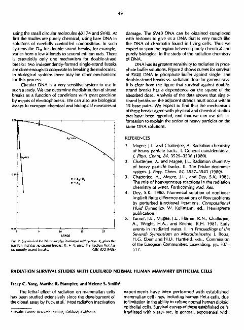
49
using the small circular molecules <j>X174 and SV40. At first the studies are purely chemical, using bare DNA in solutions of carefully controlled composition. In such systems the D „ for double-strand breaks, for example, varies from a few kilorads to several million rads. There is essentially only one mechanism for double-strand breaks: two independently-formed single-strand breaks are close enough to cooperate in breaking the molecules. In biological systems there may be other mechanisms for this process.
Circular DNA is a very sensitive system to use in such a study. We can determine the distribution of strand breaks as a function of conditions with great precision by means of electrophoresis. We can also use biological assays to compare chemical and biological measures of
ODlt 1 1 1 i -0 5 10 15 20
kRAOS
Fig. 2. Survival of X-l 74 molecules irradiated withy-rays. Xagivesthe fraction that has no strand breaks; X„ + Xt gives the fraction that has no double strand breaks. IXSL 625-94561
Tracy C. Yang, Martha R. Stampfer, and Helene S. Smith*
The lethal effect of radiation on mammalian cells has been studied extensively since the development of the clonal assay by Puck et al.' Most radiation inactivation
• Peralla Cancer Research Institute, Oakland, California
damage. The SV40 DNA can be obtained complexed with histones to give us a DNA that is very much like the DNA of chromatin found in living cells, Thus we expect to span the region between purely chemical and purely biological in the study of the radiation chemistry of DNA.
DNA has its greatest sensitivity to radiation in phosphate buffer solutions. Figure 2 shows curves for survival of SV40 DNA in phosphate buffer against single- and double-strand breaks vs. radiation dose for gamma rays. It is clear from the figure that survival against double-strand breaks has a dependence on the square of the absorbed dose. Analysis of the data shows that single-strand breaks on the adjacent strands must occur within 15 base pairs. We expect to find that the mechanisms of these breaks agree with physical and chemical studies that have been reported, and that we can use this information to explain the action of heavy particles on the same DNA solutions.
REFERENCES
1. Magee, I.L. and Chatterjee, A. Radiation chemistry of heavy particle tracks. I. General considerations. J. Phys. Chem. 84, 3529-3536 (1980).
2. Chatterjee, A. and Magee, J.L. Radiation chemistry of heavy particle tracks. II. The Fricke dosimeter system. ) . Phys. Chem. 84, 3537-3543 (1980).
3. Chatterjee, A., Magee, J.L., and Dey, 5.K. 1983. The role of homogeneous reactions in the radiation chemistry of water. Forthcoming Rad. Res.
4. Dey, S.K. 1980. Numerical solution of nonlinear implicit finite difference equations of flow problems by perturbed functional iterations. Computational Fluid Dynamics, W. Kollmann, ed., Hemisphere publication.
5. Turner, J.E., Magee, J.L., Hamm, R.N., Chatterjee, A., Wright, H.A., and Ritchie, R.H. 1981. Early events in irradiated water. II. In Proceedings of the Seventh Symposium on Microdosimetry, J. Booz, H.G. Ebert and H.D. Hartfield, eds., Commission of the European Communities, Luxemborg, pp. 507-517.
experiments have been performed with established mammalian cell lines, including human HeLa cells, due to limitation in the ability to culture normal human diploid epithelial cells. Survival curves of these established cells irradiated with x rays are, in general, exponential with
RADIATION SURVIVAL STUDIES WITH CULTURED NORMAL HUMAN MAMMARY EPITHELIAL CELLS

50
a shoulder region at low doses. The survival curve of x-rayed normal diploid human cells, however, has been found to be different. By plating cells on feeder layers, Cox and Masson2 obtained a high plating efficiency and studied the radiosensitivity of normal human fibroblasts to x rays. They found that tha survival curves of cultured diploid human fibroblasts in exponential growth were unshouldered and exponential throughout. Since most human cancers are of epithelial origin and since women exposed to x radiation showed a higher incidence of breast cancer than the nonexposed female population,1
it is important to know how normal epithelial cells respond to radiation. Recently we have developed techniques to grow normal human mammary epithelial cells in mass culture for 1 to 6 passages and in a highly efficient clon-ogenic assay. These technical advances have enabled us to study the radiation sensitivity of these cells. This report presents our recent experimental results with normal human mammary epithelial cells at second passage.
Figures 1, 2 and 3 show the experimental results of normal human mammary epithelial cells from donors of three different ages, ranging from young {age 28} to old (age 56). The percent of cell survival is plotted as a
X-RAYS (225 kVp) PE. • 4.7% 00=105 RADS
function of x-ray dose. A total of eight doses, up to 900 rad, were used for each experiment. The survival curves appeared to be unshouldered and exponential. Experimental results are summarized in Table 1. The extrapolation number (n) cf survival curve was about 1.0 for all cells studied. The D„ varied from - 1 0 5 rad to ~135 rad, and the 10% survival dose (D,J ranged from - 2 5 0 rad to - 3 2 0 rad. Cells from a 56-year-old donor (HI 91E) appeared to be slightly more sensitive to x rays than cells from younger ones, i.e., H164E and H208E. There was, however, no consistent trend in radiosensitivity with the age of donor.
For testing whether or not the normal human mammary epithelial cells can repair sublethal damage, we did fractionation experiments. Cells (H208E) were irradiated either with single doses or with a first dose of 225 rad, incubated for 4.5 hours at 37°C, and then exposed to second doses of 75, 150, 225, and 300 rad. Figure 3 shows the experimental results. For an equal dose, there was no significant difference in survival between cells exposed to single or split doses. For cells irradiated with split doses, a first dose of 225 rad and a second dose of 225 rad after 4.5 hours incubation at 37°C, for example, gave a survival about the same as that receiving single dose of 450 rad, namely ~ 3 % . Results therefore suggest that normal human mammary epithelial cells may not be able to repair sublethal radiation damage.
- S3
SS3
I ' l l
X-RAYS (225 kVp) PE - 30 5B% n= 10 Do' 130 RADS
;
! ~-J3
\
:
t
\
|
i mil
\ - \ -"
• i
" 0 100 200 300 400 500 600 700 BOO
DOSE (RAD) 400 600
DOSE I RAD I
fig. T fig. 2 (XBL 827-7707;

H208E X-RAYS
0 225 RODS * SECOND DOSE JJ SINGLE DOSE
T>4.5 HOURS n - 10 RE.«5.9% Do • 135 RADS
Table I. Radiosensitivity of normal human mammary epilhcl-al cells to 225 kVp x-rays.
300 400 500 600 DOSE (RADS)
Fig. 3
700 600 900 1000
(XBL 827-769S;
An unshouldered exponential curve appears to be the best fit of these 13 data points wi:h a D„ of about 135 rad.
Our experimental data have demonstrated that the survival curve of normal human epithelial cells irradiated with x rays is an exponential one with no shoulder and with a D 0 of 1P5-135 rad. These results are comparable to those reported for normal human fibroblasts. For example, Weichselbaum et al. < reported that normal human diploid skin fibroblasts showed a D a of 132 rad.
The shoulder of a survival curve has been commonly interpretated as an accumulation of sublethal damage before the lethal effect can be expressed. The repair of sublethal damage in mammalian cells has been investigated extensively by measuring the increase in survival observed following administration of two fractionated doses compared to the survival after an acute exposure to the same total dose. This repair Is termed "Elkind recovery." In cells that do not have a shouldered survival curve, no sublethal damage repair is usually observed with the split-dose technique. Since the survival curve of normal human mammary epithelial cells is unshouldered and exponential, one would expect no sublethal
Cell specimen Age of donor (yr) n D0 tradl D „ trad)
H164E 28 1.0 130 300
H208E 45 1.0 135 3>0
H191E 56 1.0 105 250
damage repair in these cells, and our results from split-dose experiments are consistent with this prediction.
In summary, our data indicate that the x-ray survival curve is an exponential one with no shoulder, that there is no consistent change in radiosen's'tivity of cells with the age of donor, and that there is no detectable sublet, . j l damage repair in normal epithelial cells.
Since the survival curve is unshouldered and exponential, our resu'ts suggest that there may be no threshold dose of x rays for killing normal human epithelial cells. More experiments, however, need to be done before one can make a final conclusion. In the coming year, we are planning to investigate the lethal effect of low dose rate, the potential lethal damage repair, and the potential mutagenic effects of radiation on these cells. This information may be valuable for our understanding of and estimating the potential radiation risk at low dose level.
ACKNOWLEDGMENT
We wish to acknowledge the excellent technical assistance of Ms. A. Pang, Ms. L. Craise, and Mr. S. Sylvester.
REFERENCES
1. Puck, T.T. and Marcus, P. I. A rapid method for viable cell titration and clone production with HeLa cells in tissue culture: the use of x-irradiated cells to supply conditioning factors. Proc. Natl. Acad. Sci. USA 41, 432-437 (1955).
2. Cox, R. and Masson, W.K. Changes in radiosensitivity during the in vitro growth of diploid human fibroblasts. Int. I. Radial Biol., 26, 193-196 (1974).
3. Brass, I.D.J, and Blumenson, L. Screening random asymtomatic women under 50 by annual mammographies: Does it make sense?/ Surg. Oncl. 8,437-445 (1976).
4. Weichselbuum, R.R., Epstein, J., Little, J.B. and Kornlith, P.L. Invitro cellular radiosensitivity of human malignant tumors. European 1. Cancer 12, 47-51 (1976).

52
CARCINOGENIC AND MUTAGENIC EFFECTS OF ENERGETIC SILICON IONS ON CULTURED MAMMALIAN CELLS
Tracy C. Yang, Laurie M. Craise, Jerry Howard, and Cornelius A. Tobias
The potential carcinogenic and mutagenic effects of energetic heavy ions, which are present in cosmic rays, are not well understood. In our laboratory, we are studying the effects of energetic heavy particles on somatic mutation and malignant transformation of mammalian cells to obtain quantitative information on the dose-response curves and RBE values for heavy ions with various energies and LETs, to determine the kinetics and nature of potential transformation damage repair, and to understand the fundamental mechanism(s) of neoplastic cell transformation by heavy ions. The present report describes experimental resulb we have obtained with high energy silicon ions accelerated at the Bevalac.
The cell system used for these studies is C3H10T1/ 2, an established line of mouse embryo eel Is widely used to assay neoplastic transformation in vitro. Well confluent C, cells were irradiated with x rays or a silicon ion beam. The initial energy of the silicon beam was 320 MeV/u, and the residual range in water, the ion energy, and the LET of the beam at the cell sample position were about 4.5 cm, 240 MeV/u, and 84 keV/|im respectively. Immediately after irradiation cells were plated into 100-
7-1-81 EXPT 142 cattir jT^ ( C I B / 1 4 )
^ T - £ 3 i
- \ a. 1 1 1 _
: % \ - \ S3.
-
sk
\ \ \
\ 3 -~ \ \ 3 -
CELLS PLATED BIGHT - AFTER IRRA0IATI0N : - £3 229 kVp X-RAYS -_ O 320 MtV/n SILICON ION
v is.")
1 1 1 1 i
-
0 100 200 300 400 500 600 TOO DOSE IRADS)
hti. I. Survival curves of C, cells irradiated by x rays or energetic silicon ions. Cells were plated right after irradiation.
IXBL 0110-71121
mm tissue culture dishes at low density. The dishes were incubated at 37°C for two weeks to determine the colony-forming ability and for six weeks to check the transformation frequency. After carefully scoring all dishes, data were subsequently analyzed and plotted.
A comparison between x rays and silicon ions in causing cell killing is shown in Fig. 1. Data gave an RBE value of about 3.0 at 50% survival level. For neoplastic cell transformation, a linear plot of experimental results is given in Fig. 2. The RBE determined at the level of transformation frequency induced by a 50% survival of x-ray dose is also about 3.0.
The role of repair in radiation induced lethality and cell transformation was also studied. Part of the lethal damages induced by silicon ions appears to be repairable in confluent G, cells. Survival is about 40% higher in cells irradiated by a silicon beam and incubated for one day before being trypsinized and plated at a low density (Fig. 3). The repair ratio (the ratio of doses between cells plated immediately and 1 day later at a given survival level) is about \ .4 at both 50% and 10% survival levels. Cells irradiated with x rays showed a higher repair ratio, about 1.7 (Fig. 4). Lethal damage induced by silicon ions, therefore, seems to be less repairable in C, cells than that induced by x rays.
A significant amount of repair of transformation lesions in x-rayed cells plated one day after irradiation was
eXPT. 142 C3HIOTJt (CI8/I4)
T~ T~ 240 MtV/n SILICON ION
Fig, 2. Neoplastic cell transformation induced by x rays or an energetic silicon beam. Cells were plated right after irradiation.
(XBl 8D0-7W9)

53
observed, as shown in Fig. 5. Cells plated about one day after irradiation gave a transformation frequency per buivival about half as much as that plated right after irradiation. This large decrease of transformation frequency in cells incubated for one day before being plated is in good agreement with the experimental results on the kinetics of repair in x-rayed cells obtained and reported earlier. A very interesting finding from this silicon experiment is that no repair of transformation damage could be detected in cells irradiated by silicon ions. The cell transformation dose response curves for silicon ion irradiated cells plated immediately or one day after irradiation are the same, as shown in Fig. 6. This observation suggests that the RBE for cell transformation wil l be significantly greater for slow growing cells than for fast proliferating ones irradiated with heavy ions with LET greater than 100 keV/|xm. These results also indicate that cell transformation lesions may not relate to potential lethal damage at all. Recently repairable double strand DNA break (d.s.b.) has been suggested as possible potential lethal damage (p.l.d.).' If this hypothesis is correct, our results then indicate that the transformation lesion is not a simple repairable d.s.b. Some unidentified mis-repaired DNA damage may be responsible for the radiogenic cell transformation.
The mutagenic effect of 320 MeV/u silicon ions was studied with a .-.oiine of C3H10T1/2 cells—SV10T1/2.
7 1-81 EXPT 142 CJHIOT4 (CI8/14)
>̂ ̂<* ' ' 1 1 1 1 _ — — — ^ £ 3 ' - * — — \ .\„
£3 *
- - I . 6 T -
- \ \
N*\ -
" \ N« -
\ \ z \ * v
- | £ CELLS PLATED 24 MRS " AFTER IRRADIATION \ -
- « CELLS PLATED RIGHT *"* AFTER IhRADIATION -
E25 kVp X-RAYJ.
1 1 1 1 1 | 1 430 600 DOSE (RADS)
7 1-81 EXPT 142 C3HIOTjfc 1CIB/I4)
V i 1 1 1 1
1 1
1 1
\>< m CELLS PLATED 24 HRS ^ AFTER IRRADIATION
{-> CELLS PLATE0 RIGHT -135 V AFTER IRRADIATION
-\><
320 MaWn SILICON ION -I S i " )
-\ V °s \ . * \ , _
- Q \ '* ~ -
\ X " -
1 1 i i i i O IOO LOO 300 400 300 600 700
DOSE (RADS) Fig. 3. Potential lethal damage repair in C cells irradiated with silicon particles. (XBL 8110-71I M ;
0.30
0.2B
0 26
I 0.24
0 2 2
0.Z0
: 0.18
0.16
, 0.14
. 0.12
; 0.10
0 0 6
0 0 6
0 0 4
0.02
000
C 3 H I 0 T ^ (CI 9/14)
_ 2 2 5 kVp X-RAYS
T
J I I I
Fig. 4. Repa'r of potential lethal damages, due to delayed plating, in x-rayed confluent cells. IXBL 81W-7114)
400 600 DOSE (RADS>
Fig. 5. Repair of transformation lesions in x-rayed cells. The transfer* mation frequency decreased significantly when cells were plated one day after irradiation. iXBL 8110-/W8I

54
7-1-8 EXPT 148 C3HI0TJ; ICI8/I4)
0 36 - i 1 1 1 1
• / : • 0 32 / #
£ / - 0 28 _ / _ w u / o z > 0 2 4 / — > SU
R
/ -£ 0 20 ' _ a
" < CMS _ 3 o /o — | 0 . 2 o o
0/ -0 06 / 240 MtV/n S i " —
o*c • O CELLS PLATED RIGHT — v AFTER IRRADIATION _o / O CELLS PLATED RIGHT — v AFTER IRRADIATION
004 • ' / A CELLS PLATED 24HRS
~ AFTER IRRADIATION _ • ' A CELLS PLATED 24HRS ~ AFTER IRRADIATION _
7 i 1 1 1 I 0 100 200 300 400 500 600
DOSE (RAD5)
Fig. b. Comparison between cells plated right utter and one day after silicon ion irradiation. Delayed plating showed no effect on the transformation frequency. tXBL 8tt0-7t07i
This subline was established by isolating a clone of C3H10T1/2 cells transformed by SV40 viruses. In terms of cell killing, SV10T1/2 cells showed no difference in radiosensitivity from their parental line—nontransformed C3H10T1/2 cells. Using the respread technique, we investigated the induction of 6-thioguanine-resistant mutation by silicon particles. Some preliminary results are shown in Fig. 7. High-LET silicon ions, for a given dose, appeared much more effective than x rays in producing deletion type HGPRT (hypoxanthine-guanine-phosphor-ibosyltransferase) mutation in mammalian cells.
CSHIOTfc CELLS (SVIOTfc)
£3 225 ItVp X-RAYS
Q 240MtV/n Si**
500 600 700 200 300 400 DOSE (RADS)
Fig. 7. Induction of 6-thioguanine-resistant mutation by x rays or high energy silicon particles. . (XBL 823-8342)
We are keenly interested in the fundamental aspects of the cell transformation lesion and believe that some insights can be obtained through the heavy-ion cell transformation and mutation studies. An understanding of the molecular nature of the cell transformation lesion is clearly important for elucidating the mechanismls) of radiation carcinogenesis and for assessing the radiation risk.
REFERENCE
1. Frankenberg, D. and Frankenberg-Schwager, M.; International f. Radial Biol. 59:617-631, (1981).

55
CELLULAR AND MOLECULAR RADIOBIOLOGY OF HEAVY-ION BEAMS
Cornelius A. Tobias, Eleanor A. Blakely, Ruth J. Roots, Tracy C. H. Yang, Polly Y. Chang, Leora Lomniel, Lauri M. Craise, Michael |. Yezzi, .nrl Peter M. Martin
The role of the cellular and molecular radiobiology group is to develop a fundamental understanding of the manner in which heavy ions exert their effects on biological systems, to apply our knowledge for a rational approach to therapy with heavy ions, and to quantitate the dose-effect relationships for acute and chronic effects in order to assure safe treatments. Since we do not fully understand the differences between tumor cells and normal cells, these tasks are challenging.
We are now engaged in investigating specific aspects of cell injury produced by radiation lesions, and in com
paring cell injury at different phases of the cell division cycle. Figure 1 is a schematic drawing of the cell division cycle and the effects of heavy ions and x rays at the different phases. One should visualize that the cells of the tissues are progressing in time clockwise as represented in the graph. However, many normal cells in differentiated tissues reside in the C 0 phase, represented here as following mitosis, and these cells may not participate in the full cycle. Many tumor cells are in Gl phase at any given time, but because the G 0-C, boundary either does not exist or is weak, tumor cells are more free to follow the
COMPARISON OF X-RAY AND HEAVY-ION EFFECTS IN THE CELL CYCLE
f;g. /. Schematic drawing of the cell division cycle. The inner ring shows the phases of the cycle and the DNA activity during each specific phase. The middle ring shows the effects at each phase if the cells are irradiated with x rays: the outer ring shorn the effects of exposure to heavy ions. fXBL 9212-4302)

56
cycle. Generally, resistance to x rays is highest in S and early C, phases. Heavy ions increase radiosensitivity through the entire cycle, and particularly reduce repair in G, and S phases.
We already have evidence that small doses of heavy ions can produce more lethal effects than equivalent absorbed doses of x rays. We also know that heavy ions are particularly effective in reducing repair in the G, phase, and somewhat less effective in reducing S phase repair. X rays delay progression through the S and G2
phases, whereas heavy-ion irradiation is characterized by a prolonged block in G,, just before mitosis. When normal cells are in contact, they mutually inhibit each other's progression through the cell cycle, frequently resulting in a piling up of cells in G, phase; tumor cells are not so inhibited.
The studies described in the paragraphs that follow represent progress in our understanding of the various details of these complex cellular phenomena.
FRAGMENTATION EFFECTS ON RBE AND OER Fragmentation of primary heav> charged particles
into secondary particles of predominantly lower charge and mass occurs as a result of nuclear interaction events that take place when particles traverse absorbing targets. We have investigated the effects of fragmentation events on the aerobic and hypoxic cell survival of asynchronous human T-l cells in vitro under conditions where the dose-average LET of the primary ions is approximately 118
SILICON
V ' 4 - • 320 MeV/u
A 456 MeV/u T 530MeV/u • 670MeV/u
1
..i Nr4... I I I T i , t • ill 0 l I 0 2 I 0 5
PRIMARY BEAM LET (keV/>m) I til J KUt ,inrf (JLfi mi'dnurenilYlf* nu\df ,11 i ,inom rewlUxtl r.lngel mi >tlti un bt:im> tiinizmu in tntthil energies mini iSft hi 670 MeV u
>X0L ajll-I'llii
keV/|im, as confirmed by several dosimetric methods. Particle beams of two different initial energies, but
with the same atomic number, have been studied in track segment experiments. A beam with an initial low energy usually has a small fragmentation component, whereas a high initial energy of the same primary ion can be selected which has a larger fragmentation component. Measurements of relative biological effectiveness (RBE) and oxygen enhancement ratio (OER) were made at a residual range of 0.54 cm of water in neon beams of two energies (225 MeV/u and 670 MeV/u), and at a residual range of 1.87 cm of water in silicon beams of two energies (320 MeV/u and 670 MeV/u).
Preliminary results indicate that the dilution of the primary beam dose average LET by the secondary particles of low LET resulted in a reduced effective total beam LET. The effect of this change in the physical characteristics of the high energy beams was to decrease the RBE but not to significantly change the OER, relative to the measurement of these values in the low energy beams. This is demonstrated in Fig. 2 for RBE and OER measurements made at various residual ranges in silicon beams with initial energies ranging from 320 MeV/u to 670 MeV/u.
HEAVY-ION CELL CYCLE EFFECTS
Synchronized cell populations of Chinese hamster V-79 and human T-l cells have been studied to characterize the cell ages thgt are sensitive and resistant to heavy-ion damage. The data collected have confirmed that high-LET radiation diminishes the differences in ra-diosensitivities between cell ages, which results in more similar dose responses for cell killing at all phases of the cycle of DNA synthesis, especially at low doses. The human and hamster cells were selected for this study because a large body of accumulated information on their asynchronous response to Bevalac accelerated heavy ions already exists, and because we have an interest in comparing cell lines with abbreviated and prolonged G, phases. The longer G, phase is characteristic of many human cell lines; it is also characterized by a low-LET radioresistance that is fundamentally of interest from a target point of view and may also have clinical significance in terms of evaluating dose fractionation schedules.
Figure 3 shows whole x-ray and neon-ion dose-response curves of synchronized human T-l cells. Computer assisted least-squares fits to the linear quadratic model are shown. At 10% survival the RBE values are 1.4 for mitotic cells, 3.2 for G,/S phase cells, and 2.8 for late S-phase cells. Two separate experiments are represented for each curve, except for the mitotic neon curve which represents data from a single experiment.
Consistent with the age response profile, the G,/ early S-phase cells are more sensitive than the mitotic/ early G, phase cells. This is seen more clearly in Fig. 4,

57
1.0 NEON
1.0 & MITOTIC V ^ v G| /S phase V ^ ^ t v S-phase
0.1 — * $ - — - R S ^ 0 > M i a 3 - . - X - - 3_- -RB£|0» 3.210.6 - --°-V ^~RBEi0'2B±Q&
< > 0 . 0 I \ \ X \ V \
0.001 SYNCHRONIZED
• HUMAN T-l CELLS • X-ray o 425MeV/uNE0N
0 2 4 6 8 10 12 2 4 6 8 10 12 2 4 6 8 10 12 DOSE (Gy)
Fig. 3. X-ray and neon-ton dose-response curves for synchronized human T-l cells. fXBL 824-37521
n 1 SYNCHRONIZED HUMAN T-l CELLS
\ 4E5MeWu NEON \ X 225kVp X-RAY
\ N
\ \ \
\ \ ^lOhr X-ray
\ < \ O.Jhr \
V X - r o y \ 8.8 t l r J \ \ \
NEON V - A \ \ 0 . 6 l » ' - \ \ \ \ NEON
H U M A N T - l C E L L S
S 10 12 6 DOSE IGy)
Fig. 4. Synchronized human T-1 cells exposed to \ rays or heavy Ions: the dose response curx'es show that the Cl/eariy 5 phase cells are more sensitive to Bragg peak neon ions than are the mitntic/early CI phase cells; whereas the reverse is true for the x-ray responses.
IXBL 824-3746)
which shows that the mitotic x-ray and neon curves have an intermediate radiosensitivity between the very sensitive G|/S phase neon response and the approximately same aged G/S phase x-ray response.
Figure 5 depicts the age response profile for an x-ray and a neon-ion experiment, each of which kill to approximately the 1% survival level, but with a much lower neon ion dose. The main conclusion of this preliminary report of synchronized human coU response to neon ions is that beyond the G,/S border (indicated by points to the right of the cross-hatched line), both x rays and neon ions show a radioresistant S phase population. In contrast, however, at cell ages before the CJS border
0.0I
0.01
0.001
10.001
t—I—I—I—I—I—I—I— \ .
I I I I I 0 4 8 12 16 20 24
TIME AFTER SYNCHRONIZATION (hr) Fig. 5. Synchronized human T-1 celt age-response profile for an x-ray and a neon-Ion experiment. (XBL 824-3745)
(indicated to the left of the cross-hatched line), the age response profile of mitotic/early G, phase cells is quite different for neon and x-ray irradiations. We are planning to examine these early cycle differences in greater detail in the future, ami we will be analyzing the cross-sectional dependence of the radioresponse of synchronized human cells.

58
REPAIR OF POTENTIALLY LETHAL DAMAGE IN CONFLUENT CELLS
The question "how much radiation damage can be repaired in mammalian cells" is a fundamental one. Survival curves obtained by conventional techniques usually do not give information on either the initial amount of injury or the maximum repair of damage in irradiated cells. However, recently it has been possible to Inhibit some of the enzymes responsible for repair of the ra-diolesions, and such experiments shed light on the yield of radiolesions as well. In the experiment described below, we inhibit a DNA beta polymerase with B-D-arabino-furanosyladenine (B-araA). To determine the amount of repairable and irrepairable damage, we used mouse embryo cells IC3H10T1/2). Confluent cells were x-rayed, then treated in three different ways: one group of cells was seeded at low density in regular medium, one group was incubated at confluent state for one day before being plated into regular medium, and one group was plated into (J-araA medium {100 (xm) that was replaced with regular medium one day later. Colonies formed after two weeks of incubation were counted, and the results are shown in Fig. 6. The survival curve for cells treated with 0-araA is an exponential one with no shoulder, and the LDS„ and LD,„ are about 100 and 360 rad, respectively. At the 50% survival level, B-araA treated cells appear to be about five times more radiosensitive than cells allowed maximum repair. The dose modifying factor, however, varies with the survival level: decreasing with increasing doses.
Cells trated with B-araA also showed a higher frequency of neoplastic cell transformation than control cells. Results are given in Fig. 7. This re rult suggests that a radiation-induced cell transformation lesion may be in the DNA.
REPAIR OF X-RAY DAMAGE IN A MAMMALIAN CELL MUTANT
A protein synthetic mutant of a Chinese hamster ovary cell line designated TSH1 and the wild type cell designated SCI have been characterized with respect to their ability to carry out protein synthesis when they are treated with mild hyperthermia.1 The mutant cell line but not the wild type has been shown to rapidly shut down protein synthesis when it is transferred from its normal growth temperature of 35°C to 40°C. Our first goal was to examine the effect, if any, that interruption uf protein synth ;sis had on the rapair of sublethal x-ray damage (SLD). We discovered that the repair of SLD could be reduced if the cells were first placed at a high lemperature for a minimum of 2 hours before the first dose of x rays and if the inhibition of protein synthesis was continued until the second dose; was given. We interpreted this to mean that a pool of protein was nec-
400 600 800 Dose (rod)
1000 1200
Fig. 6. Survival of C3H10V/.> celts exposed to 225 kVp x rays, and then M immediately plated. (•] delayed plated, or {A) treated with p-araA IXBL 8212-4293)
x-X-roys<225kVp> • = x-rays-t-0-aroA •
I I00(i M)
200 400 Oose (rod)
600
Fig. 7. Cell transformation as a function of dose with and without the addition of^-araA. C3HIOV/: cells wen? either exposed to 225 kVp x rays alone (x) or exposed to x-rays and then treated with fi-araA (•) The cells were then incubated, and the colonies were examined for neoplastic ce// transformation. IXBL 8212-4294)

59
essary for repair and only upon depletion of it would any effect on cell survival be seen.
Our second goal was to obtain a clearer understanding of the relationship between molecular and cellular events going on inside the cell, after exposure to x rays. Since DNA is a primary target for radiation damage leading to cell lethality, we decided to observe what effect the lack of protein synthesis, which affected cell survival, would have on the rejoining of breaks in DNA. S. Okada (Tokyo, Japan) brought to our attention a technique for observing breaks in DNA after low Joses of x rays (less than 1000 rad) patterned after the work of B. Rydberg.2
Briefly, the technique involves exposing irradiated cells prelabled with "C-TdR to an alkali solution to disrupt the cells and to cause unwinding and denaturation of the DNA at the sites of breakage. The nixture is then neutralized and sonicated to break up the DNA. The single and double strands of DNA are separated from each other on a hydroxylapatite column by eluting through different concentrations of phosphate buffer. As the dose of radiation increases, the fraction of single stranded DNA coming off the column increases. The amount of each type of DNA can also be followed as a function of time after exposure to a fixed dose of x rays.
Because this technique allows the use of doses in the range of cell survival measurements, we felt that a more meaningful correlation between DNA rejoining and cell survival could be made. In the split dose x-ray experiments shown in Fig. 8, we used the same temperature and time scheme for the DNA rejoining as the earlier cell survival experiments. We did not see any effect on DNA rejoining as we did for cell survival when protein synthesis was inhibited. We interpret this to mean that the enzymes involved in rejoining DNA are not affected by the lack of protein synthesis that reduced the repair of SLD. Another possible role of protein synthesis in the repair of SLD may involve proteins that are essential for 1he correct expression of the DNA. We intend to explore this idea with the use of synchronized cells.
INDUCTION AND REPAIR OF DNA LESIONS: IN-VITRO AND IN-VIVO WORK WITH THE SIMIAN VIRUS 40
Some of our molecular work during the past year has conccntraled on the LET-dependent induction of double-strand breaks in DNA. For this purpose we have used the simian virus 40ISV40) genome. Chromatographic assay systems developed in our labratory allow distinct separation of single- and double-strand break yields, either from the SV40 DN A - j lut ion or the SV40 DNA recovered from a mammalian host cell.
Irradiation of SV40 DNA in solution with accelerated particles of different masses and/or velocities, giving a spectrum of ionization densities, show that about 25
Dow (Gioy l
0.11 I I I I I I I I I ' ' ' I 0 1 2 3
Time after second dose Ihr) Fig. 8. Split x-ray dose cellular and molecular data using the Chinese hamster CHO-TSHI mutant protein synthesis cell line.
IXBL 8J1-J538)
single-strand breaks are produced for each double-strand break after gamma-ray irradiations. As the ionization density increases, this ratio changes such that at very high LET, about 10 single-strand DNA breaks are made for each double-strand break.
It is, however, of greater interest to determine these DNA lesions in cellular DNA, and such studies are currently under investigation. In a mammalian host cell where SV40 propagation goes on, the SV40 genome is structurally identical to mammalian chromatin, except for its much smaller size. Figure 9 shows the effects of x-ray irradiation on intracellular SV40 chromatin. In the electrophoretograms, the peaks of mammalian DNA are shown in the cross hatched area. In the second electro-retogram from the top only a very minor peak for the mammalian DNA is present in the uninfected cell lysate, and it is only after irradiation that the host cell DNA becomes noticable. The third electrophoretogram from the top shows the SV40 DNA profile from the lysate of infected African green monkey cells, the large peak representing the maximally supercoiled molecule and the small peak representing molecules that have one or more single-strand breaks. After irradiation this peak increases.

60
X-IRRADIATION OF INTRACELLULAR SIMIAN VIRUS 40 DNA
The third peak in the middle represents linear DNA, that is, molecules having a double-strand break. We are currently measuring the fraction of DNA molecules with these two types of lesions (single-strand breaks and double-strand breaks) to determine the number of single-strand breaks formed per double-strand break as the breaks occur inside the cell nucleus.
MFECTED CELL LYSATE
fj
hff. 9- After dgnrou' y>t>l electrophoresis, the SV40 DNA shows a characteristic profile from which the factions of undamaged DNA, DNA with single-strand breaks, and DNA with double-strand breaks can be quantitied <X8L 823-8627)
STUDIES OF RADIOSENSITIZATION BY D1AMIDE
Cellular x-ray survival studies with the chemical radiosensitizing agent diamide have been conducted in order to determine how diamide achieves its radiosensitizing effect. This agent, best known as a rapid oxidant of sulfhydryl compounds (notably glutathione) and of the pyridine nucleotides {NADH and NADPH), has also been shown to have radiochemical reactivity comparable O other hypoxic cell sensitizers that have a nitroheterocyclic structure. Both a slope increase and a shoulder removal of the mammalian cellular survival curve have been previously reported for diamide under selected conditions. These observations suggest that diamide may act as an electronaffinic agent, which could increase the free radical mediated damage resulting from radiation, and/or it may act as a biochemical oxidizing agent, which could prevent cellular repair processes.
it was the intent of the present work to determine just what conditions were needed to observe shoulder reduction of the cellular survival curve, and to resolve the mode, or primary modes, of action of diamide. in these experiments, conducted mainly with Chinese hamster V79 cells and 225 kVp x rays, the radiosensitizing effects of a 15-minute incubation with 0.2mM diamide were determined at 4°C, 21°C, and 37°C, under hypoxic and aerobic conditions, and when diamide was added just after irradiation, as well as 5-10 minutes before irradiation. In all cases, a reduction in the slope of the survival curve was seen. The aerobic radiosensitization of diamide was consistently dose modifying, with an enhancement ratio of 1.6 or less. The most dramatic reduction occurred in hypoxia, as expected, at 4°C and 21°C, with diamide present during irradiation. Removal of the shoulder of the hypoxic survival curve occurred at 4°C when diamide was added before or just after irradiation, and at 21°C when diamide was added shortly before irradiation.
The age response of synchronous T-l cells with 400 rad of x rays and diamide was also studied. Results depicted in Fig. 10 indicated a uniform decrease in survival over the cell cycle. No cell age dependence of diamide radiosensitization was evident.
These studies indicate that 1) the degree of radiosensitization by diamide is strongly dependent on temperature and timing of drug administration, 2) removal by diamide of the shoulder of the survival curve occurs

61
Age Response with Diamide ' ' ' ' ' Ci Controls' ' '
+ Dlomioeolone X X- rays (400rods) a x-royltriiamlde
100 r t + 0 + O + Prj t P o °
X „ „ X XX x x x x x m x X
: 10 r ° B B a ° a 8 ° B B
|im > 1
1
" , 1 1 1 1
; o — i
+
i —
P r
&
-\—T i 1 r 0 Control + Diamide, ISmln.
+ r
i + P + I i ,°o, , , o , , '
100 80
; 60 40 20
8 12 16 Cell age (hr)
Fig. 10. (Upper} Percent survival of human T.J cells to 225 kVp x rays with and without dia.. ..'e treatment as a function of cell age in hours after mitotic selection, (Lower) Percent of lH-TdR labeled 7-1 cells as a function of cell age in hours. The diamide treatment was al a concentration of 0.2 mM for 15 min starting 5 min oefore irradiation, and extending to about 5 min after irradiation. (XBL 8212-4301)
only under limited conditions, and 3) radiosensitization from diamide treatment immediately after irradiation is comparable to radiosensitization from diamide treatment during irradiation.
The results are interpreted to mean that diamide acts primarily by inhibiting repair processes. Diamide presence just after aerobic irradiation resulted in only a small change in the slope of the survival curve, compared to its effect when present during irradiation. Diamide presence just after, as well as during hypoxic irradiation at 4°C and 2 "C resulted in shoulder removal. The preliminary age r( sponse result suggests that the effect of diamide may be different than that of another well-known radiosensitizer and sulfhydryl reagent, N-ethylmaleimide.
MODELING CELLULAR AND TISSUE RESPONSES TO DELETERIOUS AGENTS
In order to achieve a quantitative understanding of tiie mechanism by which deleterious and carcinostatic agents act, it is important to have mathematical models. Many of the radiobiological models in the literature have stressed the physical interactions of radiation with matter but less attention has been given to the repair responses of cells to injury.
The repair-misrepair (RMR) model developed in our laboratory separates the interactive processes that produce key molecular lesions from the enzymatic repair processes that occur in the postexposure period. The model is
I .
* *,« 1MB- 90 I .,„„. 0.1
8.0 t-N \ A 0.01 r * r
• 670Ne-P
0.001 - ~ o '50kvpx-<a»-
0 2 4 6 0 2 4 6 8 0 2 4 S OQSE 16*1
Fig. } 1. Neon-ion and x-ray survival curves for three human celt lines: (left! human T-l kidney cells that have an efficient repair capacity: (middle) normal human cells (IMR-90): (right) human cells that are genetically deficient in repair obtained from a patient suffering i: om ataxia telangiectasia. (XBL 8211-42461
based on the simple assumption that the enzymatic in-formational apparatus of the cell recognizes molecular modes of injury and, in the course of time, attempts to enzymaticalty repair the damage. The fidelity of repair is impo' int, and mistakes made in the repair processes lead to ' re consequences of injury: lethality, cell transformation, or the appearance of variants. The RMR model can be used at three different levels to aid research: At the biochemical level it helps to analyze the chemical yield of molecular lesions, for example strand breaks produced in DNA. At the cellular level, by analysis of split dose experiments, its use can determine the time rate of repair; because different repair processes usually proceed at different rates, one can sometimes separate various processes from each other. In Fig. 11, we illustrate these principles by showing x-ray survival curves of several strains of mammalian cells: cells that are genetically deficient in repair from a patient suffering from ataxia telangiectasia, from normal human cells (IMR-90) and from human kidney cells that have an efficient repair capacity. In addition, normal mouse cells have been studied wherein repair has been inhibited by application of an inhibitor of the enzyme beta-DNA polymerase, which is believed to be involved in the repair of potentially lethal damage.
Various human tissues show an array of sensitivities to the same deleterious agent. For example, cells from gut epithelium ive substantial repair capaicty, bone marrow colony forming units repair less, and repair sterns to be absent from spermatogonia! cells of the testes. Neurons, whic . have a long life span in the body, seem to have very efficient repair system*. A possible explanation lies in the assumption that the expression of repair systems is controlled to various degrees in various differentiated tissues. In certair tumor cells ifor example, ir human breast cancer) repair seems to proceed at a greater rate than in normal cells from the same tissue, and this may explain the refractoriness of cancer cells to standard ra-

62
diation and chemotherapy. At the present time we do not understand the cellular control mechanisms that are operative in relation to intracellular repair, however, the application of accelerated heavy ions can drastically re-
Table /. Interaction of modalities.
INDEPENDENCE: S 11.2) = 5(1)* 5(2), no interaction of lesions IU) or repair (r)
SIMPLE SYNERGISM: UU), UI2) additive same repair system (r)
COMPLEX SYNERGISM: U(1) different from U(2I modality (I) affects r{2); modality (2) affects rl l) in an asymmetric manner
INTERACTIVE MODALITIES: U(t) different from Ul2) interaction in course of repair results in new U(3I
S = Survival 1 = Modality A U = Lesion 2 = Modality B r = Repair system 3 = Modality C
Stanley B. Curtis
A formulation of cell inactivation has been developed that incorporates several of the major ideas of past and present hypotheses of how individual mammalian cells are inactivated or rendered nonclonogenic by ionizing radiation. Elements from the repair-misrepair, lethal-po-tentially lethal, sublesion interaction, and tract structure models are combined to produce a single set of mutually compatible hypotheses. This unified model can be used to explain shoulders on survival curves, the exponential behavior both at low dose rates and at high LET, as well as to predict the kinetics of enzymatic cellular repair.
The basic elements of the unified model are: 1. Long-lived "sublesions" or "potentially lethal"
lesions (half-lives of many minutes) are formed in the cell by the radiation field. The lesions can interact over large distances (micrometers) during the enzymatic repair process to form lethal lesions via "misrepair" processes. These can be identified with "intertract interactions" or "gamma ki l l , " since they dominate at low LET. These lesions also have a finite probability for being repaired correctly.
2. One charged particle track can form lesions, some of which, depending on ionization density, may be created in such close proximity that two or more can interact strongly (i.e., in short times) to form lethal or irrepairable lesions. These can be identified with "intratrack inter-
duce repair efficiency. This is illustrated in Fig. 11, where we compare the survival curves for x rays and high-LET neon ions. The RMR model has furnished a basis for classification of the types of injuries produced by various deleterious agents on the basis of the interaction between these injuries and the injuries produced by ionizing radiation. Table 1 is a simplified version of this classification scheme. The basis for the classification is the nature of the injury produced and whether or not one agent modifies the repair apparatus for injury by another agent. Usually one obtains information on possible synergisms by applying the two agents together to the small cell, either simultaneously or in sucession.
REFERENCES
1. Thompson, L.H., Harkins, I.L., and 5tanners, C.P. Proceedings of the National Academy of Science 70, No. 11, 3094-3098(1973).
2. Rydberg, B. Radiation Res. 61, 274-287 (1975).
actions" or "ion k i l l , " because they are caused by a sinple charged particle track.
3. The sublesions are produced by the very close proximity of several ionizations (perhaps as many as 6 to 10 are needed for each sublesion within a distance of 3 nm). The important physical parameters to describe the radiation field become the fluence of particles and the mean free path for creation of such "clusters" of ionizations along the track.
The chemical environment within the cell nucleus will determine whether a number of ionizations in close proximity will become a "potentially lethal" lesion, which the cell must deal with later via the enzymatic repair process.
Several consequences arising from this formulation are:
1. The most "fundamental" cellular survival data are obtained with non-dividing (stationary phase) cell cultures using either very low dose rates, or "delayed plating" procedures to allow maximum repair. From the former, values for the irrepairable component of damage can be obtained. Here, the long-range interaction of the sublesions can be neglected because the lesions can repair during the irradiation time and their concentration at any given time is small. The delayed plating experiments (at conventional dose rates) yield the amount of long-
IDEAS ON THE UNIFICATION OF RADIOBIOLOGICAL THEORIES

63
range lesion interaction, which causes a shoulder on the survival curve. "Immediate plating" curves (even of stationary phase cultures) are more difficult to interpret since •hi fre*!1 medium placed on the cells initiates the mitotic cycle am 1 causes a "f ixation" of damage before the cell has had a chance to complete the repair process. Thus, either interaction between a lesion and the chemical components of the medium, or "fixation points" within the cell cycle may cause repair inhibition and/or damage "fixation."
2. The survival cuive at low LET only approximates a linear-quadratic expression. The parameter B has no fundamental significance. It varies with LET and goes to zero as LET increases. At low LET, the parameter j * : n-creasesjinearly with LET because it varies as const/(LTXfl, where L, is the track-average LET and X, is the mean free path along the track between "sublesions." To the extent that \ , is inversely proportional to ionization density (hence, to LET), a wil l increase linearly with LET.
3. The extrapolation number, n, of a low LET survival curve has no fundamental significance. The slope of the curve does, however, approach a constant at high dose. This limiting slope is a measure of the production rate (per unit dose) of the total number of lethal and "potentially lethal" lesions initially produced by the radiation.
This work was supported in part by a Senior international Fellowship (1-F06-TW00642-01) awarded by the Fogarty International Center of the National Institute? of Health and in part by the Office of Health and Environmental Resources of the U.S. Dept. of Energy under contract No. DE-AC03-76SF0009B. The work was carried out during a 10-month Professional Research Leave, eight months visiting Prof. Wolfgang Pohlit at the Biophysics institute, GSF, Frankfurt, West Germany, and two months visiting Herwig Paretzke, Institute of Radiation Protection GSF, Neuherberg, West Germany.
CELL AGE DISTRIBUTION IN MULTICELL TUMOR SPHEROIDS
Adrian Rodriguez, Edward L. Alpen, and Randy I. DeGuzman
During the past four years we have characterized the radiobiological response of multicell tumor spheroids (MTS) of a rat brain tumor cell line (9L). The studies have been done with x rays and accelerated heavy ions. We have shown that the radiation survival of MTS cells is enhanced compared to monolayer cells for low LET radiation.' With heavy-ion radiation the enhanced survival of MTS cells is reduced, and at an LET between 70-80
keV/(im there are no differences in survival between monolayer and MTS cells.2 We have also observed that the RBE-LET relationship of MTS is similar to normal tissues.1' Figure 1 shows the RBE-LET relationships for testes, gut, and MTS. There is a monotonic increase in RBE as the LET increases to 100 keV/n.m. The highest RBE appears to be associated with an LET of around 100 keV/p.m, which is lower than for monolayer cultured
1 IT| ' ' ' « Coition GUI
~ D neon Spheroids - a Silicon •
- * A',0n ft\ ~
*T777i7,7 , ,
A ? L
fi w \ ~
*T777i7,7 , , ml 1 1 100
Dote overage LET IkeV-Vm)
f ig. t. The RBE-LET relationship for two mouse normal tissues (gut and testes) and multicell tumor spheroids. These data were obtained from plateau irradiation (0.0 cm weter absorber) in heavy ion beams. (XBL 829-4090)
40 Channel number
Fig. 2. Cell size distributions of'MTS (A) and monolayers (B) determined with a Coulter counter and channetyzer. (XBL 82 J2-4286)

64
(A)
1 Whole Spheroids 1 G, = 74.5
S > 16.2 I G2M • 9.3
GV - 7,7
Exponential Monolayer Gi = 47.7 S = 42.5
CzM - 10.1
Cnonnel number » FIR. i. 0\A distributions of cef/s grown as (Af MTS and(B/exponentially Wouing monohnen. The MTS cells were derived from whole spheroids thai were completely dispersed. Channel number is proportional to DSA content. IXBL 8212-4288/
cells.' These data were determined for well defined LET distributions in the plateau regions of ionization of accelerated heavy ions.
We are presently characterizing 9L MTS with respect to cell age distribution and growth conditions. We have made preliminary observations on cell age distribution and cell size in MTS compared to monolayer cells. Cells from MTS are smaller in size than monolayer cells (Fig. 2). The difference in cell size can be attributed to alterations in the cytoskeleton due to the physical constraints of being closely packed together and to a large fraction of the cell population being in G, or G,, phase of the cell cycle.
On fixed and stained cross sections of MTS we have observed that there are three distinct regions. There is a central core region of necrosis surrounded by a rim of viable cells. The rim of viable cells can be subdivided
1 Fl FZ F3 F4
Aii \ V V. Channel number — »
Fig. 4. DNA distributions of cells sequentially dispersed from MTS (500 urn in diameter). F, - f 4 represents fractions of cells from the surface of the MTS to the necrotic core. (XBL 8212-4287)
into an outer proliferating region with many mitoses and an inner nonproliferating region with few mitoses. Labeling experiments with JH-thymidine have determined that the growth fraction for spheroids greater than 40Gym in diameter is about 40%. Comparison of DNA histogram of monolayer and MTS cells by flow cytofluorimetry (FACS IV, Becton-Dic, 'nson) indicates, as shown in Fig. 3, that the cell population of MTS is primarily G, with few S and G 2 M cells. Exponential growing monolayers have more S cells.
Sequentially dispersed layers of cells from MTS have been analyzed by flow cytofluorimetry as is shown in Fig. 4. Each fraction o f cells (F,-Fj) represents a deeper layer of cells in the spheroid. Fraction 1 (F t), the outer cell layer, is representative of an exponentially growing population as shown for monolayer cells in Fig. 3. Fractions F2 and F, show an increasing population of G, or G n cells with fewer G^M and S phase cells. These cells are either cycling slowly or, more likely, are arrested in G,. The prominent peak in fraction 4 represents dead cells and debris in the necrotic core and is characterized by a peak channel lower than the G, peak in the previous fractions of cells.
These studies indicate that spheroids are similar to growing solid tumors and tissues with respect to cell age distribution and the presence of necrotic regions. The spheroids thus afford a unique model system for tissue and tumor radiobiological responses. The various populations of cycling and noncycling cells in MTS can be readily isolated and examined independently in the hope that these studies wil l shed light on the mechanisms of radiore5istance of cells grown as MTS. Future studies will not only involve characterization of the radiation survival of these populations but also the radiation repair capacity.

65
REFERENCES
1. Rodriguez, A. and Alpen, E.L. Feeder cells and cell survival in spheroids and monolayers. Int. I. Radial Biol. 41, 111-117(1982).
2. Rodriguez, A. and Alpen, E.L. Cell survival in spheroids irradiated with heavy-ion beams. Radiat Res. 35,24-37(1981).
3. Alpen, E.L., Powers-Risius, P. and McDonald, M. Survival of intestinal crypt cells after exposure to
high Z, high-energy charged particles. Radial. Res. 83, 677-687 (1980). Alpen, E.L. and Powers-Risius, P. The relative biological effect of high-Z, high-LET charged particles for spermatogonia! killing. Radiat. Res. 88, 132-143 (1981). Blakely, E.A., Tobias, C.A., Yang, T.C.H., Smith, K.C. and Lyman, J.T. Inactivation of human kidney cells by high-energy monoenergetic heavy-ion beams. Radiat. Res. 80, 122-160 (1979).
INDUCTION OF HARDERIAN GLAND TUMORS IN MICE BY HEAVY ION IRRADIATION
Edward L. Alpen, Patricia Powers-Risius, R.|. Michael Fry,' E. John Ainsworth, Randy ). DeGuzman, Linda D. Harrison, and Virginia C. Havens
This project was undertaken as part of the program to evaluate the biological effects of charged particle beams generated by the LBL Bevelac and 184-Inch Synchrocyclotron. Experiments have been designed to investigate the relationship of LET to the effectiveness of radiation of different qualities to induce tumors; and to study the factors that may influence the shape of the dose-response curve for cancer induction by high-LET radiation. We have chosen as a model tumor system the Harderian gland in mice. Although the total number of cells in these glands is small and the natural incidence is low (—2.7%) they are reasonably susceptible to the induction of tumors by irradiation. Fry et al . ' observed that Harderian gland cells initiated by radiation could be promoted with elevated protactin levels provided by pituitary isografts, leading to increased tumor incidence in hormone treated animals. About two weeks prior to irradiation, pituitaries from B6CF,/ANL mice are put under the capsule of the spleen of 100 day old syngeneic recipients. Figure 1 illustrates the time sequence of our procedure. The transplanted isografts are not under the normal control of the hormones released by the hypothalamus into the portal system of the hypophysis; the transplanted pituitaries grow and release prolactin, raising the concentration of this hormone above normal blood levels (unpublished work, this laboratory, Clemens and Garcia). The elevated prolactin levels increase the amount of mammary tissue, the normal target for this hormone, and the identification of mammary gland hyperplasia at the completion of the experiment is a simple and reliable indicator of the activity of the isografts. The use of pituitary isografts has two advantages. Firstly, the tumors appear earlier than without the isografts, thus shortening the length of the experiment and secondly, the elevated prolactin level appears to
* Biology Division, Oak Ridge National Laboratory, Oak Ridge, TN 37830.
maximize the expression of induced lesions, thus increasing the response and reducing the number of mice required to obtain significant increase in tumor numbers above the nomiai incidence, if, as we assume, the pituitary isografts maximize the expression of the transformed cells, then the dose-response curves obtained under these ex-
DONOR MICE
Fig. 1. Schematic of pituitary implant procedure. Two piluilaries from B,,CF,/ANL mice are injected into the spleen of 100 day old syngeneic female recipients. About 2 weeks later the implanted mice are irradiated; 16 months later they are sacrificed. IXBL BIS-3BI I)

66
perimental conditions should reflect the dose-response relationships for initiation of the cancer process. It is the comparative effectiveness of the various heavy-ion beams for initiation that is of particular interest. A further advantage of this model tumor system is that we have data for tumor incidences following exposure to different energy neutrons at the IANUS reactor and the FERMI facilities.2
The relative effectiveness of cobalt gamma radiation is being compared to helium, carbon, neon, argon, and iron ions, representing an LET range of <1 keV/u,m for M C o to 650 keV/u.m for argon ions. Figure 2 shows the Bragg curves for the beams we used; the arrows indicate the position at which the mouse was irradiated in the spread Bragg peak. Because of the low dose rate of the iron beam the plateau region of the unmodified iron beam was used.
Table 1 lists the total number of animals committed to this study to date. Not all groups have reached the
I I t . t I I
EXTENDED BRAGG PEAKS m zza MtV/cmu 400M«Womu 425MiV/amu 57OMW/0HW 600M»V/omu
^ >
IG IS 20 22 20
Fig. 2. Depth-dose distributions for the ion beams used in this study. The irradiation positions in the spread Bragg peaks are indicated by the arrows for all the ions except ^Fe. In the case of u'Fe the arrows indicate the plateau region, which was the part of the beam used. The estimates of the dose-average LET values were 'He: 6 keV/^m; "C: P0 keV/}i.m; "Ne: 150 keV/u.m: " f t . ISO keVly-m. and "Ar: 650 keV/ ti.rn.tLET values were obtained from a computer calculation developed by Stan B. Curtis, LBL.) IXBL 815-3797)
appropriate time for sacrifice. Since we use prevalence of tumors as the endpoint, the mice are sacrificed at a fixed time, 16 ± 0.5 months after irradiation. The Har-derian glands are removed, histologically processed, and the number of tumors are counted microscopically. Since this work is still in progress, with some animal sacrifices yet to be completed and much histological scoring to be done, it is premature to describe in detail the analysis of the data. The final prevalence value wil l be based on the percent of mice with active isografts accounted for at sacrifice that have microscopically confirmed Harderian tumors.
The preliminary data obtained so far are shown in Fig. 3. It can be seen that there is a general trend of an increase in the initial slopes of the curves with increasing LET. The curves for carbon, neon, argon, and iron all show the bending over at relatively low doses. Since the dose-averaged LET for the argon beam is well above 100 keV/u,m (about 650 keV/u,m) the finding of very steep initial slope for the tumor prevalence as a function of 4 0 Ar dose is perhaps at variance with predictions. If the RBE for carcinogenesis in relation to LET behaved similarly
200 240 Dose (rod)
Fig. 3. Prevalence of Harderian gland tumors as a function of dose for the irradiations indicated. The estimated LET values are given in the caption for Fig. 2. IXBL S25-3BIV
Table I. Number of mice committed to study groups.'
Dosetrao
5 10 15 >0 40 80 160 320 700
•"Co — — _ - 189 182 180 181 90 Helium — — — — 201 203 200 210 40 Carbon 144 100 — — 194 112 109 63 — Neon 226 191 97 195 121 144 I I I 65 — Argon — — — — 102 74 74 — — Iron 6 24 - 24 25 - - - -
* Unirradiated implant controls: 250

67
to mutation and cell killing we would expect some decrease in RBE at very high LET values. The data are not sufficiently precise to delineate the RBE-LET relationship, but it is possible that the RBE value may plateau rather than peak at LET values of 100 keV/u.m or greater. The marked effectiveness of the " r e beam in producing tumors is of interest. We have noted that irradiation with this beam produces multiple tumors in the individual Har-derian glands and in both glands with a higher frequency than with any other of the beams. The interactions of 56Fe ions with tissue elements are complex and the LET value for such a beam may be misleading. However, it is clear that further investigations of the carcinogenic effects of this heavy ion would be productive.
The spread Bragg peaks are not homogenous beams with respect to LET spectra, and are less suitable than the plateau region of the beam for biological experiments. It may now be possible to obtain the appropriate range of LET values with the plateau regions of the beams for future experiments. It has been noted that for cell killing in mouse tissues the variation in radiation response between heavy ions cannot be accounted for on the basis of differences in LET alone and that the particle mass or
charge is important. 3 , 1 We must conclude that heavy ions have a high carcinogenic potential in the B6CF,/ANL mouse Harderian gland promoted with pituitary implants.
REFERENCES
1. Fry, R.J.M., Garcia, A.G., Allen, K.H., Sallese, A., Staffeldt, E., Tahmisian, T.N., Devine, R., Lombard, L.S. and Ainsworth, E.J. in Biological and Environmental Effects of Low-Level Radiation, Vol, I, IAEA, Vienna, 1976, p. 213-227.
2. Fry, R.j.M. Experimental radiation carcinogenesis: What have we learned! Radiat. Res. 87, 224-239 (1981).
3. Alpen, E.L., Powers-Risius, P. and McDonald, M. Survival of intestinal crypt cells after exposure to high Z, high energy charged particles. Radiat. Res. 83, 677-687 (1980).
4. Alpen, E.L, and Powers-Risius, P. The relative biological effect of high Z, high-LET charged particles for spermatogonia! killing. Radiat. Res. 88,132-143 (1981).
CATARACT PRODUCTION IN MICE BY FRACTIONATED DOSES OF "C PARTICLES OR "CO GAMMA RADIATION
E.|. Ainsworth and |.G. lose
We have reported previously on the murine cataract response following exposure to 225 kVp x-rays, and ' !C, ! 0 Ne, and 4 0Ar ions under circumstances in which animals received upper body exposures, and in the case of charged particles, were irradiated in the plateau portion of the Bragg curve.'-2 Our results were compared with data reported earlier by others who exposed animals to photons or various neutron energies to explore relationships between relative biological effectiveness (RBE) and linear energy transfer (LET). While considerable information exists concerning the murine cataract response to single doses of photons or high LET radiations, relationships between RBE and LET are not yet defined fully, and a dearth of information exists concerning the extent to which cataract risk is reduced when doses of high or low LET radiations are fractionated. Bond et al. have reported no sparing effects when paired doses of high LET neutrons are given; whereas, cataractogenic hazard is reduced significantly when paired doses of photons are given.3 The objective of our study was to evaluate the sparing effects of long-term dose fractionation with high LET "C ions and 6 0 Co gamma radiation,
Because cataract examinations could be performed nondestructively, mice dedicated to studies of life short
ening and carcinogenic response to photons and heavy charged particles were available for study. The project's staffing was such that slit-lamp biomicroscopic examinations could be performed only on CB6F, male mice that received single or fractionated doses of 225 MeV/ amu " C ions (0.4, 0.8, and 1.2 Gy) and animals that received a single or fractionated dose of ""Co gamma radiation of 4.17 Gy. A total of 24 fractionated doses were administered weekly. Using a semiquantitative scoring system described elsewhere,' the severity of cataract response was evaluated at thiee sample times following the single dose or first fractionated dose.
Figure 1 summarizes the results obtained with unirradiated controls, and mice that received total doses of 1.2 , Z C rarl or 4.17 gamma rad as a single dose or in 24 weekly fractions. The most important feature of this figure is that fractionation of the , 2 C ion dose produces no reduction of cataract response, whereas, fractionation of the gamma radiation dose produced a marked sparing effect, especially at the first two sample times. A very important aspect of these results is the increase in cataract score at the third sample time in animals that received fractionated gamma doses. Clearly, inferences pertaining to the sparing effects of gamma radiation would be in-

68
124 weekly fractions'
-Single dose ~40days after
UtFx
Sample 3 Sample I 334-360doys i 2 r / 420d otter single i j , f529d after single after single dose H460d after FK ^ I r e r - o B e d or I st fraction r ^^ 428d a * r t r ^ x
328 eyes 301 eyes r 553-557d 190 eyes
Sample 2
Fig. I. Average opacification scores (cataract incidence) comparing unirradiated control mice with mice that received total doses of 1.2 '-'C rad or 4. 17 gamma rad either .is a single dose or in 24 weekly fractions. (XBL 824-3736)
fluenced drastically by the time after irradiation at which the evaluations were made, and the same would be true tor estimates of RBE under conditions of dose fractionation. Fractionation of the '-C ion dose resulted in a significantly higher cataract score than did the same total single dose al the first sample time, but this result was not confirmed at the subsequent samples.
Comparison of the effect of , J C ions and ""Co gamma radiation on time of cataract expression (latency) is an important consideration after both single or fractionated doses. Over the time span of this study, cataract scores among animals that received single or fractionated doses of '-'C ions showed no time-dependent increase. In contrast, marked time depent ent increase was observed among animals that received fractionated gamma doses, and the increase in cr.taract score from approximately 3.0 to 4.0 among animals that received a single dose is probably significant. We conclude that important latency differences exist, and as mentioned above, estimates of cataractogenic hazard in RBE can be influenced materially. Risk assessment considerations emphasize the effects of fractionated exposures or doses received continuously at very low dose rates. Present results from animals that
received fractionated exposures indicate that estimates of RBE would decrease as the time at which cataract examinations are conducted is extended. For example, a mimimum estimate is of the order of 3.5 based on data collected at sample 3; whereas, much higher RBE would be inferred from results collected at sample 1. The single dose results are consistent with an RBE estimate of 3.5 or less at all sample times. Because a broad range of responses (and doses) is needed to confidently estimate RBE, estimates rendered here based on comparisons between single doses are considered preliminary.
REFERENCES
1. Jose, J.C. and Ainsworth, E.J. 1982. Cataract production in mice by heavy charged argon, neon and carbon particlec. Radiation Research, in press.
2. Ainsworth, E.J., Jose, J., Yang, V.V. and Barker, M.E. 1981. Cataract production in mice by heavy charged particles. Lawrence Berkeley Laboratory Report LBL-12564.
3. Bond, V.P. 1968. Biophysical Aspects of Radiation Quality, IAEA, Vienna, p. 149.
RESPONSE OF MOUSE MARROW COLONY FORMING UNITS (CFU-S) TO HEAVY CHARGED PARTICLES
E. |ohn Ainsworlh, Lynn J. Mahlmann, and John C. Prioleau
A fundamental objective of the Bevalac Biomedical Program is to characterize biological responses in relation to the physical properties of heavy charged particles. Information collected with a variety of biological systems
should contribute to the exploitation of the unique properties of heavy charged particles for radiotherapy and other medical applications. Definition of relationships between relative biological effectiveness (RBE) and linear

69
energy transfer (LET) have been pursued using various in vitro and in vivo model systems, Prior work has shown that the maximum RBE occurs at a higher level of dose-averaged LET for several in vitro systems than is the case for certain in vivo model systems, and that a significant effect of particle mass/charge exist, independent of estimated dose-averaged LET.1 The bone marrow transplantation methods developed by Till and McCullough provides a highly suitable in vivo model system to evaluate cell killing, repair of radiation injury and proliferation, and definition of how homeostasis is perturbed on a sustained basis and manifested as a persistent late effect of radiation.
Relationships between RBE for CFU-S killing and LET were the first questions addressed in our studies. The procedure used was to harvest bone marrow from the femurs of animals within two hours after total-body irradiation, preparation of appropriate aliquots, and injection of marrow suspensions into animals that had received a supralethal dose of ""Co gamma radiation. Recipients of bone marrow transplants are sacrificed at B-10 days, spleens excised and placed in an appropriate fixitive, and spleen colonies greater than 0.5 mm accounted with a dissecting microscope. Survival curves are fitted utilizing a program developed by Dr. E. L. Alpen, and in cases where CFU-S content for femur was the information sought, averaged CFU-S content is computed by routine statistical methods. Details of these procedures are described elsewhere.*
Figure 1 summarizes the available data on RBE-LET relationships for CFU-S killing. As the figure legend indicates, two strains of mice, the LAF, and CB6F,, have been used, and studies have been performed both in the plateau portion of the Bragg curve and under circumstances where the Bragg curve has been spread to dimension of 4 cm using a spiral ridge filter. Two data points in Fig. 1 were obtained by irradiation of bone marrow cell suspensions in vitro. The maximum RBE at 10% survival observed, 2.1 for plateau argon ions, was obtained using an in vitro procedure. The RBEio estimate for plateau argon ions at 100 keV/u,m is from a single experiment, and is being reevaluated using in vivo irradiation procedures. The results in Fig. 1 are consistent with a maximum RBE at 80-100 keV/u,m, as has been observed previously for testes weight loss and intestinal microcolony responses.1'1 The present data are not yet sufficient to ascertain the importance of mass/charge, independent of LET for CFU-S RBE. We plan to collect more data with <°Ar, "S i , J 0 Ne, and s'Fe accelerated to various energies to define RBE-LET relationships and identify any effects of mass/charge independent of LET over an LET range of approximately 10-200 keV/u.m. All future studies wil l emphasize irradiations performed in the plateau portion of the Bragg curve.
Two o ' the data points presented in f ig . I are for
Comporl!.on of Morrow CFU-S RBE ond L£T t f
—1—I—1 1 1 "1 1
0 1
S0BP 0> Plataou
Ploftou-
-• SI
• SI
°c » N e
&• In vitro Plottou -CFU-£
"
" • SI
•*'„ " °SI
0 A r
-
—1 1—1 1 1 Ml 1 . . M i l
-
Dose averaged LEla
Fig. I. Relationships between RBF at 10% survival for CFU-S and dose-averaged LET. Solid symbols indicate results from CBbF, mice and open symbols indicate results from LAF, mice. For comparisons, unpublished results of CFU-E from Alpen et al. are shown in the figure. IXBL 8212-42991
M Si ions where irradiations were performed either in the plateau portion of the Bragg curve or in the distal 4 cm of a Bragg peak spread to 12 cm using a spiral ridge filter. This comparison was made in support of the potential use of ! BSi ions for radiotherapy, and the question addressed was the RBE10 at various positions in Bragg curve. Previous preliminary results obtained for the killing of intestinal microcolonies indicated the RBE was higher in the plateau portion of the Bragg curve than in the spread Bragg peak (personal communications, Drs. L. Goldstein and E. L. Alpen). Fig. 2 shows results from replicated experiments where D„ in plateau portion of the Bragg curve is significantly lower than in the distal pe ,k. The RBE estimates shown in Fig. 2 are considered preliminary becaus? more data are being added currently to the survival curve for "Co gamma radiation.
Biological responses to fractionated exposure are an important fundamental question and alio have applicability to the radiotherapeutic applications of ionizing radiations. The CFU-S lias only a meager capacity to repair sublethal damage following photon-irradiation, and other in vivo model systems are better suited to address questions of SLD repair. However, the CFU-S is a model system well suited to explore the effects of daily fractionation on femur CFU-S content and on repopulation following completion of a course of fractionated exposures. When fractionated exposures are given, the CFU-S content is influenced by any repair of SLD, repair of potentially-lethal damage (PLD), any proliferation that occurs in the CFU-S compartment, and the relative allocation of CFU-S either to the stem cell compartment or to the differ-

70
entiating compartments. When doses of photons and HZE particles are selected to produce the same cell killing per dose fraction, the hypothesis is that femur CFU-S content should be the same if there are no important LET dependence effects on SLD, PLD, or the initiation of proliferation. Moreover, if residual lesions in surviving stem cells are either absent or are the same, and if damage to hematopoietic control systems is also the same, femoral repopulation should proceed on a similar temporal basis. We observed previously that nine fractionated doses of fission-spectrum neutrons from the IANUS Reactor produced greater CFU-S depression/femur than did nine fractionated gamma doses under circumstances where the dose/fraction was calculated to produce equal CFU-S killing per fraction.5 Additionally, CFU-S repopulation proceeded more rapidly during the first 7-9 days following gamma radiation than was the case following completion of the nine-fraction neutron sequence. Those results indicated an effect of fission neutrons on PLD repair and/ or the onset of proliferation that accounted for the higher RBE during a course of fractionated exposure, and the delay in onset of proliferation following completion of fractionated exposures was consistent with the same interpretation.
We have initiated similar studies with CB6F, mice irradiated with 670-MeV ! 0 Ne ions and 6 0 Co gamma radiation because the CFU-S methodology is eminently suited to test specific hypothesis pertaining to accumulation of damage and repopulation under conditions with dose fractionation. CB6F, mice received total doses of 5.76 ! 0 Ne Gy or 9.04 *°Co gamma Gy in eight fractions over two weeks. The daily fractions of 0.72 and 1.13 Gy reflect the RBE for CFU-S killing inferred from single dose CFU-S survival curves. The results in Fig. 3 indicate no delay in onset of CFU-S proliferation among animals that received fractionated doses of 2 DNe, and in fact, the femur CFU-S content was depressed to a greater extent (9 CFU-S per femur) for o 0Co gamma radiation than was the case for the fractionation sequence with ! "Ne ions (60 CFU-5/femur). The statistically significant excess killing produced by fractionated doses of gamma radiation probably indicates that our RBE for 2 0 Ne ions was slightly underestimated, but that underestimate is doubtless well within the 95% confidence limits of the D„ estimate. Although the -'"Ne irradiated animals had a significantly increased CFU-S content per femur during the first week after irradiation, this advantage disappeared during the third week when the CFU-S content of gamma irradiated animals was significantly higher. More information is being collected on CFU-S repopulation after 10 days, and if the present trend shown in Fig. 3 is maintained, we will infer an LET dependence on the second phase of CFU-S repopulation following fractionated exposures. The hypotheses that all subsequent repopulation phenomena should be the same and that animals that ex-
SURVIVAL CURVES FOR MARROW CFU-S
Fig. 2. CFXJ-S survival in femoral n,artow of CBbF, female mice exoosed to 670 MeV/amu "Si 1.1 the plateau or in the distal 4 cm of a Jragg peak spread to 12 cm. Also shown is the comparable survival curve for "Co gamma radiation. IXBL 6211-4219)
perienced less CFU-S depression following fractionated doses of neon irradiation should enjoy the advantages of an earlier repopulation are not confirmed. When a definitive data set with 2 0 Ne ions is obtained we wil l proceed with further studies using fractionated doses of w A r or 2"Si ions that are characterized by a higher RBE,„ for cell killing than is the case for 2 0 Ne ions.
Biological systems are remarkably endowed with the capacity to reestablish a "normal steady state" following damage produced by ionizing radiations or other physical-chemical agents. Processes by which homeostasis are reestablished and long term perturbations or adjustments in homeostasis are poorly understood. Our prior studies were the first to demonstrate convincingly a significant decrease in the CFU-S content in femurs of age-irradiated mice.' Neither the mechanism nor the functional significance of this alteration in bone marrow CFU-S content is understood, and further work in this area will be rewarding. Our prior studies have demonstrated no dependence of JJ on this late effect, other than for cell killing, and or ?omg work in this area wil l be confined to photon-irradiated animals until a rationale for studies with heavy charged particles emerges. Pilot studies have been done in our laboratory and in collaboration with

71
Drs. David A. Crouse at the University of Nebraska Medical School and Michael Hagen at the Armed Forces Radiobiology Research Institute. Limited data have been collected from mice that received whole-body exposures to 670-MeV !°Ne particles and the results show a significant decrease in CFU-S content at 19 months after irradiation. This result is not unexpected based on our prior studies with photon and fission-neutron irradiated animals.
,\ more interesting result comes from studies conducted on survivors of studies in which acute skin responses (flank skin) were evaluated in Balb/c mice given single or fractionated doses to a significant portion of the right leg, including approximately half of the femur and the tibia-fibula. The original rationale for these studies were to assess the extent to which comparatively high locally applied doses of x-lrradiation influence the femoral stroma and could thereby affect the extent to which CFU-S surviving in the irradiated femur could repopulate, or the irradiated femur could be reseeded from other sources such as CFU-S present in the blood. Part of the motivation for this study came from peripheral hematological results from a prior associate who had received partial body radiotherapy following a mastectomy and showed a persistent anemia and thrombocytopenia for more than 1
nEPOPULATION OF MASROW CFU-S 670MeV e 0 Ne 10cm SOBP E 5.76Gy (8X.72I
6 0 C o laOIGy (8»I.I3) HB6«I.57
Hypothesis: Equol depression and repopulation
Untreated controls
25 30 10 15 20 Doys otter challenge
ftli. .1. Femur CFU-S content and 95% confidence limits in C8dFl male mice fiwn eifilit fractionated doses of "Co samma radiation or ™Ne urns m the distal J cm of a spread Mass peak- iXBL ail 1-42131
year after the irradiation (E. J. Ainsworth and M. Miller, unpublished observations, 197S). Subsequently, Werts ef al. published results on alterations in erythropoiesis and the induction of extramedullar/ hematopoiesis induced in mice following the administration of high single photon doses to one leg.' Their results clearly demonstrated a significant systemic response to single-leg irradiation, but no information on response of CFU-S was reported. We have found a significant and sustained systemic response to right-leg only irradiation that influences the CFU-S content in the contralateral (unirradiated) femur. The results in Fig. 4 show a significantly reduced CFU-S content in the femurs of both the irradiated and unirradiated leg, in comparison with aged unirradiated control animals at 14 months after local x-irradiation. The most interesting and potentially important aspect of ihse observations is to study the mechanisms whereby systemic responses are produced. Pursuant to this, in vitro culture procedures are being used by Drs. Crouse and Hagen to evaluate alterations in stromal elements and changes in cycle kinetics of CFU-S. Because the entire femur was
WBI 670MeV//i 20Ne Ploteou 19 Mo After 320cGy
10,000
6000
" | £ 6 0 0 0
4000
AFFRI Colloborolion More CFU-S in cycle Shortened cvcle time X ±95%C.L.
0 Control »Ne 225Wp n-toy 14 mo
System response to riahl leg only
l 5 0 0 c G > Irradiation-
control lea l«Q
Fig, A. Late effects on marrow CFU-S content. Top Panel: Femur CFU-, S content in CB6F, male mice at 19 months after a total body dose of 320 Cy of'vNe ions. Mice were exposed in the plateau portion of the Bragg curve. Bottom Panel: Femoral CFU-S content m 8a!b/c female mice in which the right leg was irradiated with 225 kVp x rays 14 months previously. (XBL 62It-4220}

72
not irradiated in Balb/c animals involved in the skin tolerance studies, the estimates of CFU-S content per femur are considered conservative. Our future studies wil l concentrate on demonstration and isolation of humoral factors that may be involved in effecting the sustained reduction in the size of the CFU-S repopulation in locr.Ily irradiated animals. We conclude that the sustained late damage to the stroma and humoral factors influencing hematopoietic control systems may be involved.
REFERENCES
1. Blakely, E. L , Ngo, F. Q. H., Curtis, S. B. and Tobias, C. A. Radiation biology of heavy ions: Cellular studies. In: Advances in Radiation Biology,J. T. Lett, Ed., Academic Press, New York, in press, (1982).
2. Ainsworth, E. J., Kelly, L. S., Mahlmann, L. I., Schooley, |. C . Thomas, R. H., Howard, j . and Alpen, E. L. Response of colony forming units— spleen (CFU S) to heavy charged particles. Submitted for publication to Radial. Res., 1982.
3. Alpen, E. L., Powers-Risius, P. and McDonald, M. Survival of intestinal crypt cells after exposure to high-Z, high energy charged particles. Radial Res. 83, 677-687 (1980).
4. Alpen, E. L. and Powers-Risius, P. The relative biological effect of high-Z, high LET charged particles for spermatogonia! killing. Radiat. Res. 88,132-143 (1961).
5. Ainsworth, E. J. Effect of single cr fractionated doses of neutron or gamma radiation on hematopoietic stem cells. Radiat. Res. 59, 49 (1974) (abstract).
6. Ainsworth, E. J., Fry, R. ). M., Brennan, P. C , O'Malley, M. P., Miller, M., Cooke, E. M. and Jordan, D. L. Studies of long-term irradiated survivors and aged mice. Annual Report, Division of Biological and Medical Research, Argonne National Laboratory (ANL-7870), pp. 26-29 (1979).
7. Werts, E. D., Johnson, M. J. and DeCowin, R. L. Abscopal suppression of bone marrow erythropoiesis. Radiat. Res. 76, 206-218 (1978).
LIFE-SHORTENING EFFECTS OF HEAVY CHARGED PARTICLES: A STATUS REPORT
E. John Ainsworth, |ohn C. Prioleau, and Lynn |. Mahlmann
The purpose of this project is to test hypotheses on relationships between relative biological effectiveness (RBE) for life span shortening and linear energy transfer (LET). The approach is to determine the RBE for life shortening over a broad range of estimated dose-averaged LETs. Data collected from this pilot project using comparatively high doses wil l permit inferences concerning RBE-LET relationships for life shortening, and potentially, for lumorigenesis, in comparison with RBE-LET relationships for other short-term endpoints such as cell killing. Pathology support is being provided for this project by Dr. Lizbeth Kraft of the NASA-Ames Laboratory, and the life-shortcning/tumorigenesis data from this project will be critical for the design of future low dose experiments with muth larger samples of mice, where the objective is to define shapes of dose-response curves for various tumors.
The fundamental question concerns the most appropriate RBE or quality factor to be used for assessment ot t arcinogenic risk following exposure to heavy charged particles characterized by different LETs. Whereas an Klili estimate for life shortening, or dose-response relationships defined for life shortening, do not provide estimates of RBE for induction/promotion of various tumors or described shapes of dose-response curves for specific tumors, RBE estimate for life shortening may provide boundary condition estimates of RBE for tumorigenesis.
It is felt that at low doses, much of the life shorteni ig is attributable to induction/promotion of neoplastic diseases. Consequently, if the RBE estimate for life shortening is 10-20, that RBE obtains for whatever disease processes are responsible for life shortening. Phenomena now associated with high-LET tumorigenesis, namely a nonlinear dose-response curve, and an enhancement phenomenon associated with at least an earlier appearance time of some tumors when doses of high-LET doses are fractionated were first described for life-shortening response.12
Data forthcoming from this project compliment studies on Harderian gland carcinogenesis, and they should assist with the development of predictive models (none exist currently) for carcinogenic hazard following exposure to high-LET radiation over a range of LETs. The Bevalac is eminently well suited for these studies because of the broad range of dose-averaged LETs obtained in the plateau portion of the Bragg curve where the range of LETs is narrow and inferences about RBE-LET relationships can be drawn confidently.
The approach and current status of our project is shown in Table 1. The animals used are male Balb/c x C57/b1b(CB6F,), a reciprocal hybrid of the mouse strain used in large-scale studies of I fe shortening in t m o r i -genesis following exposure to fission-spectrum nt jtrons or b l'Co gamma radiation.1 * The original approach was to expose mice to graded doses of heavy charged particle

73
Table 1. Skyhook status update - Sept. 27. 1982.
Radiation
Est. LET keW urn
No. of replicates
No. irradiations
completed
Total irradiations
planned No. of exits
No. alive
No. needed to comp.
Days at risk
Min. Max.
"C 225 MeV/amu Mem SOBP) - 80 Single Dose
Controls 4 2
354 33
330 50
312 31
42 2
0 0
69 942 942
24 Weekly Fractions Controls
1 1
346 53
310 50
346 53
0 0
0 0 _ 1169
1166
Single Dose + ""Co -y Controls
2 2
76 32
150 30
72 32
4 0
74 0
941 1187 1106
"•Cert Single Dose Controls
> 1 5 4
316 50
330 so
316 50
0 0
14 0
- 1185 1091
24 Weekly Fractions Controls
1 2
265 58
330 50
265 57
0 0
65 0 nd
1217 1116
"'Ne 557/670 MeV/amu (Plateau) - 30 Single Dose Controls
5 4
525 74
330 50
138 9
387 43
0 0
88 88
1033 537
<"Ne 557 MeV/amu (4 cm SOBP) -130
Single Dose Controls
3 3
320 57
330 50
192 25
128 32
10 0
537 537
1032 1032
"Si 320 MeV/amu (Plateau) ~ 80 Single Dose Controls
3 2
360 70
330 50
24 1
336 59
0 0
109 109
453 433
*"Ar 330 MeV/amu (Plateau) -140 Single Dose Controls
1 1
78 10
330 50
0 0
78 to
252 40 -
87 87
(0.40-3.20 Gy) or "Co gamma rays (2.68-7.88 Cy) using groups of 50 animals/dose (including zero dose) at all levels except the lowest dose where the sample size programmed was 80 mice. The same sample sizes were used to evaluate the effects of weekly dose fractionation for 225 MeV/amu "C particle and ""Co gamma radiation where the weekly dose ranges were 1.6 to 13.3 radand 17.3 to 79 rad, respectively. One hypothesis tested was that 225 MeV U C ions characterized by an estimated LET of 80 keV/m would be equally effective as fission-spectrum neutrons for production of life shortening. The estimated LET for fission neutrons from the )ANUS reactor is in the - ime range.4 Although not all animals exposed to single doses of 225 MeV/amu " C ions are dead, the results available at this time indicate that this hypothesis is not confirmed. Data current in hand indicate that "C ions are appreciably less effective than are fission-spectrum neutrons for production of life shortening, but as is the case for fission-spectrum neutrons, dose fractionation produces no sparing effect on life shortening (Fig. 1).
Although it was felt initially that sample sizes in the range of 50-80/group would be adequate for this pilot study, the lower than expected effectiveness of "C ions produced results where no significant life shortening was produced at doses less than 126 to 160 rad, and in some
cases mean survival time varied independently of dose. The doses selected for this study, based on the hypothesis stated above, had been found to be "too close together"
MSTis .q in) Con„ol. x . 766.2 [192.6 Il69
Singltdoi. 320>oo . • 683.3111603? 42 2 4 w l l k , I C liaetwn.A' 667.37h93£ I 46 J/
of 13.3 rad
Don a> filk
fig. I. Cumulative mortality in C8tF, male mice given single or 24 weekly fractionated doses of 225-MeV '-'C ions. Mice were exposed in the approximate center of a 4-cm spread Bragg peak.
I.VBt 3212-4300)

74
and the range of doses explored has been extended by 5.0 Gy to define more accurately the effectiveness of 225 MeV 1 JC particles for production of life shortening. vVe are conferring with our statistical consultant, Mr. S 'Ivanus 1 vler, about the relative advantages of decreasing t',e number of radiation doses and increasing the sample sizes for any subsequent replications. Sample sizes in the range of 100 animals/dose should yield more accurate estimates of dose-response relationships for life shortening and could provide far more significant information regarding tumor responses than would be obtained based on the current sample sizes.
The range of LET explored for heavy charged particles is from - 3 0 keV/|i.m for plateau 557/670 MeV/amu -°Ne ions to — 140 keV/fjtm for 330 MeV/amu *°Ar ions. Our early single dose studies with I J C and J 0 Ne ions involved the use of Bragg peaks extended to 4 cm by use of spiral ridge filters, but more recent studies have involved the use of the plateau portions of Bragg curves to obtain a more narrow range of estimated LET. Because data available currently indicate that LET alone does not predict the cell killing response to heavy charged particles and, at least at high levels of LET, an effect of mass/charge independent of LET exists, animals have been exposed in the plateau portion of the Bragg curve to 320 MeV/ amu J"Si ions characterized by a dose-averaged LET 180 keV/ml similar to that estimated for 325 MeV '-'C particles for animals where exposed in a 4-cm spread Bragg peak. Another hypothesis tested concerns the independence of radiation damage produced by heavy charged particles and photons for p,oduction of life shortening. Earlier studies have demonstrated a lack of total independence, over short time frames of a few hours, for cell killing when a photon dose was given shortly after a dose of high-LET -"'Ne ions. We elected 'o test the hypothesis of independence by administering first a '-'C dose of 40 rad followed by a single gamma dose of 26B rad where t^e gamma dose was given within 1-2 hours of the , JCdose. The results available to date are not sufficient for conclusions to be drawn, but because of the less than expected life shortening for '-C ions, we doubt that the experiment, as designed, would permit detection of an interaction term. Under consideration is the matter of testing the hypothesis of damaged interactions using a higher LET panicle than I J C ions where animals are exposed in the plateau portion of the Bragg curve; plateau ;"Si (320 McV'amu) or plateau *"Ar i i 30 MeV/amu) are candidate
particles. The damage interaction question is an important one in the sense of complex radiation fields in which personnel are exposed both in terrestrial and space radiation environments. During 1983 the plan is to extend the range of LETs to which animals are exposed to ~-180-200 keV/m using high energy "Fe ions. Because of the absence of a sparing effect of dose fractionation with l 2 C ions where the RBE for life shortening is lower than expected, consideration is currently being given to the initiation of dose fractionation studies with plateau 670 MeV -°Ne ions to determine the extent to which there is a sparing effect on life shortening and carcinogenic response curves. That decision wil l be deferred until life shortening results from animals given single doses of -°Ne ions are adequate to provide at least preliminary estimates of the life shortening effect.
The contribution of Mr. Andy Shih and Ms. Louise Chere to these studies is gratefully acknowledged.
REFERENCES
1. Ainsworth, E.)., Fry, R. J. M., Grahn, D., Williamson, F. S., Brennan, P. C , Steamer, S. P., Carrano, A. V. and Rust, J. H. Late effects of neutron or gamma irradiation in mice. In Biological Effects of Neutron Irradiation, pp. 359-379. International Atomic Energy Agency, Vienna, 1974.
2. Ainsworth, E. J., Fry R. |. M., Brennan, P. C , Steamer, S. P., Rust, J. H. and Williamson, F. S. Life shortening, neoplasia, and systemic injuries in mice after single or fractionated doses of neutron or gamma radiation. In: Biological and Environmental Effects of Low-Level Radiation, Vol. 1, pp. 77-92. International Atomic Energy Agency, Vienna, 1976.
3. Thomson, ). R., Williamson, F. S., Grahn, D. and Ainsworth E. ]. II. Duration-of-life and long-term fractionated exposures. Radiat. Res. 86, 573-579 11981).
4. Ainsworth, E. J., Fry, R. J. M., Williamson, F. S., Brennan, P. C , Steamer, S. P., Yang, V. V„ Crouse, D. A., Rust, ). H. and Bovak, T. B. Dose-effect relationships for life shortening, tumorigenesis and systemic injuries in mice irradiated with fission neutron or "Co gamma radiation. Proc. IVth International Congress, International Radiation Protection Ass'n., Vol. 4, pp. 1143-1151, 1977.

75
TREATMENT OF CANCER WITH HEAVY CHARGED PARTICLES
loseph R. Castro, William M. Saunders, George T. Y. Chen, Cornelius A. Tobias, |. Michael Collier, Samuel Pilluck, Kay A. Woodruff, Ranu Grewal-Bahl,* Theodore L. Phillips, Aude Cartigny, Todd Richards, William Dedo, Jacquelyn St. John, and Robert E. WaHon'
This project has been granted continuing National Cancer Institute support from July 1982 through July 1987, which together with DOE support should permit continued exploration of the role of heavy charged particles in clinical cancer care. The Northern California Oncology Group and the Radiation Therapy Oncology group together with the Bay Area Heavy Ion Association wil l continue to provide support for protocol design and implementation, patient referral, data collection and analysis, and statistical services for the clinical trial. Continued patent accrual during the past year has included irradiation with helium ions at the 184-Inch Cyclotron (342 patients) and irradiation with heavy particles at the Bevalac (119 patients).
Since 1975 461 patients have been irradiated with particles and 30 patients irradiated as controls in randomized studies (Table 1).
Current protocols are ongoing for the following tumor sites:
Description Prolocol No.
1. Localized squamous cell LBL-NCOG 3E8I/RTOC 79-09 carcinoma OI the esophagus. nonrandomized, (helium and heavy ions) 2. Localized ocular melanoma, LBL-NCOG 7081/RTOG 79-08 nonrandomized (helium) 3. Phase I—Phase II Study of LBL-NCOC 0R81/RTOC T> 11 miscellaneous locally advanced tumors, nonrandomized, (helium and heavy ions) 4. Localized unresectable LBL-VASOC carcinoma of the pancreas, nonrandomized, (helium and heavy ions)
PRECISION HIGH DOSE RADIOTHERAPY WITH HELIUM ION RADIOTHERAPY (184-INCH CYCLOTRON)
During the past year we have continued our studies of helium-ion precision radiation therapy in ocular tumors, and tumors close to the spinal cord and base of brain in order to test the potential clinical advantage of improved dose localization. Our obse.vation is that for tumors adjacent tu critical organs such as these we are able to deliver a higher dose to the tumur with helium-ion radiotherapy than can be delivered with standard mega-
'K.iiliultu'faiml. Icllt/rman Gt'iu'ral Hospital, frci t t iu of San Jr.iniisu). tLni'rfiy Ami I f thni ia l Serviifs Division IMt th jnka l Technology Group). Ull
voltage irradiation, while keeping doses to normal tissues at acceptable levels. These tumors often present a severe problem for radiotherapy because the doses required for successful control of a tumor exceed the tolerance of critical nearby structures, as in the central nervous system. With charged-particle radiotherapy the physical characteristics of the beam permit doses of 60 to 75 CyE to be delivered successfully to the tumor target while keeping spinal cord and brain doses at safe levels. In a small group of patients irradiated at our facility, tumor control to date has been excellent with a low incidence of normal tissue damage (See Table 2).
Table ) . Heavy particle clinical trial patient*, juh 197S to September 1982.
Anatomic region Heaw-panicle
Head/neck
Intracranial
Ocular
ThurdCIC
Abdomen
Retroperitoneal
Pelvis
Skin and subcutaneous
Totals Low LET photon control patients (randomized pancreas trial) Total patients in trial
Table 2. Dose localization with helium ions for jux :aspinal or base of skull tumors.
Mean Site Status ^ Tumor dose survival
Number of No Evidence Patients of Disease (GyE) (months)
Sacral Chordoma
Clival Chordoma
Base ot Skull
Meningioma
I u Ma spina I
Tumors
Totals
ib 75
25

76
OCULAR MELANOMA
As of March 1, 1982 we had completed treatment in 62 patients with ocular melanomata, with followup to 49 months (Table 3). Twenty patients received 70 GyE in five fractions, with failure to contro' , o "mor in one patient due to presumed resistan* ta " -L " i in two patients from marginal misses in > _.-i a ,, jn., jn of the tumor was incompletely i* - '..d. One additional patient in this dose group had a multifocal ciliary body tumor with disease recurring outside of the irradiated volume. The average survival at this dose level is 25 months with 11 of 20 patients retaining useful vision.
Forty-two patients received 80 GyE in five fractions with failure to control the tumor in one patient. A second patient required enucleation because of glaucoma following irradiation of a very large lesion. Although there was shrinkage of the tumor, it is unclear whether control would have been achieved. The average survival in this group of patients is only 10 months with useful vision preserved in 29 of 42 patients.
The results in irradiation of ocular melanomata indicate the potential of significantly increasing the tumor dose relative to the dose to normal tissues. Although a longer followup is needed before drawing firm conclusions in this study of a slowly evolving tumor, a tumor control rate of better than 90% with preservation of useful vision in most patients is quite promising.
CARCINOMA OF THE PANCREAS
We have now completed a pilot study and a randomized trial of helium-ion therapy in over 100 patients with carcinoma of the pancreas. There is approximately a 15% incidence of control within the irradiated area using these techniques, but only a very small level of long term remission (about 3-5%). The randomized prospective study (LBL-NCOG 3P81/RTOG 79-10) did not show a significant advantage for helium-ion therapy over standard low LET irradiation. Therefore we have started a Phase l-ll trial incorporating heavy-particle irradiation together with multidrug chemotherapy in hopes of improving overall results in this disease.
CARCINOMA OF THE ESOPHAGUS
Twenty-two evaluable patients were accrued in a Phase II nonrandomized prospective study for helium-
Table 3. Ocular melanomata treated with helium ion radiotherapy.
Dose Number of patients Failure of Useful Mean Level treated radiation vision survival
(GyE) (months)
70 20 4 11/20 25
80 42 2 29/42 10
Totals 62 6(9.7%) 40/62 17
ion irradiation of localized carcinoma of the esophagus (NCOG protocol 3E81/RTOG 79-09). Only 18% of these patients have had local control of their tumors, with a median survival of 9 months. This is not an improvement over historial results with standard irradiation techniques. We are now continuing this trial using heavy-particle irradiation so as to study an additional 25 patients in the next two to three years. If some evidence of improved results is seen a prosepctive Phase III trial will be planned.
HEAVY PARTICLE PHASE Ml STUDIES
During the next two years we wil l concentrate on Phase l-ll heavy particle studies of cancer of the head and neck area, esophagus, stomach, pancreas, and malignant glioma of the brain. In these studies we wil l be preparing for Phase III prospective trials by evaluation of acute and subacute response of normal tissues and tumors, development of effective treatment techniques, and clinical evaluation of the physical and biological dose distributions possible with heavy particles.
We plan to complete these Phase l-ll studies with heavy particles by late 1983 or early 1984 so that prospective Phase III trials can begin. This wil l coincide with the upgrading of the local particle injector so as to facilitate the clinical trial by freeing it from reliance on the SuperHILAC.
Current patient accrual through the NCOG, RTOG and Bay Area Heavy Ion Association is running at about 110 patients per year with plans to increase this to about 150 per year if possible. Continued support for this project has been made possible by the NCI and DOE.
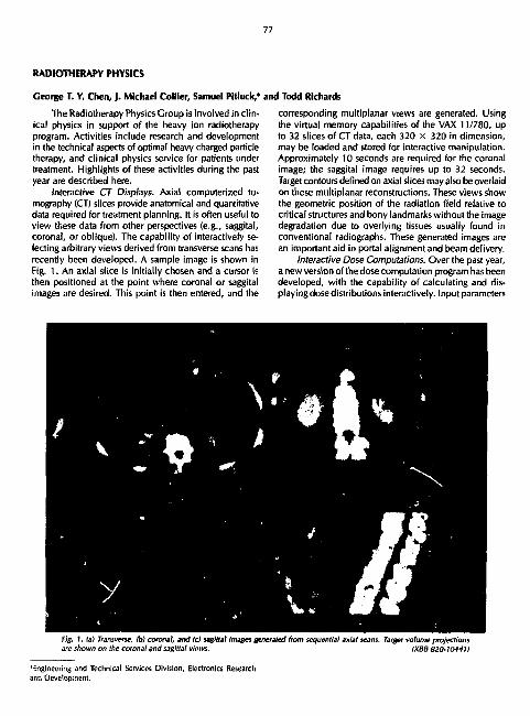
77
RADIOTHERAPY PHYSICS
George T. Y. Chen, |. Michael Collier, Samuel Pitluck,* and Todd Richards
The Radiotherapy Physics Group is involved in clinical physics in support of the heavy ion radiotherapy program. Activities include research and development in the technical aspects of optimal heavy charged particle therapy, and clinical physics service for patients under treatment. Highlights of these activities during the past year are described here.
Interactive CT Displays. Axial computerized tomography (CT) slices provide anatomical and quantitative data required for treatment planning. It is often useful to view these data from other perspectives (e.g., saggital, coronal, or oblique). The capability of interactively selecting arbitrary views derived from transverse scans has recently been developed. A sample image is shown in Fig. 1. An axial slice is initially chosen and a cursor is then positioned at the point where coronal or saggital images are desired. This point is then entered, and the
Engineering and Technical Services Division, Electronics Research and Development.
corresponding multiplanar views are generated. Using the virtual memory capabilities of the VAX 11/780, up to 32 slices of CT data, each 320 x 320 in dimension, may be loaded and stored for interactive manipulation. Approximately 10 seconds are required for the coronal image; the saggital image requires up to 32 seconds. Target contours defined on axial slices may also be overlaid on these multiplanar reconstructions. These views show the geometric position of the radiation field relative to critical structures and bony landmarks without the image degradation due to overlying tissues usually found in conventional radiographs. These generated images are an important aid in portal alignment and beam delivery.
Interactive Dose Computations. Over the past year, a new version of the dose computation program has been developed, with the capability of calculating and displaying dose distributions interactively. Input parameters
fig. 1. la) Transverse, tb) coronal, and tc) sagittal images generated from sequential axial scans. Target volume projections are shown on the coronal and sagittal views. IXBB 820-10441)

78
for each beam include ion type, field weight, angle, size and position, block compensators, target contours, etc. A subset of these parameters may be automatically extracted from target contour information (e.g., beam width, spread Bragg peak and field location). Calculation times are approximately 10 seconds per beam. The plan output is displayed via colored isodose lines overlaid on the CT image, as shown in Fig. 2. Point dose readouts may be obtained interactively through use of the cursor. Single field dose distributions are easily obtained to examine each field's contribution to the composite treatment plan.
Dose Volume Histograms. Isodose distributions on single or selected CT levels yield partial information on dose to structures. Frequently, dose gradients exist across a specific organ of interest. For example, as shown in Fig. 2, the dose to the left kidney ranges from 100% to 0%, depending on sampling location. Furthermore, dose distributions at one level give no information on the irradiated kidney volume at other levels. We have been investigating the use of dose volume histograms to evaluate the radiation burden to critical structures and uniformity of dose to the target. A differential dose volume histogram
is generated by tabulating the dose in all volume elements of a three dimensional structure. The differential dose volume histogram for the left kidney is shown in Figure 3a. The ordinate plots the percentage of organ volume, while the abscissa delineates the dose normalized to target dose. This histogram points out that a substantial fraction of the left kidney is irradiated to approximately 50% of the tumor dose. An integral form of the dose volume histogram is shown in Fig. 3b. The ordinate plots that fraction of the organ that receives at least the dose found on the abscissa as defined by the curve. In this example, over 75% of the left kidney is irradiated to 25% of the tumor dose, and about 20% of the left kidney volume is irradiated to a dose greater than 50% of the tumor dose. However, summing both right and left kidney dose volume histograms, it is clear that kidney function is not at risk. Figure 3c shows the differential dose volume histogram for the target volume. The target is virtually entirely irradiated to the desired 100% level over its three dimensional volume.
Dosimetry. Research and development efforts are underway to improve clinical dosimetry capabilities. Beam
Fig. 2. Composite treatment plan for a pancreatic tumor (small contour). Pan of the treatment also included the liver ttarge contour). The cone-down volume was treated with compensated anterior and lateral fields. The liver and target field was irradiated with a compensated anterior field. (XBB 820-10443)

79
48
40
32
24
16
8
0
ICO
I 80 3 O > 60
8 40 v a.
20
0
100 80
D ia L
(A)
r I i •••;
H 1 I 1̂ I
20 40 60 80 Dose
100 120 140
f ig. I. (a) Differential dose vofume histogram of the /eft kidney, lb) Integral dose volume histogram of the left kidney. A given point on the curve indicates the fractional volume of left kidney receiving more than the corresponding fraction of target dose. Ic) Differential dose volume histogram of target, indicating uniformity of dose received by the target volume in three dimensions. tXBL 8211-4265)
characteristics at the 184-Inch Cyclotron make three-dimensional ion chamber measurements feasible and practical, and efforts are underway to computerize this task. We believe that nuclear track detectors at the Bevalac, as developed by Dr. Eugene Benton of University of San Francisco, are an elegant technique i'or clinical dosimetry that yields both dose and radiation quality data. Collaborative efforts to develop these detectors are underway. In the interim, film dosimetry studies have been conducted to establish the precision with which chatged-particle beams may be shaped under clinical conditions. Figure
Fig, 4. fa) Experimental setup includes the phantom (with circular inserts of inhomogeneities) compensator {above phantom) intended to stop beam along a straight horizontal tine and film dosimeter showing actual slopping region (boundary between dark and light regions downstreamofphantom). (b)Catculatedstoppingdistribulionanddose distribution of the beam when compensator is aligned.
la: XB8 820-10442) (b: XBB 820-10440)
4 is a photograph of a phantom used in preliminary dosimetry studies. The phantom is constructed or Rando type material, and includes a curved entrance surface, air, compact bone and muscle inserts. The objective of this study is to determine with what accuracy a charged

60
Fig. 5. fa) Calculated dose distribution if compensator is misaligned by 3 mm. (bl Film data showing actual pattern of stopping beam tboundary between light and dark region). Note qualitative similarity os predicted and calculated beam penetration, (a: XBB 620-10439)
lb: XBB 820-10444)
particle beam penetration can be calculated and controlled in the presence of inhomogeneities. A rectangular target volume has been defined as shown in Fig. 4b. The beam enters from above the phantom in Fig. 4a. Ideally, for th.s target volume, the beam should be stopped along a straight horizontal line if the appropriate compensating bolus is constructed and aligned. Fig. 4a also shows a photographic film exposed on edge behind the phantom. The boundary between the darkened film and the lighter area defines the stopping region of the beam. These data show that the beam does not stop along a straight horizontal line. Variations of approximately 4 mm are seen in the region of the air/bone shadow. These variations are an approximate measure of the accuracy with which compensation can be used to control penetration. This particular experimental setup is one of the more stringent situations encountered clinically. The partial alignment
of an air cavity and dense bone of 2.5 cm thickness is infrequent. Thus this test is of a rather extreme case. Figure 5a shows the corresponding calculated dose distribution if a 3-mm compensator misregistration is intentionally entered. Figure 5b shows the experimentally measured stopping region under these circumstances. Agreement between calculation and experiment are within 2 mm. Film, although fast and having high spatial resolution, is sensitive to LET variations of the beam and to particle tunnelling, and is a measure of the stopping distribution of charged particle beams rather than a measure of dose. Other measurements in the abdomen suggest that charged particle beams may be stopped with confidence to within 2-3 mm of the desired point.
HEAVY-ION RADIOGRAPHY
lacob I. Fabrikant, Cornelius A. Tobias, William R. Holley, Eugene V. Benton, Kay H. Woodruff, and Eric W. MacFarland
High energy, heavy-ion beams offer superior discrimination of tissue electron densities at very low radiation doses. This characteristic has potential for diagnostic medical imaging of neoplasms arising in the soft tissues and organs because it can detect smaller inhomogeneities than x rays. Heavy-ion imaging may also increase the accuracy of cancer radiotherapy planning involving use
of accelerated charged particles. In the current physics research program of passive heavy-ion imaging, critical modulation transfer function tests are being carried out in heavy-ion projection radiography and heavy-ion computerized tomography. The research goal is to improve the heavy-ton imaging method until it reaches the limits of its theoretical resolution dc-iined by range straggling,

81
multiple scattering, and other factors involved in the beam quality characteristics. Clinical uses of the imaging method include the application of heavy-ion computerized tomography to heavy-ion radiotherapy planning, to the study of brain tumors and other structures of the head, and to low-dose heavy-ion projection mammography, particularly for women with dense breasts where other methods of diagnosis fail. The ions used are primarily 300 to 570 MeV/amu carbon and neon ions accelerated at the Lawrence Berkeley Laboratory Bevalac.
PASSIVE HEAVY-ION RADIOGRAPHY
The geometry used for heavy-ion radiography is shown in Fig. 1. The external beam channel is long; the beam is brought to a focus near the entrance of the room and allowed to expand until it is about 40 cm wide. If it is assumed that scattering is negligible, then the extent of deviation at the detector is smaller than 0.2 mm for any location in the patient's tissues. If the beam is moderated by an interposed absorber in order to put the stopping points of the particles to a desired depth in the detector, then this value is increased due to multiple elastic scattering. The best position of the attenuator is at the focal spot (beam focus), and the optics of the beam remain essentially unchanged if a relatively thin absorber is inserted there. We have designed a binary filter for range adjustments in radiography that is placed adjacent to the focal spot. A special device, the particle fluence propeller, is also placed into the beam; it makes the particle fluence uniform in the cross section of the beam. At the expense of beam intensity, it is possible to reduce the focal spot size further by an interposed aperture.
These principles are being applied for better diagnosis of soft tissue tumors. In heavy-ion mammography, for
GEOMETRIC CONSIDERATIONS IN HEAVV ION RADIOGRAPHY
BfOlf Poiltlon of OtttCtOf locui olltnualor tlgeh
Fig. I. Geometry of the heavy particle beams for diagnostic Imaging. The beam is brought to a focus of less than I cm at A. The object-focus distance, a. is about 15 m. The detector stacfc is in immediate contact with the object, or with a water bath surrounding it, so that the object detector distance is 0 to 40 cm. A stack of attenuators that control the range are placed at distance X. (XBl 829-40671.
example, the mean electron density of normal breast tissue is about 0.98 of that of water, adipose tissue has an electron density of 0.91 to 0.96, and tumor densities range from 1.02 to about 1.06. Heavy-ions demonstrate these densities with ample contrast; however, often a dense region for heavy-ions is undetectable with x rays at diagnostic dose levels. Both heavy io"- and x rays have characteristic blind regions in wh... i water and tissues of varying composition cannot be readily distinguished on the image. Unfortunately, these regions often conceal tumors from diagnosis. Figure 2 illustrates the additivity of electron densities and of attenuation coefficients for different components of tissue. It is assumed that tissue consists of three components: water (w, I = 33%), adipose components (1, II = 33%), and supporting connective and parenchymal tissues (s, III = 33%) containing denser protein and structural components. Generally, the heavy-ion number (T) and the Hounsfield number (H) are different from each other and approach equality only if very high energy x rays are compared to heavy ions. Figure 2 (upper panels) illustrates the relationship of the contribution of components of electronic stopping power to total stopping for heavy ions and the contribution of partial attenuation to total attenuation for x rays. The middle panels '"'istrate the blind spot for x
Fig. 2. Illustration of the existence of blind regions for x rays and for heavy ions at certain critical tissue compositions {see text for details).

82
rays that occurs at a particular ratio of the three components of tissue. However, at the x-ray blind spot, the heavy ions detect a difference from water. Finally, the lower panels indicate the location of the blind spot for heavy-ions; however, the x-ray attenuation per electron is different from that of water. We believe that combined use of x rays and of heavy ions might be quite important for the early detection of cancer.
The graphs (Fig. 2) illustrate the existence of blind regions for x rays and for heavy ions at certain critical tissue compositions. It is assumed that tissue is made up of three components: water (I), adipose components (II) and protein structural components (III). Upper panels: It is assumed that the arbitrary tissue composition I = II = III = 33%. in the left panel, the rate of energy loss is different in each component, so that the range ratio ICR. < 1, where R is the residual range of heavy ions in water (w) and tissue (t). In the right panel, for the same composition, the attenuation ratio A,/Am > 1, where A is the x-ray attenuation coefficient per electron in tissue (t) and water (w), and | i is the x-ray absorption coefficient in tissue (t) and water (w). Middle Panels: It is assumed that here the arbitrary composition of tissue components is I = 33%; II = 50%; III = 16%. In the left panel RJ R, = 1, so that heavy ions detect a difference. In the right panel, the attenuation in tissue is the same as in water A,/A„ = 1 so this is a blind region for x rays, L ver Panels: Here, I = 33%; II = 24%; III = 42%. I: :he left panel, RJR, = I, the range of a heavy partK ;_• is the same in tissue as in water, so this is a blind ^ion for heavy particles. In the right panel, A,/Aw = , so x rays can detect a difference. In actual tissues, <e concentration of water is much higher and th observed ratios are nearer to unity than shown ht
HEAVY-ION MAMMOGRAPHY
Soon after the initial techniques of examination of the female breast by x-ray mammography had been developed, it became apparent that normal individuals presented with differing parenchymal patterns. By 1967, attention was drawn to the association between the parenchymal pattern of the breast on x-ray mammography and the presence of carcinoma of the Lreast. Subsequently, the incidence of the development of carcinoma in the breast with various parenchymal patterns was studied. Specification of the parenchymal pattern has potential use in limited situations to identify the risk category of well women with respect to the future development of carcinoma. Until recently, only little attention has been paid to the differing parenchymal patterns demonstrated by x-ray mammography, and the limitations of conventional x-ray mammography in the assessment of differing patterns in association with early neoplastic change.
Since 1976, all heavy-ion mammograms carried out at the Lawrence Berkeley Laboratory have been classified using a system similar to that used for conventional x-ray mammography. Various associations between the parenchymal patterns and other factors are being identified. The value of the identification of the parenchymal pattern in the mammogram interpretation are being examined, and the variations in heavy-ion density distributions are being quantified. The useof carbon and neon ions for heavy-ion mammography has now been evaluated in 39 patients. In three of these patients, heavy-ions detected dense tissue regions not seen by x-ray mammography that were later proven to be cancer.
The heavy-ion technique appears to be particularly of value in patients with dense breasts; 20 millirad of carbon particles are sufficient to produce high contrast images of diagnostic quality of the human breast. It is important, however, to use optimal image reconstruction and display techniques. In addition to displaying isodensity contours, it is useful to display density gradient images and to prepare density distribution spectra. As these analyses and display techniques improve, heavy-ion mammography will be performed on patients with dense breasts where other diagnostic methods have failed to give an unambiguous diagnosis. Quantitative electron density comparisons are made between left and right breasts.
Stopping Power Density Distribution Histogram Analyses
The stopping power density distributions determined as a consequence of range distributions are used in heavy-ion mammography in a visual display so that diagnostic information can be derived from them. However, it is possible to carry out statistical analyses of the density distribution patterns for mathematical analysis of the internal topography of breast tissues. The distribution of normal density patterns between left and right breasts of the same individual exhibit a degree of symmetry, while these patterns are markedly different from one individual to the next. For example, abnormal densities due to cysts often appear most clearly when deviations between density patterns of the abnormal and the normal breast are studied.
The quantitative nature of heavy-ion range data provides opportunities for further image processing. In each heavy-ion mammographic examination, a grayscale image, electron isodensity contour map, and electron density gradient map heavy-ion number frequency distribution histogram of each breast is created. Each of these data formats provides a dilferent representation of the electron density data for quantitative analyses. The gray scale mammographic image (Fig. 3a, upper left) is produced by assigning decreasing tones of gray to increasing electron density. The average electron density at each pixel is displayed »s its corresponding gray tone. In this image,

83
ISCOttJITY CIWTIM HOT
^3 ^ % / ^ ^ ^ ^ ^ ^
wwictM "tea ^ ^ w ^ ^ ^ ^ ^ ' f a a
HCMMCM MMEI "'\7'^ • • .11,00 t • -to.oa 0 • • ).Q0 [ • tlO.OO f • •H.OO G • tM.OO H • 00 .00
//S- 3a. Computer-synthesized carborhhn {250 MeV/amu) mammogram . of left breast of patient with breast cancer; this Is the gray-scale representation of electron density changes. A small region of increased density in breast parenchyma was detected on the x ray mammogram, but the nature of the density could not be determined. The a.* of suspected pathological tissue are readily identified on the carbon-ion mammogram. (XBB 8111-10208).
Fig. 3b. tsodensity contour map of the gray-scale carbon-ion mammogram. Isodense regions are outlined and the residual range of the charged particles, after passing through the breast, is identified by letters near each contour. Alow residual range value indicates a region of high tissue electron density. The areas of suspicious pathological breast tissue are noted. IXBL 828-109271.
w w - i m NUWR IX) w»t«
Fig. 3c. Computer-produced electron density gradient map derived from stopping-power values in the carbon-ion mammogram. This is a gray-scale image representation that enhances contour borders and quantifies electron density distribution of normal and abnormal regions. IXBB 826-5179).
i tu t t l Fig. 3d. Stopping-power frequency-distribution histogram of heavy-ion (rl numbers derived from the computer-synthesized carbon ion mammogram. Normal and abnormal breast tissues appear as separate peaks with different ranges of heavy-ion (ij numbers.
IXBL 8111-43351
electron-dense tissue near the center of the breast is abnormal and is the site of suspected pathological (e.g., cancerous) breast tissue; it appears light relative to the surrounding normal tissue. On the other hand, normal breast tissue, due to its lipid content and structure, has an electron density and heavy-ion number (T) lower than water. The gray scale image is displayed on a television monitor where the contrast level and window may be altered for diagnostic interpretation (assessment of characteristic parenchymal patterns of normal and pathological breast tissues).
Figure 3b, (upper right) is the electron isodensity contour map of outlining regions of constant average electron density. In patients where the contrast of the
gray scale image does not reliably delineate detailed structure, e.g., small tumors hidden in dense tissue, this quantitative representation can be of value. This representation provides characteristic radiological patterns and quantitative information for the density values along each contour. The isodensity contour map can be compared with the gray scale image confirming borders and quantifying the electron density of abnormal regions. Values of normal and abnormal electron density can be obtained from measured stopping power values. The stopping power values of normal and neoplastic tissues derived from digitized heavy-ion radiographs of pathologic specimens demonstrate the potential for diagnosis of benign and malignant neoplasms of the breast by diagnostic

84
imaging and accurate quantitative densitometry of the stopping power distribution in the breast tissue. From studies on formalin-fixed mastectomy specimens using heavy-ion radiography, the calculated stopping power values relative to wuier were: normal breast tissue, 0.957 ± 0.004; mammary dysplasia, 1.016 ± 0.011; and infiltrating ductal carcinoma, 1.064 ± 0.003. The large difference in stopping power between mammary dysplasia and carcinoma may lead to a quantitative method for differentiating between benign disease and breast cancer using heavy-ion radiography. Furthermore, improved depth resolution may be specific-tissue heavy-ion dependent and may prove to be more accurate with heavier ions, e.g., neon and argon, that have improved resolution characteristics.
As a further aid in delineating the boundaries of tissue structures, an electron density gradient map is produced (Fig. 3c, lower left). Here, the absolute value of the first derivative of the electron density w ith respect to position is plotted. Rapid changes in electron density values at boundaries are readily displayed and identified.
The frequency of occurrence of specific heavy-ion numbers (T) may be recorded on a distribution histogram
Cancer patients are now being routinely treated with energetic heavy charged particles. If the energy of the particles is properly determined during treatment planning, it is possible to deliver a maximal dose lo the treatment volume while sparing the adjacent normal cells or vital organs. At present, treatment ptenn'tng is accomplished by using a CAT scanner. In this technique there can be an error of 2 to 5 mm in localizing the Bragg peak, depending upon certain therapeutic situations. The objective of the pk'sent research is to obtain a better accuracy than provided by the CAT scanning method. Such an accuracy can be attained by using high energy beams of radioactive nuclei that decay by positron emission at the stopping point of the beam. Hence by detecting the positron-annihilated 7-rays in coincidence mode (using a suitable positron camera), the range-energy relationship in a heterogeneous object, such as in a patient, can be determined with 1-mm accuracy. The purpose of the proposed work is to develop this idea in a practical way and demonstrate its usefulness in the treatment of cancer patients.
• Instrument Siieme Seiliun, Enfcinet'iiriK & lech. Servile* Divisiun
(Fig. 3d, lower right). Because living tissue is heterogenous, precise electron density values have limited meaning. Statistical analysis of the distribution of average density values is presently under investigation; thus far, 28 heavy-ion mammogram breast patient studies have been plotted. The presence of relatively large portions of dense (high T values) tissue compared to the known normal distribution may lead to the identification of areas of pathologic breast tissue. The density values read from the plot can be used for assigning contour values in the isodensity contour map to help identify the tissue regions responsible for the peaks. The low density portions are predominantly due to adipose tissue. High density peaks (T greater than normal) result from high density regions seen in the grayscale image and in the isodensity contour map. Tissue studies have shown the T values of normal breast tissue to be in the range of T = -10 .00 to more than 0.00 and, for carcinomas, in a much higher range of T. In this study, the pathological breast ti-,ue had a heavy-ion number distribution that peaked in the range of about T = 25.00 and, far normal skin, T was greater than 30.00.
High energy radioactive beams are obtained from the Bevalac as secondary particles. Nuclei of stable isotopes are accelerated in the synchrotron and made to undergo nuclear fragmentation through a beryllium block placed inside the accelerator ring. There are many components in the fragmented beam but only one is the desired radioactive beam. This beam can be magnetically filtered, and through proper beam optics it is brought to a focal point in the therapy cave. Through this procedure, we have been able to obtain beams of "C (naif-life: 20 min) and "Ne (half-life: 17 sec) in sufficient flux to proceed with the studies for diagnostic applications.
Several studies involving the physical characteristics of radioactive beam particles have to be completed before the beams can be used in clinical trials. Some of these studies and related measurements are described below.
PHYSICAL MEASUREMENTS
Water-equivalent thickness of a silicon diode. The purpose of this measurement was to estimate the water-equivalent thickness of a silicon diode. Data were taken with both a -"Ne primary beam and a '^Ne secondary beam. From the stopping power theory it is well known
DIAGNOSTIC STUDIES WITH HIGH ENERGV RADIOACTIVE BEAMS
Aloke Chatterjee, William M. Saunders, lanis S. Scherer, Edward L. Alpen, Jorge Llacer,* and George T. V. Chen

85
that the water-equivalent thickness should be independent of the particle charge, mass, or energy.
There are two main reasons for making these measurements. First, the " N e beam is produced from the peripheral nuclear fragmentation collision between the parent beam of :°Ne and a 1-in. thick beryllium target. Thus, "Ne can be produced at any depth in the beryllium target. Also, the secondary fragment " N e can have any velocity according to the Gaussian distribution with the most probable value being the velocity of the parent particles. Hence, the "Ne beam has more energy spread than the parent M N e beam. For the parent beam the energy spread is only 0.1 %. The question we wished to answer was, wil l this spread in energy have any influence on the determination of water-equivalent thickness?
Secondly, silicon diodes wil l be used routinely in the development stage of the radioactive beam diagnostic
10.0 19.0 PENETRATION DISTANCE (cm OF WATER)
. a.n : (b) : 1.00 |
a 1 B0 P 3 * ; g 1.00 : c , • : p l.SD
1 1.00
; 0.BQ ' :
k. ; ***«. 0.00
PENETRATION DISTANCE, (CM OF WATER!
Fig. 1. The Vrags lommion cune lor neon-20: (a) * i* estimated with Ion chmbrrs; 1l>l as eslimarecj with a silicon diode. tXBl 819-7003;
XBl BI9-7O04)
technique. They wil l be used, through ionization measurements, to confirm the Bragg peak localization estimates using PE6A. For ionization measurement with diodes, one has to make a correction to estimate the actual depth of beam stopping by taking into consideration the water-equivalent thickness of a silicon diode. These detectors have the advantage of cheapness, responsiveness, and adaptability for implantation in animal tissues.
In order to determine water-equivalent thickness of the silicon diode, depth vs. dose ionization curves were measured using gas-filled ion-chambers and a variable water absorber. Measurements were then made substituting the diode detector for the ion chamber. The difference in depth at which the Bragg peak occurs in the two cases yields the water-equivalent thickness of the solid state detector.
Various Bragg ionization curves were obtained using both 2 0Ne and collimated "Ne beam. Measurements were made by using ion chambers as well as silicon diodes. The Bragg ionization curve for i 0 Ne at 383 MeV/amu, as estimated with ion chambers, is seen in Fig. la . The Bragg peak occurs at 15.2 cm. For the same beam, when a silicon diode is used, the peak in ionization occurs at 14.94 cm as seen from Fig. l b . The Bragg ionization curve for the z 0 Ne beam is quite sharp, indicating a very small spread in energy for the primary 2 a Ne beam. The difference between 15.2 cm and 14.94 cm is 0.26 cm, which is the water-equivalent thickness of the silicon diode as measured with M N e .
Similar Bragg curves were obtained with the s.c-ondary "Ne beam and the results are shown in Figs. 2a and b. When only ion chambers are used, the Bragg peak appears at 10.23 cm in contrast with the silicon diode measurement, for which the peak was at 9.90 cm. The difference between these two readings is 0.33 cm, the water-equivalent thickness of the silicon diode as measured with the "Ne beam. Thus, we find that there is a difference of 0.07 cm between the measurements made with primary *°Ne and the secondary " N e beam. It is well known that the water-equivalent thickness should be independent of the particle charge or energy. We attribute this difference to a rather large spread in beam energy of the " N e ions compared to the Bfvalac primary particles, such as in the i 0 N e beam.
Although there is a difference, the usefulness of the high energy radioactive beam wil l in no way diminish because the accuracy desired in most therapeutic situations is of the order of 0.1 cm.
A comparison of water equivalent thickness measurements. Silicon diodes were- implanted in the brain, neck, thorax, and lower and upper abdomen of a frozen dog. In each of the regions, water-equivalent thicknesses, were measured by a computerized axial tomography method. With various heavy particle beams RHe, 'JC,
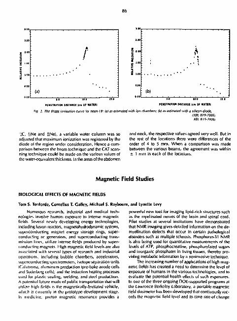
86
' : 2.10 ;
| -
i : : •
• 4
••1
0.10
(b) :
0.00
PENETRATION OIITANCE (cm OF WATER) PENETRATION OIITANCE (cm OF WATER)
Fig. 2. The Bragg ionization curve for neon-19: (ai as estimated with ion chambers; lb) as estimated with a silicon diode. IXBL 819-7005; XBL 319-7006)
'JC, ffiNe and !2Ne), a variable water column was so adjusted that maximum ionization was registered by the diode of the region under consideration. Hence a comparison between the beam technique and the CAT scanning technique could be made on the various values of the water-equivalent thickness. In the areas of the abdomen
and neck, the respective values agreed very well. But in the rest of the locations there were differences of the order of 4 to 5 mm. When a comparison was made between the various beams, the agreement was within ± 1 mm in each of the locations.
Magnetic Field Studies
BIOLOGICAL EFFECTS OF MAGNETIC FIELDS
Tom S. Tenforde, Cornelius T. Gaffcy, Michael S. Raybourn
Numerous research, industrial and medical technologies involve human exposure to intense magnetic fields. Several newly developing energy technologies, including fusion reactors, magnetohydrodynamic systems, superconducting magnet energy storage rings, superconducting ac generators, and superconducting transmission lines, utilize intense fields produced by superconducting magnets. High magnetic field levels are also associated with several types of research and industrial operations, including bubble chambers, accelerators, superconducting spectrometers, isotope separation units iCalutrons), aluminum production Ipre-bake anode cells and Soderberg cells), and the induction heating processes used for plastic sealing, welding, and steel production. A potential future mode of public transportation that wil l utilize high fields is the magnetically-levitated vehicle, which is currently in the prototype development stage. In medicine, proton magnetic resonance provides a
i, and Lynette Levy
powerful new tool for imaging lipid-rich structures such as the myelinated nerves of the brain and spinal cord. Pilot studies at several institutions have demonstrated that NMR imaging gives detailed information on the de-myelination defects that occur in certain pathological disorders such as multiple sclerosis. Phosphorus-31 NMR is also being used for quantitative measurements of the levels of ATP, phosphocreatine, phosphorylated sugars and inorganic phosphates in living tissues, thereby providing metabolic information by a noninvasive technique.
The increasing number of applications of high magnetic fields has created a need to determine the level of exposure of humans in the various technologies, and to evaluate the potential health effects of such exposures. In one of the three ongoing DOE-supported programs at the Lawrence Berkeley Laboratory, a portable magnetic field dosimeter has been developed that continously records the magnetic field level and its time rate of change

87
for periods of up to 8 hours (co-principal investigators: F. S. Coulding and T. S. Tenforde). This microprocessor-controlled dosimeter, the final prototype of which is approximately the size of a cigarette package and contains 4096 12-bit words of memory, has been used to monitor the magnetic field levels to which operators are exposed at se'.eral accelerator facilities in Berkeley, Stanford and Davis, California, and at the Kaiser Aluminum Test Facility in Fermanente, California. The highest field levels observed to date were recorded near a large aperture superconducting solenoid (LASS) at the Stanford Linear Accelerator. Fields up to 5700 gauss, with time rates of change approaching 2700 gauss/sec, were recorded at operator locations in the LASS facility. The results of these dosimeter field tests are providing useful data for a second LBL magnetic field program in which a retrospective epidemiological survey is being conducted using the health records of workers routinely exposed to magnetic fields at various national laboratories (principal investigator: T.F. Budinger). This study, in which medical data has been collected on approximately 600 occupationally-exposed subjects and a matched control group, is currently in the final stages of data analysis.
A third LBL magnetic field program has been focused on the analysis of magnetic field effects on physiological functions in experimental animals and selected organ and tissue systems (principal investigator: T.S. Tenforde). A major research effort has used electrical recording techniques to detect functional alterations in the cardiovascular, neural, and visual systems during the application of stationary magnetic fields. These systems involve ionic conduction processes and are therefore potentially sensitive to electrodynamic interactions with an applied magnetic field. In the case of the visual system, magnetic interactions could also arise through orientational effects on the magnetically anisotropic photopigment molecules within retinal photoreceptor ceils. The following paragraphs provide a brief summary of results obtained during the past year in selected research areas within the magnetic field bioeffecls program.
Cardiovascular Studies. Previous studies in our laboratory have demonstrated that large electrical potentials (on the order of millivolts) are induced in the central circulatory system of animals during exposure to strong stationary magnetic fields. These potentials originate through an interaction of the field with electrolytes in the flowing blood, and their magnitude has been demonstrated to be a linear function of the magnetic field strength. Recent studies with primates (baboons and monkeys) have been directed toward determining whether these magnetically-induced polentialsexert a perturbing influence on major cardiovascular functions. Measurements of several indices of cardiac performance, including heart rate, heart muscle bioelectric activity, and intraarterial blood pressure, have not demonstrated any ab
normality associated with the exposure of primates to magnetic fields up to 15 kilogauss intensity. The absence of measurable hemodynamic perturbations is consistent with theoretical predictions, which indicate that the magnetohydrodynamic interaction with blood flow is extremely weak in fields of this magnitude.
Retinal Blectrophysiology. The occurrence of visual phosphenes during exposure of human to time-varying magnetic fields has been studied in several laboratories since this phenomenon was first observed in the latter part of the nineteenth century. Although stationary magnetic fields have not been found to elicit phosphenes in human subjects, previous work in our laboratory using in vitro turtle retina preparations has demonstrated that stationary fields can alter the electrical activity recorded from the retina subsequent to photon absorption. This effect has been shown to occur only during the light-to-dark transition phase, when the retinal photoreceptor cells are undergoing rapid shifts in their physiological and metabolic activities. Research in this area during the past year has further documented the diurns' dependence of the retinal response to magnetic fields. U ,,as also been demonstrated that the principal effect of the magnetic field interaction with retinal photoreceptors is a reversible suppression of synaptically-mediated electrical activity, without an associated change in the state of visual adaptation to photic stimuli.
Animal Physiology Studies. General physiological functions and their circadian oscillations are being studied in rodents subjected to prolonged exposures to stationary magnetic fields with intensities up to 15 kilogauss. By using noninvasive transducer techniques, it is possible to continuously monitor physiological variables such as core body temperature, heart rate, respiration, body mass, food intake and locomotor activity. The circadian waveforms of these physiological and behavioral parameters hsve been shown in previous studies to provide a sensitive measure of the stressor effects imposed by a variety of chemical (e.g., drugs) and physical (e.g., light, sound, heat, and radiation) agents. Techniques have been developed in our program for noninvasively monitoring each of the parameters named above, and representative experimental results are illustrated here from measurements of the circadian waveform in core body temperature.
The abdominal temperatures of mice have been continuously recorded for periods as long as 80 days using implanted FM radiotelemeters. These small silicone-coated radiotransmitters record the abdominal temperature by means of a miniature thermistor element, and transmit this information as a continuous stream of pulse-interval-modulated radiowaves in the FM band (88-108MHz). The FM signals are received by an antenna surrounding the rodent cage, from which they are transmitted to a receiver-demodulator unit that decodes the data to produce an analog voltage output. By appropriate calibration,

88
-K CHANNEL H I - 2
AVERAGE -38 381
A M P L I T U D E - 1.343*0.210 ACROPHA5E-22 3 9 3 * 0 . 9 1 9
DAY 3S4 CHANNEL H I - 2
AVERAGE ^35 092 AMPLITUDE- 0 974*0.199 ACR0PHASE-32. 643*0.994
CHANNEL H I - 2 AVERAGE -38 34* AMPLITUDE- 0.903*0.1 ACR0PHASE-23.58*0 .183
DAY 356 CHANNEL III - 2
AVERAGE -SB 587 AMPLITUDE- 1 113*0 122 ACRCXHASE-33 940*0 370
OAT 357 CHANNEL III - 2
AVERAGE -ae.oea AMPLITUDE- 1.349*0. I ACROP*AS£-Z2.7IO*0.<
i-r-T[M[ (MH)
MQUSl TEMfEHATjfiL yfl| ft^.trWITa
sen.t/ d,tyt> ot < ««' body temperature data measureti by telemetry ,vn plotted for an adult female LAFn'i mouse, in rhythm of this animal was entrained on a 12-hr tight/12-dark illumination schedule. The smooth curves represent •inadu'i iv.ii'fJarm hvr Fifi. 2) And the 9(/5'.Vi lonlidcnce limit,. (XBL 626-W537A)
the* voltage output can be scaled to have a linear one-to-one correspondence with body temperture over the range J4-4UT.
An example of data obtained by the telemetry technique r ithuwn in Fig. 1, in which five serial days of recorded tore body temperatures are presented for a mouse entrained on a 12-hr light/ 12-hr dark cycle. It is
evidu -. that the core body temperature exhib'b a strong arcadian periodicity with a peak-to-trough variation of approximately 2°C. In order to quantitatively describe the circadian waveform, the temperature data are fit by computer to a single harmonic function with four adjustable parameters, namely, the mean temperature, and the amplitude, pha*e, and period of the circadian os-

89
TIME SERIES ANALYSIS
T(t[)= T0 + f cos(uti + + )
T0 = level
T = amplitude w - angular frequency
= 3 6 0 ° / T where T = period
•p = acrophase
00:00 06:00 12:00 18:00 24:00 Clock hour
Fig. 2. Schematic representation of the circadian oscillation in deep-body temperature of a rodent entrained on a J 2-hr light/12-hr dark schedule. The data are fit by computer to a cosine curve using the time series analysis technique developed by Halberg et at.' The terminology for the parameters defining the circadian waveforms is the same as that used by Halberg. For circadian rhythms with a 24-hr periodicity, the level is identical to the daily average of the body temperature. The acrriphase is defined as the clock hour at which the temperature reaches its maximum value. 1XBL 826-3901)
dilation (Fig. 2). Three of these parameters (the mean temperature, amplitude, and phase) are fit by regression analysis, and the period is obtained by an iterative technique that minimizes the variance between the experimental data and the best-fit circadian waveform. For rodents entrained on a 12-hr light/12-hr dark cycle, the period of the circadian oscillation in core body temperature is consistently 24 hr, as shown by the period spectra in Fig. 3.
In order to apply telemetry measurements of this nature to the analysis of potential stressor effects resulting from prolonged exposure to high magnetic fields, it was necessary to fabricate transmitters containing no ferromagnetic components. Using these custom-made telemetry transmitters, measurements have been made of the circadian waveforms of It) rodents subjected to either continuous or intermittent exposures to a 15 kilogauss
T iVE SERIES PEBOO 5"ECTPf l
Fig. 3. tvriodograms generated from a time series analysis of the circadian oscillations in deep-body temperature are shown for six temate lA f , / I mice. The maximum amplitude in the temperature circadian rh) '••n occurs when the period is assigned a value of 24 * 0.25hr. With this choice of period, the variance betiveen the experimental data and the bef fit cosine curve is minimized. IXBL 82f>-39071

90
EXPERIMENT I
MEAN BODY TEMPERATURE
22
h°*H -' 1 1 1 1
' * t { i i
; , -15 -10 - 5 0 5 10 15
•
• * . f t
-, . , . , -15 -10 - 5 0 5 10 IS
r
i •
1
, 1 | 1 -IS -10 - 5 0 5 10 15
. I
•
1
* 1
; f . -10 10
EXPERIMENT n
-15 10 Days Days
Fig. 4. Parameters describing the core body temperature circadian waveform are plotted as a function of time during two telemetry experiments of 23 and 31 days duration, respectively. In each experiment, groups of three female LAF,/J mice were monitored before, during and after a 5-day continuous exposure to a 15 kilogauss magnetic field. The points represent mean values ± 1 SO. of the circadian parameters determined by a time series analysis of temperature data for consecutive 5'day intervals during the course of each experiment. An analysis of variance of the four circadian parameters revealed no statistically significant changes associated with the mdgnetic field exposure. (XBL 8212-4271)
3 8 -' 1 1 1 1 -4
37 36 \\ i i * £ i : 35 34 : , ( 35 34
-15 -10 - 5 0 5 10 15
1
0 :{ f i • • f " 1
0 -15 -10 - 5 0 5 10 15
02:00 1 1 1 1 1 : 22:00 : » * - * " « : 18-00 -
-15 -10 5 0 5 10 15
2 5 - ' ' 1 ' . 2 4 • • i I -2 3 _ i
i , "
stationary magnetic field. Parameters describing the circadian waveforms of rodents exposed for a continuous 5-day period to a 15 kilogauss field are shown in Fig. 4. An analysis of variance revealed no significant changes in the average body temperature or the amplitude, phase, and period of the circadian waveform during the preexposure, exposure, and post-exposure intervals. Similar results have been obtained with rodents exposed to a 15 kilogauss field in an 8-hr on/16-hr off exposure regiment for 10 consecutive days. As exemplified by these data, the results of telemetry experiments conducted to date have indicated that general physiological variables such as core body temperature are not significantly perturbed by exposure to stationary magnetic fields with intensities greater than the levels to which humans are routinely exposed.
Hematology and Immunology. In order to evaluate potential effects of high magnetic fields on the hematological profiles of experimental animals, complete blood cell counts and chemical analyses of serum proteins, lipids, electrolytes, and nonelectrolytes have been performed on blood samples from animals exposed to a 15
kilogauss stationary magnetic field. The results of these tests have shown no abnormalities in the blood composition of lodents exposed to the field for periods up to 10 days, and similar results have been obtained for primates (baboons and monkeys) subjected to acute exposures of several hours duration.
Collaborative studies have been initiated during the past year with M. Shifrine of the Laboratory for Energy Related Healin Research at UC Davis to evaluate the immunocompetenceof mice following chronic exposure to a 15 kilogauss stationary magnetic field. These studies are still in a preliminary stage, but initial results indicate that continous exposure to a 15 kilogauss field for 6 days does not significantly alter the level of the immune response elicited by a foreign antigen challenge (sheep red blood cells). Similarly, the response of spleen lymphocytes to mitogen stimulation is not altered in the exposed animals relative to matched controls.
REFERENCES
Halberg, et al. Physiology Teacher / (4): 1-11 (1972).

91
Biophysical Studies
FUNCTIONAL ANALYSIS OF LASER-INDUCED OCULAR DAMAGE
Michael S. Raybourn and Robert L. Kong
The goal of our research efforts has been twofold: 1) the reassessment of laser damage thresholds to retinal tissues using functional endpoints, and 2) the examination of laser damage mechanisms in retinal tissues. Our experimental approach has been to study the effects of different wavelength lines of laser light on retinal function as determined by monitoring the following electrophysiological signals: electroretinogram (ERG), early receptor potential (ERP), and intracellularly-measured light evoked responses in an in vitro retinal eyecup preparation.'•'
In the past year we have examined both short wavelength (helium-cadmium, 441 nm) and long wavelength (helium-neon, 632 nm) visible light, as well as near infrared (gallium arsenide diode laser, 904 nm) light effects in order to distinguish between photochemical and thermal damage mechanisms. Although experiments with the gallium arsenide laser are still in progress, our results to date are as follows:
1. Helium-neon (HeNe) laser exposures of up to 290 joules/cm2 average power (80 mW/or f retinal ir-radiance, 60-min exposure duration, 1-mm spot diameter) showed no significant effects on either ERG b-wave amplitude or waveform. Moreover, no effects were seen in the dynamic capacity of the retina to undergo visual adaptation as assessed by the intensity-response function analysis.2
2. Gallium arsenide (GaAs) exposures of up to 54 joules/cm2 (pulse repetition frequency = 1.7-23 kHz, 200-nsec pulse duration, 60 mW/cm 2 retinal irradiance, 15-min exposure duration, 0.02-cm 2 laser image area) also showed no effects at the level of the ERG b-wave.
3. Helium-cadmium (HeCd) laser exposures of 260 joules/cm2 (290 mW/cm 2 retinal irradiance, 15-min exposure duration, 1.5-mm spot diameter) resulted in both histological and electroph/siological effects on the retina. Light microscopic pictures from both laser irradiated and nonirradiated (control) regions of the same retina are presented in Fig. 1C and 1D. (These data were obtained in collaboration with Dr. William Spencer, Pacific Medical Center, San Francisco, California.) The lased portion of the retina shows clear signs of severe disruption in the photoreceptors and melanin particle-containing pigment epithelium. ERG measurements obtained before and following HeCd laser irr igation are also shown in Fig. 1
in the form of intensity-response curves. The ERG response amplitudes at the lased sites are diminished at all test stimulus intensities following laser irradiation. Intensity-response curves for the nonirradiated (control) region of the same retina, also shown in Figs 1A and 1B, by comparison show little effect.
Our finding that short wavelength laser light (441 nm) elicited changes in retinal function while longwave-length laser light (632 nm) of comparable energy had no effect(s), supports the photochemical damage theory of laser-retinal interactions at these exposure levels. The maximum output of the GaAs diode laser (at room temperature) was too low to test this at even longer wavelengths. Efforts are now underway to attempt to increase the output energy of this laser device using cryogenic cooling of the diode junction.
Our results showing that severe, localized morphological damage to photoreceptors and pigment epithelium is accompanied by only moderate alterations in the ERG b-wave amplitudes raises doubts about the sensitivity of the ERG as a tool for assessing retinal dysfunction in small areas. The histological evidence suggests that the actual spatial extent of the damage site may comprise only a small fraction of the entire laser-irradiated area of the retina. Since the ERG samples spatially summed the response of the entire irradiated area in our experimental paradigm, it may not be sufficiently localized to reflect the more spatially limited extent of the affected area. In order to circumvent this problem, we are currently designing experiments In which we will monitor the effects of laser irradiation on individual retinal neurons by means of intracellular recording techniques. This experimental approach should provide an even more sensitive and direct means of assessing laser effects at the cellular level.
REFERENCES
1. Raybourn, M.S. and Kong, R.L. A Functional Approach to the Ocular Hazards of Lasers. LBL-13428, October 1981.
2. Raybourn, M. S., Kong, R. L. and Been, D. O. A Functional Approach to the Assessment of Ocular Hazards of Lasers. In Biology and Medicine Division Annual Report 1980-1981, LBL-13501, April 1982, pp. 116-119.

92
J 0.50
0 1.00
F>g. I. Electrophysiological and histological effects of photochemical (i.e., nonthermal) damage by a helium-cadmium (HeCd, 44 J r-n) laser irradiation of the vertebrate retina (Pseudemys scripta elegans}. Retinal response (V/V^,) was monitored at the times indicated in A and B by recording k.^roelectrodes at a control site and at the laser irradiated site. The retinal irradiance of the HeCd laser beam (1.5 mm diameter) was 290 mW/cm' and the exposure duration was 15 min. |A) Intensity-response (l-R) function for the ERG b-wave obtained from the control site. IB) l-R function for the laser irradiated retinal site located 5 mm from the control site. The post-irradiation curves at
10 and 60 min show a clear and long lasting depression of retinal response. (C) Light micrograph of an unirradiated (control) retinal site. ID) Light micrograph of the lased retinal site demonstrating extensive disruption of the photoreceptors and pigment
epithelium. (X6B 820-892 7A)
FUNDAMENTAL AND APPLIED STUDIES ON CELL-MEMBRANE SYSTEMS
Howard C. Mel, Stephen P. Akeson, Gary Richieri, and Frank Kooi
Our laboratory research this year falls into two categories: an ongoing physical and technical study of the multipurpose experimental apparatus and methodology called resitivite pulse spectroscopy (RPS), and the application of RPS to problems uf interest in both basic membrane biophysics and clinical hematology.
PHYSICAL-TECHNICAL STUDIES
As noted previously, RPS is an outgrowth of traditional "Coulter-type" electronic cell sizing. Last year we reported
major advances in the understanding of the electrical interactions of ceiis with the sensing orifice-transducer (Fig. 1). We have now refined this work and developed standard protocols for precise measurement of voltage-dependent particle resistivity, as well as protocols for determination of standard, nonconducting cell or particle size. With these tools in hand, coupled with our improved knowledge of the way in which particles interact with the electric sensing field (shaded area in Fig. 1), we have now been able to return to the questioi. of rheologicai interactions between particles and the orifice.

93
Fig. 1. The sizing orifice, divided into three idealized zones of differing hydrodynamic characteristics. The two lines with arrows represent different trajectories that a cell might take; the dashed line, or "separatrix" divides regions of qualitatively different trajectories.
(XBL 8211-4208)
Fig. 3. (a) Apparent-size distribution for native RBC. showing a "deformability shoulder," and (b) the resulting first derivative (from Yee, J 979). The parameters indicated for the derivative curve (b) serve as the components of the RPS bimodality (deformability) index. These curves are typical for the conditions of 300 mOm, normal (fast) flow, and low electric field (0.69 kV/cm). (XBL 827-3924)
Q: O 0 Chonnel number 6 3
("size")
75N/25F
(b)
50N/50F 25N/75F
k: k: Fig. 2. Apparent-size distributions for different percentage admixtures of native IN) and fixed if} rat red blood cells in 300 mOsm PBS. In sequence from pure native (a) to pure fixed ie). Average velocity through thcorifice: 4.7 m/sec. Field strength: 0.69kV/cm. Cell concentration: 2 X W cells/ml. tXBL 827-3925)
A representative example of these experiments is shown in Fig. 2, which depicts the apparent-size distributions obtained when admixtures of fixed and native cells are studied ("sized") at fast flow, under conditions where all cells, both native and fixed, are in effect perfect insulators.
The problem of analysis of complex curve shapes such as these, or importance for quantitative studies on cell deformability, was first approached in our laboratory by Dr. fames Yee, who proposed a dimensionless index of bimodality.' The calculation of this index begins with the evaluation of the first derivative of the original apparent-size distribution. A representative spectrum and its corresponding first derivative are shown in Fig. 3. The actual value of the index is calculated from the slopes at the inflection points in the first derivative curve as follows:
i = |a3| + |c| + e a, - e
(See Fig. 3b for the source of the various parameters.) When the index is evaluated for the admixtures of fixed and native cells, the results are as shown in Fig. 4. The index is seen to decrease monotonically as the fraction

94
o I i i ' • 0 0.2S 0.50 0.75 1.00
Fraction of fixed cetls Fig. 4. Bimodalitv index vs. the fraction of fixed cells in a mixed population of native and glutaraldehyde-fixed rat erythrocytes. Points are the means, plus or minus one standard error, for at least 6 measurements. The straight line is a linear regression fit to the means, and has r = 0.99. tXBL 8211-4207)
of fixed cells increases, and the relationship is apparently linear. Put somewhat differently, there is a strong correlation between the degree of bimodality of the spectral curve and the fraction of fixed cells in the population.
Other recent rheological studies in the laboratory have quantified the effects of flow rate on the index, where similar results are obtained in terms of monotonic variation of the index. However, in a regime of both high shear and high electric field strength, the simple relationship between bimodality and the rheological interactions with the orifice becomes more complicated. When we repeat an earlier experiment measuring the effects of changing osmolarity on the bimodality index,- we confirm the existence of an apparent maximum in the index, occurring in the range of isotonicity {about 280 mOsm). However, when the same measurements are made at high electric field 5 the position of maximum bimodality is shifted to much lower osmotic pressures, about 220 mOsm. The explanation of this result must lie in the phenomenon of cell-membrane dielectric breakdown, since the electric field strengths inside the orifice are far too high for any ceil to remain a perfect insulator. It may well be that we are observing, in part, the effects of differential stretching of the cells as a function of the trajectory they take through the orifice. The curious red cell-membrane phenomenon known as tank-treading may also be involved (Fischer et al.1). We are now pursuing quantitative analysis of the apparent-size distributions obtained in these experiments, to determine the role of osmotically-induced changes in cell shape on the selection of trajectories by cells passing through the orifice.
APPLICATIONS
In a sense the experiment described above could also be included in this second class of biophysical experiments on cell-membrane function. A better example,
however, is in the application of the voltage-dependent resistivity protocol to new systems. A system on which we have already worked extensively is osmotically induced hemolysis. 4 5 Using the concepts that at low field strength the cell is a perfect insulator, while high voltage gives zero resistance in the membrane. Fig. 5 shows the basic data set used to evaluate the kinetics of resistivity change in ghosts, as compared to the changes in intact cells.
When the apparent-size results are transformed into resistivities, the results are as shown in Fig. 6. The slowly increasing resistivity of the intact-cell spherocytes is interpreted as a reflection of the so-called Gardos effect (calcium-induced potassium leak). For the ghosts, on the other hand, the kinetic* of the changes appear to be dominated by the kinetics of hemoglobin loss (Anderson and Lovrien6).
The voltage-dependent-resistivity protocol has also been applied to mammalian tissue culture lines for the
D SIC 25 50 100 150 200 215 315 Time in seconds
Fig. 5. Apparent-size kinetics of ghosts and intact-cell spherocytes, during the first 5 minutes following suspension in hypotonic PBS. Open symbols = low electric field strengths; solid symbols = high electric field strengths. (XBL P24-3771)
5 100 -a
a-
• I I I I I I I Is, U 0 S10 2S SO 100 ISO 200 215 315
Time in seconds Fig. 6, Kinetics of specific-resistivity, for ghosts and intact cells at high fields, calculated using differences between apparent-sizes, as measured al low and high electric fields DatafromFig. 5. Chostsin tOOmOsm = circles; spherocytes in 160mOsm = squares. (XBL 824-3770)

95
first time this year, and the results indicate the procedure should be applicable to any membrane-bounded particle. This area represents a major new tool for cell-membrane physiology studies, for example, in investigating changes in such properties as breakdown potential and cytoplasmic resistivity as a function of cell type or metabolic state (e.g., transformed vs. native state), and is expected to be an active research area for the coming year.
REFERENCES
1. Yee, J.P. Development and application of "Resistive Pulse Spectroscopy"; Studies on the size, form and deformability of red blood cells. Ph.D. thesis. University of California, Berkeley (1979).
2. Mel, H.C., et al. (1979). Cell-membrane biophysics and environmental agents. Annual Report, Biology and Medicine Division, LBL-10022 (Ian. 1980).
3. Fischer, T.M., Stohr-Liesen, M. and Schmidt-Schbn-bein, H. The red cell as a fluid droplet: tank treadlike motion of the human erythrocyte membrane in shear flow. Science 202, 894-896 (1978).
4. Yee, J.P. and Mel, H.C. Cell-membrane responses during dynamic osmotic hemolysis and human erythrocytes and repair of ghosts, as studied by resistive pulse spectroscopy. Biorheology 15, 321-339 (1970).
5. Akeson, S.P. and Mel, H.C. Osmotic hemolysis and fragility: a new model based on membrane disruption, and a potential clinical test. Biochim. Biophys. Acta 718, 201-211 (1982).
6. Andersen, P.C. and Lovrien, R.E. Human red cell hemolysis in the subsecond to second range. Sio-physical). 20, 181-191 (1977).

5. STRUCTURAL BIOPHYSICS
INTRODUCTION
This group is composed of investigators with ex-tr< mely diverse interests tanging from studies on cells ar d tissues to those involving macromolecules such as DNA and lipoproteins. The employment of biophysical techniques is a common bonding factor in this group. !n a broad sense, the research carried out within the Structural Biophysics Group can be divided into four areas into which the reports have been grouped. These include: electron microscopy, cell biology and carcinogenesis, DNA and genetic studies, and lipoprotein studies.
Development of high resolution electron microscopy techniques utilizing both scanning and transmission electron microscopes continues to be a major endeavor with the Structural Biophysics Group. These methods are being elegantly employed to study structure-function relationships in cells and tissues. The scanning electron microscope was used by Dr. Hayes and his associates to study the structure and function of alveolar macrophages. In the present report these investigators literally take a look at what happens when these lung cells are challenged with asbestos. Asbestos is a known toxic substance to the lung and continual exposure has serious consequences to the health of the individual. The studies with bovine alveolar macrophages in culture focused on the effect of unaltered versus heat and x-ray pretreated asbestos on ceil morphology and integrity. Asbestos fiber size was not correlated with changes but fiber number was directly correlated with cellular changes. Approximately 3% of cells at high asbestos exposure showed cytotoxic effects. This study establishes a more sensitive method for assessing asbestos damage in lung cells. Dr. Forte and Richard Thrift have developed a method for determining LDL receptor site distribution on cell surfaces. This new technique utilizes colloidal gold-LHL complexes as markers for the receptor sites and cell surface replication as a means for assessing receptor distribution. This technique used in conjunction with transmission electron microscopy has the potential for locating and determining receptor siies under various metabolic conditions.
Sturiies on carcinogenic potency is one of the major emphases of the Group. Predicting the carcinogenic potency of various compounds is based mainly upon studies using short-term assay systems for mutagenesis such as the Salmonella (Ames) test and mammalian cell culture test. The numbers of such tests and the variations between tests for the same compounds in different laboratories is enormous. Evaluating this rapidly ''nlarging data base and drawing significant conclusions is a challenging task that has been undertaken by Dr. McCann and her group.
Important studies are reported that indicate that literature data from various sources can be analyzed and provide useful information on mutagenic potency at different dose levels.
In the area of cell biology some very elegant biophysical approaches are being developed to study packing of DNA material in nuclei and bacteriophages and to determine macromoiecular events during cell antibody interaction. In another area of cell biology, important studies are being carried out on the carcinogenic effects of benz(a)pyrene (BaP) on human breast epithelial cells. Culture of these cells under well defined conditions is described by Drs. Bartley and Stampfer. These cells metabolize BaP (a procarcinogen) to its carcinogenic epoxide derivative. The latter was shown to increase proliferation of normal epithelial cells and to cause chromosomal abnormalities. The work represents an important breakthrough since it demonstrates that a ubiquitous environmental pollutant (BaP) can transform human cells and ultimately this could lead to tumor formation. Furthermore, preliminary studies by these researchers suggests that polyunsaturated fats may potentiate the BaP effect.
Factors controlling expression of tumorigenesis under different conditions are a prime focus of Dr. Bissell and her associates. Chick embryos were used to study the expression of the v-src gene as a function of developmental stage. An interesting observation in these studies is that there is a difference in cell response deoending on time of inoculation of the chicks. The results suggest that presence or absence of specific cellular products can influence gene expression. The latter hypothesis has been demonstrated by these investigators in their studies on the influence of extracellulor proteins on the phenotype of mammary epithelial cells. Extracellular matrix appears to influence the synthesis of tissue specific proteins such as the various caseins.
Staff members working on molecular aspects of DNA recombination and genetic analysis continue to make significant contributions in their respective fields. Dr. Hosoda and her associates were able to isolate two of the genes (uvsX; uvsY) that are responsible for error-prone .epair in bacteriophage T4. Using chromatography and 2-dimensional electrophoresis they were able to identify these gene products as proteins of 42000 and 17000 daltons tor uvsX and uvsY, respectively. These products have been partially purified and preliminary experiments suggest that the gene products have functional differences in their interaction with |hr> go32 gene. Clearly, these studies are approaching ir,e molecular level in our understanding of control of g"ne activity. Structural-functional relationships between genes and their products are also being sought by investigators using yeast cells.
97

98
Dr. Mortimer and his associates have succeeded in cloning several genes in yeast in order to obtain gene products associated with specific mutations. In particular, they have uncovered a plasmid that complements the yeast mutant that lacks a photoreartivation enzyme necessary for the repair of pyrimidine dirr.ers. They are presently mapping this yet unmapped gene. Together with Dr. Schild, this group has recently updated the genetic map of all known genes (415) of the yeast, S. cerevisiae. This feat, of no small proportions, wil l be an invaluable tool to all yeast geneticists in their work on recombinant DNA. In a somewhat different focus on yeast genetics, Dr. Esposito and his associates have been comparing meiotic and mitotic recombinations in diploid yeast cells homozygous for the spoll-1 mutation. In their laboratory they have succeeded in constructing a hyperhaploid strain of yeast, LBL1, which can be used as an experimental tool to study intergenic recombination, gene conversion and other genetic properties. Their new strain wil l be useful for detecting recombination effects of suspected carcinogens and teratogens. A challenge of yet another dimension is the study of genetic mechanisms in mammalian cells. This task is being carried out by Dr. Goth-Goldstein, who is using the Chinese hamster ovary cell line in which to study DNA repair after exposure ot cells to toxic and mutagenic alkylating agents.
The Structural Biophysics section concludes with a summary of selected projects being carried out by the Lipoprotein Group. This group consisting of Drs. Lindgren,
The physical and ch»mical characteristics of asbestos and its associated biological toxicity have attracted a good deal of study. While physical factors such as fiber length and surface area may affect the biological response, recent findings suggest that surface charge properties play an important role in asbestos toxicity. To investigate the role of these factors, cultured bovine alveolar macrophages (BAM) were exposed to Canadian chrysotile asbestos samples pretreated by various means.' It was found that heat pretreatment of asbestos reduced cytotoxicity to BAM compared with untreated asbestos. Interestingly, subsequent x-irradiation of heat pretreated asbestos restored cytotoxicity to original (untreated) levels. Scanning electron microscopic evaluations were carried out to determine if pretreatment altered the size distribution of fiber fragments or if BAM interacted with different pretreatments in different ways.
•Battelle Memorial Laboratory, Columbus, Ohio.
Nichols, Forte, and Krauss, continues a long tradition of investigations in lipoprotein structure and metabolism in health and disease. Strong emphasis has been placed on upgrading methods used for determining physical parameters of the particles as well as parameters for lipoprotein quantitation. Studies in the past year indicate that substantial strides have been made in both areas. Most significant, for all laboratories studying lipoproteins, is the finding that LDL and HDL lipoproteins can be stored at — 70°C for 2 years with little alteration of their physical and chemical properties. This will be a boon for those requiring good lipoprotein standards. Two of the summaries take a look at aberrant lipoprotein metabolism in the hope of understanding the underlying defect and to understand genesis of the normal adult lipoprotein pattern. In familial combined hyperlipidemia it was found that the disease correlates with a particular subspecies of low density lipoproteins (Band 3, LDL). Studies on the disease lecithinxholestero! acyltransferase deficiency and in model systems that mimic this disease are expected to provide new insights into formation of high density lipoproteins. A relatively new area in the lipoprotein research groups is the study of lipoproteins in the newborn. Umbilical cord blood provided a new finding, i.e., that the fetus possesses high levels of a particular apolipoprotein (apoE) that is found in small amounts in adults. The possibility exists, but still remains to be proven, that apoE may have immunosuppressant activity in the developing fetus.
Canadian asbestos fibers from four treatment groups (untreated, heat-treated at 200°C, x-irradiated, and x-irradiated after treatment) were dispersed in filtered distilled water, briefly sonicated, and deposited onto 0.4-u.m nu-cleopore filters. Metal coated specimens were examined in an AMRay 1000A SEM at 20 kV accelerating voltage. Examination revealed many long, coiled fibers with fibrous subunits and numerous fiber fragments in all samples (Fig. 1). Small fragments were often in the process of breaking off from larger fibers and were frequently observed on the filter. Individual fiber fragments were size-classified by direct measurement from micrographs at 10,000 times magnification (Fig. 2) using diffraction gratings for SEM magnification calibration. Between 43 and 74 fragments per sample were measured for length and width. The fiber size distribution appeared logarithmically normal for all samples, and no significant differences were observed between the count median lengths for
SCANNING ELECTRON MICROSCOPIC STUDIES OF CULTURED ALVEOLAR MACROPHAGES AND CHRYSOTILE ASBESTOS
Gregory L. Finch, Thoi.ias L. Hayes, Rudy Valentine,* and Gerald L. Fisher*

99
Fig. I. Chrysolite asbestos treated at 20CFC. Notice the long, coiled nature of the fibers. The large fiber is clearly composed of fibrous subunit strands {arrows). Bar = 10 u.m. original magnification 2000% IXBB 820-9036)
Fig. 2. Asbestos fiber fragments treated with both heat and x-irradialion and dispersed onto .1 nut teopore filter. Micrographs such as this were used to determine fragment size distributions. Bar = 1 u.m. original magnification 10.000% IXBB 820-90371
each asbestos pretreatment (Table 1). Similarly, no differences were observed between fiber widths or length to width ratios. Fiber fragment widths were normally distributed with a mean value of 0.12 u.m for all samples.
To study BAM interactions with asbetos, BAM obtained by lung lavage and cultured on glass coverslips were fixed in glutaraldehyde, dehydrated in ethanol, and critical point dried using standard SEM preparative techniques. Cells were exposed to the four pretreatments and examined at two dose levels (0.01 and 0.02 mg/ml medium) 44 hours after addition to BAM cultures. In addition to characterizing the surface features of BAM, the percentage of cells physically associated with long (greater than 20 p.m) fibers versus free BAM was quantified from low magnification micrographs. Between 200 and 500 cells per sample were counted and no significant differences were observed in the ratio of free to fiber-associated BAM for the various pretreatments. However, a significant dose-dependent increase in the percentage of cells attached to fibers was observed, with 39% and 74% of BAM associated with long fibers for the low and high doses respectively.
Analyses of stereo-pair SEM images suggested that three morphological classes of BAM-fiber interactions exist. The two most common classes were elongated cells stretched along fibers with predominantly smooth surfaces and adjacent, ro':nded cells displaying various surface features (Fig. 3). Cell morphoiogy at the cell/ fiber interface was variously characterized by the presence of smooth surfaces, ruffles, and/or blebs. The third morphological class consisted of relatively few BAM (less than 3% in all samples) that exhibited a loss of membrane integrity, typified by numerous holes and a lack of surface features (Fig. 4). This mottled appearance has been observed after cell exposure to NijS a, 2 and exposure to quartz.3 In all exposures, small fibers and fiber fragments were not seen on the coverslips; they were probably internalized by BAM within the 44-hour culture period.
Scanning electron microscopic evaluation of these samples provides useful information not available by standard in vitro or light microscopic techniques. We
Table I. Fiber fragment length distributions otprelreated chrysolite asbestos determined from SEM micrographs.
Asbestos Count median Geometric standard Number of presentment' length (u.ml deviation fibers
counted
Untreated 0.71 1.80 43
HT-200 0.64 1.81 47
x-ray 0.56 1.94 74
HT-200 + x-ray 0.63 1.64 70
' Treatments depicted ore; HT-200, heat treated at JOCfCfor i hours; x-ray, irradiated with 4-MeV x rays, approximately 4500 rad.

100
Fig. 3. Bovine alveolar macrophages cultured 44 hours with untreated asbestos fibers. Several cell morphologies are observed: elongated celts stretched along fibers (E), rounded cells associated with fibers (Ri, and occasional free cells IF) distant from long fibers. 8ar= Wu.m, original magnification lOOOx (XBB 820-9034)
observed that fiber morphology and fragment size was unaltered for various pretreatments and that morphologically BAM interact similarly with all tested asbestos pretreatments, although different levels of cytotoxicity were expressed. These findings therefore downplay the suggested role of physical alterations and argue indirectly for an electronic configuration interpretation of asbestos fiber toxicity.
Fig. 4. Several asbestos fibers, previously x-irradiated, are inside this cell displaying a damaged membrane. Typical surface features are absent and numerous holes in the membrane give this cell a mottled appearance typical of exposure to other toxic substances. Bar = 2 l im, original magnification I0.O00X IXBB 820-9035)
REFERENCES
1. Valentine, R., Chang, M. J. W., Hart, R. W., Finch, G. L. and Fisher, G. L. 1982. Submitted to Environmental Health Perspectives.
2. Finch, G.L., Hayes, T. L , Fisher, G. L., McNeill, K. L. Scanning Electron Microscopic Investigations of Interactions between Pulmonary Macrophages and Toxic Particles, Biology and Medicine Division Annua! Report 1980-1981, LBL-13501:130-132, 1982.
3. Hil l , J. O., Gray, R. H., DeNee, P. B., and Newton, G. J. Environmental Research 27, 95 (1982).
THE USE OF LOW DENSITY LIPOPROTEIN (LDL) COLLOIDAL GOLD COMPLEXES TO STUDY THE DISTRIBUTION OF LOW DENSITY LIPOPROTEINS ON CELL SURFACES
Richard Thrift and Trudy Forte
Cholesterol in atherosclerotic lesions (hardening of the arteries) is present not only in the extracellular matrix, but also in large numbers of cells so distended with cholesterol-containing droplets (foam) that the original cell type is difficult to discern. Long thought to be derived from the smooth muscle cells of the artery wall, foam cells are now considered to be recruited from monocytes circulating in the blood. These presumably enter the artery wall in response to injury, then differentiate into macrophages that function as scavenger cells, removing dead cells and effete material.
It is not yet known how cholesterol deposits develop. One possibility is that macrophages in the artery wall that are exposed to blood may become engorged on circulating lipoproteins, die, and deposit their cholesterol content. Although macrophages, like other cells, have receptors for low density lipoproteins (LDLs), which have been shown epidemiological^ to contribute to atherosclerosis, these receptors are subject to feedback inhibition. They cannot mediate a gross accumulation of lipid. Much interest in the macrophage has been stimulated by the discovery that this eel I type has receptors for three

101
modified lipoproteins: 0-VLDL, acetyl-LDL, and a complex of high molecular weight dextran sulfate with LDL. f3-VLDL ip-migrating very low density lipoprotein) can be induced in dogs fed a very high cholesterol diet. Acetyl-LDL is a chemically modified LDL, which however may have physiological relevance, since a product of platelet metabolism can produce a similar modification in vitro. The LDL-dextran sulfate complex may reflect the behavior of LDL bound to glycosaminoglycans in basement membranes. The fact that dextran sulfate-LDL uptake cannot be blocked by acetyl-LDL has been taken as evidence that these two binding sites are distinct. Interestingly, the acetyl-LDL receptor is less specific: binding is competitively inhibited by dextran sulfate and a variety of other polyanions.'
We are developing a method to observe the distribution of receptor sites for these various forms of LDL on the surface of macrophages and other cell types. It is known that the LDL receptors of fibroblasts are located in coated pits (cell surface specializations involved in endocytosis and delivery of ligands to lysosomes). We would like to know whether the modified forms are also internalized via coated pits or by other mechanisms such as phagocytosis. To this end we have borrowed a procedure from freeze-fracture methodology. Electron mi-croscopists usually prepare cells in ultrathin sections, such that only a very narrow strip of a celi's surface can be examined at one time. The technique oi iuiface replication, on the other hand, oilows large aieas of cell surface to be viewed. Cell cultures to be examined are fixed, then carefully dehydrated in order to withstand high vacuum. Under vacuum the cells are "shadow-coated" with a thin layer (about 20 A) of platinunVcarbon that provides contrast, followed by a thicker layer of pure carbon for support The cells are then dissolved, leaving a replica of the cell surfaced To allow conclusive identification of the LDL (and thus binding sites) these particles can be labeled with colloidal gold. Each gold particle accumulates a rosette of lipoproteins (seen by negative staining, Fig. 1) that can still be recognized by the receptor. This method of labeling lipoproteins has been used in thin section,' but it provides a particular advantage with the replica technique. Although the replica preserves only the shape of the LDL (the organic material being dissolved away), the gold particle itself is retained by the replica, providing spectacular contrast.
While adapting these techniques to our purposes, we have been working with a Chinese hamster ovary (CHO) cell line provided by Dr Refine Goth-Goldstein. The binding of LDL-gold to these cells can be abolished by a simple chemical modification of the LDL (Fig. 2); thus binding of the probe is specific and mediated by LDL. However, in contrast to the binding of LDL to normal fibroblasts, the CHO cell receptors for our probes do not
Fig. 1. LDL-gold prepared by the method of Handley et at.- Black spheres are colloidal gold paiiicles White spheres clustered around the gold are LDL. Contrastfor '>eLDL is provided by negative staining Bar represents 0.2 micrometer. (XBB 8212-10673)
appear to be located in pits as determined from analyses of cell surfaces in stereopairs (Fig, 3). It is possible that over many years of passage, the LDL receptors in this cell line have become dissociated from the coated pits. Such a mutation is known to have occurred in some humans (Type II, receptor defective). We are now investigating this possibility, and concurrently applying these techniques to the study of human macrophages.
REFERENCES
Note: Stereopairs were obtained on the |EM 100CX at the Electron Microscope Laboratory, University of California at Berkeley.
1. Brown, M.S., Bosu, S.K., Falck, J.R., Ho, Y.K., and Goldtein, J.L. The scavenger cell pathway. /. Supra-molec. Struct. 13, b7-%\ (1980).
2. Heuser, ). and Evans, L. Three-dimensional visualization of coated vesicle formation in fibroblasts. /. Cell Biol. 84, 560-583 (1980).
3. Handley, D.A , Arbeeny, C M . , Witte, L.D., and Chien, S. Colloidal gold-low density lipoprotein conjugates as membrane receptor probes. Proc. Natl. Acad. Sci. 78, 368-371 (1981).

102
Fig. 2. (A) Numerous LDl-gold complexes (small white particles) are bound to CHO cell surfaces (a,b); some particles are also attached to cover slip (c) on which the cells were grown. (B) LDL reductively methylated, then complexed to gold does not bind to cell surfaces (a,b) but does bind to the cover slip (c). Mefhylation is known to inhibit binding to the LDL receptor. Bars represent I micrometer. Black shadows. - (XBB 82 J2-10674)
Fig. 3. A stereo pair showing two ceils (a,b) which have different densities of binding sites. Bar represents 0.5 micrometer. Black shadows. (XBB 8212-10675)

103
DIFFERENTIAL SCATTERING OF CIRCULARLY POLARIZED LIGHT BY THE HELICAL SPERM HEAD FROM THE OCTOPUS ElEDONE CIRRHOSA
Marcos F. Maestre, Carlos Bustamente,* Thomas L. Hayes, and Ignacio Tinoco, Jr.*
Circular dichroism studies of complex biological macrostructures sometimes produce anomalies in the measured spectra. Circular dichroism signals are observed outside the absorption bands of the chromophores, thus making the interpretation of the spectra difficult.1 These artifacts have been shown to arise from a difference in scattered intensities when left and right circularly polarized light is incident on the sample. This differential scattering produces a differential extinction, which is recorded by the circular dichroism spectrophotometer as if it were differential absorption. Experimental techniques exist to characterize these spurious scattering effects and to eliminate them. However, recent theoretical work 3 - 5 has shown that tho differential scattering contains detailed structural information. The theory shows that the angular dependence of the circular intensity differential scattering, CIDS = (I, - IRI/(IL + U), reflects the structure of the chira! macromolecule. Here I, and lR are the intensities of the scattered light at a given angle when left and right circularly polarized light is incident. Application of the general theory to helices shows that polar plots of CIDS vs. scattering angle give lobes whose sign, number and position are characteristic of sense, pitch and radius of the helix.
We have built an instrument to measure the angular dependence of differential scattering of circularly polarized light (CiDS), and wish to report some preliminary results that indicate the usefulness of such measurements. The
Fig. I. Scanning c'.Ttron micrograph of a freeze-dried sperm head from the octopus Eledi.ne ciirhusa. 7tie sperm head is a left-handed helix with a oitch ofabo/t 0.65 \im; the bar is 1 LI/?J.
(XBB 810-1)204)
•Chemistry Department, Laboratory of Chemical Brodynamics, University of California, Berkeley.
nuclei of sperm cells from the octopus Eledcne cirrhosa were chosen for initial study, since their large helical structure ensures large CiOS values, and their chirality and dimensions could be measured independently.6 Figure 1 illustrates the remarkable size and complexity of the helical head of the mature sperm from E. cirrhosa. These rigid nuclei have been measured by light microscopy to be 43 (im long and to form a left-handed helix with a pitch of 1 u,m and radius of 0.5 (j-m. Our measurements in the scanning electron microscope gave values for the pitch of 0.65 |i,m, with an outside diameter of 0.60 to 0.65 j im, and an inner diameter of 0.25 ftm. The difference in the two sets of values is probably due to the difference in techniques. Upon suspending purified nuclei in a buffer solution, intense light scattering is observed. Figure 2 shows the circular dichroism spectrum of the suspended nuclei. The curve, labeled PM far, is the usual experimental 2rrang?~3nt in which the photomultiplier mainly detects the transmitted beam; only light scattered very close to this beam is also detected. Notice that the circular dichroism signal ts very strong at long wavelengths, where
FLUOHSCAT FDCD
200 220 240 260 280 300 320 3 4 0
WAVELENGTH (NANOMETERS)
Fig. 2. Measured circular dichroism spectra in arbitrary units of E. cirrhosa sperm heads measured by techniques having different solid angles of detection of scattered light. (—)PM far is the conventional configuration; (- -) PM close has the photomultiplier detector close to the sample to detect most of the forward scattered light; (- - -) fluorscat method; (—} fluorescence detected circular dichroism, FDCD, method. Sample contained 1 mg lyophilized heads in 10 ml of A 0. J M NaCI, 10 mM Tris, pH 7.5 buffer. (XtiL 824-9146)

104
the components of the nuclei (DNA and protein) do not absorb light; this suggests a large contribution from differential scattering of circularly polarized light. Of particular interest is the fact that this differential scattering effect is essentially eliminated by bringing the photo-multiplier close to the sample and thus increasing the angle of acceptance of the scattered light (Fig. 2, curve labeled PM close). Considering the minor corrections to the measured circular dichroism by the fluorscat and FDCD techniques (Fig. 2), we conclude that the sperm heads mainly scatter light differentially in the forward direction.
Figure 3 shows the resulting polar plots of CIDS vs. scattering angle for the sperm heads; a reference sample of polystyrene beads shows no differential scattering under the same conditions. This plot shows that the CIDS does occur mostly in the forward scattering direction (with a maximum at 35° from the incident beam), and also that left circularly polarized light is scattered more intensely than right circularly polarized light, that is, the forward lobe is positive. There is evidence of small negative differential scattering to the sides (90°) as is predicted from the minor correction of the fluorscat method (Fig. 2). The results presented here are preliminary and have been obtained with a maximum angular resolution of 5°; however they are very reproducible. We are currently working on improving the resolution of the apparatus to obtain more detailed information about the angular dependence of the differential scattering intensities.
The data shown in Fig. 3 are of the type expected from theory for a helix whose pitch or radius is larger but similar to the wavelength of incident light. The preferential forward scattering of left circularly polarized light indicates a left-handed helix. We have measured differential scattering from T4 and T7 bacteriophages that are very different from each other and from the octopus sperm. We think that differential scattering of circularly polarized light can be very useful in determining the packing of DNA in bacteriophages, and the higher order structures of DNA in chromosomes.
REFERENCES
1. Tinoco, jr., I., Bustamante, C. and Maestre, M.F. Ann Rev. Biophys. Bioeng. 9, 107-141 (1980).
Meosured CIDS of sperm cells 01 Eledone cirrhosa
Fig. 3. Plots of CIDS - Hi - '«W, + U vs. scattering ar.gle for E. cirrhosa sperm heads at a concentrtion of 0.25 mglml in the buffer of Fig. 2; the arrow illustrates direction of incident beam (wavelength = 442 mm). Triangles, ±, mark positive values and circles, # , negative values; (—) denotes measured data; (—) is constructed by symmetry. The plot has a maximum positive value of 1.4 x IQ-- at about 35°; the positive value means left circularly polarized tight is preferentially scattered. (XBL 821-7601)
2. Bustamante, C , Maestre, M.F. and Tinoco, I., Jr. I. Chem. Phys. 73, 4273^1281 (1980).
3. Bustamante, C , Maestre, M.F. and Tinoco, I., Jr. A Ckem. Phys. 73, 6046-6055 (1980).
4. Bustamante, C , Tinoco, I. Jr. and Maestre, M.F. /. Chem. Phys. 74, 4839-4850 (1981).
5. Bustamante, C , Tinoco, I., Jr. and Maestre, M.F. /. Chem. Phys. 76, 3440-3446 (1982).
6. Maestre, M.F., Bustamante, C , Hayes, T.L, Subirana, J.A., and Tinoco, I., Jr. Eledone cirrhosa. Nature 298, 773-774(1982).

105
IDENTIFICATION AND PARTIAL PURIFICATION OF THE ERROR-PRONE REPAIR GENE PRODUCTS OF BACTERIOPHAGE T4
Junko Hosoda and Herb Moise
Error-prone repair of bacteriophage T4 is mediated by at least three genes, uvsX, uvsY and uvsW. The observations linking these genes to an error-prone repair pathway are that mutant alleles of these genes typically exhibit decreased uv- and gamma-induced mutagenesis and increased sensitivity to uv- and gamma-induced in-activation.'•' We were able to identify the uvsX and uvsY gene products by two-dimensional gel electrophoresis, and succeeded in their partial purification by gene 32 protein affinity gel chromatography.
Figure 1 is a fluorogram of a gel in wh ich ' 'C-labeled T4 prereplicative proteins are separated. They were synthesized by an amber mutant in the uvsX gene in a surMessor-minus host, thus the uvsX protein is absent; an open circle indicates where it would run, if it is present. The X protein is an acidic protein with a molecular weight Oi' 42,000, produced in a fairly large quantity during the early infection period.
We employed a double gradient, acrylamide/sucrose, for the second dimension electrophoresis to improve resolution of low molecular weight proteins and extend the range of fractionation to less trun 10,000.' This improvement was necessary to identify the uvsY gene product, marked as "Y," which has a molecular weight around 17,000. The identification was made by use of mutants in the uvsY gene: two amber mutants, neither of which made the Y protein in a suppressor-minus host; and one temperature sensitive mutant which made a Y protein more basic than the wild type protein. Even though the uvsY gene (as well as the uvsW) is located in the middle of a late gene cluster, the Y protein is an early protein and is made by mutants that cannot turn on late protein synthesis. In addition to the X and Y proteins, we have identified 17 other spots in the gel in Fig. 1. Most of them are gene products essential for T4 DNA metabolism.
In our last annual report we have shown that many
© FIRST DIMENSION - NEPHGE 0
.sgteJiV
Xom-l
.f, ~ —
<n> = Internal standard proteins (stained) Fig. I- Fluorogram of a two-dimensional get pattern of uC-labeled T4 early proteins.
si
ii tXBB 827-6580B)

106
T4 prereplicative proteins have specific affinity for the single-strand specific DNA binding protein made by gene 32 (gp32) and bind to an agarose gel column to which gp32 molecules had been covalently attached. We have used this gp32-affinity column chromatography to partially purify X- and Y-proteins. Figure 2 shows elution profiles of the "C-labeled prereplicative proteins from three different columns. Both the X and Y protein bound to the gp32 colur.n but not to the control BSA column to which bovine serum albumin instead of gp32 was attached. The majority of the X protein eluted in the 0.2M NaCI peak and the Y protein in the front fraction of the 0.6M NaCI peak. As Figs. 3a and b indicate, the specific concentration of each protein was greatly increased by the column chromatography.
Our previous studies4-4 indicated that the gp32 molecule has domain A at its carboxy terminal, which is specifically involved in its interactions with other proteins related to DNA metabolism. Many gp32 binding proteins no longer bound to a *l column that was made of gp32
l 4 C - E o r l y Soluble Proteins from Wilo Type 14 Infected Ceils
e
dda
15-
2 0 4 0 Fraction number
Fig. 2. Elution profiles of "C-labeled soluble T4 early proteins from affinity gel columns. IXBL 827-4006)
©
100-80-
£ 6 0 -X
I 4 0" a> ' 30-
j^-uvsx" ' ^ T * " • • 3 2 • 3 2
. 4 5
15-
Fig. 3. Fluorograms of "C-T4 proteins from gp32 affinity column fractions separated by two-dimensional gel electrophoresis. <a) Proteins eluted by 0.2M NaCI. lb) Proteins eluted by 0.6M NaCI.
la: XBB 820-93UA) b: XBB 020-9311 A)

107
* l , a proteolytic fragment lacking the A domain, instead of the intact gp32. The X protein showed no affinity for the "I column while the Y protein did, indicating the difference in their interactions with gp32. Further preliminary studies have shown that the Y protein, not the X protein, was membrane-bound in the absence of intact gp32.
REFERENCES
1. Green, R.R. and Drake, ).W. Misrepair mutagenesis in Bacteriophage T4. Genetics 78, 81-89 11974).
2. Ccnkling, M.A., Grunau, J.A. and Drake, I.W. Gamma-ray mutagenesis in Bacteriophage T4. Genetics 82, 565-575(1976).
Robert K. Mortimer, David Schild, Isabel L. Calderon/ lohn Johnston'
Several different DNA repair pathways have been identified in the yeast Saccharomyces cerevisiae, including excision repair, error-prone associated repair, photo-reactivation and a presumptive recombinational repair pathway (reviewed in Refs. 1 and 2). The genes in the RAD50 to RAD57 epistasis group are thought to be involved in recombinational repair, since mutations in some of these genes decrease both repair and recombination. '-1 Although many phenotypes have been associated with mutations in the genes of this pathway, no enzyme or structural protein (such as a DNA binding protein) has been determined to be the product of any of these genes. In order to study these genes at the molecular level and to isolate and characterize their primary gene products, we have cloned several of them. We have also cloned the yeast photoreactivation gene, as discussed later.
CLONING OF RAD52
The RAD52 gene is the best characterized of the genes in this group of genes and many phenotypes of the rad52 mutation have been observed. The rad52-J mutation was originally identified as causing extreme sensitivity to x rays, but only slight sensitivity to uv light. Strains carrying this mutation have also been shown to be partially or completely defective in meiosis and spor-ulation, meiotic recombination, mitol.v. gene conversion, double strand break repair, homothallic switching, and maintenance of chromosome stability, resulting in chro-
•Departmenl of Biophysics and Medical Physics, University of California. Berkeley. tDepartment or Isioscience and Biotechnology. University of Strathclyde, Glasgow. Scolland.
3. Burke, R.L., Formosa, T., Cook, K.S., Seasholtz, A.)., Hosoda, J., and Moise, H. The use of two-dimensional polyacrylamide gels for the identification of T4 prer-eplicative proteins. Bacteriophage T4, ed. E. Kutter, in press.
4. Burke, R.L., Alberts, B.M., and Hosoda,). Proteolytic removal of the carboxyl terminus of the T4 gene 32 helix-destabilizing protein alters the T4 in vitro replication complex. ;. Biol. Chem., 255, 11484-11493 (1980).
5. Hosoda,)., Burke, R.L., Kubota, I., and Tsugita, D. The control of T4 gene 32 helix-destabilizing protein activity in a NDA replication complex. ICN-UCLA Symposium Mol. Cell Biol. 19, 505-513 (1980). Academic Press, New York.
mosome loss. In addition, vegetative cells with the rad52-1 mutation have been shown to have eievated rates of spontaneous mutation, and rad52/rad52 diploids in rr eiosis have been shown to accumulate single strand breaks during prrmeiotic DNA synthesis.
Here we report on the cloning of this gene. A plasmid containing a 5.9-kb yeast DNA fragment inserted into the flamHI site of the YEp13 vector lias been isolated and shown to complement the x-ray sensitive phenotype of the rar/T2-/ mutation. This plasmid has been shown not to complement either the uv sensitivity or the recombination defect of the E. coli recA mutation. From this insert various fragments have been subcloned into the YRp7 vector (see Fig. 1). A spontaneous integration event of one of the subclones has tx^n genetically mapped to the chromosomal location of R.' 052, indicating that the structural gene has been cloned. A 1.97-kb SamHI fragment subcloned into Yf!p7 in one orientation complements the rad52-1 mutation, while the same fragment in the opposite orientation fails to complement (Fig. 1). Various other subclones indicate that a BglH site, within the BamH1 fragment, is in the RAD52 gene. This BgMI site has been deleted by S1-nuclease digestion and the resulting deletion inactivates the RAD52 gene. BAL31 deletions from one end of a 1.9-kb Sa/1 -BamH 1 fragment have been isolated; up to 0.9 kb can be deleted without loss of RAD52 activity, indicating that the RAD52 gene is approximately 1 kb or less in length.
CLONING OF RAD51, RAD54 and RAD55
Mutations in the other genes in the R/tD50epistasis group share many, but not all, of the phenotypes of the
ISOLATION AND CHARACTERIZATION OF YEAST DNA REPAIR GENES
Rebecca Contopoulou,* and

)08
•onHt/ BUS &»«•" /•rnpT-tt-fJUCM] II.OTHHwHI ^ N
mtll-^r-E™"! fell M l Sail "* , . vwp7-c9 orrmnwii
Pitt M I
Sgix YM7-»3 [!.*«.* Iwirlt
•II fill
=3fo mer-MUKiHMri)
tamA|Y'E»M ir«»T-cz-anig<i97»i k»ytl
Fig. 1. Subclones with and without RAD52 aclUty. Va'tnus segments of the original 5.9-kb fragment subclones into the SattorBamtil site of the YRp7 vector (pBRS22 with the yeast TRP1-ARS1 region;. The inserts in the Vffp7-C2-(RAD52/ and YRp7-C9 plasmids are identical except for orientation. (XBt. 626-3862}
rad52 mutation, such '.s x-ray sensitivity. Plasmids that complement the yeast .mutations rad50-1, rad51-1, radS4-3 and rad55-3 were obtained by transforming strains that carried a Ieu2 marker and the particular rad mutation, with YEp13 plasmids containing near random yeast DNA inserts. Mutant strains containing a complementing plasmid are no longer very sensitive to x rays (for example, see Fig. 2). Integration of these plasmids or of fragments of these plasmids was accomplished. Genetic studies using the integrants established the presence of the genes RAD51, RAD54 and RAD55 in the respective plasmids. However, a 8amHI subclone of the rad50-1 complementing plasmid failed to integrate at the RAD50 locus, indicating that no homology exists between this fragment and the RADSO gene. A 2-kb BamHI fragment from the RAD54 plasmid was shown to be internal to the RAD54 gene: its integration within a wild type copy of RAD54 results in a disrupted copy of this gene and causes the cell to become Rad' its excision is x-ray inducible and restores the Rad* phenotype. Since cells bearing a disrupted copy of RAD54 are able to survive, we conclude that this gene is not essential. We also conclude that the RAD54 gene is at least 2 kb in length.
In our attempts to clone RAO50 we encountered another unexpected finding. We observed that most of the Rad* transformants were also Trp* (the transformed strain was Ieu2 rad50-1 trp1-2B9). We found that both rad50-1 and trpl-289 were amber suppressive and that
the plasmid carried an amber suppressor. This suppressor was shown to be on the insert and to be expressed only in high copy number. When the plasmid was integrated into chromosome IV, suppressor activity was lost. Since we have shown that one of the mutations in the RADSO gene is a nonsense mutation, it is likely that RADSO also is not an essential function.
CLONING OF A PHOTOREACTIVATION GENE
Most organisms, including yeast, can directly repair pyrimidine dimers by splitting them into monomers using a photoreactivation enzyme and visible light. A mutation in yeast, phrl, has been isolated that lacks the ability to photoreactivate dimers." We have recently isolated a plasmid that complements the phrl mutation, restoring photoreactivation. A restriction map of this plasmid is shown in Fig. 3. We have integrated this plasmid into the yeast genome and mapped the integration 2vent to the right arm of chromosome VII. We are currently testing to see if phr l , which has not previously been mapped, maps at this location.
10 2 0 30 4 0 50 10 2 0 3 0 4 0 5 0 X Roy dose (Krad)
Fig. 2. Survival of different strains, with or without the lYEpl 3-RAD54-216AI plasmid, to increasing doses of x rays. Since the mutation rad54-3 is thermosensitive, the experiments were carried out at 23°C (left) and at 36°C (right). Symbols: (x) wild type; ®) Rad~ strains without plasmid; M) Rad- strains with plasmid; (- - -) calculated survival of Rad- strains with plasmid, assuming all the cells had plasmids at the time of irradiation. (XBl 829-4064)
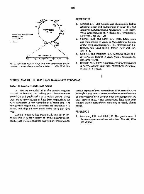
109
f YEpl3-[eMB!l \ B „ I 17.1 kb T " *
' S o ; V ^ "ECOBI1
^ S MindDI
, ' PHftl inMri • 6.4 kb \ -H 1 1 H 1 1 1 > p»rj Bqil! PvuQBginEoiRI BglD Pvull BflU
EeoRI
F'g. i . Restriction maps of the plasmid with complements the phrl mutation, restoring photoceaclivating activity. tXBL 82W-4I86)
Robert K. Mortimer and David Schild
In 1980 we compiled ail of the genetic mapping data of the brewing and baking yeast Saccharomyces cerevisiae and published it as a review article.1 Since then, many new yeast genes have been mapped and we have completed a new compilation of these data. The new genetic map in Fig. 1 describes the location of 415 genes, including 98 new genes added since our 1980 map.
Genetic mapping has traditionally played an important role in genetic studies of various organisms. Recently, such mapping has been particularly important for
REFERENCES
1. Lemontt, J.F. 1980. Genetic and physiological factors affecting repair and mutagenesis in yeast. In DNA Repair and Mutagenesis in Eukaryotes, F.J. de Serres, W.M. Generoso, and M.D. Shelby, eds. Plenum Press, New York, pp. 85-120.
2. Haynes, R.H. and Kunz, B.A. 1981. DNA repair and mutagenesis in yeast. In The Molecular Biology of the Yeast Saccharomyces, I.N. Strathern and J.R. Broach, eds. Cold Spring Harbor, New York, pp. 371-414.
3. Game, J. and Mortimer, R.K. A genetic study of X-ray sensitive mutants in yeast. Mufaf. Research 24, 281-292(1974).
4. Resnick, M.A. 1969. A photoreactivationless mutant of Saccharornyces cerevisiae. Photochem. Photobiol. 9, 307-312 (1969).
various aspects of yeast recombinant DNA research. One example is that several genes have been cloned because of knowledge of their position near another gene on the yeast genetic map. Yeast centromeres have also been isolated on the basts of their proximity to readily cloned genes.
REFERENCE
1. Mortimer, R.K. and Schild, D. The genetic map of Saccharomyces cerevisiae. Microbiol. Rev. 44, 519-571 (1980).
GENETIC MAP OF THE YEAST SACCHAROMYCES CEREVISIAE

110
GENETIC MAP OF SACCHAROUTCES CCREVISIAe
£®« 7
;Cdc2J.t'.* I eji.*
•Um874Q otHH
•SUF5
u3(hi»8)
timOII5
Lffl',
tsmOIZO
-oin>2 SUPI5.16
= {..
Miiotie linkage Tiitomlc linkoge
( I Sequent* noi established with genes ouisiae parenthesis
Fig. J, Genetic Map of Saccharomyces Cerevisiae. IXBL829-4132)

111
COMPARATIVE ANALYSIS OF MITOTIC AND MEIOTIC RECOMBINATION IN SACCHAKOMYCES CEREVISIAE
Michael S. Esposito, Carlo V. Bruschi, Dimitrios T. Maleas, and Kathleen Bjomstad
Previous studies provided evidence that mitotic and meiotic recombination in yeast cells differ in a number of basic properties. I _ l The most striking difference observed involves the timing of mitotic vs. meiotic recombination between homologous chromosomes in diploid cells. In mitosis most, if not all, spontaneous exchange between homologs is initiated prior to replication of the chromosomal regions that undergo recombination. In meiotic cells, however, recombination between genetically marked homologous chromosomes occurs after their replication and involves nonsister chromatids. Sister chromatid exchange occurs in both mitosis and meiosis of yeast, thus the major difference between mitotic and meiotic recombination is the timing of the interaction of homologous chromosomes. Our research during the past year has focused upon a comparative analysis of mitotic and meiotic recombination in diploids homogyzous for the spoil-1 mutation which affects both mitotic and meioti. recombination and confers a phenotype predicted by the prereplicational model of mitotic recombination.5 The characterization of the spo77-1 mutation suggests that addition rec mutations would aid in understanding the mechanisms of mitotic and meiotic recombination. A hyperhaploid strain, LBL1, disomic for chromosome Vtl, constructed in our laboratory'* has been employed for this purpose. The LBL1 strain is useful both for the isolation of rec mutations and for f ^m in ing the effects of suspect mutagenic and carcinogenic agents on mitotic gene conversion, intergenic recombination, and chromosomal lo;s. ' -
MITOTIC AND MEIOTIC RECOMBINATION IN spo77-7/spo7J-7 DIPLOIDS
The prereplicational mitotic versus postreplicational meiotic model of recombination predicts the existence of rec mutants exhibiting hyper-rec activity in mitosis and hypo-rec activity in meiosis with respect to the rates of intergenic recombination.' Mutations that prevent en-donucteolytic cleavage of Holliday structures are expected to exhibit this phenotype. In mitosis, cleavage of Holliday structures by wild type cells reduces the detectable rate of intergenic recombinants which are recovered when Holliday structures, or similar intermediates, are resolved by DNA replication during the S phase of mitosis. In meiosis, cleavage of Holliday structures or related intermediates by wild type cells is required for detection of intergenic recombinants and normal meiotic segregation of chromosomes because intergenic recombination occurs after chromosomal replication. A mutant defective in endonucleolytic scission of recombinational intermediates
in both mitosis and meiosis is expected to exhibit mitotic hyper-rec activity and meiotic hypo-rec activity as well as ascosporal inviability due to meiotic chromosomal loss, breakage and nondisjunction. The phenotype of spo77-7/spo77-7 cells precisely fulfills these predictions.
Diploid hybrids homozygous for the spo77-7 mutation exhibit high levels of meiotic chromosomal nondisjunction, 0 .02% ascospore survival, diminished meiotic intergenic recombination and enhanced mitotic intergenic recombination when compared to wild type spo11/spo11 congenic controls. The effect of the spo77-7 mutation is summarized in Fig. 1, which compares mitotic and meiotic map distances for chromosome VII. Spontaneous mitotic intragenic recombination was also measured in spo77-7/ spo77-7 and SP011/SP011 cells. No differences between the mutant and control cells were observed. This result was anticipated since intragenic recombination which results from gene conversion by mismatch repair is unaffected by the presence or absence of endonucleolytic scission of Holliday structures in mitotic cells.s
MEIOTIC MAP?
OOCentimorqans
SPOII SP0I1
MITOTIC MAPS
I Mit.event/io4 Mitoses
Fig. I. Meiolic and mitotic maps of chromosome VII intervals of the vrfftffypeSPOH/SPOII and thespolt-t/spotl-t mutant hybrids.
IXBL 826-39171

112
MITOTIC GENE CONVERSION, INTERGENIC RECOMBINATION AND CHROMOSOMAL LOSS IN N + 1 CELLS DISOMIC FOR CHROMOSOME VII.
Mitotic chromosomal loss, nondisjunction and recombination of spontaneous and induced origin have been recognized as sources of genotypic changes that may predispose to cancer and teratogenesis. We have constructed a hyperhaploid strain, LBL1, that permits simultaneous detection oi ,\.-;titution of haploidy due to chromosomal loss or nondisjunction, intergenic recombination and gene conversion.7 The strain is disomic for chromosome VII and incorporates six heterozygous markers distributed on both arms of the disomic chromosomes (Fig. 2). Restitution of haploidy, intergenic recombination a,id gene conversion all result in rare cycloheximide resistr.nl colonies (due to expression of the recessive cyhZ mutation conferring cycloheximide resistance) that can be recovered selectively and subsequently distinguished from one another. Restitution of haploidy was found to be the most common source of spontaneously arising cycloheximide resistant segregants (Table 1). The LrJLI strain was exposed to ultraviolet
AD£5 CYHa5 THP5 leul YE A0E6 CLYB
ode5 cyh2 r trp5 LEUI ode6 ClyS
MATa nt XX ode2
Fig. 2. Genotype of the chromosome VII disomic strain LBL1. The map distances shown are the standard meiolic values.
(XBL 824-3774)
light, a recognized mutagenic and carcinogenic agent, to determine the classes of genetic events which are stimulated (Table 2), Cycloheximide resistant segregants diagnostic of gene conversion comprised the most frequent class of ultraviolet-induced events. Intergenic recombination was also stimulated. The least frequently induced event was that of chromosomal loss of nondisjunction, which accounts for the majority of spontaneously arising cycloheximide resistant colonies. The results of this study demonstrate that the LBL1 system provides a sensitive method for detection of the aneugentc and recombinagenic effects of suspect carcinogens and teratogens.
Table 1. Rates of spontaneous mitotic restitution of haploidy, gene conversion and intergenic recombination of chromosome Vtt markers.
Event detected Phenotypic
class Mitotic rates: Cyh medium
Mitotic rates: Leu"Trp -Cyh
medium
Chromosomal loss or nondisjunction
Gene conversion of CYH2' to cyh2'
Recombination in the interval CYH2 - TRP5
Recombination in the interval TRP5 ADE6
Total of all events3
A 1.37x10"' B 1.45x10- 2.03x10"' C 3.66x10- 3.30xr0-D 4.47X10-'
2.44 + 0.24X10-' 5.33*0.64X10
*A total of 25 colonies of LBL1 grown on synthetic complete medium at 24°C were plated on both cycloheximide synthetic complete medium and leucineless tryptophanes cycloheximide medium. The total rates per cell division of the ensemble of events that result in Cyh' and Leu' Trp-Cyh' resistant segregants were determined by the median method of Lea and Coulson. 7A total of 1033 colonies recovered on Cyh medium and 314 colonies recovered on Lew Trp-Cyh medium were further characterized to determine the rates for each phenotypic class. Classes A, B, C and D accounted for 98% of the events detected on Cyh medium. Classes B and C accounted for 98% of the events detected on Lew Trp- Chy medium/
Table 2. Ultraviolet light induced restoration of haploidy, gene conversion and intergenic recombination of chromosome VII markers.
Event detected
Chromosomal loss of nondisjunction
Gene conversion of CYH2> to cyh2'
Recombination in the interval CYH2 - TRP5
Recombinal'on in the interval TRP5 ADE6
Phenotypic No/101 No/10' Class UV survivors' Control
A 12 8
B 66 1
C 12 2
0 8 0
'Cells of the LBL f strain were exposed to UV tight for 40 sec at 3 dose rate of 16 ergs/mm-/sec resulting in 34% survival/

113
Table 3. Mitotic recombination mutants isolated following ultraviolet light mutagenesis ofLBL J.
„, , , , Average Spontaneous Mitotic Recombination N°- M u l a n t s Frequencies-
Phenolypic Groups Obtained Cyh' Conv./IO8 Cyh'Inter. Rec/lO 6
cells cells
I. Conv. Recip. Exch.- 13 t.5 0.8
II. Conv.'Recip. Exch.- 24 22.8 0.8
III. Conv.-Recip. Exch.--- 13 15.3 121.9
IV. Conv. - Recip. Exch. • I 0.8 19.7
V. Conv. • • • Recip. Exch.-• - 3 94.7 1006.4
Control LBL1 125 colonies) 19.4 29.1
'The average for each phenotypic group of mutants represent the results offour independent determinations per mutant. Foreach determination a 2.5-mm colony grown on synthetic complete medium was resuspended and plated on leucineiess tryptophanless cycloheximide medium to determine the frequency of red Leu- Trp'Cyh' convertants and white Leu' Trp'Cyh' intergenic recombinants. Phenotypic symbols are as follows: - = hypo-rec; + = wildtype; and + + + = hyper-rec.
ISOLATION OF HYPO-RIC AND HYPER-REC MUTATIONS
Molecular studies of DNA recombination and genetic analyses suggest the existence of a variety of recombi-national intermediates and modes of resolution.2 Several rec mutations have been isolated in yeast but the present collection does not appear to include variants defective in each step of recombination. We have employed the LBL1 strain to isolate a broad spectrum of rec mutations.6
The genotype of the LBL1 strain permits detection of both hyper-rec and hypo-rec mutations affecting gene conversion and/or intergenic recombination. Rec mutations were isolated following ultraviolet light mutagenesis of strain LBL1. Fifty-four mutants that exhibit a reproducible hypo-rec or hyper-rec phenotype were obtained (Table 3). Five phenotypic groups were observed. Mutants in Group I exhibit reduced levels of both spontaneous mitotic gene conversion and intergenic recombination .This mutant class demonstrates that gene conversion and intergenic recombination are under coordinate genetic control. Group II mutants retain the capacity for gene conversion but exhibit a reduced level of intergenic recombination. The existence of this class indicates there are REC gene products required for detection of intergenic recombination that are not required for gene conversion. Mutants of Group III display normal levels of spontaneous gene conversion and enhanced intergenic recombination. The existence of Group III, like that of Group II, demonstrates that intergenic recombination is affected by gene products that are not involved in gene conversion.
Groups IV and V contain the fewest number of mutants. Groups IV, containing one mutant, fails to exhibit conversion but does exhibit intergenic recombination. The phenotype of Group IV demonstrates that gene conversion involves processes that are not required for in
tergenic recombination. Group V mutants exhibit enhanced levels of both gene conversion and intergenic recombination. Like Group I, Group V demonstrates the existence of RfCgene products that coordinately control the level of gene conversion and intergenic recombination in mitotic cells.
The rec mutants isolated in LBL1 are currently being studied to determine their effects on meiotic chromosomal recombination as well as recombinational events involving the 2-u.m DNA plasmid of yeast. The 2-u.m plasmid exhibits both intramolecular and intermolecular recombination and provides an approach for studying the molecular phenotypes of rec mutants. One rec mutant isolated in strain LBL1 appears to alter the linking number of the 2-u.m plasmid and thus may reflect a mutation affecting a topoisomerase.
REFERENCES
1. Esposito, M.S. Evidence that spontaneous mitotic recombination occurs at the two-strand stage. Proc. Natl. Acad. Sci. USA 75, 4436-4440 (1978).
2. Malone, R.E., Colin, J.E., and Esposito, M.S. Mitotic versus meiotic recombination in Saccharomyces cerevisiae. Current Genetics 1, 241-248(1980).
3. Esposito, M.S., and Wagstaff, J.E. Mechanisms of mitotic recombination, pp. 341-370. In: Strathern, J.N., Jones, E.W., and Broach, J.R. (eds), Molecular Biology of the Yeast Saccharomyces: Life Cycle and Inheritance. Cold Spring Harbor Laboratory, Cold Spring Harbor, N.Y., 1981.
4. Golin, J.E., and Esposito, M.S. Mitotic recombination: mismatch correctici. and replicational resolution of Holliday structures formed at the two strand stage in Saccharomyces. Mol. Cen. Genet. 183, 252-263 (1981).

114
Bruschi, C.V., and Esposito, M.S. Recombination processes in a spol iat ion defective mutant of 5. cerevisiae: role of Holliday structure resolution. Rec. Adv. Yeast Mol. Biol. 1, 254-268(1982). Esposito, M.S., and Bruschi, C.V. Molecular mechanisms of DNA recombination: testing mitotic and meiotic models. Rec. Adv. Yeast Mol. Biol. 1, 242-253 (1982).
Esposito, M.S., Maleas, D.T., Bjornstad, K.A., and Bruschi, C.V. 1982. Simultaneous detection of changes in chromosome number, gene conversion and intergenic recombination during mitosis of Sac-charomyces cerevisiae: spontaneous and ultraviolet light induced events. Current Genetics 5, in press.
EFFECT OF CHEMICAL CARCINOGENS ON MAMMALIAN CELLS IN CULTURE
Regine Goth-Goldstein, Mildred Hughes, and Bonnie P. Tincknell
ALKYLATING AGENT-INDUCED DAMAGE Regine Goth-Goldstein and Mildred Hughes
In recent years various structural alterations in DNA by chemical carcinogens have been identified and related to specific biological perturbations. Many of these modified DNA bases are removed by cellular repair systems. The ability to repair specific lesions varies depending on cell type, tissue and species and plays an important role in the cytotoxic, mutagenic and carcinogenic effect of an agent. We are especially interested in defining the role of DNA repair on the action of alkylating agents such as N-methyl-N'-nitro-N-nitrosoguanidine (MNNG) or methylnitrosourea (MNU). To quantify the toxic and mutagenic effects of these agents, we work with a simple mammalian cell system, a well established Chinese hamster ovary (CHO) cell line. Cell killing by an agent is determined by reduction in colony-forming ability, and its mutagenicity by testing for three different genetic markers, resistance to 6-thioguanine (6TC), ouabain (OUA'I and diphtheria toxin (DT'l.
We examined these cells for recovery from potentially lethal and potentially mutagenic damage after MNNG treatment when kept in a growth-arrested state. Plateau-phase cells were treated with MNNG and, after various lengths of time, were replated at lower densities to assay for survival and to allow for expression of mutations. A gradual increase in survival is seen over 2 days (Fig. 1). In contrast, the mutation frequency for all three markers does not change during the time in confluency. Alkylating agents are thought to cause mutations by alkylating ex-ocyclic oxygens of DNA bases, especially the O 6 of guanine, which leads to mispairing during DNA replication. CHO cells are unable to repair O'-alkyl-guanine. The finding that the mutation frequency in these cells is independent of the time in the growth-arrested state is consistent with the hypothesis that O'-alkylguanine is the mutagenic lesion formed by alkylating agents.
When synchronous populations with cells 1 hr apart in the cycle are treated with MNNG or MNU, the induction
of mutants to TG', OUA' and DT' is independent o' cell cycle time (Fig. 2). These results correspond to the flat response that we had observed after treatment with ethyi-
0.8 ^g /ml
Fig. I. Recovery from potentially lethal damage in MN'NC treated CHO cells. Plateau phase cells were treated with MNNG and released from the growth arrested state at different times afterwards.
IXBL 821 \->227)

115
6 0 J ' 40 >
20
60
20
20
- i i 1 r
^ A H V
A 4 ~^V H 1 1 1 H
'vH>vf*4^N_ H 1 1 1 1 h
_ 1 _ - l _ _ l _ 0 2 4 6 8 10 12 14
Time after mitotic selection (hr) F;g. -'. The age response for induction of mutants resistant to 6-thiog-uanine. ouabain and diphtheria toxin by MMNG. Sychronous populations of CHO cells were obtained by mitotic detachment and treated with 0. 25 u-wml MNNG at various limes afterward.
IXBL 3211-4226)
4-NITROQUINOLINE-1-OXIDE INDUCED DAMAGE Bonnie Presson Tincknell and Regine Goth-Goldstein
4-Nitroqutnoline-l -oxide (4NQO) mimics ultraviolet light (uv) induced damage by vaiious criteria, one of them being that uv-sensitive cells are also more sensitive to 4NQO. Therefore, it is believed that the majr" cause of killing and mutations by 4NQO are lesions in DNA that can be removed by the excision repair system responsible for excising pyrimidine dimers.
The cell cycle response of CHO cells for killing and mutation induction by uv has been studied extensively in this laboratory in previous yenrs and has been determined as follows: cell killing is highest in early S, induction of mutations to ouabain resistance (OUA') and 6-thiog-uanine resistance (6-TG') is only slightly enhanced in early S, whereas induction of diphtheria toxin (DP) is greatly enhanced in late G,. These cycle-related variations seem to be partly due to varying amounts of repair occurring during the cell cycle. Therefore it seemed of interest to examine if the uv mimetic a jent 4NQO shows similar varitions in k i l l ing and mutation induction throughout the cell cycle. The results were unexpected and very different from those obtained with uv.
Cell killing by 4NQO is highest in G,. At the specific dose used in these experiments, survival increases from 5-10% in early G, to the survival of control cells in late S. The mutation of all three markers tested, 6TG', QUA and DT', is highest in G, and is a mirror image of the cell age response of survival.
It is well known that 4NQO is altered metabolically before binding to DNA purine bases. Our data suggest that the metabolic activation of 4NQO occurs preferentially in G 1 ( leading to high cell killing and mutation induction at this stage in the <—-'i cycle.
nitrosourea and seem to be general for alkylating agents in this cell line. They might be related to the inability of these cells to repair the potentially mutagenic O'-alkyl-guanine formed by alkylating agents. In E. colt and yeast, MNNG preferentially induces mutations at the replication point. This so-called growing point mutagenesis has been thought to be the result of an enhanced reactivity of MNNG with the replication fork region. We conclude from our results that it originates from an efficient repair system that excises mutagenic lesions before replication occurs. In f. coli, which are efficient in repairing O 6-•Ikylguanine, only lesions at the growing point are fixed as mutations. If O'-alkylguanine cannot be repaired by the cells, as in CHO cells, the mutation frequency is independent of Ihe time interval between formation of the DNA adduct and the replication of this part of the genome.
INHIBITION OF DNA SYNTHESIS BY NITROSOUREAS Regine Goth-Goldstein
All DNA-damaging agents inhibit DNA synthesis in mammalian cells. Inhibition of DNA synthesis can occur depending on the type of lesion, either because replicon initiaiion is blocked or because DNA chain elongation is blocked. This can be analyzed by determining the sizes of newly synthesized DNA in alkaline sucrose gradient.'
In previous work I had investigated DNA synthesis inhibition by the potent carcinogen ethylnitrosourea (ENU).2 ENU strongly inhibits DNA synthesis already at sublethal doses. It seems not only to inhibit repltcon initiation but also to slow down the fork displacement rate. This later effect is very unusual and has so far only been reported for one otl.er agent, adryomycin, which is an intercalating agent.

116
It is of great interest to analyze the mechanism by which cells replicated DNA damage by carcinogens because mutations might be induced in this process and malignant transformation might be initiated. I therefore tried to relate the unusual effect of ENU to its specific chemical reactivity. Under physiological conditions nitrosoureas decompose spontaneously to yield a carboniui n ion and an isocyanate. The highly reactive carbonium ion alkylates DNA, proteins and other cellular constituents. The isocyanate reacts almost exclusively with proteins by a carbamoylation reaction. My idea was that the car-bamoylating and alkylating activity of ENU might h?-/e different effects on DNA synthesis. To test this hypothesis, I investigated the inhibition of DNA synthesis by nitrosoureas that had been shown to have either only alkylating or only carbamoylating activity. Streptozotocin, a glucose derivative of methylnitrosourea. does not carbamoylate cellular proteins, because the isocyanate formed reacts by internal cyclization. 1,3-bis (trans-4-hydroxycycloh-exyl)-1-nitrosourea, a compound synthesized by T. Johnson from the Southern Research Institute, is a purely carbamoylating agent and does not react with nucleic acids.
When cells are treated with a nontoxic dose of either of these agents a:.J the size of newly synthesized DNA is analyzed in an alkaline sucrose gradient, both agents give a similar profile as ENU. Independent of their alkylating and ca; uamoylating activity, all nitrosoureas cause a reduction of 3H-activity relative to control DNA in each point of the gradient, which is interpreted as slowing down the fork displacement rate. Because both the al-kylation and carbamoylation reaction occur with cellular
Efforts to use animal bioassays in the evaluation of the potential health risk of chemicals to humans have been hampered by the lack of a standardized method to compare experimental results. Experimental protocols as well as the type of information reported in the literature are quite diverse. Moreover, quantitative estimates of carcinogenic potency have not been applied to the results of tests on a broad range of chemicals. Our Carcinogenic Potency Database is an attempt to quantify and standardize the animal bioassay literature and to organize it systematically.
proteins, enzymes involved in DNA synthesis might be inactivated by either reaction and lead to a slower DNA chain growth.
REFERENCES
1. Painter, R.B. and Young, B.R. Biochim. Biophys. Acta 413, 146-153(1976).
2. Goth-Goldstein, R. and Painter, R.B. Carcinogenesis 2, 1267-1271 (1981).
l ist of Publications, 198 7-82:
Goth-Goldstein, R. "Cell Cycle Patterns of Mutation Induction and Their Relationship to DNA Repair." In Chromosome Damage and Repair, Seeberg, E., ed., Plenum Publishing Corporation, 19P1, p. 535-538.
Goth-Goldstein, R., and Painter, R.B. Effect of caffeine on cell killing, mutation induction, DNA repair, and DNA synthesis after treatment with •»thyl-nitrosourea. Carcinogenesis 2, 1267-1271 (1981).
Goth-Goldstein, R. Cell killing by various nitrosoureas and the potentiating effect of caffeine. Mutation Res. 94, 237-244 (1982).
Wood, R., and Burki, H.J. Repair capability and the cellular age response for killing and mutation induction after uv. Mutation Research 95, 505-514(1982).
Wood, R., De Veciana, M., and Presson-Tincknelt, B. Postirradiation properties of a uv-sensitive variant of CHO. Photochem. Photobiol. 36, 169-174 (1982).
TD S 0 : A NUMERICAL INDEX OF CARCINOGENIC POTENCY
As an index of carcinogenic potency, we have developed the TD 5 0 , or Tumorigenic Dose-rate for 50% of the test animals. Briefly, TD S Q is defined as the chronic-dose to induce tumors in half the test animals. This numerical index permits comparisons of diverse test results and improves past efforts to estimate carcinogenic potency in two ways. First, our calculation takes into account whatever spontaneous tumor incidence occurs in control animals. Second, where data are available about the
Carcinogenesis and Cell Biology
CARCINOGENIC POTENCY
Lois Swirsky Gold, Deborah Ang, Georganne Backman, Margarita de Veciana, Holly K. Hurcl, Patricia Kato, Robert H. Levinson, Ted Liou, Renae I. Magaw, Charles b. Sawyer, and Bruce N. Ames.

117
time of death and tumor incidence of each animal, we estimate a lifetable TD S 0 using this information; because animals given high doses of chemicals frequently die early due to chemical toxicity, failure to account for this early mortality could lead to underestimates of the true potency. A full statistical description of TD S 0 is given in Sawyer.'
We have found that the rang j of TD 5 0 's is more than a million-fold. The range of carcinogenic potency for female mice from the bioassays conducted by the National Cancer Institute and the National Toxicology Program (NCI/NTP) are shown in Fig. 1. At the two extremes are the TDs[1's for 2,3,7,8-tetrachlodibenzo-p-dioxin (TCDD), T D ^ = 526 nanograms, and for 4-chloro-o-phenyle-nediamine, TD 5 0 = 2.24grams. Both of these chemicals were evaluated as carcinogenic in the NCI/NTP Technical Reports, and the statistical significance of TD 5 0 is less than 0.01.
THE CARCINOGENIC POTENCY DATABASE
Construction of the database, chemical by chemical, has continued during the past year, in addition to the more than 1100 experiments on 216 chemicals entered previously, we have now completed entering another 482 experiments on 162 chemicals. {We define an experiment as one sex in a species in one bioassay.) The database now includes long-term bioassays of all compounds on the Candidate List of the Occupational Safety and Health Administration as well as those chemicals evaluated in IARC (international Agency for Research on Cancer) monographs, volumes 1-20, as having sufficient evidence of carcinogenicity in experimental animals.
The results of our estimates of TD S I 1 and the standardization of the bioassay literature are summarized and presented graphically in a plot format. An example of the plot for one experiment of 1,2-dibromoethane from the bioassays of the NCI/NTP is presented in Fig. 2. The plot summarizes most of the information from the database. At the top of Fig. 2 is a header line describing the type of information in each field, and a set of numbers which refer to the legend.
lOOng
! / i g
lOfig
a, -o lOOj l tg-
I m g -
I0mg-.
lOOmg--
l g . -
ESTRADIOL MUSTARD 1,2-DI OHOJIO-3-CHLOHCPPHOPANI HKPTACilLOn
ETHYLENE DIBHOHIDE
HYDRAZOOENZENE CHLOH0F0R1! TETRACHLOROETHYLEHE 5-CHLORO-o-TOLUIDINE
e-ANlSIDINE HCL l-CllLORO-it-PHENYLENE PIAHINE TWC.ILOROETHYLENE l-CIILOHO-e-PHENYLENE PIAHINE
I0g Fig. 1. Range of carcinogenic patency far female mice.
(XBL 8212-4295}
Two papers that fully describe the calculation of TD S 0 as an index of potency and the database itself have been submitted for publication. 2 3 The database paper include a plot of all NCI/NTP bioassays for which Technical Reports were published through July 1981, experiments from the published literature on those same NCI/NTP compounds, and a set of experiments on twenty-two aromatic amines.
Preliminary analyses currency being performed using the potency database include investigation of 1) differences in carcinogenic potency across species, sexs, and strains of animal, and 2) effects of lifetable analysis on estimates on carcinogenic potency.

118
mo>« (5) It) i.i-DiimonitANE I M f bfcc | i * MCB MU S3*7I.i • M r k»c !•• no iac 53»i«ti k M I ate ••> Inn MCA S3*7Sit i M ( Mc ••> run a/a SJwTStt 1 m l » c t»t TBA MtB J3w7Si. » M l ( tc ( i r H i MKB 53»78tt r M l t i c f t r | a n MKB 53w7*it
TO 01 JTHI Pail 50 t ip P-Val OaIn
(1J> (14)115)1*)
, . 10 S.74mm ' K.OSOS 4.»7mj * K.MOSe is.4n* • K.eeisc J7.Jaaj • Pc.ODBSc 4 ;*•« • K . O M J
15.41 • K .04
1,2-DlBRCMKTHANE ICI C0052I tSOSli CO0S12 eOOSll cOOSll cOOSll
1.5«n* S.SSmi l.Hlmt >-Blnt S.O»n« 31 .1m 1.85M I I . 7 m l.«3n« 7.l3nc
1 . 0 4 B * n . i . i .
0/21 e/zi » / 2 l 0/18 0/28
l i NCI T W M : C00523 l i NCI/NTT TUMI 44756 52, On* J i /S I 46750 52.On* 21/51 11/50 52. On* * / 5 l 10/50 52. In* * /S I 47/50 52.Saw 31/50
1/50 52. In* 0/50 It.On* 11/58 5 2 . I i * 4750
Fig. 2. Summary of information from database. (1) Chemical name: 1,2-dibromoethane. Line number: 1 indicates
a single experiment. Each letter indicates a TDS0 for a new site-histopathology in the experiment.
(2) Species: M = mouse (3) Sex: f = female (4) Strain: b6c = B6C3F1 (5) Route of administration: gav = gavage (6) Site: MXB = all animals with tumors evaluated by NCI/NTP in
the Technical Report as evidence for carcinogenicity sto = stomach lun = lung TBA = alt tumor bearing animals Hv = liver
(7) Histopathology: MXB = all animals with tumor types described in (27) sqc = squamous cell carcinoma MXA = a combination of tumors evaluated as evidence for
carcinogenicity in NCl/NTP Report a/a = alveolar/bronchiolar adenoma
(8) Duration of exposure to compound: w = weeks 19) Duration of experiment to terminal sacrifice, in weeks
(10) Note codes: s — poor survival due to toxicity or disease v — variable dosing, e.g., a change in concentration of dose
during the experiment (11) Logarithmic scale for values of TDi0 (in units/kg/day for a standard
2-year-lifespan) extending from 100 nanograms to 10 grams (12) The most potent TDi0 for the experiment = 3.74 mg/kg/day
+ = p-value less than 0.01 : = 99% confidence interval for TDS0
(TD's and confidence intervals for other sites and histopathology are not plotted as points, but their values are given under (13)
.(20) and (2V) (13) The value of TDS0 in mg/kg/day for standard lifespan no dre =
no dose related effect (14) Shape of dose-response curve:
' = consistent with linearity blank = no dose-related effect
(15) Two-tailed p-value for TDso (16) Published opinion in NCI/NTP Technical Report:
c = evidence that compound is carcinogenic at the site and for histopathology given under (6) and (7)
blank = no opinion stated (17) Repeat of chemical name and line numbers as in (1) (18) Synonym for compound name, followed by Chemical Abstracts
Service registry number (19) Reference number used by NCI/NTP = C00522 (20) Lower 99% confidence limit for TDX in mg/kg/day (21) Upper 99% confidence limit for 7"0W in mg/kg/day (22) Proportion of animals in control group with tumors listed under
(6) and (7) (23) Average daily dose for length of experiment in mg/kg for low-
dose group (24) Proportion of animals in low-dose group with tumors listed under
(6) and (7) (25) Average daily dose for length of experiment in mg/kg for high-
dose group (26) Proportion of animals in high-dose group with tumors listed under
(6) and (7) (27) Site and histopathology of TD^'s that combine more than one
site or hislopathology (28) Berkeley code for TDK's created for database:
c = combination of all tissues and tumors evaluated in NCI/ NTP Technical Report as evidence for carcinogenicity blank = all others IXBL8212-4296, XBL 8212.4297)
REFERENCES
1. Sawyer, C , Peto, R., Bernstein, L, and Pike, M.C. 1982. Calculation of Carcinogenic Potency from Long-Term Animal Carcinogenesis Experiments. Biometrics, to appear 1983.
2. Peto, R„ Pike, M.C., Bernstein, L , Gold, L.W., and Ames, B.N. 1982. The TD S 0 : A proposed general convention for the numerical description of the carcinogenic potency of chemicals in chronic-exposure animal experiments. Submitted to Cancer Res.
Gold, L.S., Sawyer, C.B., Hooper, N.K., Havender, W.R., Bernstein, L , Peto, R., Pike, M.C., and Ames, B.N. (with assistance of D. Ang, G.M. Backman, L. Bertucelli, M. DeVeciana, A. Friedman, K. Gould, E. Higgins, P. Kato, R. Levinson, T. Liou, P.M. MacLeod, M. Needels, M. Nichols, M. Rosenfeld and W.E. Rouse). 1982. A carcinogenic potency database of the standardized results of animal bioassays. Submitted to Cancer Res.
119
COMPARATIVE ANALYSIS OF DATA FROM SHORT-TERM TESTS FOR CARCINOGENS AND MUTAGENS
loyce McCann, Laura Horn, John KaSdor, Bob Siggel, and Barbara Levine*
The use of short-term tests, such as the Salmonella (Ames) test,' for predicting the carcinogenic potential of chemicals has expanded so rapidly that development of systematic, quantitative approach for analyzing and interpreting results has become essential. There are many different short-term testing methods.2 It is now generally agreed that for thorough evaluation a chemical should be tested by more than one of these short-term tests, but it is not clear which tests shuld be used nor how conflicting results from different tests should be interpreted. Since different short-term tests appear to detect carcinogens with varying degrees of sensitivity and specificity, the anaiysis and interpretation of results can be complex (for recent discussion, see Refs. 3-6).
The potential of short-term tests to provide quantitative information about carcinogenicity is much less certain than their potential to provide qualitative information. There is, however, suggestive evidence that some quantitative correlation in potency between short-term tests and animal cancer tests may exist, and it is important to thoroughly explore this possibility for practical as well as scientific reasons. Short-term tests of thousands of synthetic and natural chemicals have identified far more potential carcinogens than can be confirmed by conventional animal cancer tests. The carcinogenic potency of different chemicals can vary by more than a millionfold. Clearly, if potency in short-term tests could be used to make rough predictions of potency in animal cancer tests, risk evaluation could be more easily focused on chemicals with the greatest potential hazard.
A DATA BASE FOR COMPARATIVE, QUANTITATIVE ANALYSIS
We have constructed7 a data base of short-term test results that wil l provide a powerful resource for resolving issues of comparability, both between various short-term tests and between short-term and animal cancer tests, by allowing quantitative comparisons to be made.
We use a general-purpose data-base management system, SPIRES, that was developed at Stanford University. It can operate on a variety of IBM computer systems and is used extensively throughout the United States and abroad.
Information on each short-term test is contained in a series of separate data bases. We have completed the design of data bases for Salmonella test, mammalian cell mutagenesis assays, and sister chromatid exch.-oge assays. The Salmonella data base contains results from over
•Computer Applications. Engineering and Technical Services Division.
25,000 experiments from the published literature, from unpublished data provided to us by the NCI/INTP, and from our own laboratory results.
Output routines have been developed for displaying various parts of the data base. Output routines to summarize the data and the results of the statistical analysis for the Salmonella (Ames) test (described below) are shown in Figs. 1A and IB. The mutagenic potency of Captan, a common fungicide, estimated from results obtained in four different laboratories using the tester strain TA100 is indicated in Fig. 1 A. Given the wide range of potencies of different mutagens, these results appear, in general, to be sufficiently reproducible to permit useful comparisons to be made. We are currently completing a systematic study on interlaboratory variability.
AN EMPIRICAL APPROACH TO STATISTICAL ANALYSIS OF SALMONELLA TEST DATA
In the Salmonella mutagenicity assay, histidine aux-otrophs are exposed to the test chemical on a petri dish, and the only measurement made is the number of revertant colonies on each plate. The Salmonella assay is thus not a precise means of measuring mutation rates, since the estimation of a mutation rate requires knowledge of the number of cells at risk of mutation and the number of mutant cells resulting from exposure.
The shape of the dose-response curve typically depends on the range of doses used. For most mutagens at low doses the curve increases linearly; then, as the dose increases, the curve may flatten and finally turn downward, due primarily to effects of toxicity.
Several authors have based their proposed statistical analyses of Salmonella dose-response data on models of the entire dose-response curve. However, modeling the entire curve is a difficult undertaking because of the single measurement made in the assay, and because different mechanisms of mutagenesis and toxicity may be operating for different chemicals.
We have taken a different approach.6 We assume that the initial part of the curve is linear and base the statistical analysis solely on this region, reasoning that it contains most of the only interpretable information about the mutagenesis dose response. We have described both a formal approach for chcosing doses that are on this initial linear part of the dose-response curve and a procedure for estimating the initial slope of the curve and testing it for statistical significance. We have also described a simple goodness-of-fit test for linearity ot response in the defined region.

120
0 A t S S R 3 N P T 0 S D E R
l _ E O X E a K X - e L O I 0 4 / 2 1 / 6 1
j * i O 1 0 0 l l
CAPTAN E 193 0 3 A Ft NS L I V 5 0 C 3 3 3 S A M NS L I V N5 C 7 2 1 5 S P R SUA LEV NS C 7 1 5 S P N NHR LEV NS C 3 3 3 5 S A » NS L I V NS [ 7 2 3 7 S P R SUA LEV NS [ 7 1 7 S P H NNR L I V NS C 3 3 1 7 S A 11 NS L I V NS £ 7 2 3 e S P R SDA L I V NS C 7 ] e 3 P N I W L I V NS C 3 3 3 8 S A n NS L I V NB [ 2 3 3 3 S t 6 3 0 3 C 193 0 S I 13 S C 333 0 S E 6 3 S £ 1363 5 S C 5 3 5 S E 1363 5 S £ 2 3 3 3 5 S t 6 3 5 3 C 7 2 3 5 S £ 13 5 S C 3 3 3 5 s f 7 2 3 7 s [ 3 3 3 7 s £ 723 I 3 3 3 e S C 2 3 3 3 2 s
CAPTAN [ 193 O a F l o r a . 1 9 7 8 .
4 3 [ 3 3 ] S i i m o n « t a l . . 7 1 1 t 7 2 J M a r s h a l l a t a l
1 7 [ 7 3 H a r b o l d , B o » * m 4 5 [ 3 3 ] S l M i o n a t a l . .
7 0 8 E 7 2 ] M a r s h a l l a t a l 1 0 C 7 ] H t r - b o l d . B u s a l 4 7 [ 3 3 ] SlRiBon « t a l , ,
7 0 S [ 7 2 3 M a r s h a l 1 a t a l 1 9 E 7 ] H a r b o l i l , B u t a l n 4 9 C 3 3 ] S i m e o n a t a l . .
1 3 2 1 E 2 3 3 ] A M * . 1 9 7 6 . / / 2 2 5 [ 6 3 n c C a n n a t a l . . 1 0 8 E 193 D a F l o r a . 1 9 7 8 .
1 5 E 1 ] S h i r a i u * t a l . 4 2 E 3 3 3 S l a a i e n a t a l . .
2 2 4 E 6 3 H c C a n n a t a l . . 7 2 3 C 1 3 6 ] H a l l a t t a t a l .
2 0 C S ] S a l l a r . 1 9 7 5 . 7 2 4 E 1 3 6 ] H a l l a t t a t a l .
1 3 2 2 C 2 3 3 ] A a , » » . 1 9 7 6 . / / 2 2 6 E 6 ] H c C a n n a t a l . • 7 1 0 E 7 2 ] m r * h a 1 1 a t a l
14 [ 1 ] S h i r a i u a t a l . 4 4 E 3 3 ] S i n n o n a t a l • •
7 0 9 C 7 2 ] M a r s h a l l a t a l . 4 6 £ 3 3 ] S l n a i o n a t a l , .
7 0 7 E 7 2 ] M a r » h a l 1 a t a l . 4 8 E 3 3 ) S i n * o n a t a l . .
1 3 2 3 E 2 3 3 ] A m i * . 1 9 7 6 . / /
17ua 0 . 0 0 0 0 * 4 6 u a 0 . 0 0 0 0 • 12ua fi 0 . 0 0 0 0 *
J £ l _
> 0 .0542 N X
t 0.0029 -» 0.0000 N • O.0000 • > 0.0000 • * 0.0000 * t 0.0000 » I 0.0000 N » 0.0000 *
i 0 .0000 * I 0 .0000 N I & 0 .0000 * » 0 .0000 *
NATU.271.M55// 0 .204 .6 / 1.25.223/ 2 .5 .284 / 5 . 4 2 1 / 10.645/ E5.5] 1977. EPA.NB// 0 , (99> / 1 . (141) / 5 .1210) / 10.£285}/ 15.£340)/ 25 .£330) / 50.<704) / E7.7J . .1976. JAFC24.P560// 0 .14/ 23.117/ 50 .231 / C3.3) nalar.1976. MURE,40.p73//t03.t1303/ £13.£29753/ E103.C17.53/ £203.£0]/ £43 1977. EPA.NS// 0 . ( 1 4 ) / 1 , {20) / 5 . ( 6 0 ) / 10.E113>/ 15,£55) / 25 ,£71) / 50 . (143 ) / [ 7 , 7 ] . ,1976. JAFC.24.PS60// 0 . 9 / 25.148/ 50,289/ £3,33
1976. MURE.40,r73//EO).£331/ £11,£345]/ £10],£03/ £20].CO]/ [43 1977. EPA.NS// 0 . ( 3 ) / 1 , (23 / 5 .C2) / 10.C2)/ 15.CO)/ 2 5 , ( 0 ) / 5 0 . t l ) / £7]
1976. JAFC.24.P360// 0 .32 / 23.32/ 3 0 , 5 1 / C3.3 1 1
malar.1976. MUHE,40.r73//t03.£21.53/ £11,£302.53/ .03 . [03 / £20] ,£01 / 0 , ( 2 5 ) / 1.119}/ 5 . ( 2 2 ) / 10.E26)/ 1 5 . ( 2 1 ) / 2 5 . ( 4 6 ) / SO.£44)/ £7.71 0.224/ 3.2973/ 20.4644/ 50.2593/ 14.3] 0.154/ 1,302/ 10.972/ 50.1716/ 100,262/ E5.3) 0 .226 .3 / 1.25.325/ 2 .5 ,397 / 5.570/ 10.1115/ ES.53 0,206/ 1.3.412/ 2 .6 .882 / 10-2528/ 25.1970/ tO.119/ £6.43 0 . ( 7 2 ) / 1 , (211) / 3 . ( 5 3 2 ) / l u . ( 6 2 2 ) / 15.£820)' 25.1720) / 5 0 . ( T l / C7, 0 .27 / 5,292/ 10.349/ 50 ,211 / [ 4 . 3 ]
197S. Chanlcal Control RQ.4/ 1 .11 / 10,12/ 25.20/ 50 ,66 / 100.180/ [6 .61 PCST, 17.1-398// £03.£13/ E9.9E-04], £ 1. 243/ £0. 01 3. E 1.83] / £0 .11 . C4.3?l/ CD. [3.733.
975. ChamteaI Control R0.3/ 1,6/ 10.10/ 25 ,18/ 50 .67/ 100.84/ 250.2000/ [ 7 , 7 ] 0 ,28 / 5.697/ 20.1722/ 30.366/ £4,33 0 .16/ 1,35/ 3.106/ 10.163/ 50.275/ £5.33 0 ,10 / 10.248/ 25.320/ 50.396/ £4.33 O.NS/ 5.118/ 10.265/ 25,382/ 50,118/ C53 0,<19>/ 1 . (29 ) / 5 , ( 8 0 ) / 10 , (76 ) / 15 . (104} / 2 5 , ( 6 0 ) / 5 0 . ( T ) / [ 7 , 5 ] 0 . 7 / 10,335/ 25 ,47/ 5 0 , 0 / C43 0.(7)/ 1,(2)/ 5,£3)/ 10,(0]/ 15,£0)/ 25.(0)/ 50.(03/ C7) 0.32/ 10,19/ 23,11/ 50.0/ C4J 0 , ( 8 1 / 1 . ( 7 ) / 5 ,£14 ) / 10.(163/ 1 5 , ( 2 6 ) / 25.C6)/ 50. (22) / C7.7] 0 . 6 1 / 3 ,67 / 20 ,63/ 50.118/ [4 .41
1977. EPA.NS//
975. PNAS.72.C979// NATU.271.P433// 1977. 0HCA.A.P267// 977. EPA.NS//
1975. PNAS.72.r979//
1973. PNAS.72.F979// .1976. JAFC.24,i^60// 1977. 0MCA.A.C267//
,977. EPA.NS// ,1976. J A F C , 2 4 , P 5 6 0 / /
1977. EPA.NS// 1976. JAFC.24.*560//
1977. EPA.NS//
Fig, 1. Summary output for Salmonella test data, including results of statistical analyses. Each line of (A) and (B) represents a single dose-response experiment. (A) Columns are, from left to right: REF = a bibliographic number. ORG = Salmonella tester strain used (0 = TA100, 9 = TA98.5 = TA1535, 7= TA1S37, 8= TA1538TA92). ASS = type of assay (S = standard plate test). IND = inducer, if an external metabolic activation system was used (A = Arocblor, P = phenobarbital). SPE = animal species used as source of metabolic activation (R = rat, M = mouse). STR = strain of animal (a series of three-letter codes, or four-letter codes for Fl hybrids; NS = not specified). TIS = tissue used, also indicated by three-letter codes (e.g., LIV = liver). ACT = microliters S-9 per petri plate. PRO = mg per ml of protein in the S-9. (NS = not specified). Potencies, calculated as dose increasing revertant background 5 times, are plotted on a log scale using one of three symbols (+, ±, >), depending on the one-sided P-value obtained from the statistical test on the significance of the slope: + = P<0.01;± = 0.01 <P*s0.05; > = P>0.05. Associated lower and upper confidence bounds are plotted as ":". The rectangle in 2(A) indicates results obtained by four different laboratories, using the tester strain TAIOO (see text). POT = calculated potency value followed in some cases by qualifying or error codes; A = estimated slope was negative; B = confidence intervals could not be calculated: V == spontaneous revertants not specified; X = non-standard response units; PVL = one-sided P-value testing whether slope of dose-response curve is significantly different from zero. QUA = author's opinion that chemical was positive (+), negative (-), or borderline (W) IN = '••>: specified!, EXP = "experiment number" assigned to dose-response curve. (B) Frcm left to right: experiment number; bibliographic reference number (Ref. 223 unpublished; Ref, 6, dose-response curve not published); bibliographic reference (four-letter codes are used for journal names); dose/response data from which potency was calculated (ff = dose or response units are not \ig/plate or revertants/plate); number of dose-response pairs in experiment, followed by number used by statistical program to estimate initial slope, (A) XBL 8110- J1963; (8) XBL 8110-11962
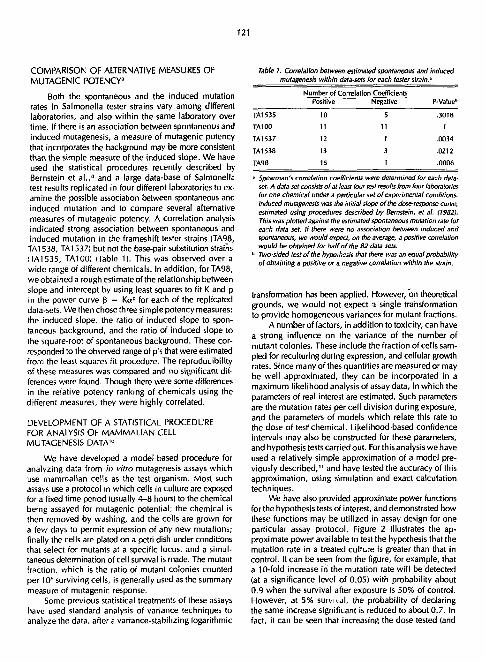
121
COMPARISON OF ALTERNATIVE MEASURES OF MUTAGENIC POTENCY'
Both the spontaneous and the induced mutation rates in Salmonella tester strains vary among different laboratories, and also within the same laboratory over time. If there is an association between spontaneous and induced mutagenesis, a measure of mutagenic potency that incorporates the background may be more consistent than the simple measure of the induced slope. We have used the statistical procedures recently described by Bernstein et al.," and a large data-base of Salmonella test results replicated in four different laboratories to examine the possible association between spontaneous and induced mutation and to compare several alternative measures of mutagenic potency. A correlation analysis indicated strong association between spontaneous and induced mutation in the frameshift tester strains (TA98, TA1538, TA1537) but not the base-pair substitution strains (TA1535, TA100) (Table 1). This was observed over a wide range of different chemicals. In addition, for TA98, we obtained a rough estimate of the relationship between slope and intercept by using least squares to fit K and p in the power curve 3 = Ka p for each of the replicated data-sets. We then chose three simple potency measures: the induced slope, the ratio of induced slope to spontaneous background, and the ratio of induced slope to the square-root of spontaneous background. These corresponded to the observed range of p's that were estimated from the least squares fit procedure. The reproducibility of these measures was compared and no significant differences were found. Though there were some differences in the relative potency ranking of chemicals using the different measures, they were highly correlated.
DEVELOPMENT OF A STATISTICAL PROCEDURE FOR ANALYSIS OF MAMMALIAN CELL MUTAGENESIS DATA1 0
We have developed a model-based procedure for analyzing data from in vitro mutagenesis assays which use mammalian cells as the test organism. Most such assays use a protocol in which cells in culture are exposed for a fixed time period (usually 4-8 hours) to the chemical being assayed for mutagenic potential; the chemical is then removed by washing, and the cells are grown for a few days to permit expression of any new mutations; finally the cells are plated on a petri dish under conditions that select for mutants at a specific locus, and a simultaneous determination of cell survival is made. The mutant fraction, which is the ratio of mutant colonies counted per 106 surviving cells, is generally used as the summary measure of mutagenic response.
Some previous statistical treatments of these assays have used standard analysis of variance techniques to analyze the data, after a variance-stabilizing logarithmic
Table I. Correlation between estimated spontaneous and induced mutagenesis within data-sets for each tester strain.'
Positive Negative
TA1535 10 5
TA100 11 11
TA1537 12 1
TA1S38 13 3
TA98 15 1
* Spearman's correlation coefficients were determined for each data-set. A data-set consists of at least four test results from four laboratories for one chemical under a particular set of experimental conditions. Induced mutagenesis was the initial slope of the dose-response curve, estimated using procedures described by Bernstein, et al. (19821 This was plotted against the estimated spontaneous mutation rate for each data set. If there were no association between induced and spontaneous, we would expect, on the average, a positive correlation would be obtained for half of the 82 data sets.
b Two-sided test of the hypothesis that there was an equal probability of obtaining a positive or a negative correlation within the strain.
transformation has been applied. However, on theoretical grounds, we would not expect a single transformation to provide homogeneous variances for mutant fractions.
A number of factors, in addition to toxicity, can have a strong influence on the variance of the number of mutant colonies. These include the fraction of cells sampled for reculturing during expression, and cellular growth rates. Since many of thes quantities are measured or may be well approximated, they can be incorporated in a maximum likelihood analysis of assay data, in which the parameters of real interest are estimated. Such parameters are the mutation rates per cell division during exposure, and the parameters of models which relate this rate to the dose of test1 chemical. Likelihood-based confidence intervals may also be constructed for these parameters, and hypothesis tests carried out. For this analysis we have used a relatively simple approximation of a model previously described, " a n d have tested the accuracy of this approximation, using simulation and exact calculation techniques.
We have also provided approximate power functions for the hypothesis tests of interest, and demonstrated how these functions may be utilized in assay design for one particular assay protocol. Figure 2 illustrates the approximate power available to test the hypothesis that the mutation rate in a treated cultLre is greater than that in control. It can be seen from the figure, for example, that a 10-fold increase in the mutation rate wil l be detected (at a significance level of 0.05) with probability about 0.9 when the survival after exposure is 50% of control. However, at 5% survival, the probability of declaring the same increase significant is reduced to about 0.7. In fact, it can be seen that increasing the dose tested (and

122
u | 5 10 15 20 K P
Fig. 2. Power curves for the test of the hypothesis that a treated culture exhibits a higher mutation rate than a control. Protocol variables are as in the mouse lymphoma assay of Clive et at. (19791. Kp = ratio of mutation rate in the treated culture to that in the control. K^t = ratio of mutant fraction in the treated culture to that in the control. Power = the probability that a difference in mutation rates between the two cultures will be declared significant at the 0.05 level. The curves represent the power at five levels of cell survival (indicated as percentages of control) as functions of KB and K^.
IXBL 824-3796)
presumably the mutation rate) can actually result in a decrease in the power available to detect the chemical as a mutagen, if there is a sufficiently large increase in toxicity. Similar investigations were made for spacing and allocation of doses when more than one dose of test chemical is tested, and it was found that for a linear dose-response curve, the design that optimizes both the test for nonzero slope, and the power of test for linearity is one with two ncr>*ero doses. The optimal spacing of the nonzero doses depends on the relationship between dose, mutation rate and toxicity, which of course is not known for most c h e t : als in advance; however, equal spacing provides greater power than the n lore commonly used exponential doss spacing, and the higher nonzero dose can be placed so that survival is as low as 15% without risking large increases in variability.
REFERENCES
1. Ames, B.N., McCann,)., and Yamasaki, E. Methods for detecting carcinogens and mutagens with the Salmonella/mammalian-microsome mutagenicity test. Mut. Res. 31, 347-364 (1975).
2. Hollstein, M., McCann, J., Angelosanto, F.A., and Nichols, W.W. Short-term tests for carcinogens and mutagens. Mut. Res. 65, 133-226 (1979).
3. McCann, J. Short-term tests and cancer policy. In Advances in Occupational Health, L. McDonald, ed., Vol. 1. Churchill-Livingstone, London, 1981, pp. 149-161.
4. Magaw, R. and McCann,). Short-term tests used to detect mutagens and their effects in body fluids. In Proceedings of an Interdisciplinary Cancer-Study Conference. North-Holland, Amsterdam, in press.
5. McCann,). In vitro test methods to detect carcinogens, in Trends in BioassayMethodology: In Vivo, In Vitro, and Mathematical Approaches. National Institutes of Health. 1981, pp. 145-161.
6. Ames, B.N. and McCann, J. Validation of the Salmonella test: A reply to Rinkus and Legator. Cancer Research 41, 4192-4203 (1981).
7. McCann,)., Horn, L., Litton, C , Kaldor, J., Magaw, R., Bernstein, L. and Pike, M. 1982. In Press. Short-term tests for carcinogens and mutagens: A database designed for comparative, quantitative analysis. In Structure Activity Correlation as a Predictive Tool in Toxicology. Fundamentals, Methods, and Application. L. Goldberg, ed., Hemisphere Press, in press.
8. Bernstein, L., Kaldor, J., McCann, J., and Pike, M.C. An empirical approach the statistical analysis of mutagenesis data from the Salmonella test. Mutation Pes. 97, 267-281 (1982).
9. Horn, L., Kaldor,)., and ). McCann. A comparison of alternative measures of mutagenic potency in the Salmonella (Ames) test. Mutation Res. in press.
10. Kaldor, ). Statistical procedures for the design and analysis of in vitro mutagenesis assay. Ph.D. Thesis. 1982. University of California, Berkeley.
11. Tan, W.Y. The probability distribution of the number of mutanti in the CHO/GHPRT assay. Presented at the National Toxicology Program Workshop on the Statistical Analysis of Mutagenesis Assays. Chapel Hil l , North Carolina, April 21-22, 1981.

123
PROPERTIES OF HUMAN MAMMARY EPITHELIAL CELLS IN CULTURE
Martha R. Stampfer and Jack C. Bartley
We have improved our system for growing human mammary epithelial cells in culture'•' by collaborating with Dr. Richard Ham of the University of Colorado to develop a serum-free defined medium for these cells. Using a modification of Ham's medium MCDB202, plus several growth factors, we can now grow normal and tumor-derived cells in mass culture for 10-16 passages. Clonal growth is also supported by this medium. These results greatly expand the usefulness of this system, because practically unlimited numbers of cells can now be generated from individual specimen donors. However, it is most important to determine whether this long-term growth in culture has in any way altered the properties of these ceiis. This is especially true since they can be observed to undergo a selection process around passage 3, in which most of the cell population appears to senesce,
while a minority population maintains the same growth rate and morphology. Thus far, we have determined that the higher passage cells maintain a normal karyology, keratin and fibronectin distribution, and pattern of glucose metabolism. However,theydoappeartohavedecreased levels of some of the enzymes involved in metabolism of benzo(a)pyrene.
REFERENCES
1. Stampfer, M.R., Hallowes, R., an'' Hackett, A.J. Growth of normal human mammary epithelial cells in culture. In Vitro 16, 415^125 (1980).
2. Stampfer, M.R. Cholera toxin stimulation of human mammary epithelial cells in culture. In Vitro 18, 531-337(1982).
IN VITRO TRANSFORMATION OF HUMAN MAMMARY EPITHELIAL CELLS
Martha R. Slampfer and Jack C. Bartley
We have initiated studies to develop an assay to detect carcinogen-induced transformation in vitro. Such an assay could be a valuable method of detecting environmental carcinogenic agents, as well as furthering our understanding of the mechanisms involved in malignant transformation. The development of this assay has been made possible by previous work in developing the human mammary cell culture system,1 ,2 in defining properties associated with tumor derived cells/ and in demonstrating that these mammary cells can readily convert thf procarcinogen, benzo(a)pyrene (BaP) to its car-cinov nic product. 4 5 We have utilized BaP for our initial stuc1 es since it is the most common environmental pollutant in its class present in our atmosphere, and it and related compounds are known to readily induce mammary carcinoma in rodents.
Cells in primary culture have been exposed to BaP, and cells in subsequent passages analyzed. Preliminary results with cells from one individual rvv: teen promising. When grown in our originally devised mammary medium, MM, 1 - 2 control cells display total senescence by passage 5 to 6. However, the cells exposed to BaP continue proliferation beyond this point. We currently have a clonally isolated cell line actively growing in 32nd passage. These cells appear to be partial transformants in that they do not yet display the tumor-associated property of anchorage-independent growth in methocel. White they maintain normal epithelial patterns of fibronectin and keratin distribution, they do display alterations in glucose
metabolism and expression of the human milk fat globule antigen, and some abnormaties in chromosome number. We are presently examining these cells for other properties that may be tumor-cell associated and attempting to induce further malignant changes by exposing them to potential promoting agents.
REFERENCES
1. Stampfer, M.R., Hallowes, R., and Hackett, A.J. Growth of normal human mammary epithelial cells in culture. In Vitro 16, 415-425 (1980).
2. Stampfer, M.R. Cholera toxin stimulation of human mammary epithelial cells in culture. In Vitro 18, 531-537(1982).
3. Stampfer, M.R., Hackett, A.J., Hancock, M.C., Leung, J.P., Edgington, T.S., and Smith, H.S. Growth of human mammary epithelium in culture and expression of tumor specific properties. Cold Spring Harbor Conferences on Cell Proliferation 9, 819-830, 1982.
4. Stampfer, M.R., Bartholomew, J.C., Smith, H.S., and Bartley, J.C. Metabolism of benzo(a)pyrene by human mammary epithelial cells: toxicity and DNA adduct formation. Proc. Natl. Acad. Sci. 78, 6251-6255 (19B1).
5. Bartley, J.C., Bartholomew, J.C., and Stampfer, M.R. Metabolism of benzo(a)pyrene by human epithelial and fibroblastic cells: Metabolite patterns and DNA adduct formation. J. Cell Biochem. 18, 135-148 (1982).

124
BENZO(A)PYRENE METABOLISM IN HUMAN MAMMARY EPITHELIAL CtLLS
lack C. Bartley and Martha R. Stampfer
We have been utilizing the system, developed by Martha Stampfer, for growing breast epithelial cells in culture1 '2 to study a possible role of chemical carcinogenesis in breast cancer. The epithelial cell is the origin of 99% of all breast malignancies.
Our published results3'* with cells grown out from a small number of normal breast specimens ate compatible with chemicals as initiating factors in breast cancer. The epithelial cells rapidly metabolize benzo(a)pyrene (BaP), a common environmental carcinogen produced during fossil fuel utilization, to a wide variety of products inc lud ing the documented u l t imate carc inogen, benzo(a)pyrene diolepoxide. The result of this metabolism is covalent bonding of the ultimate carcinogen to protein, RNA and DNA. The type and extent of DNA modification is comparable to that observed in cells transformable by treatment with BaP.
A more extensive survey of BaP metabolism by epithelial cells grown out from 30 breast specimens, both normal and malignant, has revealed that, as expected, interindividual variation in BaP metabolism is greater than that observed in studies with cells from syngeneic animals, but not as great as that observed by others studying cancer prone human tissues in organ culture. The possibility that this interindividual variation is related to the familial risk of breast cancer is currently under investigation.
The variation in BaP metabolism between cells from different specimens is small enough to reveal a distinct, quantitatively unique metabolite pattern for breast epithelial cells. This pattern is characterized by tetraol formation, even at one hour post exposure to BaP, and by conjugation of metabolites, particularly with glutathione. These results provide evidence that the oxidase system acting on BaP in the human epithelial ceils has a similar ability to form epoxide from BaP and from 7,8-dihydrodiol while that in fibroblasts preferentially utilizes BaP. This unique oxidative activity in human epithelial cells may be due to the presence of prostaglandins endoperoxide synthetase which has been reported to be capable of oxidizing 7,8-dihydrodiol-BaP to the ultimate carcinogen in the presence of arachidonic acid. One of our current aims is to investigate this possibility further.
The flexibility and comparative advantages of a system in which samples or specimens can be stored for some time, as in the present case, are obvious. For example, studies on the same sample can be repeated years later in the light of new information or techniques. Care must be taken with such systems, however, that the results generated with the frozen samples are as valid as those from the fresh ones. We have demonstrated that the
variation between results from cells cultured from fresh and from frozen organoids over a year apart is only slightly greater than that from replicate incubations done on the same day on cells cultivated from the frozen organoids.
Because of the implications in both animal and epidemiological studies that fatty acids, particularly unsaturated ones, enhance the risk of breast cancer, we have examined the effect of fatty acids on BaP metabolism in cell culture. An effect of polyunsaturated fatty acids (PUFA) on BaP metabolism was obvious: a marked reduction in conjugation observable by 16 hours after BaP exposure and a consistent increase in organosoluble metabolite yield. Of the material in the cell pellet, 22-40% was associated with the nuclear fraction in PUFA-treated cells, but only 14% in control cultures. The five-fold increase in the extent of DNA adduct formation in PUFA treated cells was likely a reflection of the increase in organosoluble metabolite yield and in BaP metabolites in the nuclear fraction.
A comparable set of experiments to those with PUFA, using a mixture of saturated fatty acids of similar carbon chain length, revealed that saturated fatty acids had no effect on BaP metabolism or on the extent of DNA adduct formation.
The modulating effect of PUFA appears to be specific for epithelial cells in the human mammary gland; the BaP metabolite pattern of breast fibroblasts was unchanged by the addition of PUFA.
Current studies are designed to clarify how the alterations brought about by PUFA might relate to the enhancing effect of these fatty acids on the incidence of breast cancer.
REFERENCES _.
1. Stampfer, M.R., Hallowes, R., and Hackett, A.|. Growth of normal human mammary epithelial cells in culture. In Vitro 16, 415-425 (1980).
2. Stampfer, M.R. Cholera toxin stimulation of human mammary epithelial cells in culture. In Vitro 18, 531-537(1982).
3. Stampfer, M.R., Bartholomew, J.C., Smith, H.S., and Bartley, j .C. Metabolism of benzo(a)pyrene by human mammary epithelial cells: toxicity and DNA adduct formation. Proc. Natl. Acad. Sci. 78, 6251-6255 (1981).
4. Bartley, J.C., Bartholomew, J.D., and Stampfer, M.R. Metabolism of benzo(a)pyrene by human epithelial and fibroblastic cells: Metabolite patterns and DNA adduct formation. J. Ceil Biochem. 18, 135-348 (1982).

125
THE ANOMOLOUS INABILITY OF ROUS SARCOMA VIRUS TO CAUSE TUMORS IN AVIAN EMBRYOS
David S. Dolberg, Henry I. Nesis, and Mina) . BhseU
One of the most important conclusions to emerge from the last 50 years of cancer research is that the progression of most cells or tissues from a normal phen-otype to malignancy is a multistep process. Progression through the early stages results in cells that are frequently distinguishable from normal, but not yet malignant. Continued progression results in an irreversible malignancy. The recognition of the existence of multiple stages wil l surely accelerate our understanding of tumorigenesis, but at present little is known about the interrelation of the various stages.
Until recently, the major exception to this general pattern has been the malignancies caused by the acute C-type sarcoma viruses typified by Rous sarcoma virus (RSV). This virus causes a rapidly growing sarcomatous tumor in newborn chicks that is palpable within one week of inoculation. Infection of chick embryo fibroblasts (CEF) in culture results in a rapid transformation, characterized by altered morphology, loss of growth control, increase in sugar transport, alterations in surface antigens, decreased synthesis of collagen, and the acquisition of a tumorigenic potential. Based on genetic studies it has been suggested that the presence of the product of a viral gene, designated "v-src," may be both necessary and sufficient for transformation. This gene codes for a 60,000 dalton phosphoprotein termed pp60v-src, which functions as a phosphokinase and has the unique property of phos-phorylating tyrosine residues. Uninfected cells possess a similar gene, "c-src," which also codes for a tyrosine •jpecific phosphokinase. However, while phosphotyrosine accounts for only 0.03% of the phosphorylated residues in uninfected cells, this level increases 10 fold after transformation by RSV. It has therefore been suggested that transformation, and possibly tumorigenesis, may result from the quantitative increase in this family of tyrosine specific phosphokinases.
However, recent observations in several laboratories suggest that the process of RSV-induced transformation and tumorigenesis may in fact be multistage, although, in most cases the stages are tightly coupled. If indeed RSV-induced tumorigenesis were also a multistep, but tightly coupled process, then RSV-induced malignancies would become an excellent system for the study of multistep carcinogenesis. This is because most other multistage systems have long latency periods in which an undefined number of stages may occur, and advancement may depend on randomly occurring events. This makes it difficult to manipulate the system from the initial to terminal stages. RSV-induced malignancies, on the other hand, usually have a very short latency. Thus, questions of
cause and effect between stages can be posed and quickly evaluated.
With this in mind, we have chosen to study an avian RSV system in which infected cells appear to have undergone progression, but are not yet malignant. As early as 1912 Murphy and Rous reported that RSV failed to cause tumors in chicken embryos. This observation was expanded in a series of papers by Duran-Reynals and coworkers in the 1940s from which he concluded that RSV caused tumors only in second semester embryos and only in the extraembryonic membranes. However, he did find that the virus-caused multiple non-neoplastic lesions in various endothelial tissues resulted in local hemorrhaging. He designated these hemorrhagic lesions. No explanation had been proposed that adequately explained the refractility of early embryos to the tumorigenic potential of RSV. It seems somewhat surprising that this fascinating observation became lost in the literature during the last 30 years. Perhaps this was due to the development of tissue culture and a shift from the study of RSV tumorigenesis in vivo to RSV transformation in culture. Nevertheless, we have now repeated the findings of Duran-Reynals and put them in the context of the recent advances of RNA tumor virology.
In brief, we microinject the limb bud of day-4 (stage 22-24) avian embryos with RSV, and generally, the embryos are assayed 10 days after inoculation. We have now inoculated over 500 embryos in this manner with up to 100,000 ffu of RSV and we have not found any evidence of malignancy. Furthermore, in most cases the limb bud continues to develop into a normal embryonic wing. On the other hand, if we inoculate embryos on day 12 or later, the embryos do develop tumors as do the chicks inoculated after hatching. We can demonstrate that the first semester embryos are infected because the embryo develops an acute viremia and we are able to recover large quantities of infectious transforming virus from the inoculated limb as well as various tissues peripheral to the site of inoculation.
We have also found that infected embryos contain elevated levels of virus-associated kinase activity (Fig. 1) and this activity can be detected as eaiiy as 2 days after inoculation (data not shown). In kinase-positive embryos, elevated levels of kinase are present in all tissues that have been assayed (Fig. 2), although tissue at the site of inoculation contains appreciably higher levels of kinase activity than proximal tissues. This suggests that while the entire embryo becomes viremic, the infection remains localized. In general, we find three typas j f response in inoculated 4-day embryos based on gross

126
1 2
/GG— m*
N T
i • • # -+-IGG
NORMALIZED , 0 ~ Q M 7 3 , 3 £ ) ( M 3 6 3
FOiD
Fig. 1. Analyses of virat associated kinase in the limb bud of 14 day old embryos inoculated on day 4. Embryos 1-8 were inoculated with 100,000 ffu of SR-D strain of RSV in the right limb bud on day 4 of development (stage 22). On day \4 embryos were sacrificed and the limb buds were quick frozen. Subsequently, the tissue was minced and incubated with antisera prepared by inoculating newborn rabbits with RSVfTBR antisera, designated T); or with normal rabbit serum (designated N}. TBR antisera recognizes all viral proteins including the pp60src kinase. The complex was then incubated with UP ATP and the mixture was electrophoresised in a 10% acrylamide gel. igC associated kinase will phosphorylate the heavy chain of the IgC complex and this is designated ICC. Sample 1 is the limb bud of a control embryo inoculated with virus-free medium; samples 2 and 3 are the limb buds from Class I embryos; samples 4, 6, and 7 are the limb buds from Class II embryos; samples 5 and 8 are the limb buds from Class ill embryos; and sample 9 is tumor tissue from the wing of a 10 day old chick inoculated with 100,000 ffu of virus at hatching. Normalized fold increase is the fold increase of radioactivity in the ICC band in each lane relative to the normal control and normalized for the amount of tissue in each assay as determined by DNA content. (XBB 820-10575)
1 2 3 M T N T N T
IGG-*-
NORMALIZED FOLD 3.7 g.6 5 tO
INCREASE Fig, 2. Analysis of viral associated kinase in several tissues of a Class HI embryo. The embryo was inoculated and analysed as in Fig. I. Normalized fold increase was calculated as in Fig. I and the same control tissue was used in both gels. Thus the fold Increase from both gels are comparable. Inoculation was in the right limb bud. Sample I is tissue from the left limb bud, sample 2 is liver, and sample 3 is right limb bud. (XBB 820-10576)
morphology and virus production when assayed 10 days after inoculation. Viral kinase ajsays of these three types are demonstrated in Fig. 1. Class I emoryos have normal morphology and are not viremi'. (lanes 2 and 3). These embryos probably were not infected or were infected at
a very low levels and they show virtually no increase in kinase activity. Class II embryos have a normal morphology, but are viremic (lanes 4, 6, and 7). Cells cultured on day 14 from the wings of these animals appear normal at first but begin to appear transformed within 48 hours. These cultures are virus positive. Class III embryos have hemorrhagic lesions as well as a systemic viremia (lanes 5 and 8). The level of kinase activity in Class III embryos does not differ significantly from Class II. Thus, the level of viral kinase is nota determining factor in the formation of hemorrhagic lesions. Cells from Class III embryos behave like the cells from Class II.
We are now trying to quantitate the specific infectivity of the virus in the embryos of these three classes. Two methods are being employed. First, we are using a modified infectious center assay. After digesting the inoculated limbs of day 14 embryos, dilutions of cell suspensions are plated on a lawn of normal CEF and foci are scored one week later. This provides us with a means of calculating the percentage of infected cells. Second, we are evaluating sections of embryonic limbs by immunohis-tological staining using antibodies specific for viral antigens. This enables us to determine the distribution of infected cells throughout the limb. In another study we are trying to determine whether embryos infected on day 4 wil l eventually develop tumors if allowed to hatch. This study has been hampered by the low survival of embryos due to the trauma of inoculation and the high incidence of anemia. However, we have recently increased the survival of these embryos by altering the egg

127
incubation technique and we are now proceeding with this study.
Why are these embryos refractile to tumorigenesis? One possibility is that the targets of the pp60src are developmentally regulated. Several of the target proteins for transformation have been identified in culture and we wil l look for their presence and degree of phosphorylation in ovo. Another possibility is that other factors are required for tumorigenicity and these factors are developmentally regulated. To test this we wil l implant tissue from infected embryos in newborn quail and ask
Cordon Parry, Eva Lee, and Mina J. Bissell
The concept that expression of genetic information may be regulated by extracellular matrix components has recently gained support from several sources,1 ?nd has prompted consideration of the possibility that tissue-specific differentiation and maintenance of a differentiated phenotype may be influenced by the substratum to which cells are attached. An experimental system that we have chosen to investigate this idea is that of cultured mouse mammary epithelial cells, because these cells respond to their attachment substratum in a striking manner. Cells can be isolated from mammary glands of mid to late pregnant mice by using digestive enzymes, and maintained as viable cultures in the presence of lactogenic hormones. If these cultures are maintained on plastic tissue culture surfaces the cells are flattened and morphologically undifferentiated. However, if the cultures arc maintained on collagen gels that are made to float in the culture medium they adopt a columnar epithelial morphology characteristic of secretory mammary epithelia and assemble an array of intracellular organelles required for protein secretion. Early studies by Emerman et a l . z demonstrated that cells cultured on the floating gels were able to retain morphological differentiation. They also showed thy fhe cells could secrete one of the caseins, -y casein, a component of milk.
Our research has addressed three questions: 1) To what extent can cells cultured on floating gels
exhibit tissue-specific protein synthesis? The tissue-specific products of mammary epithelial cells are milk proteins, a large set of proteins that have many different functions. We addressed this question by examining the set of proteins present in mouse milk using 2-dimensional gel electrophoresis to resolve them and then compared them with the set of proteins secreted by cultured cells. The gel pattern we observed for skim milk proteins is seen in Fig. 1. The proteins with molecular weights of 38K, 23K,
whether the embryonic chicken tissue can form tumors in an otherwise permissive host. Quail cells are distinguishable from chick cells by histological examination. Thus the origin of tumor tissue can be determined. We shall also prepare extracts of late embryos and newborns and look for promotor function in early infected embryos.
In summary, we intend to exploit the refractility of early avian embryos to the tumorigenic potential of RSV as a means of demonstrating and beginning to understand the multistage nature of i-irally-induced avian sarcoma formation.
22K and 36K were identified as a, p, 7, and K caseins respectively, while those with molecular weights of 67K and 15.5K were identified as albumin and a lactalbumin respectively. The 80K protein and several minor components were seen consistently but not identified. Cells cultured on released, floating collagen gels produced all of these proteins with the exception of a lactalbumin (Fig. 2), clearly demonstrating that a large number of the milk proteins (though not all) were produced in the released gel cultures. Cells cultured on either plastic surfaces or
NEPHGE
Acidic Basic
S D S P a g e * " • • - • • * *
Fig. I. 2-dimensional electrophoretic analysis of mouse skim milk proteins. First dimension was nonequitibirum pH gradient eteclropho-resis. pH 3.5-10, 10% pH 5-7. Left side is acidic and right side is basic. The molecular weight values on the side denote the molecular weight of proteins referred to in the text. The gel was stained with Coomassie Blue to visualise the protein spots. (XBB 826-5501)
EXTRACELLULAR MATRIX INFLUENCES ON GENE EXPRESSION: THE ROLE OF MATRIX COMPONENTS IN MODULATING THE DIFFERENTIATED PHENOTYPE OF MAMMARY EPITHELIAL CELLS

128
«t*
.% V
~wrrr
Plastic
\ Gel
h A-%
tfrr^r
Released Gel Fig. 2. "S-methionine containing polypeptides secreted by cells cultured on various substrata. 4 Indicates proteins that are present in milk and released gel cultures only; t indicates proteins that are present in all cultures; 4 indicates proteins produced by cells on plastic substrata. (Note: only selected examples are marked with arrows.) {XBB B26-5504)
on attached gels did not produce significant quantities of these proteins other than the 80K protein, demonstrating a very specific influence of the substratum.
2) The second question we have been concerned with is to what extent nonmilk proteins are produced by these cultures. Interestingly, several proteins were secreted by cells on floating gels that were not found in milk (Fig. 2), notably a set of proteins with molecular weights of about 20K. The secretion of these proteins was not dependent on the substratum in the same way as was secretion of tissue-specific proteins and these products were released by cultures on plastic attached gels and released gels.
3} The third question we have addressed is whether ceils produce their own extracellular matrix components in culture and whether their ability to do so is dependent on the substratum to which they are attached. It is possible, based on information obtained in other systems, that the newly synthesised matrix components, rather than the prepared substrata on which the cells are plated, are the signals for modulation of gene expression. As a first step in approaching this question, we have used detergents
to prepare matrices synthesised by the cultured cells. When these are analysed by 1-dimensional gel electrophoresis (Fig. 3) several bands corresponding to known extracellular matrix components can be identified, including fibronectin, type IV collagen, and laminin. The relative proportions of these three components differ depending upon the culture substraum; and significantly more type IV collagen is found in cultures on released collagen gels. Thus, correlating with changes in expression of tissue specific-proteins, we see distinct changes in the composition of the extracellular matrix. Future experiments wil l consider whether these are both expressions of the differentiated state of the epithelial cells or whether de novo synthesis of specific matrix components is required for expression of tissue-specific characteristics.
REFERENCES
1. Bissell, M.J., Hall, G.H., and Parry, G. 1982. /. Theoret. Biol, (in press).
2. Emerman, J.T., Enami, | „ Pitelka, D.R., and Nandi, S. Proc. Natl. Acad. Sci. 74, 4466-4470 (19771.

129
ml-
200 —
116 —
92 —
i
66 —
45 —
P G f ig. 3. 'H-pro/Jne fabe/ed polypeptides present in the matrix of ceils cultured on various substrata. Top arrow marks (ibronectin, the 2nd and 3rd arrows mark type IV collagen, although the identity of these bands has not' een proven rigorously The lower arrow marks a protein that is present only on cultures maintained un collagen.
IXBB 826-5499)
A NEW METHOD FOR THE STUDY OF ENERGY METABOLISM IN LIVINC CELLS USING NUCLEAR MAGNETIC RESONANCE
Greg S. Karczmar,* Alan P. Koretsky,* Mina J. Bissell, and
The biochemistry of glycolysis and respiration is well understood, but we do not yet have a good understanding of how these processes are regulated. Steady state rates of turnover for ATP and glycolytic intermediates are difficult to determine using traditional biochemical methods. In addition, it has been difficult to study the Woy in which these rates, and the steady state levels of
•Chemical Biodynamks Division, LBL
Melvin P. Klein*
metabolites, are related to pH and other physical characterises of the cell. Changes in glycolysis are known to occur con.comittantly with malignant transformation; thus an understanding of the regulation of glycolysis may improve our ability to decipher the process of transformation.
We study high energy metabolism using secondary cl.ickei. embryo fibroblasts (CEFs) grown in culture. Viral

130
transformation of CEFs has been well characterized, and the accompanying changes in glycolysis have been studied.1 It is known that transport of glucose into transformed cells is enhanced and that the activities of hexokinase and phosphofructokinase increase by a factor of two or more. Our experiments compare glucose metabolism in normal and transformed cells. We would like to determine how increased glycolytic activity in transformed cells is related to the steady state level of ATP, and whether normal cells are different in this respect.
Our studies of glycolysis are based on the use of 3 , P nuclear magnetic resonance <NMR). NMR gives us access to information that cannot be obtained using traditional biochemical techniques. Because our measurements are made on living cells they complement (but do not yet substitute for) measurements made on cell extracts. We are able to measure concentrations of phosphorylated metabolites and the intracellular pH. In addition measurements of transverse and longitudinal relaxation times can be used to infer certain characteristics of the intracellular environment. Saturation transfer measurements can sometimes be used to determine steady state rates of turnover of various metabolites.
Because NMR is relatively insensitive, cells must be studied at extremely high densities. Thus investigators have been forced to maintain cells in a magnet for long periods of time. In early studies of aerobic cells, oxygen was supplied from a bubbling apparatus, with no attempt to supply nutrients or remove waste products on a con-
Fiber input
tinuous basis.2 Recently methods have been developed that allow the study of anchorage-dependent cells that are on solid supports and are continously perfused with oxygenated media.3-4 A disadvantage of these approaches is that the solid supports occupy most of the sensitive volume, and as a result there is a significant loss m sensitivity. Neither technique has been used with cells that are not anchorage-dependent.
We have developed a system that maintains cell suspensions in a steady state at densities necessary for NMII experiments and is directly compatible with conventional NMR probes. Using hollow fiber dialysis tubing to pe:fuse the cells with oxygenated medium, we have obtained 3 , P spectra of normal and transformed chicken embryo fibroblasts. Figure 1 is a schematic illustration -of our apparatus. A bundle of cellulose acetate fibers, occupying approximately one sixth of the sample volume, is sealed into the ends of polyethylene tubing using water resistant epoxy (2 ton epoxy, Devco). The fibers are then inserted into an NMR tube whose top, for technical reasons, must be modified so that a pressure-tight seal can be made around the tubing. Nutrient rich medium is pumped through the fibers at a rate of 5-10 ml/min by a peristaltic pump. The fibers are semipermeable, and allow molecules with a molecular weight of less than one thousand daltons to pass in and out of the cell pellet. This device maintains CEFs for up to 48 hours while experiments are done.
When the amount of glucose delivered to the ce(L is changed, ATP and Pi levels are adjusted. The data in Fig. 2 summarize the effects of glucose starvation on metabolite levels. Glucose is removed from the perfusing medium at time zero. The steady state concentration of ATP decreases while Pk increases proportionately. Although one might expect that ADP and AMP would be the primary
Inorganic and Sugor Phosphates
• Fig. 1. Schematic drawing of apparatus used for cell suspensions. About 60 3-in. long strands of kidney dialysis fibers are used to perfuse the cells. The polyethylene tubing is run through pressure sealed ports so that virus can be contained. The tube can be kept under pressure (with the vent closed) so that water level does not rise.
(XBL 8211-4267)
Fig. 2. Effect of glucose on metabolite concentrations. These data summarize the effects of glucose starvation on metabolite levels. Glucose was removed from the perfusing medium at time zero. The steady state concentration of ATP decreased while the concentration of P'increased proportionately. No ADP was detected at any time. Sugar-6-phosphate i *a$ unaffected by glucose starvation, and the pH of th .• cytoplasm w*s rnnstant at 7.3. The total integrated 3'P intensity was constant throughout the experiment. The cells are at 25°C. Intensities are given as percent of total "P intensity. (XBL 8211-4268) '

131
^jj^^^
30 20 10 20
Fig. 3. Effect of glucose on metabolism of CEFs. "P spectra were taken #t 109 MHz using 45 degree pulses and a recycle time of 200 msec. Each spectrum is the average of 3000 free induction decays and an exponential filter of 20 Hz was applied, (a) CEFs perfused with glucose-free Medium 199. (b) 5 mgs/ml glucose added to the perfusate. Peak assignments are: til glucose-6-phosphate and fructose-6-phosate; (2) inorganic phosphate; Ci! gamma NTP; 14) alpha NTP; (5) beta NTP.
(XBL 8210-4192)
products of ATP hydrolysis, neither of these compounds is detected as the ATP signal disappears. The sugar phosphate level is unaffected by glucose starvation, and the pH of the cytoplasm is constant at 7.3. The integrated "P intensity is constant throughout the experiment, even though its distribution between ATP and P, is not. These data imply that under some conditions, the amount of free phosphate present in the cell may place an upper limit on the cell's steady state concentration of ATP. Figure 3 shows spectra of cells with and without glucose.
We have been able * compare the longitudinal relaxation times of phosphoryfated metabolites in normal and transformed cells, and our preliminary results are shown in Fig. 4. In general the relaxation times are much shorter in transformed cells. The greatest difference is in the Tl of the beta ATP which decreases by 65% when the cells are transformed, while the Tl of the alpha AT V
321 ±44
Trontfarnwd T, (mi) 310130 320*52
• ATP Normol T, (mi) 550*97
0 ATP 459140
156*26
S-6-P P 16501250 1731 ±270
1110*26 9631 ISO
_ 1 _ - L i -4 M C 0 1 2 3 4 H C
T, dotofor^ATPol normal CEFS T, data forfiATPof/troMfOrmtdCEFS
Fig. 4. The results of measurements of T, by the inversion-recovery method are shown in the table above. The data come from' one experiment on normal CEFs and one experiment on CEF transformed with Rous Sarcoma Virus. The cells were at 25°C. The raw data (o) and the exponential fits (solid lines) for the beta ATP resonances are shown. (XBL 8211-4266)
is unaffected. The differences in TTs may be due to physical differences between the cytoplasms of normal and transformed cells. These resuits, however, are very preliminary and need to be reproduced.
A continuation of this work wil l give us a detailed picture of high energy metabolism in normal and transformed CEFs. This wil l include a comparison of levels of phosphorylated metabolites in normal and transformed ceils, a study of the changes in pH that are associated with changes in ATP levels, and a study of the response of cells to changes in the levels of glucose and other nutrients. We also expect to obtain information about the environment of phosphorylated metabolites from T l and T2 measurements, and to study rates of turnover of glycolytic intermediates using saturation transfer techniques.
REFERENCES
1. Bissell, M.J., White, R.C., Hatie, C , and Bassham, J.A. PNAS70, 2951-2955 (1973).
2. Navon, C , Ogawa, S., Shulman, R.G., and Yamane, T. PNAS74, 888-891 (1977).
3. Ugurbil, K., Guernsey, D.L., Brown, T.R., Glynn, P., Tobkes, N., and Edelman, I.S. PNAS 78, 4843-4847(1981).
4. Gonzalez-Mendez, R., Wemmer, D., Wade-Jar-detzky, N., and Jardetzky, O. Biochem. Biophys. Acta 720, 274-280(1982).

132
BIOPHYSICAL STUDIES OF IMMUNE RECOGNITION
|ohn C. Owicki Our research focuses on the basic mechanisms of
interactions involving biological surfaces. The general problem addressed is: how do the specificity and strength of biological recognition processes depend on the physical and chemical states of the macromolecular assemblies whose interactions underlie the phenomena! Such questions have broad significance in several biological disciplines. Our own interests primarily concern the immune system, where the problem relates to the complex interactions among cells and antibodies that regulate immune responses.
As is common in biophysics, the complexity of native biological systems precludes convenient investigtion. Using a model system is the most efficient route to understanding the molecular bases of the recognition process. During the past year we have concentrated on constructing and testing such a model system.
Aqueous dispersions of phospholipids spontaneously aggregate into lipid bilayers that can be made to form spherical vesicles 0.1 to 10 micrometers in diameter. These vesicles are excellent models of cell membranes
CALIBRATION OF THE ANALYTIC ULTRACENTRIFUCE
The analysis of lipoproteins involves reproducible methods for isolation, methods for characterization, and methods for quantification. One such method is the analytical ultracentrifuge (AnUC), which provides a Schlieren pattern utilizing the invariant physical measurement of refractive increment. Such results give essentially a molecular size distribution and corresponding refractive increments and thus the concentrations among lipoprotein subfractions. With quantitative refinement in the specific refractive increments, the analytic ultracentrifuge may provide a more stable absolute standardization for l ipoprotein and lipoprotein subfraction analysis.
We have used a Beckman standard calibration wedge to routinely establish and maintain calibration, after a drive change or any manipulation or adjustment of the optical system. Usual changes are less than 1 - 2 % of the previous calibration. The current calibration is that used for our computer analysis of the Schlieren patterns. Since this method has never been independently validated, we
in many respects. They can be made into realistic targets for recognition by the immune system if a small fraction of the phospholipids are substituted by phospholipids to which an antigen has been linked covalently sc that it is exposed to the aqueous phase at the membrane surface.
We have synthesized such antigen-derivatized lipids, reconstituted them into vesicles, and demonstrated recognition of the vesicles by antibodies directed against the antigen. The antigen ,s a fluorescent hapten, fluorescein; the antibodies are monoclonal murine immunoglobulin G (IgC r'i-.-cted against fluorescein. The recognition (binding) or we fluorescein by the antibodies is conveniently monitored with fluorescence spectroscopy, since the fluorescence of the bound haptens is almost completely quenched.
Experiments are currently in progress to characterize the equilibrium and kinetic properties of the model recognition system. Factors of probable biological significance include the valence of the antibodies, the lateral density of the haptens on the membranes, and the lateral mobility of the haptens in the membranes.
chose to construct a special boundary cell. It utilizes a double sectored 2-'/2° centerpiece scribed one third from the top on both sides as shown in Fig. 1. One side is Vi filled with 1.17% sucrose and the other is filled completely with distilled HjO. During acceleration to approximately 8,000 rpm a sharp boundary is formed that diffuses with time. As shown in Fig. 1, with standard magnification, the calibration wedge gives a rectangular area between the scribed lines and the vertical deviation of 114.86 cm 2 corresponding to 0.007638 An . Thus, the extrapolated 0 time of 32.03 cm 2 (on a standard 5 x enlarged tracing) gives an An = 0.002130. Independent precision refractometry (Na D line) between water and the sucrose solution gave a An = 0.002108 or within 1% of the wedge value. Substituting a Hg source (as used in the AnUC) gave a An within the error of measurement, i.e., ± 0.00002 An , or ± 1% of the above Na D line ADn value.
Lipoprotein Studies
Frank T. Lindgren, Virgie Shore, Gerald L. Adamsen, Laura A. Glines and Talwinder S. Kahlon

133
ANUC calibration
Standard calibration cell (scribed optical wedge)
Boundary cell
• A r .
d Z & = « 1.17% sucrose sol.
AG- AR (0.00918 rad)(0.9984 cm) 1.20 cm Area calib cell * T
114.86 cm 2 corresponds to 0.007638 Arj
• - Distilled H , 0
0 time area 32.03 c m 2
AT? = 0.002130
Precision ref actometry, &? - 0.002108 N a D line (Hg line measurement within error of measurement, ± 0.00002 AR
Fig. L Two means for calibration of the analytic uitracentrifuge: (1) by the standard calibration cell, and (2) by the use of a synthetic boundary celt in which both sides of (he centerpiece are scribed at the same height IXBl 8211-4248)
Evaluation of specific refractive increment requires precise lipoprotein mass measurements. To achieve this we employed CHN elemental analysis (accurate to less than 1 %) and total lipid and protein composition analysis (See Table 1). Also, since lipoprotein fractions are isolated in the presence of the serum small molecule background containing glucose, amino acids, etc., all fractions were dialyzed against the NaBr salt background of p2„ - 1.0631 g/ml and 1.2030 g/ml as used for AnUC of LDL and HDL, respectively.
The results shown in Table 2 suggest some revision of the refractive increments used for AnUC. The lower
values obtained from Schlieren analysis in the analytic uitracentrifuge probably reflect nonfloating HDL and LDL of density close to 1.20 g/ml and 1.061 g/ml, respectively. However, if one uses the An value; derived from the AnUC values for both LDL and HDL this will compensate, on the average, for the nonfloating lipoproteins present in each fraction. However, there wil l be individual variations as estimated by the mass of nondialyzable 4VLJ and WLH^subnatant fractions given in Table 1. No totally satisfactory solution to this problem is evident at this time.
Table 1. Total lipoprotein mass by CHN elemental analysis and composition analysis.
(LDL) IHDL) Sample No. 2VL! 4VL; 2VLH? 4VLH,'S
6569 4.19(4.031' 0.29 (0,40) 8.00 (7.72) 0.58 I )
6661 4.47 14.15) 0.21 10.28) 8.38 17.34) 0.53 (0.26)
6662 5.18(4.81) 0.29 (0.46) 8.85 (8.40) 0.79 10.73)
7072 9.37 18.43) 1.32 1 ) 12.94(12.86) 3.41 ( )
Mean Difference (93.5%) (94.6%)
' Values in parentheses are composf'n'on analysis, i.e., folaf protein, triglycerides, free cholesterol, cholesteryl esters, and P-containing phospholipids. No carbohydrates or the 5-6% of lipids (free fatty acids and : ertain phospholipids) are analyzed.

134
Table 2. Evaluation of lipoprotein specific refractive increments (&n/g 100 mi'1).
Total LDL Total HDL
Sample No. AnUC In = 1) Refractometry AnUC Refractometry
6569 0.001339 0.001754 0.001447 (n = 4) 0.001617
6661 0.001421 0.001579 0.OO1252 (n = 2) 0.001326
6662 0.001552 0.001502 0.001333 (n = 3) 0.001322
7072 0.001284 0.001446 0.001227 (n - 1) 0.001405
Mean 0.0O1399 0.001570 0.001315 0.001417
% An by refractometry 69.1% 92.8%
Correction factor to old data' 1.101 1.133
'Old values for total LDL = 0.00154 and for HDL = 0.00149 b.n/g/100 ml. New values include total mass evaluatedbyCHN elementalmicroanalysis andby refractive incrementevaluatedby AnUCschlieren An.
PLASMA LIPOPROTEIN STABILITY AND, STANDARDIZATION
Frank T. Lindgren, Steven B. Hulley,* Gerald L. Adamson, and Talwinder S. Kahlon
With recent interest in HDL and its subtractions, there is increased need for some form of a stable lipoprotein-lipid standard. For example, in longitudinal diet and exercise studies some data may need comparison and standardization during periods of some two years or more. Thus, small drifts in the measurements of total HDL cholesterol (HDL,) and/or other lipoprotein-lipid measurements might seriously compromise valid comparison of the experimental data.
Some time ago we evaluated the stability of frozen serum with regard to the integrity of lipoprotein patterns over a period of some two years. Serum samples studied were from 1) a type V, 2) a type IV, 3) a pattern with moderate VLDL, and 4) a pattern having a high HDL (i.e., HDL, > 60 mg/100 ml) and a low triglyceride (less than50mg/100ml), and a low VLDL. Aliquots of serum were stored at -70°C under N 2 in teflon-sealed vials. All samples were studied by analytic ultracentrifugation after a single freeze-thaw cycle after 1 day, 1 month, 3 months, 1 year, \-'A years, and 2 years. The unfrozen fresh serum analytic schlieren patterns are given in Fig. 1 ("a" and " b " sections for each serum pool). Subfigures I, II, III and IV show the serum pools 2, 4, 3, and 1, respectively, arranged in order of increasing VLDL and decreasing HDL concentrations.
All the patterns of the six samples that were freeze-thawed from each pool were averaged, and the initial unfrozen pattern subtracted from it, giving a mean difference nlot. Thus in Fig. 1, for each serum pool, "c"
•U.S. Public Health Service Hospital, San Francisco, CA.
(the area below the baseline) represents loss of lipoprotein, and " d " (the area above the baseline) represents gain in lipoprotein concentration in specific parts of the lipoprotein spectra. In general, this distortion in pools 1, 3, and 4 was substantial and somewhat irregular in each of the six frozen and thawed samples. Specifically, there was great loss of VLDL, usually a net gain in LDL, and a net gain in HDL. Also,-there were both losses and gains within a given lipoprotein class. It would thus appear that VLDL was orimarily vulnerable to fragmentation and/ or degradation in the freeze-storage-thaw process. In marked contrast, pool 2, having minimal VLDL and high levels of HDL, exhibited minimal distortion of the LDL and HDL after freeze-thaw. The actual levels of mean loss of lipoproteins in pool 2 were - 3 . 3 mg/100 ml for LDLC and only - 1.8 mg/100 ml for HDLC. Moreover, there was very little variation and no age trend among the six patterns studied after being frozen from 1 day to 2 years. These results would suggest that a frozen sample such as pool 2 stored at - 70°C might make a most useful long-term cholesterol standard as well as lipoprotein standard for LDL and HDL. Indeed, we have routinely used such a standard for several years, initially stored at - 26°C and now at - 70°C, in an internally standardized quantitative microelectrophoresis procedure.'
Recently, we have begun freeze-thaw studies on three different pools similar to pool 2 above. Although only results from 3 months since freezing are available, comparable results from our previous study showing minimal degradation of both LDL and HDL have been observed. Perhaps with additional experiments with

400 100 20 0 20 9 0 400 100 20 0 20 9 0
C j d + ^ _ _ _ _ , „ , _ _ _ _ ^ , , (P00L4) U ! d
Fig. 1. Comparisons of analytic ultracentrifuge schlieren patterns for unfrozen fresh serum and freeze-thawed serum samples. Subfigures I, II, III, and IV are for serum pools 2, 4, 3, and 1, respectively, arranged in order of increasing VLDL and decreasing HDL concentrations. (a, b) The initial unfrozen serum samples for each pool, showing the low and high density spectra, respectively. (c, d) Difference plots of the mean of six freeze-thaw patterns obtained over a period of two years and the initie' unfrozen fresh sera. The area above the baseline indicates gain of lipoprotein, and the area below the baseline represents loss of lipoprotein within a specific portion of the lipoprotein spectra. (XBL 821-39; 821-40)

136
changing the rate of freeze-thaw and/or the incorporation REFERENCES of substances such as glycerol, preservation of LDL and HDL may be enhanced. However, from these studies it would appear that nearly all the lipoprotein degradation resulted from the freeze-thaw cycle and not with short or long term storage under N; at - 70°C.
1. Lindgren, Frank T., Adamson, Gerald L., and Krauss, Ronald M. "Electrophoresis of Lipoproteins." In Handbook of Electrophoresis and Chromatography, L.A. Lewis and J.J. Opplt, Eds., Vol. I, Chemical Rubber Company, Cleveland, O H , 1980.
SEDIMENTATION EQUILIBRIUM OF HUMAN LOW DENSITV LIPOPROTEIN SUBTRACTIONS
Talwinder S. Kahlon, Gerald L. Adamson, Laura A. Glines, and Frank T. Lindgren
The molecular weights of low density lipoprotein (LDL) subtractions were determined precisely by meniscus depletion sedimentation equilibrium.' Equilibrium speeds ranged from 9743 to 5896 rpm. These values were compared with those obtained by sedimentation velocity at 37,020 and 52,640 rpm (corrected for concentration dependent discrepancies inherent in sedimentation velocity data).
The average molecular weights of various LDL subfractions of S,"values9.49, 7.94, 6.42, 5.17, and 3.71 determined by sedimentation equilibrium were 2.97 x 10 s; 3.13 x 10 6; 2.89 X 1C6: 2.45 x 10 6; and 2.61 X 10 s daltons, respectively; and their respective densities at 1 g were 1.0267, 1.0306, 1.0358, 1.0422, and 1.0492 g/ml. Minimum hydrated molecular weights for these fractions determined by flotation velocity at 37,020 rpm were 2.57 X 10 6; 2.37 x 10 6; 2,09 X 10 6 ; 1.94 X 10 6; and 1.81 X ICdaltons; whereas similar molecular weights determined at 52,540 rpm were 2.53 x 10 6; 2.27 x 10 s; 1.99 X 10 6; 1.86 X 10 6; and 1.74 X 10 6
daltons for the respective LDL subtractions. Significantly lower molecular weights obtained by flotation velocity data, as compared to sedimentation equilibrium data, suggest substantial molecular distortion of these LDL molecules under high g-force (104,283 g and 210,849 g) in high salt solutions and/or differences in compressibility between the lipoprotein molecules and the solvent. Higher molecular weights of fractions 2 and 5 as compared to their adjacent fractions 1 and 4 by sedimentation equilibrium are of great interest, This discrepancy may relate to the possible metabolic interconversion or noninter-conversion of LDL subfractions during LDL catabolism. The fricrional ratio W calculated from sedimentation equilibrium and flotation velocity (sedimentation equilibrium mol. wt. + flotation velocity mol. w t . ) M in this study results in values ranging from 1.10 for fraction 1 (p = 1.0267 and S1} = 9.49) to 1.31 for fraction 5 (p = 1.0492 and S° = 3 71) suggesting hydration and/or asymmetry of LDL SMI action molecules, Mean f/f° for flotation velocity data at 37,020 and 52,640 rpm is 1.20 ± 0.03 and 1.23 ± 0.03, respectively. All lipoprotein
density values from ir|F° vs. p data are iower than corresponding 1-g densities of LDL subfractions. These discrepancies might be due to the differences in compressibility of LDL molecules as compared to the compressibility of the corresponding salt solutions. LDL molecies redistributed under low g-force (2.5 atmospheres) at equilibrium may be considered essentially spherical (Fig. 1). In contrast, particle diameters obtained on similar LDL subfractions by electron microscopy gave LDL molecules wit'i axial ratios ranging from 2.21/1.00 to 1.30/1.00 for S° J.5 to S° 3.5, respectively.2 The average axial ratio obtained by electron microscopy, assuming sedimentation equilibrium to be correct, was approximately 1.65/1.00. Similarly, laser light scattering data represented axial ratios of ! .52/1.00 to 1.03/1.00 for the respective S? rates of LDL molecules of corresponding normal individuals.3 It is possible that the shape of LDL molecules undergoing
l i i i i i i i | i [ i i i o v Eleclton Mieroqropfi (Shenetaf.,19811 *——« LourLiqni-Scoltcrtng{PackardetaL.I9T3
-280 |n>4) * * Sedimentation Equilibrium luting 1.9 VI
O—-O Flotation VBloeily.3T020HPM a a Flotation Velocity, 52640RPM "
[n-21 *——+ Sedimentation equilibrium uting irue 0
„ 260 K '"V -"̂ " " \ \ t *~v^ V | 240
-~ 2EO v .
\
eon
pi
r T V ^ A -a ZOO x x v "
160 ^ - y t . -
Fie. J, Comparison of mean partide diameters by different techniques with both flotation S1,,.^ values and sedimentation equilibrium results. Further studies Snot shown in figure) where i> is determined by using a six-place density meter and CHN-mass analysis reveal similar but slightly different patterns of sedimentation equilibrium particle diameters for various LDL subfractions. (XBL 813-3737)

137
flotation velocity determinations may be distorted in high g-force conditions. Such distortion may be consistent with the high f / f values obtained ultracentrifugally and may also be a basis for structural rearrangement and/or lipoprotein degradation with prolonged preparative ul-tracentrifugation at high g-force and pressure.
REFERENCES
1. Kahlon, Talwinder S., Adamson, Gerald L , Shen, Mason M.S., and Lindgren, Frank T. Sedimentation
Trudy M. Forte, Paul A. Davis, and Conrad B. Blum*
Apolipoprotein E (apo E), with a molecular weight of approximately 37,000 daltons, is a minor apolipoprotein constituent in adult plasma lipoproteins. This apolipoprotein, like apolipoprotein B, is a ligand recognized by specific lipoprotein receptor sites (B-E receptors) on cell surfaces. We have recently shown that a pronounced apo E band appears in umbilical cord blood low-denf.lty (LDL) lipoproteins' and also in high density (HDLj l ipoproteins.2 Densitometric scans of Coomassie blue G-250 stained polyacrylamide gels suggested that apo E was probably elevated in cord blood lipoproteins. To pursue this suggestion, apo E in cord blood was quantitated by radioimmunoassay3 and correlated with cord blood lipid levels. In addition, apo E levels in 20 normal adult volunteers were also examined.
Umbilical cord blood lipid and apo E levels compared with those of adults are summarized in Table 1. Two points are noteworthy: 1) cord blood plasma cholesterol levels are extremely low compared wiih adults, and 2) apo E levels in cord blood are almost two-fold greater than that of adults. The apo E level in cord blood plasma is similar to that reported for hypercholesterolemic subjects (63 |ig/ml),' but in fact the neonate is hypocholesterolemic compared to the adult.
The relationship between cord blood cholesterol and apo E is shown in Fig. 1, together with the corresponding values for the 20 adult subjects. This figure demonstrates the extreme difference between the newborn and adult; there is clearly no overlap ^yetween the two populations. The figure shows a strcig correlation (r = 0.61) between cord blood cholesterol and apo E levels. No such correlation is present in the case of adults; however, previous work indicates that in adults the strongest correlation is between triglyceride and apo E.3
Triglyceride concentration in cord blood did not correlate with apo E levels. This lack of correlation probably relates
"Arteriosclerosis Research Center, Columbia University, New York.
equil ibr ium of human low density lipoprotein subtractions. Lipids 17, 323-330 (1982).
2. Shen, M.M.S., Krauss, R.M., Lindgren, F.T., and Forte, T.M. Heterogeneity of serum low density l i poproteins in normal human subjects. I. Lipid Res. 22, 236-244 (1981).
3. Packard, C.J., Shepherd, J., Joerns, S., Gotto, A.M., Jr., and Taunton, O.D. Very low density and low density lipoprotein subfractions in type III and type IV hyperlipoproteinemia. Biochim. Biophys. Acta 572, 269-282 (1979).
to the fact that triglyceride-transporting particles, particularly very low density lipoproteins, are present in very low quantities in the fetus.
The metabolic implications of elevated apo E in cord blood have yet to be elucidated. However, one can speculate on two possible roles for apo E in the fetus:
1) Fetal cord blood plasma contains very low levels of LDL, the major cholesterol-transporting ma-cromolecule in adults. The apparent reason for
Table 1. Cord blood lipid and apolipoprotein E levels vs. adult levels.
Total cholesterol Triglyceride Apo E N mg/dl mg/dl (tg/ml
Umbilical Cord bloon 51 67.8 ± 17.9 35.5 ± 58.0 ±
10.3 25.0
Adult 20 197.4 ± 28.8 93.5 ± 3 i . i ± 28.7 8.9
APO £ LEVELS »« PLASMA CHOLESTEROL
J 0 10 80 ISO 160 200 240 Plasma cholesterol [mg'dl)
Fig- I. Comparison of relationships between apolipoprotein E and plasma cholesterol hvets in umbilical cord blood and in normal adult Wood. IXBL 6211-4203)
APOLIPOPROTEIN E IN UMBILICAL CORD BLOOD PLASMA

138
low LDL in the fetus is that LDL are avidly utilized by the fetal adrenal gland for the production of hormonal precursors required for intrauterine survival. This being the case, apo E particles could be used as an alternate system to target lipoproteins and cholesterol to receptors on peripheral and liver cells.
2) Apo E is believed to possess immunosuppressant activity. Since the fetus is an allograft to the mother, immunosuppressant activity of apo E could again be potentially useful for survival of the fetus.
Familial combined hyperlipidemia (FCHL) is characterized by elevated plasma apolipoprotein B (apo B) levels, variable lipoprotein phenotype, and increased incidence of premature coronary artery disease. Studies were carried out to determine whether specific distributions of plasma low density lipoprotein (LDL) and high density lipoprotein (HDL) subspecies occur in this disorder. LDL of d 1.019-1.063 g/ml were analyzed by pore-gradient gel electrophoresis (GGE) and equilibrium density gradient ultracentrifugation (DGU) in 13 relatives of probands with FCHL (ten affected, three normal). GGE of unfractionated LDL from affected relatives showed 5 discrete major species of diameter 252 ± 2 A (mean ± S.E.M.) vs. a iart,°r major LDL species of 258 ± 4 A in unaffected relatives. After DGU, LDL was isolated from four distinct density Dands in fractions of d (g/ml): (1) 1.J31; (2) 1.036; (3) 1.041; and (4) 1.049. In affected relatives 23 ± 2% (mean ± S.E.M.) of apo B measured by radioimmunoassay was in band 2, and 46 ± 2% was in band 3. In contrast, in unaffected relatives 41 ± 3% was in band 2, and 34 + 5% in band 3 (p < 0.005, p
REFERENCES
1. Davis, Paul A. and Forte, Trudy M. Neonatal umbilical cord blood lipoproteins; Isolation and characterization of intermediate density and low density lipoproteins. Arteriosclerosis 2, 37-43 (1982).
2. Davis, Paul A. and Forte, Trudy M. Isolation and characterization of high density lipoproteins from human cord blood. Arteriosclerosis >, 391a (1981).
3. Blum, C.B., Aran, L., and Sciacca, R. Radioimmunoassay studies of human apolipoprotein E. /. Clin. Invest. 66, 1240-1250 (1980).
< 0.02, respectively). Other DGU fractions contained smaller amounts of apo B. In all subjects, LDL in band 3 had a lower lipid to protein ratio than band 2. Diameters of LDL by GGE were 261 ± 1 A in band 2 and 252 ± 2 A in band 3 (p < 0.001), values similar to those for the major species observed by GGE in unfra'-tionated LDL for unaffected and affected subjects, respectively. Thus, in subjects with FCHL, the predominant LDL subspecies is smaller, more dense and apo B enriched compared with the major LDL in unaffected relatives.
Studies of HDL and HDL subspecies in the same group of subjects revealed lower levels of HDL-cholesterol in affected and nonaffected subjects, as well as a reduced ratio of apo'ipoproteins A-l to A-ll, the major HDL protein constituents. Analytic ultracentrifugation and GGE of HDL revealed that these differences were related to lower levels of the hDL j subspecies in affected individuals.
The changes in distribution of LDL and HDL subspecies found in FCHL may relate to the increased incidence of early coronary artery disease in this disorder.
ABNORMALITIES IN LOW DENSITY AND HIGH DENSITY LIPOPROTEIN SUBSPECIES IN FAMILIAL COMBINED HYPERLIPIDEMIA
Ronald M. Krauss, Alex V. Nichols, John Albers,* and John Brunzell*
'University of Washington, Seattle.

139
COORDINATE CHANGES IN LEVELS OF SERUM LOW AND HIGH DENSITY LIPOPROTEIN SUBCLASSES
Ronald M. Krauss, Frank T. Lindgren, Paul Williams,* and Peter Wood*
Subspecies of serum low density lipoproteins (LDL) in normal humans have been shown recently to exhibit highly significant correlations with levels of high density lipoprotein (HDL) subclasses and very low density l ipoproteins (VLDL).1 -' The purpose of the present study was to determine whether changesin levels of four major LDL subclasses in 72 healthy middle-aged men over a one year period were correlated with one another or with changes in levels of HDL subclasses or VLDL. Changes in levels of the largest, most buoyant LDL sub-: lecies {LDL-I, of flotation or S° rate 7-10} were inversely rt lated to changes in the denser LDL-lll species of S° 3 -5 Furthermore, changes in levels of HDL-cholesterol and the HDL., subclass, which are reciprocally related to the risk of coronary artery disease, were correlated directly with changes in LDL-I and inversely with changes
Alex V. Nichols, Elaine L. Gong, and Patricia |. Blanche
Plasma high density lipoproteins (HDL) appear to play an important role in controlling the accumulation of cholesterol in human arterial tissues. Using gradient gel electrophoresis we have identifier' fi' ubpopulations h particle size distributions of normal human plasma HDL. The relative distribution of HDI. mass among the different subpopulations is strongly associated with the risk of arterial disease and may be indicative of processes involved in the control of arterial cholesterol. One possible process in the control of arterial cholesterol involves uptake of cholesterol by cholesterol-poor discoidal HDL precursor particles, conversion of this cholesterol to cho-lesteryl esters by the action of the plasma enzyme lec-ithinxholesterol acyltransferase (LCAT) and clearance of the cholesteryl ester-enriched HDL species from the bloodstream. The ability or discoidal HDL precursor species to transport cholesterol has been previously shown by studies on HDL isolated from subjects with a rare genetic deficiency in plasma LCAT activity.
Recently, we have characterized the particle size distribution of HDL from LCAT-deficient subjects by gradient gel electrophoresis and have found it to differ markedly from that encountered for HDL of normal subjects (Fig. I). In these patterns we find sharp peaks representing discrete classes of HDL particles both in the particle size range of normal HDL as well as in a size range considerably
'Stanford University.
in LDL-lll. A major implication of these findings is that, contrary to previous reports based on standard HDL and LDL cholesterol measurements, differences in coronary disease risk associated with variation in HDL levels may depend in part on associated changes in LDL subspecies.1-2
REFERENCES
1. Krauss, R.M., Lindgren, F.T., and Ray, R.M. Interrelationships among subgroups of serum lipoproteins in normal human subjects. Clinica Chimica Acta 104, 275-290(1980).
2. Shen, M.M.S., Krauss, R.M., Lindgren, F.T., and Forte, T.M. Heterogeneity of serum low density l ipoproteins in normal human subjects. ]. Lipid Res. 22, 236-244(1981).
larger than that of normal HDL. When these HDL are incubated with LCAT isolated from r armal subjects they undergo rapid transformation to spheroidal particles that appear in the particle size range of normal HDL species.
Unfortunately, the limited availability of plasma from LCAT-deficient subjects has restricted detailed investigation of the nature of the precursor-product relationship between discoidal and spherical particles and evaluation of factors determining the particle size distribution of the product HDL species. To meet this need, we have recently developed a detergent-facilitated method for the production of model discoidal complexes comprised of the major HDL apolipoprotein (apoA-l), phospholipid, and cholesterol. Our work indicates that only specific classes of complexes are formed when mixtures of differing molar ratios of lipid and protein components are used.
Gradient gel electroplioretic patterns showing formation of preferred classes of complexes, composed of apoA-l and phospholipid, are presented in Fig. 2. When cholesterol is incorporated into the model complexes, the total electrophoretic pattern now shows at least five peaks that closely correspond to major components in the LCAT-deficient HDL pattern (Fig. 3). We have been able to isolate and characterize several of these model complexes and have found them similar to those of the corresponding species in LCAT-deficient HDL.
Based on these studies, it wil l now be possible 1) to investigate in detail the LCAT-induced transformation
MODEL PRECURSORS TO HIGH DENSITY LIPOPROTEINS

140
PATTERN OF NORMAl SUBJECT'S HDL IBPHEROIDAL PARTICLES) PATTERNS OF MODEL DISCOIDAL COMPLEXES WITHOUT CHOLESTEROL
PATTERN OF LCAT-OEFICIENT SUBJECT'S HDL (PREDOMINANTLY DISCOIOAL PARTICLES)
Fig. I. Gradient gel electrophoresis patterns of HDL from a normal subject (upper) and from a subject with LCAT-deficiency (lower). Regions indicated betow the baselines represent the particle size ranges of the subpopulations of normal HDL (HDL.^ HDL .„ HDLu, HDL,*, HDL J. Roman numerals above the peaks in the tCAT^ieficient pattern designate the major peaks observed in the pattern. Areas *jnder curve are proportional to concentration of HDL subpopulations. (XBL B2U-A3IS)
of specific precursor particles to spheroidal HDL particles, and 2) to determine the role of this process in the origins of the normal HDL particle size distribution and to evaluate its potential contribution to the control of cellular cholesterol.
Molar ratio (EYPC: apoA-1)
A (0:1) N B (25:1}
C (87.5:1)
D (150:1)
Rangi ol normal HDL tgbptfalititni
Fig. 2. Gradient gel electrophoresis patterns of apoA-l alone and of modelj!scoidal complexes composedofegg yolk phosphatidylcholine (EYPC) ana 3poA-l. Numbers above peaks identify unique complex classes which are formed at the indicated molar ratios for each of the patterns. HDL subpopulation particle size ranges are indicated for reference. (XBL 8212-43) 6)

141
PATTERN OF MODEL DISCOIDAL COMH.EXES WITH CHOLESTEIIOL
Fig. 3. Gradient gel electrophoresis pattern of model discotdal complexes, containing phospholipid, cholesterol, and apoA-l, formed at a molar ratio of 150:75: t, respectively. Roman numerals indicate peaks which correspond to those observed in the pattern of LCAT-deficient HDL (see fig. 1). (XBL 8212-4314)

APPENDICES
Appendix A. List of Contracts and Grants Supporting Portions of Work Presented in this Annual Report
INVESTIGATOR
E.J. Ainsworth
E.L. Alpen
E.L. Alpen
B.N. Ames
B.N. Ames
|.S. Bastacky
M.E. Bentfield-Barker
C. Brecher
T.F. Budinger
T.F. Budinger
T.F. Budinger
H.I. Burki
J.R. Castro
A. Chatterjee
S.B. Curtis
•C.H. Downing
P.W. Durbin
P.W. Durbin
CONTRACT OR GRANT
NASA P.O. *T3516-G Life-Shortening Effects of HzE Particles on Mice/Heavy Ion Cell Transformation
PHS Grant #CA 24789 Heavy Ion Radiation & Multicellular Spheroids
PHS Grant #JA 30236 Advanced Design Research Heavy Ion Medical Accelerator
PHS IAG# 222-Y01-AS-10066 Quantitative Species Extrapolation in Carcinogenesis
PHS IAC.# N01-CP-15791-72 Comparative Carcinogenesis Data and Base Quantitative Species Comparison
American Lung Association Positional Morphology of Human Lung
PHS G.ant* HL 24577 Megakaryocyte Differentiation
PHS Grant* AM 27454 Kinetics of Transfused Stem Cells in Normal Mice
PHS Grant* HL 25840 Cardiovascular Flow and Metabolism
PHS Grant* HL 07367 Quantitative Cardiovascular Research, Training Grant
VAP.O.#612-D10586 Cyclotron Target Fabrication
PHS Grant* CA 14310 In Vitro Study of Mammalian Cells Damaged by Tritium
PHS Grant* CA 19138 Treatment of Cancer w/Heavy Charged Particles
PHS Grant* CA 27024 Bragg Peak Localization by Radioactive Beams
PHS Grant* CA 17411 Response of Rat Rhabdomyosarcoma to Heavy Ions
PHS Grant* GM 27291 DNA Structure by Electron Diffraction & Microscopy
NRCIAC* 60-82-165 Metabolism of Actinide Elements in Monkeys
PHS Grant* ES 02698 Biological Testing of New Actinide-Chelating Agents
143

144
S.N. Ebbe
S.N. Ebbe
M.S. Esposito
M.S. Esposito
T.M. Forte
J.F. Garcia
R.M. Glaeser
R. Goth-Goldstein
J.W. Goodman
T.L. Hayes
A.J. Hackett
). Hosoda
J. Hosoda
R.M. Krauss
F.T. Lindgren/ A.V. Nichols
F.T. Lindgren
).T. Lyman
M.F. Maestre
). McCann
T.A. Musliner
M.S. Raybourn
T.W. Sargent
P H S G r a n t # A M 2 1 3 5 5 Kinetics of Megakaryocyte and Platelet Turnover
PHS Gran t * A M 07349 Hemopoietic Cellular Proliferation/Regulation, T.C.
PHS Grant# CM 29002 Comparative Analysis of Mitot ic and Meiotic Recombination
PHS G r a n t * ES 027S6 Genetic Effects of Carcinogens in Mitosis and Meiosis
PHS G r a n t * HL 07279 Lipoprotein Methodology, Structure and Function Training Grant
PHS Gran t * HL 22469 Radioassay of Erythropoietin
PHS Gran t * G M 23325 Biological Structure Analysis by Electron Microscopy
PHS Gran t * ES 01916 Alkylating-Carcinogens Mutagenesis in Mammalian Cells
PHS Gran t * CA 28430 Factors Regulating Hemopoietic Progenitors in Marrow
:PRI Contract* RP 1639 Tra. e Element Interactions In Metal Carcinogenesis
PHS Gran t * CA 25418 Murine Virus Cross-Reacting Antigen in Human Tissue
PH^ ^ ^ n t * G M 16841 r ,o le of Ligase i n d Other Enzymes in DNA Replication
PHS Gran t * f . 123563 The Structure of Helix Destabilizing Protein
PHS Gran t * HL 24740 Dietary Eff cts on Serum High Density Lipoproteins
PHS Gran t * HL 18574 Lipoprotein Methodology and Biomedical Applications
PHS I A G # Y O l - H V 0035 Dietary Effects on Serum High Density Lipoproteins
P H S I A G * Y O l - C M - 2 0 1 1 0 Evaluation of Treatment Planning for Particle Beam Radiotherapy
PHS Gran t * A l 08427 Physical Structure of Viruses, Chromosomes & Cell Nuclei
EPA 80-D-X0935 Plan for a Research and Information Center for the Analysis and Interpretation of Data from Short-Term Tests
American Heart Association Metabolism of Plasma Lipoproteins by Human Lipolytic Enzymes
U.S. Army I A G * 0011 Radiation Damage to Vertebrate Photoreceptors
V.A. Medical Center, Martinez Purch. Order V612P-1014 Cerebral Metabolic Indices

145
W. Schimmerling
W. Schimmerling
H.S. Smith
H.S. Smith
M.R. Stampfer
C.A. Tobias
C.A. Tobias/J.I Fabrikant
PHS Grant* CA 23247 Physical Characteristics of Heavy Ion Beams
NASA P.O.* L22395A To Measure the Production of Neutrons by High Energy Heavy ions
PHS Grant* CA 25630 Studies of Malignant Progression Using Human Cells
PHS Grant* 29993 Effects of X-rays on Human Mammary Epithelial Cells
PHS Grant* CA 24B44 Characterization of Human Mammary Cells
PHS Grant* CA 15184 Heavy Ion Radiobiology Related to Oncology
PHS Grant* CA 27021 Heavy Ion Radiography and Cancer

Appendix B: 1982 Publications
C O N T R I B U T I O N S T O JOURNALS
Ames, B.N., McCann, J. Validation of the salmonella test: a reply to Rinkus and Legator. Cancer Research 41, 4192-4203 (1981).
Barttey, Jack, Bartholomew, James C , Stampfer, Martha R. Metabolism of benzo(a)pyrene by human epithelial and f i broblastic cells: Metabolite patterns and DNA adduct formation. }. Cellular Biochem. 18, 135-348 (1982).
Behr, S.R., Patsch, J.R., Forte, T., Bensadoun, A. Plasma l i poprotein changes resulting from blocked lipolysis. J. Lipid Res. 22, 443-451 (1981).
Bennett-Clark, S., Atkinson, D., Hamilton, J.A., Forte, T., Russell, B., Feldman, E.B., Small, D.M. Physical studies of D < 1.006 g/ml lymph lipoproteins from rats fed palmitate-rich diets. /. Lipid Res. 23, 28-41 (1982).
Bentfeld-Barker, M.E., Bainton, D.F. Identification of primary lysosomes in human megakaryorytes and platelets. Blood 59, 472^m (1982).
Benton, E.V., Tobias, C.A. Tunga joner avslojar tumorer. Forskning och Framsteg 5, 34-37 (1982).
Bissell, M.J., Hall, H.G., Parry, G. How do extracellular matrices direct gene expressions? /. Theoretical Biology 99, 31-68 11982).
Black, J.A., Forte, T.M., Forte, J.C. Inhibition of HCI secretion and the effects of ultra structure and electrical resistance in isolated piglel gastric mucosa. Gastroenterology 81, 5 0 9 -519(1981).
Brecher, G„ Ansel), J.D., Micklem, S.H., Tjio, J.H., Cronkite, E.P. Special proliferative sites are not needed for seeding and proliferation of transfused bone marrow cells in normal syngeneic mice. Proc. Natl. Acad. Sci. (USA) 79, 5085-5087 (1982).
Brownell, Gordon L, Budinger, Thomas F., Lauterbur, Paul C , McGeer, Patrick L. Positron tomography and nuclear magnetic resonance imaging. Science215, 619-626(1982).
Budd, M., Mortimer, R.K. Repair of double-strand breaks in a temperature conditional radiation-sensitive mutant of Sac-charomyces cerevist'ae. Mutation Research 103, 19-24(1982).
Budinger, T.F., McNeil, B.J., Aldei-son, P.O. Perspectives in nuclear medicine: pulmonary studies. J. Nucl. Med. 23, 6 0 -65 (1982).
Bustamante, C , Tinoco, I., Jr., Maeslre, M.F. Circular intensity differential scatteringof light: IV. Randcmly oriented species. J. Chem. Phys. 76, 3440-3446 (1982).
Cecil, R.A., Anderson, B.D., Baldw'ft, A.R., Madey, R., Schim-merling, W., Kast, J.W., Ortendahl, D. Measurements of inclusive neutron | luction by 337 MeV/A neon ions on carbon, aluminum, :opper and uranium. Physical Rev. C 24, 2U13-2029 11981).
Chatterjee, A „ Saunders, W., Alpen, E.L., Alonso, J., Scherer, | . Physical measurements with high-energy radioactive beams. Radiation Research 92, 230-244 (1982).
Clemons, G.K., Garcia, J.F. Neuroendocrine effects of acute nickel chloride administration in rats. Toxicology and Appl. Pharmacology 6 h 343-348 (1981).
Colin, J.E., Esposito, M.S. Mitotic recombination: mismatch correction and replicational resolution of Holl iday structures formed at the two strand stage in Saccharomyces, Mol. Gen. Genet. 183, 252-263 (1981).
Curtis, S.B., Schilling, W.A., Tenforde, T.S., Crabtree, K.E., Tenforde, S.D., Howard, J., Lyman, J.T. Survival of oxygenated and hypoxic tumor cells in the extended-peak regions of heavy charged-particle beams. Radiation Research 90, 2 9 2 -309 (1982).
Davis, J., Yano, Y„, Cahoon, J., Budinger, T.F. Preparation of "C-methyl iodide and L-[S-methyl-"C| methionine by an automated continuous f low process. Int. J. Appl. Radiat. Isotopes 33, 363-369 (1982).
Davis, Paul A., Forte, Trudy M. Neonatal umbil ical cord blood lipoproteins: isolation and characterization of intermediate density and low density lipoproteins. Arteriosclerosis 2, 37— 43 (1982).
Downing, K., Grano, D.A. Analysis of photographic emulsions for electron microscopy of two-dimensional crystalline specimens. Ultramicroscopy 7, 381-403 (1982).
Qumont, M.E., Wiggins, J.W., Hayward, S.B. Location of platinum binding site on Bacteriorhodopsin by electron diffraction. Proc. National Acad. Sci. (USA) 78, 2947-2951 (1981),
Ebbe, S., Phalen, E. Macromegakarycytosis after hydroxyurea. Proc. Soc. Experimental Bioi. andMed. 771,151-157 11982).
Echlin, Patrick, Lai, C.E., Hayes, T.L. Low temperature x-ray microanalysis of the differentiating vascular tissue in root tips of Lemna minor L. }. Microscopy 126, 285-306 (1982).
Echlin, P., Lai, C , Hayes, T.L. The distribution and relative concentration of potassium in the root-tips of Lemna minor L analysed by low-temperature X-ray microanalysis. Scan. Elect. Micros. 2, 489^*98 (1981).
Epstein, E.J., Jr., Krauss, R.M., Shackleton, C.J.L. X-liriked ichthyosis—increased blood cholesterol sulfate and low density lipoprotein electrnphoretic mobility. Science 214, 659-660(1981) .
Esposito, M.S., Bruschi, C.V. Molecular mechanisms of DNA recombination: testing mitotic and meiotic models. Rec. Adv. Yeast Mol. Biol. 1, 242-253 (1982).
Esposito, M.S., Maleas, D.T., Bjornstad, K.A., Bruschi, C.V. Simultaneous detection of changes in chromosome number, gene conversion and intergenic recombination during mitosis of Saccharomyces cerevisiae: Spontaneous and ultraviolet light induced events. Current Genetics 6, 5-11 (1982).
Fabrikant, Jacob I. Estimation of risk of cancer induction in populations exposed to low-level radiation. Investigative Radiology 17, 342-349 (1982).

148
Finch, Gregory L., Fisher, Gerald, Hayes, Thomas L , GoMe, David W. Surface morphology and functional studies of human alveolar macrophages from cigarette smokers and nonsmokers. RES:). Reticuloendothelial Society 32, 1-23(1982).
Forte, | .G . , Black, J.A., Forte, T.M., Machen, T.E., Wolosin, J.M. Ulttastructural changes related to functional activity in gastric oxyntic cells. Am.}. Physiol. 241, C349-G358 (1981).
Forte, T.M., Davis, P.A., Nordhausen, R.W., Gfueck, C.\. The electron microscopic structure of human umbilical cord blood lipoproteins. Artery TO, 223-236 (1982).
Fujita, T.Y., Tenforde, T.S. Portable magnetic field dosimeter wi th data acquisition capabilities. Rev. Sci. Instruments 53, 326-331 (1982).
Caffey, C.T., Tenforde, T.S. Alterations in the rat electrocardiogram induced by stationary magnetic fields. Bioelectro-magnetics 2, 357-370 (1981).
Garcia, Joseph F., Ebbe, Shirley N., Hollander, Lester, Cutting, Hunter O. , Milter, Marilyn E.r Cronkite, Eugene P. Radioimmunoassay of erythropoietin: circulating levels in normal and polycythemic human beings. ) . Lab. Clin. Med. 99, 624-635(1982) .
Goth-Goldstein, R. Cell ki l l ing by various nitrosoureas and the potentiating effect of caffeine. Mutation Res. 94, 237-244 (1982).
Goth-Goldstein, R-, Painter, R.B. Effect of caffeine on cell kill ing, mutation induction, DNA repair, and DNA synthesis after treatment wi th ethylnitrosourea. Carcinogenesis 2, 1267-1271 (1981).
Guichard, M., Tenforde, T., Curtis, S-, Malaise, E.P. Effect of combined misonidazole and accelerated neon ions on a human melanoma transplanted into nude mice. Radioiogy 142, 219-223(1982) .
Hall, H.G., Farson, D.A., Bissell, M.J. Lumen formation by epithelial cell lines in response to collagen overlay: a mor-phogenetic model in culture. Proc. Natl. Acad. Sci. (USA) 79 ,4672-4676(1982) .
Hall, H.G., Vacquier, V.D. The apical lamina of the sea urchin embryo: major glycoproteins associated wi th the hyaline layer. Developmental Biology 89, 168-178(1982).
Holley, W.R., Fabrikant, | . l . , Tobias, C.A., Benton, E.V. The physics of heavy-ion radiography and heavy-ion computerized tomography. In Proceedings of International Workshop on Physics and Engineering in Medical Imaging, Pacific Grove, CA March 15-18 ,19B2. IEEE Computer Society Press. 1982
Horiot, | . C , Bone, M.C., Ibrahim, E., Castro, |.R. Systematic dental management in head and neck irradiation. Int.). Rad. Oncol. Biol, and Physics 7, 1025-1029 (1981).
Hunter, fames A., Shahrokh, Zahra, Forte, Trudy M., Nichols, Alex V. Aggregation of low density lipoproteins wi th unilamellar phosphatidylcholine vesicles. Biochem. Biophys. Research Commun. 105, 828-834 (1982).
Johnston, John, Hilger Francois, Mortimer, Robert. Variation in frequency of transformation by plasmid YRp7 in Saccha-romyces cerevisiae. Gene 16, 325-329 (1981).
Kahlon, T.S., Adamson, G.L., Sben, M.M.S., Lindgren, F.T. Sedimentation equil ibrium of human low density lipoprotein subfractions. Lipids 17, 323-330 (1982).
Krauss, R.M. Effects of progestational agents on serum lipids and l ipoproteins.). Reproductive Med. 27, 503-510 (1982).
Krauss, Ronald M. and Burke, David J. Identification of multiple subclasses of plasma low density lipoproteins in normal humans. I Lipid Research 23, 97-104 (1982).
Lan, 5., Smith, H.S., Stampfer, M.R. Clonal growth of normal and malignant human breast epithelia. ) . Surgical Oncol. W, 317-322 (1981).
Leith, John T., McDonald, Mara, Powers-Risius, Pattt, Bliven, Sarah F., Howard, Jerry Response of rat spinal cord to single and fractionated doses of accelerated heavy ions. Radiation Research 89, 176-193 (1982).
Long, Michael W., Williams, Neil, Ebbe, Shirley Immature megakaryocytes in the mouse: physical characteristics, cell cycle status, and responsiveness to thrombopoietic stimulatory factor. Blood 59, 569-575 (1982).
Maestre, M.F,, Bustamante, C , Hayes, T.L., Subirana, I.A., Tinoco, I., Jr. Differential Scattering of Circularly Polarized Light by the Helical Sperm Head of the Octopus Eledone cirrhosa. Nature 298, 773-774 (1982).
Maestre, M.F., Kate,}. A circular dichroism microspectropho-tometer. Biopolymers 21, 1899-1908 (1982).
Mages, N.L., Castro, JR., Fobatr, P., Hall, J., Harrison, I., Mendelsohn, G., Wolfeon, A. Patterns of psychosocial response to cancer: Can effective adaptation be predicted? Intl. J. Rad. Oncol., Biol. Phys. 7, 385-392 (1981).
Marmiroli, N., Tassi, F., Bianchi, L , Algeri, A., Puglisi, P., Esposito, M. Erythromycin and cycloheximide sensitivities of protein and RNA synthesis in sporulating cells of Sac-charomyces cerevisiae: Environmentally induced modif ications controlled by chromosomal and mitochondrial genes. Current Genetics 4, 51-62 (1981).
Martin, C , Jelinsky, p., Lampton, M,, Mali ' ..F., Anger, H.O. Wedge-and-strip anodes for centroid-fir ,g position-sensitive photon and particle detectors. Rev. SL,. (nstrum. 52, 1067-1074(1981).
Mazzeo, R.S., Brooks, G.A., Budinger, T.F., Schoeller, D.A. Pulse injection, , J C tracer studies of lactate metabolism in humans during rest and two levels of exercise. Biomedical Mass Spectrometry 9, 310-314 (1982).
Mel, H.C. Some remarks on red cell shape and on surface and volume relationships. Nouv. Rev. Fr. Hematol. 22, 267-74 (1980).
Mel, H.C., Reed, T.A. Biophysical responses of red cell-membrane to very low concentrations of inorganic mercury. Cell Biophysics 3, 233-250 (1981).
Miller, M.E., Garcia, J.F., Cohen, K.A., Cronkite, E.P., Moccia, G., Acevedo, f. Diurnal levels of immunoreactive erythropoietin in normal subjects and subjects wi th chronic lung disease. Brit. ) . Haematol 49, 189-200 (1981).
Olson, I.E., Schimmerling, W., Gundy, G.C., Tobias, C.A. Laser microirradiation of cerebellar neurons in culture: Electro-physiology and morphological effects. Cell Biophysics 3, 349-371 (1981),
Owicki, J.C. Interactions between lipids and intrinsic membrane proteins. Comments on Molecular and Cellular Biophysics 1, 83-92(1981) .

149
Parry, G., Blenis, J., Hawkes, 5.P. Detection of transformed cells using a fluorescent probe: The molecular basis for the differential reaction of fluorescamme wi th normal and transformed cells. Cytometry 3, 97-103 (1982).
Rodriguez, A., Alpen, E.L Feeder cells and cell survival in spheroids and monolayers. Int. }. Radiat. Biol. 41, W 1—117 (1982).
Roots, R., Chatterjee, A., Blakely, E.A., Chang, P.Y., Smith, K., Tobias, C.A. Radiation responses in air-, nitrous oxide-, and nitrogen-saturated mammalian cells. Radiation Res. 92, 245 -254(1982).
Roy, 5., Krauss, R.M., Mishell, D.R., |r., Casagrande, |., Pike, M.C. The effect on lipids and lipoproteins of a contraceptive vaginal ring containing levonorgestrei and estradiol. Contraception 24, 429^149 (1981).
Schaefer, E.J., Anderson, D.W., Zech, L.A., Lindgren, F.T., Bronzert, T.B., Rubalcaba, E.A., Brewer, H.B., !r- Metabolism of high density lipoprotein subfractions and constituents in Tangier disease fol lowing the infusion of high density l ipoproteins. J. Lipid Res. 22, 217-228 (1981).
Shahrokh, Zahra, Nichols, Alex V. Particle size interconversion of human low density lipoproteins during incubation of plasma with phosphatidylcholine vesicles. Biochemical and Biophysical Research Communications 108(2), 8R8-895 (1982).
Shore, V.G., Forte, T.M., Licht, H., Lewis, S.B. Plasma and urinary lipoproteins and apolipoproteins in human nephrotic syndrome. fy.Jtabolism 31, 258-268 0982) .
Shore, V., Salen, G., Cheng, F.W., Forte, T„ Shefer, S., Tint, G.S. Abnormal high density lipoproteins in cerebrotendinous xanthomatosis. /. Clin. Invest. 68, 1295-1304 (1981).
Skrable, K.W., Chabot, G.E., French, CS., Wrenn, M.E., Lipstein, J., LoSasso, T., Durbin, P.W. Blood-organ transfer kinetics. Health Physics 39, 193-209 (1980).
Smith, H.S., Hackett, A.J., Lan, S., Stampfer, M.R. Use of an efficient method for culturing human mammary epithelial cells to study adriamycin sensitivity. Cancer Chemotherapy and Pharmacology 6, 237-244 (1981).
Smith, H.S„ Lan, S„ Ceriani, R., Hackett, A.J., Stampfer, M.R. Clonal proliferation of cultured nonmalignant and malignant human breast epithelia. Cancer Research 41, 4637-4643 (1981).
Stampfer, M.R. Cholera toxin stimulation of human mammary epithelial cells in culture. In Vitro 18, 531-537 (1982).
Tenforde, T.S., Afzal, S.M.J., Parr, S.S., Howard, | „ Lyman, J.T., Curtis, S.B. Cell survival in rat rhabdomyosarcoma tumors irradiated in vivo wi th extended-peak silicon ions. Radiat. Res. 92, 208-216(1982) .
Tenforde, T.S., Curtis, S.B., Parr, S.S., Afzal, S.M.J., Howard, | „ Lyman, J.T. Increased enhancement of tumor response to X rays and high-LET neon ion;, by desmethylmisonidaEole relative to misonidazole. Int. J. Radiat. Biol. 41, 677-684 11982).
Thomson, I.R.. Williamson, F.S., Grahn, D „ Ainsworth, E.J. Life shortening in mice exposed to fission neutrons and gamma rays. II. Duration-of-life and long term fractionated exposures. Radiation Res. 86, 573-579 (1981).
Thomson/ J.R., Williamson, F.S., Grahn, D., Ainsworth, E.J. Life shortening in mice exposed to fission neutrons and gamma rays. I. Single and short-term fractionated exposures. Radiation Res. 86, 559-572 (1981).
Widness, |.A., Susa, J.B., Garcia, J.F., Singer, D.B., Seghal, PI, Oh, W., Schwartz, R,, Schwartz, H.C. Increased erythropoiesis and elevated erythropoietin in infants born to diabetic mothers and in hyperinsulinemic Rhesus fetuses, J, Clin. Invest. 67, 637-642(1981) .
Wood, R., Burki, H.J. Repair capability and the cellular age response for kill ing and mutation induction after UV. Mutation Research 95, 505-514 (1982).
Wood, R., DeVeciana, M., Presson-Tincknell, B. Postirradiation properties of a UV-sensitive variant of CHO. Photochem. Photobiol. 36, 169-174 (1982).
Yang, V.V., Ainsworth, E.J. Late effects of heavy charged particles on the fine structure of mouse coronary artery. Radiat Res. 91, 135-144(1982) .
Yen, C-K, Yano, Y., Budinger, T.F., Friedland, R.P., Derenzo, S.E., Huesman, R.H., O'Brien, H.A. Brain tumor evaluation using Rb-82 and positron emission tomography. [Preliminary Note] j. Nucl. Med. 23, 532-537 11982).
Zaider, M. , Decetlo, J.F., Brenner, D.J., Takai, M., Raju, M.R., Howard, J. Microdosimetry of range-modulated beams of heavy ions: I. Determination of the yield of projectile fragments from microdosimetric spectra for neon-10 beams. Radiation Res. 87, 511-520(1981) .
C O N T R I B U T I O N S T O BOOKS A N D PROCEEDINGS
Alpen, E.L., Powers-Risius, P., Fry, J.M., Ainsworth, E.J. Harderian gland carcinogenesis by heavy ions. Proceedings, International Workshop on Pion and Heavy Ion Radiotherapy. Vancouver, B.C., 1981.
Ames, B.N., Gold, L.S., Sawyer, C.B.,Havender,W. Carcinogenic potency.Pages 663-670 in Proceedings of Third International Conference on Environmental Mutagens; T. Sugimura, S. Kondo and H. Takebe, Eds.; Univ. Tokyo Press/Alan R. Liss Inc., New York; 1982.
Bastacky, J., Hayes, T.L. Scanning electron micrograph. In "New Pathways to Asthma Cure," E.A. Steinkamp, Current Science, Vol. 66 (6): 4 - 5 , 1980.
Benton, E.V., Henke, R.P., Tobias, C.A„ Holley, W.R., Fabrikant, J.I., Henshaw, D.L. Charged-particle radiography and computerized tomography. Pages 661-666 in "Solid State Nuclear Track Detectors" (Proceedings, Tenth International Conference on Solid State Nuclear Track Detectors, Lyon, France) P.H. Fowler and V M . Clapham, Eds. Pergamon Press, 1982.
Blakely, E.A. Choice of pauicle species. Pages 47-93 in proceedings Maria Design Symposium, Volume II , "Radiation Oncology." Publ. Medical Accelerator Research Institute in Alberta, Edmonton, Alberta, Canada, 1981.
Budinger, T.F. Single Photon Emission Tomography. In "Current Developments in Medical Imaging—an Evaluation." Proceedings of a Symposium held in Colorado Springs, Colorado, (August 5 -7 , I960), 1981.

150
Budinger, Thomas F. Revival of Clinical Nuclear Medicine Brain Imaging; . Nuct. Med. 22, 1094-1097 (1981). (Editorial)
ttudinger, T.F. Physical attributes of single photon tomography. Pages 6-25 in Emission Computed Tomography: The Single Photon Approach, P. Paras, E.A. Eikman, Eds. U.S. Dept. of Health and Human Services, Food and Drug Administration, Bureau of Radiological Health, Rockvilie, Maryland HHS Publ. FDA 81-8177, Aug. 1981.
Budinger, T.F. Preface: Instrumentation for imaging receptor binding sites. Pages 87-94 in Receptor Binding Radiotracers Vol . I I ; W.C. Eckelman, Ed.; CRC Press, Boca Raton, Florida 1982.
Budinger, T.F. Dynamic transmission computed tomography, emission tomography and nuclear magnetic resonance methods measure different physiological parameters. Pages 66-77 in Proceedings Earl H. WoodSymposium; Mayo Clinic, Rochester, Minnesota; (August 28-29 , 1981) Mayo Clinic Proc. 57 (Supplement): 67 -77 , July 1982.
Budinger, T.F. Medical applications of NMR scanning: some perspectives in relation to other techniques. Pages 51-64 in "NMR Imaging," Proceedings of International Symposium on Nuclear Magnetic Resonance Imaging (Winston-Salem, North Carolina, Oct. 1-2, 1981) R.L. Witcofski, N. Karstaedt, C.L. Partain, Eds.; Bowman Gray School of Medicine of Wake Forest University, Winston-Salem, N.C. 1982.
Budinger, T.F. Nuclear magnetic resonance in the study of atherosclerosis. Pages 151-154 in Noninvasive Techniques for Assessment of Atherosclerosis in Peripheral, Carotid, and Coronary Arteries. T.F. Budinger, A.S. Berson, I. Ringqvist, M.B. Mock, J.P. Watson, R.S. Powell, Eds. Raven Press, New York, 1982.
Budinger, T.F. Emission tomography: ar overview of instrument limitations and potentials. Pages 155-159 in Noninvasive Techniques for Assessment of Atherosclerosis in Peripheral, Carotid, and Coronary Arteries. T.F. Budii:ger, A.S. Berson, I. Ringqvist, M.B. Mock, ).P. Watson, R.S. Powell, Eds. Raven Press, New York. 1982.
Budinger, T.F. Coronary arteries. Pages 217-222 in Noninvasive Techniques for Assessment of Atherosclerosis in Peripheral, Carotid, and Coronary Arteries. T.F. Budtnger, A.S. Berson, I. Ringqvist, M.B. Mock, ).P. Watson, R.S. Powell, Eds. Raven Press, New York. 1982.
Budinger, T.F. Single photon emission tomography. Pages 1159-1177 in "Nuclear Medicine and Biology," Proceedings of Third World Congress of Nuclear Medicine, (Paris, France; Aug. 29-Sept. 2, 1982) Vol I. 1982.
Budinger, T.F. Three-dimensional display techniques: description and critique of methods. Pages 2185-2191 in "Nuclear Medicine and Biology," Proceedings of Third World Congress of Nuclear Medicine and Biology (Paris, France; Aug. 2 9 -Sept. 1, 1982) Volume II . 1982.
Budinger, T.F., Yano, Y., Mayer, B.R., Mathis, C.H., Ganz, E., Huesman, R.H., Derenzo, S.E. Positron emission tomography of the heart. Pages 2338-2341 in "Nuclear Medicine and Biology," Proceedings of Third World Congress of Nuclear Medicine and Biology. (Paris, France; Aug. 29-Sept. 2,1982). Volume III. 1982.
Budinger, T.F., Derenzo, S.E., Huesman, R.H., Caihfon, J.L. Positron emission tomography: Instrumentation perspectives.
Pages 3-13 in Proceedings International Workshop on Physics and Engineering in Medical Imaging, March 15-18, 1982; Asilomar. (U.S. Govt, work) IEEE Computer Society P.*ess, Catalog No. 82CHr751-7.
Budinger, T.F., Derenzo, S.E., Huesman, R.H., Gaboon, l.L. Medical criteria for the design of a dynamic positron tomograph for heart studies. IEEE Trans. Nucl. Sci. NS-29(1), 4B8-432 (1982).
Castro, J.R., Hendrickson, C , Quivey, J.M., Saunders, W.M., Hannigan, J.F., Silverberg, I.J., and Torti, F.M. Heavy charged particle radiotherapy for localized esophageal squamous cell carcinoma. Pages 450 in Proceedings of Amen Society Clinical Oncology, Volume 22, 1981.
Castro, j.R., Curtis, S.B. The application of particle beams to radiation therapy. Pages 265-285 in "Radiation Biology"; D.J. Pizzareilo, Ed. CRC Series in RaJ.otracers in Biolr^y and Medicine; CRC Press Inc., Boca Raton, Fla. 1982.
Castro, J.R., Saunders, W.M., Tobias, C.A., Chen, G.T.Y., Curtis, S. r Lyman, J X , Collier, J.M., Pitluck, S., Woodruff, K.A., Blakely, E.A., TenfoMq, T., Char, D., Phillips, T.L., AI pen, E.L. Treatment of cancer with heavy charged particles. Interntl. J. Radial Oncol/Biol/Phys 8, 2191-2198 (1982). (Special Issue: "Particle Accelerators in Radiation Therapy"; Proceedings of the CROS/RTOG Part III International Workshop, Houston, Texas, Feb. 1 0 - 1 1 , 1982).
Chu, W.T., Alonso, J.R., Tobias, C.A. Imaging wi th a multiplane multiwire proportional chamber using heavy ion beams. Pages 221-224 in Proceedings, International Workshopon Physics and Engineering in Medical Imaging, March 15-18, 1982, Asi lcnar, California. IEEE Computer Society Press, Los Angeles, CA 1982.
Cohen, H.A., Chiu, W., Hosoda, J. Reconstruction of two-di mensional projection of Gp32*l . Pages 38-39 in 39th Annual Proc. Electron Microscopy Society of America, Atlanta, Georgia, 1981. G.W. Bailey, Ed. 1981.
Derenzo, S.E. Detectors, sampling shielding, and electronics for positron emission tomography. Trans. Amer. Nucl. Soc. 39(5), 16-18(1982) .
Derenzo, S.E., Budinger, T.F., Huesman, R.H., Cahoon, j . L Dynamic positron emission tomography in man using small bismuth germanante crystals in Proceedings Sixth international Conference on Positron Annihilation, Fort Worth, Texas, April 3-7, 1982; P.G. Coleman, S.C. Sharma, L.M. Diana, Eds. North-Holland Pub. Co. , 1982.
Derenzo, S.E., Riles, J.K. Monte Carlo calculations of the optical coupling between bismuth germanate crystals and photo-multiplier tubes. IEEE Trans. Nucl. Sci. NS-29(1), 191-195 (1982).
Fabrikant, J.I. The BEIR-III Report and its implications for radiation protection and publ ic health pol icy. Pages 1213-1232, in Radiation Protection. A Systematic Approach toSafety, Volume 2. Pergamon Press, New York, 1981.
Fabrikant, J.I. Epidemiological studies on radiation carcinogenesis in human populations fol lowing acute exposure: nuclear explosions and medical radiation. YaleJBiol. andMed. 54, 457-469(1981) .
Fabrikant, | . I. fcOOK REVIEW: "Influence of Dose and its Distribution in Time on Dose-response Relationships for low-LET Radiations," NCRP Report 64, Issued 1 Apri l 1980.

151
National Council on Radiation Protection and Measurements, Washington D.C. (216 pages) Health Physics 42: 392-393, 1982.
Fabrikant, J.I. The Effects of tow-level radiation on human health: Epidemiological studies. Pages 451-455 in Diagnostic Radiology 1981, A.R. Margulis, G.A. Gooding, Eds.; University of California, 5an Francisco, 1981.
Fabrikant, J.I. Risk estimation and decision-making: The health effects on populations of exposure to low levels of ionizing radiation. Pages 400-413 in Pediatric Dentistry, Volume 3, Special Issue 2/Radiology Conference; American Academy of Periodontics, 1982.
Fabrikant, | . l . , Holley, W.R., McFarland, E.W., Tobias, C A , Radiation carcinogenesis in man: Influence of dose-response models and risk projection models in the estimation of risk coefficients fol lowing exposure to low-level radiation. Pages 39 -47 in "Radiological Protection—Advances in Theory and Practice," Proceedings Third International Symposium: Society for Radiological Protection (Inverness, Scotland; June 6 - 1 1 , 1982) Vol. 1 . 1982.
Fabrikant, J.I., Holley, W.R., McFarland, E.W., Tobias, C.A. Heavy-ion radiography and heavy-ion computed tomography. Pages 175-180 in "Radiological Protection—Advances in Theory and Practice," Proceedings Third international Symposium: Society for Radiological Protection. (Inverness, Scotland; June 6 - 1 1 , 1982) Vol. 1. 1982.
Fabrikant, J.I., Lyman, J.T. Estimates of radiation doses in tissues and organs and risk of excess cancer in the patients treated for ankylosing spondylitis in England and Wales. Pages 3 9 8 -404 in "Radiological Protection—Advances in Theory and Practice," Proceedings, Third International Symposium: Society for Radiological Protection. (Inverness, Scotland; June 6 - 1 1 , 1982) Vol. 1. 1982.
Fogel, S., Mortimer, R.K., Lusnak, K. Mechanisms of Meiotic Gene Conversion, or "Wanderings on a Foreign Strand." Pages 289-339, in "The Molecular Biology of the Yeast Sac-charomyces: Life Cycle and Inheritance," J. Strathern, E.W. Jones, J.R. Broach, Eds., Cold Spring Harbor Lab., Cold Spring Harbor, New York, 1981.
Ganz, E. Diagnostic studies in neurosurgery. Pages 1469-1473 in Textbook of Surgery, D.C. Sabislon, Jr., Ed.; W.B. Saunders, Philadelphia, 1981.
Goodman, Joan W., Goodman, Diana R. Involvement of cells of the immune system in regulation of erythropoiesis. Pages 59-80 in "Current Concepts in Erythropoiesis"; C.D.R. Dunn, Ed.; John Wiley & Sons, New York, 1982.
Goth-Goldstein, R. Cell cycle patterns of mutation induction and their relationship to DNA repair. Pages 535-538 in "Chromosome Damage and Repair," E. Seeberg, Ed.; Plenum Publ. Corp., New York, 1981.
H o . j d a , J,, Moise, H. The role of T4 Gene 32 protein terminal domains on protein—protein interactions, as examined by affinity chromatography. Page 31 in Proceedings Bacteriophage Meeting, August 24-29, 1982, Cold Spring Harbor, N.Y. 1982.
Huesman, R.H., Derenzo, S.E., Budinger, T.F. A two-position sampling scheme for positron emission tomography. Pages 542-545 in "Nuclear Medicine and Biology," Proceedings of Third World Congress of Nuclear Medicine and Biology, Paris, France, August. 29-Sept. 2, 1982. Volume I. 1982.
Krauss, R.M. Regulation of high density lipoprotein levels. Medical Clinics of North Amer. (on Lipid Disorders), Vol . 66, 403-430 (1982). R.J. (Havel, Ed., W.B. Saunders, Co., Philadelphia, PA.
Uszlo, A, r Bissell, M,| . Tumor viruses transforming genes and tumor promoters do not act via common pathway. Page 232 in Cold Spring Harbor Symposium on RNA Tumor Viruses, CSH Laboratory, New York, 1981.
Moore, S.C., Parker, I.A., Zimmerman, R.E., Budinger, T.F., Holman,B.L. The effect of angular sampling on image quality of the Harvard multi-detector ECT brain scanner. Pages 5 3 1 -534 in "Nuclear Medicine and Biology," Proceedings of Third World Congress of Nuclear Medicine and Biology (Paris, France, Aug. 29-Sept. 2, 1982) Volume I. 1982.
Phillips, T.L., Ross, G.Y., Goldstein, L.5., Ainsworth, J., Alpen, E. In vivo radiobiology of heavy ions. International J. Radiat Oncol/Biol/Phys. 52121-2125(1982). (Special Issue: "Particle Accelerators in Radiation Therapy"; Proceedings of the CROS/ RTOG Part III Internationa! Workshop, Houston, Texas, Feb. 1 0 - 1 1 , 1982).
Sargent, I I I , T., Shulgin, A.T., Mathis, C.A., Budinger, T.F. A new iodo-amphetamine for rapid positron tomographic measurement of brain blood f low w i th ' " I . Pages 646-649 in "Nuclear Medicine and Biology," Proceedings Third World Congress of Nuclear Medicine and Biology (Paris, France, Aug. 29-Sept. 2, 1982) Volume I. 1982.
Slampfer, M.R., Hackett, A.)., Leung, J.P., Edgmgton, T.S., Smith, H.S. Growth of human mammary epithelium in culture and expression of tumor specific properties. Cold Spring Harbor Conferences on Cell Proliferation 9, 819-830 (1982). Cold Spring Harbor Symposium, New York.
Tinoco, Jr., I,, Bustamante, G , Maestre, M.F. Chiro-optical methods and their applications to biomolecular systems. Page 269 in "Structural Molecular Biology," D.B. Davies and S.S. Danyluk, Eds., Plenum Press, 1982.
Tobias, C.A. Heavy-ion radiography. Encyclopedia of Technology, McGraw-Hi l l , New York, 1982.
Tobias, C.A, The future role of heavy-ion radiography and tomography in cancer research. Pages 206-235 in proceedings, Maria Design Symposium, Vol. t l , "Radiation Oncology." Publ. Medical Accelerator Research Institute in Alberta, Edmonton, Alberta, Canada, 1981.
Tobias, C.A., BJakely, E.A., Alpen, E.L., Castro, J.R., Ainswr.rth, E.J., Curtis, S.B., Ngo, F.Q.H., Rodriguez, A., ?W.*, R.J., Tenforde, T., Yang, T.C.H. Molecular and cellular radiobiology of heavy ions. International}. Radiation Oncohgy/Biol/Phys. 8, 2109-2120 (1982). (Special Issue: "Particle Accelerators in Radiation Therapy," Proceedings of the CROS/RTOG Part III International Workshop, Houston, Texas, Feb. 1 0 - 1 1 , 1982.)
Turner, J.E., Magee, J.L., Hamm, R.N., Chatterjee, A., Wright, H A , Ritchie, R.H. Early events in irradiated water. Pages 507-517 in Proceedings, Seventh Symposium on Micro-dosimetry, Vol. I. |. Booz, H.G. Ebert, H.D. Hartfiel, Eds. Harwood Academic Publishers, Ltd., Oxford, U.K. 1981.
Yano, Y. Effect of solute structure on transport of radiotracers. Pages 147-156 in Biological Transport of Radiotracers, L.G. Colombetti, Ed.; CRC Press, Boca Raton, Florida; 1982.

152
Yano, Y. Effect of transport on distribution of radioions and radiometabolites. Pages 157-187 in Biological Transport of Radiotracers, L.G. Colombetti, Ed., CRC Press, Boca Raton, Honda 1982.
Yam, Y. Trapping and metabolism of radioions by the thyroid. Chap. 9, in Mechanisms of Localization of Radiotracers: Study of Cellular Function using Radiotracers, M.W. Billin-ghurst, Ed., CRC Press, Boca Raton, Florida, 1982.
Yano, Yukjo. Generators for short-lived radionuclides. In Proceedings of 11th International Hot Atom Chem. Symp., June 27-Suiy2, 1982, University of California, Davis, XV-6.
Yano, Y., Budinger, T.F. Short-lived positron emitters from radioisotope generators. Pages 170-172 in Proceedings, fourth Intl. Symp. Radiopharmaceutical Chemistry, JulichCFR, August 23-27, 1982.
LBL REPORTS ISSUED
Budd, M.E. The Genetics of X-Ray Induced Double Strand Break Repair in Saccharomyces Cerevisiae, LBL-14684, Ph.D. Thesis, 160 p., July 1982.
Chatterjee, A., Saunders, W., Alpen, E.L., Alonso, J., Scherer, |. Physical Measurements with High Energy Radioactive Beams, LBL-13606, 38 p., Nov. 1981.
Chu, W.T., Alonso, |.R.r Tobias, C.A. Imaging with a Multiplane Multivvire Proportional Chamber Using Heavy Ion Beams, LBL-14143, 4 p., March 1982.
Derenzo, S.F.. Comparison of Detector Materials for Time-of-Flight Positron Tomography, LBL-14544, 6 p., June 1982.
Derenzo, S.E., Budinger, T.F., Huesman, R.H., Cahoon, (.1. Dynamic Positron Emission Tomography in Man Using Small Bismuth Cennanate Crystals, LBL-14308,11 p., April 1982.
Derenzo, S.E., Riles, f .K. Monte Carlo Calculations of the Optical Coupling between Bismuth Germanate Crystals and Pho-tomultiplier Tubes, LBL-13486, 5 p., Oct. 1981.
Esposito, M.S., Mortimer, R.K., Bruschi, C , SchiM, D., Her-skowilz, I. (Berkeley Workshop Committee) Berkeley Workshop on "Recent Advances in Yeast Molecular Biology: Re-combinanat DNA," LBL-14657, Vol. 1 (CONF-820592), 355 p., Sept. 1982.
Fabrikant, | . l . Mathematical Models and Epidemiological Surveys: Their Value in Risk Assessment of the Health Effects on Populations of Exposure to Low Levels of Ionizing Radiation. LBL-13448, 26 p., Sept. 1981.
Fabrikant, | . l . Is Nuclear Energy an Unacceptable Hazard to Health? LBL-13586, 9 p., Nov. 1981.
Fabrikant, |acob I. Ra Jiation Carcinogenesis in Man: influence of Dose-response Models and Risk Projection Models in the Estimation of Risk Coefficients Following Exposure to Low-Level Radiation, LBL-14016, 10 p., Feb. 1982.
Fabrikant, |acob I. Decision-Making and Radiological Protection at Three Mile Isfand: Response of the Department of Health Education and Welfare, LBL-14021, 9 p., Feb. 1982.
Fabrikant, 1.1. The Effects of the Accident at Three Mile Island on the Mental and Behavioral Responses of the General Population and the Nuclear Workers, LBL-14846, 24p., Feb. 1982.
Fabrikant, J.I. Carcinogenesis and Low-Level Ionizing Radiation with Special Reference to Lung Cancer and Exposure to Radon Daughters, LBL-14772, 45 p., April 1982.
Fabrikant, J.I. The Health Effects in Women Exposed to Low-Levels of Ionizing Radiation, LBL-14198, 30 p., June 1982.
Fabrikant, lacob l„ Hilberg, Albert W. Cost-Benefit Analysis in Decision Making for Diagnostic Radiology. LBL-14000, 8 p., Feb. 1982.
Fabrikant, J.I., Holley, W.R., McFarland, E.W., Tobias, C.A. Heavy-Ion Radiography and Heavy-Ion Computed Tomography, LBL-14001, 7 p., Feb. 1982.
Fabrikant, 1.1., Lyman, IT. Estimates of Radiation Doses in Tissue and Organs and Risk of Excess Cancer in the Single-Course Radiotherapy Patients Treated for Ankylosing Spondylitis in England and Wales, LBL-13999, 8 p., Feb. 1982.
Hathaway, G.W. Thermotolerance in 9L-G!iosarcoma Cells, LBL-12778, Ph.D. Thesis, 153 p. May 1982.
Holley, W.R., Fabrikant, | . l . , Tobias, C.A., Benton, E.V. The Physics of Heavy-ion Radiography and Heavy-Ion Computerized Tomo-raphv, LBL-14316, 16 p., March 1982.
Undis, D A , Lu, S-L., Schimmerling, W. Amplifier-Preamplifier System for Relativistic Heavy Ion Detectors, LBL-14156, April 1982.
Lyman, |.T. Heavy Charged-Particle Beam Dosimetry, LBL-14779, 21 p., )une 1982.
Raybourn, M.S., Kong, R.L., Been, D. A Functional Approach to the Assessment of Ocular Hazards of Lasers, LBL-13428, 21 p., Oct. 1981.
Roots, R.I., Kraft, G.H., Farinato, R.S., Tcr.fcrde, T.S. Elcclro-phoretic and Electrooptical Studies on the Conformation and Susceptibility to Psoralen Cross-Linking of Magnetically Oriented DNA. LBL-13601, 18 p., Aug. 1982.
Schimmerling, W., Kaplan, S.N., Gator, C , Subramanian, T.S., McDonald, W.J., Sadoff, A. Beam Analyses Spectrometer for Relativistic Heavy Ions, LBL-14439, 32 p., July 1982.
Schimmerling, W„ Kaplan, S., Subramanian, T.A., McDonald, W.J., Alpen, F..L Measurements of W for High Energy Heavy Ions, (Proceedings International Conference on Microdosi-metry, lulich, Germany, September 1982). LBL-14672, 1982.
Tenforde, T.S., Curtis, S.B., Parr, S.S., Afzal, S.M.j., Howard, |., Lyman, |.T. Increased Enhancement of Tumor Response to X Rays and High-LET Neon Ions by Desmethylmtsonidazole Relative to Misonidazole, LBL-13677, 17 p., Nov. 1981.
Webber, R.I., Schooley, I.e. Synthesis of a-1 -Protease Inhibitor by Cultured Hepatocytes from Adult Rats, IBL-14193, 16 p., March 1982.
Wood, Richard D. Mutation and repair in an Ultraviolet-Sensitive Chinese Hamster Ovary Cell Line, LBL-13622, Ph.D. Thesis, 122 p., Nov. 1981.

Appendix C: Biology and Medicine Division Staff September 30, 1982
DIVISION HEAD
Edward L. Alpen Thomas L. Hayes, Deputy
DIVISION ADMINISTRATION STAFF
Janice C. DeMoor De A. Eggers Wendell Horn Allan W. Long Georgia A. Peterson Robert W. Springsleen Baird Whaley H( rbert Wiener Mary L, Worth
DIVISION SCIENTIFIC STAFF
Clara Adrados *S. laved Afzal t". John Ainsworth
tBruct1 N. Ames -Hal O. Anger
Mary E. Barker John C. Bariley John B. Bassel S. Jacob Bastacky
•Alan I. Hcarden Eugene V. Benton Mina |. Bissell Eleanor A. Blakely Caridad Borras
§George Brecher Michael Bruno Carlo V. Bruschi
•Thomas F. Budinger David J. Burke
•Gail E. Bulterfield
Marco Capovilla Joseph R. Castro Aloke Chaiterjee Lai-Man Chan George T.Y. Chen Gisela K. Clemons
•Ruth A. Cohen I.Michael Collier Gerald M. Connell Prise ilia K. Cooper Stanley B. Curtis Hunter O. Cutting
•Jefferson W. Davis
Paul A. Davis Stephen E. Derenzo Suhrit K. Dey Melvyn R. Dickson David S. Dolberg Kenneth H. Downing Patricia W. Durbin
§Shirley N. Ebbe Michael 5. Esposito
SJacob I. Fabrikant Trudy M. Forte
•Michael Freeling ||Robert P. Friedland Jack M. Frumm
Cornelius T. Gaffey Edward Ganz |ohn C. Game
tjoseph F. Garcia §Grant E. Gauger Peter S. Geissler
^Robert M. Glaeser Lois S. Gold Joseph D. Goldstein Robert F. Goldstein Joan W. Goodman Regine Goth-Goldstein
•Martin H. Graham David A. Grano
"Adeline). Hackett H. Glenn Hall Julian D. Harrison
•William R. Havender Stephen B. Hayward Lester Hollander William R, Holley
§Yostiio Hosobuchi Junko Hosoda Jerry Howard Ronald H. Huesman
Bing K. lap Lin C. Jensen
•Jule G. lose
'Left Biology and Medicine Division prior to September 30, 1982. tDr. Joseph F. Garcia died September 20, I982. •Faculty UCB IFacuhy UCSF|| Faculty UC Davis
153

Talwinder S. Kahlon Robert L. Kong
SRonald M. Krauss Kennelh Krohn
'Andrei Laszlo i john H. Lawrence Robert >H. Levinson Frank T. Lindgren
•Helen Londe Isabel Lopez-Calderon John T. Lyman
Marcos F. Maestre John L. Magee
•Chester A. Mathis Joyce C. McCann
tHoward C. Mel tRobert K. Mortimer John P. Murname Thomas A. Musliner
tAlex V. Nichols Neela Noorani
Yin Lie O •Shigefumi Okada tWill iam G. Owen tjohn C. Owicki
Gordon Parry Rukmani Pennathur-Das
•Natalia V. Perova §Theodore L. Phillips
Nancy A. Pope
Rose M. Ray Michael S. Raybourn Adrian Rodriguez Ruth J. Rools
Leona D. Samson Thorton W. Sargent, III
jjWilliam M. Saunders Charles B. Sawyer David Schild Walter Schimmerling John C. Schooley
•Helen P. Shu Alexander T. Shulgin
tjerome R. Singer •Helene S. Smith Martha R. Stampfer Susan G. Stanton Henry H. Stauffer
•T.S. Subramanian
Tom S. Tenforde (Cornelius A. Tobias Sylvanus A. Tyler
154
Robert J. Webber Margaret R. White Paul T. Williams Kay H. Woodruff Mervyn Wong
•Larry D. Woods
Tracy C. Yang Yukio Yano Chi-Kwan Yen
DIVISION SUPPORT STAFF
Linda D. Abe Frederick E. Abrams Gerald L. Adamson Steve P. Akeson Hilda M. Alexander Christopher D. Alafi Cathryne C. Allan Lilia L. Ancheta Sharon Anderson
•Deborah Ang
John Babiak Georganne Bachman Violet Barghesharghi Josephine L. Ban-Judy E. Barule Paul A. Bash
"Kathleen T. Becky David O. Been Sindy E. Berger William S. Bialek Kathleen A. Bjornstad Patricia J. Blanche S. Kay Bristol
•Patricia A. Bronnenberg Gerald L. Brooks
•Martin E. Budd
John L. Cahoon Dorothy A. Carpenter Betsy C. Can-Eileen T. Castro
•Emanuela N. Catena Chung-Fu Chang Polly Y. Chang Larry J, Chevez Steven Chin Huey-Ltng C. Chu
•Sarah Colley C. Rebecca Contopoulou Maria R. Costin Laurie M. Craise Freddie L. Crenshaw, Jr.
•Lindsey A. Criswell •Walter J. Crossier Mark S. Crowder Christopher Cullander

William R. Detio Randy J. DeGuzman Marguerite DeVeciana Martha L. Dixon Odilon M. DoCanto Dennis P. Duncan Frank J. Durante
Diana E. Fajardo Deborah A. Farson Gregory L. Finch Sherry L. Fitzsimmons Michael B. Frzer Brian L. Fletcher
"Noel Fonj> Carol A. Fong-Lum Myrtle L. Foster Marilyn A, Fowler Roscoe Frazier Charlie M. Fuller
Patricia A. Garbutt Richard P. Gelinas Christine Giutas Queen E. Gipson Laura A. Clines
•Wendy B. Gnllop Elaine L. Gong Sara P. Goolsby Joan M. Graham
*Paul J. Graham Elizabeth G. Guy
Beth A. Hall 'Miriam E.C. Hancock Linda D. Harrison Helen F. Hart Carroll H 'ie Virginia C. Havens
"Christopher G. Hawkins Lilian E. Hawkins
*Lorin M. Hawley Linda G. Hayashi Unit,i HciH' Lynn R. Hlatky Minghsiu Ho Laura |. Horn Mildred K. Hughes
"Kimberly E. Huhta 'Andrew C-H Hui lames A. Hunter Holly K. Kurd
"Philip D. Hutcherson
Helen M. henberg
Jules S Jaffa Lee M Jensen Nylan M. Jeung Patricia A. Johnson
155
James W. Judnick
John M. Kaldor Karen A. Kalman Aaron B, Kantor Elfzabeih Kenney
'Karla M. Kerlikowsi Susan B. Klein Brian L. Knittel Frank Kooi
•Boyana B. Konforli •Michael H. Koval Natalia Kusubov
Clifford E. Lai "Praveena K. Lai Wendy Lambert
"Francine A. Lauderdale Ann Lee Yue-Hwa Pan Lee
•Niles E. Lehman Cyril Y. Leung Gerri A. Levine Lynette L. Levy
"lohathan G. Lieber *Yin Y. Lim •Sheila LT. Ling Diana M. Lininger Leora Lcmmel Janet S. Lowe Linda J. Lutgens
Laurence J. McCabe Eric W. McFarland Tommy J. McKey May F. McKoon Velma B. McNeal Ian S. Madfes Renae I. Magaw Lynn J. Mahlmann Dimilrios T. Maleas
"Lisa W. Martin Peter M. Martin Kristine Mathews Marc S. Mendonca John C. Mikesell Herbert W. Moise, III Victor Montoya Jeanette Mowery Brian R. Moyer Maureen Morford Kimberly Mulcahy Michael A. Munier
"Henry I, Nesis David Ng Carolyn Nishi Robert W. Nordhausen Rudi Nunlist

Virginia I. Obie Lucinda S. Olney
•Glen E. Orcutt Joseph R. Orr
Beverly S. Packard Annie C. Pang Shannon S. Parr Celeste A. Paz Marc Y. Pellzer Steven J. Perakis Cart F. Perez
•Theresa C. Peters Ashot Petrossian Elizabeth A. Phalen Mary C. Pirruccello
•Anne K. Poley James R. Potter Patricia P. Powers-Rislus John C. Prioleau
'Sarah 5. Rabinovici Raymond Ranken Louise Ray Dahlia Remler Clement L. Ren Todd Richards Gary V. Richieri
•John K. Riles
•Delores L. Sandefur Janis S. Scherer James B. Schmidt Annelie D. Schumann Thea B. Scott-Garner Janet F. Selmek Victor Sevilla
•Susan W. Shattuclc Zahra Shahrokh Andy Shih Sarah G. Shinpock Robert D. Siegel Mohindar Singh Charlotte P. Smith Gina E. Sosinsky
•Rodrigo C. Soto Dorothy L. Spencer Dorothy S. Sprague L. Michael Sprague Lore S. Stein Linda L. Stimson Jacquelyn St.John
•Daniel Straus •Norine S. 5tirpe Shirley C. Sutton Susan F. Sweigert
•Stephen S. Sylvester
John N. Takakuwa Joseph R. Taylor
156
Richard Thrift 'Bonnie J. Tincknell Julia A. Twitchell
Maia N. Veres
Davie D, Wei 'Richard D. Wood Vernon Wong Wennie H. Wu
Michael J. Yezzi
COMMITTEES
Division Advisory Committee
Edward i. Alpen Eleanor A. Blakely Thomas F. Budinger Joseph F. Carcia Thomas L. Hayes Robert K. Mortimer Tom S. Tenforde Cornelius A. Tobias
Waird Whaley
Division Staff Committee
Shirley N. Ebbe, Chair Aloke Chatterjee Trudy M. Forte John T. Lyman Tony W. Sargent
Equipment Committee
loseph F. Garcia, Chair Aloke Chatterjee Shirley N. Ebbe Cornelius T. Gaffey Tony W. Sargent
Space Committee
Thomas L. Hayes, Chair E. John Ainsworth Gisela K. demons Michael S. Esposito Allan W. Long Tracy C, Yang Yuklo Yano
tEx Officio, nonvoting member "Executive Officer

157
Salary Committee
Edward L. Alpen, Chair Thomas F. Budinger Joseph F. Garcia Thomas L. Hayes Robert K. Mortimer Tom S, Tenforde Cornelius A. Tobias
figaird Whaley
Human Use Committee
Henry H. Stauffef, Chair Thomas F. Budinger Shirley N. Ebbe Kathleen E. Mondron Walter Sihimmerling
**|,inke C. DeMoor rBaird Whaley
Ruduuictive Drug Research Committee
latob I. Fahrikanl. Chair tdwarcl L. Atpen
Aloke Chatterjee Stephen B. Lewis Henry H. Stauffer Joseph D. Goldstein (Consultant)
"Janice C. DeMoor
Animaf Welfare Committee
Gerald M. Cortnell, Chair Herman Bona»ch Joseph F. Garcia Joan W. Goodman John C. Schootey
URobert W. Springsteen
ponner Library Committee
Robert M. Glaeser, Chair Edward L. Bennett Thomas F. Budinger Gerald M. Connell Thomas L. H-ves Frank T. Lindyren
^Dorothy E. Denney ^Gloria L. Haire
GPO 0P7-IIB / A 7

I This report was done with luppori i':om the I | Department of Energy. Any conclusions or opinions ! i expressed in Ihis report represent solely those ol ihc
auihor(s) and nol ncccssarii ' those of The Repents of the University ol California, ihc Lawrence Berkeley Laboratory or ihe Dcpartmer! of Energy.
Reference 10 a company or product name does not imply approval or recommendation of ihe product by the University of California or the U.S. Department of Energy 10 [he exclusion of others lhat
| may be suitable
Related Documents