Biol Res 42: 5-12, 2009 BR Leptospirosis research: fast, easy and reliable enumeration of mobile leptospires STEFAN SCHREIER 1 , WANNAPONG TRIAMPO 2, 3, *, GALAYANEE DOUNGCHAWEE 4 , DARAPOND TRIAMPO 2, 3, 5 , and SUDARAT CHADSUTHI 2, 3 1 Department of Bioengineering, Faculty of engineering physics, University of Applied Science Munich, 80335, Germany. 2 Group of Biological and Environmental Physics, Department of Physics, Faculty of Science, Mahidol University, Bangkok 10400, Thailand. 3 Center of Excellence for Vectors and Vector-Borne Diseases, Faculty of Science, Mahidol University, Salaya Campus, Nakhon Pathom 73170, Thailand. 4 Department of Pathobiology, Faculty of Science, Mahidol University, Bangkok 10400, Thailand. 5 Department of Chemistry (R3/1), Faculty of Science, Mahidol University, Salaya Campus, Nakhon Pathom 73170, Thailand. ABSTRACT Leptospirosis caused by Leptospira interrogans is the most widespread zoonosis and a major public health problem worldwide. Based on light-scattering and absorption, quantification of leptospires using UV-VIS spectroscopy was used as an indirect counting technique by measuring the optical density and comparing this to automated direct counting using a counting chamber in combination with imaging and analyzing software. Two serovars, Bangkok and Copenhagenii, from log-phase growth were used for the establishment of standard curves. They were found to be linear and slightly different in gradient for each serovar. The ease, rapidity, and reliability of these two adapted and optimized counting techniques may provide a useful alternative enumeration technique for leptospirosis research. Key terms: leptospirosis, leptospira, enumeration, spectroscopy, indirect counting. Author for correspondence: Center of Excellence for Vectors and Vector-Borne Diseases, Faculty of Science, Mahidol University, Salaya Campus, Nakhon Pathom 73170, Thailand. E-mail: [email protected] Received: February 22, 2008. In Revised form: November 26, 2008. Accepted: December 11, 2009 INTRODUCTION Leptospirosis is the most widespread zoonosis worldwide, particularly in warm and humid regions, caused by pathogenic spirochete of the genus Leptospira [1]. The disease is transmitted via indirect contact with contaminated water and soil [2] or direct exposure to infected animals and their products [3,4], mainly urine. Both wild and domestic animals, such as rodents, heifers, and canines are the natural hosts. The estimated annual worldwide number of leptospirosis cases is 350,000-500,000 according to the World Health Organization’s (WHO) - International Leptospirosis Society (ILS) survey [5]. The problem is more severe in humid tropical and subtropical countries such as Thailand [6], Nicaragua [7] and India [8]. Typically, leptospires are aerobic and motile spirochetes with helical or spiral structures and a unique (among the spirochetes) hook at both ends (Fig. 1). They are about 0.1-0.2 μm wide and 6-20 μm long and have a helical amplitude of approx 0.1- 0.15 μm and wavelength about 0.5 μm [1,9]. Their ultra-structure comprises a double cytoplasmic membrane, in close contact with a peptidoglycan layer and an outer membrane [10]. The composition of lipopolysaccharide (LPS) of their outer membrane is similar to other gram-negative bacteria [11], but has lower endotoxic activity [12]. Optimal conditions for the growth of this organism are well documented [1,13]. The most suitable conditions for their survival outside the host are a moist environment with a neutral pH and a temperature range between 20-32°C.

Welcome message from author
This document is posted to help you gain knowledge. Please leave a comment to let me know what you think about it! Share it to your friends and learn new things together.
Transcript
5SCHREIER ET AL. Biol Res 42, 2009, 5-12Biol Res 42: 5-12, 2009 BRLeptospirosis research: fast, easy and reliableenumeration of mobile leptospires
STEFAN SCHREIER1, WANNAPONG TRIAMPO2, 3, *, GALAYANEEDOUNGCHAWEE4, DARAPOND TRIAMPO2, 3, 5, and SUDARAT CHADSUTHI2, 3
1 Department of Bioengineering, Faculty of engineering physics, University of Applied Science Munich,80335, Germany.2 Group of Biological and Environmental Physics, Department of Physics, Faculty of Science, MahidolUniversity, Bangkok 10400, Thailand.3 Center of Excellence for Vectors and Vector-Borne Diseases, Faculty of Science, Mahidol University,Salaya Campus, Nakhon Pathom 73170, Thailand.4 Department of Pathobiology, Faculty of Science, Mahidol University, Bangkok 10400, Thailand.5 Department of Chemistry (R3/1), Faculty of Science, Mahidol University, Salaya Campus, Nakhon Pathom73170, Thailand.
ABSTRACT
Leptospirosis caused by Leptospira interrogans is the most widespread zoonosis and a major public healthproblem worldwide. Based on light-scattering and absorption, quantification of leptospires using UV-VISspectroscopy was used as an indirect counting technique by measuring the optical density and comparing thisto automated direct counting using a counting chamber in combination with imaging and analyzing software.Two serovars, Bangkok and Copenhagenii, from log-phase growth were used for the establishment of standardcurves. They were found to be linear and slightly different in gradient for each serovar. The ease, rapidity,and reliability of these two adapted and optimized counting techniques may provide a useful alternativeenumeration technique for leptospirosis research.
Key terms: leptospirosis, leptospira, enumeration, spectroscopy, indirect counting.
Author for correspondence: Center of Excellence for Vectors and Vector-Borne Diseases, Faculty of Science, MahidolUniversity, Salaya Campus, Nakhon Pathom 73170, Thailand. E-mail: [email protected]
Received: February 22, 2008. In Revised form: November 26, 2008. Accepted: December 11, 2009
INTRODUCTION
Leptospirosis is the most widespread zoonosisworldwide, particularly in warm and humidregions, caused by pathogenic spirochete of thegenus Leptospira [1]. The disease istransmitted via indirect contact withcontaminated water and soil [2] or directexposure to infected animals and their products[3,4], mainly urine. Both wild and domesticanimals, such as rodents, heifers, and caninesare the natural hosts. The estimated annualworldwide number of leptospirosis cases is350,000-500,000 according to the WorldHealth Organization’s (WHO) - InternationalLeptospirosis Society (ILS) survey [5]. Theproblem is more severe in humid tropical andsubtropical countries such as Thailand [6],Nicaragua [7] and India [8].
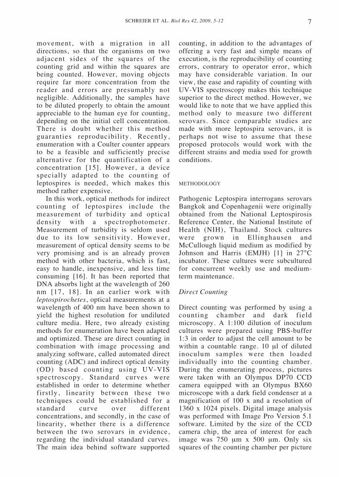
Typically, leptospires are aerobic andmotile spirochetes with helical or spiralstructures and a unique (among thespirochetes) hook at both ends (Fig. 1). Theyare about 0.1-0.2 μm wide and 6-20 μm longand have a helical amplitude of approx 0.1-0.15 μm and wavelength about 0.5 μm [1,9].Their ultra-structure comprises a doublecytoplasmic membrane, in close contact witha peptidoglycan layer and an outer membrane[10]. The composition of lipopolysaccharide(LPS) of their outer membrane is similar toother gram-negative bacteria [11], but haslower endotoxic activity [12]. Optimalconditions for the growth of this organism arewell documented [1,13]. The most suitableconditions for their survival outside the hostare a moist environment with a neutral pHand a temperature range between 20-32°C.
SCHREIER ET AL. Biol Res 42, 2009, 5-126
Leptospirosis research often requires thequantitative determination of bacterialpopulations. Enumerating microbialpopulations is also important for evaluatingsuch products as antibiotics, vitamins, andpreservatives. Quantification is alsonecessary to prepare inocula for bioassaysand tests. There are numerous well-knowndirect, as well as indirect methods, forenumerating bacteria, for instance countingby means of a counting chamber, platecounting, filtration, optical or turbidimetricprocedures, and the Coulter counter [14].For leptospires, the well-accepted methodof enumerating bacteria, which remains thegold standard, is direct counting thespirochetes using a direct counting chamberand dark-field microscopy [2]. However,there are three main features that handicapthe execution and might distort the outcome
of the counting experiment. These aremovement, the low refractive index, whichis equal to water, and the necessity to breedthe cells in more or less a liquid media. Ifnot prevented, the movement of leptospirescauses clumping into cultures, so that thecolonies cannot be traced back to one singlecell [1]. Other problems include regulationof humidity and oxygenation. The plateshould not dry out and too much oxygeninhibits the growth of the leptospires. Aswell, the largely disadvantageousincubation period of 10 to 20 days preventsfast quantification of samples. Becauseleptospires are less visible under a darkfield microscope with a counting chamberslide than with a routine glass slide, themovement of the bacteria makes manualcounting rather difficult. Counting onlyworks with the assumption of random
Figure 1: Pictures of a lepto spire: (a) side view schematic representation of the structure ofLeptospira (b) SEM micrographs of spiral shape taken using scanning electron microscope (Hitachi,Japan) with 15 kV [Magnification 6000x].
7SCHREIER ET AL. Biol Res 42, 2009, 5-12
movement, with a migration in alldirections, so that the organisms on twoadjacent sides of the squares of thecounting grid and within the squares arebeing counted. However, moving objectsrequire far more concentration from thereader and errors are presumably notnegligible. Additionally, the samples haveto be diluted properly to obtain the amountappreciable to the human eye for counting,depending on the initial cell concentration.There is doubt whether this methodguaranties reproducibility. Recently,enumeration with a Coulter counter appearsto be a feasible and sufficiently precisealternative for the quantification of aconcentration [15]. However, a devicespecially adapted to the counting ofleptospires is needed, which makes thismethod rather expensive.
In this work, optical methods for indirectcounting of leptospires include themeasurement of turbidity and opticaldensity with a spectrophotometer.Measurement of turbidity is seldom useddue to its low sensitivity. However,measurement of optical density seems to bevery promising and is an already provenmethod with other bacteria, which is fast,easy to handle, inexpensive, and less timeconsuming [16]. It has been reported thatDNA absorbs light at the wavelength of 260nm [17, 18]. In an earlier work withleptospirochetes, optical measurements at awavelength of 400 nm have been shown toyield the highest resolution for undilutedculture media. Here, two already existingmethods for enumeration have been adaptedand optimized. These are direct counting incombination with image processing andanalyzing software, called automated directcounting (ADC) and indirect optical density(OD) based counting using UV-VISspectroscopy. Standard curves wereestablished in order to determine whetherfirstly, l inearity between these twotechniques could be established for astandard curve over differentconcentrations, and secondly, in the case oflinearity, whether there is a differencebetween the two serovars in evidence,regarding the individual standard curves.The main idea behind software supported
counting, in addition to the advantages ofoffering a very fast and simple means ofexecution, is the reproducibility of countingerrors, contrary to operator error, whichmay have considerable variation. In ourview, the ease and rapidity of counting withUV-VIS spectroscopy makes this techniquesuperior to the direct method. However, wewould like to note that we have applied thismethod only to measure two differentserovars. Since comparable studies aremade with more leptospira serovars, it isperhaps not wise to assume that theseproposed protocols would work with thedifferent strains and media used for growthconditions.
METHODOLOGY
Pathogenic Leptospira interrogans serovarsBangkok and Copenhagenii were originallyobtained from the National LeptospirosisReference Center, the National Institute ofHealth (NIH), Thailand. Stock cultureswere grown in Ellinghausen andMcCullough liquid medium as modified byJohnson and Harris (EMJH) [1] in 27°Cincubator. These cultures were subculturedfor concurrent weekly use and medium-term maintenance.
Direct Counting
Direct counting was performed by using acounting chamber and dark fieldmicroscopy. A 1:100 dilution of inoculumcultures were prepared using PBS-buffer1:3 in order to adjust the cell amount to bewithin a countable range. 10 μl of dilutedinoculum samples were then loadedindividually into the counting chamber.During the enumerating process, pictureswere taken with an Olympus DP70 CCDcamera equipped with an Olympus BX60microscope with a dark field condenser at amagnification of 100 x and a resolution of1360 x 1024 pixels. Digital image analysiswas performed with Image Pro Version 5.1software. Limited by the size of the CCDcamera chip, the area of interest for eachimage was 750 μm x 500 μm. Only sixsquares of the counting chamber per picture
SCHREIER ET AL. Biol Res 42, 2009, 5-128
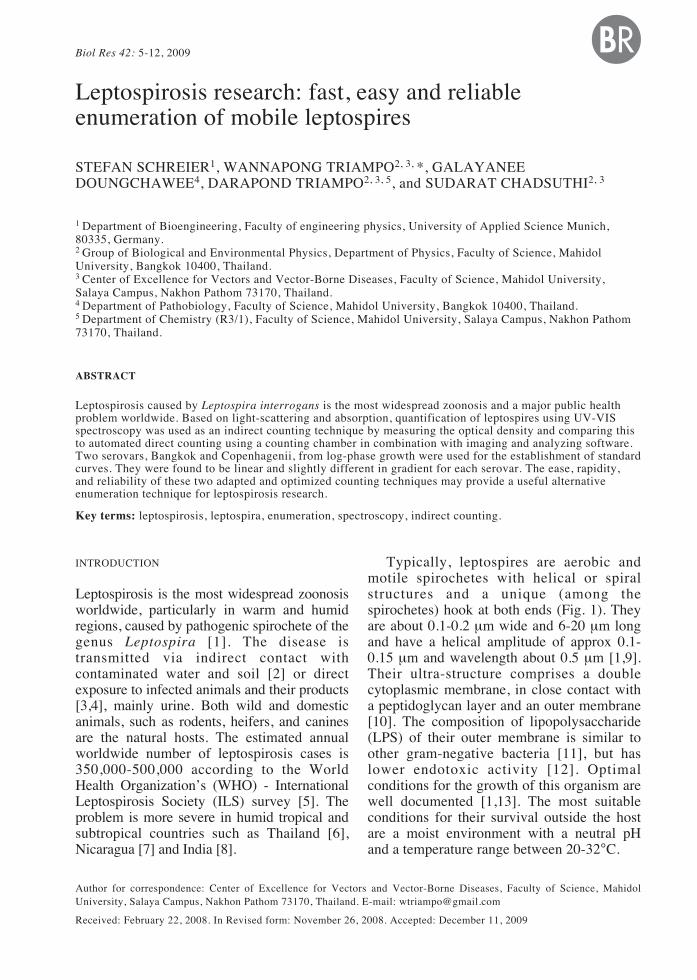
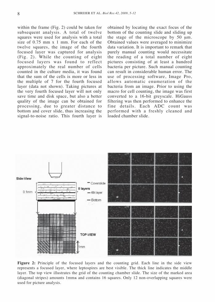
within the frame (Fig. 2) could be taken forsubsequent analysis. A total of twelvesquares were used for analysis with a totalsize of 0.75 mm x 1 mm. For each of thetwelve squares, the image of the fourthfocused layer was captured for analysis(Fig. 2). While the counting of eightfocused layers was found to reflectapproximately the real number of cellscounted in the culture media, it was foundthat the sum of the cells is more or less inthe multiple of 7 for the fourth focusedlayer (data not shown). Taking pictures atthe very fourth focused layer will not onlysave time and disk space, but also a betterquality of the image can be obtained forprocessing, due to greater distance tobottom and cover slide, thus increasing thesignal-to-noise ratio. This fourth layer is
obtained by locating the exact focus of thebottom of the counting slide and sliding upthe stage of the microscope by 50 μm.Obtained values were averaged to minimizedata variation. It is important to remark thatpurely manual counting would necessitatethe reading of a total number of eightpictures consisting of at least a hundredbacteria per picture. Such manual countingcan result in considerable human error. Theuse of processing software, Image Pro,allows automatic enumeration of thebacteria from an image. Prior to using themacro for cell counting, the image was firstconverted to a 16-bit greyscale. HiGaussfiltering was then performed to enhance thefine details. Each ADC count wasperformed with a freshly cleaned andloaded chamber slide.
Figure 2: Principle of the focused layers and the counting grid. Each line in the side viewrepresents a focused layer, where leptospires are best visible. The thick line indicates the middlelayer. The top view illustrates the grid of the counting chamber slide. The size of the marked area(diagonal stripes) amounts 1mm≤ and contains 16 squares. Only 12 non-overlapping squares wereused for picture analysis.
9SCHREIER ET AL. Biol Res 42, 2009, 5-12
Indirect Counting
Indirect counting using UV-VISspectroscopy (V-530 UV/VIS spectrometer,code number 6736-J004A, JascoInternational Co., Ltd.) was performedusing five tubes for each strain.Measurements were obtained using a quartzcuvette (SUPRASIL 200- 2500 nm, lightpath 10 mm, type number 100.600-QG,Hellma Co.) prior to OD measurement ofthe real sample, a baseline of the solutionwas first measured. The baseline solutioncontained no leptospires and was preparedin the same way as the sample solution. Thespectrum ranging from 230 - 450 nm wasrecorded. The wavelengths of interest wereat 260 and 400 nm. The absorbance wascalculated following the Lambert-Beer law[19]. The sample was firstly centrifuged forat least 30 minutes at 14000 rpm. Thesupernatant of the process was used asbaseline solution. The dilution of theEMJH-media with phosphate bufferedsaline (PBS) 1:7 was necessary only for ODmeasurements at the wavelength of 260 nm.Before centrifugation, 0.5 ml of PBS wasadded to each microtube as a pre-dilutionstep of the media. After centrifugation, 1.0ml from the culture was carefully obtainedand used as the initial baseline solution.After that, the packed cells are completelyresuspended in the remaining 0.5 ml ofculture media. 220 μl was then taken anddiluted with 1ml of PBS. The exact sameprocedure was repeated for the baselinesolution. At 400 nm, it is possible to workwith the pure media for baseline correctionof the OD measurement and the sampleitself. As there were also proteins in themedia but no nuclei acid, the measurementat 260 nm is far more sensitive to determinelow cell concentration. The experimentswere aborted after the OD reached thehighest value of 0.6 absolute value, due tosignificant non-linearity beyond thisthreshold.
RESULTS AND DISCUSSION
The measurements were made with the aimof demonstrating the ease, rapidity, and
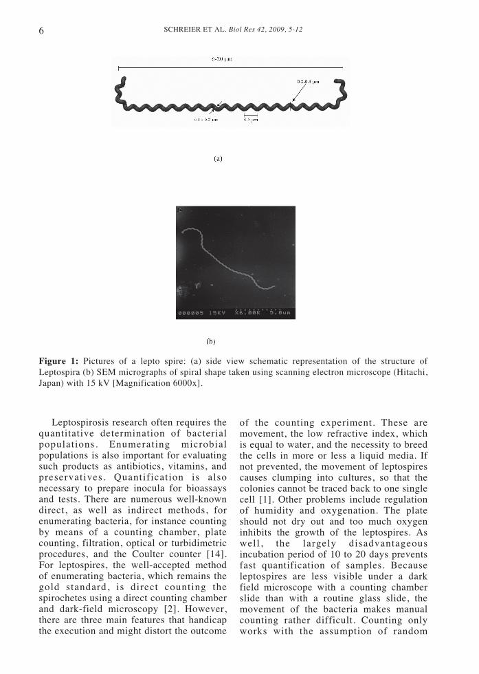
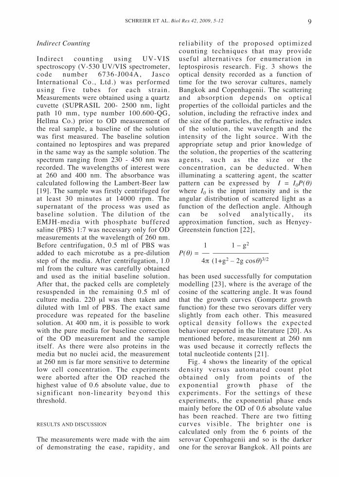
reliability of the proposed optimizedcounting techniques that may provideuseful alternatives for enumeration inleptospirosis research. Fig. 3 shows theoptical density recorded as a function oftime for the two serovar cultures, namelyBangkok and Copenhagenii. The scatteringand absorption depends on opticalproperties of the colloidal particles and thesolution, including the refractive index andthe size of the particles, the refractive indexof the solution, the wavelength and theintensity of the light source. With theappropriate setup and prior knowledge ofthe solution, the properties of the scatteringagents, such as the size or theconcentration, can be deducted. Whenilluminating a scattering agent, the scatterpattern can be expressed by I = I0P(θ)where I0 is the input intensity and is theangular distribution of scattered light as afunction of the deflection angle. Althoughcan be solved analytically, i tsapproximation function, such as Henyey-Greenstein function [22],
1 1 – g2
P(θ) =4π (1+g2 – 2g cosθ)3/2
has been used successfully for computationmodelling [23], where is the average of thecosine of the scattering angle. It was foundthat the growth curves (Gompertz growthfunction) for these two serovars differ veryslightly from each other. This measuredoptical density follows the expectedbehaviour reported in the literature [20]. Asmentioned before, measurement at 260 nmwas used because it correctly reflects thetotal nucleotide contents [21].
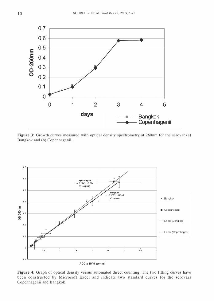
Fig. 4 shows the linearity of the opticaldensity versus automated count plotobtained only from points of theexponential growth phase of theexperiments. For the settings of theseexperiments, the exponential phase endsmainly before the OD of 0.6 absolute valuehas been reached. There are two fittingcurves visible. The brighter one iscalculated only from the 6 points of theserovar Copenhagenii and so is the darkerone for the serovar Bangkok. All points are
SCHREIER ET AL. Biol Res 42, 2009, 5-1210
Figure 3: Growth curves measured with optical density spectrometry at 260nm for the serovar (a)Bangkok and (b) Copenhagenii.
Figure 4: Graph of optical density versus automated direct counting. The two fitting curves havebeen constructed by Microsoft Excel and indicate two standard curves for the serovarsCopenhagenii and Bangkok.
11SCHREIER ET AL. Biol Res 42, 2009, 5-12
derived from five independent samples withone left out, so that the twelve points in thechart are the average of four measurements.The averaged points fit to a high degree(R≤ = 0.998) to the fitting curves, so thatlinearity can be assumed. The results for thestationary phase are especially consistentwith former works [14], where the opticalcharacteristics of Escherichia coli havebeen explored. Limitations of this studyinclude the lack of evaluation of thistechnique on different types of cultures ordirectly from infected tissues or fluids.Future work should include a moreextensive evaluation of the UV-VIS forleptospirosis enumeration, to include fromother sources such as tissue culture anddirectly from experimentally infectedanimals. For precise measurements withUV-VIS spectrometry, it is necessary togather the background spectrum before theactual measurement of the real sample toavoid manual inaccuracy. However, thecells in solution need to be washedcompletely from the media, which isinapplicable for the leptospire cells. Due totheir marginal weight, it is necessary tocentrifuge them at least for 30 minutes at14000 rpm. The total cell-washing stepwould take at least two hours. A fastermethod would be to dilute the media to thenecessary limit for the device and measurethe baselines of each sample gained duringpreparation of the solutions. A disadvantagehowever, is the resolution limit forconcentrations lower than 107.Centrifugation in the matter of countingcells should be regarded with care as cellsmay be lost or destroyed. Counting with acounting chamber in contrast does not needa centrifugation step at all and enables thequantification of leptospira cells at theconcentrations ranging from 105 to 108.
In summary, leptospirosis research iscomplicated by the lack of an efficienttechnique for quantification. This studydemonstrates the use of UV-VISspectroscopy as an indirect countingmethod and automated direct countingmethod as alternatives to determine numberof the leptospire. Limitations of this studyinclude the lack of evaluation of thesetechniques on different types of cultures or
directly from infected tissues or fluids.Future work should include a moreextensive evaluation of both techniques forleptospira enumeration, to include fromother sources such as directly fromexperimentally infected tissues or usingvarious kinds of serovars.
ACKNOWLEDGMENTS
This work has been supported in part by theThailand Center of Excellence in Physics,the Thailand Research Fund (TRF), theCommission on Higher Education (CHE),the National Center for Engineering andBiotechnology (BIOTEC) Thailand, theAcademy of Sciences for the DevelopingWorld (TWAS), and the University ofApplied Science, Munich.
REFERENCES
[1] FAINE S, ADLER B, BOLIN C, PEROLAT P (1999)Leptospira and Leptospirosis, 2ed. Melbourne Victoria,Australia: MediSci: 173-174
[2] LEVETT PN (2001) Leptospirosis. Clin Microbiol Rev14: 296-326
[3] TANGKANAKUL W, THARMAPHORNPIL P,PLIKAYTIS BD, BRAGG S, POONSUKSOMBAT D,CHOOMKASIEN P, KINGNATE D, ASHFORD DA(2000) Risk factors associated with leptospirosis innortheastern Thailand, 1998. Am J Trop Med Hyg 63:204-208
[4] DOUGLIN CP, JORDAN C, ROCK R, HURLEY A,LEVETT PN (1997) Risk factors for severeleptospirosis in the parish of St. Andrew, Barbados.Emerg Infect Dis 3: 78-80
[5] INTERNATIONAL LEPTOSPIROSIS SOCIETY (ILS).In: Proccedings of the fourth ILS 2005 abstracts,Chiang Mai, Thailand, November 14-16, 2005
[6] WATT G, JONGSAKUL K, SUTTINONT C (2003)Possible scrub typhus coinfections in Thai agriculturalworkers hospitalized with leptospirosis. Am J TropMed Hyg 68: 89-91
[7] BRANDLING-BENNETT AD, PINHEIRO F (1996)Infectious diseases in Latin America and theCaribbean: are they really emerging and increasing.Emerg Infect Dis 2: 59-61
[8] World Health Organization. Leptospirosis, India:Report of the investigation of a post-cyclone outbreakin Orissa, Wkly Epidemiol Rec WHO, Nov. 1999, pp.217-223
[9] GOLDSTEIN SF, CHARON NW (1990) Multiple-exposure photographic analysis of a motile spirochete.Proc Natl Acad Sci (USA) 87: 4895-4899
[10] HAAKE DA (2000) Spirochaetal lipoproteins andpathogenesis. Microbiology 146: 1491-1504
[11] VINH T, ADLER B, FAINE S (1986) Ultrastructureand chemical composition of lipopolysaccharideextracted from Leptospira interrogans serovarCopenhagenii. J Gen Microbiol 132: 103-109
SCHREIER ET AL. Biol Res 42, 2009, 5-1212
[12] SHIMIZU T, MATSUSAKA E, TAKAYANAGI K,MASUZAWA T, IWAMOTO Y, MORITA T,MIFUCHI I, YANAGIHARA Y (1987) Biologicalactivities of lipopolysaccharide-like substance (LLS)extracted from Leptospira interrogans serovar Canicolastrain Moulton. Microbiol Immunol 31: 727-735
[13] FAINE S. Guideline for control of leptospirosis,World Health Organization, Geneva 1982, Vol. 67, pp.129
[14] PHILIPP G, KRIEG NR, MURRAY RGE, WOODWA (1994) Methods for general and molecularbacteriology. American Society for Microbiology: 261-266
[15] HUMBERD CM, MURRAY CK, STUART SK, REEBBA (2005) Enumerating leptospires using the CoulterCounter. Am J Trop Med Hyg 73: 962-963
[16] ALUPOAEI CE, GARCIAUBIO LH (2004) GrowthBehavior of Microorganisms using UV-VISspectroscopy: Escherichia coli. Biotechnol Bioeng 86:163-167
[17] FREIFELDER D (1982) Physical Biochemistry,Applications to Biochemistry and Molecular Biology,2nd ed, WH Freeman and Company, New York: 504
[18] INAGAKI T, HAMM RN, ARAKAWA ET, PAINTERLR (1997) Optical and dielectric properties of DNA inthe extreme ultraviolet. J Chem Phys 61: 4246- 4250
[19] PERKAMPUS HH (1992) UV-VIS spectroscopy andits application. New York: Springer-Verlag BerlinHeidelberg
[20] WONG-EKKABUT J (2007) Effects ofelectromagnetic f ields on biological systems :Leptospira. Thesis (Ph.D. (Physics)) MahidolUniversity, CALL NUMBER: Thesis J61e 2007(Reserve)
[21] FREIFELDER D (1982) In: Physical biochemistry,2nd ed. San Francisco: W.H. Freeman & Company:504
[22] HENYEY LG, GREENSTEIN JL (1941) Diffuseradiation in the galaxy. Astrophys J 193: 70-83
[23] MOURANT JR, FREYER JP, HIELSCHER AH, EICKAA, SHEN D, JOHNSON TM (1998) Mechanisms oflight scattering from biological cells relevant tononinvasive optical-tissue diagnostics. Applied Optics37: 3586-3593
Related Documents