Lenalidomide therapy in treatment refractory cutaneous lupus erythematosus: Histologic and circulating leukocyte profile and potential risk of a systemic lupus flare I Braunstein, MD 1,2 , NG Goodman, MPH 3 , M Rosenbach, MD 2 , J Okawa, RN 1,2 , A Shah, MD 1,2 , M Krathen, MD 1,2 , CL Kovarik, MD 1,2 , E Luning Prak, MD,PhD 3 , and VP Werth, MD 1,2 1 Department of Dermatology, Philadelphia Veterans Affairs Medical Center, Philadelphia, PA 2 Department of Dermatology, University of Pennsylvania, Philadelphia, PA 3 Department of Pathology & Laboratory Medicine, University of Pennsylvania, Philadelphia, PA Abstract Background—Lenalidomide is a thalidomide analogue that may serve as an adjunctive therapy for treatment refractory cutaneous lupus erythematosus (CLE). Objectives—We evaluate the use of lenalidomide in CLE and describe the skin and circulating leukocyte profile of treatment refractory patients before and after treatment. Patients/Methods—Five subjects were treated with lenalidomide in an unblinded open-label study. Immunohistochemistry of skin was performed for T-cell markers, glycosaminoglycans and CXCL10, an interferon (IFN)-inducible chemokine, before and after treatment. Immunophenotyping and measurement of IFN-inducible genes from peripheral blood mononuclear cells was also performed before and after treatment. Results—Four subjects demonstrated clinical improvement of their skin, however one of these responders subsequently developed symptoms of systemic lupus erythematosus. Small changes in rare circulating leukocyte subsets, plasmacytoid dendritic cells and regulatory T-cells, were observed with treatment and may correlate with clinical response. Treatment was associated with increased circulating HLA-DR expression and decreased markers of IFN-mediated pathways, regardless of clinical response. Limitations—Our results are limited by small sample size and the measurement of rare populations of circulating cell subsets. Conclusions—Lenalidomide may have utility as therapy for severe, treatment refractory CLE. However, our preliminary data suggest that lenalidomide may activate T-cells and trigger systemic disease in some CLE patients. We also saw a unique histologic and circulating leukocyte phenotype in the nonresponding subject. Further characterization of the skin and circulating leukocyte profile of treatment refractory patients will improve our understanding of CLE. Corresponding Author: Victoria P. Werth, M.D., Department of Dermatology, Hospital of the University of Pennsylvania, PCAM Suite 1-330S, 3400 Civic Center Blvd., Philadelphia, PA 19104, Tel. 215-898-4208, Fax 866-755-0625, [email protected]. Prior presentation of part of this manuscript at the Society of Investigative Dermatology 2009 annual meeting and American College of Rheumatology 2009 annual meeting Conflict of Interest: The authors have no conflict of interest to declare. NIH Public Access Author Manuscript J Am Acad Dermatol. Author manuscript; available in PMC 2013 April 1. Published in final edited form as: J Am Acad Dermatol. 2012 April ; 66(4): 571–582. doi:10.1016/j.jaad.2011.01.015. NIH-PA Author Manuscript NIH-PA Author Manuscript NIH-PA Author Manuscript

Welcome message from author
This document is posted to help you gain knowledge. Please leave a comment to let me know what you think about it! Share it to your friends and learn new things together.
Transcript

Lenalidomide therapy in treatment refractory cutaneous lupuserythematosus: Histologic and circulating leukocyte profile andpotential risk of a systemic lupus flare
I Braunstein, MD1,2, NG Goodman, MPH3, M Rosenbach, MD2, J Okawa, RN1,2, A Shah,MD1,2, M Krathen, MD1,2, CL Kovarik, MD1,2, E Luning Prak, MD,PhD3, and VP Werth, MD1,2
1Department of Dermatology, Philadelphia Veterans Affairs Medical Center, Philadelphia, PA2Department of Dermatology, University of Pennsylvania, Philadelphia, PA3Department of Pathology & Laboratory Medicine, University of Pennsylvania, Philadelphia, PA
AbstractBackground—Lenalidomide is a thalidomide analogue that may serve as an adjunctive therapyfor treatment refractory cutaneous lupus erythematosus (CLE).
Objectives—We evaluate the use of lenalidomide in CLE and describe the skin and circulatingleukocyte profile of treatment refractory patients before and after treatment.
Patients/Methods—Five subjects were treated with lenalidomide in an unblinded open-labelstudy. Immunohistochemistry of skin was performed for T-cell markers, glycosaminoglycans andCXCL10, an interferon (IFN)-inducible chemokine, before and after treatment.Immunophenotyping and measurement of IFN-inducible genes from peripheral bloodmononuclear cells was also performed before and after treatment.
Results—Four subjects demonstrated clinical improvement of their skin, however one of theseresponders subsequently developed symptoms of systemic lupus erythematosus. Small changes inrare circulating leukocyte subsets, plasmacytoid dendritic cells and regulatory T-cells, wereobserved with treatment and may correlate with clinical response. Treatment was associated withincreased circulating HLA-DR expression and decreased markers of IFN-mediated pathways,regardless of clinical response.
Limitations—Our results are limited by small sample size and the measurement of rarepopulations of circulating cell subsets.
Conclusions—Lenalidomide may have utility as therapy for severe, treatment refractory CLE.However, our preliminary data suggest that lenalidomide may activate T-cells and trigger systemicdisease in some CLE patients. We also saw a unique histologic and circulating leukocytephenotype in the nonresponding subject. Further characterization of the skin and circulatingleukocyte profile of treatment refractory patients will improve our understanding of CLE.
Corresponding Author: Victoria P. Werth, M.D., Department of Dermatology, Hospital of the University of Pennsylvania, PCAMSuite 1-330S, 3400 Civic Center Blvd., Philadelphia, PA 19104, Tel. 215-898-4208, Fax 866-755-0625, [email protected] presentation of part of this manuscript at the Society of Investigative Dermatology 2009 annual meeting and American Collegeof Rheumatology 2009 annual meetingConflict of Interest: The authors have no conflict of interest to declare.
NIH Public AccessAuthor ManuscriptJ Am Acad Dermatol. Author manuscript; available in PMC 2013 April 1.
Published in final edited form as:J Am Acad Dermatol. 2012 April ; 66(4): 571–582. doi:10.1016/j.jaad.2011.01.015.
NIH
-PA Author Manuscript
NIH
-PA Author Manuscript
NIH
-PA Author Manuscript

IntroductionApproximately 10% of patients with cutaneous lupus erythematosus (CLE) are refractory toestablished therapies, including thalidomide1. CLE has been reported to have the sameincidence as systemic lupus erythematosus (SLE), approximately 3 per 100,000,highlighting the need for improved therapies2. Lenalidomide, a thalidomide analogue withmore potent T-cell, IL-2 and Interferon (IFN)-γ stimulating effects in vitro compared tothalidomide, is a potential alternative or adjunctive treatment for severe generalized discoidlupus erythematosus (DLE)3. It is an immunomodulatory drug shown to stimulate T-cells,natural killer (NK)-cells and anti-inflammatory cytokines and inhibit tumor necrosis factoralpha (TNF-α) production and angiogenesis4.
The current CLE pathogenesis model implicates IFN-inducible proteins and chemokines inthe recruitment and activation of inflammatory cells in the skin5-8. Plasmacytoid dendriticcells (pDCs) accumulate in CLE skin and produce type I IFNs which may initiate theinflammatory cascade8-11. IFN-inducible chemokines, like CXCL9, CXCL10 and CXCL11,are highly expressed around lymphocytes in CLE lesions, supporting a potential role in cellrecruitment5, 7, 8. The lymphocytic infiltrate also expresses cutaneous lymphocyte antigen(CLA), a skin-homing receptor 12. CLE lesions have increased glycosaminoglycans (GAG)content and are characterized by low numbers of regulatory T-cells (Tregs).13
There is evidence of B- and T-cell activation in the peripheral blood of patients with DLEand subacute cutaneous lupus erythematosus (SCLE)14-17. In DLE absolute circulatinglymphocyte numbers are similar to healthy controls, but SCLE patients have decreasedabsolute circulating CD4+ and CD8+ T-cells18-20. No difference in circulating Treg countshave been seen in DLE, SCLE or tumid lupus erythematosus (TLE)13. Investigators haveobserved severe scarring DLE to have increased expression of circulating CCR4+ and CLA+ cells (skin-homing receptors) and decreased CXCR3+ expression, a chemokine receptorassociated with IFN-signaling6, 21.
These circulating changes support the growing evidence of systemic alterations in theinflammatory cascade in CLE. Recently we found elevated IFN-inducible genes in bloodfrom patients with DLE and SCLE22. A polymorphism in Interferon regulatory factor 5(IRF5) in DLE and SCLE has been described23. Additionally, mutations in TREX1, a DNAexonuclease implicated in SLE and Aicardi-Goutieres syndrome, have been reported in afamilial form of chilblain lupus, suggesting a shared pathogenesis between these conditionsmanifesting with elevated serum IFN-α24, 25.
Here we investigate the use of lenalidomide in five patients with CLE. We examinedhistology, circulating leukocyte subsets and markers of IFN-mediated signaling, in the skinand blood, before and after treatment to better characterize the pathophysiology of thisrefractory patient subset and gain insight into the mechanism of action of lenalidomide. Wealso describe one patient who developed SLE during lenalidomide therapy.
Materials and methodsPatients
Five patients were enrolled according to the following inclusion criteria: 1) DLE or SCLEdiagnosis, 2) no response to 3 months of hydroxychloroquine and 3) participation inRevAssist®, a distribution program run by the manufacturers of lenalidomide26. Patientswith SLE (by ACR criteria), who were pregnant, had thrombocytopenia, lymphopenia,neutropenia or a history of deep venous thrombosis or pulmonary embolism were excluded.Informed consent was obtained. Subjects received 5 mg oral lenalidomide daily during the
Braunstein et al. Page 2
J Am Acad Dermatol. Author manuscript; available in PMC 2013 April 1.
NIH
-PA Author Manuscript
NIH
-PA Author Manuscript
NIH
-PA Author Manuscript

first six weeks of treatment. Patients were maintained on all baseline medications. The studyprotocol was approved by the University of Pennsylvania Institutional Review Board.
Skin biopsy collectionA punch biopsy of lesional skin was taken at weeks 0, 2, and 6 of treatment. A biopsy ofnon-lesional skin was taken at week 0. Half of each biopsy was fixed in formalin and theother half frozen in liquid nitrogen. Week 2 biopsies were refused by subjects 4 and 5.
Assessment of clinical disease activityDisease activity was assessed using the CLASI (cutaneous lupus area and severity index)activity score27, 28. A partial response reflects a four-point decrease in CLASI activity score(Klein et al. in press). VPW or MR performed all CLASI scoring.
Immunohistochemical stainingImmunohistochemical (IHC) staining was performed on formalin-fixed, paraffin-embeddedskin biopsy samples using: monoclonal mouse anti-CD3 (Novacastra, Clone PS1, Newcastleupon Tyne, UK), anti-CD4 (Novacastra, Clone IF6), anti-CD8 (Dako, Clone C8/144B,Glostrup, Denmark), rat anti-CLA (BD Pharmingen, Clone HECA-452, San Jose,California), and polyclonal goat anti-CXCL10/IP-10, (interferon-γ inducible protein ten)(R&D Systems, Minneapolis, MN). Isotype control reactions were performed with mouseIgG1 (Sigma, Clone MOPC 21, St. Louis, MO) for CD4 and CD8, mouse IgG2a (Dako,Clone DAK-G05) for CD3, goat total IgG (R&D systems) for CXCL10, and rat IgM, Kisotype (BD Pharmigen, Clone R4-22) for CLA. Indirect IHC staining was performed withDako LSAB™ system, except for CLA where biotinylated mouse anti-rat IgM (IgG1) (BDPharmigen, Clone G53-238) was the secondary antibody. Staining was visualized usingNovoRed chromogen (Vector Labs, Burlingame, CA) or DAB chromogen (Dako).Hematoxylin and Eosin (H&E) and Hale’s stain were performed using establishedprotocols29.
Immunohistochemical staining quantificationCXCL10 staining was quantified using ImagePro (MediaCybernetics, Bethesda, MD).Percent staining was calculated from six high-power-field (HPF) images (400x). CD3, CD4,CD8, and CLA staining was quantified using the cell counter plugin on ImageJ Software(NIH, Bethesda, MD).
Peripheral blood collection and flow cytometryPeripheral blood cells were obtained at weeks 0, 2, and 6 of treatment. A complete bloodcount with differential and immunofluorescence was performed. Monoclonal antibodieswere added directly to 150 μl aliquots of whole blood and incubated at room temperature for20 minutes. Red blood cells were lysed with ammonium chloride lysing buffer (BD PharmLyse). Remaining mononuclear cells were washed using wash buffer (Dulbecco’s PBS with5% fetal calf serum and sodium azide). Cells were fixed in 1% paraformaldehyde (ElectronMicroscopy Sciences, Hatfield, PA).
The following antibodies were used: FITC-HLA-DR (clone L243), FITC-CLA (cloneHECA-452), PE-CD123 (clone 9F5), PE-CD127 (clone hIL-7R-M21), PerCP-CD4 (cloneSK3), APC-HLA-DR (clone L243), APC-CD25 (clone M-A251), APC-CD27 (clone L128),PE-Cy5-CD40 (clone 5C3), and PE-Cy7-CD19 (clone SJ25C1) (all BD Pharmigen). APC-Cy5.5 -CD38 (clone HIT2), APC-Alexa Fluor 750-CD3 (clone S4), PE-TR-CD4 (cloneS3.5), and PE-Cy5.5-CD5 (clone 5D7) (all Caltag/Invitrogen, Carlsbad, CA). Alexa Fluor700 -CD33 (clone WM-53) and PE-Cy7-CD11c (clone 2.9) (eBioscience, San Diego, CA).
Braunstein et al. Page 3
J Am Acad Dermatol. Author manuscript; available in PMC 2013 April 1.
NIH
-PA Author Manuscript
NIH
-PA Author Manuscript
NIH
-PA Author Manuscript

Dead cell exclusion was performed using the Live/Dead Fixable Violet Dead Cell Stain Kit(Invitrogen, Carlsbad, CA). The sample preparation, instrument operation and data analysiswas performed by the same operator to control for inter-operator variability. Flow cytometrywas performed on the same instrument (Becton Dickinson LSR2 Benchtop Flow Cytometer)to control for machine variability. Data was analyzed using v.8.2 Flow Jo software (TreestarInc, Eugene, OR).
Type I IFN-inducible gene expressionFive IFN-inducible genes were studied: lymphocyte antigen 6 complex, locus E (LYGE),Hs00158942_m1; 2’,5’-oligoadenylate synthetase 1, 40/46 kd (OAS1), HS00242943_m1;2’,5’-oligoadenylate synthetase-like (OASL), Hs00388714_m1; myxovirus resistance 1(MX1), Hs00182073_m1 and interferon-α-inducible protein (clone IFI-15K) (ISG15),Hs00192713_m1 (Applied Biosystems, Foster City, CA). Total RNA was extracted fromperipheral blood using RNA Later, Qiagen RNeasy Minikit and Qiashredder Minicolumns(Qiagen, Valencia, CA). Total RNA was reverse transcribed into complementary DNA(Invitrogen First-Strand cDNA Synthesis Kit). RNA was stored at -70°C and cDNA samplesstored at -20°C. Gene expression was measured by real time PCR using ABI Prism 7000sequence detection system and normalized to GAPDH (Taqman assay: Hs99999905_m1)using ABI Prism SDS 7000 software, version 1.0 (Applied Biosystems). Experiments wereperformed in triplicate in 25 μl of TaqMan Universal PCR Master Mix (AppliedBiosystems). Samples were denatured for 2 minutes at 50°C then 10 minutes at 95°C,followed by 40 cycles at 95°C for 15 seconds and combined primer annealing and extensionat 60°C for 1 minute. IFN scores were calculated using published methods30.
Statistical analysisA Wilcoxon signed rank test was performed to evaluate changes in IFN score and IHCstaining with treatment.
ResultsWe present the clinical response to lenalidomide in five subjects and describe the histologicfeatures and circulating leukocyte profile before and after treatment. Expression levels ofIFN-inducible genes in blood before and after treatment are shown as a measure of systemicIFN-mediated signaling.
Clinical ResponseAge, sex, race/ethnicity, diagnosis and medications are listed for each subject (Table 1).Four subjects demonstrated satisfactory clinical improvement in their skin. The CLASIactivity scores at weeks 0, 2 and 6 are shown for each patient. All subjects demonstrated atleast a four-point drop from week 0 to 6, consistent with a partial response; however, foursubjects had more pronounced changes, with a drop in CLASI activity score of at least 8points and will be referred to as responders. Subject 2, a female with generalized DLE, had afive-point decrease in her CLASI score, however this was felt to be clinically unsatisfactoryand she was withdrawn from the study at week 12. She will be referred to as thenonresponder. Among the responders were three females with DLE (subjects 1, 4 and 5) andone male with a mixed clinical phenotype of SCLE and TLE (subjects 3). Although subject5 experienced improvement in her skin lesions, she developed new-onset proteinuria and anexacerbation of arthralgias at week 20 prompting withdrawal from the study.
Braunstein et al. Page 4
J Am Acad Dermatol. Author manuscript; available in PMC 2013 April 1.
NIH
-PA Author Manuscript
NIH
-PA Author Manuscript
NIH
-PA Author Manuscript
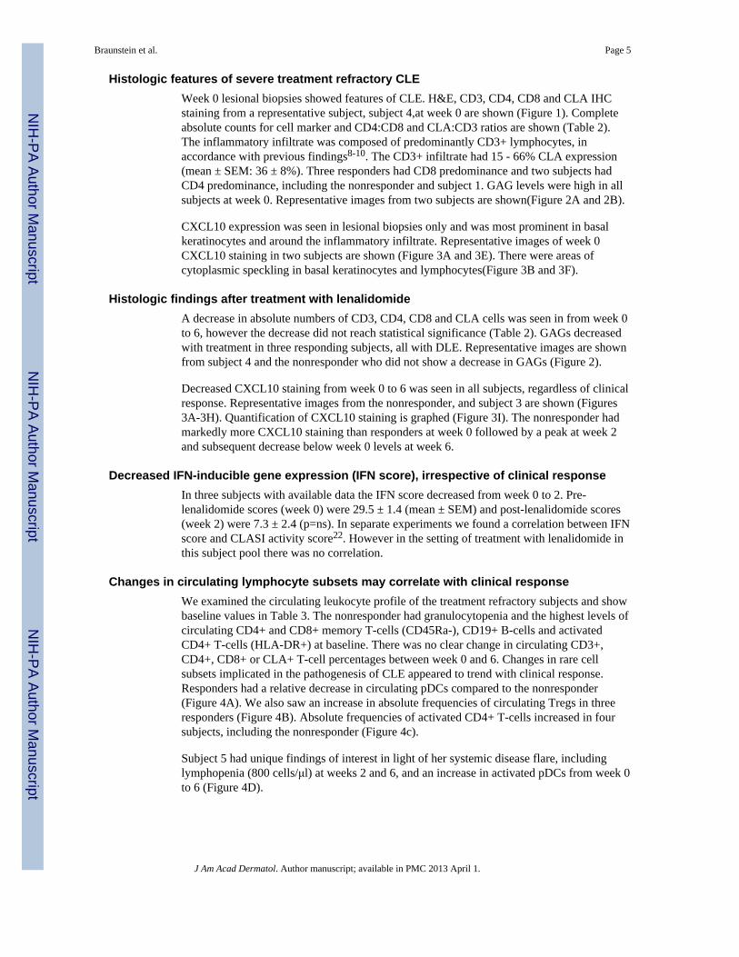
Histologic features of severe treatment refractory CLEWeek 0 lesional biopsies showed features of CLE. H&E, CD3, CD4, CD8 and CLA IHCstaining from a representative subject, subject 4,at week 0 are shown (Figure 1). Completeabsolute counts for cell marker and CD4:CD8 and CLA:CD3 ratios are shown (Table 2).The inflammatory infiltrate was composed of predominantly CD3+ lymphocytes, inaccordance with previous findings8-10. The CD3+ infiltrate had 15 - 66% CLA expression(mean ± SEM: 36 ± 8%). Three responders had CD8 predominance and two subjects hadCD4 predominance, including the nonresponder and subject 1. GAG levels were high in allsubjects at week 0. Representative images from two subjects are shown(Figure 2A and 2B).
CXCL10 expression was seen in lesional biopsies only and was most prominent in basalkeratinocytes and around the inflammatory infiltrate. Representative images of week 0CXCL10 staining in two subjects are shown (Figure 3A and 3E). There were areas ofcytoplasmic speckling in basal keratinocytes and lymphocytes(Figure 3B and 3F).
Histologic findings after treatment with lenalidomideA decrease in absolute numbers of CD3, CD4, CD8 and CLA cells was seen in from week 0to 6, however the decrease did not reach statistical significance (Table 2). GAGs decreasedwith treatment in three responding subjects, all with DLE. Representative images are shownfrom subject 4 and the nonresponder who did not show a decrease in GAGs (Figure 2).
Decreased CXCL10 staining from week 0 to 6 was seen in all subjects, regardless of clinicalresponse. Representative images from the nonresponder, and subject 3 are shown (Figures3A-3H). Quantification of CXCL10 staining is graphed (Figure 3I). The nonresponder hadmarkedly more CXCL10 staining than responders at week 0 followed by a peak at week 2and subsequent decrease below week 0 levels at week 6.
Decreased IFN-inducible gene expression (IFN score), irrespective of clinical responseIn three subjects with available data the IFN score decreased from week 0 to 2. Pre-lenalidomide scores (week 0) were 29.5 ± 1.4 (mean ± SEM) and post-lenalidomide scores(week 2) were 7.3 ± 2.4 (p=ns). In separate experiments we found a correlation between IFNscore and CLASI activity score22. However in the setting of treatment with lenalidomide inthis subject pool there was no correlation.
Changes in circulating lymphocyte subsets may correlate with clinical responseWe examined the circulating leukocyte profile of the treatment refractory subjects and showbaseline values in Table 3. The nonresponder had granulocytopenia and the highest levels ofcirculating CD4+ and CD8+ memory T-cells (CD45Ra-), CD19+ B-cells and activatedCD4+ T-cells (HLA-DR+) at baseline. There was no clear change in circulating CD3+,CD4+, CD8+ or CLA+ T-cell percentages between week 0 and 6. Changes in rare cellsubsets implicated in the pathogenesis of CLE appeared to trend with clinical response.Responders had a relative decrease in circulating pDCs compared to the nonresponder(Figure 4A). We also saw an increase in absolute frequencies of circulating Tregs in threeresponders (Figure 4B). Absolute frequencies of activated CD4+ T-cells increased in foursubjects, including the nonresponder (Figure 4c).
Subject 5 had unique findings of interest in light of her systemic disease flare, includinglymphopenia (800 cells/μl) at weeks 2 and 6, and an increase in activated pDCs from week 0to 6 (Figure 4D).
Braunstein et al. Page 5
J Am Acad Dermatol. Author manuscript; available in PMC 2013 April 1.
NIH
-PA Author Manuscript
NIH
-PA Author Manuscript
NIH
-PA Author Manuscript

DiscussionFour of five patients with treatment refractory CLE had a clinically satisfactory response tolenalidomide. Two of the responders (subjects 1 and 3) and the nonresponder previouslyfailed thalidomide therapy. Lesional biopsies showed histologic features of CLE includingCD3+ infiltrate and GAG accumulation. Two subjects had a CD4 predominant infiltrate, inkeeping with prior reports9, 10. Recent investigation suggests CD4 predominance bettercharacterizes SCLE and TLE, while CD8 predominance characterizes scarring subsets likeDLE and lupus profundus7, 21, 31, 32. Two subjects with CD8 predominance had a scarringDLE phenotype and the other had SCLE/TLE overlap. CLA staining of the infiltrate wasvariable in accordance with previous findings, suggesting a range of CLA+ expression inCLE lesions12.
We saw decreased GAG content in three responding subjects with DLE. We find thisinteresting given the immunologic activity of GAGs, specifically their ability to stimulatedendritic cells via toll-like receptors33, 34. Further study into the role of these molecules inthe pathogenesis of CLE is warranted.
To assess the predominant model of IFN-mediated signaling in CLE pathogenesis, weanalyzed CXCL10 expression in the skin and the IFN-inducible gene expression in theperipheral blood before and after treatment. CXCL10 gene expression increases inkeratinocytes after stimulation with IFN-α; however, the same response has been seen inapoptotic keratinocytes treated with IFN-γ5, 6, 35. This suggests CXCL10 may also reflecttype II IFN-mediated signaling, however, CXCL10 is expressed at significantly higherlevels in CLE lesions than in other inflammatory dermatoses, suggesting specificity for CLEpathogenesis5.
All lesional biopsies had significant CXCL10 staining. The nonresponder had the highestlevels, peaking to involve greater than 15% of the section at week 2 (Figure 3). However, allsubjects had lower CXCL10 staining at week 6 than baseline. Also, the IFN score of threesubjects decreased with treatment irrespective of the clinical response.
A small body of literature characterizing the circulating leukocyte phenotype in CLE exists.We add characteristics of severe treatment refractory CLE (Table 3). Prior studies showedincreased circulating CLA+ CD4+ and CD8+ T-cells in generalized DLE compared tohealthy controls21. Here we observed less circulating CLA+ leukocytes than described usingthe same antibody clone. Also we noted the nonresponder to have unique baseline findingsincluding granulocytopenia, and higher levels of B cells, and memory and activated T cells.Further study is needed to determine if these findings have prognostic value.
No significant changes in the circulating leukocyte profile was seen with treatment, howeverrare circulating subsets appeared to trend with clinical response. Responders had a decreasein circulating pDCs and an increase in circulating Tregs from week 0 to 6 (Figure 4A and4B). Although these represent rare subsets and the changes represent small changes inabsolute number, both cell types have been implicated in the pathophysiology of CLE. Thedecrease in pDCs in responders is interesting given their ability to produce type I IFNs. Priorstudies showed normal numbers of circulating Tregs in CLE. Here we show an increase inTregs with response to treatment. We used CD25++, CD127- as Treg markers leaving thepossibility activated T-cells were counted. Lenalidomide has been shown to increase CD4+Foxp3 cells in patients with multiple myeloma, suggesting a potential drug specific effect36.
Absolute numbers of circulating HLA-DR+ CD4+ T-cells, a rare circulating cell-type in ourstudy, increased in four subjects, including the nonresponder (Figure 4C). A correlationbetween CLE clinical activity and HLA-DR expression on CD4+ and CD8+ T cells has been
Braunstein et al. Page 6
J Am Acad Dermatol. Author manuscript; available in PMC 2013 April 1.
NIH
-PA Author Manuscript
NIH
-PA Author Manuscript
NIH
-PA Author Manuscript

described. We believe this correlation requires further investigation in the setting oftreatment.
Finally, we saw unique changes in subject 5, the patient whose skin responded butdeveloped systemic symptoms of SLE at week 20 of the study (Figure 4D). This patient hada robust increase in activated pDCs which may correlate with systemic symptoms, as SLE isassociated with high levels of serum IFNαwhich activated pDCs produce30. We cannotexclude a predisposition to develop SLE. She was the only subject to report arthralgias atbaseline and was lymphopenic at weeks 2 and 6. Lymphopenia has been described as amarker for SLE development in CLE patients37. In light of this event and evidence of cellactivation, we caution of the risk of systemic disease activation with lenalidomide.
In summary, lenalidomide reduced CLE activity in four of five subjects. The histology andcirculating leukocyte profile of five patients with severe treatment refractory CLE isdescribed, including CD8 predominant infiltrate in three subjects. The nonresponder hadhigh CXCL10 expression in the skin and increased activated circulating CD4+T-cells.Responders with DLE had a decrease in GAG content. Clinical response was also associatedwith a trend towards increased circulating Tregs and decreased circulating pDCs. Inaddition, we noted a decrease in IFN-mediated gene expression in the peripheral blood andIFN-inducible chemokine expression in the skin that did not correlate with clinical response.Finally, we documented evidence of cellular activation and caution that lenalidomide mayactivate T-cells and pDCs, triggering systemic disease in a subset of CLE patients.
The small subject pool limits recommendations on patient selection and monitoringparameters. However patients with signs of systemic disease such as arthralgias may not becandidates. In considering the use of this medication for treatment refractory CLE it wouldbe prudent to monitor for signs and symptoms of SLE, including a CBC with differential tolook for cytopenias. Additional study is required to understand the predictive value of morespecialized laboratory studies in predicting response to lenalidomide therapy. Further workto identify SLE susceptibility risk factors in CLE patients may be informative in safelyselecting patients that would benefit from lenalidomide.
AcknowledgmentsThis work was supported in part by the Alliance for Lupus Research, a Merit Review Grant from the Department ofVeterans Affairs Veterans Health Administration, Office of Research and Development, Biomedical LaboratoryResearch and Development and by the National Institutes of Health (NIH K24-AR 02207) to VPW.
Funding sources: This study was supported by the Alliance for Lupus Research. Celgene Corporation provided thedrug free of charge.
References1. Moghadam-Kia S, Chilek K, Gaines E, Costner M, Rose ME, Okawa J, et al. Cross-sectional
analysis of a collaborative Web-based database for lupus erythematosus-associated skin lesions:prospective enrollment of 114 patients. Arch Dermatol. 2009; 145:255–60. [PubMed: 19289753]
2. Durosaro O, Davis MD, Reed KB, Rohlinger AL. Incidence of cutaneous lupus erythematosus,1965-2005: a population-based study. Arch Dermatol. 2009; 145:249–53. [PubMed: 19289752]
3. Shah A, Albrecht J, Bonilla-Martinez Z, Okawa J, Rose M, Rosenbach M, et al. Lenalidomide forthe treatment of resistant discoid lupus erythematosus. Arch Dermatol. 2009; 145:303–6. [PubMed:19289762]
4. Kotla V, Goel S, Nischal S, Heuck C, Vivek K, Das B, et al. Mechanism of action of lenalidomidein hematological malignancies. J Hematol Oncol. 2009; 2:36. [PubMed: 19674465]
Braunstein et al. Page 7
J Am Acad Dermatol. Author manuscript; available in PMC 2013 April 1.
NIH
-PA Author Manuscript
NIH
-PA Author Manuscript
NIH
-PA Author Manuscript

5. Meller S, Winterberg F, Gilliet M, Muller A, Lauceviciute I, Rieker J, et al. Ultraviolet radiation-induced injury, chemokines, and leukocyte recruitment: An amplification cycle triggering cutaneouslupus erythematosus. Arthritis Rheum. 2005; 52:1504–16. [PubMed: 15880822]
6. Wenzel J, Worenkamper E, Freutel S, Henze S, Haller O, Bieber T, et al. Enhanced type I interferonsignalling promotes Th1-biased inflammation in cutaneous lupus erythematosus. J Pathol. 2005;205:435–42. [PubMed: 15685590]
7. Wenzel J, Zahn S, Mikus S, Wiechert A, Bieber T, Tuting T. The expression pattern of interferon-inducible proteins reflects the characteristic histological distribution of infiltrating immune cells indifferent cutaneous lupus erythematosus subsets. Br J Dermatol. 2007; 157:752–7. [PubMed:17714558]
8. Flier J, Boorsma DM, van Beek PJ, Nieboer C, Stoof TJ, Willemze R, et al. Differential expressionof CXCR3 targeting chemokines CXCL10, CXCL9, and CXCL11 in different types of skininflammation. J Pathol. 2001; 194:398–405. [PubMed: 11523046]
9. Tebbe B, Mazur L, Stadler R, Orfanos CE. Immunohistochemical analysis of chronic discoid andsubacute cutaneous lupus erythematosus--relation to immunopathological mechanisms. Br JDermatol. 1995; 132:25–31. [PubMed: 7756149]
10. Wenzel J, Uerlich M, Worrenkamper E, Freutel S, Bieber T, Tuting T. Scarring skin lesions ofdiscoid lupus erythematosus are characterized by high numbers of skin-homing cytotoxiclymphocytes associated with strong expression of the type I interferon-induced protein MxA. Br JDermatol. 2005; 153:1011–5. [PubMed: 16225615]
11. Farkas L, Beiske K, Lund-Johansen F, Brandtzaeg P, Jahnsen FL. Plasmacytoid dendritic cells(natural interferon- alpha/beta-producing cells) accumulate in cutaneous lupus erythematosuslesions. Am J Pathol. 2001; 159:237–43. [PubMed: 11438470]
12. Magro CM, Dyrsen ME. Cutaneous lymphocyte antigen expression in benign and neoplasticcutaneous B- and T-cell lymphoid infiltrates. J Cutan Pathol. 2008; 35:1040–9. [PubMed:18681860]
13. Franz B, Fritzsching B, Riehl A, Oberle N, Klemke CD, Sykora J, et al. Low number of regulatoryT cells in skin lesions of patients with cutaneous lupus erythematosus. Arthritis Rheum. 2007;56:1910–20. [PubMed: 17530636]
14. Kind P, Lipsky PE, Sontheimer RD. Circulating T- and B-cell abnormalities in cutaneous lupuserythematosus. J Invest Dermatol. 1986; 86:235–9. [PubMed: 3489051]
15. Wouters CH, Diegenant C, Ceuppens JL, Degreef H, Stevens EA. The circulating lymphocyteprofiles in patients with discoid lupus erythematosus and systemic lupus erythematosus suggest apathogenetic relationship. Br J Dermatol. 2004; 150:693–700. [PubMed: 15099365]
16. Wenzel J, Henze S, Brahler S, Bieber T, Tuting T. The expression of human leukocyte antigen-DRand CD25 on circulating T cells in cutaneous lupus erythematosus and correlation with diseaseactivity. Exp Dermatol. 2005; 14:454–9. [PubMed: 15885081]
17. Wangel AG, Johansson E, Ranki A. Polyclonal B-cell activation and increased lymphocyte helper-suppressor ratios in discoid lupus erythematosus. Br J Dermatol. 1984; 110:665–9. [PubMed:6234013]
18. Gilliam JN, Hurd ER. Comparison of circulating T and B lymphocytes in discoid versus systemiclupus erythematosus. Clin Immunol Immunopathol. 1976; 6:149–55. [PubMed: 1085683]
19. Wenzel J, Bauer R, Boehm I. Fluorescence-activated cell sorter analysis in patients with cutaneouslupus erythematosus. Arch Dermatol. 1999; 135:720–1. [PubMed: 10376710]
20. Wenzel J, Bauer R, Bieber T, Boehm I. Discoid and subacute cutaneous lupus erythematosus:detection of differences in peripheral lymphocyte numbers. Acta Derm Venereol. 2000; 80:456.[PubMed: 11243649]
21. Wenzel J, Henze S, Worenkamper E, Basner-Tschakarjan E, Sokolowska-Wojdylo M, Steitz J, etal. Role of the chemokine receptor CCR4 and its ligand thymus- and activation-regulatedchemokine/CCL17 for lymphocyte recruitment in cutaneous lupus erythematosus. J InvestDermatol. 2005; 124:1241–8. [PubMed: 15955100]
22. Braunstein, IKR.; Rosenbach, M.; Okawa, J.; Werth, VP. The IFN-inducible Gene Signature isElevated in SCLE and DLE and correlates with the CLASI score. The 73rd Annual ScientificMeeting of the American College of Rheumatology; Philadelphia PA. 2009.
Braunstein et al. Page 8
J Am Acad Dermatol. Author manuscript; available in PMC 2013 April 1.
NIH
-PA Author Manuscript
NIH
-PA Author Manuscript
NIH
-PA Author Manuscript

23. Jarvinen TM, Hellquist A, Koskenmies S, Einarsdottir E, Koskinen LL, Jeskanen L, et al. Tyrosinekinase 2 and interferon regulatory factor 5 polymorphisms are associated with discoid andsubacute cutaneous lupus erythematosus. Exp Dermatol. 2009
24. Rice G, Newman WG, Dean J, Patrick T, Parmar R, Flintoff K, et al. Heterozygous mutations inTREX1 cause familial chilblain lupus and dominant Aicardi-Goutieres syndrome. Am J HumGenet. 2007; 80:811–5. [PubMed: 17357087]
25. Goutieres F, Aicardi J, Barth PG, Lebon P. Aicardi-Goutieres syndrome: an update and results ofinterferon-alpha studies. Ann Neurol. 1998; 44:900–7. [PubMed: 9851434]
26. Castaneda CP, Zeldis JB, Freeman J, Quigley C, Brandenburg NA, Bwire R. RevAssist: acomprehensive risk minimization programme for preventing fetal exposure to lenalidomide. DrugSaf. 2008; 31:743–52. [PubMed: 18707189]
27. Albrecht J, Taylor L, Berlin JA, Dulay S, Ang G, Fakharzadeh S, et al. The CLASI (CutaneousLupus Erythematosus Disease Area and Severity Index): an outcome instrument for cutaneouslupus erythematosus. J Invest Dermatol. 2005; 125:889–94. [PubMed: 16297185]
28. Bonilla-Martinez ZL, Albrecht J, Troxel AB, Taylor L, Okawa J, Dulay S, et al. The cutaneouslupus erythematosus disease area and severity index: a responsive instrument to measure activityand damage in patients with cutaneous lupus erythematosus. Arch Dermatol. 2008; 144:173–80.[PubMed: 18283174]
29. Lillie, R.; Fullmer, H. Histopathologic Technic and Practical Histochemistry. New York: McGraw-Hill Book Co; 1976.
30. Feng X, Wu H, Grossman JM, Hanvivadhanakul P, FitzGerald JD, Park GS, et al. Association ofincreased interferon-inducible gene expression with disease activity and lupus nephritis in patientswith systemic lupus erythematosus. Arthritis Rheum. 2006; 54:2951–62. [PubMed: 16947629]
31. Alexiades-Armenakas MR, Baldassano M, Bince B, Werth V, Bystryn JC, Kamino H, et al. Tumidlupus erythematosus: criteria for classification with immunohistochemical analysis. ArthritisRheum. 2003; 49:494–500. [PubMed: 12910555]
32. Kuhn A, Sonntag M, Lehmann P, Megahed M, Vestweber D, Ruzicka T. Characterization of theinflammatory infiltrate and expression of endothelial cell adhesion molecules in lupuserythematosus tumidus. Arch Dermatol Res. 2002; 294:6–13. [PubMed: 12071156]
33. Tesar BM, Jiang D, Liang J, Palmer SM, Noble PW, Goldstein DR. The Role of HyaluronanDegradation Products as Innate Alloimmune Agonists. American Journal of Transplantation. 2006;6:2622–35. [PubMed: 17049055]
34. Termeer C, Benedix F, Sleeman J, Fieber C, Voith U, Ahrens T, et al. Oligosaccharides ofHyaluronan activate dendritic cells via toll-like receptor 4. J Exp Med. 2002; 195:99–111.[PubMed: 11781369]
35. Klunker S, Trautmann A, Akdis M, Verhagen J, Schmid-Grendelmeier P, Blaser K, et al. A secondstep of chemotaxis after transendothelial migration: keratinocytes undergoing apoptosis releaseIFN-gamma-inducible protein 10, monokine induced by IFN-gamma, and IFN-gamma-induciblealpha-chemoattractant for T cell chemotaxis toward epidermis in atopic dermatitis. J Immunol.2003; 171:1078–84. [PubMed: 12847282]
36. Minnema MC, van der Veer MS, Aarts T, Emmelot M, Mutis T, Lokhorst HM. Lenalidomidealone or in combination with dexamethasone is highly effective in patients with relapsed multiplemyeloma following allogeneic stem cell transplantation and increases the frequency ofCD4+Foxp3+ T cells. Leukemia. 2009; 23:605–7. [PubMed: 18784738]
37. Wenzel J, Bauer R, Uerlich M, Bieber T, Boehm I. The value of lymphocytopenia as a marker ofsystemic involvement in cutaneous lupus erythematosus. Br J Dermatol. 2002; 146:869–71.[PubMed: 12000386]
Braunstein et al. Page 9
J Am Acad Dermatol. Author manuscript; available in PMC 2013 April 1.
NIH
-PA Author Manuscript
NIH
-PA Author Manuscript
NIH
-PA Author Manuscript

Figure 1.Representative images from a responding subject, subject 4, baseline (week 0) lesional skin.(a) Hematoxylin & Eosin (H&E) stain, (b) CD3 (c) CLA (d) CD4 (e) CD8. Scale bar = 500μm for H&E, panel (A), and Scale bar = 100 μm for remaining stains, panels (B)-(E).
Braunstein et al. Page 10
J Am Acad Dermatol. Author manuscript; available in PMC 2013 April 1.
NIH
-PA Author Manuscript
NIH
-PA Author Manuscript
NIH
-PA Author Manuscript

Figure 2.Decrease in Hale’s staining in responding subjects with DLE. Hale’s staining highlightsglycosaminoglycan (GAG) content (blue). Images from subject 2, nonresponder, and subject4, a responder with DLE, before and after treatment are shown. Subjects 1, 4 and 5 hadsimilar decrease in GAG staining at week 6. Week 0, lesional: (A) subject 2, (B) subject 4;and Week 6, lesional: (C) subject 2, (D) subject 4. Scale bar = 500 μm.
Braunstein et al. Page 11
J Am Acad Dermatol. Author manuscript; available in PMC 2013 April 1.
NIH
-PA Author Manuscript
NIH
-PA Author Manuscript
NIH
-PA Author Manuscript

Figure 3.CXCL10 expression in lesional skin decreases in basal keratinocytes and aroundlymphocytes with lenalidomide treatment. Panels (a-h): Representative images of higheststaining for subjects 2 and 3 are shown for lesional skin at week 0, week 2, and week 6.Subject 2: (A) week 0, (B) week 0 high power, (C) week 2, (D) week 6, Subject 3: (E) week0, (F) week 0 high power, (G) week 2, and (H) week 6. Scale bar = 500 μm, except scale bar= 100 μM for high power. Panel (I) Graphic representation of the change in percentCXCL10 staining area with treatment.
Braunstein et al. Page 12
J Am Acad Dermatol. Author manuscript; available in PMC 2013 April 1.
NIH
-PA Author Manuscript
NIH
-PA Author Manuscript
NIH
-PA Author Manuscript

Figure 4.Changes in the circulating leukocyte profile that correlate with clinical response. (A)Percent-change in plasmacytoid dendritic cells (pDCs) (CD4- CD33-CD123+ CD11c-) frombaseline to week 6. Subjects who experienced skin improvement had an average 74%decrease in pDC percentage (mean ± SEM: -74% ± 7%) vs. a 28% increase in thenonresponder; (B) Absolute numbers of regulatory T-cell (Treg) (CD4+ CD25++ CD127-)increased from week 0 to week 6 in responders. Subjects who experienced skinimprovement had a mean 4.1-fold increase in Treg percentage (mean ± SEM: 4.1-foldincrease ± 1.84-fold increase) vs. a 0.13-fold increase in the nonresponder; © Absolutenumbers of activated CD4+ T-cells increased (CD3+ CD4+ HLA-DR+ CD25+) from week0 to week 6. (D) Increase in activated pDCs (CD4- CD33- CD123+ CD11c- CD40+ HLA-DR+) in subject 5. Subject 5 had a robust and unique increase in activated pDCs.
Braunstein et al. Page 13
J Am Acad Dermatol. Author manuscript; available in PMC 2013 April 1.
NIH
-PA Author Manuscript
NIH
-PA Author Manuscript
NIH
-PA Author Manuscript

NIH
-PA Author Manuscript
NIH
-PA Author Manuscript
NIH
-PA Author Manuscript
Braunstein et al. Page 14
Tabl
e 1
Subj
ect b
asel
ine
char
acte
ristic
s and
clin
ical
out
com
e
Subj
ect
Age
Sex
Rac
e/E
thni
city
Dia
gnos
isM
edic
atio
nsC
linic
al O
utco
me
CL
ASI
act
ivity
scor
eW
eek
02
6
147
FW
hite
/Cau
casi
an n
on-h
ispa
nic
Gen
eral
ized
DLE
Dap
sone
25
mg
twic
e da
ilyPa
rtial
resp
onse
2015
8
235
FW
hite
/Cau
casi
an n
on-h
ispa
nic
Loca
lized
DLE
,H
yper
troph
ic L
EQ
uina
crin
e 10
0 da
ilyPa
rtial
resp
onse
, but
uns
atis
fact
ory
clin
ical
resp
onse
. The
pat
ient
was
with
draw
n at
wee
k 12
of t
he st
udy.
2321
18
Met
hotre
xate
25
mg
subc
utan
eous
lyw
eekl
y
Aza
thio
prin
e 75
mg
thre
e tim
es d
aily
Chl
oroq
uine
250
mg
daily
343
MW
hite
/Cau
casi
an n
on-h
ispa
nic
SCLE
/TLE
Parti
al re
spon
se22
189
437
FW
hite
/Cau
casi
an n
on-h
ispa
nic
Loca
lized
DLE
, SC
LEPr
edni
sone
30
mg
daily
for w
eeks
1an
d 2,
then
20
mg
daily
for w
eeks
3-6.
Parti
al re
spon
se16
158
Myc
ophe
nyla
te M
ofet
il 50
0 m
gtw
ice
daily
Chl
oroq
uine
250
mg
daily
Qui
nacr
ine
100
mg
daily
542
FW
hite
/Cau
casi
an n
on-h
ispa
nic
Gen
eral
ized
DLE
Hyd
roxy
chlo
roqu
ine
200
mg
twic
eda
ilyPa
rtial
resp
onse
2618
11
Qui
nacr
ine
100
mg
daily
Aza
thio
prin
e 75
mg
daily
F, fe
mal
e; M
, mal
e; D
LE, d
isco
id lu
pus e
ryth
emat
osus
; LE,
lupu
s ery
them
atos
us; S
CLE
, sub
acut
e cu
tane
ous l
upus
ery
them
atos
us; a
nd T
LE, t
umid
lupu
s ery
them
atos
us.
A p
artia
l res
pons
e is
def
ined
by
a de
crea
se in
cut
aneo
us lu
pus a
rea
and
seve
rity
inde
x (C
LASI
) act
ivity
scor
e of
four
or m
ore
poin
ts.
J Am Acad Dermatol. Author manuscript; available in PMC 2013 April 1.

NIH
-PA Author Manuscript
NIH
-PA Author Manuscript
NIH
-PA Author Manuscript
Braunstein et al. Page 15
Tabl
e 2
CD
4: C
D8
ratio
and
CLA
:CD
3 ra
tio a
t wee
k 0
and
wee
k 6
in le
sion
al sk
in
Wee
k 0
Wee
k 6
CD
4C
D8
CD
4:C
D8
CD
4C
D8
CD
4:C
D8
Subj
ect 1
286
139
2.1
3126
1.2
Subj
ect 2
144
951.
513
590
1.5
Subj
ect 3
1125
0.4
2813
50.
2
Subj
ect 4
143
194
0.7
2570
0.4
Subj
ect 5
4017
50.
28
280.
3
Ave
rage
±SE
M in
all
125±
4812
6±30
1±0.
345
±23
70±2
00.
7±0.
3
Ave
rage
±SE
M in
res
pond
ers
120±
6213
3±38
0.9±
0.4
23±5
65±2
60.
5±0.
2
CL
AC
D3
CL
A:C
D3
CL
AC
D3
CL
A:C
D3
Subj
ect 1
8053
40.
142
560.
8
Subj
ect 2
100
271
0.4
130
319
0.4
Subj
ect 3
5315
30.
366
157
0.4
Subj
ect 4
252
875
0.3
5187
0.6
Subj
ect 5
214
324
0.7
6623
70.
3
Ave
rage
±SE
M in
all
140±
3943
1±12
70.
4±0.
171
±15
171±
480.
5±0.
1
Ave
rage
±SE
M in
res
pond
ers
150±
4947
2±15
50.
4±0.
156
±613
4±40
0.5±
0.1
Cel
ls w
ere
coun
ted
man
ually
usi
ng th
e ce
ll co
unte
r plu
gin
on Im
ageJ
softw
are.
A h
igh
pow
er fi
eld
repr
esen
ting
the
area
of g
reat
est s
tain
ing
was
cou
nted
. Sim
ilar h
igh
pow
er fi
elds
from
sequ
entia
l sec
tions
wer
e us
ed a
t eac
h tim
epoi
nt.
CLA
, cut
aneo
us ly
mph
ocyt
e an
tigen
; and
SEM
, sta
ndar
d er
ror o
f mea
sure
men
t.
J Am Acad Dermatol. Author manuscript; available in PMC 2013 April 1.

NIH
-PA Author Manuscript
NIH
-PA Author Manuscript
NIH
-PA Author Manuscript
Braunstein et al. Page 16
Tabl
e 3
Circ
ulat
ing
leuk
ocyt
e pr
ofile
in se
vere
, tre
atm
ent r
efra
ctor
y C
LE
WBC
subs
ets (
tho/μl
)
Lym
phoc
ytes
Nl r
ange
(0.8
-3.9
)G
ranu
locy
tes N
l ran
ge (1
.5-7
.8)
Mon
ocyt
es N
l ran
ge (0
.2-.9
5)
Wee
k 0
Wee
k 6
Wee
k 0
Wee
k 6
Wee
k 0
Wee
k 6
subj
ect 1
1.6
1.6
5.9
5.7
0.4
0.4
subj
ect 2
2.2
1.6
0.8
1.9
0.5
0.4
subj
ect 3
1.5
1.2
2.7
3.5
0.3
0.3
subj
ect 4
2.1
2.1
4.6
3.2
0.8
0.9
subj
ect 5
1.1
0.8
3.0
3.8
0.4
0.3
Lym
phoc
yte
subs
ets (
tho/μl
)
T-ce
lls (C
D3+
)B
-cel
ls (C
D19
+)N
K c
ells
(CD
3- C
D16
+ C
D56
+)
Wee
k 0
Wee
k 6
Wee
k 0
Wee
k 6
Wee
k 0
Wee
k 6
subj
ect 1
1.3
1.3
0.1
0.08
0.4
0.3
subj
ect 2
1.3
1.0
0.3
0.2
0.6
0.4
subj
ect 3
1.4
1.0
0.1
0.03
0.3
0.6
subj
ect 4
1.6
1.7
0.1
0.09
1.4
1.4
subj
ect 5
1.0
0.7
0.02
0.01
0.4
0.1
T-ce
ll su
bset
s (th
o/μl
)
subs
ets
CD
4+ C
D8-
CD
8+ C
D4-
CD
4+ C
D8+
CD
4-C
D8-
Wee
k 0
Wee
k 6
Wee
k 0
Wee
k 6
Wee
k 0
Wee
k 6
Wee
k 0
Wee
k 6
subj
ect 1
1.0
1.0
0.2
0.2
<0.0
10.
010.
020.
03
subj
ect 2
0.9
0.8
0.3
0.2
0.02
0.02
0.07
0.02
subj
ect 3
0.8
0.6
0.5
0.3
0.02
0.02
0.06
0.04
subj
ect 4
0.9
0.9
0.6
0.6
0.01
0.02
0.09
0.07
subj
ect 5
0.8
0.5
0.2
0.1
0.01
0.01
0.02
0.01
B-ce
ll su
bset
s (th
o/μl
)
Tran
sitio
nal (
CD
19+
CD
27-C
D38
++)
naïv
e m
atur
e (C
D19
+ C
D27
-CD
38+)
rest
ing
mem
ory
(CD
19+
CD
27+
CD
38-)
mat
ure
activ
ated
(CD
19+
CD
27+
CD
38+)
Wee
k 0
Wee
k 6
Wee
k 0
Wee
k 6
Wee
k 0
Wee
k 6
Wee
k 0
Wee
k 6
subj
ect 1
<0.0
1<0
.01
0.08
0.06
<0.0
1<0
.01
0.02
0.01
J Am Acad Dermatol. Author manuscript; available in PMC 2013 April 1.

NIH
-PA Author Manuscript
NIH
-PA Author Manuscript
NIH
-PA Author Manuscript
Braunstein et al. Page 17
WBC
subs
ets (
tho/μl
)
Lym
phoc
ytes
Nl r
ange
(0.8
-3.9
)G
ranu
locy
tes N
l ran
ge (1
.5-7
.8)
Mon
ocyt
es N
l ran
ge (0
.2-.9
5)
Wee
k 0
Wee
k 6
Wee
k 0
Wee
k 6
Wee
k 0
Wee
k 6
subj
ect 2
0.01
<0.0
10.
20.
1<0
.01
<0.0
10.
050.
04
subj
ect 3
<0.0
1<0
.01
0.08
0.03
<0.0
1<0
.01
0.01
<0.0
1
subj
ect 4
<0.0
1<0
.01
0.07
0.05
0.01
<0.0
10.
020.
03
subj
ect 5
<0.0
1<0
.01
0.01
<0.0
1<0
.01
<0.0
1<0
.01
<0.0
1
Treg
s, ac
tivat
ed T
-cel
ls a
nd p
DC
s (ce
lls/μ
l)
Treg
(CD
4+ C
D25
++ C
D12
7-)
activ
ated
CD
4+ (C
D4+
HLA
-DR
+ C
D25
+)pD
Cs (
CD
4-C
D33
-CD
123+
CD
11c-
)
Wee
k 0
Wee
k 6
Wee
k 0
Wee
k 6
Wee
k 0
Wee
k 6
subj
ect 1
25
735
0.6
0.2
subj
ect 2
33
3965
11
subj
ect 3
47
1645
20.
3
subj
ect 4
0.5
5na
747
1
subj
ect 5
819
2553
40.
7
CLA
+ T
-cel
ls su
bset
s (th
o/μl
)
CD
4 m
emor
y (C
D4+
CLA
+ C
D45
RA
-)C
D4
naiv
e (C
D4+
CLA
+ C
D45
RA
+)C
D8
mem
ory
(CD
8+ C
LA+
CD
45R
A-)
CD
8 na
ive
(CD
8+ C
LA+
CD
45R
A+)
Wee
k 0
Wee
k 6
Wee
k 0
Wee
k 6
Wee
k 0
Wee
k 6
Wee
k 0
Wee
k 6
subj
ect 1
0.03
0.02
0.05
0.2
<0.0
10.
010.
030.
2
subj
ect 2
0.03
0.03
0.05
0.1
0.01
0.01
0.02
0.09
subj
ect 3
0.01
0.01
0.01
0.03
0.01
0.01
0.01
0.04
subj
ect 4
0.10
0.04
<0.0
11.
70.
010.
030.
051.
8
subj
ect 5
0.02
0.01
0.7
0.7
0.02
0.01
0.2
0.6
WB
C, w
hite
blo
od c
ell;
Nl r
ange
, nor
mal
rang
e; N
K, n
atur
al k
iller
; Tre
g, re
gula
tory
T-c
ell;
and
CLA
, cut
aneo
us ly
mph
ocyt
e an
tigen
.
J Am Acad Dermatol. Author manuscript; available in PMC 2013 April 1.
Related Documents