Lecture 3 Lecture 3 CATARACTS CATARACTS

Lecture 3 CATARACTS
Jan 02, 2016
Lecture 3 CATARACTS. Classification of cataracts: By age: congenital, juvenile, age-related (senile) By location opacities in the lens as seen with slit beam : cortical, nuclear, anterior subcapsular, posterior subcapsular - PowerPoint PPT Presentation
Welcome message from author
This document is posted to help you gain knowledge. Please leave a comment to let me know what you think about it! Share it to your friends and learn new things together.
Transcript

Lecture 3Lecture 3
CATARACTSCATARACTS


Classification of cataracts:Classification of cataracts:
By age: By age: congenital, juvenile, age-related congenital, juvenile, age-related (senile)(senile)
By location opacities in the lens as seen By location opacities in the lens as seen with slit beamwith slit beam:: cortical, nuclear, anterior cortical, nuclear, anterior subcapsular, posterior subcapsularsubcapsular, posterior subcapsular
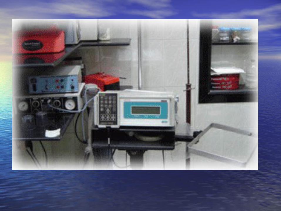
By maturity: By maturity: early, immature, mature, early, immature, mature, hypermature (Morgagnian cataract)hypermature (Morgagnian cataract)
By pattern: By pattern: cuneiform (typical senile type), cuneiform (typical senile type), zonular, polar, pyramidal (congenital types)zonular, polar, pyramidal (congenital types)


DIFFERENTIAL DIAGNOSISThe same sign is progressive (not acute) painless visual loss
SignCATARACT GLAUCOMA
MACULAR DEGENERATION
Visual acuity
is decreased
Field of vision
is not damaged constriction of nasal visual field, Bjerrum’s scotoma
may be central scotoma
Intraocular pressure
normal,if increased-secondary phakogenic glaucoma
increased normal
Lens opaque transparent,if opaque– complicated cataract
transparent, if opaque– complicated cataract
Fundus If is seen, not damaged.If damaged - complicated cataract
optic disc changes - dislocation of vessels, pale colour, increased cupping
degenerative patches in central area


Artificial lensArtificial lens

Artiphakia is a condition of eye with artificial lens (IOL).
Aphakia is a condition of eye without lens. The visual acuity without correction is very poor – 0,02-0,04. Iridodenesis (iris vibration) is typical.Thick plus glasses are needed for vision: for far distance – sph convex 10,0-12,0 Dfor near distance – sph convex 13,0-15,0 D.
Secondary cataract occurs eventually in about 20 % cases after cataract surgery. It is opacity of natural posterior capsule. It can be treated by YAG laser capsulotomy.
Attention! The term «complicated cataract» is used to describe a lens opacity which occurs as a result of some other disease of the eye. Longstanding uveitits, an untreated retinal detachment or an intraocular tumour are all examples of an associated disease.


CONGENITAL CATARACTS :
I degree – visual acuity is 0,3 and more; the size of opacity is less then 1,5 mm; the surgery may be done at the age of 14-16 years.
II degree– visual acuity is 0,05-0,2; the surgery is usually done at the age of 3-4 years.
III degree– visual acuity is less then 0,05; the surgery must be done during first year of life.


Ectopia lentis (displacement of the lens) may be partial (subluxation) or complete (luxation).
Aethiology: trauma, familial ectopia lentis (may be associated with ectopic pupil), associated with other ocular disorders (aniridia and buphtalmos), Marfan’s and Weill-Marchesani syndromes, metabolic (homocystinuria and hyperlysinaemia).
Clinical features: iridodenesis (vibration of iris) and not proportional depth of anterior chamber.
Marfan’s syndrome: tall person with partial displacement of the lens (subluxation), fragile bones and arachnodactyly.


THANK YOU FOR THANK YOU FOR ATTENTION !ATTENTION !
Related Documents