Latrunculin A and Its C-17-O-Carbamates Inhibit Prostate Tumor Cell Invasion and HIF-1 Activation in Breast Tumor Cells ⊥ Khalid A. El Sayed †,* , Hassan M. Shallal † , Mohammad A. Khanfar † , A. Muralidharan † , Bhushan Awate † , Diaa T.A. Youssef ‡ , Yang Liu § , Yu-Dong Zhou § , Dale G. Nagle § , and Girish Shah † † Department of Basic Pharmaceutical Sciences, College of Pharmacy, University of Louisiana at Monroe, 700 University Avenue, Monroe, Louisiana 71209 ‡ Department of Pharmacognosy, Faculty of Pharmacy, Suez Canal University, Ismailia 41522, Egypt § Department of Pharmacognosy, School of Pharmacy, University of Mississippi, University, Mississippi 38677-1848 Abstract The marine-derived macrolides latrunculins A (1) and B, from the Red Sea sponge Negombata magnifica, have been found to reversibly bind actin monomers, forming a 1:1 complex with G-actin and disrupting its polymerization. The microfilament protein actin is responsible for several essential functions within the cell such as cytokinesis and cell migration. One of the main binding pharmacophores of 1 to G-actin was identified as the C-17 lactol hydroxyl moiety that binds arginine 210 NH. Latrunculin A-17-O-carbamates 2–6 were prepared by reaction with the corresponding isocyanates. Latrunculin A (1) and carbamates 4–6 displayed potent anti-invasive activity against the human highly metastatic human prostate cancer PC-3M cells in a Matrigel™ assay at a concentration range of 50 nM-1 μM. Latrunculin A (1, 500 nM) decreased the disaggregation and cell migration of PC-3M-CT+ spheroids by three-fold. Carbamates 4 and 5 were two and half and five-fold more active than 1, respectively, in this assay with less actin binding affinity. Latrunculin A (1, IC 50 6.7 μM) and its 17-O-[N-(benzyl)carbamate (6, IC 50 29 μM) suppress hypoxia-induced HIF-1 activation in T47D breast tumor cells. Latrunculin A (1) and B are macrolides reported by Kashman and coworkers from the Red Sea sponge Negombata magnifica Kelly-Borges and Vacelet (Podospongiidae). 1 Latrunculins are reported to decrease intraocular pressure and increase outflow facility without corneal effects in monkeys.2 , 3 Latrunculin B and analogs showed antiangiogenic, antimetastatic, and antimicrobial activities.4 The most important biological effects of latrunculins are their abilities to disrupt microfilament organization and inhibit microfilament-mediated processes without affecting the organization of the microtubular system. 5 The latrunculins bind reversibly to the cytoskeleton actin monomers, forming 1:1 complexes with G-actin and disrupting polymerization. 5 Actin-active agents are attracting more attention in the field of cancer chemotherapy because microfilament and microtubule proteins form versatile dynamic polymers that can define cell polarity, organize cytoplasmic organelles, control cell shape and promote stable cell-cell and cell-matrix adhesions, and generate protrusive forces required for migration. 6–8 These functions usually fail and become abnormal in cancer cells. 6–8 ⊥ Dedicated to Dr. G. Robert Pettit of Arizona State University for his pioneering work on bioactive natural products. * To whom correspondence should be addressed. Tel: 318-342-1725. Fax: 318-342-1737. [email protected]. NIH Public Access Author Manuscript J Nat Prod. Author manuscript; available in PMC 2010 August 31. Published in final edited form as: J Nat Prod. 2008 March ; 71(3): 396–402. doi:10.1021/np070587w. NIH-PA Author Manuscript NIH-PA Author Manuscript NIH-PA Author Manuscript

Welcome message from author
This document is posted to help you gain knowledge. Please leave a comment to let me know what you think about it! Share it to your friends and learn new things together.
Transcript
Latrunculin A and Its C-17-O-Carbamates Inhibit Prostate TumorCell Invasion and HIF-1 Activation in Breast Tumor Cells⊥
Khalid A. El Sayed†,*, Hassan M. Shallal†, Mohammad A. Khanfar†, A. Muralidharan†,Bhushan Awate†, Diaa T.A. Youssef‡, Yang Liu§, Yu-Dong Zhou§, Dale G. Nagle§, and GirishShah††Department of Basic Pharmaceutical Sciences, College of Pharmacy, University of Louisiana atMonroe, 700 University Avenue, Monroe, Louisiana 71209‡Department of Pharmacognosy, Faculty of Pharmacy, Suez Canal University, Ismailia 41522,Egypt§Department of Pharmacognosy, School of Pharmacy, University of Mississippi, University,Mississippi 38677-1848
AbstractThe marine-derived macrolides latrunculins A (1) and B, from the Red Sea sponge Negombatamagnifica, have been found to reversibly bind actin monomers, forming a 1:1 complex with G-actinand disrupting its polymerization. The microfilament protein actin is responsible for several essentialfunctions within the cell such as cytokinesis and cell migration. One of the main bindingpharmacophores of 1 to G-actin was identified as the C-17 lactol hydroxyl moiety that binds arginine210 NH. Latrunculin A-17-O-carbamates 2–6 were prepared by reaction with the correspondingisocyanates. Latrunculin A (1) and carbamates 4–6 displayed potent anti-invasive activity againstthe human highly metastatic human prostate cancer PC-3M cells in a Matrigel™ assay at aconcentration range of 50 nM-1 µM. Latrunculin A (1, 500 nM) decreased the disaggregation andcell migration of PC-3M-CT+ spheroids by three-fold. Carbamates 4 and 5 were two and half andfive-fold more active than 1, respectively, in this assay with less actin binding affinity. LatrunculinA (1, IC50 6.7 µM) and its 17-O-[N-(benzyl)carbamate (6, IC50 29 µM) suppress hypoxia-inducedHIF-1 activation in T47D breast tumor cells.
Latrunculin A (1) and B are macrolides reported by Kashman and coworkers from the Red Seasponge Negombata magnifica Kelly-Borges and Vacelet (Podospongiidae).1 Latrunculins arereported to decrease intraocular pressure and increase outflow facility without corneal effectsin monkeys.2,3 Latrunculin B and analogs showed antiangiogenic, antimetastatic, andantimicrobial activities.4 The most important biological effects of latrunculins are their abilitiesto disrupt microfilament organization and inhibit microfilament-mediated processes withoutaffecting the organization of the microtubular system.5 The latrunculins bind reversibly to thecytoskeleton actin monomers, forming 1:1 complexes with G-actin and disruptingpolymerization.5 Actin-active agents are attracting more attention in the field of cancerchemotherapy because microfilament and microtubule proteins form versatile dynamicpolymers that can define cell polarity, organize cytoplasmic organelles, control cell shape andpromote stable cell-cell and cell-matrix adhesions, and generate protrusive forces required formigration.6–8 These functions usually fail and become abnormal in cancer cells. 6–8
⊥Dedicated to Dr. G. Robert Pettit of Arizona State University for his pioneering work on bioactive natural products.*To whom correspondence should be addressed. Tel: 318-342-1725. Fax: 318-342-1737. [email protected].
NIH Public AccessAuthor ManuscriptJ Nat Prod. Author manuscript; available in PMC 2010 August 31.
Published in final edited form as:J Nat Prod. 2008 March ; 71(3): 396–402. doi:10.1021/np070587w.
NIH
-PA Author Manuscript
NIH
-PA Author Manuscript
NIH
-PA Author Manuscript
Based on X-ray crystallography, the binding site of 1 has been located between subdomains IIand IV, in the vicinity of the ATP binding cleft of the protein target in the actin monomer.9,10 The binding pharmacophores of 1 to G-actin were identified as follows: C-1 carbonyl oxygenthrough water to glutamate 214 carboxy, C-17 lactol hydroxyl to arginine 210 NH (majorbinding), C-17 pyran oxygen to tyrosine 69 hydroxy, thiazolidinone NH to aspartate 157carboxy, and thiazolidinone C-20 carbonyl oxygen to threonine 186 hydroxy.9,10 Only thethiazolidinone NH group acts as a hydrogen bonding donor while the rest of the bindingfunctions act as hydrogen bonding acceptors.9,10
Semi-synthetic carbamoylation products of the C-17 lactol group in 1 were produced to studythe pharmacological effects of the addition of hydrogen bond donors and acceptors at this keyposition. This study reports the anti-invasive and HIF-1 inhibitory activities of latrunculin A(1) and its semi-synthetic analogs (2–6).
Results and DiscussionReflux of latrunculin A (1) in toluene with chloroethyl, bromoethyl, chloropropyl, benzyl, andphenyl isocyanates in the presence of catalytic amounts of triethylamine afforded the C-17-O-carbamates, 2–6, respectively.
The HRMS analysis of compound 2 revealed the molecular formula, C25H35ClN2O6S, with[M]+ and [M+2]+, 3:1, isotopic clusters characteristic for a monochlorinated compound. Thedownfield shift of the C-17 carbon signal in 2 (+1.1 ppm) compared with that of the startingmaterial 1 suggested possible carbamoylation at this position.11 The 1H and 13C NMR datafurther supported this fact and were closely comparable to those of 1 except in the signals ofan additional C-17-O-[N-(2-chloroethyl)carbamoyl] moiety.11 The carbonyl carbon at δC 152.5was assigned to C-1´. This was based on its 3J-HMBC correlation with the methylene multipletH2-2´ (δH 3.61), which, in turn, showed 1H-1H COSY coupling with the H2-3´ multiplet (δH3.63).
Interpretation of the HRMS data of 3 suggested the molecular formula, C25H35BrN2O6S, withcharacteristic isotopic clusters for a monobrominated compound. The 1H and 13C NMR datawere similar to those of 2 with the replacement of chlorine at C-3´ by bromine. The methylenemultiplet H2-3´ (δH 3.69), which correlated with the methylene carbon at δC 42.1, was assignedbased on its 1H-1H COSY coupling with the H2-2´ multiplet (δH 3.47, δC 48.0).
Analysis of the HRMS and 1H and 13C NMR data indicated 4 to be nearly identical in structureto 2, except for the presence of an additional methylene carbon in the carbamate side chain.The broad methylene multiplet H2-2´ (δH 3.47, δC 37.7) showed 1H-1H COSY coupling withthe H2-3´ multiplet (δH 1.42, δC 31.8). The latter protons showed a 1H-1H COSY couplingwith the H2-4´ triplet (δH 3.59, δC 42.3).
Interpretation of the HRMS and 1H and 13C NMR data of 5 indicated the presence of a 17-O-[N-(phenyl)carbamoyl] side chain. The broad doublet H2-3´/7´ (δH 7.47, δC 121.5) showedCOSY coupling with the H2-4´/6´ double doublet (δH 7.33, δC 129.1). The latter protonsshowed coupling in the COSY spectrum with the H-5´ multiplet (δH 7.15, δC 124.5).
Analysis of the HRMS and 1H and 13C NMR data indicated that the structure of 6 is closelysimilar to 5, except that the phenyl group in 5 was replaced by a benzyl group in the carbamateside chain. The methylene doublet H2-2´ (δH 4.39) showed a 3J HMBC correlation with thecarbonyl carbon at δC 153.5 (C-1´), confirming the 17-O-[N-(benzyl)carbamoyl] side chain.
Metastasis is the predominant cause of cancer mortality. The potential antimetastatic effectsof compounds 1–6 against a human highly metastatic human prostate cancer PC-3M-CT+ cell
El Sayed et al. Page 2
J Nat Prod. Author manuscript; available in PMC 2010 August 31.
NIH
-PA Author Manuscript
NIH
-PA Author Manuscript
NIH
-PA Author Manuscript

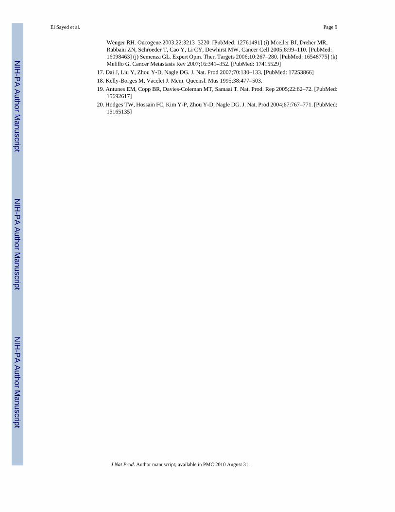
line overexpressing calcitonin (CT) were evaluated using two in vitro models: linear invasionof cells through the Matrigel™ barrier; and spheroid disaggregation.13a–d Latrunculin A (1)(100 nM) remarkably inhibited baseline and CT-stimulated invasion in the linear Matrigel™assay (Figure 1). However, 1 was cytotoxic at doses higher than 500 nM (data not shown).There is evidence to suggest that cell migration and invasion involve multiple processesregulated by various signaling molecules.14a–d The actin cytoskeleton and its regulatoryproteins are crucial for cell migration in most cells.15a,b During cell migration, the actincytoskeleton is dynamically remodeled, and this reorganization produces the force necessaryfor cell migration.15a,b Since latrunculin A (1) attenuated invasiveness of PC-3M cells, itseffect was examined on the actin cytoskeleton of PC-3M cells. PC-3M cells display an invasivephenotype and they usually attach to the surface, exhibiting stretched actin fibers with longexon-type extensions (Figure 2A). Upon stimulation with 50 nM CT, the cells displayed rapidreorganization of the actin cytoskeleton, leading to visible changes in cell morphology within10 minutes. The CT-stimulated cells demonstrated long actin-rich extensions resemblingfilopodia or microspikes or web-like actin-containing fibers linking these filopodium-likestructures. However, the addition of latrunculin A (1) resulted in rapid changes in cellmorphology and actin distribution as characterized by the remarkable shrinkage of actin stressfibers, possibly caused by the depolymerization disruption of actin (Figure 2B). Compound1 also abolished CT-induced changes in the actin cytoskeleton. These results are consistentwith the actions of latrunculin on PC-3M cell invasion (Figure 1), and suggest that anti-invasiveactions of 1 are mediated through the disruption of actin cytoskeleton remodeling.
Spheroid disaggregation provides the measure of cell disaggregation as well as cell migration,and this model simulates the process of tissue disaggregation and invasion of cells in vivo.13b,15c This assay is based on disaggregation of cancer cell spheroids, and radial migration ofreleased cells on extracellular matrix (ECM).13a–c,14d Current evidence has shown that tumorcells from primary tumors in vivo are generally released in clumps, which attach to a favorableECM and are later released gradually to migrate in all directions.13a–c,14d Therefore, thespheroid disaggregation model is closer to in situ tumor metastasis than the the linearMatrigel™ invasion assays. The results shown in Figure 3A demonstrate that latrunculin A(1, 500 nM) decreased disaggregation and cell migration of PC-3M-CT+ spheroids three-fold.Figure 3B shows the antimetastatic actions of latrunculin A (1) with analogs 4–6 at multipledoses. Analogs 4–6 also attenuated PC-3M-CT+ spheroid disaggregation/cell migration, andthis response was dose-dependent in all three analogs tested. Based on the results shown inFigure 3B, analog 5 was the most potent. Only a dose of 100 nM of analog 5 was required togenerate inhibition of PC-3M-CT+ spheroid disaggregation/cell migration to the extent of thatproduced by 500 nM of 1. Compound 4 required 200 nM to produce the equivalent effect.Analog 6 was equipotent to latrunculin A. Halogenated ethylcarbamates 2 and 3 were inactive,suggesting the significance of conjugation and extension of the carbamate side chain. Thephenylcarbamate 5 was two-fold more potent than benzylcarbamate 6, suggesting theimportance of optimum distance between the aromatic ring and the carbamate functionalityfor activity.
Relatively uncontrolled proliferation of cells within a tumor outstrip the capacity of existingvasculature to supply oxygen. This results in the formation of hypoxic regions within tumors.The occurrence of tumor hypoxia is associated with a poor prognosis in cancer patients.16a Nodrugs have been approved clinically to specifically target hypoxic tumor cells. Thetranscription factor hypoxia-inducible factor-1 (HIF-1) regulates hypoxia-induced geneexpression in tumor cells that are subjected to hypoxic conditions.16b Disruption of HIF-1-mediated hypoxic adaptation/survival reduces tumor growth in animal models.16c–i HIF-1inhibitors represent potential anticancer drug leads that may selectively target hypoxic tumormasses.16j–k The effects of 1–6 on hypoxia (1% O2)-induced HIF-1 activation were examinedusing a T47D human breast carcinoma cell-based luciferase reporter assay,17 and compared to
El Sayed et al. Page 3
J Nat Prod. Author manuscript; available in PMC 2010 August 31.
NIH
-PA Author Manuscript
NIH
-PA Author Manuscript
NIH
-PA Author Manuscript
those observed on 1,10-phenanthroline-induced HIF-1 activation. Compound 1 inhibitedhypoxia-induced activation (IC50 6.7 µM, Figure 4) and 1,10-phenanthroline induced HIF-1activation (IC50 25 µM, data not shown). Compound 6 weakly inhibited hypoxia-inducedHIF-1 activation (IC50 29 µM). None of the other compounds (2–5) exerted greater than 50%inhibition of hypoxia-induced HIF-1 activation at the highest concentration tested (30 µM).The effects of 1–6 on the expression of luciferase from a control construct were examined ina T47D cell-based reporter assay and hypoxic cell viability/proliferation in a sulforhodamineB-based cell viability assay. None of the latrunculin analogs significantly inhibited the activityof a constitutively expressed pGL3 control reporter or significantly suppressed T47D cellviability under experimental conditions (less than 20% inhibition at the concentration of 30µM). Therefore, latrunculin-based actin depolymerization disruptors appear to have the abilityto selectively inhibit hypoxia-induced HIF-1 activity in tumor cells at concentrations belowthose evident for the inhibition of other critical cellular processes. The role of actin in HIF-1signaling has never been documented. These results represent the first observation of the abilityof actin inhibitors to inhibit HIF-1 activation at concentrations that also inhibit actinpolymerization and these findings may indicate that actin polymerization plays a vital role inHIF-1 signaling.
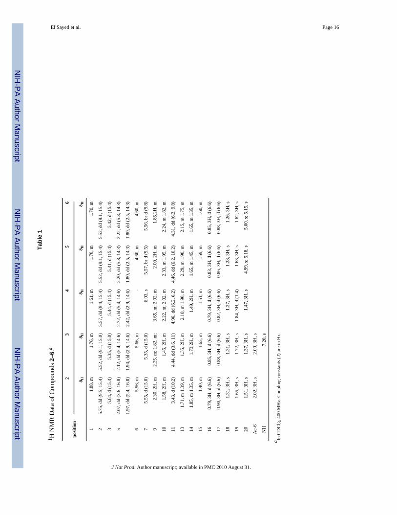
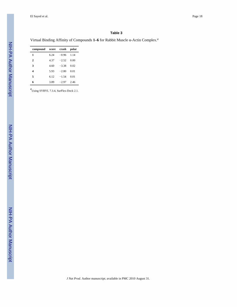
To further correlate the anti-invasive activity with the actin binding affinity, docking of 1–6toward rabbit muscle δ-actin was implemented using the SYBYL 7.3.4 and SurFlex-DockPrograms. Docking results were expressed in three functions: total score, crash, and polar. Thetotal score was expressed in -log(Kd) units to represent binding affinities. Crash is the degreeof inappropriate penetration by the ligand into the protein and of interpenetration betweenligand atoms that are separated by rotatable bonds. Crash scores close to 0 are favorable.Negative numbers indicate penetration. The Polar score is the contribution of the polar non-hydrogen bonding interactions to the total score, may be useful for excluding docking resultsthat involve no hydrogen bonds. The docking procedure was validated using the sameconditions to dock latrunculin A (1) into the binding pocket of α-actin. The docking simulationresulted in a very close model to the crystallographic structure.9,10 The docking data aresummarized in Table 1.
The parent compound, latrunculin A (1), as well as the phenylcarbamate (5), showed the highestscores and molecular interaction, consistent with the observed anti-invasive activity (Table 1,Figure 5). The carbonyl oxygen of the carbamoyl moiety in 5 makes a strong H-bondinginteraction with the guanidine amino group of arginine (ARG) 210 of actin (Figure 5). A similarinteraction has been observed for carbamates 3 and 6. Therefore, ARG 210 seems to play acentral role in the binding of latrunculin A (1) and analogs within the actin monomers.9,10
Although structure 6 showed anti-invasive activity, the scoring was the lowest amonglatrunculin A derivatives. Such deviation could be attributed to the activity of structure 6 onprotein target(s) other than actin.
Experimental SectionGeneral Experimental Procedures
Measurements of optical rotation were carried out on a Rudolph Research Analytical AutopolIII polarimeter. IR spectra were recorded on a Varian 800 FT-IR spectrophotometer. The 1Hand 13C NMR spectra were recorded in CDCl3, using TMS as an internal standard, on a JEOLEclipse NMR spectrometer operating at 400 MHz for 1H and 100 MHz for 13C. The HREIMSexperiments were conducted at the University of Michigan on a Micromass LCT spectrometer.TLC analysis was carried out on precoated silica gel 60 F254 500 µm TLC plates, using thedeveloping systems n-hexane/EtOAc (1:1) or CHCl3-MeOH (9:1). For columnchromatography, silica gel 60 (particle size 63–200 µm) or Bakerbond octadecyl (C18), 40 µmwere used. For Sephadex LH-20 column chromatography, n-hexane-CHCl3 (1:3), CHCl3, and
El Sayed et al. Page 4
J Nat Prod. Author manuscript; available in PMC 2010 August 31.
NIH
-PA Author Manuscript
NIH
-PA Author Manuscript
NIH
-PA Author Manuscript
CHCl3-MeOH (9:1) systems were used. For column chromatography, silica gel 70–230 meshwas used.
Biological MaterialThe sponge Negombata magnifica Kelly-Borges and Vacelet (order Poecilosclerida, sub-orderMycalina, family Podospongiidae) was collected as red long finger-like strips by SCUBA fromthe sand-covered bottom at −10 to −15 m at Hurghada, on the Egyptian Red Sea coast, in June2003.18,19 The sponge was identified by Mr. Tamer Helmy, Suez Canal University. A voucherspecimen (03RS3) was deposited in the Department of Basic Pharmaceutical Sciences, Collegeof Pharmacy, University of Louisiana at Monroe, Louisiana.
Extraction and IsolationThe frozen sponge (6 kg) was coarsely minced and extracted with CHCl3 (5 × 1000 mL) atroom temperature. The CHCl3 extract was then concentrated under vacuum and subjected toliquid chromatography, as described elsewhere, to afford 1, 1.2 g (0.0002%).1,4,11
Preparation of Carbamates 2–6To solutions of 50 mg of 1 in toluene (2 mL), 59 µL of 2-chloroethyl isocyanate, 60 µL of 2-bromoethyl isocyanate, 60 µL of 3-chloropropyl isocyanate, 40 µL of phenyl isocyanate, or36 µL of benzyl isocyanate were added, respectively, and separately mixed with 10 µL ofEt3N. Each solution was separately stirred at room temperature for 1 h (12 h for benzylisocyanate only). Water (10 mL) was then added and the product of each reaction mixture wasextracted with EtOAc. Each EtOAc extract was dried over anhydrous Na2SO4 and concentratedunder reduced pressure. Crude products were then purified by column chromatography onsilica gel 60 using EtOAc-n-hexane 1:9 to give compounds 2 (20.0 mg, 40% Rf 0.60, silica gel,CHCl3-MeOH 9.5:0.5) and 4 (13.2 mg, 26.4%, Rf 0.64),or EtOAc-n-hexane 2:8 to givecompounds 3 (13.3 mg, 26.6%, Rf 0.57) and 6 (19.4 mg, 38.8%, Rf 0.55) or using CHCl3-MeOH (9.5:0.5) to afford compound 5 (13.6 mg, 27.2%, Rf 0.51).
17-O-[N-(2-Chloroethyl)carbamoyl]-latrunculin A (2)
colorless oil, (c 0.51, CHCl3); IR νmax (neat) 3564, 3331, 2953-2856, 1761, 1708,1680, 1542, 1355, 1164 cm−1; 1H NMR (CDCl3) δH 3.61 (2H, m, H2-2´), 3.63 (2H, m, H2-3´), 8.48 (1H, brs, exchangeable, NH); 13C NMR δC 152.5 (qC, C-1´), 42.2 (CH2, C-2´), 43.2(CH2, C-3´); HRESIMS m/z 549.1808 (calcd for C25H35ClN2O6SNa, 549.1802 [M+Na]+).
17-O-[N-(2-Bromoethyl)carbamoyl]-latrunculin A (3)
colorless oil, (c 0.55, CHCl3); IR νmax (neat) 3563, 3327, 2995-2857, 1735, 1713,1560, 1356, 1301, 1128 cm−1; 1H NMR (CDCl3) δH 3.47 (2H, m, H2-2´), 3.69 (2H, m, H2-3´), 8.49 (1H, t, J = 5.2 Hz, exchangeable, NH); 13C NMR δ 152.2 (qC, C-1´), 48.0 (CH2, C-2´), 42.1 (CH2, C-3´); HRESIMS m/z 593.1306 (calcd for C25H35BrN2O6SNa, 593.1297 [M+H]+).
17-O-[N-(3-Chloropropyl)carbamoyl]-latrunculin A (4)
colorless oil, (c 0.47, CHCl3); IR νmax (neat) 3564, 3335, 2953-2856, 1740, 1704,1545, 1356, 1288, 1163 cm−1; 1H NMR (CDCl3) δH 3.47 (2H, m, H2-2´), 1.42 (2H, m, H2-3´), 3.59 (2H, t, J = 6.2 Hz, H2-4´), 8.23 (1H, t, J = 5.8 Hz, exchangeable, NH); 13C NMR δC152.4 (qC, C-1´), 37.7 (CH2, C-2´), 31.8 (CH2, C-3´), 42.3 (CH2, C-4´); HRESIMS m/z563.1953 (calcd for C26H37ClN2O6SNa, 563.1959 [M+H]+).
El Sayed et al. Page 5
J Nat Prod. Author manuscript; available in PMC 2010 August 31.
NIH
-PA Author Manuscript
NIH
-PA Author Manuscript
NIH
-PA Author Manuscript
17-O-[N-(Phenyl)carbamoyl]-latrunculin A (5)
colorless oil, (c 0.14, CHCl3); IR νmax (neat) 3570, 3286, 2927-2853, 1710, 1601,1680, 1554, 1446, 1296, 1160 cm−1; 1H NMR (CDCl3) δH 7.47 (2H, brd, J = 8.1 Hz, H-3´/H-7´), 7.33 (2H, dd, J = 8.1, 8.0 Hz, H-4´/H2-6´), 7.15 (1H, m, H-5´); 13C NMR δC 149.6 (qC,C-1´), 137.1 (qC, C-2´), 121.5 (CH, C-3´/C-7´), 129.1 (CH, C-4´/C-6´), 124.5 (CH, C-5´);HRESIMS m/z 563.2200 (calcd for C26H37N2O6SNa, 563.2192 [M+Na]+).
17-O-[N-(Benzyl)carbamoyl]-latrunculin A (6)
colorless oil, (c 0.55, CHCl3); IR νmax (neat) 3564, 3334, 2953-2857, 1730, 1703,1680, 1542, 1455, 1357, 1282, 1166 cm−1; 1H NMR (CDCl3) δ 4.39 (2H, d, J = 5.4 Hz, H2-2´), 7.27 (2H, brd, J = 8.0 Hz, H-4´/H-8´), 7.30 (2H, dd, J = 8.0, 7.8 Hz, H-5´/H2-7´), 7.32 (1H,dd, J = 8.0, 7.8 Hz, H-6´); 13C NMR δC (151.2, qC, C-1´), 41.5 (CH2, C-2´), 141.5 (qC, C-3´), 127.0 (CH, C-4´/C-8´), 129.1 (CH, C-5´/C-7´), 126.5 (CH, C-6´); HRESIMS m/z 577.2360(calcd for C30H38N2O6SNa, 577.2348 [M+Na]+).
Invasion AssayThese experiments were conducted in 24-well, two compartment, Matrigel™ invasionchambers (Becton Dickinson, Bedford, MA).13a–13d The PC-3M-CT+ cells were grownexponentially under serum-starved conditions for 24 h [basal RPMI medium containing noserum or growth factors but containing 0.1% BSA, 10 mM HEPES, 4 mM L-glutamine, 100IU mL−1 penicillin G, and 100 mg mL−1streptomycin]. The cells were then harvested, andseeded at a density of 25 × 103 cells per well in the upper insert of the Matrigel™ invasionchamber. The lower chamber received the chemo-attractant medium, which consisted of 90%basal RPMI medium and 10% conditioned medium from the cultures of PC-3M-CT+ cellsexpressing constitutively active Gas protein. The incubation was carried out for 24 h, afterwhich the Matrigel™ (along with non-invading cells) was scraped off with cotton swabs, andouter side of the insert was fixed and stained using Diff Quick staining (Dade BehringDiagnostics, Aguada, Peurto Rico). The number of cells migrated on the outer bottom side ofthe insert counted under the microscope in six or more randomly selected fields (magnification:100×). Final results are expressed as mean +/− SEM per 100× field. Each experiment wasperformed in triplicate, and the experiment was repeated twice.13a–13d
Growth CorrectionSince some cell lines can exhibit high proliferation rates, it is likely that the cells migratingduring the early part of the 24 h incubation period could proliferate during the remaining periodof incubation, leading to a slight overestimation of the final results. To correct this probability,the growth rate of PC-3M cells was determined under identical culture conditions. Twenty-five thousand cells were plated at hourly intervals in six-well dishes and cultured with/withoutCT (50 nM) for 1–24 h. Mean percent increase in the cell number was determined at the endof the incubation period by counting the net increase in the number of cells. The relative CT-induced increase of the pooled results of all time points was found to be 1.19 (vehicle control= 1). This correction was applied to the results of invasion assays.13b,15d,e
Spheroid Disaggregation AssaySpheroids were prepared from single cell suspension of prostate cell lines as described before.13b,13d In brief, 5 × 10⁴/mL cells in RPMI 1640 serum-free medium were placed on 96-welllow-attachment tissue culture plates. The plates were rocked on a gyrorotatory shaker in aCO2 incubator at 37 °C for 2 days, at the end of which the spheroids measuring 150–300 µmin diameter (~4 × 10⁴ cells/spheroid) were formed. A single spheroid was then placed in thecenter of each well of extracellular matrix (ECM)-coated 24-well microplate in 200 mL of
El Sayed et al. Page 6
J Nat Prod. Author manuscript; available in PMC 2010 August 31.
NIH
-PA Author Manuscript
NIH
-PA Author Manuscript
NIH
-PA Author Manuscript
serum-free medium. From previous studies, it was determined that 1 h is an appropriate timefor spheroids to begin adhering to an ECM. Thus t = 0 was set as 1 h from initial plating, sothat if the plate was not disturbed, the spheroids would not move from their location at the timeof plating. Spheroids were photographed digitally at t = 0, cultured at 37 °C for 48 h, and thenre-photographed. The spheroids were then fixed, stained with Diff-Quik™ (Dade Behring,Newark, DE) and examined under light microscopy. The diameter of the area covered withcells migrated from the spheroids was measured in a microscope calibrated with a stage andocular micrometer. The radial distance of migration was calculated after subtraction of themean initial spheroidal diameter at t = 0. Values shown represent the average percent increasein surface area of spheroids.
Cell Proliferation/Viability AssayHuman breast carcinoma T47D cells were grown in DMEM/F12 medium with L-glutamine(Mediatech) supplemented with 10% (v/v) fetal calf serum (FCS, Hyclone), 50 units mL−1
penicillin G (Na salt), and 50 µg mL−1 streptomycin sulfate (referred to as "Pen/Strep")(Invitrogen) in a humidified atmosphere (5% CO2/95% air) at 37 °C. Exponentially growncells were plated at a density of 30,000 cells per well into 96-well tissue culture plates (Corning)in a volume of 100 µL of DMEM/F12 medium with 10% FCS and Pen/Strep. The cells wereincubated at 37 °C overnight.17,20 Test compounds were diluted in DMEM/F12 medium withPen/Strep and added in a volume of 100 µL per well. Following a 30-min incubation, thecompound treatment continued for another 16 h (or 48 h) at 37 °C under hypoxic (5% CO2/1%O2/94% N2) or normoxic (5% CO2/95% air) conditions. Cell proliferation/viability (performedin triplicate) was determined as previously described.17,20 The absorbance at 515 nm wasmeasured on a BioTek Synergy HT microplate reader with correction wavelength at 690 nm.The data were normalized to the untreated control. The following formula was used to calculate% inhibition of cell proliferation/viability: % inhibition = 1 - OD515(treated)/OD515(control).
Cell-Based Reporter Assay for HIF-1 ActivityThe transfection, compound treatment, exposure to hypoxic conditions (5% CO2/1% O2/94%N2), normoxic conditions (5% CO2/95% air), and a hypoxia mimetic (10 µM 1,10-phenanthroline), and luciferase activity determination were performed as previously described.20 Emetine was used as a positive control (IC50 0.11 µM).
Molecular ModelingDocking and scoring modeling studies were performed using SYBYL 7.3.4 (Tripos DiscoveryInformatics, St. Louis, MO) installed on a Dell desktop workstation equipped with a 1.86 GHzIntel® Xeon® processor and the Red Hat Enterprise Linux (version 4) operating system. The3D coordinates of rabbit muscle α-actin complex with latrunculin A (1) was retrieved from theProtein Data Bank (PDB code: 1esv). The selected structure is of the best 3D resolution (2.0Å). The protein structure was utilized in subsequent docking experiments without energyminimization. Explicit water molecules were removed from the structure. Chemical structuresof 1–6 were drawn in SYBYL 7.3.4 and assigned Gasteiger partial charges and energyminimized using Energy Force Field. Docking simulation of structures 1–6 were carried outusing SurFlex-Dock (version 2.1). SurFlex-Dock identifies the active site of the protein andconstructs a docking target (protomol) to which molecules match. Protmol was generated bythe ligand based method, setting the Threshold and Bloat parameters as default value (0.5 and0.0, respectively).
El Sayed et al. Page 7
J Nat Prod. Author manuscript; available in PMC 2010 August 31.
NIH
-PA Author Manuscript
NIH
-PA Author Manuscript
NIH
-PA Author Manuscript
AcknowledgmentsThis investigation was made possible through the support of NIH grant number P20RR16456 from the BRIN Programof the National Center for Research Resources, grant R01CA96534 (G.V.S.), and grant CA098787 (D.G.N.-Y.D.Z.).Its contents are solely the responsibility of the authors and do not necessarily represent the official views of NIH.
References and Notes1. Kashman Y, Groweiss A, Shmueli U. Tetrahedron Lett 1980;21:3629–3632.2. Okka M, Tian B, Kaufman PL. Arch. Ophthalmol 2004;122:1482–1488. [PubMed: 15477460]3. Kaufman, PL.; Geiger, B. U. S. Patent. 772,412. 2002.4. El Sayed KA, Youssef DTA, Marchetti D. J. Nat. Prod 2006;69:219–223. [PubMed: 16499319]5. Spector I, Shochet NR, Blasberger D, Kashman Y. Cell Motil. Cytoskel 1989;13:127–144.6. Newman DJ, Cragg GM. J. Nat. Prod 2004;67:1216–1238. [PubMed: 15332835]7. Nakaseko Y, Yanagida M. Nature 2001;412:291–292. [PubMed: 11460149]8. Gachet Y, Tournier S, Millar JB, Hyams JS. Nature 2001;412:352–355. [PubMed: 11460168]9. Yarmola EG, Somasundaram T, Boring TA, Spector I, Bubb MR. J. Biol. Chem 2000;275:28120–
28127. [PubMed: 10859320]10. Morton WM, Ayscough KR, McLaughlin PJ. Nat. Cell. Biol 2000;2:376–378. [PubMed: 10854330]11. Groweiss A, Shmueli U, Kashman Y. J. Org. Chem 1983;48:3512–3516.12. Blasberger D, Carmely S, Cojocaru M, Spector I, Shochet NR, Kashman Y. Liebigs Ann. Chem
1989;12:1171–1188.13. (a) Shah GV, Noble MJ, Austenfeld M, Weigel J, Deftos LJ, Mebust WK, Winston K. Prostate
1992;21:87–97. [PubMed: 1409122] (b) Thomas S, Chigurupati S, Anbalagan M, Shah G. Mol.Endocrinol 2006;20:1894–1911. [PubMed: 16574742] (c) Chien J, Ren Y, Wang YQ, Bordelon W,Thompson E, Davis R, Rayford W, Shah G. Mol. Cell Endocrinol 2001;181:69–79. [PubMed:11476942] (d) Thomas S, Chiriva-Internati M, Shah GV. Clin. Exp. Metastasis 2007;24:363–377.[PubMed: 17487556]
14. (a) Cooper CR, Chay CH, Pienta KJ. Neoplasia 2002;4:191–194. [PubMed: 11988838] (b) GhoshPM, Ghosh-Choudhury N, Moyer ML, Mott GE, Thomas CA, Foster BA, Greenberg NM, KreisbergJI. Oncogene 1999;18:4120–4130. [PubMed: 10435593] (c) Chen Y, Wang Y, Yu H, Wang F, XuW. Exp. Biol. Med 2005;230:731–741. (d) Alroy I, Yarden Y. FEBS Lett 1997;410:83–86. [PubMed:9247128] (d) Hoevel T, Macek R, Swisshelm K, Kubbies M. Int. J. Cancer 2004;108:374–383.[PubMed: 14648703]
15. (a) Byers HR, Etoh T, Doherty JR, Sober AJ, Mihm MC. Am. J. Pathol 1991;139:423–435. [PubMed:1867326] (b) Hotulainen P, Paunola E, Vartiainen MK, Lappalainen P. Mol. Biol. Cell 2005;16:649–664. [PubMed: 15548599] (c) Cascone I, Giraudo E, Caccavari F, Napione L, Bertotti E, Collard JG,Serini G, Bussolino F. J. Biol. Chem 2003;278:50702–50713. [PubMed: 12972426] (d) YoshiokaK, Nakamori S, Itoh K. Cancer Res 1999;59:2004–2010. [PubMed: 10213513] (e) Gondi CS, LakkaSS, Yanamandra N, Olivero WC, Dinh DH, Gujrati M, Tung CH, Weissleder R, Rao JS. Cancer Res2004;64:4069–4077. [PubMed: 15205313]
16. (a) Tatum JL, Kelloff GJ, Gillies RJ, Arbeit JM, Brown JM, Chao KS, Chapman JD, Eckelman WC,Fyles AW, Giaccia AJ, Hill RP, Koch CJ, Krishna MC, Krohn KA, Lewis JS, Mason RP, Melillo G,Padhani AR, Powis G, Rajendran JG, Reba R, Robinson SP, Semenza GL, Swartz HM, Vaupel P,Yang D, Croft B, Hoffman J, Liu G, Stone H, Sullivan D. Int. J. Radiat. Biol 2006;82:699–757.[PubMed: 17118889] (b) Semenza GL. Nat. Rev. Cancer 2003;3:721–732. [PubMed: 13130303] (c)Maxwell PH, Dachs GU, Gleadle JM, Nicholls LG, Harris AL, Stratford IJ, Hankinson O, Pugh CW,Ratcliffe PJ. Proc. Natl. Acad. Sci. U. S. A 1997;94:8104–8109. [PubMed: 9223322] (d) Ryan HE,Lo J, Johnson RS. EMBO J 1998;17:3005–3015. [PubMed: 9606183] (e) Ryan HE, Poloni M,McNulty W, Elson D, Gassmann M, Arbeit JM, Johnson RS. Cancer Res 2000;60:4010–4015.[PubMed: 10945599] (f) Kung AL, Wang S, Klco JM, Kaelin WG, Livingston DM. Nat. Med2000;6:1335–1340. [PubMed: 11100117] (g) Kung AL, Zabludoff SD, France DS, Freedman SJ,Tanner EA, Vieira A, Cornell-Kennon S, Lee J, Wang B, Wang J, Memmert K, Naegeli HU, PetersenF, Eck MJ, Bair KW, Wood AW, Livingston DM. Cancer Cell 2004;6:33–43. [PubMed: 15261140](h) Unruh A, Ressel A, Mohamed HG, Johnson RS, Nadrowitz R, Richter E, Katschinski DM,
El Sayed et al. Page 8
J Nat Prod. Author manuscript; available in PMC 2010 August 31.
NIH
-PA Author Manuscript
NIH
-PA Author Manuscript
NIH
-PA Author Manuscript
Wenger RH. Oncogene 2003;22:3213–3220. [PubMed: 12761491] (i) Moeller BJ, Dreher MR,Rabbani ZN, Schroeder T, Cao Y, Li CY, Dewhirst MW. Cancer Cell 2005;8:99–110. [PubMed:16098463] (j) Semenza GL. Expert Opin. Ther. Targets 2006;10:267–280. [PubMed: 16548775] (k)Melillo G. Cancer Metastasis Rev 2007;16:341–352. [PubMed: 17415529]
17. Dai J, Liu Y, Zhou Y-D, Nagle DG. J. Nat. Prod 2007;70:130–133. [PubMed: 17253866]18. Kelly-Borges M, Vacelet J. Mem. Queensl. Mus 1995;38:477–503.19. Antunes EM, Copp BR, Davies-Coleman MT, Samaai T. Nat. Prod. Rep 2005;22:62–72. [PubMed:
15692617]20. Hodges TW, Hossain FC, Kim Y-P, Zhou Y-D, Nagle DG. J. Nat. Prod 2004;67:767–771. [PubMed:
15165135]
El Sayed et al. Page 9
J Nat Prod. Author manuscript; available in PMC 2010 August 31.
NIH
-PA Author Manuscript
NIH
-PA Author Manuscript
NIH
-PA Author Manuscript
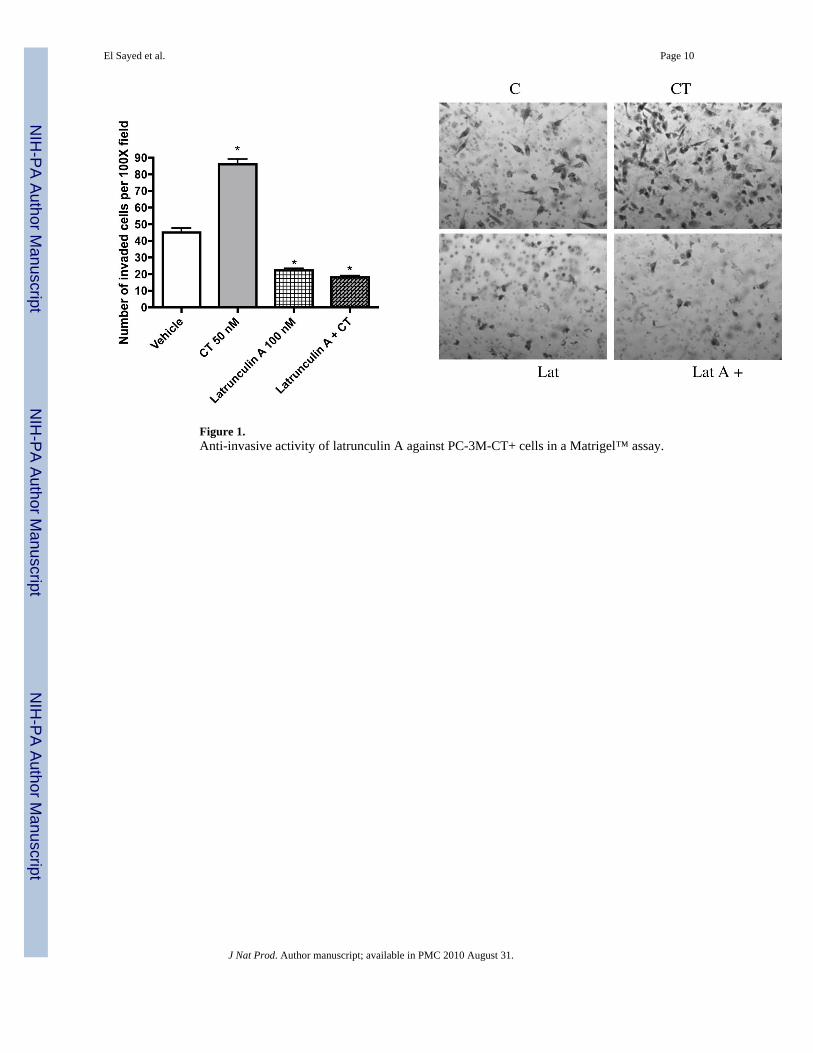
Figure 1.Anti-invasive activity of latrunculin A against PC-3M-CT+ cells in a Matrigel™ assay.
El Sayed et al. Page 10
J Nat Prod. Author manuscript; available in PMC 2010 August 31.
NIH
-PA Author Manuscript
NIH
-PA Author Manuscript
NIH
-PA Author Manuscript
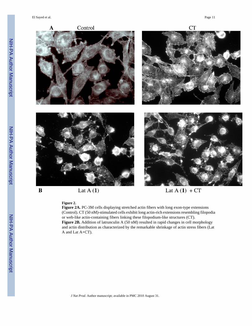
Figure 2.Figure 2A. PC-3M cells displaying stretched actin fibers with long exon-type extensions(Control). CT (50 nM)-stimulated cells exhibit long actin-rich extensions resembling filopodiaor web-like actin-containing fibers linking these filopodium-like structures (CT).Figure 2B. Addition of latrunculin A (50 nM) resulted in rapid changes in cell morphologyand actin distribution as characterized by the remarkable shrinkage of actin stress fibers (LatA and Lat A+CT).
El Sayed et al. Page 11
J Nat Prod. Author manuscript; available in PMC 2010 August 31.
NIH
-PA Author Manuscript
NIH
-PA Author Manuscript
NIH
-PA Author Manuscript
Figure 3.Figure 3A. Spheroid disaggregation of PC-3M-CT+ cells using 500 nM latrunculin A (1).Figure 3B. Spheroid disaggregation of PC-3M-CT+ cells comparison of different doses ofcarbamates 4–6 with the activity of latrunculin A.
El Sayed et al. Page 12
J Nat Prod. Author manuscript; available in PMC 2010 August 31.
NIH
-PA Author Manuscript
NIH
-PA Author Manuscript
NIH
-PA Author Manuscript
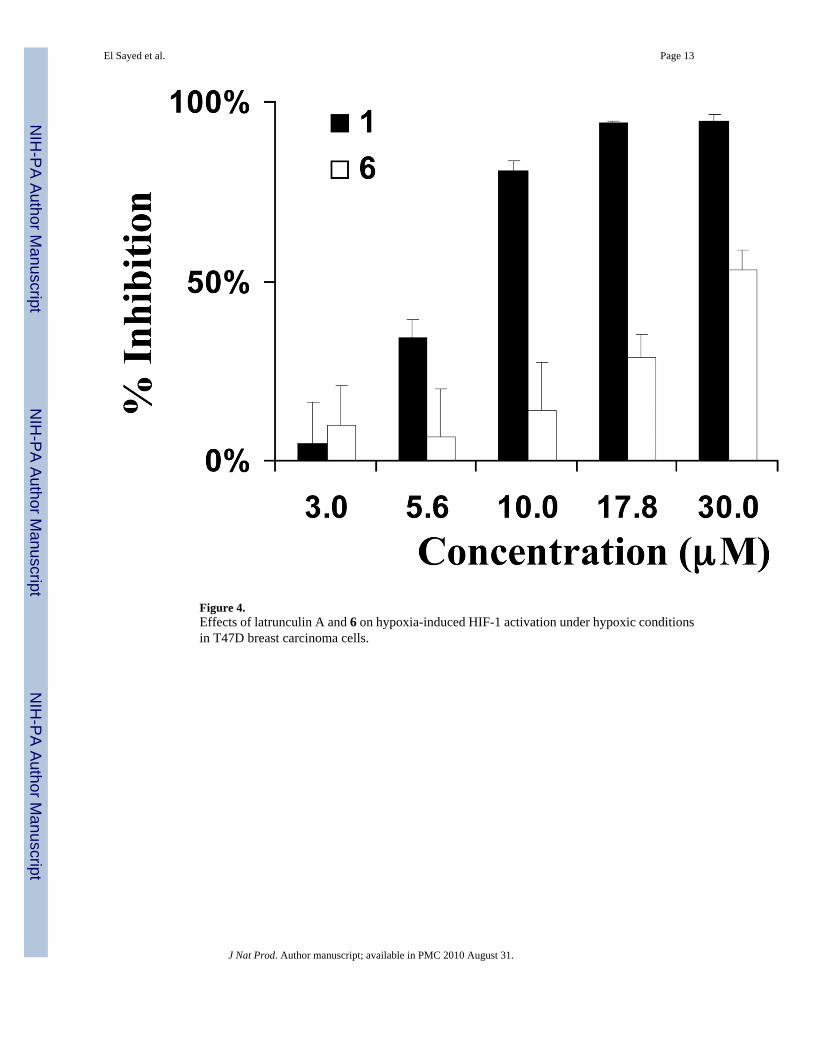
Figure 4.Effects of latrunculin A and 6 on hypoxia-induced HIF-1 activation under hypoxic conditionsin T47D breast carcinoma cells.
El Sayed et al. Page 13
J Nat Prod. Author manuscript; available in PMC 2010 August 31.
NIH
-PA Author Manuscript
NIH
-PA Author Manuscript
NIH
-PA Author Manuscript
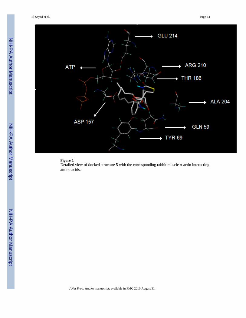
Figure 5.Detailed view of docked structure 5 with the corresponding rabbit muscle α-actin interactingamino acids.
El Sayed et al. Page 14
J Nat Prod. Author manuscript; available in PMC 2010 August 31.
NIH
-PA Author Manuscript
NIH
-PA Author Manuscript
NIH
-PA Author Manuscript
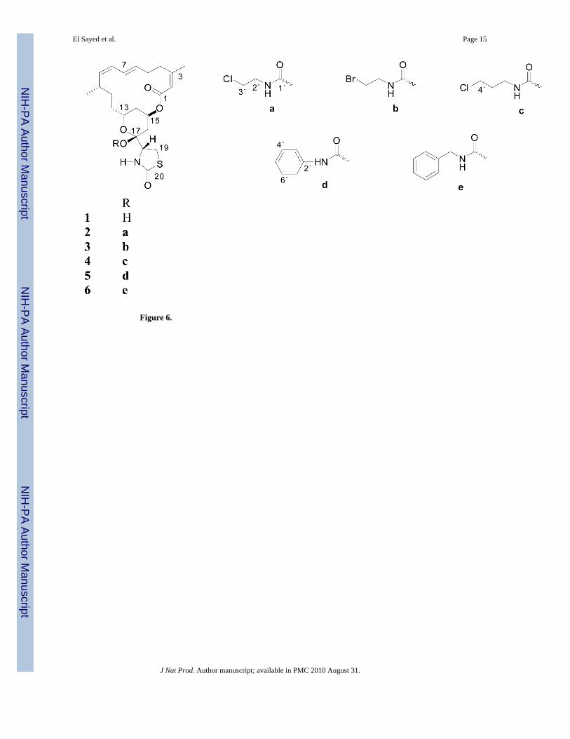
Figure 6.
El Sayed et al. Page 15
J Nat Prod. Author manuscript; available in PMC 2010 August 31.
NIH
-PA Author Manuscript
NIH
-PA Author Manuscript
NIH
-PA Author Manuscript
NIH
-PA Author Manuscript
NIH
-PA Author Manuscript
NIH
-PA Author Manuscript
El Sayed et al. Page 16
Tabl
e 1
1 H N
MR
Dat
a of
Com
poun
ds 2
–6.a
posi
tion
23
45
6
δ Hδ H
δ Hδ H
δ H
11.
88, m
1.76
, m1.
61, m
1.70
, m1.
70, m
25.
75, d
d (9
.5, 1
5.4)
5.52
, dd
(9.1
, 15.
0)5.
57, d
d (8
.4, 1
5.4)
5.52
, dd
(9.1
, 15.
4)5.
52, d
d (9
.1, 1
5.4)
35.
64, d
(15.
4)5.
35, d
(15.
0)5.
44, d
(15.
4)5.
41, d
(15.
4)5.
42, d
(15.
4)
52.
07, d
d (3
.6, 1
6.8)
2.12
, dd
(5.4
, 14.
6)2.
72, d
d (5
.4, 1
4.6)
2.20
, dd
(5.8
, 14.
3)2.
22, d
d (5
.8, 1
4.3)
1.97
, dd
(5.4
, 16.
8)1.
94, d
d (2
.9, 1
4.6)
2.42
, dd
(2.9
, 14.
6)1.
80, d
d (2
.5, 1
4.3)
1.80
, dd
(2.5
, 14.
3)
65.
56, m
5.66
, m-
4.60
, m4.
60, m
75.
55, d
(15.
0)5.
35, d
(15.
0)6.
03, s
5.57
, br d
(9.5
)5.
56, b
r d (9
.8)
92.
30, 2
H, m
2.25
, m; 1
.82,
m;
3.65
, m; 2
.02,
m2.
00, 2
H, m
1.85
,2H
, m
101.
58, 2
H, m
1.45
, 2H
, m2.
22, m
; 2.0
2, m
2.33
, m 1
.95,
m2.
24, m
1.8
2, m
113.
43, d
(10.
2)4.
44, d
d (3
.6, 1
1)4.
96, d
d (6
.2, 6
.2)
4.46
, dd
(6.2
, 10.
2)4.
31, d
d (6
.2, 9
.8)
131.
71, m
1.3
9, m
1.35
, 2H
, m2.
10, m
1.9
0, m
2.29
, m 1
.90,
m2.
15, m
1.7
5, m
141.
85, m
1.3
5, m
1.73
,2H
, m1.
49, 2
H, m
1.65
, m 1
.45,
m1.
65, m
1.3
5, m
151.
40, m
1.65
, m1.
51, m
1.59
, m1.
60, m
160.
79, 3
H, d
(6.6
)0.
85, 3
H, d
(6.6
)0.
79, 3
H, d
(6.6
)0.
83, 3
H, d
(6.6
)0.
85, 3
H, d
(6.6
)
170.
90, 3
H, d
(6.6
)0.
88, 3
H, d
(6.6
)0.
82, 3
H, d
(6.6
)0.
86, 3
H, d
(6.6
)0.
88, 3
H, d
(6.6
)
181.
31, 3
H, s
1.31
, 3H
, s1.
27, 3
H, s
1.28
, 3H
, s1.
26, 3
H, s
191.
65, 3
H, s
1.72
, 3H
, s1.
84, 3
H, d
(1.4
)1.
63, 3
H, s
1.62
, 3H
, s
201.
51, 3
H, s
1.37
, 3H
, s1.
47, 3
H, s
4.99
, s; 5
.18,
s5.
00, s
; 5.1
5, s
Ac-
62.
02, 3
H, s
2.00
, 3H
, s
NH
7.20
, s
a In C
DC
l 3, 4
00 M
Hz.
Cou
plin
g co
nsta
nts (
J) a
re in
Hz.
J Nat Prod. Author manuscript; available in PMC 2010 August 31.
NIH
-PA Author Manuscript
NIH
-PA Author Manuscript
NIH
-PA Author Manuscript
El Sayed et al. Page 17
Tabl
e 2
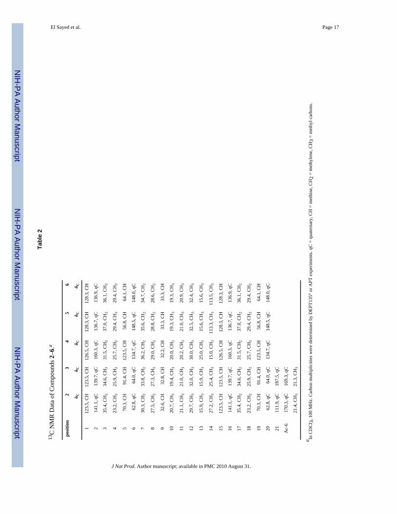
13C
NM
R D
ata
of C
ompo
unds
2–6
.a
posi
tion
23
45
6
δ Cδ C
δ Cδ C
δ C
112
3.5,
CH
123.
5, C
H12
6.5,
CH
128.
3, C
H12
8.3,
CH
214
1.1,
qC
139.
7, q
C16
0.3,
qC
136.
7, q
C13
6.9,
qC
335
.4, C
H2
34.6
, CH
231
.5, C
H2
37.0
, CH
236
.1, C
H2
423
.2, C
H2
25.9
, CH
225
.7, C
H2
29.4
, CH
229
.4, C
H2
570
.3, C
H91
.4, C
H12
3.5,
CH
56.8
, CH
64.1
, CH
662
.8, q
C64
.0, q
C13
4.7,
qC
148.
5, q
C14
8.0,
qC
730
.3, C
H2
33.8
, CH
236
.2, C
H2
35.6
, CH
234
.7, C
H2
827
.5, C
H2
27.3
, CH
229
.0, C
H2
28.8
, CH
228
.6, C
H2
932
.6, C
H32
.8, C
H32
.2, C
H33
.3, C
H33
.3, C
H
1020
.7, C
H3
19.4
, CH
320
.0, C
H3
19.3
, CH
319
.3, C
H3
1121
.1, C
H3
21.0
, CH
320
.2, C
H3
21.0
, CH
320
.9, C
H3
1229
.7, C
H3
32.0
, CH
330
.0, C
H3
32.5
, CH
332
.4, C
H3
1315
.9, C
H3
15.9
, CH
325
.0, C
H3
15.6
, CH
315
.6, C
H3
1427
.2, C
H3
25.4
, CH
315
.0, C
H3
113.
3, C
H3
113.
5, C
H3
1512
3.5,
CH
123.
5, C
H12
6.5,
CH
128.
3, C
H12
8.3,
CH
1614
1.1,
qC
139.
7, q
C16
0.3,
qC
136.
7, q
C13
6.9,
qC
1735
.4, C
H2
34.6
, CH
231
.5, C
H2
37.0
, CH
236
.1, C
H2
1823
.2, C
H2
25.9
, CH
225
.7, C
H2
29.4
, CH
229
.4, C
H2
1970
.3, C
H91
.4, C
H12
3.5,
CH
56.8
, CH
64.1
, CH
2062
.8, q
C64
.0, q
C13
4.7,
qC
148.
5, q
C14
8.0,
qC
2111
1.9,
qC
187.
5, q
C
Ac-
617
0.3,
qC
169.
3, q
C
21.4
, CH
321
.3, C
H3
a In C
DC
l 3, 1
00 M
Hz.
Car
bon
mul
tiplic
ities
wer
e de
term
ined
by
DEP
T135
° or A
PT e
xper
imen
ts. q
C =
qua
tern
ary,
CH
= m
ethi
ne, C
H2
= m
ethy
lene
, CH
3 =
met
hyl c
arbo
ns.
J Nat Prod. Author manuscript; available in PMC 2010 August 31.
NIH
-PA Author Manuscript
NIH
-PA Author Manuscript
NIH
-PA Author Manuscript
El Sayed et al. Page 18
Table 3
Virtual Binding Affinity of Compounds 1–6 for Rabbit Muscle α-Actin Complex.a
compound score crash polar
1 6.24 −0.96 1.14
2 4.37 −2.52 0.00
3 4.60 −3.38 0.02
4 5.93 −2.80 0.01
5 6.12 −1.54 0.01
6 3.89 −2.97 2.46
aUsing SYBYL 7.3.4, SurFlex-Dock 2.1.
J Nat Prod. Author manuscript; available in PMC 2010 August 31.
Related Documents