Dept. of Radiotherapy & Radiation Oncology, Klinikum rechts der Isar Technical University, Munich, Germany 1st Symposium of the SFB 824, Alpbach 2010 Tumor hypoxia: Causes, characterization and clinical implications Univ.-Prof. Dr. med. P. Vaupel, M.A.

Welcome message from author
This document is posted to help you gain knowledge. Please leave a comment to let me know what you think about it! Share it to your friends and learn new things together.
Transcript
Dept. of Radiotherapy & Radiation Oncology,Klinikum rechts der Isar
Technical University, Munich, Germany
1st Symposium of the SFB 824, Alpbach 2010
Tumor hypoxia:Causes, characterizationand clinical implications
Univ.-Prof. Dr. med. P. Vaupel, M.A.
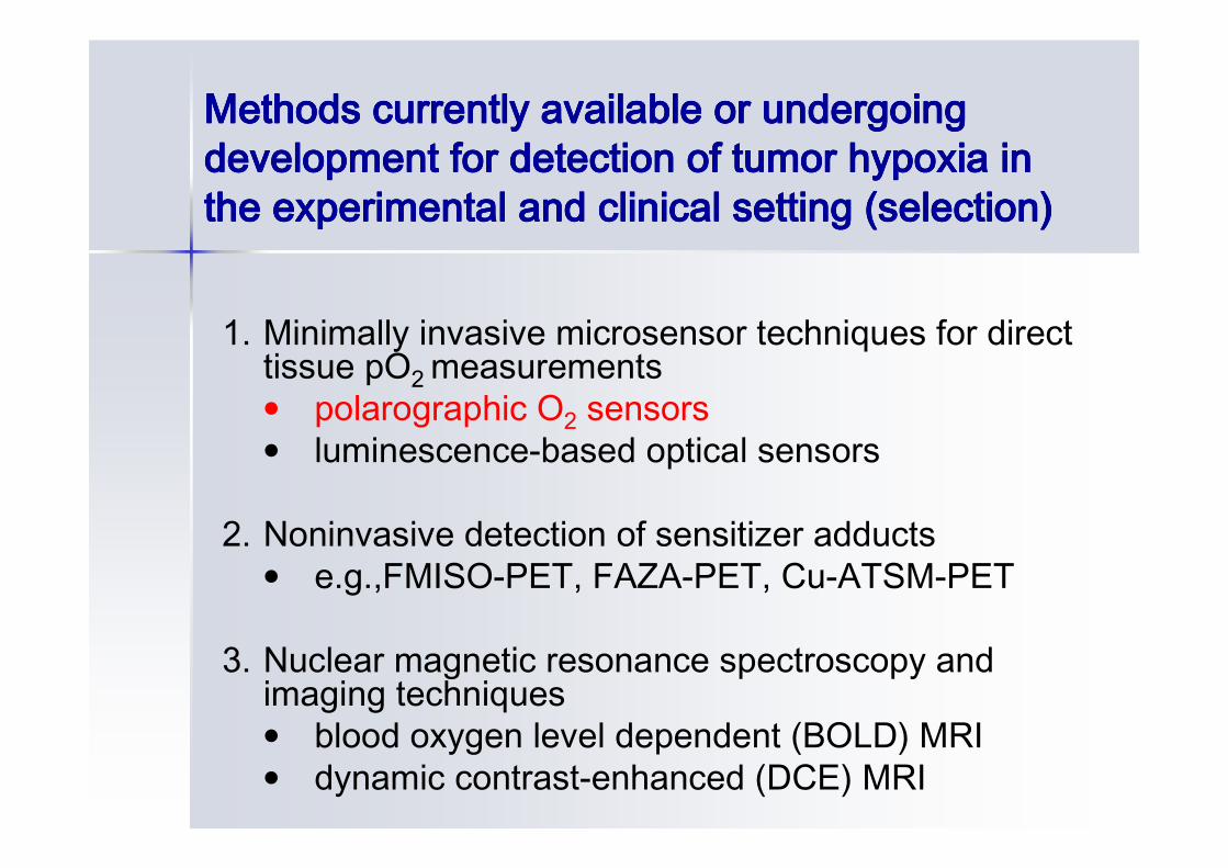
Methods currently available or undergoing Methods currently available or undergoing Methods currently available or undergoing Methods currently available or undergoing development for detection of tumor hypoxia in development for detection of tumor hypoxia in development for detection of tumor hypoxia in development for detection of tumor hypoxia in the experimental and clinical setting (selection)the experimental and clinical setting (selection)the experimental and clinical setting (selection)the experimental and clinical setting (selection)
1. Minimally invasive microsensor techniques for direct tissue pO2 measurements • polarographic O2 sensors• luminescence-based optical sensors
2. Noninvasive detection of sensitizer adducts• e.g.,FMISO-PET, FAZA-PET, Cu-ATSM-PET
3. Nuclear magnetic resonance spectroscopy and imaging techniques• blood oxygen level dependent (BOLD) MRI• dynamic contrast-enhanced (DCE) MRI
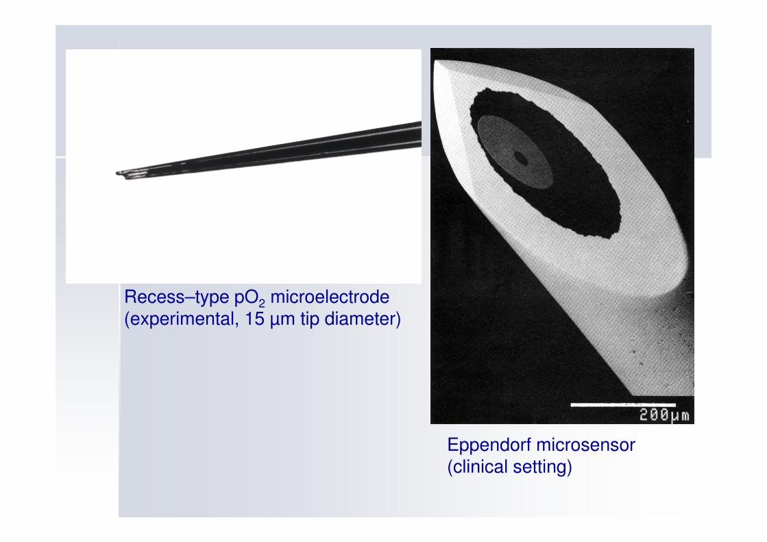
Eppendorf microsensor(clinical setting)
15 µm
Recess–type pO2 microelectrode(experimental, 15 µm tip diameter)
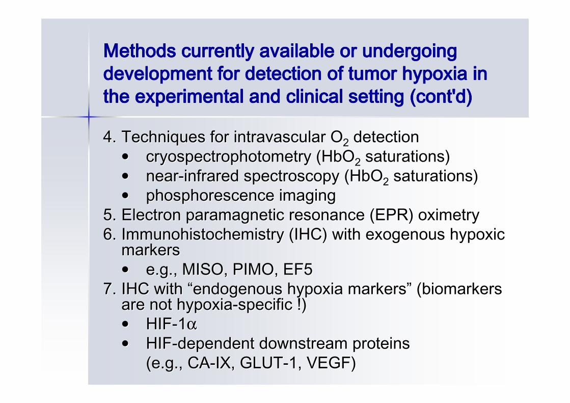
4.4. Techniques for intravascular OTechniques for intravascular O22 detectiondetection•• cryospectrophotometrycryospectrophotometry (HbO(HbO22 saturations)saturations)•• nearnear--infrared spectroscopy (HbOinfrared spectroscopy (HbO22 saturations)saturations)•• phosphorescence imagingphosphorescence imaging
5.5. Electron paramagnetic resonance (EPR) Electron paramagnetic resonance (EPR) oximetryoximetry6.6. ImmunohistochemistryImmunohistochemistry (IHC) with exogenous hypoxic (IHC) with exogenous hypoxic
markersmarkers•• e.g., MISO, PIMO, EF5e.g., MISO, PIMO, EF5
7.7. IHC with IHC with ““endogenous hypoxia markersendogenous hypoxia markers”” (biomarkers (biomarkers are not hypoxiaare not hypoxia--specific !)specific !)•• HIFHIF--11αα•• HIFHIF--dependent downstream proteinsdependent downstream proteins
(e.g., CA(e.g., CA--IX, GLUTIX, GLUT--1, VEGF)1, VEGF)
Methods currently available or undergoing Methods currently available or undergoing Methods currently available or undergoing Methods currently available or undergoing development for detection of tumor hypoxia in development for detection of tumor hypoxia in development for detection of tumor hypoxia in development for detection of tumor hypoxia in the experimental and clinical setting (cont'd)the experimental and clinical setting (cont'd)the experimental and clinical setting (cont'd)the experimental and clinical setting (cont'd)
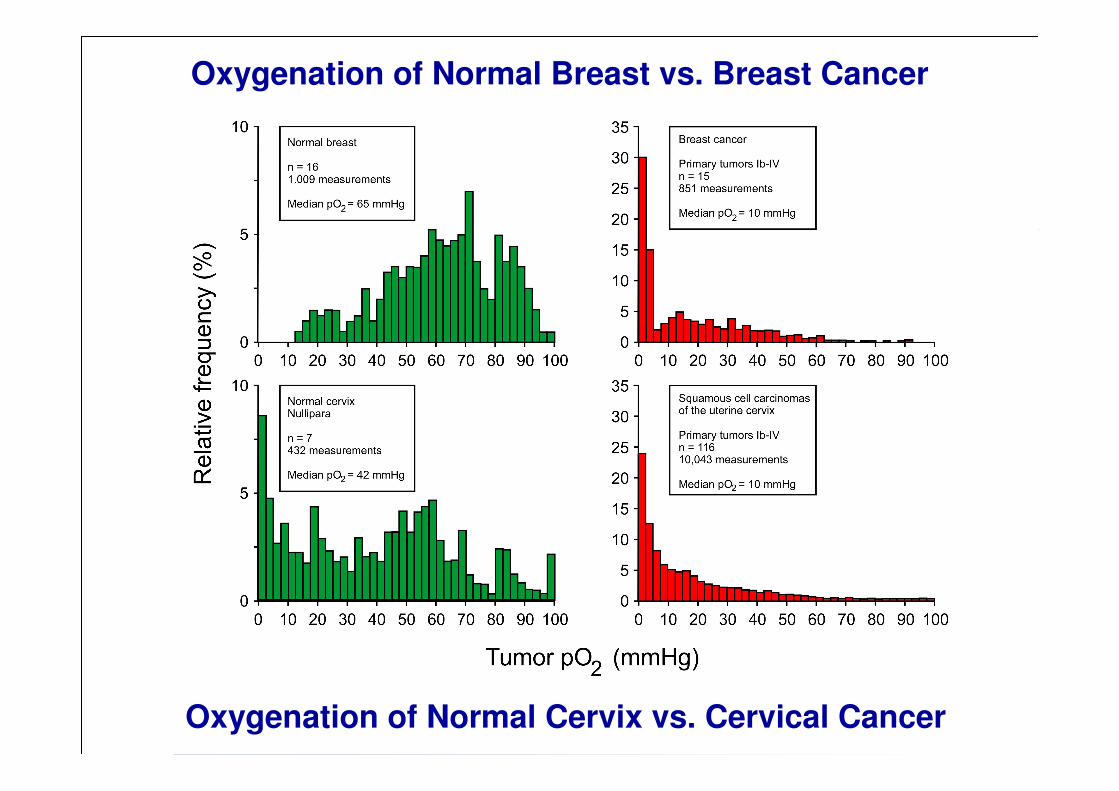
Oxygenation of Normal Breast vs. Breast Cancer
Oxygenation of Normal Cervix vs. Cervical Cancer
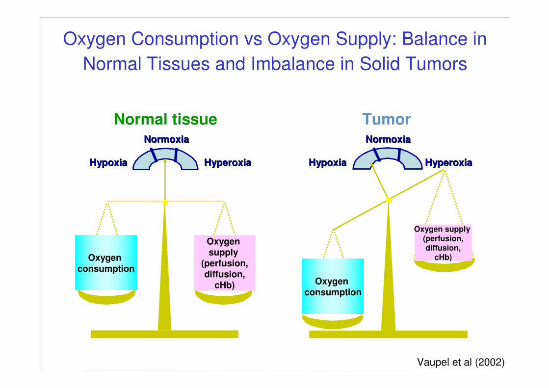
HypoxiaHypoxia
����
Oxygenconsumption
Oxygen supply
(perfusion,diffusion,
cHb)
Normal tissueNormoxiaNormoxia
HyperoxiaHyperoxia
����
TumorNormoxiaNormoxia
HypoxiaHypoxia HyperoxiaHyperoxia
Vaupel et al (2002)
Oxygen Consumption vs Oxygen Supply: Balance in
Normal Tissues and Imbalance in Solid Tumors
Oxygen supply (perfusion,diffusion,
cHb)
Oxygenconsumption
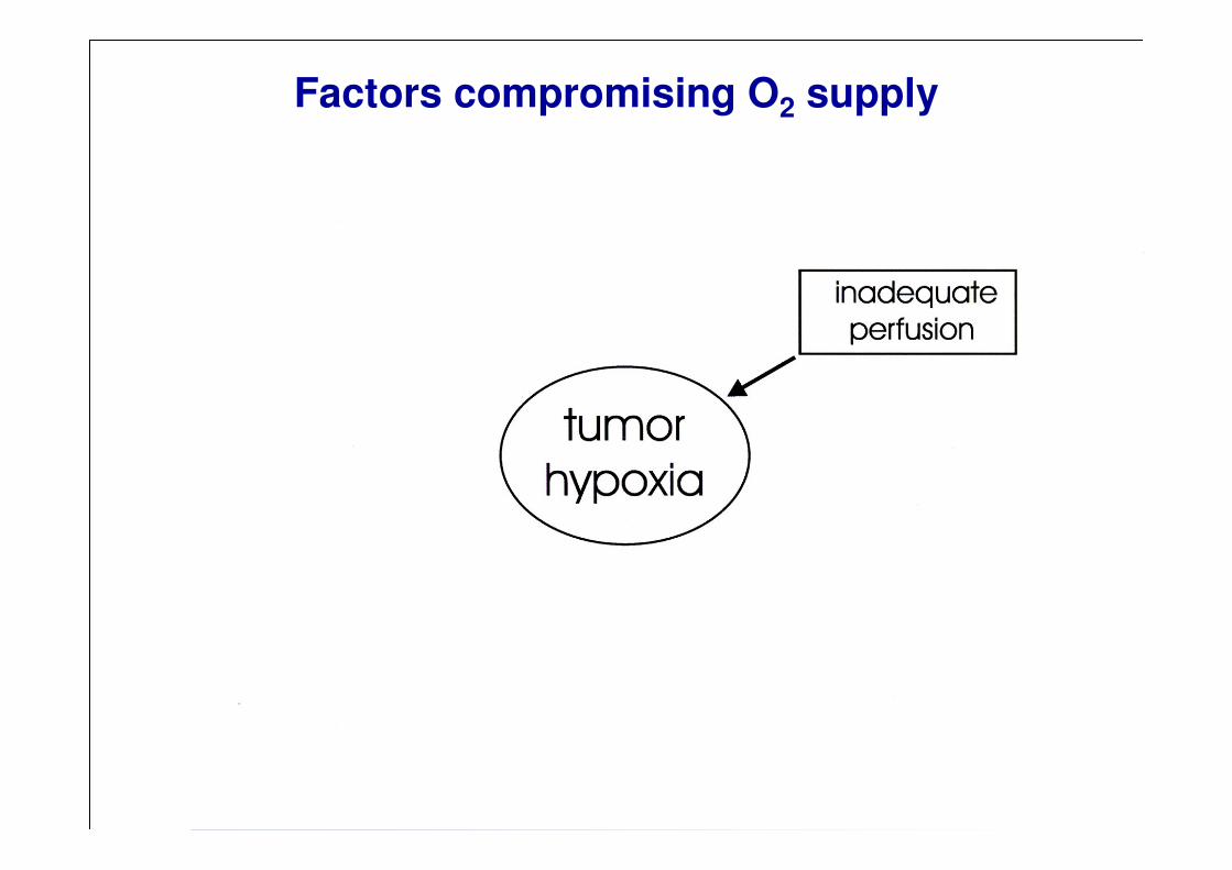
Factors compromising O2 supply
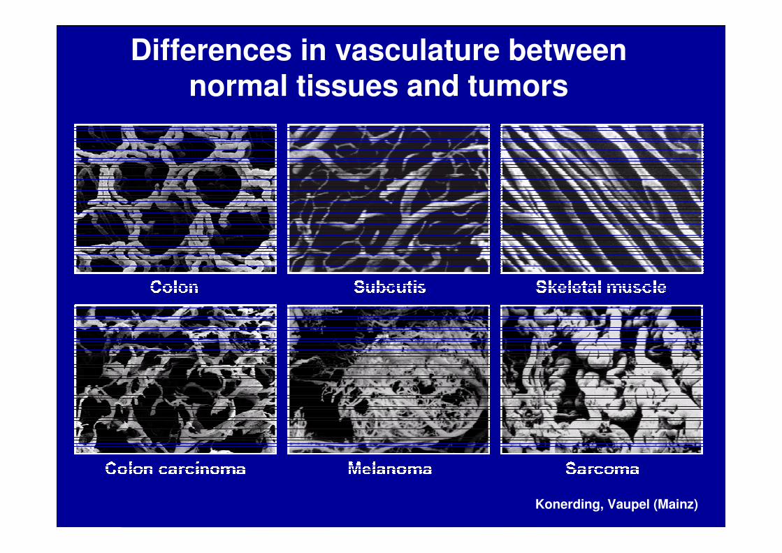
Differences in vasculature between normal tissues and tumors
Konerding, Vaupel (Mainz)
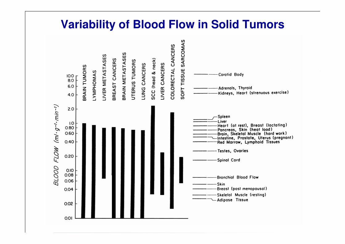
Variability of Blood Flow in Solid Tumors
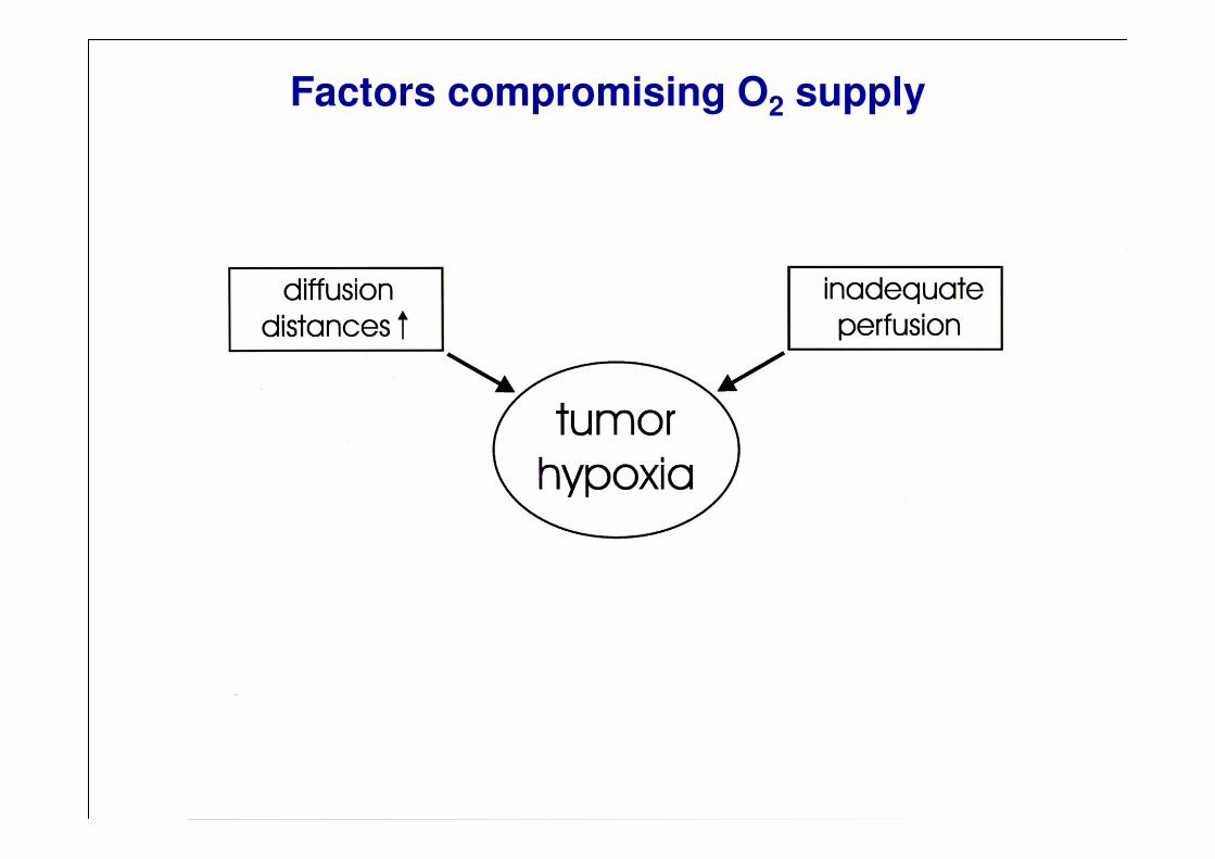
Factors compromising O2 supply
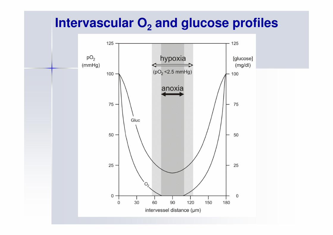
Intervascular O2 and glucose profiles
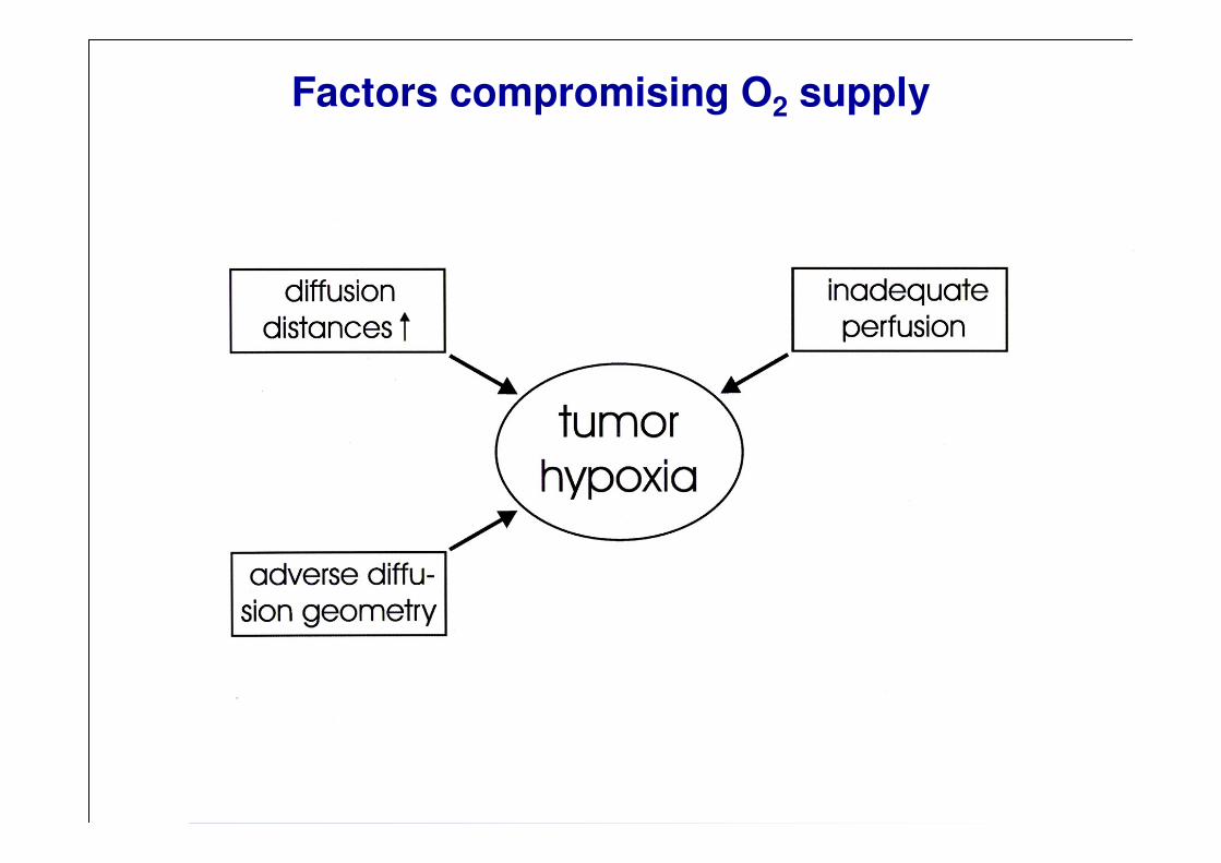
Factors compromising O2 supply
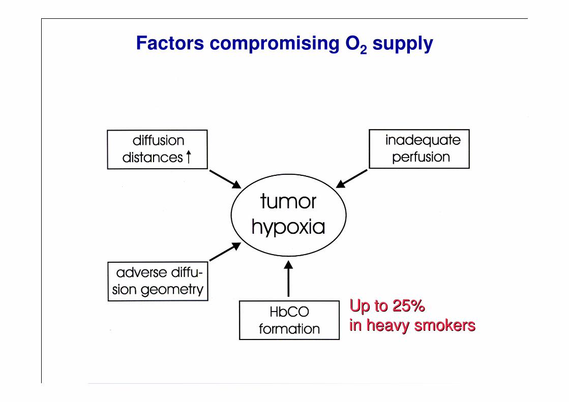
Factors compromising O2 supply
Up to 25%Up to 25%
in in heavyheavy smokerssmokers
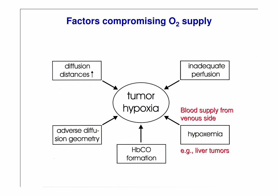
Factors compromising O2 supply
BloodBlood supplysupply fromfrom
venousvenous sideside
e.ge.g., ., liverliver tumorstumors
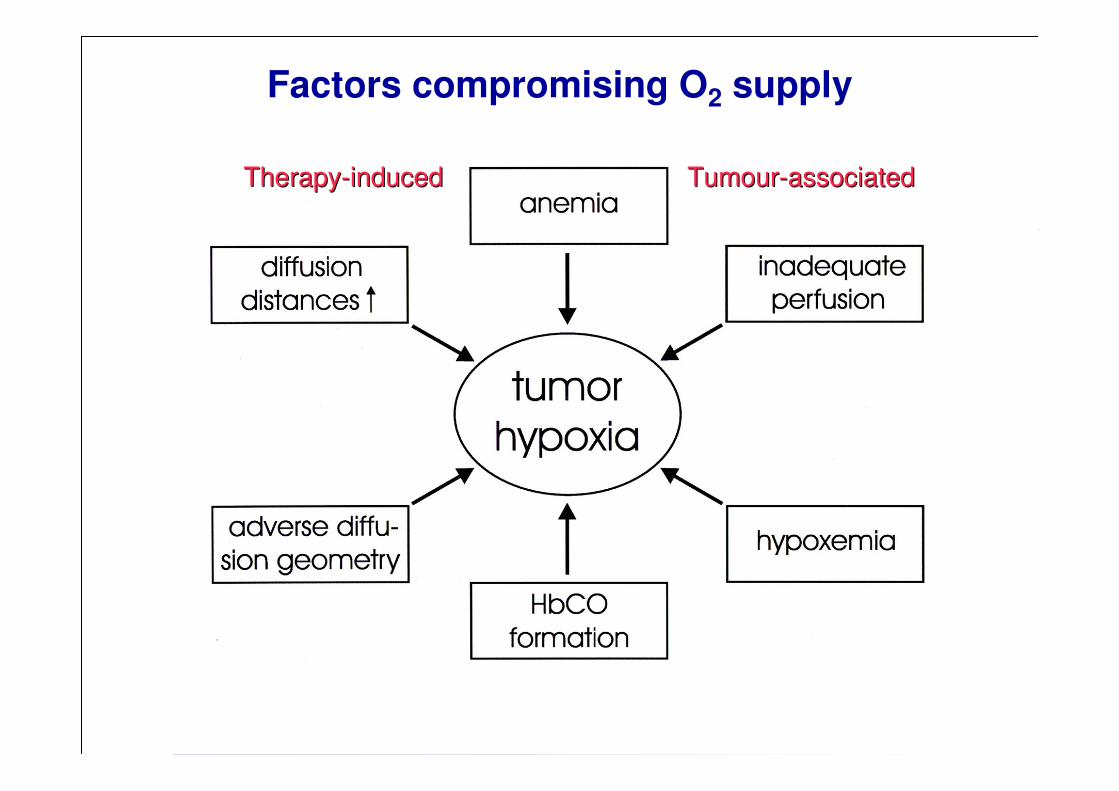
Factors compromising O2 supply
TherapyTherapy--inducedinduced TumourTumour--associatedassociated
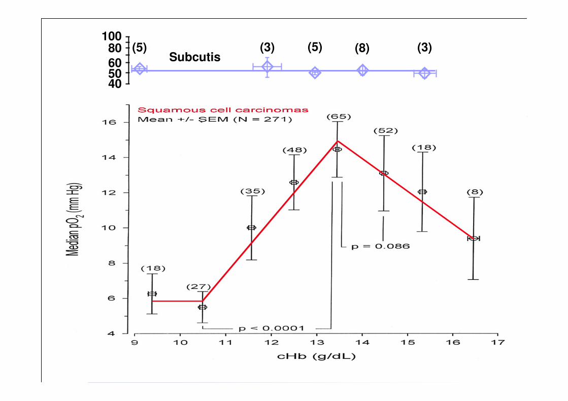
Tissue oxygenation as a function of hemoglobin concentration
1001008080
606050504040
(5)(5) (3)(3) (5)(5) (8)(8) (3)(3)SubcutisSubcutis
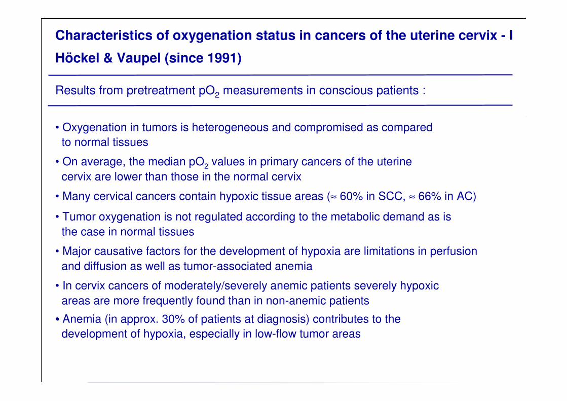
Characteristics of oxygenation status in cancers of the uterine cervix - I
Höckel & Vaupel (since 1991)
Results from pretreatment pO2 measurements in conscious patients :
• Oxygenation in tumors is heterogeneous and compromised as compared
to normal tissues
• On average, the median pO2 values in primary cancers of the uterine
cervix are lower than those in the normal cervix
• Many cervical cancers contain hypoxic tissue areas (≈ 60% in SCC, ≈ 66% in AC)
• Tumor oxygenation is not regulated according to the metabolic demand as is
the case in normal tissues
• Major causative factors for the development of hypoxia are limitations in perfusion
and diffusion as well as tumor-associated anemia
• In cervix cancers of moderately/severely anemic patients severely hypoxic
areas are more frequently found than in non-anemic patients
•• Anemia (in approx. 30% of patients at diagnosis) contributes to the
development of hypoxia, especially in low-flow tumor areas
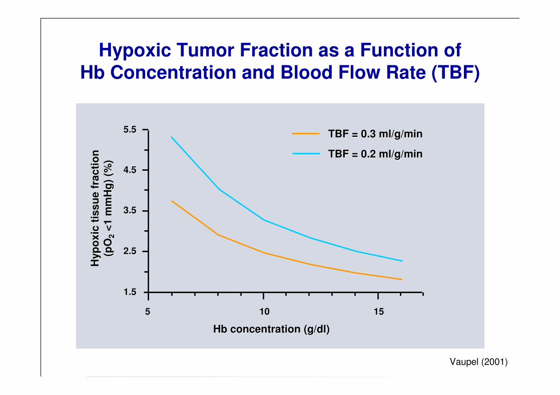
TBF = 0.2 ml/g/minTBF = 0.2 ml/g/min
5.55.5
4.54.5
3.53.5
2.52.5
1.51.5
55 1010 1515
Hb concentration (g/dl)Hb concentration (g/dl)
Hyp
ox
ic t
iss
ue
fra
cti
on
H
yp
ox
ic t
iss
ue
fra
cti
on
(pO
(pO
22<
1 m
mH
g)
(%)
<1
mm
Hg
) (%
)
TBF = 0.3 ml/g/minTBF = 0.3 ml/g/min
Vaupel (2001)
Hypoxic Tumor Fraction as a Function ofHb Concentration and Blood Flow Rate (TBF)
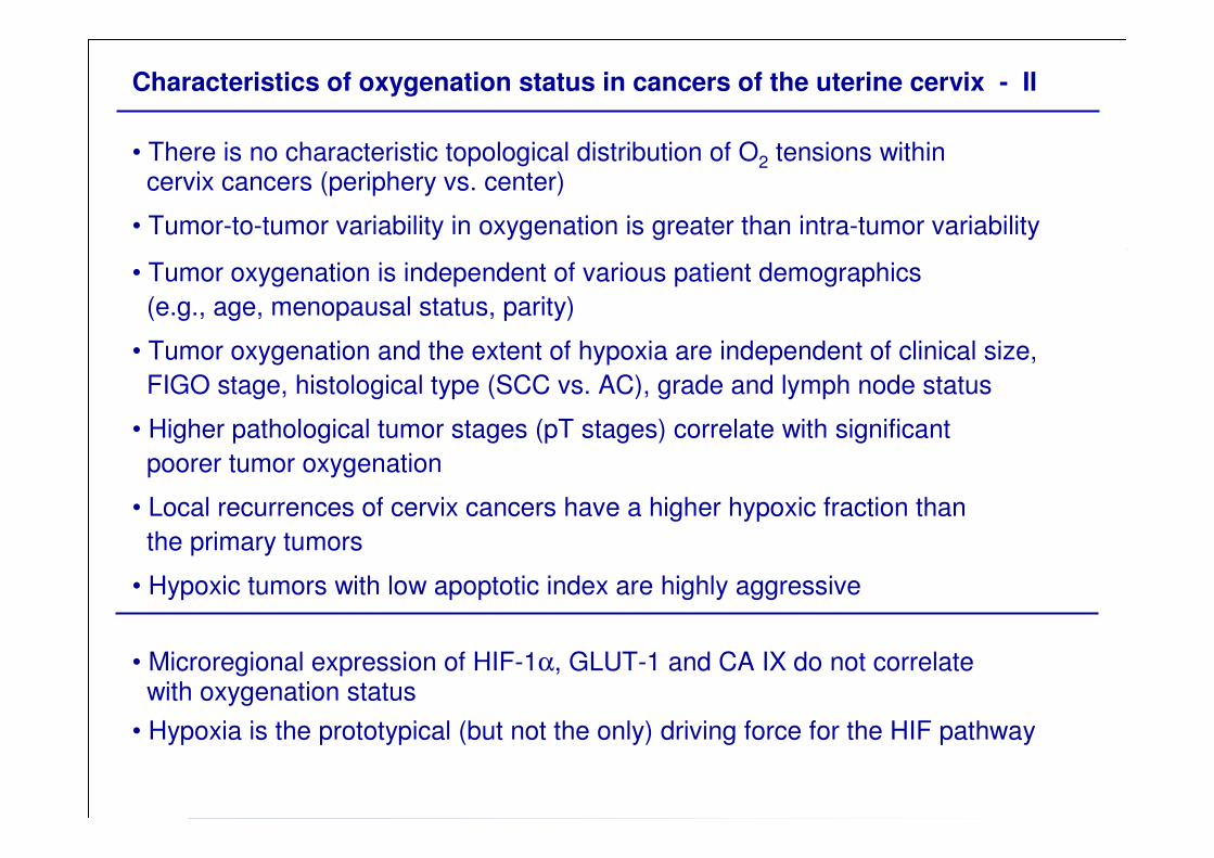
Characteristics of oxygenation status in cancers of the uterine cervix - II
• There is no characteristic topological distribution of O2 tensions within cervix cancers (periphery vs. center)
• Tumor-to-tumor variability in oxygenation is greater than intra-tumor variability
• Tumor oxygenation is independent of various patient demographics
(e.g., age, menopausal status, parity)
• Tumor oxygenation and the extent of hypoxia are independent of clinical size,
FIGO stage, histological type (SCC vs. AC), grade and lymph node status
• Higher pathological tumor stages (pT stages) correlate with significant
poorer tumor oxygenation
• Local recurrences of cervix cancers have a higher hypoxic fraction than
the primary tumors
• Hypoxic tumors with low apoptotic index are highly aggressive
• Microregional expression of HIF-1α, GLUT-1 and CA IX do not correlatewith oxygenation status
• Hypoxia is the prototypical (but not the only) driving force for the HIF pathway
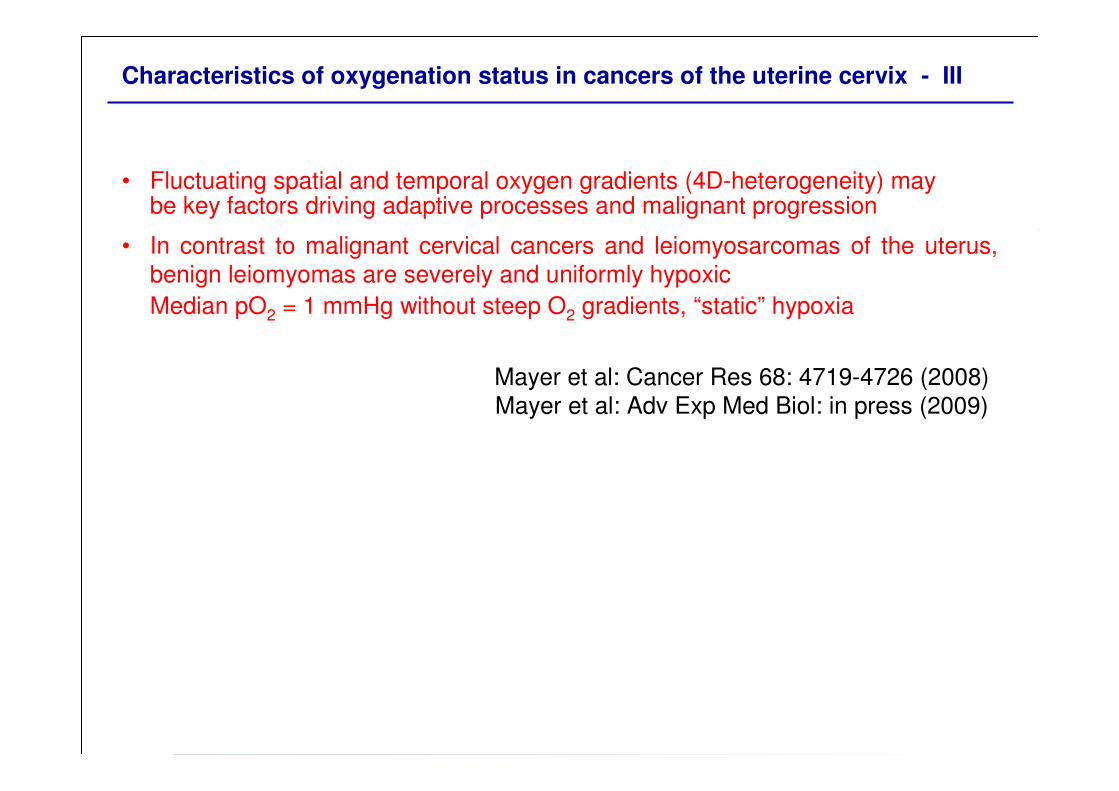
Characteristics of oxygenation status in cancers of the uterine cervix - III
• Fluctuating spatial and temporal oxygen gradients (4D-heterogeneity) maybe key factors driving adaptive processes and malignant progression
• In contrast to malignant cervical cancers and leiomyosarcomas of the uterus, benign leiomyomas are severely and uniformly hypoxic
Median pO2 = 1 mmHg without steep O2 gradients, “static” hypoxia
Mayer et al: Cancer Res 68: 4719-4726 (2008)Mayer et al: Adv Exp Med Biol: in press (2009)
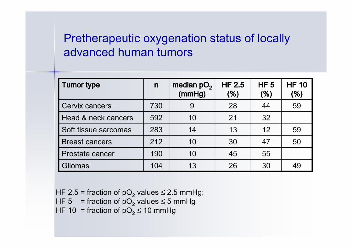
Pretherapeutic oxygenation status of locally advanced human tumors
HF 2.5 = fraction of pO2 values ≤ 2.5 mmHg;HF 5 = fraction of pO2 values ≤ 5 mmHgHF 10 = fraction of pO2 ≤ 10 mmHg
49302613104Gliomas554510190Prostate cancer
50473010212Breast cancers59121314283Soft tissue sarcomas
322110592Head & neck cancers5944289730Cervix cancers
HF 10HF 10HF 10HF 10(%)(%)(%)(%)
HF 5HF 5HF 5HF 5(%)(%)(%)(%)
HF 2.5HF 2.5HF 2.5HF 2.5(%)(%)(%)(%)
median pOmedian pOmedian pOmedian pO2222(mmHg)(mmHg)(mmHg)(mmHg)nnnnTumorTumorTumorTumor typetypetypetype
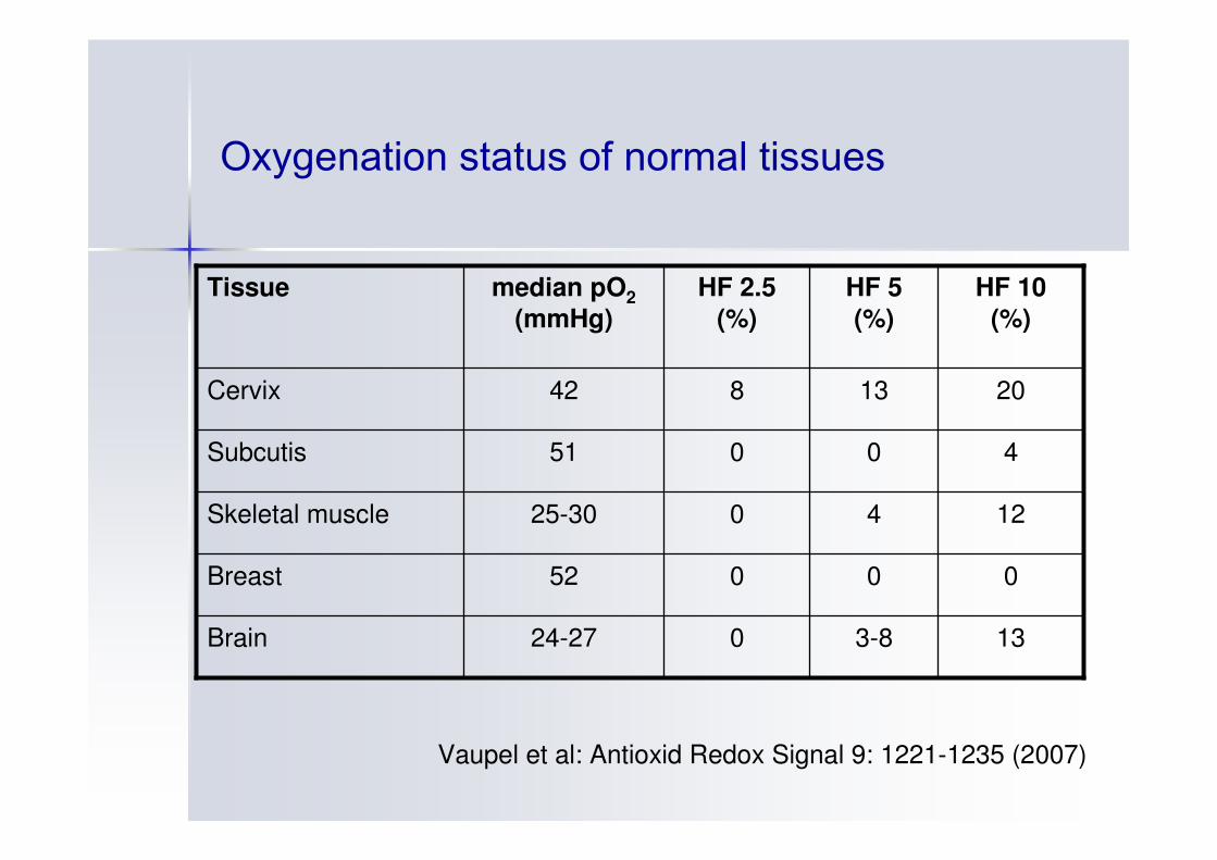
Oxygenation status of normal tissues
133-8024-27Brain
00052Breast
124025-30Skeletal muscle
40051Subcutis
2013842Cervix
HF 10
(%)
HF 5
(%)
HF 2.5
(%)
median pO2
(mmHg)
Tissue
Vaupel et al: Antioxid Redox Signal 9: 1221-1235 (2007)
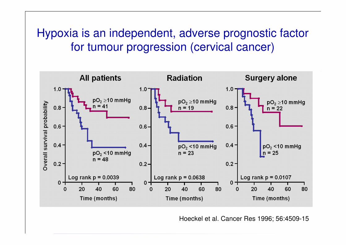
Hypoxia is an independent, adverse prognostic factor
for tumour progression (cervical cancer)
Hoeckel et al. Cancer Res 1996; 56:4509-15
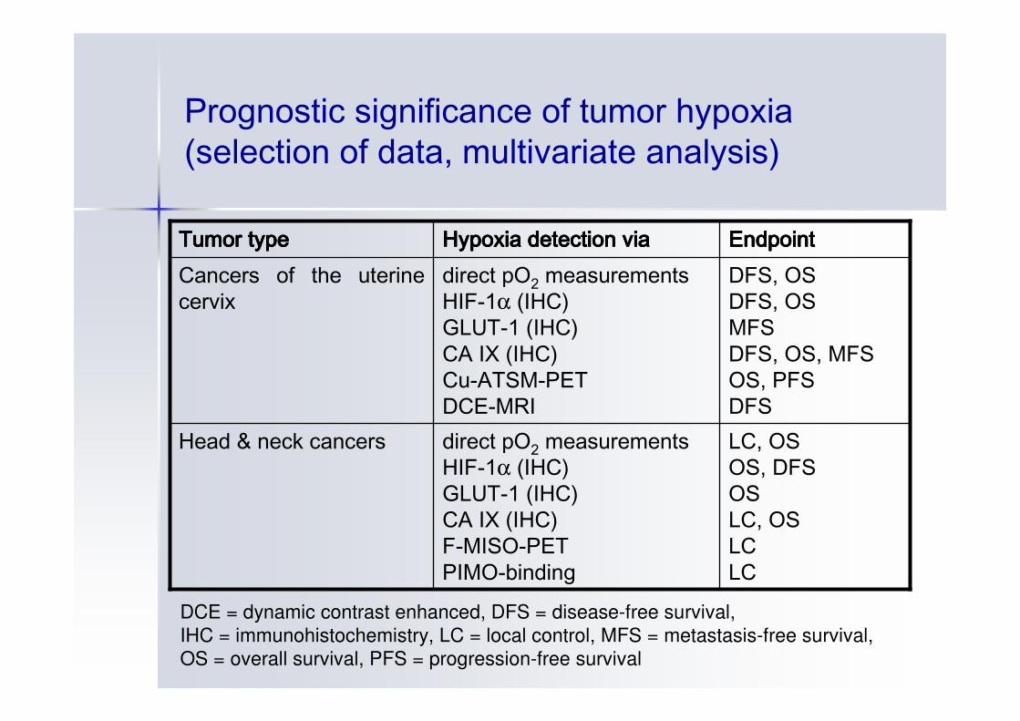
Prognostic significance of tumor hypoxia (selection of data, multivariate analysis)
LC, OSOS, DFSOSLC, OSLCLC
direct pO2 measurementsHIF-1α (IHC) GLUT-1 (IHC) CA IX (IHC)F-MISO-PETPIMO-binding
Head & neck cancers
DFS, OSDFS, OSMFSDFS, OS, MFSOS, PFSDFS
direct pO2 measurementsHIF-1α (IHC) GLUT-1 (IHC) CA IX (IHC)Cu-ATSM-PETDCE-MRI
Cancers of the uterine cervix
EndpointEndpointEndpointEndpointHypoxia detection viaHypoxia detection viaHypoxia detection viaHypoxia detection viaTumorTumorTumorTumor typetypetypetype
DCE = dynamic contrast enhanced, DFS = disease-free survival,
IHC = immunohistochemistry, LC = local control, MFS = metastasis-free survival,
OS = overall survival, PFS = progression-free survival
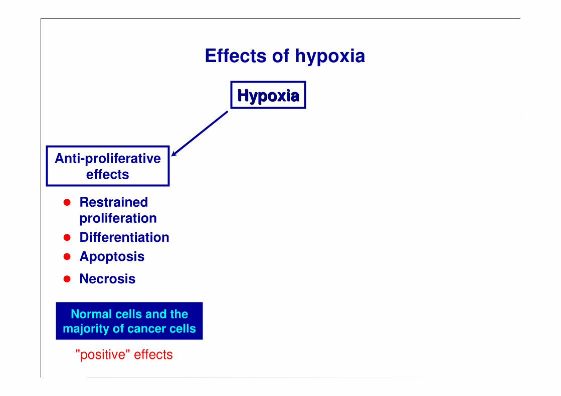
Effects of hypoxia
� Restrainedproliferation
� Differentiation
� Apoptosis
� Necrosis
Normal cells and the
majority of cancer cells
HypoxiaHypoxia
"positive" effects
Anti-proliferativeeffects
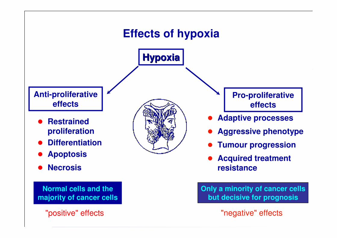
� Adaptive processes
� Aggressive phenotype
� Tumour progression
� Acquired treatmentresistance
Only a minority of cancer cells
but decisive for prognosis
"negative" effects
Normal cells and the
majority of cancer cells
"positive" effects
� Restrainedproliferation
� Differentiation
� Apoptosis
� Necrosis
HypoxiaHypoxia
Anti-proliferativeeffects
Effects of hypoxia
Pro-proliferativeeffects
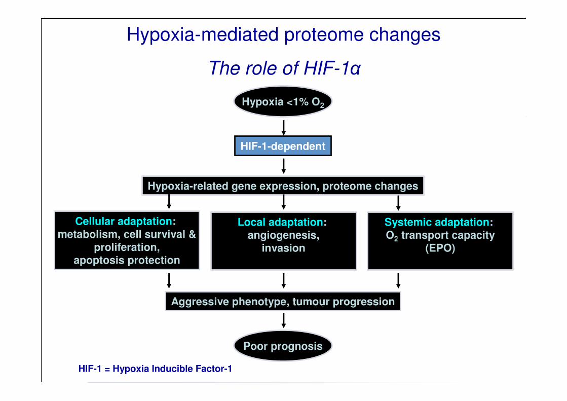
Hypoxia-mediated proteome changes
The role of HIF-1α
Hypoxia <1% O2
HIF-1-dependent
Cellular adaptation:
metabolism, cell survival &proliferation,
apoptosis protection
Poor prognosis
Hypoxia-related gene expression, proteome changes
Aggressive phenotype, tumour progression
HIF-1 = Hypoxia Inducible Factor-1
Local adaptation: angiogenesis,
invasion
Systemic adaptation: O2 transport capacity
(EPO)
Fluctuating spatial
and temporal O2 gradients
(4D-heterogeneity)
+
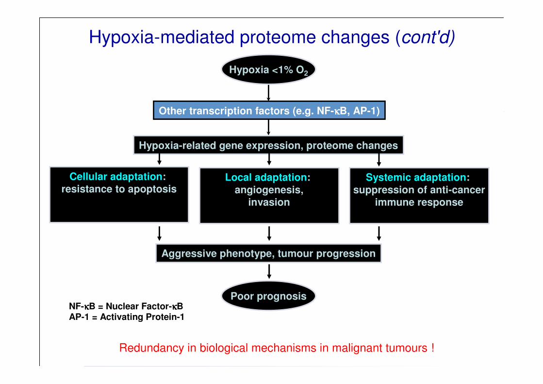
Hypoxia-mediated proteome changes (cont'd)
Hypoxia <1% O2
Other transcription factors (e.g. NF-κκκκB, AP-1)
Hypoxia-related gene expression, proteome changes
NF-κκκκB = Nuclear Factor-κκκκBAP-1 = Activating Protein-1
Cellular adaptation: resistance to apoptosis
Local adaptation:
angiogenesis,invasion
Systemic adaptation:
suppression of anti-cancerimmune response
Poor prognosis
Aggressive phenotype, tumour progression
Redundancy in biological mechanisms in malignant tumours !
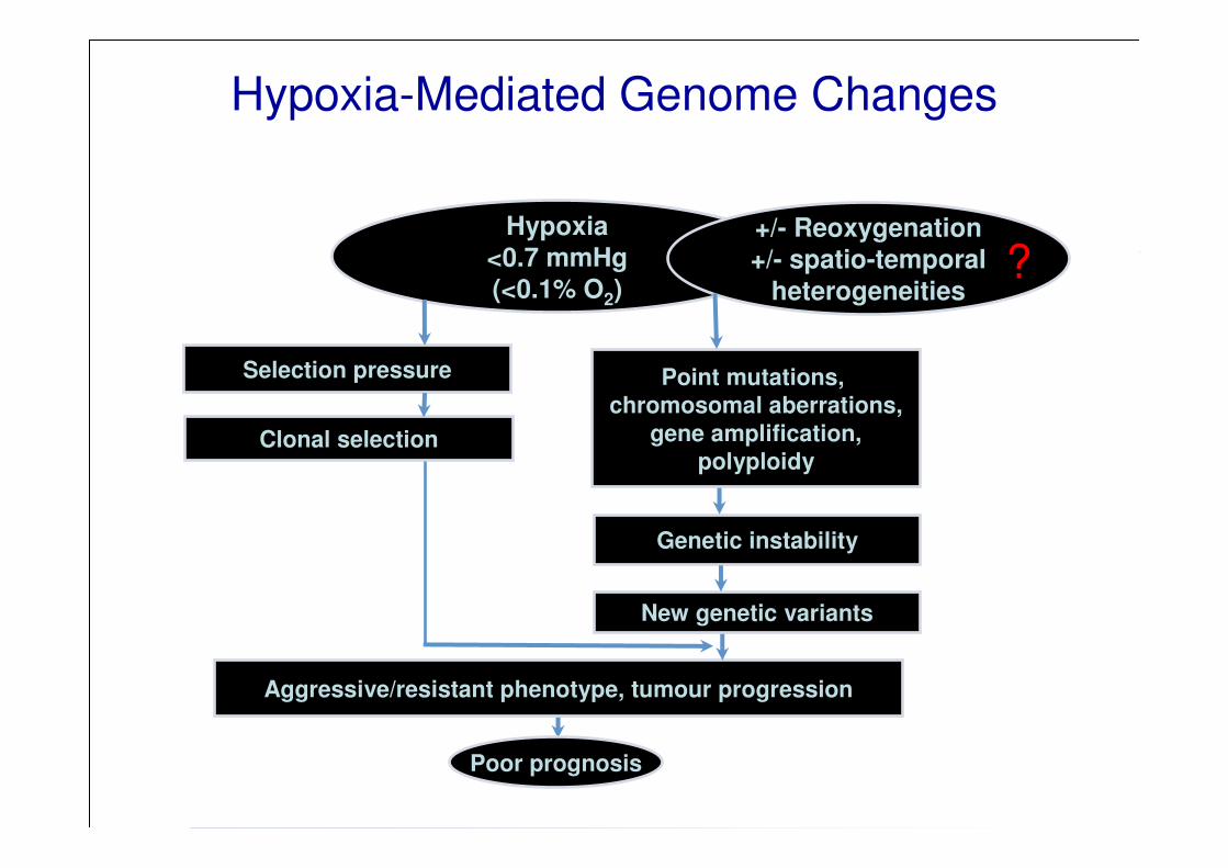
Hypoxia-Mediated Genome Changes
Hypoxia<0.7 mmHg(<0.1% O2)
+/- Reoxygenation+/- spatio-temporal
heterogeneities
Point mutations, chromosomal aberrations,
gene amplification,polyploidy
New genetic variants
Aggressive/resistant phenotype, tumour progression
Clonal selection
Selection pressure
Poor prognosis
Genetic instability
?
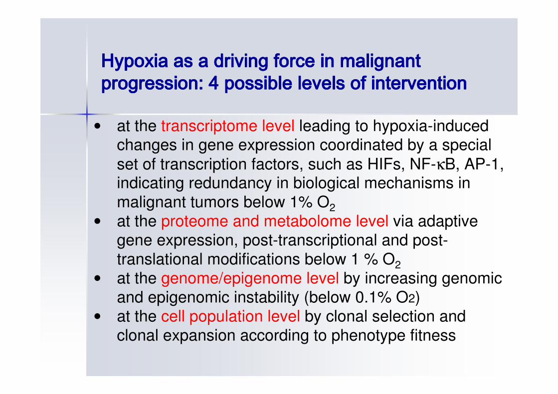
HypoxiaHypoxiaHypoxiaHypoxia as a as a as a as a drivingdrivingdrivingdriving force in force in force in force in malignantmalignantmalignantmalignantprogressionprogressionprogressionprogression: 4 : 4 : 4 : 4 possiblepossiblepossiblepossible levelslevelslevelslevels of of of of interventioninterventioninterventionintervention
• at the transcriptome level leading to hypoxia-induced changes in gene expression coordinated by a special
set of transcription factors, such as HIFs, NF-κB, AP-1, indicating redundancy in biological mechanisms in malignant tumors below 1% O2
• at the proteome and metabolome level via adaptivegene expression, post-transcriptional and post-translational modifications below 1 % O2
• at the genome/epigenome level by increasing genomicand epigenomic instability (below 0.1% O2)
• at the cell population level by clonal selection andclonal expansion according to phenotype fitness
Tumor Hypoxia
Chicken Chicken
oror
EggEgg
??
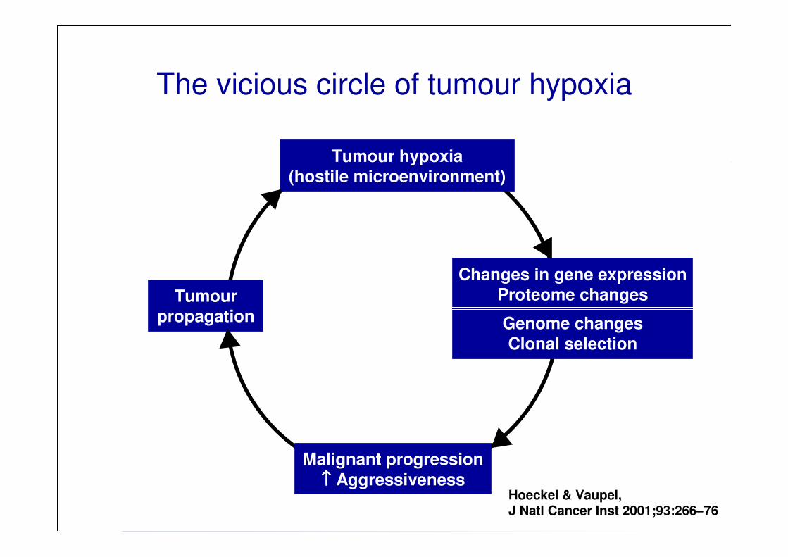
Changes in gene expressionProteome changesTumour
propagation
Malignant progression
↑↑↑↑ Aggressiveness
Genome changes
Clonal selection
The vicious circle of tumour hypoxia
Tumour hypoxia(hostile microenvironment)
Hoeckel & Vaupel,J Natl Cancer Inst 2001;93:266–76
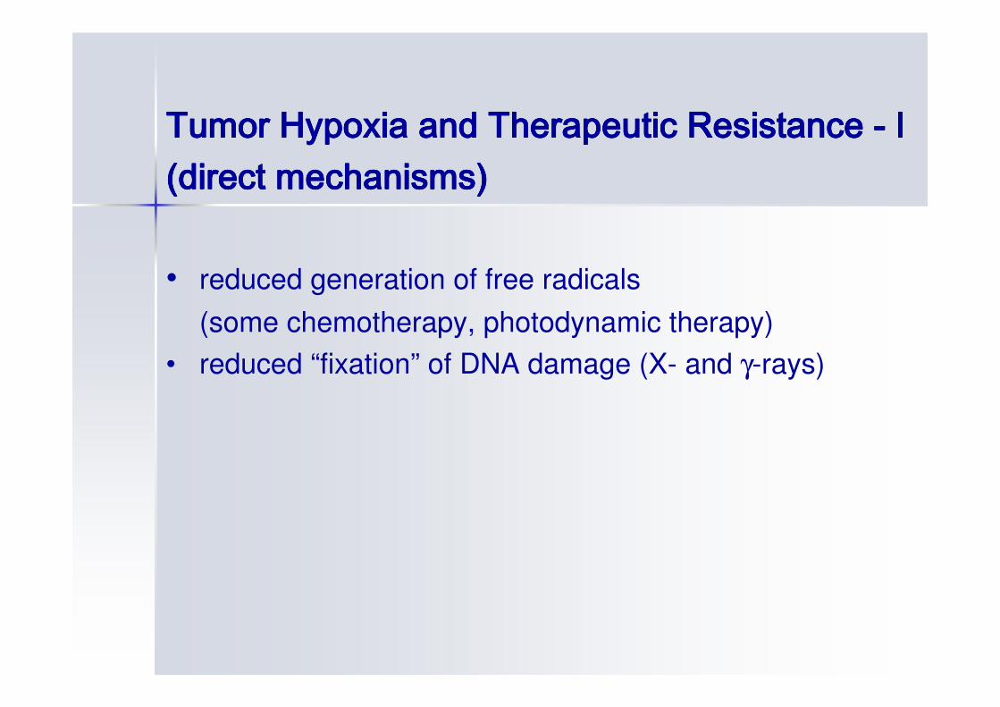
TumorTumorTumorTumor Hypoxia and Therapeutic Resistance Hypoxia and Therapeutic Resistance Hypoxia and Therapeutic Resistance Hypoxia and Therapeutic Resistance ---- IIII(direct mechanisms)(direct mechanisms)(direct mechanisms)(direct mechanisms)
• reduced generation of free radicals
(some chemotherapy, photodynamic therapy)
• reduced “fixation” of DNA damage (X- and γ-rays)
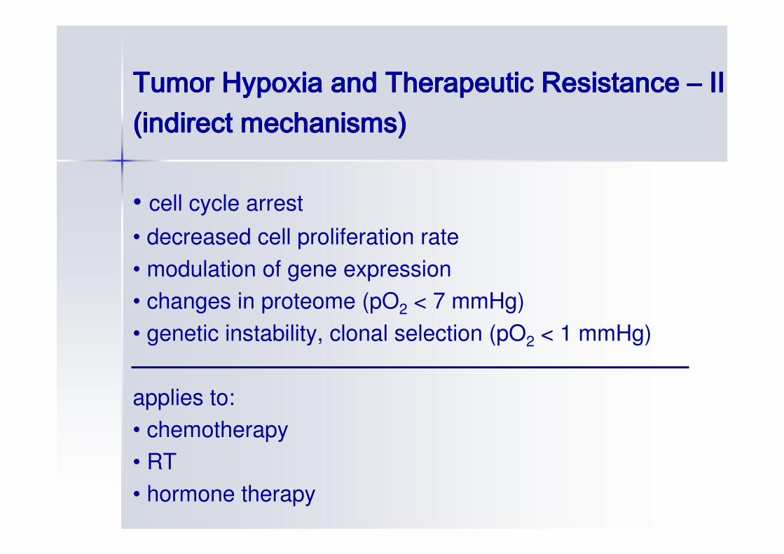
TumorTumorTumorTumor Hypoxia and Therapeutic Resistance Hypoxia and Therapeutic Resistance Hypoxia and Therapeutic Resistance Hypoxia and Therapeutic Resistance –––– IIIIIIII(indirect (indirect (indirect (indirect mechanisms)mechanisms)mechanisms)mechanisms)
• cell cycle arrest
• decreased cell proliferation rate
• modulation of gene expression
• changes in proteome (pO2 < 7 mmHg)
• genetic instability, clonal selection (pO2 < 1 mmHg)
applies to:
• chemotherapy
• RT
• hormone therapy

< 2% O2
Key information
< 2% O2
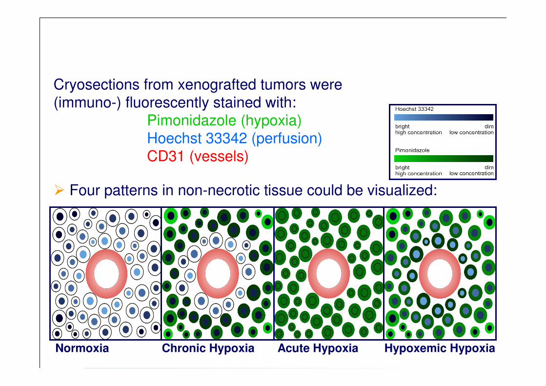
Normoxia Chronic Hypoxia Acute Hypoxia Hypoxemic Hypoxia
Cryosections from xenografted tumors were(immuno-) fluorescently stained with:
Pimonidazole (hypoxia)Hoechst 33342 (perfusion)CD31 (vessels)
� Four patterns in non-necrotic tissue could be visualized:
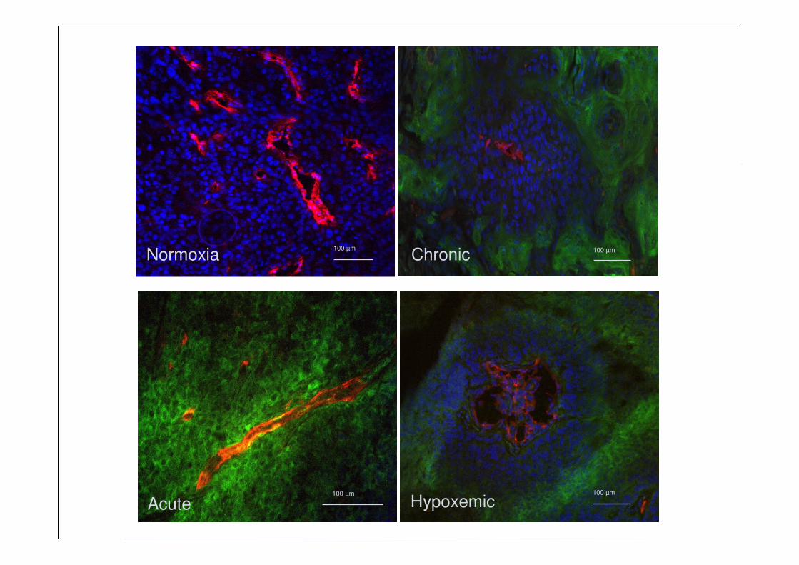
100 µm100 µm
100 µm100 µm
100 µmNormoxia
Acute
Chronic
Hypoxemic
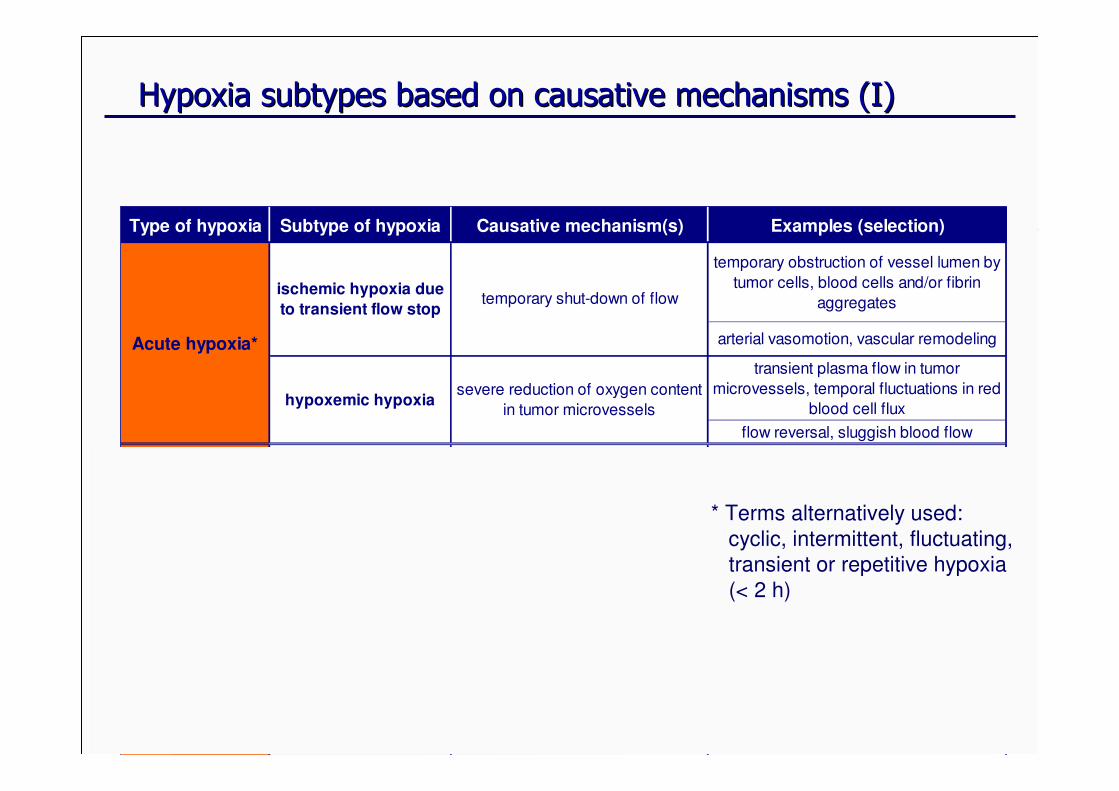
Hypoxia subtypes based on causative mechanisms (I)Hypoxia subtypes based on causative mechanisms (I)
Type of hypoxia Subtype of hypoxia Causative mechanism(s) Examples (selection)
temporary obstruction of vessel lumen by
tumor cells, blood cells and/or fibrin
aggregates
arterial vasomotion, vascular remodeling
transient plasma flow in tumor
microvessels, temporal fluctuations in red
blood cell flux
flow reversal, sluggish blood flow
surpassing the maximum oxygen
diffusion distance in the
perivascular space
expansion of the intervascular space (low
vascular density)
concurrent vs. countercurrent tumor
microvessels;
centripedal vs. centrifugal diffusion field***
(approx. 2.7 times greater hypoxic area in
the latter at otherwise identical boundary
exaggerated longitudinal
intravascular oxygen gradients
elongated, tortuous (convoluted)
microvessels
diverging oxygen-rich (arterial)
blood to the venous side of the
tumor vascular bed
shunt perfusion through arterio-venous
anastomoses
reduced oxygen content in the
blood supplying the tumor
tumor-associated and/or therapy-induced
anemia (anemic hypoxia)
temporary shut-down of flow
adverse diffusion (vascular)
geometry
severe reduction of oxygen content
in tumor microvessels
Acute hypoxia*
Chronic
hypoxia**
ischemic hypoxia due
to transient flow stop
hypoxemic hypoxia
diffusion-limited
hypoxia
* Terms alternatively used:
cyclic, intermittent, fluctuating,
transient or repetitive hypoxia
(< 2 h)
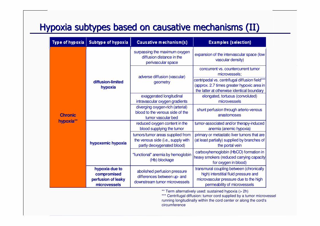
** Term alternatively used: sustained hypoxia (> 2h)
*** Centrifugal diffusion: tumor cord supplied by a tumor microvessel
running longitudinally within the cord center or along the cord‘s
circumference
aggregates
arterial vasomotion, vascular remodeling
transient plasma flow in tumor
microvessels, temporal fluctuations in red
blood cell flux
flow reversal, sluggish blood flow
surpassing the maximum oxygen
diffusion distance in the
perivascular space
expansion of the intervascular space (low
vascular density)
concurrent vs. countercurrent tumor
microvessels;
centripedal vs. centrifugal diffusion field***
(approx. 2.7 times greater hypoxic area in
the latter at otherwise identical boundary
exaggerated longitudinal
intravascular oxygen gradients
elongated, tortuous (convoluted)
microvessels
diverging oxygen-rich (arterial)
blood to the venous side of the
tumor vascular bed
shunt perfusion through arterio-venous
anastomoses
reduced oxygen content in the
blood supplying the tumor
tumor-associated and/or therapy-induced
anemia (anemic hypoxia)
tumors/tumor areas supplied from
the venous side (i.e., supply with
partly deoxygenated blood)
primary or metastatic liver tumors that are
(at least partially) supplied by branches of
the portal vein
"functional" anemia by hemoglobin
(Hb) blockage
carboxyhemoglobin (HbCO) formation in
heavy smokers (reduced carrying capacity
for oxygen in blood)
hypoxia due to
compromised
perfusion of leaky
microvessels
abolished perfusion pressure
differences between up- and
downstream tumor microvessels
transmural coupling between (chronically
high) interstitial fluid pressure and
microvascular pressure due to the high
permeability of microvessels
temporary shut-down of flow
adverse diffusion (vascular)
geometry
severe reduction of oxygen content
in tumor microvessels
Acute hypoxia*
Chronic
hypoxia**
to transient flow stop
hypoxemic hypoxia
diffusion-limited
hypoxia
hypoxemic hypoxia
Hypoxia subtypes based on causative mechanisms (II)Hypoxia subtypes based on causative mechanisms (II)
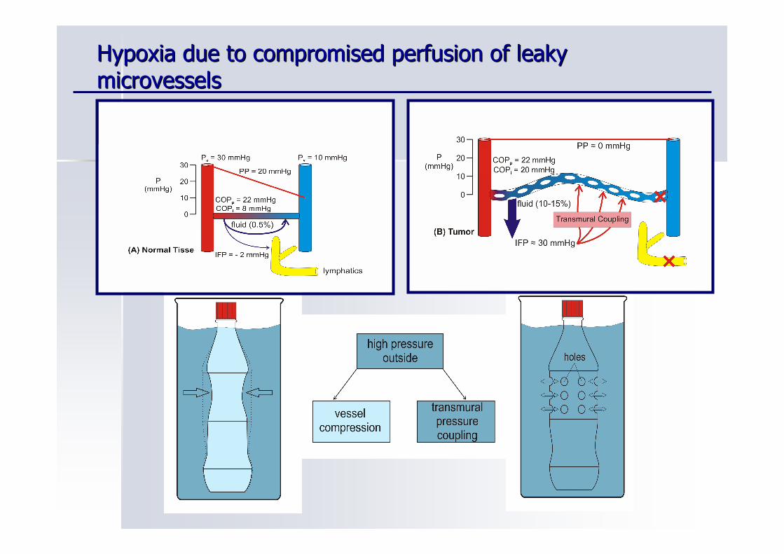
Hypoxia due to compromised perfusion of leaky Hypoxia due to compromised perfusion of leaky
microvesselsmicrovessels
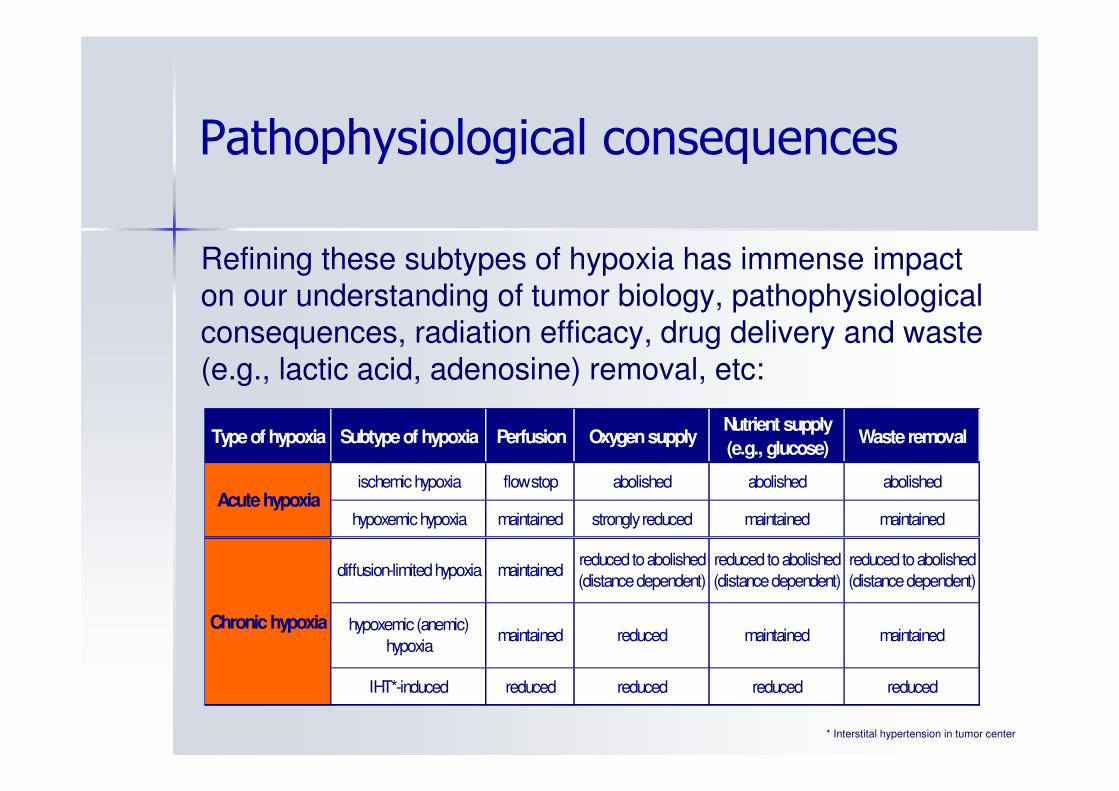
Pathophysiological consequences
Refining these subtypes of hypoxia has immense impacton our understanding of tumor biology, pathophysiological consequences, radiation efficacy, drug delivery and waste
(e.g., lactic acid, adenosine) removal, etc:
* Interstital hypertension in tumor center
Type of hypoxia Subtype of hypoxia Perfusion Oxygen supplyNutrient supply
(e.g., glucose) Waste removal
ischemic hypoxia flow stop abolished abolished abolished
hypoxemic hypoxia maintained strongly reduced maintained maintained
diffusion-limited hypoxia maintainedreduced to abolished
(distance dependent)
reduced to abolished
(distance dependent)
reduced to abolished
(distance dependent)
hypoxemic (anemic)
hypoxiamaintained reduced maintained maintained
IHT*-induced reduced reduced reduced reduced
Acute hypoxia
Chronic hypoxia
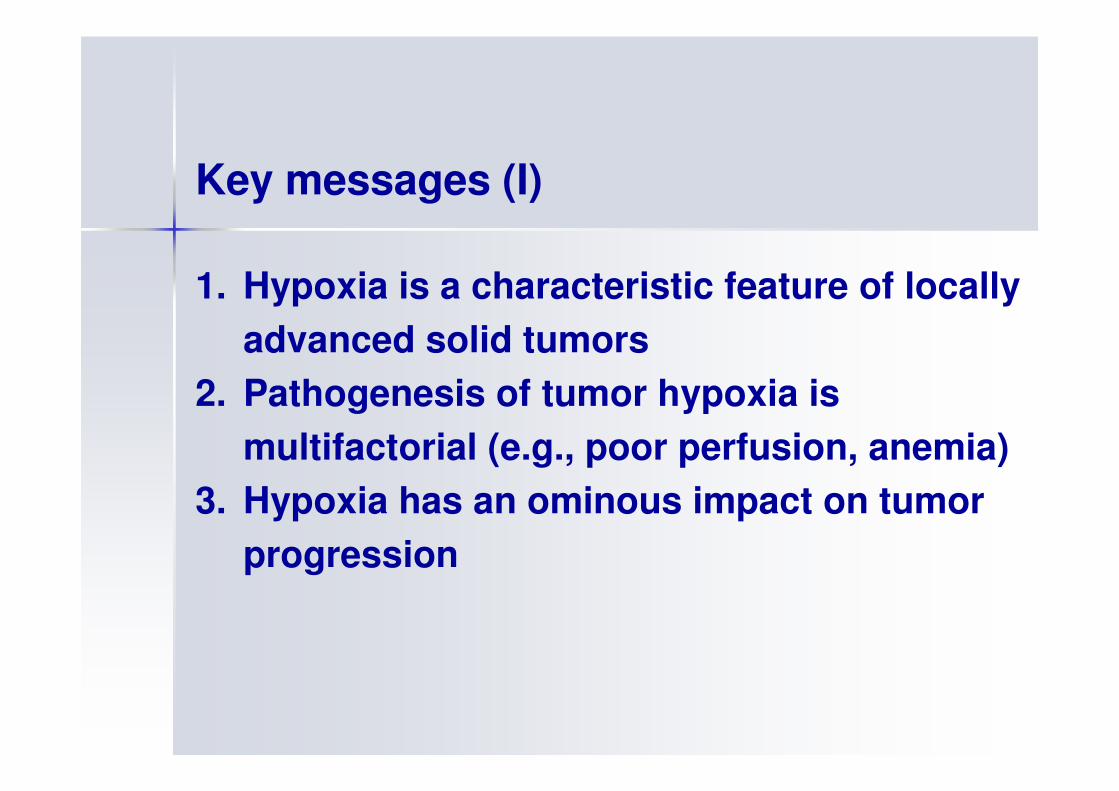
Key messages (I)
1. Hypoxia is a characteristic feature of locally
advanced solid tumors
2. Pathogenesis of tumor hypoxia is
multifactorial (e.g., poor perfusion, anemia)
3. Hypoxia has an ominous impact on tumor
progression
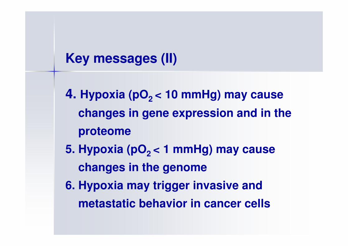
Key messages (II)
4. Hypoxia (pO2 < 10 mmHg) may cause
changes in gene expression and in the
proteome
5. Hypoxia (pO2 < 1 mmHg) may cause
changes in the genome
6. Hypoxia may trigger invasive and
metastatic behavior in cancer cells
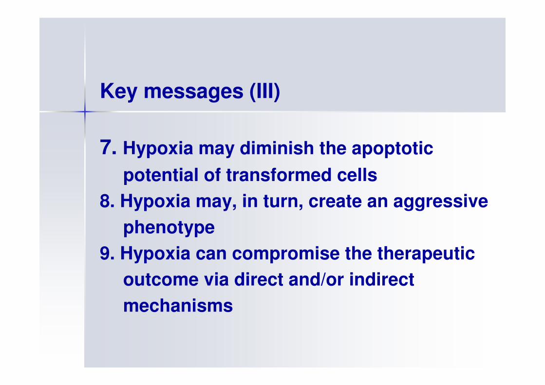
Key messages (III)
7. Hypoxia may diminish the apoptotic
potential of transformed cells
8. Hypoxia may, in turn, create an aggressive
phenotype
9. Hypoxia can compromise the therapeutic
outcome via direct and/or indirect
mechanisms
Thank you for your kind attention!
For questions not addressed in the followingdiscussion please contact:
Related Documents


