Laryngeal inhalational injuries: A systematic review Jessica A. Tang a , Grace Amadio b , Lavanya Nagappan b , Cecelia E. Schmalbach a, b, c , Gregory R. Dion d, * a Department of Otolaryngology, Head and Neck Surgery, Temple University Hospital, Philadelphia, PA, USA b Lewis Katz School of Medicine at Temple University, Philadelphia, PA, USA c Temple Head and Neck Institute, Philadelphia, PA, USA d US Army Institute of Surgical Research, Joint Base San Antonio, Fort Sam Houston, TX, USA a b s t r a c t Laryngeal inhalation injury carries a significant increase in mortality rate and often indicates immediate airway evaluation. This may be difficult in the setting of clinical deterioration necessitating immediate intubation, which itself can synergistically cause mucosal damage. Prior studies do not encompass predictive factors or long-term outcomes for the laryngotracheal complex. This systemic review of PubMed, Embase, and Cochrane identified studies investigating inhalational injuries of the upper airway. Demographic data as well as presentation, physical findings, and delayed sequelae were documented. Laryngotracheal burn patients were divided into two cohorts based on timing of laryngeal injury diagnosis (before- versus after-airway intervention). 1051 papers met initial search criteria and 43 studies were ultimately included. Airway stenosis was more common in patients that were intubated immediately (50.0%, n = 18 versus 5.2%, n = 13; p = 0.57). Posterior glottic involvement was only identified in patients intubated prior to airway evaluation (71.4%, n = 15). All studies reported a closed space setting for those patients in whom airway intervention preceded laryngeal evaluation. Laryngeal inhalational injuries are a distinct subset that can have a variety of minor to severe laryngotracheal delayed sequelae, particularly for thermal injuries occurring within enclosed spaces. Given these findings, early otolaryngology referral may mitigate or treat these effects. Published by Elsevier Ltd. a r t i c l e i n f o Article history: Accepted 5 February 2021 Keywords: Laryngeal inhalational injuries Posterior glottic stenosis Subglottic stenosis Upper airway 1. Introduction Inhalation injuries occur in about 10% of burn patients and are linked to an increased overall mortality in the range of 25% [1 5]. Diagnosis is generally made on clinical suspicion with flexible videolaryngoscopy [6 8] and imaging [9 11] employed for confirmation. Clinical deterioration may necessitate imme- diate intubation prior to upper airway evaluation. In addition, traditional criteria [12] for inhalational injury often lead to prophylactic intubation. Taken together, these factors limit quantification of laryngotracheal and pharyngeal injury pat- terns and tissue injury progression in many cases. Endotracheal intubation in the setting of an inhalation injury has been linked to long term laryngotracheal injuries ranging from dysphonia and dysphagia to tracheal and posterior glottic stenosis (PGS) [13 17]. Studies quantifying these laryngotracheal outcomes based on initial thermal injury pattern to the laryngotracheal complex and pharynx are lacking. * Corresponding author at: Dental and Craniofacial Trauma Research Department, U.S. Army Institute of Surgical Research, 3698 Chambers Pass, Bldg 3611, JBSA Fort Sam Houston, TX 78234-7313, USA. E-mail address: [email protected] (G.R. Dion). https://doi.org/10.1016/j.burns.2021.02.006 0305-4179/ Published by Elsevier Ltd. b u r n s 4 8 ( 2 0 2 2 ) 2 3 3 3 Available online at www.sciencedirect.com ScienceDirect jo u rn al h o mep age: w ww .elsevier .co m /loc ate/b u rn s Descargado para BINASSS BINASSS ([email protected]) en National Library of Health and Social Security de ClinicalKey.es por Elsevier en febrero 15, 2022. Para uso personal exclusivamente. No se permiten otros usos sin autorización. Copyright ©2022. Elsevier Inc. Todos los derechos reservados.

Welcome message from author
This document is posted to help you gain knowledge. Please leave a comment to let me know what you think about it! Share it to your friends and learn new things together.
Transcript

Laryngeal inhalational injuries: A systematic review
Jessica A. Tang a, Grace Amadio b, Lavanya Nagappan b,Cecelia E. Schmalbach a,b,c, Gregory R. Dion d,*aDepartment of Otolaryngology, Head and Neck Surgery, Temple University Hospital, Philadelphia, PA, USAb Lewis Katz School of Medicine at Temple University, Philadelphia, PA, USAc Temple Head and Neck Institute, Philadelphia, PA, USAdUS Army Institute of Surgical Research, Joint Base San Antonio, Fort Sam Houston, TX, USA
a b s t r a c t
Laryngeal inhalation injury carries a significant increase in mortality rate and often indicates
immediate airway evaluation. This may be difficult in the setting of clinical deterioration
necessitating immediate intubation, which itself can synergistically cause mucosal damage.
Prior studies do not encompass predictive factors or long-term outcomes for the
laryngotracheal complex. This systemic review of PubMed, Embase, and Cochrane identified
studies investigating inhalational injuries of the upper airway. Demographic data as well as
presentation, physical findings, and delayed sequelae were documented. Laryngotracheal
burn patients were divided into two cohorts based on timing of laryngeal injury diagnosis
(before- versus after-airway intervention). 1051 papers met initial search criteria and
43 studies were ultimately included. Airway stenosis was more common in patients that
were intubated immediately (50.0%, n = 18 versus 5.2%, n = 13; p = 0.57). Posterior glottic
involvement was only identified in patients intubated prior to airway evaluation (71.4%, n =
15). All studies reported a closed space setting for those patients in whom airway
intervention preceded laryngeal evaluation. Laryngeal inhalational injuries are a distinct
subset that can have a variety of minor to severe laryngotracheal delayed sequelae,
particularly for thermal injuries occurring within enclosed spaces. Given these findings,
early otolaryngology referral may mitigate or treat these effects.
Published by Elsevier Ltd.
a r t i c l e i n f o
Article history:
Accepted 5 February 2021
Keywords:
Laryngeal inhalational injuries
Posterior glottic stenosis
Subglottic stenosis
Upper airway
1. Introduction
Inhalation injuries occur in about 10% of burn patients and arelinked to an increased overall mortality in the range of 25%[1�5]. Diagnosis is generally made on clinical suspicion withflexible videolaryngoscopy [6�8] and imaging [9�11] employedfor confirmation. Clinical deterioration may necessitate imme-diate intubation prior to upper airway evaluation. In addition,traditional criteria [12] for inhalational injury often lead to
prophylactic intubation. Taken together, these factors limitquantification of laryngotracheal and pharyngeal injury pat-terns and tissue injury progressionin many cases. Endotrachealintubation in the setting of an inhalation injury has been linkedto long term laryngotracheal injuries ranging from dysphoniaand dysphagia to tracheal and posterior glottic stenosis (PGS)[13�17]. Studies quantifying these laryngotracheal outcomesbased on initial thermal injury pattern to the laryngotrachealcomplex and pharynx are lacking.
* Corresponding author at: Dental and Craniofacial Trauma Research Department, U.S. Army Institute of Surgical Research, 3698 ChambersPass, Bldg 3611, JBSA Fort Sam Houston, TX 78234-7313, USA.
E-mail address: [email protected] (G.R. Dion).https://doi.org/10.1016/j.burns.2021.02.0060305-4179/ Published by Elsevier Ltd.
b u r n s 4 8 ( 2 0 2 2 ) 2 3 �3 3
Available online at www.sciencedirect.com
ScienceDirect
jo u rn al h o mep age: w ww .e lsev ier . co m / loc ate /b u rn s
Descargado para BINASSS BINASSS ([email protected]) en National Library of Health and Social Security de ClinicalKey.es por Elsevier en febrero 15, 2022. Para uso personal exclusivamente. No se permiten otros usos sin autorización. Copyright ©2022. Elsevier Inc. Todos los derechos reservados.
Laryngotracheal mucosal injury with concomitant intuba-tion may initiate posterior glottic stenosis [18]. Duringintubation and with an endotracheal tube (ETT) in place, theairway is at risk of insults to delicate epithelium andsubmucosal areolar and tracheal tissues leading to edemaand subsequent airway narrowing [19]. Iatrogenic injurysecondary to ETT intubation accounts for approximately 90%of acquired subglottic stenosis (SGS) and an even higher rate ofPGS [20]. Dysregulated wound healing in response to ETT-induced ulceration helps to propagate rapidly progressivefibroplasia and pathologic scarring in subglottic injury [21]. Arecent study identified acute laryngeal injury in more than halfof patients intubated greater than 12 h, with findings persistingfor more than two months [22]. Evidence also suggests earlyintervention may be ideal in treating these patients [23]. Withthe lack of therapies to prevent these complex complications,
efforts traditionally have focused on improving treatmentsand minimizing the associated impacts. Although well studiedin other intubated populations, the prevalence and impact ofthese adverse effects of ETT are less well understood in thecohort of patients with inhalation injury.
The majority of existing inhalational injury studies focusspecifically on pulmonary injuries, as these play a direct impactin early survival outcomes. Despite numerous studies charac-terizing inhalational injuries, there is a paucity of data onthermal injury patterns or impact of inhalational injury onupper airway function. Specific to inhalational injury to thelaryngotracheal complex, it is hypothesized that the subglotticregion, comprised of respiratory epithelium, may be moresensitive to inhalational injury compared to the stratifiedsquamous epithelium above the vocal folds [15]. In addition,airflow patterns below the vocal folds may increase the area of
Fig. 1 – Search terms used in the PubMed, Embase, and Cochrane databases.
24 b u r n s 4 8 ( 2 0 2 2 ) 2 3 �3 3
Descargado para BINASSS BINASSS ([email protected]) en National Library of Health and Social Security de ClinicalKey.es por Elsevier en febrero 15, 2022. Para uso personal exclusivamente. No se permiten otros usos sin autorización. Copyright ©2022. Elsevier Inc. Todos los derechos reservados.

exposuretotoxinandthermalinjury[24].Conversely,theglotticclosure reflex, an airway protective mechanism activated byreceptors within the larynx [25], may functionally create a heatsink protecting the lower airway from thermal injury. Addition-ally, heat capacities of inhaled fluids can vary by orders ofmagnitude, such as with steam verses hot dry air, widelyaltering resulting tissue damage from inhalational injuries [26].
Although there are systematic reviews of prognosticimplications in inhalation injuries [27] and risk factors fordeveloping dysphagia after thermal burn injuries [28], these donot encompass predictive factors or long term outcomes for thelaryngotracheal complex in the setting of inhalation injury.As aresult, in this systematic review, we sought to compile existingliterature addressing thermal inhalation injuries to the laryng-otracheal complex, defining the presentation, physical find-ings, and associated short- and long-term sequelae ofinhalational injuries on the upper airway. Ultimately, thisinformation may help to optimize early consultation andintervention, where applicable, to reduce long-term morbidity.
2. Methods
A systematic review was performed in accordance with thePreferred Reporting Items for Systematic Reviews and
Meta-Analyses (PRISMA) guidelines [29,30]. Guidance wasprovided by our institution’s library information specialist(KB). A review protocol did not exist prior to this study and nostudy registration was required. Three search engines wereused to conduct the systematic review: PubMed, Embase, andThe Cochrane Library. An additional search through theClinical Trials was performed. Search terms are defined inFig. 1. The search was performed for all studies throughNovember 2019. A specific start date was not set to ensurecomprehensive review. Studies were included if they dis-cussed inhalational or thermal burn injuries specific to theupper airway. Anatomical subsites included the pharynx,oropharynx, nasopharynx, hypopharynx, larynx, supraglottis,subglottis, glottis, laryngotrachea, upper respiratory tract,upper airway, or upper aerodigestive tract.
Studies were excluded if they were non-English full texts,animal studies, in-vitro studies (only histological/molecularanalyses), review articles, or if full text was unavailable. Inorder to minimize risk of bias, we excluded any study from thesame institution that included repeat cohorts (Fig. 2). Dedicat-ed pediatric studies were excluded in order to limit anyconfounding factors attributed to differences in anatomy andphysiology in children. Additionally, studies were excluded ifthey were limited to the lower respiratory tract, describedinhalational injuries in general terms without objective upper
Fig. 2 – Flow chart of identification and screening as performed in accordance to PRISMA guidelines.
b u r n s 4 8 ( 2 0 2 2 ) 2 3 �3 3 25
Descargado para BINASSS BINASSS ([email protected]) en National Library of Health and Social Security de ClinicalKey.es por Elsevier en febrero 15, 2022. Para uso personal exclusivamente. No se permiten otros usos sin autorización. Copyright ©2022. Elsevier Inc. Todos los derechos reservados.

airway findings, reported non-inhalational mechanisms ofinjury (chemical, electric, iatrogenic airway fire), or involvedblunt/penetrating laryngeal trauma. Many studies includedlaryngeal inhalational injuries as a subset of a larger group ofburn patients; in these cases, only the patients and associateddata specific to laryngeal inhalational injuries were collectedfor analysis.
All studies were reviewed independently by two authors(JAT, LN). Studies in which inclusion and exclusion criteriacould not be determined based on abstracts alone warrantedfull text review. The bibliographies of all included studies werehand searched for additional studies potentially missingcapture in the initial search criteria. When necessary, studieswere discussed in detail to achieve consensus and consistencyfor inclusion.
All studies meeting inclusion criteria underwent dataextraction (JAT, GA) for the following variables: article year,lead author, institution, study type, demographics (age, sex,and race if specified), etiology and circumstance of burn injury,percent total body surface area (% TBSA), clinical presentation,airway management, discharge disposition, mortality, de-layed sequelae, and complications. Specifically, clinicalpresentation variables included dysphonia, dysphagia, dys-pnea, stridor, wheezes/rales, and cough. Documented physicalexam findings include singed nasal or facial hairs, cutaneousburns to the head and neck, and if endoscopic examinationwas performed, soot and/or edema, hyperemia, and hyperse-cretion. Delayed sequelae were defined as any result notpresent upon initial airway evaluation of the patient andincluded pneumonia, stricture or stenosis, posterior glotticweb or stenosis, and tracheoesophageal fistula.
Laryngotracheal burn patients were divided into twocohorts based on the timing of laryngeal injury diagnosis(before- versus after-airway intervention). This grouping wasselected to categorize patients where a formal airway evalua-tion was undertaken in a non-instrumented airway comparedto those patients where airway evaluation was only possibleafter initial intubation, as the known impacts of endotrachealintubation may confound airway findings. Airway interven-tion was defined as any procedure performed to secure theairway to include: intubation, surgical tracheostomy, andcricothryoidotomy. Patients were included in the before-airway intervention group if objective laryngeal inhalationinjury was documented before any airway interventions(intubation, etc.). Conversely, if the diagnosis of laryngealinhalation injury was diagnosed at some point after initialairway intervention, patients were included in the after group.The mechanism of laryngeal burn was also investigated basedon closed space (e.g. house fire, trailer) versus open space (e.g.outdoors).
The data was recorded using Microsoft Excel (MicrosoftCorporations, Redmond, WA). Overall outcome variableswere reported using descriptive statistics (IBM SPSS Statis-tics, Version 20, Chicago, IL). When applicable, patientpercentages were among reported only as a percentage ofthe patient population in those studies reporting thevariable of interest. Overall mortality, tracheostomy rates,and airway stenosis were reported between groups based onthe timing of laryngeal injury diagnosis (pre- or post-airwayintervention).
3. Results
A total of 1051 papers met initial search criteria of which41 studies met inclusion criteria (Fig. 2). Hand searches of thegrey literature to include bibliographies of the above 41 articlesidentified two additional papers for a total of 43 includedstudies. Initial conflicts with 23 studies were resolved throughreviewer discussion.
Included studies were published between 1973 and 2019. Intotal, there were nine prospective observational studies,16 retrospective reviews, and 18 case reports or series.Collectively, a total of 12,474 burn patients were identified,1517 of which had objective documentation of laryngealinhalation injuries (12.15%). Across 29 studies [6�12,14,31�51], 1400 patients had laryngeal inhalational injuries docu-mented prior to airway intervention (Table 1). An additional117 patients from 14 studies [13,16,52�63] were noted to havelaryngeal inhalational injuries documented after airwayintervention (Table 2). Laryngeal inhalational injuries weremost commonly identified by flexible endoscopy. A smallsubset of patients were diagnosed via virtual bronchoscopy inwhich CT scans were used to specifically assess laryngotra-cheal structures [9].
Clinical diagnosis (Fig. 3), physical exam findings (Fig. 4),and delayed sequelae (Fig. 5) are summarized based onlaryngeal findings of inhalation injury documented beforeand after airway intervention. Intubation was the mostcommon means of airway intervention (n = 483) with surgicalairway/tracheostomy and emergent cricothyroidotomy.
Patients with laryngeal inhalational injury noted prior toairway intervention had a mortality rate of 17.4%, n = 143 of824, compared to findings after airway intervention of 4.2%, n =3 of 79. Both cohorts underwent tracheotomy at similar rates(34.2%, n = 181 of 527, and 34.3%, n = 30 of 77). Airway stenosiswas more common in patients that were intubated immedi-ately (50.0%, n = 18 of 36, versus 5.2%, n = 13 of 246). Posteriorglottic involvement was only identified in patients intubatedprior to airway evaluation (71.4%, n = 15 of 21). Airway stenosiswas identified as early as one to fourteen weeks after injury[61]. All studies reported a closed space setting for the subset ofpatients in whom airway intervention preceded laryngealevaluation and environment was documented.
4. Discussion
This is the first systematic review assessing laryngotrachealcomplex sequelae from thermal upper airway injury, adding toa growing body of literature documenting the voice, swallow,and breathing effects of upper airway and inhalation burns.Results of this systematic review illustrate varying physicalexam and clinical presentations and delayed outcomes ofpatients with laryngeal thermal inhalational injuries. Longterm sequelae may be minor or may include tracheo-esophageal fistula or tracheal or posterior glottic stenosis.Appropriately, most research and patient studies to date oninhalation injuries focus on acute life-saving therapies;however, data are lacking on the laryngotracheal sequelaefrom these thermal injuries that can negatively impact quality
26 b u r n s 4 8 ( 2 0 2 2 ) 2 3 �3 3
Descargado para BINASSS BINASSS ([email protected]) en National Library of Health and Social Security de ClinicalKey.es por Elsevier en febrero 15, 2022. Para uso personal exclusivamente. No se permiten otros usos sin autorización. Copyright ©2022. Elsevier Inc. Todos los derechos reservados.

Table 1 – Study characteristics including demographics of study population for those in which laryngeal inhalational injuries were noted prior to airway intervention. Nrepresents the laryngeal inhalational injury sample size. NA, not available; No., number of; trach, tracheostomy; Avg, average; yrs, years.
Leadauthor
Institution Country Publisheddate
Studydates
Journal Study type Closed/openspace
Totalsamplesize
N No.deaths
No.intubated
No.trach
Sex(M/F)
Avgage(yrs)
TBSA
Haponiket al. [31]
Johns HopkinsUniversity
USA 1987 1982�1984 Am Rev Re-spir Dis
Prospective NA 38 36 0 6 27/9 34.8 Mean 15.7� 15%
Desai et al.[32]
Singapore GeneralHospital
Singap-ore
2019 2017 SingaporeMed J
Case Report/Series
NA 5 5 0 5 5/0 Range 0�22.5
Ching et al.[33]
Morsani College ofMedicine
USA 2016 2002�2010 J Burn CareRes
Retrospectivechart review
NA 9775 22 43.7 Mean21.74%
Bai et al. [6] Changhai AffiliatedHospital of the Sec-ond Military MedicalUniversity
China 2013 2010 Diagn Pathol Case Report/Series
Closed 20 20 0 1 7 14/6 54.2 Range 1�5%
Edelmanet al. [34]
Wayne State Univer-sity/Detroit Receiv-ing Hospital
USA 2008 2001�2006 J Burn CareRes
Retrospectivechart review
NA 11 7 0 7 7 5/2 44 Mean(range)30.7% (10�60%)
Megahedet al. [36]
Menoufiya Universi-ty Hospitals
Egypt 2008 2004�2008 Ann BurnsFire Disasters
Retrospectivechart review
NA 281 130 54 61/69
Yamamuraet al. [11]
Osaka CityUniversity
Japan 2013 Crit Care Prospective NA 37 37 4 25 0 31/6 63 Mean11.5%
Badulak et al.[12]
University ofWashington
USA 2018 2008�2013 Burns Retrospectivechart review
NA 218 10 10
Freno et al.[7]
University of SouthAlabama School ofMedicine
USA 2018 2012�2015 Burns Retrospectivechart review
Closed 210 73 10 41 Median5%(IQR2.5�9%)
Goh et al. [37] Changi GeneralHospital
Singap-ore
2006 27-Jun-05 Eur J EmergMed
Case Report/Series
Open 22 15 0 0 38.7
Rhodes et al.[38]
Mount Sinai HospitalMiami
USA 1973 1973 Am Rev Re-spir Dis
Case Report/Series
Closed 15 12 2 3/9
Muehlbergeret al. [8]
Johns HopkinsUniversity
USA 1998 1996�1997 Arch Otolar-yngol HeadNeck
Retrospectivechart review
Closed 11 11 8/3 43
Tilney [39] Albany MedicalCenter
USA 2010 Air Med J Case Report/Series
Open 1 1 1 1 1/0 80 Mean100%
Madnaniet al. [40]
Albert Einstein Col-lege of Medicine
USA 2006 1998�2003 Ear NoseThroat J
Retrospectivechart review
NA 41 41 8 28/13 36
Marek et al.[41]
Slaskie Burn Center Poland 2007 2001�2004 Burns Prospective NA 292 111
Lowery et al.[42]
Vanderbilt Universi-ty School ofMedicine
USA 2019 2012�2017 J Burn CareRes
Retrospectivechart review
NA 129 129 129 19 76/53 47.1 Mean10.5%non-LTSMean30.3% LTS
(continued on next page)
b u
r n
s 4
8 (
2 0
2 2
) 2
3 �
3 3
27
Descargado para B
INA
SSS BIN
ASSS (pedidos@
binasss.sa.cr) en National Library of H
ealth and Social Security de ClinicalK
ey.es por Elsevier en febrero 15, 2022. Para uso personal exclusivam
ente. No se perm
iten otros usos sin autorización. Copyright ©
2022. Elsevier Inc. Todos los derechos reservados.

Table 1 (continued)
Leadauthor
Institution Country Publisheddate
Studydates
Journal Study type Closed/openspace
Totalsamplesize
N No.deaths
No.intubated
No.trach
Sex(M/F)
Avgage(yrs)
TBSA
Onishi et al.[43]
Japan CommunityHealth Care Organi-zation ChukyoHospital
Japan 2017 2012�2014 Acute MedSurg
Retrospectivechart review
NA 80 71 9 59 Median9.5%
Fang-Ganget al. [44]
Beijing JishuitanHospital
China 2015 2009�2013 Burns Retrospectivechart review
NA 443 443 31 164 353/90 37.2 Mean46.63 �33.01%tracheos-tomyMean21.90 �20.49%non-tra-cheosto-my
Arakawaet al. [45]
Hamamatsu MedicalCenter
Japan 2007 2002�2004 Burns Prospective Closed 5 5 4 2 2/3 >20%
Costa Santoset al. [46]
Hospital da Prelada Portugal 2015 2009�2013 Ann BurnsFire Disasters
Retrospective NA 136 12 6 104 49.8
Moshrefiet al. [47]
Santa Clara ValleyMedical Center
USA 2019 2013�2017 J Burn CareRes
Retrospective NA 51 51 1 41/10 40.5 Mean(range)6.5% (0.5�38.0%)
Ikonomidiset al. [48]
University Hospital Switzer-land
2012 Burns Prospective NA 100 79 8 79
Yang et al.[49]
Chang Gung Memo-rial Hospital
Taiwan 1998 1987�1995 Burns Retrospective NA 7 1 0 1 1 1/0 45 Mean 80%
Ribeiro et al.[35]
Centro Hospitalar deVila Nova de Gaia
Portugal 2013 J Bronchol In-terventPulmonol
Case Study Closed 1 1 1 0/1
Lee andO’Connell[10]
Institute of Radio-logical Sciences
Ireland 1988 Clin Radiol Prospective Closed 45 13
Colice et al.[14]
Tampa GeneralHospital
USA 1986 Am Rev Re-spir Dis
Case Study Open 3 1 1 1/0
Jones et al.[50]
New York Hospital �Cornell
USA 1988 1982�1986 Ann Surg Retrospective NA 99 54 39
Gore et al. [9] Lokmanya Tilak Mu-nicipal Medical
India 2004 Burns Case Study NA 10 8 0/8 Range 25�70%
Kim et al. [51] Pusan National Uni-versity Hospital
Korea 2019 Iran J Otorhi-nolaryngol
Case Study Closed 1 1 0/1 47
28b
u r
n s
4
8 (
2 0
2 2
) 2
3 �
3 3
Descargado para B
INA
SSS BIN
ASSS (pedidos@
binasss.sa.cr) en National Library of H
ealth and Social Security de ClinicalK
ey.es por Elsevier en febrero 15, 2022. Para uso personal exclusivam
ente. No se perm
iten otros usos sin autorización. Copyright ©
2022. Elsevier Inc. Todos los derechos reservados.

Table 2 – Study characteristics including demographics of study population for those in which laryngeal inhalational injuries were noted after airway intervention. Nrepresents the laryngeal inhalational injury sample size. NA, not available; No., number of; trach, tracheostomy; Avg, average; yrs, years.
Lead author Institution Country Publisheddate
Studydates
Journal Study type Closed/openspace
Totalsamplesize
N No.deaths
No.intubated
No.trach
Sex(M/F)
Avg age(yrs)
TBSA
Gherardiniet al. [52]
Karolinska Hospital Sweden 1994 1987�1992 Eur J PlastSurg
Case Study NA 5 4 1 4 4 2/2 Range 10�50%
Bassi et al. [53] Universidade de SãoPaulo
Brazil 2014 2013 Rev Bras TerIntensiva
Case Study Closed 4 1 0 1 0 1/0 0%
Al Kassis et al.[54]
Yale New HavenHospital
USA 2014 2006�2012 J AmColl Surg Retrospective Closed 28 28 0 28 0 15/13 63.8 Mean(range) 4%(0�10%)
Cobley et al.[55]
Frenchay Hospital UK 1999 Burns Case Study NA 1 1 0 1 1 0/1 41 35%
Irrazabal et al.[56]
Hospital de ClinicasJose de San Martin
Argenti-na
2008 26-Jun-05 Burns Case Study Closed 15 13 2 13 4 8/5 22.5
Hantson et al.[57]
Hopital Fernand Wi-dal, Universite ParisVII
France 1997 1987�1992 Chest Retrospective Closed 64 34 47.3 0%
Valdez et al.[58]
Texas Children’sHospital
USA 2006 2003�2004 Laryngoscope Prospective Closed 9 9 0 9 4 2/7 35 Mean(range)19% (9�45%)
Jayawardenaet al. [59]
Vanderbilt Universi-ty Medical Center
USA 2019 J Burn CareRes
Case Study Closed 3 3 0 3 2 3/0 32 4%, 50%,10%
Cha et al. [13] Kyungpook NationalUniversity Hospital
Korea 2007 2003 Burns Prospective Closed 96 6 0 6 35.2 0%
Casper et al.[16]
State University ofNew York and theUpstate MedicalUniversity
USA 2002 J Burn CareRehabil
Prospective NA 10 7 0 7 6
Koshkarevaet al. [60]
Temple UniversityHospital
USA 2018 World J OtolHead NeckSurg
Case Study Closed 139 1 0 1 0 0/1 34 29%
Flexon et al.[61]
Massachusetts Gen-eral Hospital
USA 1989 1968�1987 Ann Otol Rhi-nol Laryngol
Case Study Closed 11 8 6 7 6/2
Furáket al. [62] University of Szeged Hungary 2011 Interact Car-diovasc Thor-ac Surg
Case Study Closed 1 1 0 1 1 1/0 22
Ashraf et al.[63]
BronxCare HealthSystem, affiliatedwith Icahn School ofMedicine at MountSinai
USA 2018 Medicine Case Study Closed 1 1 0 1 1 1/0 58
b u
r n
s 4
8 (
2 0
2 2
) 2
3 �
3 3
29
Descargado para B
INA
SSS BIN
ASSS (pedidos@
binasss.sa.cr) en National Library of H
ealth and Social Security de ClinicalK
ey.es por Elsevier en febrero 15, 2022. Para uso personal exclusivam
ente. No se perm
iten otros usos sin autorización. Copyright ©
2022. Elsevier Inc. Todos los derechos reservados.
of life in survivors. Our study identified a number of early andlate clinical signs and sequelae of these injuries.
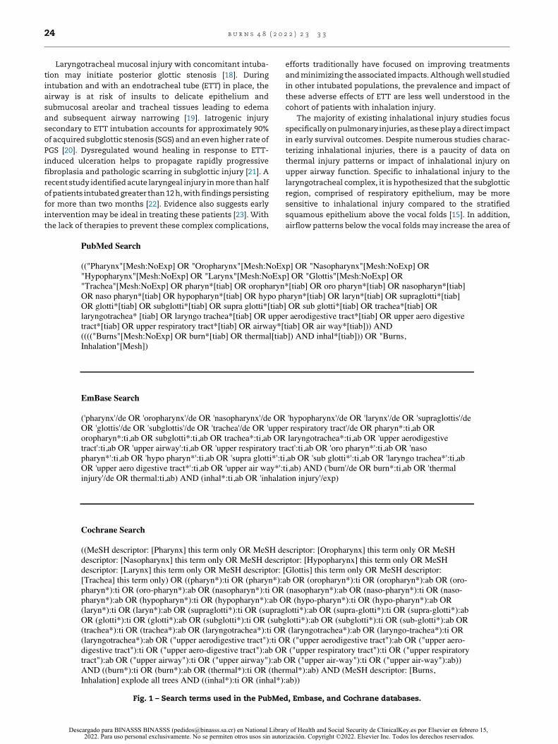
Of all the patients with objective documentation oflaryngeal inhalational injury in this review, cough followedby dysphonia were the most frequently noted clinicalpresentations. Dysphagia, dyspnea, wheezes/rales, and
stridor were also commonly reported. In general, clinical signsand symptoms were specifically mentioned in patients thatdid not receive immediate attention to secure the airway, asoften reporting of clinical signs is impossible for those patientswho remained intubated for more than a few days.
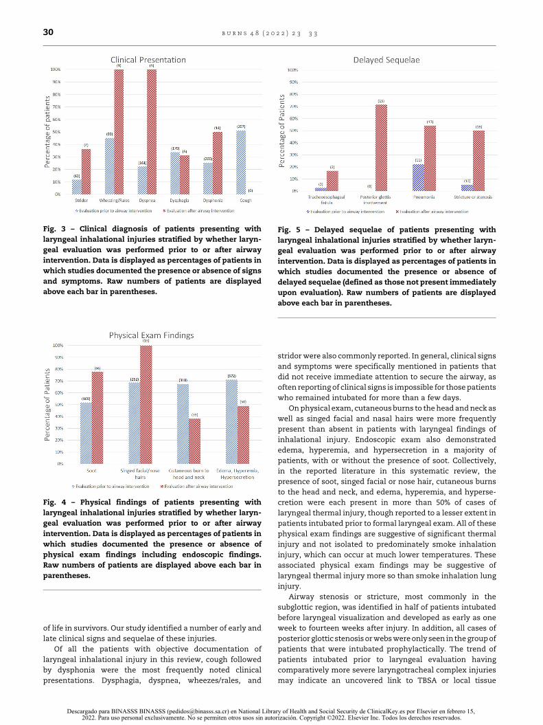
On physical exam, cutaneous burns to the head and neck aswell as singed facial and nasal hairs were more frequentlypresent than absent in patients with laryngeal findings ofinhalational injury. Endoscopic exam also demonstratededema, hyperemia, and hypersecretion in a majority ofpatients, with or without the presence of soot. Collectively,in the reported literature in this systematic review, thepresence of soot, singed facial or nose hair, cutaneous burnsto the head and neck, and edema, hyperemia, and hyperse-cretion were each present in more than 50% of cases oflaryngeal thermal injury, though reported to a lesser extent inpatients intubated prior to formal laryngeal exam. All of thesephysical exam findings are suggestive of significant thermalinjury and not isolated to predominately smoke inhalationinjury, which can occur at much lower temperatures. Theseassociated physical exam findings may be suggestive oflaryngeal thermal injury more so than smoke inhalation lunginjury.
Airway stenosis or stricture, most commonly in thesubglottic region, was identified in half of patients intubatedbefore laryngeal visualization and developed as early as oneweek to fourteen weeks after injury. In addition, all cases ofposterior glottic stenosis or webs were only seen in the group ofpatients that were intubated prophylactically. The trend ofpatients intubated prior to laryngeal evaluation havingcomparatively more severe laryngotracheal complex injuriesmay indicate an uncovered link to TBSA or local tissue
Fig. 3 – Clinical diagnosis of patients presenting withlaryngeal inhalational injuries stratified by whether laryn-geal evaluation was performed prior to or after airwayintervention. Data is displayed as percentages of patients inwhich studies documented the presence or absence of signsand symptoms. Raw numbers of patients are displayedabove each bar in parentheses.
Fig. 4 – Physical findings of patients presenting withlaryngeal inhalational injuries stratified by whether laryn-geal evaluation was performed prior to or after airwayintervention. Data is displayed as percentages of patients inwhich studies documented the presence or absence ofphysical exam findings including endoscopic findings.Raw numbers of patients are displayed above each bar inparentheses.
Fig. 5 – Delayed sequelae of patients presenting withlaryngeal inhalational injuries stratified by whether laryn-geal evaluation was performed prior to or after airwayintervention. Data is displayed as percentages of patients inwhich studies documented the presence or absence ofdelayed sequelae (defined as those not present immediatelyupon evaluation). Raw numbers of patients are displayedabove each bar in parentheses.
30 b u r n s 4 8 ( 2 0 2 2 ) 2 3 �3 3
Descargado para BINASSS BINASSS ([email protected]) en National Library of Health and Social Security de ClinicalKey.es por Elsevier en febrero 15, 2022. Para uso personal exclusivamente. No se permiten otros usos sin autorización. Copyright ©2022. Elsevier Inc. Todos los derechos reservados.

characteristics predisposing to injury. Additionally, localtissue trauma to the delicate laryngeal tissues from evencareful intubation may precipitate long term injury to thelaryngotracheal complex. Beginning at the time of injury, amyriad of patient and situational factors in the setting of aninhalation injury, from mental status to acute signs of airwayobstruction or distress, play an important role in if and howquickly an airway intervention, generally orotracheal intuba-tion, is undertaken. In this review, all scenarios whereintubation or a surgical airway was performed to secure theairway prior to formal diagnostic visualization of the laryngo-tracheal complex were burns that occurred in an enclosedspace, such as within a trailer or house fire. Many of thesepatients were intubated on the scene or during transport to thehospital. Smoke inhalation injury is more common in enclosedspaces due to the inability of smoke to dissipate quickly inenclosed spaces as it would outdoors, and inhalation of largeconcentrations of products of combustion such as hydrogencyanide and carbon monoxide can quickly lead to a reducedlevel of consciousness and subsequent inability to protect theairway [64].
Aside from an altered mental status, upper airwayobstruction has been noted in up to a third of patients withinhalational injury [65] and may necessitate immediateintubation. Inhalation injury can have variable effects aboveand below the glottis, and injury to the upper airway does notautomatically imply injury to the lower airway or vice versa[43]. In fact, Head noted that in patients with oropharyngealexams consistent with inhalational injury, bronchoscopywas negative 38% of the time [66]. Conversely, the respiratoryepithelium immediately below the vocal folds may be moresensitive to inhalational injury than the squamous epitheli-um above the vocal fold [15], potentially attributable tosubglottic airflow eddy currents that increases local expo-sure to toxic components of smoke [24]. Ultimately, eitherinjury to the supraglottic or subglottic region can necessitateintubation.
Often, intubation may be performed urgently prior tovisualization of the larynx and upper airway, especially in thesetting where impending airway compromise is anticipated.However, some studies suggest intubation may be performedmore often than necessary. In fact, Moshrefi et al. [47] reportedthat the majority of such intubations are unnecessary, with50 of 51 intubated patients having a normal flexible exam.There was one patient who was intubated only after repeatlaryngoscopy, which also speaks to the need for serialsurveillance as the initial airway exam can evolve. It has beenshown that those who undergo more than one endoscopicexam have shorter lengths of hospital stay [67].
There is likely a synergistic effect of damage frominhalational injury and intubation that could account for theincreased finding of airway stenosis in patients intubated priorto laryngeal evaluation [13�15]. When patients are ultimatelyextubated, it can be challenging to define a clear distinction orcompound impact between the contributions of inhalationalinjury and intubation to mucosal injury, stenosis, and othersoft tissue injuries in the laryngotracheal complex.
Cha et al. [13] noted a high frequency of vocal fold andtracheal stenosis in patients with isolated smoke inhalation-al injuries despite only short term endotracheal intubation.
Tracheal stenosis has even been observed as early as 64 hafter intubation, suggesting that intubation alone could nothave been the only factor contributing to such earlydevelopment of airway stenosis [14]. Furthermore, moresevere thermal injury and longer duration of intubation havebeen shown to be associated with the development oflaryngotracheal stenosis [42]. Given the potential direconsequences that can result from the combined effects ofintubation and inhalational injury, diagnostic laryngealexamination prior to intubation may provide diagnosticinformation on potential long term voice, airway, andswallowing function.
As with all systematic reviews, limitations exist in thevariability of each paper in reporting variables important tothe current study. Not every study was systematic in theirdocumentation of symptoms, and not every study docu-mented each one of our study variables of interest. Weattempted to display the data in as representative a way aspossible by only counting the population of studies thatmentioned the presence or absence of each variable whenportraying percentages and performing our statistical anal-yses, rather than counting the entire population of thestudies combined. Unfortunately, not many studies includedlong term follow up, which may subsequently underestimatethe prevalence of delayed sequelae reported in this study.
Although limited by the available data in existing literature,the findings of this review illustrate the frequency and broadrange of secondary effects from laryngeal thermal injuries,particularly coupled with early intubation. One considerationwould be more comprehensive or standardized evaluations ofthe larynx and upper airway both before intubation, whenpossible, involving immediate and repeat after extubationexamination to minimize any negative effects on thesedelicate tissues. More globally, the frequent identification ofdysphonia, dysphagia, dyspnea, and cough combined with theidentified long term sequelae of tracheal or posterior glotticstenosis or tracheo-esophageal fistula suggest that earlyOtolaryngology Head and Neck Surgery referral or consulta-tion may be warranted to supplement care and potentiallymitigate or engage in treatment of delayed sequelae. Futureprospective studies and meta-analyses may better elucidatethese complex injuries and sequelae.
5. Conclusions
Laryngeal inhalational injuries are a distinct subset ofinhalational injuries that can have a variety of minor to severelaryngotracheal delayed sequelae, particularly for thermalinjuries occurring within enclosed spaces. Given these find-ings, early Otolaryngology Head and Neck Surgery referral maybe useful to mitigate or treat these effects.
Disclaimer
The views expressed in this manuscript are those of theauthors and do not reflect the official policy or position of theDepartment of the Army, Department of the Air Force,Department of Defense, or the US Government.
b u r n s 4 8 ( 2 0 2 2 ) 2 3 �3 3 31
Descargado para BINASSS BINASSS ([email protected]) en National Library of Health and Social Security de ClinicalKey.es por Elsevier en febrero 15, 2022. Para uso personal exclusivamente. No se permiten otros usos sin autorización. Copyright ©2022. Elsevier Inc. Todos los derechos reservados.

Authorship declaration
All authors have made substantial contributions to all of thefollowing: (1) the conception and design of the study, oracquisition of data, or analysis and interpretation of data, (2)drafting the article or revising it critically for importantintellectual content, (3) final approval of the version to besubmitted.
Funding source
This research did not receive any specific grant from fundingagencies in the public, commercial, or not-for-profit sectors.
Declarations of interest
None.
Submission declaration
The work described has not been published previously and isnot under consideration for publication elsewhere. Thismanuscript has been approved by all authors.
Acknowledgement
The authors would like to acknowledge and thank KarenBurstein for her guidance in performing a comprehensive andthorough literature search for this systematic review
R E F E R E N C E S
[1] You K, Yang HT, Kym D, Yoon J, Yim Haejun, Cho YS, et al.Inhalation injury in burn patients: establishing the linkbetween diagnosis and prognosis. Burns 2014;40:1470�5.
[2] Dries DJ, Endorf FW. Inhalation injury: epidemiology,pathology, treatment strategies. Scand J Trauma ResuscEmerg Med 2013;21:31.
[3] Enkhbaatar P, Murakami K, Shimoda K, Mizutani A, Traber L,Phillips GB, et al. The inducible nitric oxide synthase inhibitorBBS-2 prevents acute lung injury in sheep after burn andsmoke inhalation injury. Am J Respir Crit Care Med2003;167:1021�6.
[4] Shirani KZ, Pruitt Jr. BA, Mason Jr. AD. The influence ofinhalation injury and pneumonia on burn mortality. Ann Surg1987;205:82�7.
[5] Walker PF, Buehner MF, Wood LA, Boyer NL, Driscoll IR, LundyJB, et al. Diagnosis and management of inhalationinjury: an updated review. Crit Care (London, England)2015;19:351.
[6] Bai C, Huang H, Yao X, Zhu S, Li B, Hang J, et al. Application offlexible bronchoscopy in inhalation lung injury. Diagn Pathol2013;8:174.
[7] Freno D, Sahawneh J, Harrison S, Sahawneh T, Patterson S,Kahn SA. Determining the role of nasolaryngoscopy in theinitial evaluation for upper airway injury in patients withfacial burns. Burns 2018;44:539�43.
[8] Muehlberger T, Kunar D, Munster A, Couch M. Efficacy offiberoptic laryngoscopy in the diagnosis of inhalation injuries.Arch Otolaryngol Head Neck Surg 1998;124:1003�7.
[9] Gore MA, Joshi AR, Nagarajan G, Iyer SP, Kulkarni T,Khandelwal A. Virtual bronchoscopy for diagnosis ofinhalation injury in burnt patients. Burns 2004;30:165�8.
[10] Lee MJ, O’Connell DJ. The plain chest radiograph after acutesmoke inhalation. Clin Radiol 1988;39:33�7.
[11] Yamamura H, Kaga S, Kaneda K, Mizobata Y. Chest computedtomography performed on admission helps predict theseverity of smoke-inhalation injury. Criti Care (London,England) 2013;17:R95.
[12] Badulak Jh, Schurr M, Sauaia A, Ivashchenko A, Peltz E.Defining the criteria for intubation of the patient with thermalburns. Burns 2018;44:531�8.
[13] Cha SI, Kim CH, Lee JH, Park JY, Jung TH, Choi WI, et al. Isolatedsmoke inhalation injuries: acute respiratory dysfunction,clinical outcomes, and short-term evolution of pulmonaryfunctions with the effects of steroids. Burns 2007;33:200�8.
[14] Colice GL, Munster AM, Haponik EF. Tracheal stenosiscomplicating cutaneous burns: an underestimated problem.Am Rev Respir Dis 1986;134:1315�8.
[15] Gaissert HA, Lofgren RH, Grillo HC. Upper airway compromiseafter inhalation injury. Complex strictures of the larynx andtrachea and their management. Ann Surg 1993;218:672�8.
[16] Casper JK, Clark WR, Kelley RT, Colton RH. Laryngeal andphonatory status after burn/inhalation injury: a long termfollow-up study. J Burn Care Rehabil 2002;23:235�43.
[17] Pfannenstiel TJ, Gal TJ, Hayes DK, Myers KV. Vocal foldimmobility following burn intensive care. Otolaryngol HeadNeck Surg 2007;137:152�6.
[18] Hillel AT, Karatayli-Ozgursoy S, Samad I, Best SR, Pandian V,Giraldez L, et al. Predictors of posterior glottic stenosis: amulti-institutional case-control study. Ann Otol RhinolLaryngol 2016;125:257�63.
[19] Soose RJ, Carrau RL. Acquired laryngeal stenosis. In: Myers EN,editor. Operative otolaryngology: head and neck surgery. 2 ed.Philadelphia, PA: Saunders Elsevier; 2008. p. 357�78.
[20] MaedaK,Ono S,BabaK.Managementof laryngotracheal stenosisininfants and children: the role ofre-do surgeryin casesof severesubglottic stenosis. Pediatr Surg Int 2013;29:1001�6.
[21] Hillel AT, Samad I, Ma G, Ding D, Sadtler K, Powell JD, et al.Dysregulated macrophages are present in bleomycin-inducedmurine laryngotracheal stenosis. Otolaryngol Head Neck Surg2015;153:244�50.
[22] Shinn JR, Kimura KS, Campbell BR, Sun Lowery A, Wootten CT,Garrett CG, et al. Incidence and outcomes of acute laryngealinjury after prolonged mechanical ventilation. Crit Care Med2019;47:1699�706.
[23] Nouraei SA, Singh A, Patel A, Ferguson C, Howard DJ, SandhuGS. Early endoscopic treatment of acute inflammatory airwaylesions improves the outcome of postintubation airwaystenosis. Laryngoscope 2006;116:1417�21.
[24] Mallory TB, Brickley WJ. Pathology: with special reference tothe pulmonary lesions. Ann Surg 1943;117:865�84.
[25] Ikari T, Sasaki CT. Glottic closure reflex: control mechanisms.Ann Otol Rhinol Laryngol 1980;89:220�4.
[26] Moritz AR, Henriques FC, McLean R. The effects of inhaled heaton the air passages and lungs: an experimental investigation.Am J Pathol 1945;21:311�31.
[27] Galeiras R, Seoane-Quiroga L, Pértega-Díaz S. Prevalence andprognostic impact of inhalation injury among burn patients: asystematic review and meta-analysis. J Trauma Acute CareSurg 2020;88:330�44.
[28] Vo E, Kurmis R, Campbell J, Greenwood J. Risk factors for andcharacteristics of dysphagia development in thermal burninjury and/or inhalation injury patients: a systematic reviewprotocol. JBI Database System Rev Implement Rep 2016;14:31�43.
32 b u r n s 4 8 ( 2 0 2 2 ) 2 3 �3 3
Descargado para BINASSS BINASSS ([email protected]) en National Library of Health and Social Security de ClinicalKey.es por Elsevier en febrero 15, 2022. Para uso personal exclusivamente. No se permiten otros usos sin autorización. Copyright ©2022. Elsevier Inc. Todos los derechos reservados.
[29] Liberati A, Altman DG, Tetzlaff J, Mulrow C, Gotzsche PC,Ioannidis JP, et al. The PRISMA statement for reportingsystematic reviews and meta-analyses of studies thatevaluate healthcare interventions: explanation andelaboration. BMJ 2009;339:b2700.
[30] Moher D, Liberati A, Tetzlaff J, Altman DG, Group P. Preferredreporting items for systematic reviews and meta-analyses: thePRISMA statement. Ann Intern Med 2009151: 264-9, W64.
[31] Haponik EF, Meyers DA, Munster AM, Smith PL, Britt EJ, WiseRA, et al. Acute upper airway injury in burn patients. Serialchanges of flow-volume curves and nasopharyngoscopy. AmRev Respir Dis 1987;135:360�6.
[32] Desai SR, Zeng D, Chong SJ. Airway management in inhalationinjury: a case series. Singapore Med J 2019 [epub ahead ofprint].
[33] Ching Ja, Ching Yh, Shivers Sc, Karlnoski Ra, Payne Wg, SmithJr Dj. An analysis of inhalation injury diagnostic methods andpatient outcomes. J Burn Care Res 2016;37:e27�32.
[34] Edelman DA, Sheehy-Deardorff DA, White MT. Bedsideassessment of swallowing is predictive of an abnormal bariumswallow examination. J Burn Care Res 2008;29:89�96.
[35] Ribeiro C, Guimarães M, Antunes A, Oliveira A, Neves S,Almeida J, et al. "The black bronchoscopy" A case of airwaysoot deposition. J Bronchol Interventional Pulmonol2013;20:271�3.
[36] Megahed MA, Ghareeb F, Kishk T, El-Barah A, Abou-Gereda H,El-Fol H, et al. Blood gases as an indicator of inhalation injuryand prognosis in burn patients. Ann Burns Fire Disasters2008;21:192�8.
[37] Goh SH, Tiah L, Lim HC, Ng EK. Disaster preparedness:experience from a smoke inhalation mass casualty incident.Eur J Emerg Med 2006;13:330�4.
[38] Rhodes ML. Early recognition of upper airway obstructionfollowing smoke inhalation. Am Rev Respir Dis 1973;108:1421�6.
[39] Tilney P. Extensive burns in an elderly man found in a brushfire. Air Med J 2010;29:98�103.
[40] Madnani DD, Steele NP, de Vries E. Factors that predict theneed for intubation in patients with smoke inhalation injury.Ear Nose Throat J 2006;85:278�80.
[41] Marek K, Piotr W, Stanislaw S, Stefan G, Justyna G, Mariusz N,et al. Fibreoptic bronchoscopy in routine clinical practice inconfirming the diagnosis and treatment of inhalation burns.Burns 2007;33:554�60.
[42] Lowery AS, Dion G, Thompson C, Weavind L, Shinn J, McGraneS, et al. Incidence of laryngotracheal stenosis after thermalinhalation airway injury. J Burn Care Res 2019;40:961�5.
[43] Onishi S, Osuka A, Kuroki Y, Ueyama M. Indications of earlyintubation for patients with inhalation injury. Acute Med Surg2017;4:278�85.
[44] Fang-Gang N, Yang C, Yu-Xuan Q, Yan-Hua R, Wei-Li D, ChengW, et al. Laryngeal morphologic changes and epidemiology inpatients with inhalation injury: a retrospective study. Burns2015;41:1340�6.
[45] Arakawa A, Fukamizu H, Hashizume I, Kasamatsu N,Nagayoshi M, Shinozuka N, et al. Macroscopic and histologicalfindings in the healing process of inhalation injury. Burns2007;33:855�9.
[46] Costa Santos D, Barros F, Frazao M, Maia M. Pre-burn centremanagement of the airway in patients with face burns. AnnBurns Fire Disasters 2015;28:259�63.
[47] Moshrefi S, Sheckter CC, Shepard K, Pereira C, Davis DJ,Karanas Y, et al. Preventing unnecessary intubations: a 5-yearregional burn center experience using flexible fiberopticlaryngoscopy for airway evaluation in patients with suspectedinhalation or airway injury. J Burn Care Res 2019;40:341�6.
[48] Ikonomidis C, Lang F, Radu A, Berger MM. Standardizing thediagnosis of inhalation injury using a descriptive score basedon mucosal injury criteria. Burns 2012;38:513�9.
[49] Yang JY, Yang WG, Chang LY, Chuang SS. Symptomatictracheal stenosis in burns. Burns 1999;25:72�80.
[50] Jones WG, Madden M, Finkelstein J, Yurt RW, Goodwin CW.Tracheostomies in burn patients. Ann Surg 1989;209:471�4.
[51] Kim Gh, Wang Sg, Lee Yw, Kwon Sb. Voice recovery in a patientwith inhaled laryngeal burns. Iran J Otorhinolaryngol2019;31:55�9.
[52] Gherardini G, Jernbeck J, Kuylenstierna R, Jonsson CE.Anatomical and functional complications after tracheostomyin patients with burns and inhalation injuries. Eur J PlasticSurg 1994;17:127�30.
[53] Bassi E, Miranda LC, Tierno PF, Ferreira CB, Cadamuro FM,Figueiredo VR, et al. Assistance of inhalation injury victimscaused by fire in confined spaces: what we learned from thetragedy at Santa Maria. Rev Bras Ter Intensiva 2014;26:421�9.
[54] Al Kassis S, Savetamal A, Assi R, Crombie RE, Ali R, Moores C,et al. Characteristics of patients with injury secondary tosmoking on home oxygen therapy transferred intubated to aburn center. J Am Coll Surg 2014;218:1182�6.
[55] Cobley TD, Hart WJ, Baldwin DL, Burd DA. Complete fusion ofthe vocal cords; an unusual case. Burns 1999;25:361�3.
[56] Irrazabal CL, Capdevila AA, Revich L, Del Bosco CG, Luna CM,Vujacich P, et al. Early and late complications among15 victims exposed to indoor fire and smoke inhalation. Burns2008;34:533�8.
[57] Hantson P, Butera R, Clemessy JL, Michel A, Baud FJ. Earlycomplications and value of initial clinical and paraclinicalobservations in victims of smoke inhalation without burns.Chest 1997;111:671�5.
[58] Valdez TA, Desai U, Ruhl CM, Nigri PT. Early laryngealinhalation injury and its correlation with late sequelae.Laryngoscope 2006;116:283�7.
[59] Jayawardena A, Lowery AS, Wootten C, Dion GR, Summitt JB,McGrane S, et al. Early surgical management of thermal airwayinjury: a case series. J Burn Care Res 2019;40:189�95.
[60] Koshkareva YA, Hughes WB, Soliman AMS. Laryngotrachealstenosis in burn patients requiring mechanical ventilation.World J Otorhinolaryngol Head Neck Surg 2018;4:117�21.
[61] Flexon PB, Cheney ML, Montgomery WW, Turner PA.Management of patients with glottic and subglottic stenosisresulting from thermal burns. Ann Otol Rhinol Laryngol1989;98:27�30.
[62] Furak J, Szakacs L, Nagy A, Rovo L. Multiple costal cartilagegraft reconstruction for the treatment of a full-lengthlaryngotracheal stenosis after an inhalation burn. InteractCardiovasc Thorac Surg 2011;13:453�5.
[63] Ashraf U, Bajantri B, Roa-Gomez G, Venkatram S, Cantin A, DI-F G. Nebulized heparin and N-acetylcysteine for smokeinhalational injury: a case report. Medicine (United States)201897:.
[64] Antonio AC, Castro PS, Freire LO. Smoke inhalation injuryduring enclosed-space fires: an update. J Bras Pneumol2013;39:373�81.
[65] Mlcak RP, Suman OE, Herndon DN. Respiratory managementof inhalation injury. Burns 2007;33:2�13.
[66] Head JM. Inhalation injury in burns. Am J Surg 1980;139:508�12.
[67] Carr JA, Phillips BD, Bowling WM. The utility of bronchoscopyafter inhalation injury complicated by pneumonia in burnpatients: results from the National Burn Repository. J BurnCare Res 2009;30:967�74.
b u r n s 4 8 ( 2 0 2 2 ) 2 3 �3 3 33
Descargado para BINASSS BINASSS ([email protected]) en National Library of Health and Social Security de ClinicalKey.es por Elsevier en febrero 15, 2022. Para uso personal exclusivamente. No se permiten otros usos sin autorización. Copyright ©2022. Elsevier Inc. Todos los derechos reservados.
Related Documents