The new england journal of medicine n engl j med 385;19 nejm.org November 4, 2021 1786 Review Article From the University of Colorado School of Medicine, Aurora. Dr. Teitelbaum can be contacted at isaac.teitelbaum@ cuanschutz.edu or at the University of Colorado School of Medicine, 12605 E. 16th Ave., Aurora, CO 80045. N Engl J Med 2021;385:1786-95. DOI: 10.1056/NEJMra2100152 Copyright © 2021 Massachusetts Medical Society. A n estimated 3.8 million people worldwide currently rely on some form of dialysis for treatment of end-stage kidney disease (ESKD). 1 Although the prevalence of peritoneal dialysis varies from country to coun- try, it accounts for approximately 11% of patients undergoing dialysis overall. 2 In developed countries, peritoneal dialysis is less expensive to deliver than hemo- dialysis. 3 Therefore, some national health care systems have implemented a “PD first” policy, with peritoneal dialysis as the preferred approach unless a medical contraindication is present. 4 There is no formal PD-first policy in the United States, although Medicare favors home dialysis over in-center dialysis. 5 Further- more, implementation of the 2019 Advancing American Kidney Health executive order 6 may increase the use of peritoneal dialysis. Many clinicians lack knowledge of and experience in using peritoneal dialysis and may not feel comfortable man- aging the care of patients who are using that type of dialysis. This review aims to address the knowledge gap. Peritoneal Anatomy The peritoneum approximates body-surface area in size. Anatomically, it is com- posed of two layers: the visceral peritoneum, which covers the abdominal organs and accounts for 80% of the total surface area, and the parietal peritoneum, which lines the undersurface of the diaphragm and the interior surface of the anterior abdominal wall. 7 Histologically, the peritoneum consists of a single layer of meso- thelial cells resting on submesothelial interstitial tissue, a gel-like matrix contain- ing fibroblasts, adipocytes, collagen fibers, nerves, lymphatic vessels, and capillar- ies (Fig. 1). 8 The endothelium of these peritoneal capillaries functions as the filter that regulates peritoneal transport. 9 Thus, the peritoneum provides a suitable membrane for the performance of dialysis. Physiology of Peritoneal Dialysis Dialytic Process In peritoneal dialysis, fluid (dialysate) is instilled in the peritoneal cavity, and solutes diffuse from the blood in the peritoneal capillaries into the dialysate, ef- fecting an exchange analogous to that of extracorporeal hemodialysis. Similarly, imposition of a transmembrane pressure gradient creates the driving force for ultrafiltration of fluid from the capillaries into the dialysate. In contrast to hemo- dialysis, in which the pressure that is applied is hydrostatic, peritoneal dialysis involves osmotic pressure created by the intraperitoneal instillation of hypertonic dialysate, usually as glucose in the form of 1.5%, 2.5%, or 4.25% dextrose (glucose monohydrate). Higher concentrations of glucose exert higher osmotic pressures and effect greater degrees of ultrafiltration. Julie R. Ingelfinger, M.D., Editor Peritoneal Dialysis Isaac Teitelbaum, M.D. CME at NEJM.org The New England Journal of Medicine ownloaded from nejm.org at CCSS CAJA COSTARRICENSE DE SEGURO SOCIAL BINASSS on November 9, 2021. For personal use only. No other uses without permission Copyright © 2021 Massachusetts Medical Society. All rights reserved.

Welcome message from author
This document is posted to help you gain knowledge. Please leave a comment to let me know what you think about it! Share it to your friends and learn new things together.
Transcript
T h e n e w e ngl a nd j o u r na l o f m e dic i n e
n engl j med 385;19 nejm.org November 4, 20211786
Review Article
From the University of Colorado School of Medicine, Aurora. Dr. Teitelbaum can be contacted at isaac . teitelbaum@ cuanschutz . edu or at the University of Colorado School of Medicine, 12605 E. 16th Ave., Aurora, CO 80045.
N Engl J Med 2021;385:1786-95.DOI: 10.1056/NEJMra2100152Copyright © 2021 Massachusetts Medical Society.
An estimated 3.8 million people worldwide currently rely on some form of dialysis for treatment of end-stage kidney disease (ESKD).1 Although the prevalence of peritoneal dialysis varies from country to coun-
try, it accounts for approximately 11% of patients undergoing dialysis overall.2 In developed countries, peritoneal dialysis is less expensive to deliver than hemo-dialysis.3 Therefore, some national health care systems have implemented a “PD first” policy, with peritoneal dialysis as the preferred approach unless a medical contraindication is present.4 There is no formal PD-first policy in the United States, although Medicare favors home dialysis over in-center dialysis.5 Further-more, implementation of the 2019 Advancing American Kidney Health executive order6 may increase the use of peritoneal dialysis. Many clinicians lack knowledge of and experience in using peritoneal dialysis and may not feel comfortable man-aging the care of patients who are using that type of dialysis. This review aims to address the knowledge gap.
Per i t one a l A nat om y
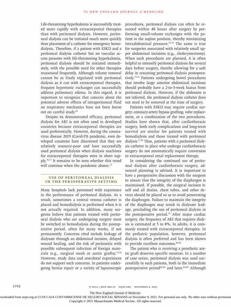
The peritoneum approximates body-surface area in size. Anatomically, it is com-posed of two layers: the visceral peritoneum, which covers the abdominal organs and accounts for 80% of the total surface area, and the parietal peritoneum, which lines the undersurface of the diaphragm and the interior surface of the anterior abdominal wall.7 Histologically, the peritoneum consists of a single layer of meso-thelial cells resting on submesothelial interstitial tissue, a gel-like matrix contain-ing fibroblasts, adipocytes, collagen fibers, nerves, lymphatic vessels, and capillar-ies (Fig. 1).8 The endothelium of these peritoneal capillaries functions as the filter that regulates peritoneal transport.9 Thus, the peritoneum provides a suitable membrane for the performance of dialysis.
Ph ysiol o gy of Per i t one a l Di a lysis
Dialytic Process
In peritoneal dialysis, fluid (dialysate) is instilled in the peritoneal cavity, and solutes diffuse from the blood in the peritoneal capillaries into the dialysate, ef-fecting an exchange analogous to that of extracorporeal hemodialysis. Similarly, imposition of a transmembrane pressure gradient creates the driving force for ultrafiltration of fluid from the capillaries into the dialysate. In contrast to hemo-dialysis, in which the pressure that is applied is hydrostatic, peritoneal dialysis involves osmotic pressure created by the intraperitoneal instillation of hypertonic dialysate, usually as glucose in the form of 1.5%, 2.5%, or 4.25% dextrose (glucose monohydrate). Higher concentrations of glucose exert higher osmotic pressures and effect greater degrees of ultrafiltration.
Julie R. Ingelfinger, M.D., Editor
Peritoneal DialysisIsaac Teitelbaum, M.D.
CMEat NEJM.org
The New England Journal of Medicine Downloaded from nejm.org at CCSS CAJA COSTARRICENSE DE SEGURO SOCIAL BINASSS on November 9, 2021. For personal use only. No other uses without permission.
Copyright © 2021 Massachusetts Medical Society. All rights reserved.
n engl j med 385;19 nejm.org November 4, 2021 1787
Peritoneal Dialysis
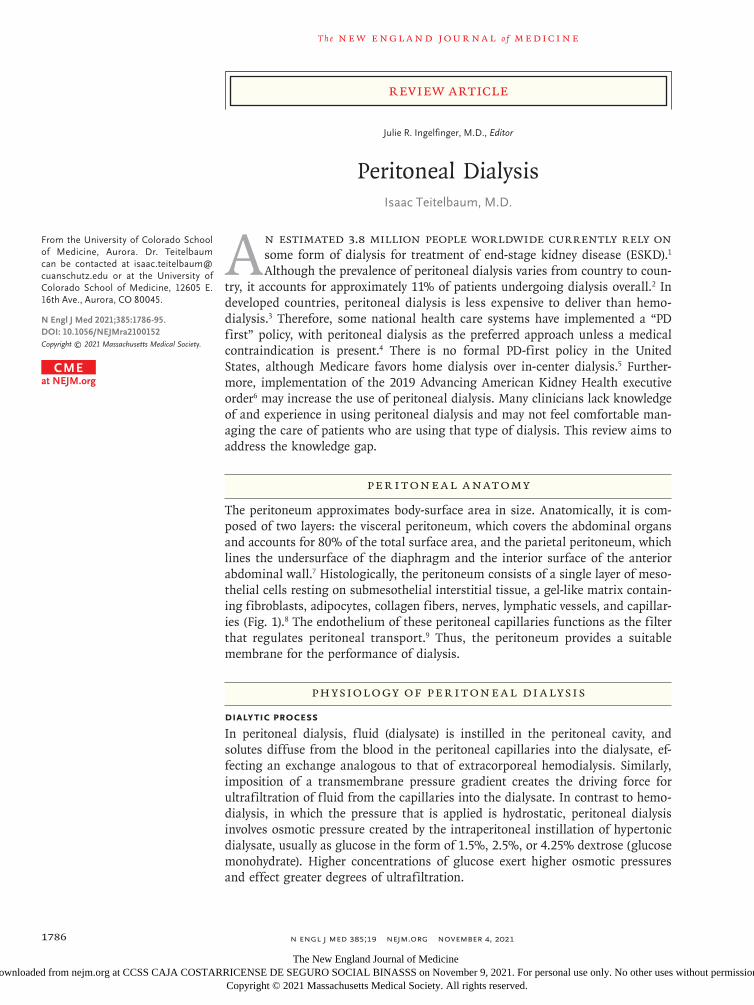
Solute transfer across the peritoneal capillar-ies is bidirectional. Solutes such as urea, creati-nine, and potassium diffuse from the blood-stream into the dialysate, whereas glucose diffuses from the dialysate into the peritoneal capillaries (Fig. 1). Diffusion of glucose out of the dialysate into the peritoneal capillaries re-sults in dissipation of the osmotic gradient and progressive slowing in the rate of ultrafiltration. The rate of solute transfer across the peritoneum depends on the concentration gradient and the degree of peritoneal vascularity,10 which varies from person to person.
In patients with less peritoneal vascularity, solutes diffuse slowly in both directions. Waste products accumulate in the dialysate slowly, and the glucose gradient favoring ultrafiltration dis-sipates slowly. Conversely, in patients with great-er peritoneal vascularity, solutes diffuse more rapidly, also in both directions. Waste products accumulate in the dialysate more rapidly, and the glucose gradient favoring ultrafiltration dis-sipates more rapidly. Such patients have poor, sometimes even negative ultrafiltration with long “dwells.” (The dwell is the time during which the dialysate remains in the abdominal cavity.) The use of a non–glucose-based fluid such as icodextrin during long dwells may be beneficial in these patients.11 Icodextrin is a col-loid osmotic agent that does not diffuse across the peritoneum; it effects ultrafiltration that is sustained for 12 to 16 hours.12 Other types of dialysate fluids (available in some countries but not in the United States) include an amino acid–based fluid and fluids that are low in glucose degradation products. These two dialysate types decrease exposure of the peritoneal membrane to glucose, and the type that is low in glucose degradation products has been shown to help preserve residual kidney function.13
Peritoneal Access
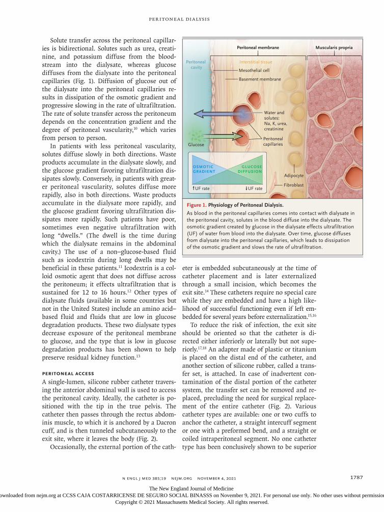
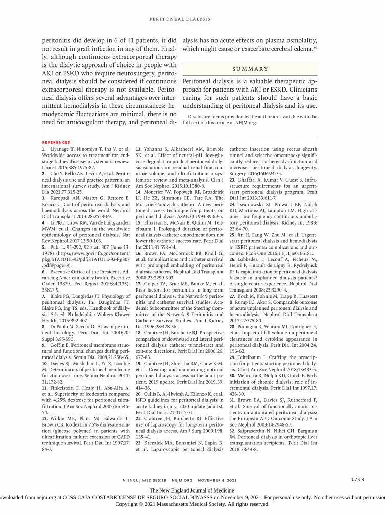
A single-lumen, silicone rubber catheter travers-ing the anterior abdominal wall is used to access the peritoneal cavity. Ideally, the catheter is po-sitioned with the tip in the true pelvis. The catheter then passes through the rectus abdom-inis muscle, to which it is anchored by a Dacron cuff, and is then tunneled subcutaneously to the exit site, where it leaves the body (Fig. 2).
Occasionally, the external portion of the cath-
eter is embedded subcutaneously at the time of catheter placement and is later externalized through a small incision, which becomes the exit site.14 These catheters require no special care while they are embedded and have a high like-lihood of successful functioning even if left em-bedded for several years before externalization.15,16
To reduce the risk of infection, the exit site should be oriented so that the catheter is di-rected either inferiorly or laterally but not supe-riorly.17,18 An adapter made of plastic or titanium is placed on the distal end of the catheter, and another section of silicone rubber, called a trans-fer set, is attached. In case of inadvertent con-tamination of the distal portion of the catheter system, the transfer set can be removed and re-placed, precluding the need for surgical replace-ment of the entire catheter (Fig. 2). Various catheter types are available: one or two cuffs to anchor the catheter, a straight intercuff segment or one with a preformed bend, and a straight or coiled intraperitoneal segment. No one catheter type has been conclusively shown to be superior
Figure 1. Physiology of Peritoneal Dialysis.
As blood in the peritoneal capillaries comes into contact with dialysate in the peritoneal cavity, solutes in the blood diffuse into the dialysate. The osmotic gradient created by glucose in the dialysate effects ultrafiltration (UF) of water from blood into the dialysate. Over time, glucose diffuses from dialysate into the peritoneal capillaries, which leads to dissipation of the osmotic gradient and slows the rate of ultrafiltration.
Water andsolutes: Na, K, urea, creatinine
Mesothelial cell
Basement membrane
Interstitial tissue
Peritonealcapillaries
Fibroblast
Adipocyte
Peritoneal cavity
Peritoneal membrane Muscularis propria
Glucose
OSMOTICGRADIENT
UF rate UF rate
GLUCOSEDIFFUSION
The New England Journal of Medicine Downloaded from nejm.org at CCSS CAJA COSTARRICENSE DE SEGURO SOCIAL BINASSS on November 9, 2021. For personal use only. No other uses without permission.
Copyright © 2021 Massachusetts Medical Society. All rights reserved.
n engl j med 385;19 nejm.org November 4, 20211788
T h e n e w e ngl a nd j o u r na l o f m e dic i n e
to the others, but double-cuffed catheters are recommended.19
When peritoneal dialysis is being performed for treatment of acute kidney injury (AKI) (dis-
cussed below), the catheter should preferably be placed as described above. However, when the availability of catheter types is restricted by limit-ed resources, rigid catheters and even unconven-tional catheters such as nasogastric tubes or Foley catheters may be used and are often lifesaving.20
The catheter can be placed surgically with the use of either an open or a laparoscopic approach. Alternatively, it may be placed percutaneously through a modified Seldinger technique, with or without fluoroscopic or ultrasound guidance. Local resources and operator experience dictate the choice of technique. However, laparoscopy is preferred, since it allows for the performance of adjunctive procedures, such as rectus sheath tun-neling, omentopexy, or adhesiolysis, that reduce the risk of mechanical complications during peri-toneal dialysis.19,21,22
It is recommended that 2 weeks elapse before the catheter is used; this delay allows the inter-nal cuff to heal into place and minimizes the risk of mechanical complications. However, if a patient with ESKD and no prior planned renal replacement therapy requires dialysis, a perito-neal dialysis catheter can be placed and used before 2 weeks have elapsed, in what is called “urgent start” peritoneal dialysis.23 To avoid leakage of dialysate around the newly placed catheter, urgent-start peritoneal dialysis is per-formed with the patient in the supine position, which minimizes intraperitoneal pressure.24 In addition, the volume of fluid infused is gener-ally smaller than the volume in standard perito-neal dialysis and is tailored to body size. There are no data from prospective, randomized trials comparing urgent-start hemodialysis with urgent-start peritoneal dialysis. However, retrospective observational data show similar survival with the two techniques,25,26 with fewer episodes of bacteremia among patients undergoing perito-neal dialysis and no increase in the incidence of peritonitis.25,27 Thus, in the absence of a life-threatening indication for immediate hemodial-ysis, patients presenting for the first time with ESKD should be allowed to choose between ur-gent initiation of hemodialysis and peritoneal dialysis.
Di a ly tic Pro cedur e
Peritoneal dialysis is performed by instilling fluid, called dialysate, into the peritoneal cavity. The
Figure 2. Peritoneal Dialysis System.
A catheter traversing the anterior abdominal wall is placed with its tip in the pelvis. Dialysate is instilled through the catheter and allowed to dwell in the abdomen, during which time solute diffusion and ultrafiltration occur. The dialysate is then drained, and the process is repeated.
Subcutaneous cuff
Smallintestine
Bladder
Greater omentum
Parietalperitoneum
Rectovesical pouch
Exit site
Tunnel
Catheter
Transfer set
Adapter
Deep cuffRectus abdominis
Visceral peritoneum
The New England Journal of Medicine Downloaded from nejm.org at CCSS CAJA COSTARRICENSE DE SEGURO SOCIAL BINASSS on November 9, 2021. For personal use only. No other uses without permission.
Copyright © 2021 Massachusetts Medical Society. All rights reserved.
n engl j med 385;19 nejm.org November 4, 2021 1789
Peritoneal Dialysis
fluid is allowed to dwell for a defined period, after which it is drained and fresh fluid is in-stilled. All exchanges are performed with the use of a sterile procedure. The volume of fluid instilled is 2 liters in most adults, although lower volumes are often used in smaller patients and higher volumes in larger patients. Volumes of up to 1.25 liters per square meter of body-surface area are generally well tolerated.28 Dur-ing the dwell period, solute diffusion and ultra-filtration occur (Fig. 1); the used dialysate is then discarded, and the cycle is repeated.
Peritoneal dialysis may be performed manu-ally, usually three or four times daily, with the dialysate dwelling in the abdominal cavity be-tween exchanges to equilibrate; this is termed continuous ambulatory peritoneal dialysis (CAPD) (Table 1). Since patients who opt for CAPD are not tethered to a machine, they can be ambula-tory at all times, if desired. Alternatively, a me-chanical device, commonly referred to as a “cycler,” may be used to perform a number of exchanges over a period of several hours in a procedure called automated peritoneal dialysis (APD). Some patients receiving APD, particularly those who still have substantial residual kidney function, will have sufficient solute removal and ultrafil-tration to warrant dialysis only at night, which is termed nocturnal intermittent peritoneal dialy-sis (NIPD). When residual kidney function has deteriorated further, such patients will often need dialysis during the day as well, in a procedure known as continuous cycling peritoneal dialysis (CCPD). Other patients, usually those who are
very muscular or have little or no residual kidney function, will require drainage of the fluid in-stilled earlier in the day, with another fluid ex-change performed later in the day. This strategy of increasing the dose of dialysis as residual kidney function decreases is referred to as incre-mental peritoneal dialysis.29,30 Even in patients with anuria (i.e., those with no residual kidney function whatsoever), peritoneal dialysis has been used successfully.31
Con tr a indic ations t o Per i t one a l Di a lysis
There are only a few absolute contraindications to peritoneal dialysis. These include an insuffi-ciently clean environment in which to perform exchanges, an inadequate cognitive or physical ability on the part of the patient or an assisting partner to learn and perform peritoneal dialysis, and lack of a suitable peritoneal cavity due to extensive scarring or adhesions. The degree of scarring often cannot be assessed until the peri-toneal cavity is visualized laparoscopically at the time of attempted catheter placement.21
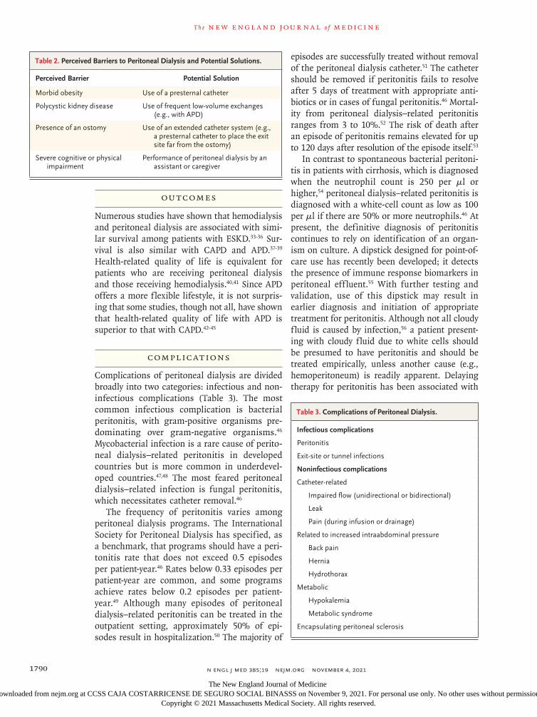
Peritoneal dialysis has been performed suc-cessfully in patients who have previously under-gone liver transplantation, with rates of perito-nitis and death that are similar to those in the general population of patients undergoing peri-toneal dialysis and with no adverse effects on the hepatic allograft.32 Other perceived barriers to peritoneal dialysis and potential solutions are listed in Table 2.
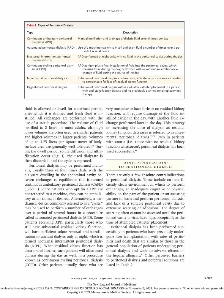
Table 1. Types of Peritoneal Dialysis.
Type Description
Continuous ambulatory peritoneal dialysis (CAPD)
Manual instillation and drainage of dialytic fluid several times per day
Automated peritoneal dialysis (APD) Use of a machine (cycler) to instill and drain fluid a number of times over a pe-riod of several hours
Nocturnal intermittent peritoneal dialysis (NIPD)
APD performed at night only, with no fluid in the peritoneal cavity during the day
Continuous cycling peritoneal dialy-sis (CCPD)
APD at night plus a final installation of fluid into the peritoneal cavity, which remains there during the day; performed with or without an additional ex-change of fluid during the course of the day
Incremental peritoneal dialysis Initiation of peritoneal dialysis at a low dose, with stepwise increases as needed to compensate for loss of residual kidney function
Urgent-start peritoneal dialysis Initiation of peritoneal dialysis within 2 wk after catheter placement in a person with end-stage kidney disease and no previously planned renal replacement therapy
The New England Journal of Medicine Downloaded from nejm.org at CCSS CAJA COSTARRICENSE DE SEGURO SOCIAL BINASSS on November 9, 2021. For personal use only. No other uses without permission.
Copyright © 2021 Massachusetts Medical Society. All rights reserved.
n engl j med 385;19 nejm.org November 4, 20211790
T h e n e w e ngl a nd j o u r na l o f m e dic i n e
Ou t comes
Numerous studies have shown that hemodialysis and peritoneal dialysis are associated with simi-lar survival among patients with ESKD.33-36 Sur-vival is also similar with CAPD and APD.37-39 Health-related quality of life is equivalent for patients who are receiving peritoneal dialysis and those receiving hemodialysis.40,41 Since APD offers a more flexible lifestyle, it is not surpris-ing that some studies, though not all, have shown that health-related quality of life with APD is superior to that with CAPD.42-45
Complic ations
Complications of peritoneal dialysis are divided broadly into two categories: infectious and non-infectious complications (Table 3). The most common infectious complication is bacterial peritonitis, with gram-positive organisms pre-dominating over gram-negative organisms.46 Mycobacterial infection is a rare cause of perito-neal dialysis–related peritonitis in developed countries but is more common in underdevel-oped countries.47,48 The most feared peritoneal dialysis–related infection is fungal peritonitis, which necessitates catheter removal.46
The frequency of peritonitis varies among peritoneal dialysis programs. The International Society for Peritoneal Dialysis has specified, as a benchmark, that programs should have a peri-tonitis rate that does not exceed 0.5 episodes per patient-year.46 Rates below 0.33 episodes per patient-year are common, and some programs achieve rates below 0.2 episodes per patient-year.49 Although many episodes of peritoneal dialysis–related peritonitis can be treated in the outpatient setting, approximately 50% of epi-sodes result in hospitalization.50 The majority of
episodes are successfully treated without removal of the peritoneal dialysis catheter.51 The catheter should be removed if peritonitis fails to resolve after 5 days of treatment with appropriate anti-biotics or in cases of fungal peritonitis.46 Mortal-ity from peritoneal dialysis–related peritonitis ranges from 3 to 10%.52 The risk of death after an episode of peritonitis remains elevated for up to 120 days after resolution of the episode itself.53
In contrast to spontaneous bacterial peritoni-tis in patients with cirrhosis, which is diagnosed when the neutrophil count is 250 per μl or higher,54 peritoneal dialysis–related peritonitis is diagnosed with a white-cell count as low as 100 per μl if there are 50% or more neutrophils.46 At present, the definitive diagnosis of peritonitis continues to rely on identification of an organ-ism on culture. A dipstick designed for point-of-care use has recently been developed; it detects the presence of immune response biomarkers in peritoneal eff luent.55 With further testing and validation, use of this dipstick may result in earlier diagnosis and initiation of appropriate treatment for peritonitis. Although not all cloudy fluid is caused by infection,56 a patient present-ing with cloudy fluid due to white cells should be presumed to have peritonitis and should be treated empirically, unless another cause (e.g., hemoperitoneum) is readily apparent. Delaying therapy for peritonitis has been associated with
Table 2. Perceived Barriers to Peritoneal Dialysis and Potential Solutions.
Perceived Barrier Potential Solution
Morbid obesity Use of a presternal catheter
Polycystic kidney disease Use of frequent low-volume exchanges (e.g., with APD)
Presence of an ostomy Use of an extended catheter system (e.g., a presternal catheter to place the exit site far from the ostomy)
Severe cognitive or physical impairment
Performance of peritoneal dialysis by an assistant or caregiver
Table 3. Complications of Peritoneal Dialysis.
Infectious complications
Peritonitis
Exit-site or tunnel infections
Noninfectious complications
Catheter-related
Impaired flow (unidirectional or bidirectional)
Leak
Pain (during infusion or drainage)
Related to increased intraabdominal pressure
Back pain
Hernia
Hydrothorax
Metabolic
Hypokalemia
Metabolic syndrome
Encapsulating peritoneal sclerosis
The New England Journal of Medicine Downloaded from nejm.org at CCSS CAJA COSTARRICENSE DE SEGURO SOCIAL BINASSS on November 9, 2021. For personal use only. No other uses without permission.
Copyright © 2021 Massachusetts Medical Society. All rights reserved.
n engl j med 385;19 nejm.org November 4, 2021 1791
Peritoneal Dialysis
an increased likelihood of treatment failure.57 Therefore, if the fluid cell count is not readily available, consideration should be given to ini-tiation of empirical therapy even in its absence. Empirical treatment should provide coverage for both gram-positive and gram-negative organ-isms. Unless the patient has signs of systemic sepsis, intraperitoneal administration of anti-biotics is preferred because it delivers the high-est concentration of drug directly to the infected site.46 This treatment can be administered either by a trained dialysis nurse or at home by the patient or caregiver. Other infections occasion-ally complicating peritoneal dialysis involve the exit site, the catheter tunnel, or both.58
Common noninfectious complications of peri-toneal dialysis include catheter-related issues such as catheter malfunction, problems with increased intraabdominal pressure, and meta-bolic consequences of the glucose-rich perito-neal dialysate.59 Mechanical complications include flow dysfunction, fluid leaks, and pain on infu-sion or draining of dialysate. Flow dysfunction is usually limited to poor outflow and is most commonly due to constipation, in which dis-tended bowel loops impinge on the catheter. Therefore, careful attention to a bowel regimen is important for patients treated with peritoneal dialysis. Less commonly, bladder distention is the cause of poor outflow. Occasionally, omentum, epiploic appendixes, or fallopian tube fimbriae impinge on the side holes of the catheter, neces-sitating laparoscopic repair. Bidirectional obstruc-tion to catheter flow is relatively uncommon, but it may be caused by kinking of the intramural portion of the catheter or intraluminal obstruc-tion (e.g., by a fibrin clot).19 Leakage of fluid around the catheter, through a hernia or other defect in the abdominal wall, or leakage into the pleural space may occur.
Metabolic complications include development of metabolic syndrome,60 with concern regard-ing the attendant weight gain, which may pre-vent or delay kidney transplantation. It is impor-tant to recognize, however, that the average weight gain after 1 year of peritoneal dialysis is reported to be only 1.3 kg61 or 2.3 kg.62 Some of this weight gain presumably reflects reversal of uremic anorexia and is therefore physiologically appropriate. Furthermore, in a large, propensity-matched cohort study of weight gain in patients treated with peritoneal dialysis as compared with
those receiving hemodialysis, weight gain was lower in the peritoneal dialysis group.63 In addi-tion, patients in the peritoneal dialysis group were more likely to undergo transplantation than those in the hemodialysis group and had equiva-lent survival. Thus, the concern about excessive weight gain and delay of transplantation appears to be unfounded.
Hypokalemia is another common metabolic complication of peritoneal dialysis. Since perito-neal dialysate contains no potassium, patients treated with peritoneal dialysis, particularly con-tinuous dialysis, are prone to hypokalemia, much more so than to hyperkalemia (which is more common in patients treated with hemodialysis). Consequently, it is important to recognize that patients receiving peritoneal dialysis are gener-ally permitted to consume a more potassium-rich diet than patients receiving hemodialysis. In fact, potassium supplementation may occasion-ally be required to maintain a normal plasma potassium level in a patient undergoing perito-neal dialysis. Finally, encapsulating peritoneal sclerosis is a severe but rare complication of long-term peritoneal dialysis (almost always occur-ring in patients treated for more than 5 years)64 that is associated with substantial morbidity and mortality. This disorder leads to progressive peritoneal fibrosis, culminating in “cocooning” of the bowel, with resultant symptoms of bowel obstruction and malnutrition.
Per i t one a l Di a lysis in Patien t s w i th Acu te K idne y Inj ur y
Use of peritoneal dialysis as therapy for AKI has increased in recent years, particularly in low-resource settings.20 This increase is due, in large measure, to the efforts of the International So-ciety of Nephrology Saving Young Lives project, which has brought peritoneal dialysis treatment for AKI to underserved regions around the world.65,66 A systematic review of studies involv-ing patients with AKI compared outcomes of those who were treated with extracorporeal ther-apies, continuous or intermittent, with outcomes of those treated with peritoneal dialysis; the re-view showed no significant difference in sur-vival between the groups.67 Peritoneal dialysis successfully treats the acidosis and most cases of hyperkalemia that occur in AKI in a manner equivalent to that of intermittent hemodialysis.68
The New England Journal of Medicine Downloaded from nejm.org at CCSS CAJA COSTARRICENSE DE SEGURO SOCIAL BINASSS on November 9, 2021. For personal use only. No other uses without permission.
Copyright © 2021 Massachusetts Medical Society. All rights reserved.
n engl j med 385;19 nejm.org November 4, 20211792
T h e n e w e ngl a nd j o u r na l o f m e dic i n e
Life-threatening hyperkalemia is successfully treat-ed more rapidly with extracorporeal therapies than with peritoneal dialysis. However, perito-neal dialysis can be initiated much more quickly than placement of a catheter for emergency hemo-dialysis. Therefore, if a patient with ESKD and a peritoneal dialysis catheter but no vascular ac-cess presents with life-threatening hyperkalemia, peritoneal dialysis should be initiated immedi-ately, with the possible need for other therapies reassessed frequently. Although volume removal cannot be as finely regulated with peritoneal dialysis as it can with extracorporeal therapies, frequent hypertonic exchanges can successfully address pulmonary edema. In this regard, it is important to recognize that concern about the potential adverse effects of intraperitoneal fluid on respiratory mechanics have not been borne out on careful study.69
Despite its demonstrated efficacy, peritoneal dialysis for AKI is not often used in developed countries because extracorporeal therapies are used preferentially. However, during the corona-virus disease 2019 (Covid-19) pandemic, even de-veloped countries have discovered that they are relatively resource-poor and have successfully used peritoneal dialysis when dialysis machines for extracorporeal therapies were in short sup-ply.70,71 It remains to be seen whether this trend will continue when the pandemic abates.72
Use of Per i t one a l Di a lysis in the Per ioper ati v e Se t ting
Many hospitals lack personnel with experience in the performance of peritoneal dialysis. As a result, sometimes a central venous catheter is placed and hemodialysis is performed when it is not actually required. In addition, many sur-geons believe that patients treated with perito-neal dialysis who are undergoing surgery must be switched to hemodialysis during the postop-erative period, often for many weeks, if not permanently. Concerns cited include leakage of dialysate through an abdominal incision, delayed wound healing, and the risk of peritonitis with possible subsequent infection of foreign mate-rials (e.g., surgical mesh or aortic grafts).73,74 However, study data and anecdotal experiences do not support such concerns. In patients under-going hernia repair or a variety of laparoscopic
procedures, peritoneal dialysis can often be re-sumed within 48 hours after surgery by per-forming small-volume exchanges with the pa-tient in the supine position, thereby minimizing intraabdominal pressure.74,75 The same is true for surgeries associated with relatively small up-per abdominal incisions (e.g., cholecystectomy). When such procedures are planned, it is often helpful to intensify peritoneal dialysis for several days before surgery, thereby allowing for a safe delay in resuming peritoneal dialysis postopera-tively.75,76 Patients undergoing bowel procedures that involve large anterior abdominal incisions should probably have a 2-to-3-week hiatus from peritoneal dialysis. However, if the abdomen is not infected, the peritoneal dialysis catheter does not need to be removed at the time of surgery.
Patients with ESKD may require cardiac sur-gery: coronary-artery bypass grafting, valve replace-ment, or a combination of the two procedures. Studies have shown that, after cardiothoracic surgery, both early complications and long-term survival are similar for patients treated with hemodialysis and those treated with peritoneal dialysis.77,78 Thus, patients with a peritoneal dialy-sis catheter in place who undergo cardiothoracic surgery do not automatically require conversion to extracorporeal renal replacement therapy.
In considering the continued use of perito-neal dialysis after cardiothoracic surgery, ad-vanced planning is advised. It is important to have a preoperative discussion with the surgeon to ensure that the integrity of the diaphragm is maintained. If possible, the surgical incision it-self and all drains, chest tubes, and other de-vices should be placed so as to avoid penetrating the diaphragm. Failure to maintain the integrity of the diaphragm may result in dialysate leak-age, precluding the use of peritoneal dialysis in the postoperative period.79 After major cardiac surgery, the frequency of AKI that requires dialy-sis is estimated at 5 to 8%. In adults, it is com-monly treated with extracorporeal therapies. In the pediatric population, however, peritoneal dialysis is often preferred and has been shown to provide excellent outcomes.80,81
The patient who is receiving a prosthetic aor-tic graft deserves specific mention. In a number of case series, peritoneal dialysis was used suc-cessfully in such patients, both in the immediate postoperative period82,83 and later.84,85 Although
The New England Journal of Medicine Downloaded from nejm.org at CCSS CAJA COSTARRICENSE DE SEGURO SOCIAL BINASSS on November 9, 2021. For personal use only. No other uses without permission.
Copyright © 2021 Massachusetts Medical Society. All rights reserved.
n engl j med 385;19 nejm.org November 4, 2021 1793
Peritoneal Dialysis
peritonitis did develop in 6 of 41 patients, it did not result in graft infection in any of them. Final-ly, although continuous extracorporeal therapy is the dialytic approach of choice in people with AKI or ESKD who require neurosurgery, perito-neal dialysis should be considered if continuous extracorporeal therapy is not available. Perito-neal dialysis offers several advantages over inter-mittent hemodialysis in these circumstances: he-modynamic fluctuations are minimal, there is no need for anticoagulant therapy, and peritoneal di-
alysis has no acute effects on plasma osmolality, which might cause or exacerbate cerebral edema.86
Summ a r y
Peritoneal dialysis is a valuable therapeutic ap-proach for patients with AKI or ESKD. Clinicians caring for such patients should have a basic understanding of peritoneal dialysis and its use.
Disclosure forms provided by the author are available with the full text of this article at NEJM.org.
References1. Liyanage T, Ninomiya T, Jha V, et al. Worldwide access to treatment for end-stage kidney disease: a systematic review. Lancet 2015; 385: 1975-82.2. Cho Y, Bello AK, Levin A, et al. Perito-neal dialysis use and practice patterns: an international survey study. Am J Kidney Dis 2021; 77: 315-25.3. Karopadi AN, Mason G, Rettore E, Ronco C. Cost of peritoneal dialysis and haemodialysis across the world. Nephrol Dial Transplant 2013; 28: 2553-69.4. Li PK-T, Chow KM, Van de Luijtgaarden MWM, et al. Changes in the worldwide epidemiology of peritoneal dialysis. Nat Rev Nephrol 2017; 13: 90-103.5. Pub. L. 95-292, 92 stat. 307 (June 13, 1978) (https://www . govinfo . gov/ content/ pkg/ STATUTE - 92/ pdf/ STATUTE - 92 - Pg307 . pdf#page= 9).6. Executive Office of the President. Ad-vancing American kidney health. Executive Order 13879. Fed Regist 2019; 84(135): 33817-9.7. Blake PG, Daugirdas JT. Physiology of peritoneal dialysis. In: Daugirdas JT, Blake PG, Ing TS, eds. Handbook of dialy-sis. 5th ed. Philadelphia: Wolters Kluwer Health, 2015: 392-407.8. Di Paolo N, Sacchi G. Atlas of perito-neal histology. Perit Dial Int 2000; 20: Suppl 3: S5-S96.9. Goffin E. Peritoneal membrane struc-tural and functional changes during peri-toneal dialysis. Semin Dial 2008; 21: 258-65.10. Davies SJ, Mushahar L, Yu Z, Lambie M. Determinants of peritoneal membrane function over time. Semin Nephrol 2011; 31: 172-82.11. Finkelstein F, Healy H, Abu-Alfa A, et al. Superiority of icodextrin compared with 4.25% dextrose for peritoneal ultra-filtration. J Am Soc Nephrol 2005; 16: 546-54.12. Wilkie ME, Plant MJ, Edwards L, Brown CB. Icodextrin 7.5% dialysate solu-tion (glucose polymer) in patients with ultrafiltration failure: extension of CAPD technique survival. Perit Dial Int 1997; 17: 84-7.
13. Yohanna S, Alkatheeri AM, Brimble SK, et al. Effect of neutral-pH, low-glu-cose degradation product peritoneal dialy-sis solutions on residual renal function, urine volume, and ultrafiltration: a sys-tematic review and meta-analysis. Clin J Am Soc Nephrol 2015; 10: 1380-8.14. Moncrief JW, Popovich RP, Broadrick LJ, He ZZ, Simmons EE, Tate RA. The Moncrief-Popovich catheter. A new peri-toneal access technique for patients on peritoneal dialysis. ASAIO J 1993; 39: 62-5.15. Elhassan E, McNair B, Quinn M, Teit-elbaum I. Prolonged duration of perito-neal dialysis catheter embedment does not lower the catheter success rate. Perit Dial Int 2011; 31: 558-64.16. Brown PA, McCormick BB, Knoll G, et al. Complications and catheter survival with prolonged embedding of peritoneal dialysis catheters. Nephrol Dial Transplant 2008; 23: 2299-303.17. Golper TA, Brier ME, Bunke M, et al. Risk factors for peritonitis in long-term peritoneal dialysis: the Network 9 perito-nitis and catheter survival studies. Aca-demic Subcommittee of the Steering Com-mittee of the Network 9 Peritonitis and Catheter Survival Studies. Am J Kidney Dis 1996; 28: 428-36.18. Crabtree JH, Burchette RJ. Prospective comparison of downward and lateral peri-toneal dialysis catheter tunnel-tract and exit-site directions. Perit Dial Int 2006; 26: 677-83.19. Crabtree JH, Shrestha BM, Chow K-M, et al. Creating and maintaining optimal peritoneal dialysis access in the adult pa-tient: 2019 update. Perit Dial Int 2019; 39: 414-36.20. Cullis B, Al-Hwiesh A, Kilonzo K, et al. ISPD guidelines for peritoneal dialysis in acute kidney injury: 2020 update (adults). Perit Dial Int 2021; 41: 15-31.21. Crabtree JH, Burchette RJ. Effective use of laparoscopy for long-term perito-neal dialysis access. Am J Surg 2009; 198: 135-41.22. Krezalek MA, Bonamici N, Lapin B, et al. Laparoscopic peritoneal dialysis
catheter insertion using rectus sheath tunnel and selective omentopexy signifi-cantly reduces catheter dysfunction and increases peritoneal dialysis longevity. Surgery 2016; 160: 924-35.23. Ghaffari A, Kumar V, Guest S. Infra-structure requirements for an urgent-start peritoneal dialysis program. Perit Dial Int 2013; 33: 611-7.24. Twardowski ZJ, Prowant BF, Nolph KD, Martinez AJ, Lampton LM. High vol-ume, low frequency continuous ambula-tory peritoneal dialysis. Kidney Int 1983; 23: 64-70.25. Jin H, Fang W, Zhu M, et al. Urgent-start peritoneal dialysis and hemodialysis in ESRD patients: complications and out-comes. PLoS One 2016; 11(11): e0166181.26. Lobbedez T, Lecouf A, Ficheux M, Henri P, Hurault de Ligny B, Ryckelynck JP. Is rapid initiation of peritoneal dialysis feasible in unplanned dialysis patients? A single-centre experience. Nephrol Dial Transplant 2008; 23: 3290-4.27. Koch M, Kohnle M, Trapp R, Haastert B, Rump LC, Aker S. Comparable outcome of acute unplanned peritoneal dialysis and haemodialysis. Nephrol Dial Transplant 2012; 27: 375-80.28. Paniagua R, Ventura MJ, Rodríguez E, et al. Impact of fill volume on peritoneal clearances and cytokine appearance in peritoneal dialysis. Perit Dial Int 2004; 24: 156-62.29. Teitelbaum I. Crafting the prescrip-tion for patients starting peritoneal dialy-sis. Clin J Am Soc Nephrol 2018; 13: 483-5.30. Mehrotra R, Nolph KD, Gotch F. Early initiation of chronic dialysis: role of in-cremental dialysis. Perit Dial Int 1997; 17: 426-30.31. Brown EA, Davies SJ, Rutherford P, et al. Survival of functionally anuric pa-tients on automated peritoneal dialysis: the European APD Outcome Study. J Am Soc Nephrol 2003; 14: 2948-57.32. Saiprasertkit N, Nihei CH, Bargman JM. Peritoneal dialysis in orthotopic liver transplantation recipients. Perit Dial Int 2018; 38: 44-8.
The New England Journal of Medicine Downloaded from nejm.org at CCSS CAJA COSTARRICENSE DE SEGURO SOCIAL BINASSS on November 9, 2021. For personal use only. No other uses without permission.
Copyright © 2021 Massachusetts Medical Society. All rights reserved.
n engl j med 385;19 nejm.org November 4, 20211794
T h e n e w e ngl a nd j o u r na l o f m e dic i n e
33. Heaf JG, Wehberg S. Relative survival of peritoneal dialysis and haemodialysis patients: effect of cohort and mode of dialysis initiation. PLoS One 2014; 9(3): e90119.34. Mehrotra R, Chiu Y-W, Kalantar-Zadeh K, Bargman J, Vonesh E. Similar out-comes with hemodialysis and peritoneal dialysis in patients with end-stage renal disease. Arch Intern Med 2011; 171: 110-8.35. Ryu J-H, Kim H, Kim KH, et al. Im-proving survival rate of Korean patients initiating dialysis. Yonsei Med J 2015; 56: 666-75.36. van de Luijtgaarden MWM, Jager KJ, Segelmark M, et al. Trends in dialysis mo-dality choice and related patient survival in the ERA-EDTA Registry over a 20-year period. Nephrol Dial Transplant 2016; 31: 120-8.37. Badve SV, Hawley CM, McDonald SP, et al. Automated and continuous ambula-tory peritoneal dialysis have similar out-comes. Kidney Int 2008; 73: 480-8.38. Mehrotra R, Chiu Y-W, Kalantar-Zadeh K, Vonesh E. The outcomes of continuous ambulatory and automated peritoneal di-alysis are similar. Kidney Int 2009; 76: 97-107.39. Michels WM, Verduijn M, Boeschoten EW, Dekker FW, Krediet RT. Similar sur-vival on automated peritoneal dialysis and continuous ambulatory peritoneal dialysis in a large prospective cohort. Clin J Am Soc Nephrol 2009; 4: 943-9.40. Purnell TS, Auguste P, Crews DC, et al. Comparison of life participation activities among adults treated by hemodialysis, peritoneal dialysis, and kidney transplan-tation: a systematic review. Am J Kidney Dis 2013; 62: 953-73.41. Zazzeroni L, Pasquinelli G, Nanni E, Cremonini V, Rubbi I. Comparison of qual-ity of life in patients undergoing hemodi-alysis and peritoneal dialysis: a systematic review and meta-analysis. Kidney Blood Press Res 2017; 42: 717-27.42. de Wit GA, Merkus MP, Krediet RT, de Charro FT. A comparison of quality of life of patients on automated and continuous ambulatory peritoneal dialysis. Perit Dial Int 2001; 21: 306-12.43. Michels WM, van Dijk S, Verduijn M, et al. Quality of life in automated and con-tinuous ambulatory peritoneal dialysis. Perit Dial Int 2011; 31: 138-47.44. Rabindranath KS, Adams J, Ali TZ, Daly C, Vale L, Macleod AM. Automated vs continuous ambulatory peritoneal dialy-sis: a systematic review of randomized controlled trials. Nephrol Dial Transplant 2007; 22: 2991-8.45. Bieber SD, Burkart J, Golper TA, Teitel-baum I, Mehrotra R. Comparative out-comes between continuous ambulatory and automated peritoneal dialysis: a nar-rative review. Am J Kidney Dis 2014; 63: 1027-37.46. Li PK, Szeto CC, Piraino B, et al. ISPD
peritonitis recommendations: 2016 update on prevention and treatment. Perit Dial Int 2016; 36: 481-508.47. Cho Y, Struijk DG. Peritoneal dialysis-related peritonitis: atypical and resistant organisms. Semin Nephrol 2017; 37: 66-76.48. Song Y, Wu J, Yan H, Chen J. Perito-neal dialysis-associated nontuberculous mycobacterium peritonitis: a systematic review of reported cases. Nephrol Dial Transplant 2012; 27: 1639-44.49. Piraino B, Bernardini J, Brown E, et al. ISPD position statement on reducing the risks of peritoneal dialysis-related infec-tions. Perit Dial Int 2011; 31: 614-30.50. Perl J, Fuller DS, Bieber BA, et al. Peri-toneal dialysis-related infection rates and outcomes: results from the Peritoneal Dialysis Outcomes and Practice Patterns Study (PDOPPS). Am J Kidney Dis 2020; 76: 42-53.51. Mehrotra R, Devuyst O, Davies SJ, Johnson DW. The current state of perito-neal dialysis. J Am Soc Nephrol 2016; 27: 3238-52.52. Bieber S, Mehrotra R. Peritoneal dialy-sis access associated infections. Adv Chron-ic Kidney Dis 2019; 26: 23-9.53. Boudville N, Kemp A, Clayton P, et al. Recent peritonitis associates with mortal-ity among patients treated with peritoneal dialysis. J Am Soc Nephrol 2012; 23: 1398-405.54. Aithal GP, Palaniyappan N, China L, et al. Guidelines on the management of ascites in cirrhosis. Gut 2021; 70: 9-29.55. Goodlad C, George S, Sandoval S, et al. Measurement of innate immune response biomarkers in peritoneal dialysis effluent using a rapid diagnostic point-of-care de-vice as a diagnostic indicator of peritoni-tis. Kidney Int 2020; 97: 1253-9.56. Rocklin MA, Teitelbaum I. Noninfec-tious causes of cloudy peritoneal dialy-sate. Semin Dial 2001; 14: 37-40.57. Muthucumarana K, Howson P, Craw-ford D, Burrows S, Swaminathan R, Irish A. The Relationship Between Presentation and the Time of Initial Administration of Antibiotics With Outcomes of Peritonitis in Peritoneal Dialysis Patients: the PROMPT study. Kidney Int Rep 2016; 1: 65-72.58. Szeto C-C, Li PK-T, Johnson DW, et al. ISPD catheter-related infection recommen-dations: 2017 update. Perit Dial Int 2017; 37: 141-54.59. McCormick BB, Bargman JM. Nonin-fectious complications of peritoneal dialy-sis: implications for patient and technique survival. J Am Soc Nephrol 2007; 18: 3023-5.60. Szeto C-C, Kwan BC-H, Chow K-M, et al. Metabolic syndrome in peritoneal dialysis patients: choice of diagnostic cri-teria and prognostic implications. Clin J Am Soc Nephrol 2014; 9: 779-87.61. Choy AS-M, Chow K-M, Kwan BC-H, et al. Weight change during the first year of peritoneal dialysis: risk factors and prog-
nostic implications. Hong Kong J Nephrol 2015; 17: 28-35.62. Kim J-K, Kim Y-S, Song YR, Kim HJ, Kim SG, Moon SJ. Excessive weight gain during the first year of peritoneal dialysis is associated with inflammation, diabetes mellitus, and a rapid decrease in residual renal function. PLoS One 2015; 10(9): e0139033.63. Lievense H, Kalantar-Zadeh K, Lukow-sky LR, et al. Relationship of body size and initial dialysis modality on subsequent transplantation, mortality and weight gain of ESRD patients. Nephrol Dial Trans-plant 2012; 27: 3631-8.64. Brown EA, Bargman J, van Biesen W, et al. Length of time on peritoneal dialy-sis and encapsulating peritoneal sclerosis — position paper for ISPD: 2017 update. Perit Dial Int 2017; 37: 362-74.65. Cullis B, Lalya F, Smoyer WE. Saving more young lives in Africa. Perit Dial Int 2020; 40: 438-40.66. Finkelstein FO, Smoyer WE, Carter M, Brusselmans A, Feehally J. Peritoneal di-alysis, acute kidney injury, and the Saving Young Lives program. Perit Dial Int 2014; 34: 478-80.67. Chionh CY, Soni SS, Finkelstein FO, Ronco C, Cruz DN. Use of peritoneal dialy-sis in AKI: a systematic review. Clin J Am Soc Nephrol 2013; 8: 1649-60.68. Gabriel DP, Caramori JT, Martim LC, Barretti P, Balbi AL. High volume perito-neal dialysis vs daily hemodialysis: a ran-domized, controlled trial in patients with acute kidney injury. Kidney Int Suppl 2008; 73: Suppl 108: S87-S93.69. Almeida CP, Ponce D, de Marchi AC, Balbi AL. Effect of peritoneal dialysis on respiratory mechanics in acute kidney injury patients. Perit Dial Int 2014; 34: 544-9.70. El Shamy O, Patel N, Abdelbaset MH, et al. Acute start peritoneal dialysis dur-ing the COVID-19 pandemic: outcomes and experiences. J Am Soc Nephrol 2020; 31: 1680-2.71. Sourial MY, Sourial MH, Dalsan R, et al. Urgent peritoneal dialysis in patients with COVID-19 and acute kidney injury: a single-center experience in a time of cri-sis in the United States. Am J Kidney Dis 2020; 76: 401-6.72. Brown EA, Perl J. Increasing peritoneal dialysis use in response to the COVID-19 pandemic: will it go viral? J Am Soc Nephrol 2020; 31: 1928-30.73. Lee MB, Bargman JM. Myths in peri-toneal dialysis. Curr Opin Nephrol Hyper-tens 2016; 25: 602-8.74. Mari G, Scanziani R, Auricchio S, Crippa J, Maggioni D. Laparoscopic sur-gery in patients on peritoneal dialysis: a review of the literature. Surg Innov 2017; 24: 397-401.75. Shah H, Chu M, Bargman JM. Periop-erative management of peritoneal dialysis patients undergoing hernia surgery with-
The New England Journal of Medicine Downloaded from nejm.org at CCSS CAJA COSTARRICENSE DE SEGURO SOCIAL BINASSS on November 9, 2021. For personal use only. No other uses without permission.
Copyright © 2021 Massachusetts Medical Society. All rights reserved.
n engl j med 385;19 nejm.org November 4, 2021 1795
Peritoneal Dialysis
out the use of interim hemodialysis. Perit Dial Int 2006; 26: 684-7.76. Chaudhry RI, Chopra T, Fissell R, Golper TA. Strategies to prevent peritoni-tis after procedures: our opinions. Perit Dial Int 2019; 39: 315-9.77. Bäck C, Hornum M, Møller CJH, Olsen PS. Cardiac surgery in patients with end-stage renal disease on dialysis. Scand Cardiovasc J 2017; 51: 334-8.78. Kumar VA, Ananthakrishnan S, Ras-gon SA, Yan E, Burchette R, Dewar K. Comparing cardiac surgery in peritoneal dialysis and hemodialysis patients: peri-operative outcomes and two-year survival. Perit Dial Int 2012; 32: 137-41.79. Teitelbaum I. Peritoneal dialysis after
cardiothoracic surgery: do it! Perit Dial Int 2012; 32: 131-3.80. Namachivayam SP, Butt W, Millar J, Konstantinov IE, Nguyen C, d’Udekem Y. Early peritoneal dialysis and major ad-verse events after pediatric cardiac sur-gery: a propensity score analysis. Pediatr Crit Care Med 2019; 20: 158-65.81. Santos CR, Branco PQ, Gaspar A, et al. Use of peritoneal dialysis after surgery for congenital heart disease in children. Perit Dial Int 2012; 32: 273-9.82. Hajarizadeh H, Rohrer MJ, Herrmann JB, Cutler BS. Acute peritoneal dialysis following ruptured abdominal aortic an-eurysms. Am J Surg 1995; 170: 223-6.83. Tzamaloukas AH, Garella S, Chazan
JA. Peritoneal dialysis for acute renal fail-ure after major abdominal surgery. Arch Surg 1973; 106: 639-43.84. Charytan C. Continuous ambulatory peritoneal dialysis after abdominal aortic graft surgery. Perit Dial Int 1992; 12: 227-9.85. Gulanikar AC, Jindal KK, Hirsch DJ. Is chronic peritoneal dialysis safe in pa-tients with intra-abdominal prosthetic vascular grafts? Nephrol Dial Transplant 1991; 6: 215-7.86. Davenport A. Treatment of combined acute renal failure and cerebral edema. In: Ronco C, Bellomo R, Kellum JA, Ricci Z, eds. Critical care nephrology. 3rd ed. Phil-adelphia: Elsevier, 2019: 800-4.Copyright © 2021 Massachusetts Medical Society.
images in clinical medicine
The Journal welcomes consideration of new submissions for Images in Clinical Medicine. Instructions for authors and procedures for submissions can be found on the Journal’s website at NEJM.org. At the discretion of the editor, images that
are accepted for publication may appear in the print version of the Journal, the electronic version, or both.
The New England Journal of Medicine Downloaded from nejm.org at CCSS CAJA COSTARRICENSE DE SEGURO SOCIAL BINASSS on November 9, 2021. For personal use only. No other uses without permission.
Copyright © 2021 Massachusetts Medical Society. All rights reserved.
Related Documents