Lack of complex N-glycans on HIV- 1 envelope glycoproteins preserves protein conformation and entry function Dirk Eggink, Mark Melchers, Manfred Wuhrer, ijs van Montfort, Antu K. Dey, Benno A. Naaijkens, Kathryn B. David, Valentin Le Douce, André M. Deelder, Kenneth Kang, William C. Olson, Ben Berkhout, Cornelis H. Hokke, John P. Moore, Rogier W. Sanders Chapter 3 Virol. 2010; 401, 236-247

Welcome message from author
This document is posted to help you gain knowledge. Please leave a comment to let me know what you think about it! Share it to your friends and learn new things together.
Transcript
Lack of complex N-glycans on HIV-1 envelope glycoproteins preserves
protein conformation and entry function
Dirk Eggink, Mark Melchers, Manfred Wuhrer, Thijs van Montfort, Antu K. Dey, Benno A. Naaijkens, Kathryn B. David, Valentin Le Douce, André M.
Deelder, Kenneth Kang, William C. Olson, Ben Berkhout, Cornelis H. Hokke, John P. Moore, Rogier W. Sanders
Chapter 3
Virol. 2010; 401, 236-247
32
Chapter 3
3
AbstractThe HIV-1 envelope glycoprotein complex (Env) is the focus of vaccine development aimed at eliciting humoral immunity. Env’s extensive and heterogeneous N-linked glycosylation affects folding, binding to lectin receptors, antigenicity and immunogenicity. We characterized recombinant Env proteins and virus particles produced in mammalian cells that lack N-acetylglucosaminyltransferase I (GnTI), an enzyme necessary for the conversion of oligomannose N-glycans to complex N-glycans. Carbohydrate analyses revealed that trimeric Env produced in GnTI-/- cells contained exclusively oligomannose N-glycans, with incompletely trimmed oligomannose glycans predominating. The folding and conformation of Env proteins was little affected by the manipulation of the glycosylation. Viruses produced in GnTI-/- cells were infectious, indicating that the conversion to complex glycans is not necessary for Env entry function, although virus binding to the C-type lectin DC-SIGN was enhanced. Manipulating Env’s N-glycosylation may be useful for structural and functional studies and for vaccine design.
33
Lack of complex N-glycans on HIV-1 envelope glycoproteins preserves protein conformation and entry function
3
IntroductionThe entry of human immunodeficiency virus type 1 (HIV-1) into target cells is mediated by the envelope glycoprotein (Env) complex, which consists of three gp120/gp41 heterodimers. The Env complex is located on the virion surface and is the target for virus-neutralizing antibodies (NAbs) that are generated by infected individuals. The protein components of the Env complex are, therefore, the focus of all HIV-1 vaccine strategies intended to induce NAbs. Such approaches most commonly involve the use of individual gp120 surface glycoproteins or gp41 transmembrane glycoproteins, fragments of these proteins, or mimetics of the Env complex created by expressing gp140 trimers containing both gp120 and the gp41 ectodomain.
Half the molecular mass of gp120 is comprised of N-linked glycans that overlie the protein backbone, and the gp41 ectodomain is also glycosylated, although to a lesser extent. Env glycosylation is important at several stages in the viral life cycle. First, N-glycans play an essential role in the oxidative folding of gp120 in the endoplasmic reticulum (ER); inhibiting glycosylation impairs the production of functional Env proteins (1). Second, N-glycans form the binding sites for ancillary receptors such as DC-SIGN and langerin that are involved in the binding of HIV-1 to, e.g., dendritic cells (DC), and that facilitate or impede its subsequent transmission to T cells (2-5). Third, the poorly immunogenic N-glycans play a critical role in HIV-1 immune evasion, by shielding underlying NAb epitopes on the gp120 and gp41 protein backbones (6). The mannose moieties on gp120 N-glycans can also induce immunosuppressive cytokine responses under certain conditions (7,8). Although mannose moieties on N-linked glycans constitute the epitope for 2G12, one of the very few relatively broadly active NAbs (9-13), the extensive glycosylation of gp120 is a hindrance to the use of Env proteins as vaccine antigens.
The extent, heterogeneity and flexibility of Env glycosylation also interfere with the formation of protein crystals, which seriously compromises attempts to determine molecular structures. Indeed, the structures of gp120 core fragments could not be obtained without enzymatic deglycosylation of the proteins to facilitate crystallization (14-17). A better understanding of Env glycosylation is therefore likely to help both vaccine and structural studies, particularly ones involving recombinant Env trimers.
We have been studying and refining a recombinant trimer construct, SOSIP gp140, that is stabilized by an intermolecular disulfide bond between gp120 and gp41 and a point substitution in gp41 (18,19). The SOSIP gp140 trimers are proteolytically cleaved at the gp120-gp41 junction, a process that alters their antigenicity and immunogenicity and that may make them better mimics of the native Env complex, compared with uncleaved trimers (18,20-22). We now wish to be able to modify the glycan content of SOSIP gp140 trimers for the reasons outlined above. As a first step, we have assessed whether
34
Chapter 3
3
Env monomers and trimers can be produced in a cell line (293S GnTI-/-) that has been engineered to prevent the production of complex type N-glycans, leaving oligomannose glycans as the only Env glycan component (23).
ResultsExpression of recombinant HIV-1 Env glycoproteins in mammalian cells lacking N-acetylglucosaminyltransferase I
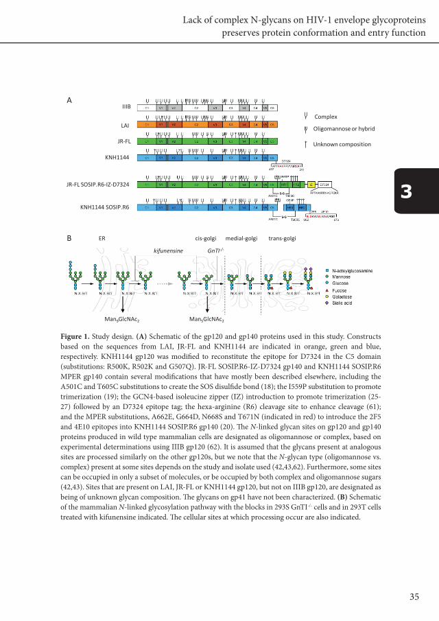
The various Env proteins used in this study are depicted schematically in Fig. 1A. Our strategy to reduce the extent and heterogeneity of Env glycosylation involved expressing monomeric gp120 and trimeric gp140 proteins in 293S GnTI-/- cells that do not express UDP-N-acetylglucosamine:α-d-mannoside-β1,2-N-acetylglucosaminyltransferase I (GlcNAc transferase I; GnTI) . The absence of this enzyme prevents the attachment of GlcNAc to Man5GlcNAc2 glycans in the medial-Golgi; as a result, complex carbohydrate chains of variable composition are not formed. Hence, glycoproteins produced in 293S GnTI-/- cells are expected to contain predominantly oligomannosidic N-glycans with 5 mannose residues (Fig. 1B) (23,24).
We first expressed three different monomeric gp120 proteins in 293S GnTI-/- cells and, for comparison, in wild type 293T cells that do express GnTI. The gp120s were derived from isolates LAI, JR-FL (both subtype B) and KNH1144 (subtype A). The KNH1144 gp120 had a minor modification from the original sequence; the C5 domain epitope for Ab D7324 was optimized to facilitate analyses by antigen-capture ELISA (Fig. 1A). All three gp120s were expressed efficiently in the 293S GnTI-/- cells (Fig. 2A). The 293S GnTI-/- cell-derived gp120s migrated more rapidly in SDS-PAGE gels than the corresponding proteins from 293T cells; their lower apparent molecular weight (~ 110 kDa compared to ~ 120 kDa) is consistent with a decrease in glycan size as a result of the blocked conversion of Man5GlcNAc2 moieties to complex glycans. We note that sialylation of complex N-glycans produced in 293T cells may influence their migration in SDS-PAGE and the ~ 10 kDa size difference may be a slight underestimation of the absolute size difference.
We next investigated whether different forms of gp140 trimers based on the JR-FL or KNH1144 sequences could be expressed efficiently in 293S GnTI-/- cells. The JR-FL SOSIP.R6-IZ-D7324 gp140 construct contains several modifications, including a heterologous trimerization domain based on GCN4 at its C-terminus (25-27) and a D7324-epitope tag (Fig. 1A). The addition of the GCN4 domain increases SOSIP gp140 trimer formation and/or stability, but interferes with cleavage at the gp120-gp41 juncture. From here on, we will refer to this construct as uncleaved JR-FL gp140. When this protein was expressed in either 293T or 293S GnTI-/- cells, it formed trimers efficiently, although they were not cleaved (Fig. 2B, and data not shown).
35
Lack of complex N-glycans on HIV-1 envelope glycoproteins preserves protein conformation and entry function
3
Figure 1. Study design. (A) Schematic of the gp120 and gp140 proteins used in this study. Constructs based on the sequences from LAI, JR-FL and KNH1144 are indicated in orange, green and blue, respectively. KNH1144 gp120 was modified to reconstitute the epitope for D7324 in the C5 domain (substitutions: R500K, R502K and G507Q). JR-FL SOSIP.R6-IZ-D7324 gp140 and KNH1144 SOSIP.R6 MPER gp140 contain several modifications that have mostly been described elsewhere, including the A501C and T605C substitutions to create the SOS disulfide bond (18); the I559P substitution to promote trimerization (19); the GCN4-based isoleucine zipper (IZ) introduction to promote trimerization (25-27) followed by an D7324 epitope tag; the hexa-arginine (R6) cleavage site to enhance cleavage (61); and the MPER substitutions, A662E, G664D, N668S and T671N (indicated in red) to introduce the 2F5 and 4E10 epitopes into KNH1144 SOSIP.R6 gp140 (20). The N-linked glycan sites on gp120 and gp140 proteins produced in wild type mammalian cells are designated as oligomannose or complex, based on experimental determinations using IIIB gp120 (62). It is assumed that the glycans present at analogous sites are processed similarly on the other gp120s, but we note that the N-glycan type (oligomannose vs. complex) present at some sites depends on the study and isolate used (42,43,62). Furthermore, some sites can be occupied in only a subset of molecules, or be occupied by both complex and oligomannose sugars (42,43). Sites that are present on LAI, JR-FL or KNH1144 gp120, but not on IIIB gp120, are designated as being of unknown glycan composition. The glycans on gp41 have not been characterized. (B) Schematic of the mammalian N-linked glycosylation pathway with the blocks in 293S GnTI-/- cells and in 293T cells treated with kifunensine indicated. The cellular sites at which processing occur are also indicated.
A
B
Complex
Oligomannose or hybrid
Unknown composition
IIIB
LAI
JR-FL
KNH1144
JR-FL SOSIP.R6-IZ-D7324
KNH1144 SOSIP.R6
ER cis-golgi medial-golgi trans-golgi
kifunensine GnTI-/-
Man9GlcNAc2 Man5GlcNAc2
Complex
Oligomannose or hybrid
Complex
Unknown composition
Fig 6.1. Study design. (A) Schematic of the gp120 and gp140 proteins used in this study. Constructs basedon the sequences from LAI, JR-FL and KNH1144 are indicated in orange, green and blue, respectively.KNH1144 gp120 was modified to reconstitute the epitope for D7324 in the C5 domain (substitutions:R500K, R502K and G507Q). JR-FL SOSIP.R6-IZ-D7324 gp140 and KNH1144 SOSIP.R6 MPER gp140 containseveral modifications that have mostly been described elsewhere, including the A501C and T605Csubstitutions to create the SOS disulfide bond (6); the I559P substitution to promote trimerization (49);the GCN4-based isoleucine zipper (IZ) introduction to promote trimerization (25, 68, 69) followed by anD7324 epitope tag; the hexa-arginine (R6) cleavage site to enhance cleavage (7); and the MPERsubstitutions, A662E, G664D, N668S and T671N (indicated in red) to introduce the 2F5 and 4E10epitopes into KNH1144 SOSIP.R6 gp140 (14). The N-linked glycan sites on gp120 and gp140 proteinsproduced in wild type mammalian cells are designated as oligomannose or complex, based onexperimental determinations using IIIB gp120 (33). It is assumed that the glycans present at analogoussites are processed similarly on the other gp120s, but we note that the N-glycan type (oligomannose vscomplex) present at some sites depends on the study and isolate used (12, 33, 71). Furthermore, somesites can be unoccupied in a only subset of molecules, or be occupied by both complex andoligomannose sugars (12, 71). Sites that are present on LAI, JR-FL or KNH1144 gp120, but not on IIIBgp120, are designated as being of unknown glycan composition. The glycans on gp41 have not beencharacterized. (B) Schematic of the mammalian N-linked glycosylation pathway with the blocks in 293SGnTI-/- cells and in 293T cells treated with kifunensine indicated. The cellular sites at which processingoccurs are also indicated.
6-28

36
Chapter 3
3
Trimeric SOSIP gp140 proteins (without C-terminal additions) are substantially (> 90%) cleaved when produced in mammalian cells in the presence of co-transfected furin (19). KNH1144 SOSIP gp140s are particularly useful for structural studies because they form trimers more efficiently than the corresponding proteins derived from various other strains such as JR-FL (28). Because KNH1144 Env proteins do not bind MAbs 2F5 and 4E10 due to sequence variation, we introduced these epitopes by mutagenesis, to create KNH1144 SOSIP.R6 MPER gp140 (Fig. 1A) (20). From here on, we refer to this construct as cleaved KNH1144 gp140. When this protein was expressed in 293S GnTI-/- cells in the presence of furin, it formed trimers efficiently (Fig. 2C). Moreover, these trimers were fully cleaved, as judged by their conversion to monomeric gp120s on reducing SDS-PAGE gels (Fig. 2C and data not shown). As observed when monomeric gp120 proteins were produced in 293S GnTI-/- cells (Fig. 2A), the gp120 component of the gp140 trimers
Figure 2. Expression of gp120 and gp140 in 293T cells and in 293S cells lacking the GnTI enzyme. (A) SDS-PAGE analysis of LAI, JR-FL and KNH1144 gp120s expressed transiently in 293T or 293S GnTI-/- cells. (B) SDS-PAGE and BN-PAGE analysis of uncleaved JR-FL SOSIP.R6-IZ-D7324 gp140, expressed in both cell types. (C) SDS-PAGE and BN-PAGE analysis of cleaved KNH1144 SOSIP.R6 MPER gp140 expressed in the presence of furin, in either 293T cells with or without kifunensine, or in 293S GnTI-/- cells. (D) Size exclusion chromatography analysis of uncleaved JR-FL SOSIP.R6-IZ gp140 trimers produced in 293T or 293S GnTI-/- cells and fractionated using a Superose-6 column. The fractions (indicated on top) were analyzed by SDS-PAGE and western blot. The peak fractions of the standard protein thyroglobulin (669 kDa), ferritin (440 kDa) and BSA (67 kDa) are indicated.
C
KNH1144JR-FLLAI
native
trimer
293T
293S
GnT
I-/-A
trimer
monomer
B
SDS
native
SDS
293T
293T
+ k
if
293S
GnT
I-/-
293T
293T
+ k
if
293S
GnT
I-/-
293T
293S
GnT
I-/-
293T
293S
GnT
I-/-
293T
293S
GnT
I-/-
gp12
0
293T
293S
GnT
I-/-
669 kDa 440 kDa 67 kDa
9 11 2515 1713 27 29 31 3319 21 23 25 35 37 39 41
293T
293S GnTI-/-
D
gp120 JR-FL SOSIP.R6-IZ-D7324 KNH1144 SOSIP.R6 MPER
JR-FL SOSIP.R6-IZ-D7324
6-29
Fig. 6.2 Expression of gp120 and gp140 in 293T cells and in 293S cells lacking the GnTI enzyme. (A) SDS-PAGE analysis of LAI, JR-FL and KNH1144 gp120s expressed transiently in 293T or 293S GnTI-/- cells. (B)SDS-PAGE and BN-PAGE analysis of uncleaved JR-FL SOSIP.R6-IZ-D7324 gp140, expressed in both celltypes. (C) SDS-PAGE and BN-PAGE analysis of cleaved KNH1144 SOSIP.R6 MPER gp140 expressed in thepresence of furin, in either 293T cells with or without kifunensine, or in 293S GnTI-/- cells. (D) Sizeexclusion chromatography analysis of uncleaved JR-FL SOSIP.R6-IZ gp140 trimers produced in 293T or293S GnTI-/- cells and fractionated using a Superose-6 column. The fractions (indicated on top) wereanalyzed by SDS-PAGE and western blot. The peak fractions of the standard protein thyroglobulin (669kD), ferritin (440 kD) and BSA (67 kD) are indicated.

37
Lack of complex N-glycans on HIV-1 envelope glycoproteins preserves protein conformation and entry function
3
migrated with an apparent molecular weight of ~ 110 kDa, slightly less than the ~ 120 kDa proteins expressed in 293T cells (Fig. 2C).
To further characterize the oligomeric states of gp140 trimers produced in 293T and 293S GnTI-/- cells, we performed analytical size exclusion chromatography using a Superose-6 column. Concentrated supernatant from the Env-transfected cell cultures were column-fractionated and then analyzed by SDS-PAGE and western blot. The uncleaved JR-FL gp140 and the cleaved KNH1144 gp140 proteins, whether produced in 293T or 293S GnTI-/- cells, were eluted in fractions between the peaks for ferritin (440 kDa) and thyroglobulin (669 kDa) (Fig. 2D and data not shown). These observations are consistent with our earlier findings (19), and imply that Env proteins produced in 293S GnTI-/- cells form trimers efficiently.
Expression of SOSIP gp140 trimers in the presence of kifunensine
Kifunensine is a mannose analog that inhibits both type I mannosidases in the endoplasmic reticulum and α-mannosidases in the Golgi, resulting in the production of glycoproteins that contain predominantly Man9GlcNAc2 carbohydrates (29). Thus, by preventing the trimming of these moieties to Man5GlcNAc2, kifunensine inhibits an earlier step in the carbohydrate-processing pathway than the one blocked in GnTI-/- cells (Fig. 1B). Because monomeric gp120 can be successfully expressed in 293T cells in the presence of kifunensine (30), this approach may be a useful alternative way to reduce glycan heterogeneity on gp140 trimers. We therefore produced cleaved KNH1144 gp140 trimers in 293T cells in the presence and absence of kifunensine, and in the presence of furin, then compared them with trimers produced in 293S GnTI-/- cells (Fig. 2C). The efficiency of trimer production was independent of kifunensine use. Furthermore, the size of the gp120 components of the trimers was unaffected by kifunensine, whereas the gp120 moieties produced in 293S GnTI-/- cells were, as noted above, ~ 10 kDa smaller (Fig. 2C). The presence of only oligomannose glycans was confirmed by EndoH digestion (data not shown).
Env trimers from 293S GnTI-/- cells contain exclusively oligomannosidic N-glycan species
To establish which N-glycans are present on cleaved KNH1144 trimers produced in 293S GnTI-/- , we first released all N-glycans by PNGase F treatment. Subsequently, the free, reducing end N-acetylglucosamine residues were reacted with 2-AA to introduce a fluorescent label. The released, labeled glycan pool was analyzed by HILIC HPLC with fluorescence detection, and by MALDI-TOF mass spectrometry. The HILIC HPLC analysis showed that a relatively simple mixture of N-glycans was present, separated into five major fractions (Fig. 3A). The MALDI-TOF mass spectrum (Fig. 3B) also indicated
38
Chapter 3
3
that five major glycan species were present. The major LC fractions (3, 5, 7, 8, and 9) were each collected and analyzed by MALDI-TOF MS (Fig. 3, panels C-G, respectively). The observed m/z values show that a single glycan species with the composition HexnNAc2-AA was present in each of these fractions, with n = 5 for the glycan in panel C, n = 6 in panel D, n = 7 in E, n = 8 in F and n = 9 in G. These data imply that Man5GlcNAc2 to Man9GlcNAc2 oligomannosidic glycans are present on KNH1144 gp140 trimers, with Man5GlcNAc2 (26.9%), Man8GlcNAc2 (24.9%) and Man9GlcNAc2 (28.6%) being the major species (see Fig. 3A). Thus, N-glycans on trimeric Env are substantially resistant to mannosidase trimming, which stands in contrast to reports that other proteins expressed in 293S GnTI-/- cells bear homogenous Man5GlcNAc2 glycans (23,24), but is consistent with the presence of oligomannose glycans on Env produced under natural glycosylation conditions.
Env proteins produced in 293T and 293S GnTI-/- cells are antigenically similar
To assess whether the antigenic structures of gp120 and gp140 proteins were affected by eliminating complex carbohydrates, we measured their reactivity with a panel of MAbs against conformational epitopes. We first used the D7324-capture ELISA to study LAI, JR-FL and KNH1144 gp120s. The 293T and 293S GnTI-/- cell-derived JR-FL and LAI gp120s bound a CD4 mimetic (CD4-IgG2) and MAb b12 to the CD4 binding site, to similar extents. GnTI-/- cell-derived gp120s showed enhanced reactivity with pooled Ig from HIV-positive individuals (HIVIg), suggesting that some unidentified antibody epitopes become more accessible when complex N-glycans are absent. This effect was subtle and was not reproduced with a different lot of HIVIg (not shown). Conformational changes in all the gp120s were induced by soluble CD4 (sCD4), as indicated by the increased binding of MAb 17b to a CD4-induced (CD4i) epitope (Fig. 4A). The binding of 17b to LAI gp120 from 293S GnTI-/- cells was, however, slightly decreased compared to its counterpart from 293T cells. KNH1144 gp120 interacted efficiently with HIVIg, CD4-IgG2 and 17b, but not with b12, consistent with sequence variation in critical b12 contact residues (P369L, V372T, M373T, P417Q, and E462K) (17,31). Again, there was little effect of the producer cell line on the reactivity of the gp120, except for the subtly enhanced HIVIg binding. Thus, it appears that monomeric gp120 folds normally in 293S GnTI-/- cells despite the manipulation of its glycan structures.
We next evaluated the uncleaved JR-FL gp140 trimers using the D7324-capture ELISA. There were no marked differences in how CD4-IgG2 or MAbs b12 and 2F5 (to gp41) bound to trimers from 293S GnTI-/- and 293T cells (Fig. 4B). The binding of HIVIg was enhanced upon expression in GnTI-/- cells, but the effect was more subtle than observed with gp120 monomers, suggesting that the (unknown) epitopes that become more exposed when complex N-glycans are absent, are affected by
39
Lack of complex N-glycans on HIV-1 envelope glycoproteins preserves protein conformation and entry function
3
trimerization. MAb 17b bound slightly less efficiently to the trimers from the 293S GNTI-/- cells, but its epitope was strongly induced by sCD4 irrespective of the producer cell type (Fig. 4B). Most of the gp120-directed MAbs and CD4-based reagents bound similarly to monomeric and uncleaved, trimeric JR-FL Env with the exception of 17b, which interacted less efficiently with trimeric gp140 (compare Fig. 4A middle panels with Fig. 4B).
The D3724-capture ELISA method cannot be used to analyze cleaved SOSIP gp140 trimers; the internal D7324 epitope is mutated and occluded, while the epitope tag added to the C-terminus of the gp41 moiety interferes with gp140 cleavage. We therefore used less quantitative immunoprecipitation assays to study MAb binding to cleaved KNH1144 gp140 trimers that had been produced in 293T cells in the presence or absence of kifunensine, or in 293S GnTI-/- cells (Fig. 4C). Irrespective of how they were produced, the trimers reacted efficiently with CD4-IgG2 and MAbs 17b and 2F5. As was seen with the corresponding gp120, b12 bound poorly to the KNH1144 gp140 trimers.
Figure 3. Glycosylation analysis of gp140 trimers produced in the absence of GnTI. (A) HILIC chromatogram and (B) MALDI-TOF MS spectrum of enzymatically released, 2-AA labeled N-glycans of KNH1144 trimers from 293S GnTI-/- cells. (C-G) MALDI-TOF MS spectra of RP-LC fractions 3,5,7,8 and 9, respectively.
aa
aa
aa
aa
aa
x 2
1 2
3
4
5
6
7
8
9
10
2002
.7
1840
.7
1354
.6
1678
.6
1516
.6
1354
.6
1516
.6
1678
.6
1840
.7
2002
.7
1200 1400 1600 1800 2000 2200m/z
B
C
D
E
F
G
A
26.9 %
0.2 % 0.7 %
28.6 %
12.8 %
24.9 %
5.7 %
25 30 35 40
0.2 %
6-30
Fig 6.3 Glycosylation analysis of gp140 trimers produced in the absence of GnTI. (A) HILIC chromatogramand (B) MALDI-TOF MS spectrum of enzymatically released, 2-AA-labeled N-glycans of KNH1144 trimersfrom 293S GnTI-/- cells. (C-G) MALDI-TOF MS spectra of RP-LC fractions 3,5,7,8 and 9, respectively.

40
Chapter 3
3
We tested the reactivity of three V3 MAbs with JR-FL gp120 (Fig. 4D) and trimers (not shown) expressed in 293T or 293S GnTI-/- cells (PA1 (32)), 39F (33), and 19b (33); Fig. 4D), but we did not observe significant differences in V3 MAb binding to recombinant monomers and (uncleaved) trimers. These results are in contrast with findings on complete virus, which becomes more sensitive to neutralization by V3 MAbs when produced in GnTI-/- cells (James Binley and RWS, unpublished observations). In addition, the V3 can become more accessible and more sensitive to proteolytic cleavage upon desialylation (34). Combined these and our observations confirm that differences exist between virus-associated Env and recombinant Env monomers and uncleaved trimers.
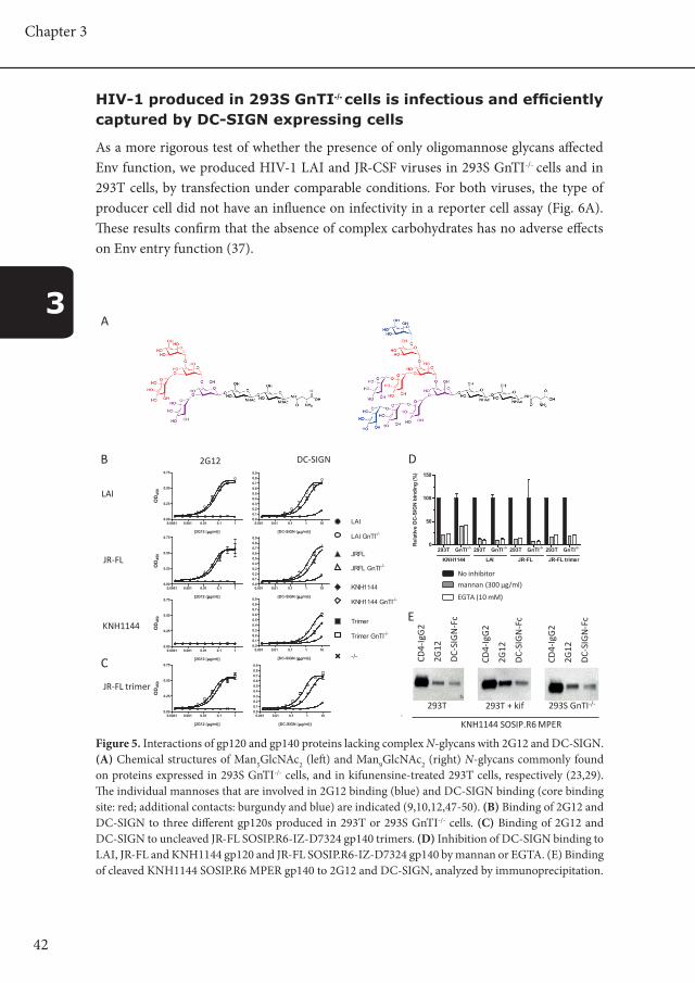
Reactivity of 2G12 and DC-SIGN with glycan-modified Env proteins
MAb 2G12 binds to gp120 via the terminal α1-2-linked mannoses on the outer branches of a cluster of oligomannose carbohydrates (Fig. 5A) (9-13). We therefore studied 2G12 reactivity with gp120 proteins expressed under the various conditions described above. MAb 2G12 bound efficiently to 293T cell-derived LAI and JR-FL gp120s but poorly to KNH1144 gp120, which lacks the glycan at residue 295 that is an important element of the 2G12 epitope (Fig. 5B) (11-13). MAb 2G12 bound equally well to the LAI and JR-FL gp120s produced in 293S GnTI-/- cells (Fig. 5B). Thus, production of gp120 in GnTI-/- cells preserves the 2G12 epitope. It is therefore likely that the untrimmed and/or incompletely trimmed Man6-9GlcNAc2 glycans that retain α1-2-linked mannoses (Fig. 3) include the ones involved in 2G12 binding.
DC-SIGN also binds to gp120 via a cluster of exposed mannose moieties (Fig. 5A) (5,8,35). The specificity of DC-SIGN binding is less well defined than that of 2G12, but there appears to be some overlap between the two binding sites (35,36). DC-SIGN bound strongly to 293T cell-expressed LAI and JR-FL gp120s, but reacted poorly with KNH1144 gp120 (Fig. 6A). All three gp120s from 293S GnTI-/- cells interacted more strongly with DC-SIGN than the same proteins produced in 293T cells (Fig. 5B).
We next compared how 2G12 and DC-SIGN bound to JR-FL gp120 monomers and uncleaved JR-FL gp140 trimers in the D7324-capture ELISA. The reactivity patterns were similar for the two types of Env protein. Thus, as was seen with JR-FL gp120 monomers, 2G12 bound with similar apparent affinity to gp140 trimers produced in 293T and 293S GnTI-/- cells, whereas DC-SIGN reacted better with the trimers from 293S GnTI-/- cells (Fig. 5C). Mannan and EGTA were able to block the binding of DC-SIGN to gp120 and trimeric gp140 proteins from both cell types (Fig. 5D).
We used immunoprecipitation methods to study 2G12 and DC-SIGN binding to cleaved KNH1144 gp140 trimers. CD4 bound similarly to trimers produced in 293T cells (+/- kifunensine) and in 293S GnTI-/- cells, again showing that the trimers were properly folded (Fig. 5E). As was observed using the corresponding gp120 in ELISA, DC-SIGN
41
Lack of complex N-glycans on HIV-1 envelope glycoproteins preserves protein conformation and entry function
3
bound poorly to cleaved KNH1144 gp140 trimers, irrespective of the producer cells (Figs. 5B and E). Even though 2G12 bound only weakly to KNH1144 gp140 (which lacks a relevant glycan at position 295), the binding was moderately increased when the trimers were produced in the presence of kifunensine (Fig. 5B). As previously proposed the explanation may be that additional binding sites for 2G12 are created (30).
Figure 4. Antigenic properties of gp120 and gp140 proteins that lack complex N-glycans. ELISA analysis of the binding of HIVIg, b12, 17b (in the absence of sCD4: gray lines and symbols; in the presence of sCD4: black lines and symbols) and CD4-IgG2 to: (A) LAI, JR-FL or KNH1144 gp120 proteins expressed in 293T cells or in 293S GnTI-/- cells; (B) uncleaved JR-FL SOSIP.R6-IZ-D7324 gp140 expressed in the same two cell types. (C) Immunoprecipitation analysis of cleaved KNH1144 SOSIP.R6 MPER gp140 expressed in 293T cells with furin and with or without kifunensine, or in 293S GnTI-/- cells. The Env proteins were precipitated with CD4-IgG2, b12 or 2F5, and then analyzed by SDS-PAGE and western blot. (D) ELISA analysis of the binding of the V3 MAbs 39F, 19b, and PA1 with JR-FL gp120 expressed in 293T or GnTI-/- cells.
C
CD4-
IgG
2
b12
17b/
sCD
4
2F5
293T 293S GnTI-/-293T + kif
CD4-
IgG
2
b12
17b/
sCD
4
2F5
CD4-
IgG
2
b12
17b/
sCD
4
2F5
B
A
KNH1144 SOSIP.R6 MPER
LAI
JR-FL
KNH1144
JR-FL trimer
CD4-IgG2 b12 HIV-Ig 17b +/- sCD4
0.0001 0.001 0.01 0.1 10.00
0.25
0.50
0.75
[CD4-IgG2 ( µg/ml)]
OD
450
0.0001 0.001 0.01 0.1 10.0
0.1
0.2
0.3
0.4
0.5
0.6
[b12 (µg/ml)]
0.0001 0.001 0.01 0.1 10.0
0.1
0.2
0.3
0.4
0.5
0.6
[HIV-Ig (µg/ml)]
0.0001 0.001 0.01 0.1 10.0
0.2
0.4
0.6
0.8
[17b (µg/ml)]
0.0001 0.001 0.01 0.1 10.00
0.25
0.50
0.75
[CD4-IgG2 ( µg/ml)]
OD
450
0.0001 0.001 0.01 0.1 10.0
0.1
0.2
0.3
0.4
0.5
0.6
[b12 (µg/ml)]
0.0001 0.001 0.01 0.1 10.0
0.1
0.2
0.3
0.4
0.5
0.6
[HIV-Ig (µg/ml)]
0.0001 0.001 0.01 0.1 10.0
0.2
0.4
0.6
0.8
[17b (µg/ml)]
0.0001 0.001 0.01 0.1 10.00
0.25
0.50
0.75
[CD4-IgG2 ( µg/ml)]
OD
450
0.0001 0.001 0.01 0.1 10.0
0.1
0.2
0.3
0.4
0.5
0.6
[b12 (µg/ml)]
0.0001 0.001 0.01 0.1 10.0
0.1
0.2
0.3
0.4
0.5
0.6
[HIV-Ig (µg/ml)]
0.0001 0.001 0.01 0.1 10.0
0.2
0.4
0.6
0.8
[17b (µg/ml)]
0.0001 0.001 0.01 0.1 10.00
0.25
0.50
0.75
[CD4-IgG2 ( µg/ml)]
OD
450
0.0001 0.001 0.01 0.1 10.0
0.1
0.2
0.3
0.4
0.5
0.6
[b12 (µg/ml)]
0.0001 0.001 0.01 0.1 10.0
0.1
0.2
0.3
0.4
0.5
0.6
[HIV-Ig (µg/ml)]
0.0001 0.001 0.01 0.1 10.0
0.2
0.4
0.6
0.8
[17b (µg/ml)]
0.0001 0.001 0.01 0.1 1 100.0
0.2
0.4
0.6
0.8
1.0
[2F5 (µg/ml)]
LAI
LAI GnTI-/-
JRFL
JRFL GnTI-/-
KNH1144
KNH1144 GnTI-/-
Trimer
Trimer GnTI-/-
Mock
2F5
PA1 39F 19b
0.001 0.01 0.1 10.0
0.1
0.2
0.3
0.4
0.5
0.6
[PA-1 (µg/ml)]
OD
450JR-FL
D
0.001 0.01 0.1 10.0
0.1
0.2
0.3
0.4
0.5
0.6
[39F (µg/ml)]0.001 0.01 0.1 1
0.0
0.1
0.2
0.3
0.4
0.5
0.6
[19b (µg/ml)]
JRFL
JRFL GnTI-/-
Mock
Fig 6.4 Antigenic properties of gp120 and gp140 proteins that lack complex N-glycans. ELISA analysis ofthe binding of HIV-Ig, b12, 17b (in the absence of sCD4: grey lines and symbols; in the presence of sCD4:black lines and symbols) and CD4-IgG2 to: (A) LAI, JR-FL or KNH1144 gp120 proteins expressed in 293Tcells or in 293S GnTI-/- cells; (B) uncleaved JR-FL SOSIP.R6-IZ-D7324 gp140 expressed in the same two celltypes. (C) Immunoprecipitation analysis of cleaved KNH1144 SOSIP.R6 MPER gp140 expressed in 293Tcells with furin and with or without kifunensine, or in 293S GnTI-/- cells. The Env proteins wereprecipitated with CD4-IgG2, b12 or 2F5, then analyzed by SDS-PAGE and western blotting. (D) ELISAanalysis of the binding of the V3 MAbs 39F, 19b, and PA1 with JR-FL gp120 expressed in 293T or GnTI-/-
cells.
6-31
42
Chapter 3
3
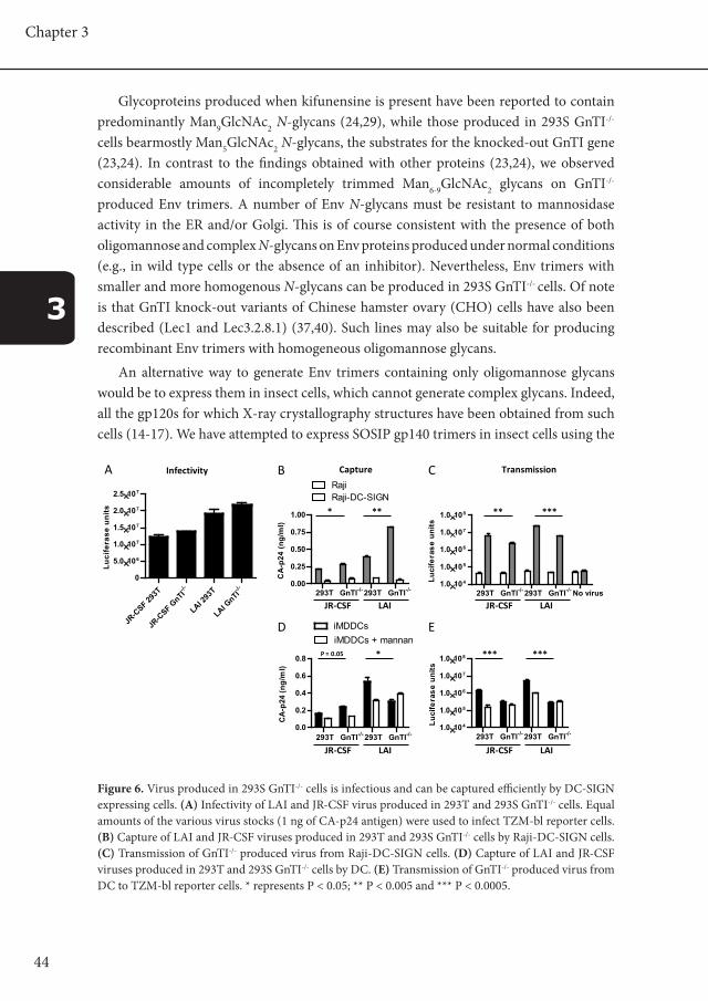
HIV-1 produced in 293S GnTI-/- cells is infectious and efficiently captured by DC-SIGN expressing cells
As a more rigorous test of whether the presence of only oligomannose glycans affected Env function, we produced HIV-1 LAI and JR-CSF viruses in 293S GnTI-/- cells and in 293T cells, by transfection under comparable conditions. For both viruses, the type of producer cell did not have an influence on infectivity in a reporter cell assay (Fig. 6A). These results confirm that the absence of complex carbohydrates has no adverse effects on Env entry function (37).
Figure 5. Interactions of gp120 and gp140 proteins lacking complex N-glycans with 2G12 and DC-SIGN. (A) Chemical structures of Man5GlcNAc2 (left) and Man9GlcNAc2 (right) N-glycans commonly found on proteins expressed in 293S GnTI-/- cells, and in kifunensine-treated 293T cells, respectively (23,29). The individual mannoses that are involved in 2G12 binding (blue) and DC-SIGN binding (core binding site: red; additional contacts: burgundy and blue) are indicated (9,10,12,47-50). (B) Binding of 2G12 and DC-SIGN to three different gp120s produced in 293T or 293S GnTI-/- cells. (C) Binding of 2G12 and DC-SIGN to uncleaved JR-FL SOSIP.R6-IZ-D7324 gp140 trimers. (D) Inhibition of DC-SIGN binding to LAI, JR-FL and KNH1144 gp120 and JR-FL SOSIP.R6-IZ-D7324 gp140 by mannan or EGTA. (E) Binding of cleaved KNH1144 SOSIP.R6 MPER gp140 to 2G12 and DC-SIGN, analyzed by immunoprecipitation.
C
B
LAI
JR-FL
KNH1144
JR-FL trimer
2G12 DC-SIGN
293T GnTI-/- 293T GnTI-/- 293T GnTI-/- 293T GnTI-/-0
50
100
150
KNH1144 LAI JR-FL JR-FL trimer
Rel
ativ
e D
C-S
IGN
bin
ding
(%)
A
E
CD4-
IgG
2
2G12
DC-
SIG
N-F
c
293T
CD4-
IgG
2
2G12
DC-
SIG
N-F
c
CD4-
IgG
2
2G12
DC-
SIG
N-F
c
293T + kif 293S GnTI-/-
D
KNH1144 SOSIP.R6 MPER
0.0001 0.001 0.01 0.1 10.00
0.25
0.50
0.75
[2G12 (µg/ml)]
OD
450
0.001 0.01 0.1 1 100.00.10.20.30.40.50.60.70.80.9
[DC-SIGN (µg/ml)]
0.0001 0.001 0.01 0.1 10.00
0.25
0.50
0.75
[2G12 (µg/ml)]
OD
450
0.001 0.01 0.1 1 100.00.10.20.30.40.50.60.70.80.9
[DC-SIGN (µg/ml)]
0.0001 0.001 0.01 0.1 10.00
0.25
0.50
0.75
[2G12 (µg/ml)]
OD
450
0.001 0.01 0.1 1 100.00.10.20.30.40.50.60.70.80.9
[DC-SIGN (µg/ml)]
0.0001 0.001 0.01 0.1 10.00
0.25
0.50
0.75
[2G12 (µg/ml)]
OD
450
0.001 0.01 0.1 1 100.00.10.20.30.40.50.60.70.80.9
[DC-SIGN (µg/ml)]
LAI
LAI GnTI-/-
JRFL
JRFL GnTI-/-
KNH1144
KNH1144 GnTI-/-
Trimer
Trimer GnTI-/-
-/-
No inhibitor
mannan (300 μg/ml)
EGTA (10 mM)
Fig 6.5 Interactions with 2G12 and DC-SIGN of gp120 and gp140 proteins lacking complex N-glycans. (A)Chemical structures of Man5GlcNAc2 (left) and Man9GlcNAc2 (right) N-glycans commonly found onproteins expressed in 293S GnTI-/- cells, and in kifunensine-treated 293T cells, respectively (17, 44). Theindividual mannoses that are involved in 2G12 binding (blue) and DC-SIGN binding (core binding site:red; additional contacts: burgundy and blue) are indicated (9, 10, 19, 20, 24, 50, 61). The sugar linkagesare also shown. (B) Binding of 2G12 and DC-SIGN to three different gp120s produced in 293T or 293SGnTI-/- cells. (C) Binding of 2G12 and DC-SIGN to uncleaved JR-FL SOSIP.R6-IZ-D7324 gp140 trimers. (D)Inhibition of DC-SIGN binding to LAI, JR-FL and KNH1144 gp120 and JR-FL SOSIP.R6-IZ-D7324 gp140 bymannan or EGTA. (E) Binding of cleaved KNH1144 SOSIP.R6 MPER gp140 to 2G12 and DC-SIGN, analyzedby immunoprecipitation.
6-32

43
Lack of complex N-glycans on HIV-1 envelope glycoproteins preserves protein conformation and entry function
3
Because we observed an increase in DC-SIGN binding to Env proteins derived from 293S GnTI-/- cells, we investigated whether viruses from these cells would be captured more efficiently by DC-SIGN-expressing cells. HIV-1 LAI and JR-FL viruses derived from 293T cells and 293S GnTI-/- cells were therefore incubated with Raji control cells or Raji-DC-SIGN cells, and virus capture was quantified by p24 antigen ELISA. HIV-1 did not bind detectably to Raji cells, whereas Raji-DC-SIGN cells captured viruses very efficiently (Fig. 6B). Moreover, viruses produced in 293S GnTI-/- cells were captured to a greater extent than ones from 293T cells. Immature monocyte-derived dendritic cells (iMDDC) also captured HIV-1 efficiently in a mannan-sensitive manner, confirming the involvement of C-type lectins such as DC-SIGN (Fig. 6D). Again, viruses from 293S GnTI-/- cells were more reactive with iMDDC than their counterparts from 293T cells. One exception was the binding of HIV-1 LAI to iMDDC. Thus, HIV-1 LAI produced in 293S GnTI-/- cells consistently interacted less efficiently with iMDDC compared to the same virus derived from 293T cells (Fig. 6D). It is therefore possible that other receptors than DC-SIGN, possibly ones with affinity for complex glycans, contribute to the binding of HIV-1 LAI, but not HIV-1 JR-CSF, to iMDDC.
We next measured the transmission of DC-SIGN-bound HIV-1 to TZM-bl target cells. Although LAI and JR-CSF viruses from 293S GnTI-/- cells bound more efficiently to Raji-DC-SIGN, they were less able to infect TZM-bl cells in co-culture systems than their counterparts from 293T cells (Figs. 6B and C). A similar trend was observed for capture and transmission of JR-CSF by iMDDCs. LAI virus produced in GnTI-/- cells, however, did not bind more efficiently to iMDDCs, suggesting that other C-type lectin receptors than DC-SIGN play a role in capture of LAI virus by iMDDCs. Nevertheless, the data suggest that the altered glycosylation profile that promotes the binding of virions to cell surface DC-SIGN impairs their subsequent transmission to target cells.
DiscussionReducing the size and heterogeneity of the N-linked glycans that shield the underlying peptide epitopes on the HIV-1 Env glycoproteins could be beneficial for the use of these proteins in structural and immunogenicity studies. Here, we have studied the properties of various monomeric gp120 and trimeric gp140 proteins that were produced in 293S GnTI-/- cells, variants of 293T cells that lack the GnTI enzyme and hence cannot synthesize complex N-glycans (Fig. 1B). As an alternative approach, we expressed the same Env glycoproteins in 293T cells, but in the presence of an inhibitor, kifunensine, that interferes with an earlier stage in the glycan-processing pathway (Fig. 1B). Importantly, the elimination of complex N-glycans did not compromise Env conformation and/or entry function, in agreement with previous studies using the mannosidase inhibitors 1-deoxynojirimycin and swainsonine (38,39).
44
Chapter 3
3
Glycoproteins produced when kifunensine is present have been reported to contain predominantly Man9GlcNAc2 N-glycans (24,29), while those produced in 293S GnTI-/-
cells bearmostly Man5GlcNAc2 N-glycans, the substrates for the knocked-out GnTI gene (23,24). In contrast to the findings obtained with other proteins (23,24), we observed considerable amounts of incompletely trimmed Man6-9GlcNAc2 glycans on GnTI-/-
produced Env trimers. A number of Env N-glycans must be resistant to mannosidase activity in the ER and/or Golgi. This is of course consistent with the presence of both oligomannose and complex N-glycans on Env proteins produced under normal conditions (e.g., in wild type cells or the absence of an inhibitor). Nevertheless, Env trimers with smaller and more homogenous N-glycans can be produced in 293S GnTI-/- cells. Of note is that GnTI knock-out variants of Chinese hamster ovary (CHO) cells have also been described (Lec1 and Lec3.2.8.1) (37,40). Such lines may also be suitable for producing recombinant Env trimers with homogeneous oligomannose glycans.
An alternative way to generate Env trimers containing only oligomannose glycans would be to express them in insect cells, which cannot generate complex glycans. Indeed, all the gp120s for which X-ray crystallography structures have been obtained from such cells (14-17). We have attempted to express SOSIP gp140 trimers in insect cells using the
Figure 6. Virus produced in 293S GnTI-/- cells is infectious and can be captured efficiently by DC-SIGN expressing cells. (A) Infectivity of LAI and JR-CSF virus produced in 293T and 293S GnTI-/- cells. Equal amounts of the various virus stocks (1 ng of CA-p24 antigen) were used to infect TZM-bl reporter cells. (B) Capture of LAI and JR-CSF viruses produced in 293T and 293S GnTI-/- cells by Raji-DC-SIGN cells. (C) Transmission of GnTI-/- produced virus from Raji-DC-SIGN cells. (D) Capture of LAI and JR-CSF viruses produced in 293T and 293S GnTI-/- cells by DC. (E) Transmission of GnTI-/- produced virus from DC to TZM-bl reporter cells. * represents P < 0.05; ** P < 0.005 and *** P < 0.0005.
JR-C
SF 293T -/-
JR-C
SF GnTI
LAI 293
T -/-
LAI GnTI
0
5.0×10⁶
1.0×10⁷
1.5×10⁷
2.0×10⁷
2.5×10⁷
Luci
fera
se u
nits
Infectivity
293T GnTI-/- 293T GnTI-/-0.00
0.25
0.50
0.75
1.00
CA
-p24
(ng/
ml)
293T GnTI-/- 293T GnTI-/- No virus1.0×104
1.0×10⁵
1.0×10⁶
1.0×10⁷
1.0×10⁸
Luci
fera
se u
nits
Capture Transmission
JR-CSF LAI JR-CSF LAI
B C
293T GnTI-/- 293T GnTI-/-1.0×104
1.0×10⁵
1.0×10⁶
1.0×10⁷
1.0×10⁸
Luci
fera
se u
nits
293T GnTI-/- 293T GnTI-/-0.0
0.2
0.4
0.6
0.8
CA
-p24
(ng/
ml)
JR-CSF LAI JR-CSF LAI
D E
A
Raji-DC-SIGNRaji
iMDDCsiMDDCs + mannan
** ***
*** ***
**
P = 0.05 *
*
6-33
Fig 6.6 Virus produced in 293S GnTI-/- cells is infectious and can be captured efficiently by DC-SIGNexpressing cells. (A) Infectivity of LAI and JR-CSF virus produced in 293T and 293S GnTI-/- cells. Equalamounts of the various virus stocks (1 ng of CA-p24 antigen) were used to infect TZM-bl reporter cells.(B) Capture of LAI and JR-CSF viruses produced in 293T and 293S GnTI-/- cells by Raji-DC-SIGN cells. (C)Transmission of GnTI-/- produced virus from Raji-DC-SIGN cells. (D) Capture of LAI and JR-CSF virusesproduced in 293T and 293S GnTI-/- cells by DC. (E) Transmission of GnTI-/- produced virus from DC toTZM-bl reporter cells. * represents P < 0,05; ** P < 0,005 and *** P < 0,0005.
45
Lack of complex N-glycans on HIV-1 envelope glycoproteins preserves protein conformation and entry function
3
baculovirus system, but gp120-gp41 cleavage is very inefficient even when human furin is co-delivered (RWS, Giel Bosman, Wim de Grip, unpublished results).
Both gp120 monomers and gp140 trimers from three different HIV-1 strains (JR-FL, LAI and KNH1144) were expressed efficiently in 293S GnTI-/- cells, and in kifunensine-treated 293T cells; under both circumstances, trimerization and cleavage of the gp140s, when appropriate, occurred normally. The glycan-modified Env proteins all interacted efficiently with HIVIg, CD4-IgG2 and various MAbs to conformational epitopes on gp120 and gp41. Hence the absence of complex glycans did not detectably affect the correct folding of these Env glycoproteins. Moreover, viruses produced in GnTI-/- cells were infectious, confirming that Env entry function was not affected.
Kifunensine did not affect the size of Env proteins, in contrast to observations that others have made about other proteins, where kifunensine reduces both the size and heterogeneity of glycans (30). Hence, the average size of the complex carbohydrates on Env is no larger than the Man9GlcNAc2 structure, which is consistent with the observation that relatively simple diantennary complex glycans predominate on gp120 (MW, DE, RWS and CHH, unpublished observations and (41-43). In contrast, Env proteins from 293S GnTI-/- cells were slightly smaller, which is consistent with mannosidase trimming of most of the Man9GlcNAc2 structures. The complete trimming of ~ 24 glycans to Man5GlcNAc2 would reduce the gp120 m.wt. by ~ 17 kDa (i.e., 4 mannose residues of ~ 0.18 kDa removed from ~ 24 glycans). The observed size difference (~ 10 kDa; Fig. 2) therefore already implied that mannose trimming was incomplete. A more direct analysis then confirmed that a large proportion of Env glycans was indeed of the Man6-9GlcNAc2 forms (Fig. 3). Thus, expressing Env in 293S GnTI-/- cells effectively results in the conversion of complex carbohydrates to Man5GlcNAc2 moieties, but does not change the composition of any oligomannose carbohydrates that are resistant to mannose trimming; those glycans remain as Man6-9GlcNAc2 structures.
We analyzed the interaction of Env proteins with two carbohydrate-binding proteins that bind to oligomannose glycans: MAb 2G12 and the C-type lectin receptor DC-SIGN. These ligands are known to have different but overlapping specificities and requirements for oligomannose glycans. The 2G12 MAb binds to a cluster of oligomannose carbohydrates containing α1-2-linked terminal mannoses (9-12) (Fig. 5A). Env proteins from 293S GnTI-/- cells interacted efficiently with 2G12, which suggests that carbohydrates involved in 2G12 binding (e.g. those attached to residues 295, 332 and 392, and possibly 386 and 448) are resistant to mannosidase activity in the ER and/or Golgi, and hence retain their α1-2-linked terminal mannoses.
DC-SIGN bound strongly to 293T cell-expressed LAI and JR-FL gp120s, but not to KNH1144 gp120. This observation is consistent with our finding that KNH1144 gp120 could not induce IL-10 from DC via an interaction with mannose C-type lectin receptors, whereas LAI and JR-FL gp120s could do so (8). The limited binding of DC-SIGN to
46
Chapter 3
3
KNH1144 gp120 may, as for 2G12, be attributable to the absence of the glycan at residue 295 that is involved in the binding of both ligands (11,12,35,44). Alternatively, or as well, the carbohydrate at position 160, which is present on gp120s from LAI and JR-FL but not KNH1144, may be responsible, as this site has also been implicated in DC-SIGN binding (45,46).
We found that Env proteins and viruses from 293S GnTI-/- cells consistently bound DC-SIGN more strongly than their counterparts from 293T cells. DC-SIGN interacts with the outer tri-mannose core on Man5-9GlcNAc2 sugars, but α1-2-linked mannose moieties that are absent from Man5GlcNAc2 structures provide additional contacts (47-50). We suggest that the natural DC-SIGN binding site(s) on oligomannose Man6-
9GlcNAc2 sugars is unaltered on gp120 moieties produced in 293S GnTI-/- cells, but that the replacement of complex glycans by Man5GlcNAc2 carbohydrates creates additional binding sites for DC-SIGN. These extra sites would probably be of lower affinity because they lack α1-2-linked mannoses.
Of note is that enhancing HIV-1 binding to DC-SIGN actually decreased its subsequent transmission from DCs to target cells (Fig. 6). An explanation is that too strong an interaction with DC-SIGN may prevent the virus from dissociating at a time and place necessary for the captured virion to reroute to virological synapses that form upon encounter of a DC with a target T cell, instead of being targeted for degradation. Thus, there may be a delicate balance between the requirements for association of virus with DC-SIGN and subsequent steps in the transmission process.
In summary, we have produced well-folded gp120 monomers and both uncleaved and cleaved gp140 trimers bearing glycan structures that are smaller and more homogeneous than is normally the case. These glycan-modified proteins may be useful for immunogenicity and structural studies.
Materials and methodsPlasmids
The pPPI4 vector system for expressing gp120 and gp140 proteins has been described elsewhere (18). Single amino acid substitutions were made using the Quickchange mutagenesis kit (Stratagene, La Jolla, CA). The various Env proteins described below are all summarized in Fig. 1A.
We modified the C5 region of KNH1144 gp120 to introduce the epitope for the polyclonal sheep anti-gp120 antibody (Ab) D7324 and thereby facilitate analysis of ligand binding to gp120 in a D7324-capture ELISA; the KNH1144 sequence APTRARRRVVGREKR was altered to APTKAKRRVVQREKR. We modified the JR-FL SOSIP.R6 gp140 construct to include a C-terminal GCN4-based trimerization domain
47
Lack of complex N-glycans on HIV-1 envelope glycoproteins preserves protein conformation and entry function
3
(isoleucine zipper; IZ) and the above D7324 epitope tag, linked by flexible spacers, to create the JR-FL SOSIP.R6-IZ-D7324 gp140 protein. Note that the internal D7324 epitope in the C5 domain of SOSIP constructs is mutated by the introduction of the cysteine at residue 501 that forms the intramolecular disulfide bond with gp41, and it is also occluded by the proximity of the gp41 subunit; hence unmodified SOSIP gp140s do not react efficiently with Ab D7324 (18,19). Because of the addition of C-terminal sequences to SOSIP gp140, SOSIP.R6-IZ-D7324 trimers are uncleaved. A more detailed description of the SOSIP.R6-IZ-tag and related constructs will be published elsewhere.
The KNH1144 SOSIP.R6 MPER gp140 construct containing engineered epitopes for MAbs 2F5 and 4E10 has been described previously (20). Note that the gp140 used here does not contain the S295N change that enhances binding by 2G12 (20).
Reagents
DC-SIGN-Fc was purchased from R&D Systems (Minneapolis, MN). HIVIg was obtained through the AIDS Research and Reference Reagent Program (ARRRP), Division of AIDS, NIAID, NIH. MAbs 2G12 and 2F5 were obtained from Hermann Katinger through the ARRRP. MAb b12 was donated by Dennis Burton (The Scripps Research Institute, La Jolla, CA). MAbs 17b and 39F were gifts from James Robinson (Tulane University, New Orleans, LA). The CD4-IgG2 and sCD4 proteins have been described elsewhere (51).
D7324 monomer and trimer ELISA
Enzyme-linked immunosorbent assays (ELISA) were performed as described previously (8), with minor modifications. Microlon 96-well plates (Greiner Bio-One, Alphen aan den Rijn, The Netherlands) were coated overnight with Ab D7324 (Aalto Bioreagents, Dublin, Ireland) at 10 µg/ml in 0.1 M NaHCO3, pH 8.6 (100 µl/well). Subsequently, gp120 or SOSIP.R6-IZ-D7324 gp140 (diluted in TBS/10% fetal bovine serum (FBS)) was captured by the coated D7324 for 2 h before addition of TSM/5% BSA to block protein-binding sites on the well surface (TSM; 20 mM Tris, 150 mM NaCl, 1.0 mM CaCl2, 2.0 mM MgCl2). Serially diluted HIVIg, MAbs, DC-SIGN-Fc or CD4-IgG2 in TSM/5% BSA was then added for 2 h followed by 3 washes with TSM/0.05% Tween-20. Horseradish peroxidase (HRP)-labeled goat-anti-human immunoglobulin G (IgG) (Jackson Immunoresearch, Suffolk, England) was added for 30 min at a 1:5000 dilution (final concentration 0.2 µg/ml) in TSM/5% BSA, followed by 5 washes with TSM/0.05% Tween-20. DC-SIGN blocking experiments were performed using 1 µg/ml DC-SIGN in the presence of 300 µg/ml mannan or 10 mM EGTA. Colorimetric detection was performed using a solution containing 1% 3,3’,5,5’-tetramethylbenzidine (TMB, Sigma-Aldrich, Zwijndrecht, The Netherlands), 0.01% H2O2 in 0.1 M sodium acetate, and 0.1 M citric acid. The colorimetric reaction was stopped using 0.8 M H2SO4 when appropriate, and absorption was measured at 450 nm.
48
Chapter 3
3
SDS-PAGE, Blue Native PAGE and western blotting
SDS-polyacrylamide gel electrophoresis (SDS-PAGE) and western blotting were performed according to established protocols using the anti-gp120 V3 loop MAb PA-1 (1:5,000 dilution to a final concentration of 0.2 µg/ml) (32) and HRP-labeled goat-anti-mouse IgG (1:5000 dilution). Luminometric detection of envelope glycoproteins was performed using the Western Lightning ECL system (PerkinElmer, Groningen, The Netherlands). Blue Native (BN)-PAGE was carried out using minor modifications to the published method (52-54). Thus, purified protein samples or cell culture supernatants were diluted with an equal volume of a buffer containing 100 mM 4-(N-morpholino) propane sulfonic acid (MOPS), 100 mM Tris-HCl, pH 7.7, 40% glycerol, and 0.1% Coomassie blue, immediately prior to loading onto a 4 to 12% Bis-Tris NuPAGE gel (Invitrogen). Typically, gel electrophoresis was performed for 2 h at 150 V (~ 0.07A) using 50 mM MOPS, 50 mM Tris, pH 7.7 as running buffer.
Immunoprecipitation assays
Twenty-fold concentrated supernatants from transiently Env-transfected 293T or 293S GnTI-/- cells were incubated overnight at 4 °C with MAbs or CD4-IgG2 in a 500 µl volume that contained 100 µl of 5-fold concentrated RIPA buffer (250 mM Tris-HCl, pH 7.0 containing 750 mM NaCl and 5 mM EDTA). Next, 50 µl of protein G-coated agarose beads (Pierce Inc./Thermo Fisher Scientific, Etten-Leur, The Netherlands) was added, with rotation-mixing, for 2 h at 4 °C. The beads were then washed extensively with ice-cold RIPA buffer containing 0.01% Tween-20. Proteins were eluted by heating the beads at 100 °C for 5 min in 50 µl of SDS-PAGE loading buffer supplemented with 100 mM dithiothreitol (DTT). The immunoprecipitates were analyzed on 8% SDS-PAGE gels (125 V for 2 h).
Analytical size exclusion chromatography
Twenty-fold concentrated, Env-containing supernatants (200 µl) from 293T or 293S GnT-/- cell cultures were fractionated using a Superose-6 10/300 GL column (GE Healthcare, Diegem, The Netherlands), equilibrated with phosphate buffered saline, using an Äkta FPLC system (GE Healthcare, Diegem, The Netherlands). Fractions were analyzed by SDS-PAGE and western blot.
Carbohydrate analysis
Purified KNH1144 gp140 trimers (620 ng in 10 µl) produced in 293S GnTI-/- cells were denatured by addition of 20 µl of 2% SDS and heating at 60 °C for 10 min. Subsequently, 10 µl of 4% NP-40 and 10 µl of PNGase F (0.5 mU) in 5× PBS were added to the samples (final: 0.1 mU PNGase F in 0.8% NP-40, 0.4% SDS, 1× PBS), which were then incubated overnight at 37 °C to allow N-glycan release. The released N-glycans were subsequently
49
Lack of complex N-glycans on HIV-1 envelope glycoproteins preserves protein conformation and entry function
3
labeled with the fluorophore 2-aminobenzoic acid (2-AA), and the 2-AA labeled glycans were purified by solid-phase extraction as described elsewhere (55).
An aliquot of 2-AA labeled N-glycans was fractionated by hydrophilic interaction liquid chromatography (HILIC HPLC), using an Ultimate LC system (Dionex, Sunnyvale, CA) controlled by Chromeleon software and equipped with a 2.0 mm × 150 mm TSK gel-Amide 80 column (3 µm; Tosoh Biosciences, Stuttgart, Germany). The 2-AA labeled N-glycans were separated using a linear gradient from 80% acetonitrile (0 min) to 53% acetonitrile (40 min) in 50 mM ammonium formate (pH 4.4) at a flow rate of 0.3 ml/min. To identify the 2-AA labeled N-glycans, a fluorescence detector (FP-2020 plus; Jasco, Easton, MD) was used, set to a 360 nm excitation wavelength and a 420 nm emission wavelength. Peak fractions were collected, dried by vacuum centrifugation, and dissolved in 50 µl of water.
MALDI-TOF-MS was carried out on an Ultraflex II MALDI-TOF/TOF mass spectrometer (Bruker Daltonics), equipped with a Smartbeam laser. A 2 µl aliquot of 2,5-dihydroxybenzoic acid (DHB) matrix (20 mg/ml in ACN:water (30:70 v/v)) and 1 µl of the test sample were applied to a polished steel target plate and left to dry. The Ultraflex was operated in reflector-negative mode, and calibration was performed on a peptide calibration standard (Bruker Daltonics). Mass spectra from 500 to 2000 laser shots were accumulated per sample.
Cell culture and transient transfection
The TZM-bl reporter cell line stably expresses high levels of CD4 and the CCR5 and CXCR4 coreceptors, and contains the luciferase and β-galactosidase genes under the control of the HIV-1 LTR promoter (56). These cells were obtained through the ARRRP (donated by Dr. John C. Kappes, Dr. Xiaoyun Wu and Tranzyme Inc., Durham, NC). One day prior to infection, the cells were plated on a 96-well plate in DMEM containing 10% FBS, MEM nonessential amino acids and penicillin/streptomycin (both at 100 U/ml).
The 293T and 293S GnTI-/- cells were transiently transfected with env genes using linear polyethylenimine (PEI, MW 25,000) as described before (57). Briefly, DNA encoding Env protein was diluted in Dulbecco’s Modified Eagle’s Medium (DMEM; Invitrogen, Breda, The Netherlands), to 1/10 of the final culture volume and mixed with PEI (0.15 mg/ml final concentration). After incubation for 20 min, the DNA-PEI mix was added to the cells for 4 h before replacement with normal culture medium containing 10% FBS (HyClone, Perbio, Etten-Leur, The Netherlands), penicillin, streptomycin and MEM nonessential amino acids (0.1 mM, Invitrogen, Breda, The Netherlands). Supernatants were harvested 48 h after transfection. When appropriate, 100 µM kifunensine was added 1 h before transfection, and the culture medium was replaced 4 h post transfection with medium containing 100 µM kifunensine.
50
Chapter 3
3
The Raji control cell line and Raji cells expressing DC-SIGN (Raji-DC-SIGN) were cultured in RPMI 1640 medium containing 10% FBS. DC-SIGN expression by Raji-DC-SIGN cells was positively selected for using neomycin (2 µg/ml), and routinely monitored by FACS analysis with a PE-labeled DC-SIGN MAb. Human immature monocyte-derived dendritic cells (iMDDCs) were prepared as previously described (58-60) Briefly, peripheral blood mononuclear cells (PBMCs) were isolated from buffy coats (Central Laboratory Blood Bank, Amsterdam) by use of a Ficoll gradient and positive CD14 selection using a MACS system (Miltenyi Biotec GmbH, Bergisch Gladbach, Germany). Purified monocytes were differentiated into iMDDCs by adding 45 ng/ml of interleukin-4 (IL-4) and 500 U/ml of granulocyte-macrophage colony-stimulating factor on days 1 and 4 (Schering-Plough, Brussels, Belgium). The resulting iMMDCs were used on day 6. The phenotype of iMDDCs was confirmed by staining for MHC class II, CD1a, CD14, CD83 and CD86, followed by flow cytometry. Low levels of CD83, CD86 and MHC class II proteins were present on the cell surface, indicative of an immature phenotype.
Virus infectivity
HIV-1 stocks were prepared by transfecting either 293T or 293S GnTI-/- cells as for recombinant protein production (see above). A fixed amount of virus (equivalent to 1.0 ng p24 antigen) was added to TZM-bl cells (at 70-80% confluency in a 96-well plate) in the presence of 400 nM saquinavir (Roche, Basel, Switzerland) to prevent multiple rounds of infection, and 40 µg/ml DEAE-dextran (Sigma) to enhance infection, in a total volume of 200 µl. The medium was removed 2 days post-infection and the cells were washed once with PBS before lysis with Reporter Lysis Buffer (Promega, Madison, WI). Luciferase activity was measured using the Luciferase Assay kit (Promega) and a Glomax luminometer, according to the manufacturer’s instructions (Turner BioSystems, Sunnyvale, CA). All infections were performed in duplicate and luciferase measurements were also performed in duplicate. Background luciferase activity was determined using uninfected cells and used as a correction factor.
Virus capture and transmission
Either 1.0 × 10⁶ Raji or Raji-DC-SIGN cells, or 3.0 × 10⁵ iMDDCs treated with or without mannan (30 µg/ml) for 30 min, were incubated for 2 h with HIV-1 (15 ng/ml of p24 antigen) that had been produced in either 293T or 293S GnTI-/- cells. The cells were washed 3 times with PBS to remove unbound virus, and then lysed in 1% empigen detergent for 1 h at 56 °C. Cell-debris was removed by centrifugation and the p24 capsid antigen content of the supernatant was determined by ELISA. For transmission experiments, 1.0 × 10⁵ Raji or Raji-DC-SIGN cells, or iMDDCs (with or without mannan treatment), were incubated for 2 h with HIV-1 (3 ng/ml of p24 antigen) derived from either 293T or 293S GnTI-/- cells. Unbound virus was removed by washing the cells three times with RPMI
51
Lack of complex N-glycans on HIV-1 envelope glycoproteins preserves protein conformation and entry function
3
1640 medium supplemented with 10% heat-inactivated FBS. The cells were co-cultured with TZM-bl reporter cells for 48 h in presence of 400 nM saquinavir. HIV-1 transfer to the reporter cells was quantified by measuring luciferase activity.
Statistical analysis
All statistical comparisons were performed using the unpaired t test (two tailed) and indicated in figures with asterisks: * P < 0.05; ** P < 0.005; *** P < 0.0005.
AcknowledgmentsWe are grateful to Ilja Bontjer and Carolien A. M. Koeleman for technical assistance.
We thank James Robinson, Philip Reeves and Dennis Burton for reagents, and James Binley for critical reading of the manuscript. This research was supported by AIDS fund (Amsterdam) grants #2005021 (BB), #2008013 (RWS) and #2009012 (RWS), by NIH grants and contracts AI45463 and AI36082 (JPM) and AI082362 (WO), and by the International AIDS Vaccine Initiative (JPM). RWS is a recipient of an Anton Meelmeijer fellowship, a VENI fellowship from the Netherlands Organization for Scientific Research (NWO), and a Mathilde Krim research fellowship from the American Foundation for AIDS Research (amfAR).
References 1. Walker, B. D., Kowalski, M., Goh, W. C., Kozarsky, K., Krieger, M., Rosen, C., Rohrschneider,
L., Haseltine, W. A., and Sodroski, J. (1987) Proceedings of the National Academy of Sciences of the United States of America 84, 8120-8124
2. de Witte, L., Nabatov, A., Pion, M., Fluitsma, D., de Jong, M. A. W. P., de Gruijl, T., Piguet, V., van Kooyk, Y., and Geijtenbeek, T. B. H. (2007) Nat Med 13, 367-371
3. Geijtenbeek, T. B. H., Kwon, D. S., Torensma, R., Van Vliet, S. J., Van Duijnhoven, G. C. F., Middel, J., Cornelissen, I. L. M. H., Nottet, H. S. L. M., KewalRamani, V. N., Littman, D. R., Figdor, C. G., and van Kooyk, Y. (2000) Cell 100, 587-597
4. Marzi, A., Mitchell, D. A., Chaipan, C., Fisch, T., Doms, R. W., Carrington, M., Desrosiers, R. C., and P÷hlmann, S. (2007) Virol. 368, 322-330
5. Lin, G., Simmons, G., Pohlmann, S., Baribaud, F., Ni, H., Leslie, G. J., Haggarty, B. S., Bates, P., Weissman, D., Hoxie, J. A., and Doms, R. W. (2003) J. Virol. 77, 1337-1346
6. Reitter, J. N., Means, R. E., and Desrosiers, R. C. (1998) Nat. Med. 4, 679-684 7. Banerjee, K., Andjelic, S., Klasse, P. J., Kang, Y., Sanders, R. W., Michael, E., Durso, R. J.,
Ketas, T. J., Olson, W. C., and Moore, J. P. (2009) Virol. 389, 108-121 8. Shan, M., Klasse, P. J., Banerjee, K., Dey, A. K., Iyer, S. P., Dionisio, R., Charles, D., Campbell-
Gardener, L., Olson, W. C., Sanders, R. W., and Moore, J. P. (2007) PLoS. Pathog. 3, e169 9. Calarese, D. A., Scanlan, C. N., Zwick, M. B., Deechongkit, S., Mimura, Y., Kunert, R., Zhu,
P., Wormald, M. R., Stanfield, R. L., Roux, K. H., Kelly, J. W., Rudd, P. M., Dwek, R. A., Katinger, H., Burton, D. R., and Wilson, I. A. (2003) Science 300, 2065-2071
10. Calarese, D. A., Lee, H. K., Huang, C. Y., Best, M. D., Astronomo, R. D., Stanfield, R. L., Katinger, H., Burton, D. R., Wong, C. H., and Wilson, I. A. (2005) Proceedings of the National Academy of Sciences of the United States of America 102, 13372-13377
11. Sanders, R. W., Venturi, M., Schiffner, L., Kalyanaraman, R., Katinger, H., Lloyd, K. O., Kwong, P. D., and Moore, J. P. (2002) J. Virol. 76, 7293-7305
12. Scanlan, C. N., Pantophlet, R., Wormald, M. R., Ollmann, S. E., Stanfield, R., Wilson, I. A.,
52
Chapter 3
3
Katinger, H., Dwek, R. A., Rudd, P. M., and Burton, D. R. (2002) J Virol 76, 7306-7321 13. Trkola, A., Pomales, A. B., Yuan, H., Korber, B., Maddon, P. J., Allaway, G. P., Katinger, H.,
Barbas, C. F., III, Burton, D. R., and Ho, D. D. (1995) J. Virol. 69, 6609-6617 14. Huang, C. c., Lam, S. N., Acharya, P., Tang, M., Xiang, S. H., Hussan, S. S., Stanfield, R. L.,
Robinson, J., Sodroski, J., Wilson, I. A., Wyatt, R., Bewley, C. A., and Kwong, P. D. (2007) Science 317, 1930-1934
15. Huang, C. C., Tang, M., Zhang, M. Y., Majeed, S., Montabana, E., Stanfield, R. L., Dimitrov, D. S., Korber, B., Sodroski, J., Wilson, I. A., Wyatt, R., and Kwong, P. D. (2005) Science 310, 1025-1028
16. Kwong, P. D., Wyatt, R., Robinson, J., Sweet, R. W., Sodroski, J., and Hendrickson, W. A. (1998) Nature 393, 648-659
17. Zhou, T., Xu, L., Dey, B., Hessell, A. J., Van Ryk, D., Xiang, S. H., Yang, X., Zhang, M. Y., Zwick, M. B., Arthos, J., Burton, D. R., Dimitrov, D. S., Sodroski, J., Wyatt, R., Nabel, G. J., and Kwong, P. D. (2007) Nature 445, 732-737
18. Binley, J. M., Sanders, R. W., Clas, B., Schuelke, N., Master, A., Guo, Y., Kajumo, F., Anselma, D. J., Maddon, P. J., Olson, W. C., and Moore, J. P. (2000) J. Virol. 74, 627-643
19. Sanders, R. W., Vesanen, M., Schuelke, N., Master, A., Schiffner, L., Kalyanaraman, R., Paluch, M., Berkhout, B., Maddon, P. J., Olson, W. C., Lu, M., and Moore, J. P. (2002) J. Virol. 76, 8875-8889
20. Dey, A. K., David, K. B., Lu, M., and Moore, J. P. (2009) Virol. 385, 275-281 21. Pancera, M., Lebowitz, J., Schon, A., Zhu, P., Freire, E., Kwong, P. D., Roux, K. H., Sodroski,
J., and Wyatt, R. (2005) J Virol 79, 9954-9969 22. Si, Z., Phan, N., Kiprilov, E., and Sodroski, J. (2003) AIDS Res. Hum. Retroviruses 19, 217-226 23. Reeves, P. J., Callewaert, N., Contreras, R., and Khorana, H. G. (2002) Proceedings of the
National Academy of Sciences of the United States of America 99, 13419-13424 24. Chang, V. T., Crispin, M., Aricescu, A. R., Harvey, D. J., Nettleship, J. E., Fennelly, J. A., Yu,
C., Boles, K. S., Evans, E. J., Stuart, D. I., Dwek, R. A., Jones, E. Y., Owens, R. J., and Davis, S. J. (2007) Structure 15, 267-273
25. Harbury, P. B., Kim, P. S., and Alber, T. (1994) Nature 371, 80-83 26. Yang, X., Farzan, M., Wyatt, R., and Sodroski, J. (2000) J. Virol. 74, 5716-5725 27. Yang, X., Florin, L., Farzan, M., Kolchinsky, P., Kwong, P. D., Sodroski, J., and Wyatt, R.
(2000) J. Virol. 74, 4746-4754 28. Beddows, S., Kirschner, M., Campbell-Gardener, L., Franti, M., Dey, A. K., Iyer, S. P., Maddon,
P. J., Paluch, M., Master, A., Overbaugh, J., VanCott, T., Olson, W. C., and Moore, J. P. (2006) AIDS Res Hum Retroviruses 22, 569-579
29. Elbein, A. D., Tropea, J. E., Mitchell, M., and Kaushal, G. P. (1990) J. Biol. Chem. 265, 15599-15605
30. Scanlan, C. N., Ritchie, G. E., Baruah, K., Crispin, M., Harvey, D. J., Singer, B. B., Lucka, L., Wormald, M. R., Wentworth Jr, P., Zitzmann, N., Rudd, P. M., Burton, D. R., and Dwek, R. A. (2007) J. Mol. Biol. 372, 16-22
31. Pantophlet, R., Ollmann, S. E., Poignard, P., Parren, P. W., Wilson, I. A., and Burton, D. R. (2003) J Virol 77, 642-658
32. Trkola, A., Dragic, T., Arthos, J., Binley, J. M., Olson, W. C., Allaway, G. P., Cheng-Mayer, C., Robinson, J., Maddon, P. J., and Moore, J. P. (1996) Nature 384, 184-187
33. Pantophlet, R., Wrin, T., Cavacini, L. A., Robinson, J. E., and Burton, D. R. (2008) Virol. 381, 251-260
34. Papandreou, M. J. and Fenouillet, E. (1998) Virol. 241, 163-167 35. Hong, P. W. P., Nguyen, S., Young, S., Su, S. V., and Lee, B. (2007) J. Virol. 81, 8325-8336 36. Binley, J., Ngo-Abdalla, S., Moore, P., Bobardt, M., Chatterji, U., Gallay, P., Burton, D., Wilson,
I., Elder, J., and de Parseval, A. (2006) Retrovirology 3, 39 37. Fenouillet, E., Miquelis, R., and Drillien, R. (1996) Virol. 218, 224-231 38. Montefiori, D. C., Robinson, W. E., and Mitchell, W. M. (1988) Proceedings of the National
Academy of Sciences of the United States of America 85, 9248-9252 39. Pal, R., Hoke, G. M., and Sarngadharan, M. G. (1989) Proceedings of the National Academy of
Sciences of the United States of America 86, 3384-3388

53
Lack of complex N-glycans on HIV-1 envelope glycoproteins preserves protein conformation and entry function
3
40. Stanley, P. (1993) Use of mammalian cell mutants to study the functions of N- and O-linked glycosylation. Cell Surface and Extracellular Role of N-linked Glycoconjugates, Academic Press, San Diego,
41. Mizuochi, T., Spellman, M. W., Larkin, M., Solomon, J., Basa, L. J., and Feizi, T. (1988) Biochem J 254, 599-603
42. Cutalo, J. M., Deterding, L. J., and Tomer, K. B. (2004) Journal of the American Society for Mass Spectrometry 15, 1545-1555
43. Zhu, X., Borchers, C., Bienstock, R. J., and Tomer, K. B. (2000) Biochem. 39, 11194-11204 44. Trkola, A., Purtscher, M., Muster, T., Ballaun, C., Buchacher, A., Sullivan, N., Srinivasan, K.,
Sodroski, J., Moore, J. P., and Katinger, H. (1996) J. Virol. 70, 1100-1108 45. Borggren, M., Repits, J., Kuylenstierna, C., Sterjovski, J., Churchill, M., Purcell, D., Karlsson,
A., Albert, J., Gorry, P., and Jansson, M. (2008) Retrovirology 5, 28 46. Lue, J., Hsu, M., Yang, D., Marx, P., Chen, Z., and Cheng-Mayer, C. (2002) J. Virol. 76, 10299-
10306 47. Feinberg, H., Mitchell, D. A., Drickamer, K., and Weis, W. I. (2001) Science 294, 2163-2166 48. Feinberg, H., Castelli, R., Drickamer, K., Seeberger, P. H., and Weis, W. I. (2007) J. Biol. Chem.
282, 4202-4209 49. Guo, Y., Feinberg, H., Conroy, E., Mitchell, D. A., Alvarez, R., Blixt, O., Taylor, M. E., Weis,
W. I., and Drickamer, K. (2004) Nat Struct Mol Biol 11, 591-598 50. van Liempt, E., Bank, C. M. C., Mehta, P., GarcI¦a-Vallejo, J. J. s., Kawar, Z. S., Geyer, R.,
Alvarez, R. A., Cummings, R. D., Kooyk, Y. v., and van Die, I. (2006) FEBS Letters 580, 6123-6131
51. Allaway, G. P., Davis-Bruno, K. L., Beaudry, G. A., Garcia, E. B., Wong, E. L., Ryder, A. M., Hasel, K. W., Gauduin, M. C., Koup, R. A., McDougal, J. S., and . (1995) AIDS Research and Human Retroviruses 11, 533-539
52. Schagger, H., Cramer, W. A., and von, J. G. (1994) Anal. Biochem. 217, 220-230 53. Schagger, H. and von, J. G. (1991) Anal. Biochem. 199, 223-231 54. Schulke, N., Vesanen, M. S., Sanders, R. W., Zhu, P., Lu, M., Anselma, D. J., Villa, A. R.,
Parren, P. W., Binley, J. M., Roux, K. H., Maddon, P. J., Moore, J. P., and Olson, W. C. (2002) J. Virol. 76, 7760-7776
55. Ruhaak, L. R., Huhn, C., Waterreus, W. J., de Boer, A. R., Neusu¦êss, C., Hokke, C. H., Deelder, A. ü. M., and Wuhrer, M. (2008) Analytical Chemistry 80, 6119-6126
56. Wei, X., Decker, J. M., Liu, H., Zhang, Z., Arani, R. B., Kilby, J. M., Saag, M. S., Wu, X., Shaw, G. M., and Kappes, J. C. (2002) Antimicrob. Agents Chemother. 46, 1896-1905
57. Kirschner, M., Monrose, V., Paluch, M., Techodamrongsin, N., Rethwilm, A., and Moore, J. P. (2006) Protein Expr. Purif. 48, 61-68
58. Sallusto, F. and Lanzavecchia, A. (1994) The Journal of Experimental Medicine 179, 1109-1118
59. Van Montfort, T., Nabatov, A. A., Geijtenbeek, T. B., Pollakis, G., and Paxton, W. A. (2007) J. Immunol. 178, 3177-3185
60. Van Montfort, T., Thomas, A. A., Pollakis, G., and Paxton, W. A. (2008) J. Virol. 82, 7886-7896
61. Binley, J. M., Sanders, R. W., Master, A., Cayanan, C. S., Wiley, C. L., Schiffner, L., Travis, B., Kuhmann, S., Burton, D. R., Hu, S.-L., Olson, W. C., and Moore, J. P. (2002) J. Virol. 76, 2606-2616
62. Leonard, C. K., Spellman, M. W., Riddle, L., Harris, R. J., Thomas, J. N., and Gregory, T. J. (1990) J. Biol. Chem. 265, 10373-10382
Related Documents
![Membrane-bound mucins and mucin terminal glycans ... · associated with higher morbidity and mortality[1-7]. Mucins, heavily glycosylated high-molecular-weight glycoproteins, are](https://static.cupdf.com/doc/110x72/5fcbfea3277df0670a5fee63/membrane-bound-mucins-and-mucin-terminal-glycans-associated-with-higher-morbidity.jpg)