Laboratory diagnostics Martina Vachová Department of Immunology and Allergology Faculty of Medicine and Faculty Hospital in Pilsen

Laboratory diagnostics Martina Vachová Department of Immunology and Allergology Faculty of Medicine and Faculty Hospital in Pilsen.
Dec 30, 2015
Welcome message from author
This document is posted to help you gain knowledge. Please leave a comment to let me know what you think about it! Share it to your friends and learn new things together.
Transcript
Laboratory diagnostics
Martina Vachová
Department of Immunology and Allergology
Faculty of Medicine and Faculty Hospital in Pilsen

Topics:
Laboratory methods of assessment of humoral immunity. Radioimmunoassay, enzyme immunoassay Laboratory measurement of specific IgE antibodies. Laboratory measurement of autoantibodies. Laboratory methods of assessment of cellular immunity. Flow cytometry - principles, practical use.

Laboratory methods of assessment of humoral immunity
all methods use an antigen (Ag) – antibody(Ab) reaction as their primary means of detection
we assess either Ag or Ab

Laboratory methods of assessment of humoral immunity
• Precipitation – immunodiffussion
immunoelectrophoresis
turbidimetry
nephelometry
• Agglutination
• Radioimmunoassay, enzyme immunoassay
• Imunofluorescence

Immunodiffusion
Diagnostic test which involves diffusion of Ag or Ab through
a substance such as agar
Two commonly known forms are:
- single radial immunodiffusion
- Ouchterlony double immunodiffusion
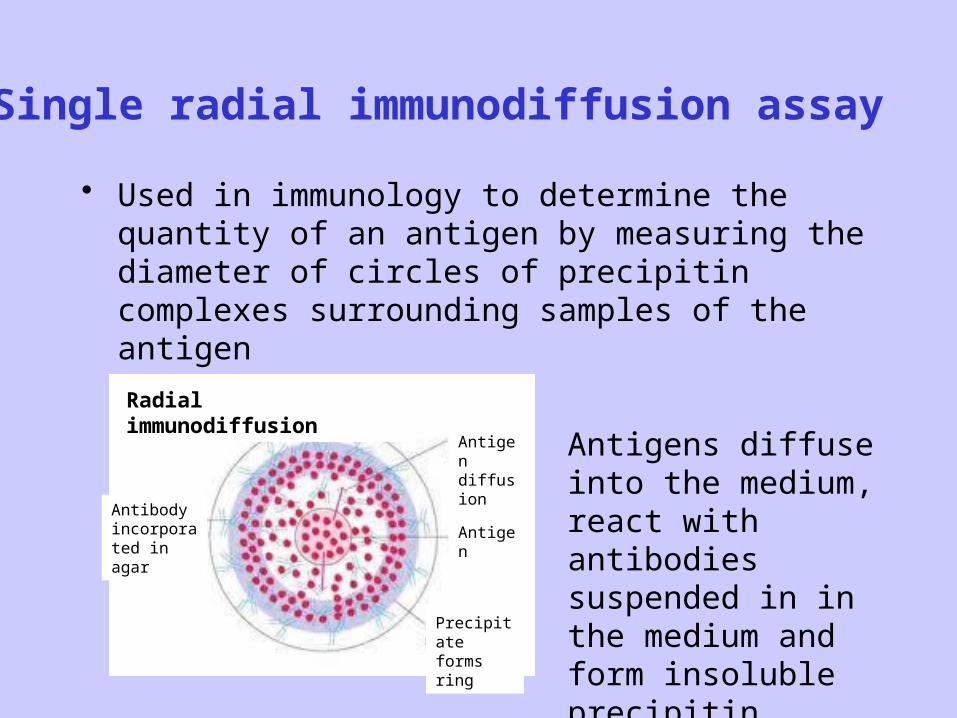
Single radial immunodiffusion assay
• Used in immunology to determine the quantity of an antigen by measuring the diameter of circles of precipitin complexes surrounding samples of the antigen
Radial immunodiffusion
Antibody incorporated in agar
Antigen diffusion
Precipitate forms ring
Antigen
Antigens diffuse into the medium, react with antibodies suspended in in the medium and form insoluble precipitin complexes.
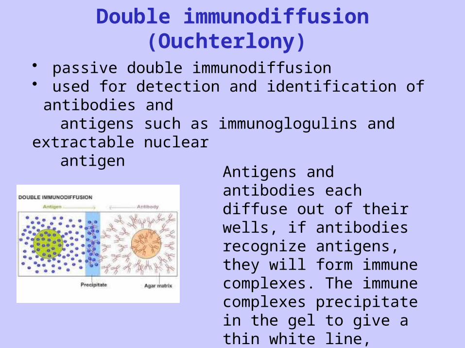
Double immunodiffusion (Ouchterlony)
• passive double immunodiffusion• used for detection and identification of antibodies and antigens such as immunoglogulins and extractable nuclear antigen
Antigens and antibodies each diffuse out of their wells, if antibodies recognize antigens, they will form immune complexes. The immune complexes precipitate in the gel to give a thin white line, which is a vizual signature of antigen recognition.

Immunoelectrophoresis
- is a laboratory technique, in which the blood serum is
placed into a gel and exposed to an electric current to separate
the serum protein components into five major fractions:
1. Serum albumin
2. Alfa-1-globulins
3. Alfa-2-globulins
4. Beta-globulins
5. Gama-globulins (imunoglobulins)

Immunofixation• Permits the detection and typing of monoclonal immunoglobulins in serum or
urine
• It is of great importance for diagnosis and monitoring of myeloma
• Immunofixation takes place in two steps:
1. separating the serum immunoglobulins on a gel under the effect of
an electric field, immunofixation requires to migrate serum tested
several times.
2. than anti-immunoglobulin antibodies are individually added to
each migration lane.
The presence of a monoclonal immunoglobulin results in the apperance of a
narrow band after staining complex precipitates.

Imunofixation of serum
Typing an M band by immunofixation. In this example, the M band found on electrophoresis (1) is identified as an IgM (type K).
Nephelometry and turbidimetry
- methods based on measuring of concentration of suspended immune complexes in a solution
- technique used in immunology to determine the levels of blood plasma proteins, for example levels of IgG, IgA and IgM, CRP, RF, C3 and C4 complement, C1 inhibitor

Turbidimetry: Definition: Light passing through a medium with dispersed
immune complexes, so the intensity of light transmitted is measured.
Arrangement of photometr: made in the same direction as the propagation of the light from the source

Nephelometry: Definition: The measurement of intensity of scattered light at
right angles to the direction of the light.
Arrangement of photometer: measure the light scattered at right angle to the direction of the light from the source
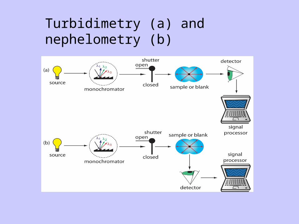
Turbidimetry (a) and nephelometry (b)

AgglutinationThe interaction between specific antibody and antigenic determinant on the surface of antigen results in visible clumping called agglutination.
Antigens include: bacteria, red blood cells, latex particles
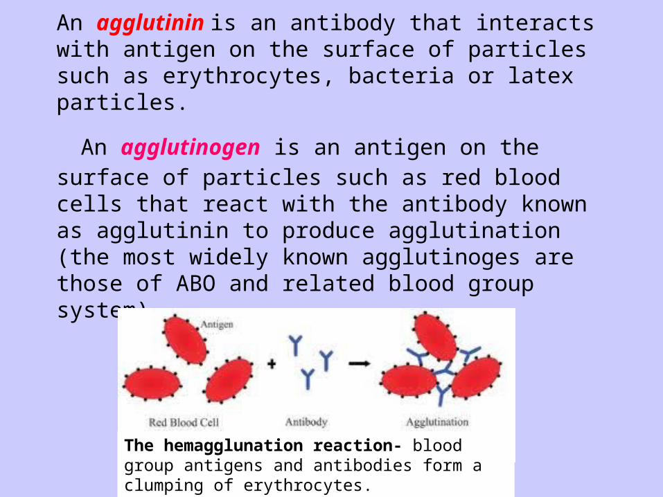
An agglutinin is an antibody that interacts with antigen on the surface of particles such as erythrocytes, bacteria or latex particles.
An agglutinogen is an antigen on the surface of particles such as red blood cells that react with the antibody known as agglutinin to produce agglutination (the most widely known agglutinoges are those of ABO and related blood group system).
The hemagglunation reaction- blood group antigens and antibodies form a clumping of erythrocytes.

Radioimmunoassay, enzyme immunoassay
very sensitive methods used to measure small concentrations of antigens (not detected by precipitation or agglutination)
labeled immunoassays
measure indirectly using a labeled antigen/antibody
antigen or antibody are labeled by
- radioactive element (radioimmunoassay)
- enzyme (enzyme immunoassay)
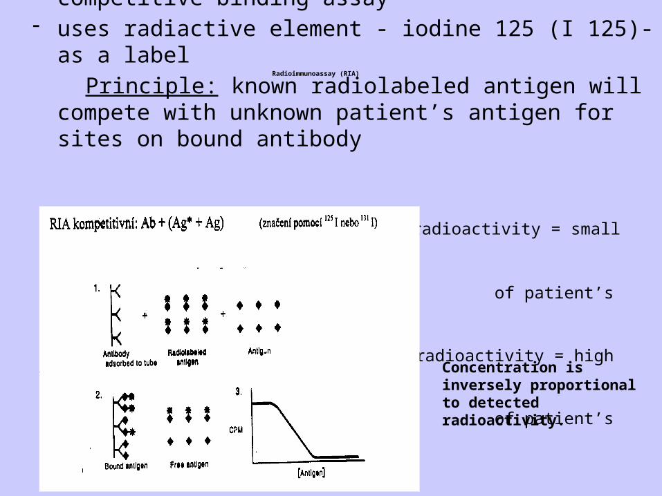
Radioimmunoassay (RIA)
- competitive binding assay- uses radiactive element - iodine 125 (I 125)- as a label
Principle: known radiolabeled antigen will compete with unknown patient’s antigen for sites on bound antibody
High radioactivity = small amount
of patient’s antigen
Low radioactivity = high amount
of patient’s antigen
Concentration is inversely proportional to detected radioactivity.

ImmunoRadioMetricAssay (IRMA)Noncompetitive binding assayPrinciple: patient’s antigens bind to adsorbed antibodies, added radiolabeled antibodies bind to bound antigens
The concentration is directy releated to detected radioactivity.
High radioactivity = high amount of patient’s antigenLow radioactivity = low amount of patient’s antigen
RIA/IRMA- andvantages and disadvantages
• Advantages:
- extremely sensitive and precise
- detects trace amount of analytes small in size• Disadvantages:
- expensive equipment necessary
- work with radioactive elements, radioactive waste
Enzyme immunoassays have largely replaced radioimmunoassay.
Enzyme immunoassay (EIA)
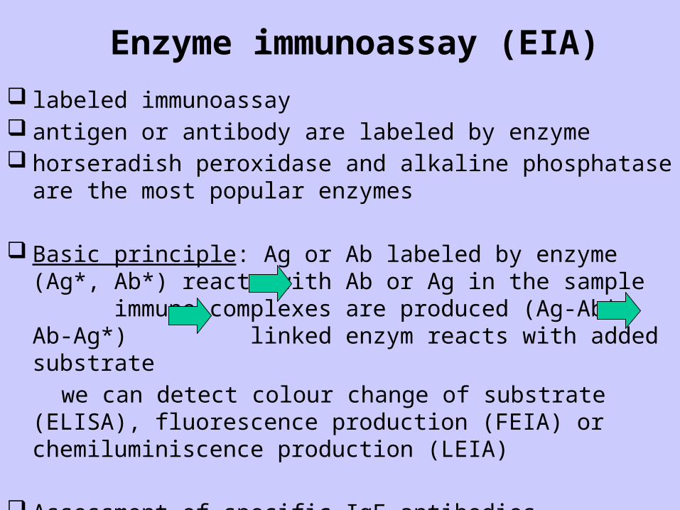
labeled immunoassay antigen or antibody are labeled by enzyme horseradish peroxidase and alkaline phosphatase are the most popular
enzymes
Basic principle: Ag or Ab labeled by enzyme (Ag*, Ab*) reacts with Ab or Ag in the sample immune complexes are produced (Ag-Ab*, Ab-Ag*) linked enzym reacts with added substrate
we can detect colour change of substrate (ELISA), fluorescence production (FEIA) or chemiluminiscence production (LEIA)
Assessment of specific IgE antibodies, cytokines, hormones, specific autoantibodies, antiinfectious antibodies (anti HIV abb)
- Použití: kvantitativní stanovení proteinů (protilátek), které se vyskytují v séru v nízkých koncentracích (autoprotilátky o známé specifitě, protil. proti očkovacím antigenům a proti infekčním činitelům, stanovení cytokinů)

EIA – enzyme immunoassayELISA - enzyme-linked immunosorbent assay- special sandwich type of EIA- can be used to quantify antigen/antibody in the sample- large no of samples can be proceed at a time- highly sensitive method- involves coating the Ag/Ab to a solid phase, the common format is to absorb the Ag/Ab to the wells of a 96- well microplate and to use substrates that produce a colored soluble product

ELISA – assessment of specific antibodies
substrate
2.Antibody (labeled by enzyme)
wash
wash
Specific antibody detected in the serum
Antigen – coated well
Spectrophotometry
Add sample to well coated with antigen. If antibody of interest is present in the sample, it will bind to the antigen. After washing add second antibody, that will specifically bind first antibody. After washing the substrate is added to the mixture. This substrate will trigger a reaction with enzyme attached to a second antibody to produce coloured substance.

Assessment of specific IgE antibodies
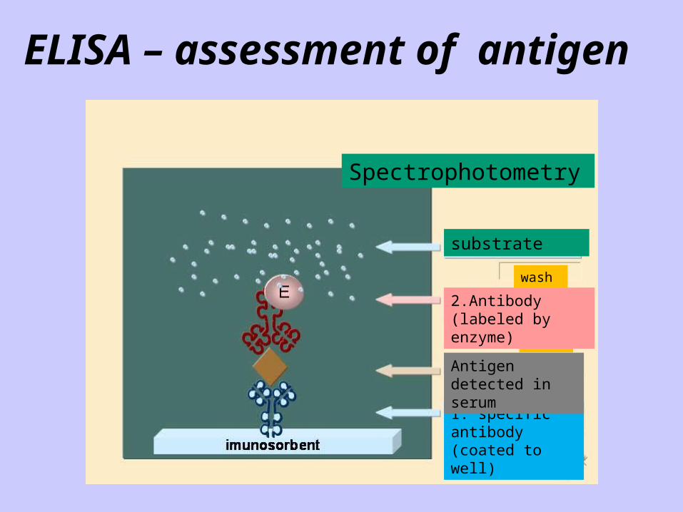
ELISA – assessment of antigen
Spectrophotometry
substrate
wash
wash
2.Antibody (labeled by enzyme)
1. specific antibody (coated to well)
Antigen detected in serum

Immunofluorescence assay Immunofluorescence is a technique allowing the vizualization of a
specific protein or antigen in tissue sections by binding a specific antibody chemically conjugated with a fluorescent dye such as fluorescein isothiocyanate ( FITC).
The specific antibodies are labeled with a compound (FITC) that makes them glow an apple-green color when observed microsopically under ultraviott light
There are two major types of immunofluorescence staining methods1. direct IF: staining in which the primary antibody is labeled with fluorescence dye2. indirect IF: staining in which a secondary antibody labeled with fluorochrome is used to detect a primary antibody
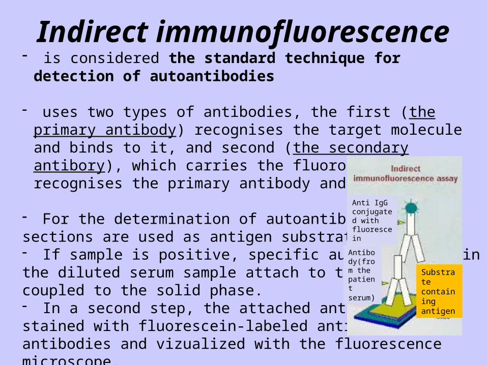
Indirect immunofluorescence- is considered the standard technique for detection of autoantibodies
- uses two types of antibodies, the first (the primary antibody) recognises the target molecule and binds to it, and second (the secondary antibory), which carries the fluorophore, recognises the primary antibody and binds to it
- For the determination of autoantibodies, tissue sections are used as antigen substrates.- If sample is positive, specific autoantibodies inthe diluted serum sample attach to the antigens coupled to the solid phase.- In a second step, the attached antibodies are stained with fluorescein-labeled anti-human antibodies and vizualized with the fluorescence microscope.
Substrate containing antigen
Antibody(from the patient serum)
Anti IgG conjugated with fluorescein
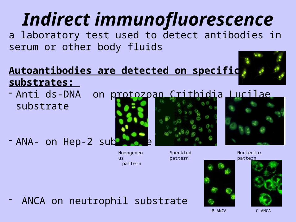
Indirect immunofluorescencea laboratory test used to detect antibodies in serum or other body fluids
Autoantibodies are detected on specific substrates: - Anti ds-DNA on protozoan Crithidia Lucilae substrate
- ANA- on Hep-2 substrate
- ANCA on neutrophil substrate
- AMA- on mouse stomach, kidney substrate- Anti LKM- on mouse liver, stomach, kidney
Homogeneouspattern
Speckled pattern Nucleolar pattern
P-ANCA C-ANCA
Asessment of cellular imunity
• Number of cells - subpopulations • Phagocytosis• Activation of lymphocytes

CD3+ T lymphocytes: mature T lymphocytes 50 - 75%
CD4+ T lymphocytes: helper T lymphocytes 30 - 60%
CD8+ T lymphocytes: cytoxic T lymphocytes 15 - 30%
CD25+ T lymphocytes: activated T lymphocytes 1 - 5%
CD19+ B lymphocytes: mature B lymphocytes 5 - 15%
CD56+ NK cells: natural killer cells 5 - 15%
CD14+ cells: monocytes
CD15+ cells: granulocytes
CD38+ cells: plasma cells
IMPORTANT LEUKOCYTE POPULATIONS

Flow cytometry- the main diagnostic tool for the assessment of cellular immunity

- method used for analysis of cells- uses direct immunofluorescence assay to identify a particular cell type
Direct immunofluorescence
Fluorescence detection
Wash out
FITC labeled monoclonal antibody
T lymphocyte with specific CD marker
UV Green light
Flow cytometry
Primary, or direct, immunofluorescence uses a single, primary antibody, chemically linked to a fluorophore (FITC, PE). The primary antibody recognizes the target molecule (antigen) and binds to a specific region called the epitope (CD 3, CD 4)
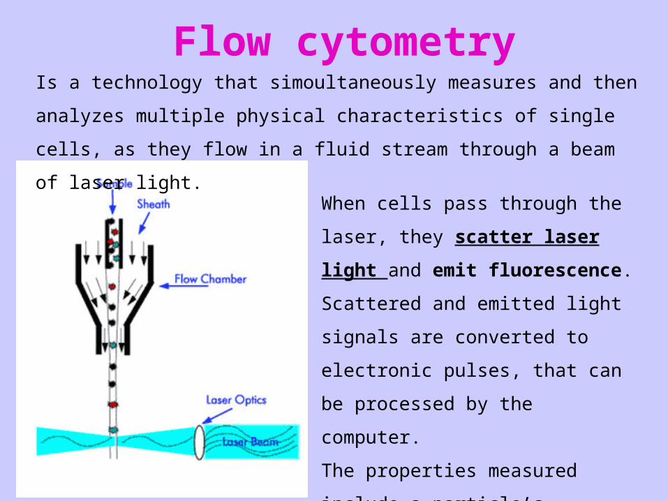
Flow cytometry
When cells pass through the laser, they
scatter laser light and emit fluorescence.
Scattered and emitted light signals are
converted to electronic pulses, that can be
processed by the computer.
The properties measured include a
particle’s relative size, relative
granularity and relative fluorescence
intensity.
Is a technology that simoultaneously measures and then analyzes multiple
physical characteristics of single cells, as they flow in a fluid stream through a
beam of laser light.
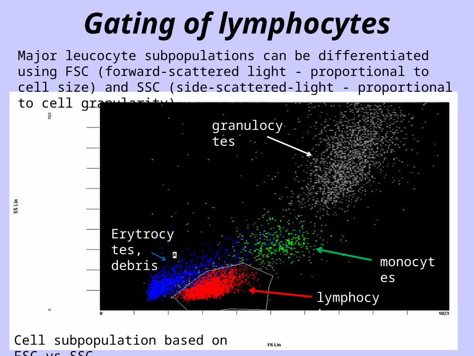
Gating of lymphocytesMajor leucocyte subpopulations can be differentiated using FSC (forward-scattered light - proportional to cell size) and SSC (side-scattered-light - proportional to cell granularity).
lymphocytes
monocytes
granulocytes
Erytrocytes, debris
Cell subpopulation based on FSC vs SSC
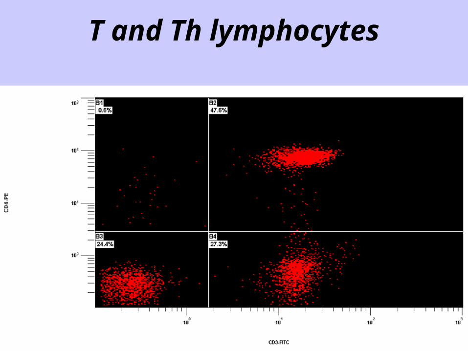
T and Th lymphocytes
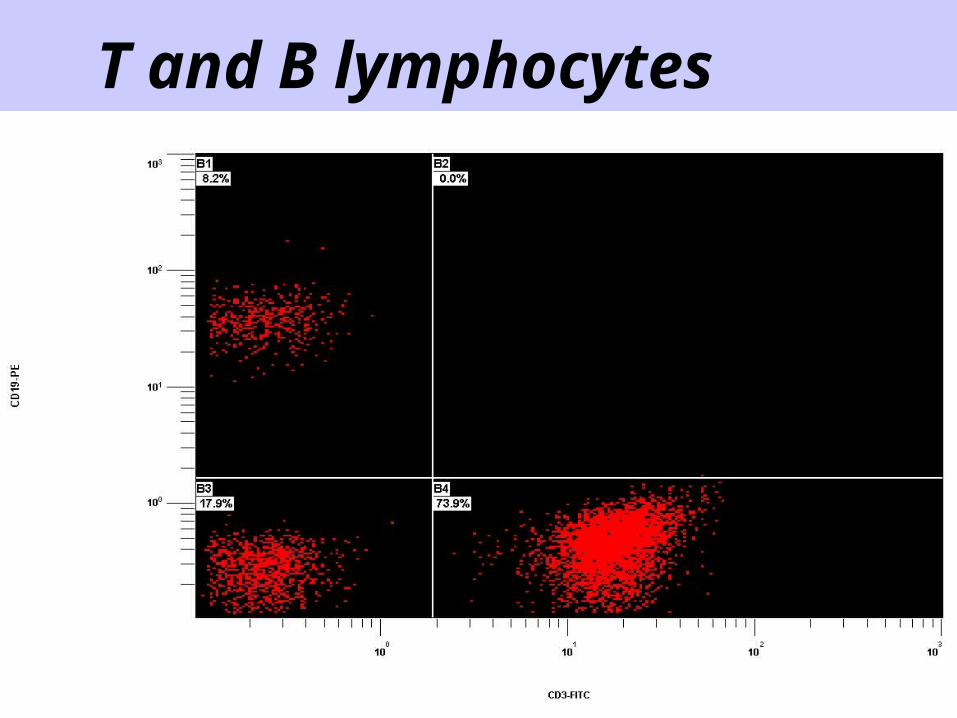
T and B lymphocytes
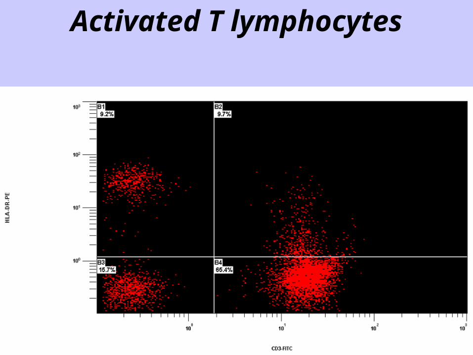
Activated T lymphocytes
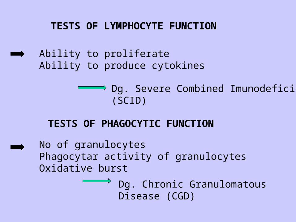
Ability to proliferateAbility to produce cytokines
TESTS OF PHAGOCYTIC FUNCTION
TESTS OF LYMPHOCYTE FUNCTION
No of granulocytesPhagocytar activity of granulocytesOxidative burst Dg. Chronic Granulomatous Disease
(CGD)
Dg. Severe Combined Imunodeficiency(SCID)

http://www.lifetechnologies.com/cz/en/home/support/tutorials.html#vid4

Thank you for your attention!
http://www.lifetechnologies.com/cz/en/home/support/tutorials.html#vid4http://www.lifetechnologies.com/cz/en/home/support/tutorials.html#vid4
Related Documents