This article appeared in a journal published by Elsevier. The attached copy is furnished to the author for internal non-commercial research and education use, including for instruction at the authors institution and sharing with colleagues. Other uses, including reproduction and distribution, or selling or licensing copies, or posting to personal, institutional or third party websites are prohibited. In most cases authors are permitted to post their version of the article (e.g. in Word or Tex form) to their personal website or institutional repository. Authors requiring further information regarding Elsevier’s archiving and manuscript policies are encouraged to visit: http://www.elsevier.com/copyright

Welcome message from author
This document is posted to help you gain knowledge. Please leave a comment to let me know what you think about it! Share it to your friends and learn new things together.
Transcript

This article appeared in a journal published by Elsevier. The attachedcopy is furnished to the author for internal non-commercial researchand education use, including for instruction at the authors institution
and sharing with colleagues.
Other uses, including reproduction and distribution, or selling orlicensing copies, or posting to personal, institutional or third party
websites are prohibited.
In most cases authors are permitted to post their version of thearticle (e.g. in Word or Tex form) to their personal website orinstitutional repository. Authors requiring further information
regarding Elsevier’s archiving and manuscript policies areencouraged to visit:
http://www.elsevier.com/copyright
Author's personal copy
Cell Calcium 46 (2009) 303–312
Contents lists available at ScienceDirect
Cell Calcium
journa l homepage: www.e lsev ier .com/ locate /ceca
L-type channel inhibition by CB1 cannabinoid receptors is mediated byPTX-sensitive G proteins and cAMP/PKA in GT1-7 hypothalamic neurons
Hanaa Hoddahb,1, Andrea Marcantonia,1, Valentina Comunanzaa, Valentina Carabelli a, Emilio Carbonea,∗
a Department of Neuroscience, NIS Center, CNISM Research Unit, Corso Raffaello 30, 10125 Torino, Italyb Department of Biology, University Abdelmalek Essaadi, Tetouan, Morocco
a r t i c l e i n f o
Article history:Received 8 August 2009Accepted 31 August 2009Available online 8 October 2009
Keywords:CannabinoidsVoltage-gated calcium channelsCB1 inverse agonistscAMP/PKA signal pathwayImmortalized GnRH neurons
a b s t r a c t
Using immortalized hypothalamic GT1-7 neurons, which express the CB1 cannabinoid receptor (CB1R)and three Ca2+ channel types (T, R and L), we found that the CB1R agonist WIN 55,212-2 inhibited thevoltage-gated Ca2+ currents by about 35%. The inhibition by WIN 55,212-2 (10 �M) was reversible and pre-vented by nifedipine (3 �M), suggesting a selective action on L-type Ca2+ channels (LTCCs). WIN 55,212-2action exhibited all the features of voltage-independent Ca2+ channel modulation: (1) no changes of theactivation kinetics, (2) equal depressive action at all potentials and (3) no facilitation following strongprepulses. At variance with WIN 55,212-2, the CB1R inverse agonist AM-251 (10 �M) caused 20% increaseof Ca2+ currents. The inhibition of LTCCs by WIN 55,212-2 was prevented by overnight PTX-incubationand by intracellular perfusion with GDP-�-S. The latter caused also a 20% Ca2+ current up-regulation.WIN 55,212-2 action was also prevented by application of the PKA-blocker H89 or by loading the neu-rons with 8-CPT-cAMP. Our results suggest that LTCCs in GT1-7 neurons are partially inhibited at rest dueto a constitutive CB1R activity removed by AM-251 and GDP-�-S. Activation of CB1R via PTX-sensitiveG proteins and cAMP/PKA pathway selectively depresses LTCCs that critically control the synchronizedspontaneous firing and pulsatile release of gonadotropin-releasing hormone in GT1-7 neurons.
© 2009 Elsevier Ltd. All rights reserved.
1. Introduction
Cannabinoids are the primary psychoactive constituents ofmarijuana that have profound effects on pain perception, neuralconvulsions, memory and motor coordination [1,2]. Their effectsare most commonly mediated by the CB1 cannabinoid receptor(CB1R), which is highly expressed in a variety of brain regions,including the hypothalamus [3–5]. The CB1R is a member of theG protein-coupled receptor superfamily [6] which acts by inhibit-ing adenylate cyclase (AC) activity [7], delaying the opening ofvoltage-gated N- and P/Q-type channels [8–11], activating K+ chan-nels [12,13] and triggering MAP kinases signal cascades [14]. Allthese effects are originated from the activation of PTX-sensitive Gi,oproteins coupled to CB1Rs, which inhibit AC activity and reducescytoplasmic cAMP levels. Inhibition of AC and reduction of cAMP isa common pathway to most CB1R-mediated effects, but curiouslyenough, this pathway does not apply to the cannabinoid-inducedmodulation of voltage-gated K+ and Ca2+ channels, which is medi-
∗ Corresponding author. Tel.: +39 011 670 8489; fax: +39 011 670 8174.E-mail address: [email protected] (E. Carbone).
1 These two authors contributed equally to the work.
ated by Gi,o proteins acting directly on the channels, regardless ofcAMP or any other diffusible messenger [15]. The only exception tothis rule is the activation of the fast inactivating IA potassium cur-rent induced by the CB1R/CB2R agonist WIN 55,212-2 in dissociatedhippocampal neurons through a PTX- and cAMP/PKA-dependentpathway [16].
Cannabinoids act also on L-type Ca2+ channels (LTCCs) but,despite much work, the results remain controversial. CB1R acti-vation could not affect [9,17], up-regulate [18,19] or inhibit LTCCs[20]. In arterial smooth muscles and in NTS (nucleus of tractus soli-tarius) neurons, activation of CB1R causes a voltage-independentinhibition of LTCCs which is prevented by PTX [21,22]. The inhib-ited current displays the same activation time course of controls (nodelay of the rising phase) and the degree of inhibition remains unal-tered after strong facilitatory prepulses, which are typical featuresof the LTCCs down-modulation in response to receptor-activatedGi,o proteins [23] (see [24] for a review). In addition, the inhibitionof LTCCs by cannabinoids in NTS is mediated by cAMP and PKA.Blocking PKA or AC activity the inhibitory action of WIN 55,212-2 onLTCCs is prevented [22]. This is consistent with the effects of otherG protein-coupled receptor-mediated signaling on neuroendocrineLTCCs that are regulated by cAMP/PKA [25] and highlights themarkedly different mechanism by which CB1R activation inhibits
0143-4160/$ – see front matter © 2009 Elsevier Ltd. All rights reserved.doi:10.1016/j.ceca.2009.08.007
Author's personal copy
304 H. Hoddah et al. / Cell Calcium 46 (2009) 303–312
L- and non-L-type channels. Despite being PTX-sensitive, the latteraction on non-L-type channels (N and P/Q) is voltage-dependentand insensitive to cAMP [15].
Given the key role that LTCCs play in the control of manybrain functions and the little knowledge on CB1R-mediated signaltransduction mechanisms targeting these channels, we thought ofinterest to study the action of CB1R on LTCCs expressed in hypotha-lamic immortalized GT1-7 neurons. GT1-7 clonal cells produce andrelease the gonadotropin-releasing hormone (GnRH) [26]. Theyalso express CB1Rs and are able to synthesize and release endo-cannabinoids [27]. GT1-7 neurons form stable networks that firespontaneously and possess high densities of LTCCs. These channelsplay an important role in the control of synchronized firings andconsequent pulsatile GnRH release [28,29]. In addition, cAMP sig-naling and a variety of G protein-coupled receptors regulate GnRHrelease in GT1-7 neurons [30]. Thus, immortalized GnRH neuronsappear to be an ideal model for studying the CB1R-mediated mod-ulation of neuronal LTCCs.
We report here that the CB1R agonist WIN 55,212-2 selectivelyinhibits LTCCs in immortalized GT1-7 neurons. This selective actionis mediated by PTX-sensitive Gi,o proteins through a cAMP/PKA sig-nal transduction pathway. Inhibition is voltage-independent andinsensitive to short facilitatory prepulses excluding a direct Gi,o-mediated action on the non-L-type channels expressed in GT1-7cells (R-type; [31]). This new modulatory action on neuroendocrineLTCCs could be critical in the control of GnRH release in hypotha-lamic neurons and broadens the possible mechanisms by whichcannabinoids could affect cell excitability, neuronal firing and Ca2+-dependent hormone release.
2. Material and methods
2.1. Tissue culture of immortalized GT1-7 neurons
Immortalized GT1-7 neurons (provided by Dr. P. Mellon, Univer-sity of California, San Diego, La Jolla, USA) were cultured in 4.5 g/lglucose Dulbecco’s modified Eagle’s medium (DMEM) supple-mented with 10% fetal bovine serum, 2% of l-glutamine, 100 �U/mlpenicillin and 100 �g/ml streptomycin in an atmosphere of 5% CO2at 37 ◦C. Morphological differentiation was optimized by culturingthe cells just after they reached confluence in a medium containingB27 serum-free supplement, 1% sodium pyruvate, 0.5% fetal bovineserum and 2% of l-glutamine. The culture medium was changedevery 3–4 days, and cells used in this study were between passages9 and 14.
2.2. Electrophysiology
The Ca2+ currents were recorded by using two configurations:the perforated-patch with amphotericin B and the whole-cell. Patchelectrodes were made of borosilicate glass capillaries and had aresistance of 1–2 M�. For the perforated patch-clamp configura-tion [25], the pipette solution contained (in mM): 135 CsMeSO3,8 NaCl, 2 MgCl2, 20 HEPES and 50–100 �g/ml of amphotericin B(pH 7.3 with CsOH); for the whole-cell configuration the solutioncontained (mM): 95 CsCl, 30 TEACl, 10 EGTA, 2 MgCl2, 10 HEPES,8 glucose, 2 ATP, 0.5 GTP, 15 phosphocreatine (pH 7.3 with CsOH).The extracellular solution contained (in mM): 135 TEACl, 10 CaCl2,2 MgCl2, 10 HEPES, 10 glucose (pH 7.4 with CsOH).
Electrophysiological recordings were performed either using anEPC-9 patch-clamp amplifier and PULSE software (HEKA Electronic,Lambrecht, Germany) [25] or using an Axopatch 200-A amplifierand pClamp 10.0 software programs (Axon Instruments Inc., FosterCity, CA, USA) [32]. Currents were sampled at 10 kHz and filteredat 1–5 kHz. Recordings were made at room temperature.
2.3. RNA extraction and PCR-RT for CB1R and CB2R in GT1-7neurons
Total RNA from GT1-7 cells was isolated with Mini RNeasy(Qiagen AG, Basel, Switzerland) as indicated in the manufac-turer’s instructions. DNase-treated total RNA was used in thereversed transcription (RT) procedure. cDNA was synthesized ina total volume of 50 �l with the High Capacity cDNA Archivekit (Applied Biosystems, Foster City, CA, USA) according to themanufacturer’s protocol. The primer sequences used were asfollows: for CB1R (U22948), 5′-TGTGGGGAGAATTTTATGGA (for-ward) and 5′-AGATTGCAGCTTCTTGCAGT (reverse); for CB2R (NM009924), 5′-GGTCCTCTCAGCATTGATTTCTTAC (forward) and 5′-TTCACATCAGCCTCTGTTTCTGTA (reverse); for GAPDH (M32599) 5′-CAACAGCAACTCCACTCTT (forward); 5′-AGGCCCCTCCTGTTATTATG(reverse). Polymerase chain reaction (PCR) was carried out in atotal volume of 25 �l containing 2 �l of cDNA from the above reac-tion, 0.5 �M of each specific primer, 0.5 U Phusion DNA polymerase,5× Phusion GC buffer, 0.2% DMSO (FinnzymesOy, Espoo, Finland),0.2 mM of each deoxynucleotide triphosphate (Invitrogen, Carls-bad, CA, USA). The reaction conditions were 98 ◦C for 30 s, followedby 30 cycles (for CB1R and GAPDH) or 35 cycles (for CB2R) at 98 ◦Cfor 15 s, 58 ◦C for 30 s, and 72 ◦C for 30 s. Positive controls (cDNAobtained from total mouse brain total RNA) and negative controls(water instead of template) were amplified in the same conditionand glyceraldehyde-3-phosphate dehydrogenase was used to eval-uate the integrity of RNA. The amplified products were separated on2% agarose gels with Gel star (Cambrex Corporation, East Ruther-ford, NJ, USA) and in the presence of a 100 bp DNA ladder as themolecular weight marker (Promega; Madison, WI, USA).
2.4. Chemicals
WIN 55,212-2, AM-251 and GDP-�-S were purchased fromTocris Bioscience (Avonmouth, UK). Nifedipine, 8-CPT-cAMP, andH89 were purchased from Sigma–Aldrich (Milano, Italy). All drugswere prepared just before use. WIN 55,212-2 and AM-251 were dis-solved in dimethyl sulfoxide (DMSO) in stocks of 10 mM and bothused at the final concentration of 1–10 �M. GDP-�-S was dissolvedin water in stocks of 50 mM and used at the final concentrationof 170–500 �M. Nifedipine was dissolved in ethanol and preparedto the final concentration of 3 �M. PTX was purchased from Cal-biochem Corporation (Darmstadt, Germany) and dissolved in waterin stocks of 50 �g/ml. GT1-7 neurons were incubated overnightwith 130 ng/ml of toxin. 8-CPT-cAMP and H89 were dissolved inwater in stocks of 10 mM and 1 mM and used at the final concen-tration of 1 mM and 5 �M, respectively.
2.5. Statistical analysis
The results are expressed as the mean ± S.E.M. for n number ofneurons. The differences were analyzed by either one-sample orpaired Student’s t-tests as indicated. Values of p < 0.05 were con-sidered statistically significant.
3. Results
3.1. Expression of CB1R and CB2R in GT1-7 neurons
The presence of cannabinoid receptors (CB1R and CB2R) inGT1-7 neurons was supported by qualitative RT-PCR experimentsperformed on the total RNA extracted from GT1-7 cells andmouse brain RNA (positive control). Total RNA extracted wasretro-transcribed and amplified with specific oligonucleotides forCB1R and CB2R. To evaluate RNA integrity we used primers forGAPDH (glyceraldehyde-3-phoshate dehydrogenase) specifically
Author's personal copy
H. Hoddah et al. / Cell Calcium 46 (2009) 303–312 305
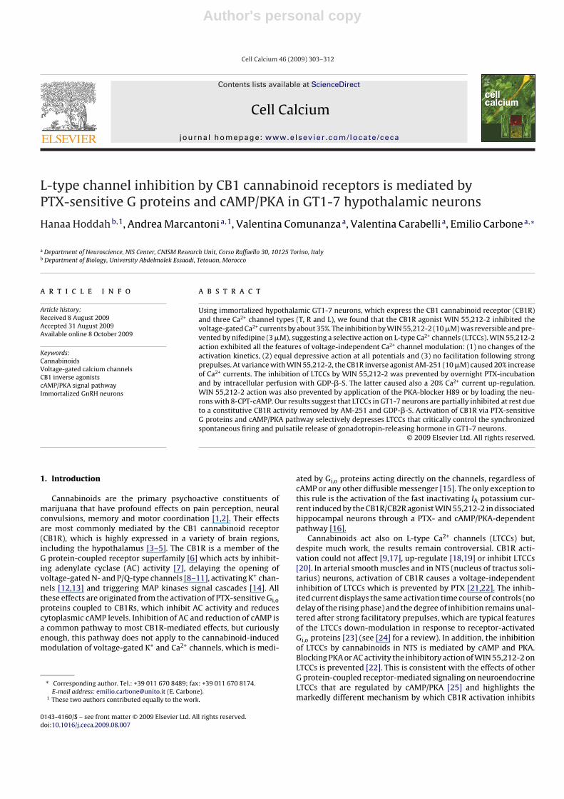
Fig. 1. RT-PCR demonstrates the presence of CB1R and CB2R mRNA in GT1-7 neu-rons. (A) RT-PCR analyses of CB1R expression in GT1-7 cells (lane 1). Lanes 1 and 2show samples from GT1-7 cells, lanes 4 and 5 are from mouse brain (positive con-trol). The enzyme GAPDH (lanes 2 and 5) was used as housekeeping gene. Productswere separated via electrophoresis on 2% agarose gel containing ethidium bromidein the presence of specific molecular weights (MW) (lane 3). (B) RT-PCR analysesof CB2R expression in GT1-7 cells (lane 1). Lanes 1 and 2 show template from GT1-7; lanes 4 and 5 show samples from mouse brain (positive control). Lane 3 is themolecular weights marker.
expressed in GT1-7 cells and mouse brain. Qualitative RT-PCRshows the presence of both cannabinoid receptors in GT1-7 neu-rons: CB1R mRNA (lane 1, Fig. 1A) and CB2R mRNA (lane 1, Fig. 1B).As positive control, expression of CB1R and CB2R was evaluated inthe mouse brain (lane 4, Fig. 1A and B) and GAPDH was used ashousekeeping gene (lanes 2 and 5; Fig. 1A and B).
3.2. LVA and HVA Ca2+ currents in GT1-7 neurons
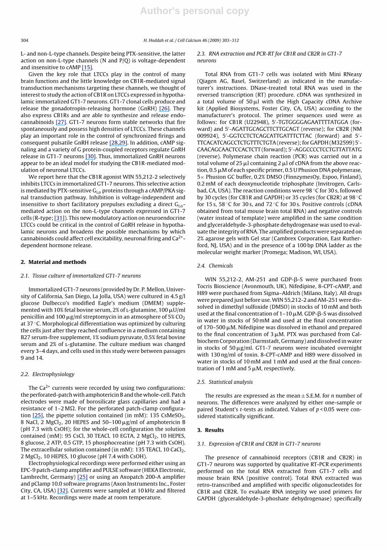
GT1-7 neurons express low-voltage activated (LVA, T-type) andhigh-voltage activated (HVA) Ca2+ channels [33]. The HVA com-ponent is further subdivided in two channel types (L and R) withpercentages of expression varying from cell to cell [31,28]. Wefound on average that 30% of GT1-7 neurons expressed fast inac-tivating T-type channels that activated from −50 mV in 10 mMCa2+ (holding potential, Vh, −80 mV) and reached maximal ampli-tudes at −10 mV (Fig. 2A and B). Slowly inactivating HVA channelswere found to activate above −40 mV (Fig. 2C) and reached maxi-mal amplitudes at +10 mV (Fig. 2D). HVA Ca2+ currents had meanamplitudes of 196.6 ± 16.6 pA at +10 mV (n = 81) that normalizedfor the cell capacitance (33.2 ± 3.2 pF; n = 81) gave mean currentdensities of 5.9 ± 0.8 pA/pF. The T-type channels had the typical gat-ing characteristics described in these [28] and other neurons [34].They activated below −40 mV, had voltage-dependent activationand fast inactivation and were inactivated at Vh around −50 mV.Their presence caused rapid inactivating currents during step depo-larization and an early current peak around −10 mV during rampcommands (Fig. 2B). In this work the T-type channels were notfurther considered.
To simplify our analysis, we tested the percentage of LTCCsexpressed by GT1-7 neurons by determining the blocking effectsof 3 �M nifedipine. We found that the DHP-antagonist on averageblocked 49.4 ± 3.0% (n = 12) of the total HVA currents activated at+10 mV from Vh = −80 mV (Fig. 3A and C). DHP block was fast andreversible and, could be complete in 30% of the cells, indicating thepresence of only LTCCs in a fraction of GT1-7 neurons (Fig. 3B). Theresidual DHP-resistant currents (∼50% of the total) were not furtherpharmacologically dissected and, from here on, will be indicated asthe non-L-type current component (Fig. 3C). In previous studiesthe HVA Ca2+ currents of GT1-7 neurons were identified mainly asLTCCs (100% of the total) [28] or carried mostly by R-type chan-
Fig. 2. Ca2+ currents in GT1-7 neurons expressing different percentages of LVA (T-type) and HVA (L- and non-L-type) channels. (A) Overlapped Ca2+ current traces recorded in10 mM Ca2+ from a GT1-7 neuron expressing high densities of T-type channels. Step depolarizations were from −50 to +10 mV. The holding potential (Vh) was −80 mV. (B) I–Vcharacteristics recorded from a GT1-7 neuron expressing T-type and HVA Ca2+ channels using a ramp command from −60 to +60 mV (Vh −80 mV). Notice the typical double-peak indicative of the presence of LVA and HVA channels. (C and D) Overlapped Ca2+ current traces and I–V characteristics recorded from two GT1-7 neurons expressingmostly HVA channels. Sequential step depolarizations in (C) were from −50 to −10 mV (Vh −80 mV); traces at more positive potentials are not shown to avoid confusingoverlapping. The ramp command was as in (B). Notice the absence of the “early shoulder” associated to T-type currents.

Author's personal copy
306 H. Hoddah et al. / Cell Calcium 46 (2009) 303–312
Fig. 3. GT1-7 neurons express different densities of LTCCs. (A and B) Two examples of Ca2+ current block recorded at +10 mV (Vh −80 mV) from neurons with differentsensitivity to nifedipine (3 �M). In (A) the DHP blocked nearly 50% of the control current (red trace) while in (B) the DHP blocked completely the total current. In both casesthe DHP action was fully reversible (blue trace). (C) Mean percentage of current amplitude in the presence of 3 �M nifedipine from GT1-7 neurons responding partially to theDHP (n = 12). The percentage of current is expressed relative to the peak control current (**p < 0.01 vs. control using a one-sample analysis Student’s t-test when comparingthe mean percentage values to hundred).
nels (75% of the total) [31]. Our data are in fair agreement withboth reports indicating significant degree of heterogeneity of Ca2+
channels expression in GT1-7 neurons, which might derive fromdifferent tissue culture conditions.
3.3. Ca2+ currents inhibition by WIN 55,212-2 isvoltage-independent
GT1-7 cells express CB1R (Fig. 1) and recent works have shownthat their activation by WIN 55,212-2 reduces the release of GnRHfrom these cells [27]. A likely mechanism is that WIN 55,212-2might block the LTCCs that control both, the rate of synchronizedaction potential firing and pulsatile GnRH release [29]. We testedtherefore the effects of WIN 55,212-2 on the Ca2+ currents activatedat +10 mV using saturating concentrations (1–10 �M) and looked
for its effects on the amplitude and kinetics of Ca2+ currents. Directapplication of WIN 55,212-2 on GT1-7 neurons produced an aver-age Ca2+ current inhibition of 34.4 ± 1.6% (n = 13; p < 0.01 respect tonormalized control) (Fig. 4A–C). The inhibition was fast, reversibleand required less than 1 min to reach maximal values (Fig. 4D).Recovery had similar kinetics to the onset of inhibition.
Ca2+ current inhibition by WIN 55,212-2 was of the same per-centage at every potential and had no marked effects on thetime course and voltage-dependence of channel activation. Thehalf-time to peak at +10 mV was 0.76 ± 0.17 ms in control and0.75 ± 0.15 ms with WIN 55,212-2 (n = 8). In addition, the I–V curvefrom −60 to +60 mV in the presence of the CB1R agonist had thesame shape of the control I–V and was simply scaled-down by a con-stant factor (Fig. 4B). This is a clear indication that WIN 55,212-2action is voltage-independent and thus different from the voltage-
Fig. 4. Inhibition the Ca2+ currents by WIN 55,212-2 in GT1-7 neurons is voltage-independent. (A) Current traces recorded at +10 mV from a GT1-7 neuron before (control,black trace), during (WIN, red trace) and after (wash, blue trace) exposure of 10 �M WIN 55,212-2. (B) I–V characteristics in control conditions and during application of10 �M WIN 55,212-2. Notice the proportional decrease of the current with WIN 55,212-2 at every potential. (C) Mean percentage of current amplitude in the presence of10 �M WIN 55,212-2 obtained from n = 13 neurons. The percentage of current is expressed relative to the peak control current (**p < 0.01 vs. control using a one-sampleanalysis Student’s t-test when comparing the mean percentage values to hundred). (D) Time course of peak Ca2+ currents at +10 mV before, during and after addition of 10 �MWIN 55,212-2. In the inset are shown the current traces recorded at the time indicated by the letters (a–c). (E) Ca2+ currents at control (black trace) and during exposure of10 �M WIN 55,212-2 (red trace) recorded using the double-pulse protocol illustrated on the top. The prepulse depolarization to +80 mV was not able to relieve the inhibitionof the Ca2+ current induced by WIN 55,212-2.
Author's personal copy
H. Hoddah et al. / Cell Calcium 46 (2009) 303–312 307
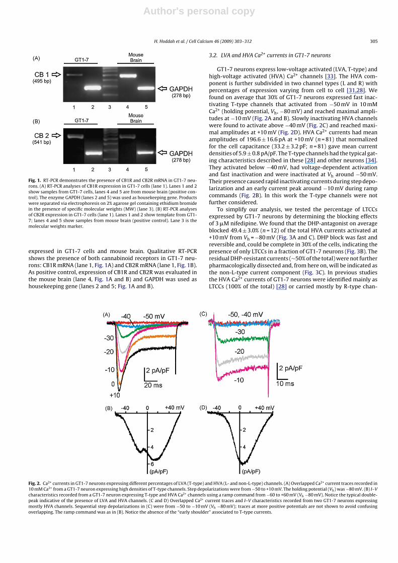
Fig. 5. The Ca2+ current inhibition induced by WIN 55,212-2 is selective for LTCCs. (A and B) Current traces recorded at +10 mV and I–V characteristics at control (blacktrace), in the presence of 3 �M nifedipine (red trace) and during application of nifedipine plus 10 �M WIN 55,212-2 (blue trace) from a GT1-7 neuron. (C) Mean peak currentamplitudes recorded at +10 mV in the presence of nifedipine and nifedipine plus WIN 55,212-2 (n = 11). The relative peak current values are normalized with respect tocontrol (**p < 0.01 vs. control using a one-sample analysis Student’s t-test when comparing the mean percentage values to hundred).
dependent inhibition induced by cannabinoids in cell preparationsexpressing N and P/Q-type channels [9]. Ca2+ current inhibition byWIN 55,212 in these cases produced a net delay of Ca2+ channel acti-vation at voltages below +10 mV that was largely relieved at morepositive potentials (>+40 mV). Strong prepulses of 50 ms to +80 mVanticipating the test pulse could relief most of the inhibition [10].In the case of GT1-7 neurons the same strong prepulses to +80 mV(n = 11) was unable to remove the inhibition induced by WIN55,212-2. Percentage of Ca2+ current depression with WIN 55,212-2(34%) was identical before and after the prepulse (Fig. 4E), indicat-ing that WIN 55,212-2 action was voltage-independent. There wasneither an acceleration of Ca2+ current activation nor an increaseof peak amplitude after the prepulse which could suggest a facil-itatory effect of voltage as for N- and P/Q-type channels (see [35]for a review).
3.4. WIN 55,212-2 and the CB1R inverse agonist AM-251 actselectively on LTCCs
The inhibitory effects of WIN 55,212-2 are selective for LTCCsand prevented by nifedipine. In the presence of the DHP (3 �M),WIN 55,212-2 had no further inhibitory effects during step depo-larization to +10 mV (Fig. 5A) or during ramp commands (Fig. 5B).This was systematically observed in eleven GT1-7 neurons in whichthe relative peak amplitude of the current with nifedipine was52.7 ± 4.9% with respect to control and decreased to 47.4 ± 4.8%after addition of WIN 55,212-2 (Fig. 5C).
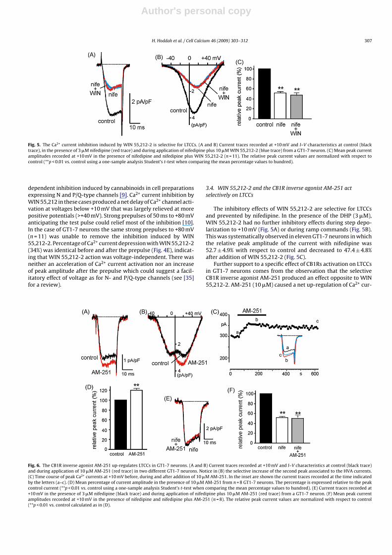
Further support to a specific effect of CB1Rs activation on LTCCsin GT1-7 neurons comes from the observation that the selectiveCB1R inverse agonist AM-251 produced an effect opposite to WIN55,212-2. AM-251 (10 �M) caused a net up-regulation of Ca2+ cur-
Fig. 6. The CB1R inverse agonist AM-251 up-regulates LTCCs in GT1-7 neurons. (A and B) Current traces recorded at +10 mV and I–V characteristics at control (black trace)and during application of 10 �M AM-251 (red trace) in two different GT1-7 neurons. Notice in (B) the selective increase of the second peak associated to the HVA currents.(C) Time course of peak Ca2+ currents at +10 mV before, during and after addition of 10 �M AM-251. In the inset are shown the current traces recorded at the time indicatedby the letters (a–c). (D) Mean percentage of current amplitude in the presence of 10 �M AM-251 from n = 8 GT1-7 neurons. The percentage is expressed relative to the peakcontrol current (**p < 0.01 vs. control using a one-sample analysis Student’s t-test when comparing the mean percentage values to hundred). (E) Current traces recorded at+10 mV in the presence of 3 �M nifedipine (black trace) and during application of nifedipine plus 10 �M AM-251 (red trace) from a GT1-7 neuron. (F) Mean peak currentamplitudes recorded at +10 mV in the presence of nifedipine and nifedipine plus AM-251 (n = 8). The relative peak current values are normalized with respect to control(**p < 0.01 vs. control calculated as in (D).
Author's personal copy
308 H. Hoddah et al. / Cell Calcium 46 (2009) 303–312
rents at +10 mV (Fig. 6A). The up-regulation was selective for theHVA Ca2+ channels (Fig. 5B), required 1–2 min to reach maximalvalues (Fig. 5C), and was systematically observed in every GT1-7 neuron tested (n = 8). The percentage of up-regulation relativeto control varied from 6 to 44% with a mean value of 19.7 ± 4.9%(p < 0.01 vs. normalized control; Fig. 6D) and was fully preventedby 3 �M nifedipine (Fig. 6E). Simultaneous application of AM-251and nifedipine caused no further decrease of the peak current thanthe DHP alone (Fig. 6E). Mean relative amplitudes of Ca2+ currentswere 53.5 ± 4.2% with nifedipine and 50.2 ± 6.7% with nifedipineplus AM-521.
3.5. WIN 55,212-2 action is prevented by PTX cell incubation andintracellular GDP-ˇ-S
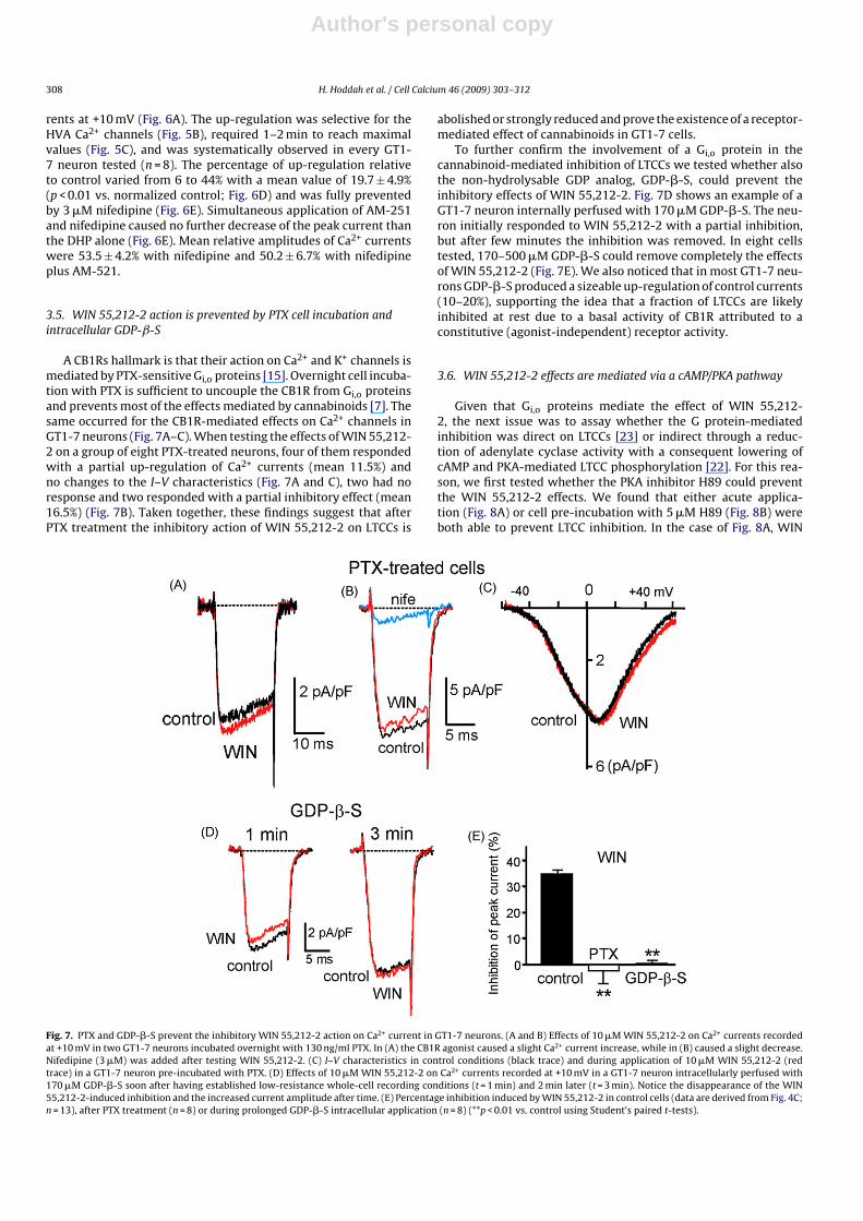
A CB1Rs hallmark is that their action on Ca2+ and K+ channels ismediated by PTX-sensitive Gi,o proteins [15]. Overnight cell incuba-tion with PTX is sufficient to uncouple the CB1R from Gi,o proteinsand prevents most of the effects mediated by cannabinoids [7]. Thesame occurred for the CB1R-mediated effects on Ca2+ channels inGT1-7 neurons (Fig. 7A–C). When testing the effects of WIN 55,212-2 on a group of eight PTX-treated neurons, four of them respondedwith a partial up-regulation of Ca2+ currents (mean 11.5%) andno changes to the I–V characteristics (Fig. 7A and C), two had noresponse and two responded with a partial inhibitory effect (mean16.5%) (Fig. 7B). Taken together, these findings suggest that afterPTX treatment the inhibitory action of WIN 55,212-2 on LTCCs is
abolished or strongly reduced and prove the existence of a receptor-mediated effect of cannabinoids in GT1-7 cells.
To further confirm the involvement of a Gi,o protein in thecannabinoid-mediated inhibition of LTCCs we tested whether alsothe non-hydrolysable GDP analog, GDP-�-S, could prevent theinhibitory effects of WIN 55,212-2. Fig. 7D shows an example of aGT1-7 neuron internally perfused with 170 �M GDP-�-S. The neu-ron initially responded to WIN 55,212-2 with a partial inhibition,but after few minutes the inhibition was removed. In eight cellstested, 170–500 �M GDP-�-S could remove completely the effectsof WIN 55,212-2 (Fig. 7E). We also noticed that in most GT1-7 neu-rons GDP-�-S produced a sizeable up-regulation of control currents(10–20%), supporting the idea that a fraction of LTCCs are likelyinhibited at rest due to a basal activity of CB1R attributed to aconstitutive (agonist-independent) receptor activity.
3.6. WIN 55,212-2 effects are mediated via a cAMP/PKA pathway
Given that Gi,o proteins mediate the effect of WIN 55,212-2, the next issue was to assay whether the G protein-mediatedinhibition was direct on LTCCs [23] or indirect through a reduc-tion of adenylate cyclase activity with a consequent lowering ofcAMP and PKA-mediated LTCC phosphorylation [22]. For this rea-son, we first tested whether the PKA inhibitor H89 could preventthe WIN 55,212-2 effects. We found that either acute applica-tion (Fig. 8A) or cell pre-incubation with 5 �M H89 (Fig. 8B) wereboth able to prevent LTCC inhibition. In the case of Fig. 8A, WIN
Fig. 7. PTX and GDP-�-S prevent the inhibitory WIN 55,212-2 action on Ca2+ current in GT1-7 neurons. (A and B) Effects of 10 �M WIN 55,212-2 on Ca2+ currents recordedat +10 mV in two GT1-7 neurons incubated overnight with 130 ng/ml PTX. In (A) the CB1R agonist caused a slight Ca2+ current increase, while in (B) caused a slight decrease.Nifedipine (3 �M) was added after testing WIN 55,212-2. (C) I–V characteristics in control conditions (black trace) and during application of 10 �M WIN 55,212-2 (redtrace) in a GT1-7 neuron pre-incubated with PTX. (D) Effects of 10 �M WIN 55,212-2 on Ca2+ currents recorded at +10 mV in a GT1-7 neuron intracellularly perfused with170 �M GDP-�-S soon after having established low-resistance whole-cell recording conditions (t = 1 min) and 2 min later (t = 3 min). Notice the disappearance of the WIN55,212-2-induced inhibition and the increased current amplitude after time. (E) Percentage inhibition induced by WIN 55,212-2 in control cells (data are derived from Fig. 4C;n = 13), after PTX treatment (n = 8) or during prolonged GDP-�-S intracellular application (n = 8) (**p < 0.01 vs. control using Student’s paired t-tests).

Author's personal copy
H. Hoddah et al. / Cell Calcium 46 (2009) 303–312 309
Fig. 8. Inhibition of PKA or cell loading with 8-CPT-cAMP prevents the inhibitory effects of WIN 55,212-2 on Ca2+ currents. (A) Time course of peak Ca2+ currents at +10 mVduring WIN 55,212-2 (10 �M), H89 (5 �M) and H89 + WIN 55,212-2 exposure, as indicated by the horizontal bars. In the inset are shown the current traces recorded at thetime indicated by the letters (a–d). (B) Effects of WIN 55,212-2 on Ca2+ currents recorded at +10 mV in a GT1-7 neuron incubated for 30 min with 5 �M H89. After testingWIN 55,212-2 effects, the Ca2+ current was assayed for its sensitivity to 3 �M nifedipine (blue trace), unmasking a nearly complete contribution of LTCCs. (C) Effects of WIN55,212-2 on Ca2+ currents recorded at +10 mV in a GT1-7 neuron incubated for 30 min with 1 mM 8-CPT-cAMP. (D) Percentage inhibition induced by WIN 55,212-2 in controlcells (data are derived from Fig. 4C; n = 13), after chronic or acute application of H89 (n = 8) or cell loading with 8-CPT-cAMP (n = 4) (**p < 0.01 vs. control using Student’spaired t-tests).
55,212-2 alone caused about a 35% inhibition of the total Ca2+ cur-rent while after applying H89 the inhibitory effect was stronglyprevented (n = 5). WIN 55,212-2 had also no effects when GT1-7neurons were incubated for 30 min in a solution containing 5 �MH89 (n = 3). In Fig. 8B, the neuron was pre-incubated with H89and WIN 55,212-2 had no effect on the Ca2+ current at +10 mVthat was fully blocked by nifedipine. On a total of eight cells pre-treated with H89 (acutely or chronically), WIN 55,212-2 caused amean inhibition of 5.1 ± 4.5% which was significantly smaller thanthe mean inhibition induced by WIN 55,212-2 alone (black bar inFig. 8E).
To further prove that CB1R activation proceeds via the inhi-bition of the cAMP/PKA pathway we also tested whether loadingGT1-7 neurons with 8-CPT-cAMP could prevent the cannabinoid-induced inhibition on LTCCs. In four cells pre-incubated for 30 minin a solution containing 1 mM 8-CPT-cAMP, WIN 55,212-2 hadpractically no inhibitory effects (Fig. 8C and D). This suggests thatmaintaining high levels of intracellular cAMP can preserve LTCCsfunctioning, regardless of any active Gi,o protein-mediated inhibi-tion of adenylate cyclase. The results of Fig. 8 do also exclude thatCB1R-activated Gi,o proteins act directly on LTCCs, as it occurs forthe CB1R-mediated inhibition of N- and P/Q-type Ca2+ channels incells expressing CB1R [9,10].
4. Discussion
We provided evidence that LTCCs of hypothalamic immortalizedGT1-7 neurons are selectively inhibited by the CB1 cannabinoidreceptor through Gi,o proteins and cAMP/PKA-mediated pathway.The mechanism of LTCCs inhibition fulfills the main features of
CB1R action which involves the activation of PTX-sensitive Gi,oproteins and the reduction of cAMP/PKA activity [7]. There is a num-ber of evidence supporting this conclusion. The inhibitory effectof the cannabimimetic agonist WIN 55,212-2 is fully preventedby nifedipine and the CB1R inverse agonist AM-251 produces anup-regulation of LTCCs. The action of WIN 55,212-2 is abolishedby PTX cell incubation, intracellular GDP-�-S application, specificPKA inhibition and by treatment with hydrolysis-resistant cAMPanalogues.
4.1. L-type versus non-L-type channel modulation bycannabinoids
Our data clearly show that WIN 55,212-2 selectively affectsLTCCs without affecting non-L-type channels. Since GT1-7 cellsmainly express T-, L- and R-type channels [28,31], this impliesthat the T- and R-type are unlikely the target of CB1R activa-tion in these neurons. These results are in good agreement withprevious works reporting no effects on native T-type channelsin the neuroblastoma–glioma NG108-15 cell line using low con-centrations of WIN 55,212-2 [9] but diverge from the effects onheterologously expressed Cav3 channels which are depressed byCB1R activation [36,37]. In this case, however, the cannabimimeticagonists act directly on the Cav3 channel isoforms and inde-pendently of CB1R activation. Also the lack of effects on R-typechannels is in agreement with previous works reporting no actionon somatic R-type channels in central neurons [22,38] but it is atvariance with the observation that presynaptic R-type channelsare effectively inhibited by cannabimimetic agonists at the gran-ule cell–Purkinje neuron synapses [39]. These contrasting results
Author's personal copy
310 H. Hoddah et al. / Cell Calcium 46 (2009) 303–312
reflect most likely the heterogeneous nature of neuronal R-typechannels [40].
At variance with the divergent findings on T- and R-type chan-nels, there is converging evidence that CB1R activation inhibits N-and P/Q-type channels in a voltage-dependent manner [9–11,15].The inhibition is rapid and mediated by PTX-sensitive Gi,o proteins.It closely resembles the one mediated by the Gi�� subunit andassociated to most G protein-coupled receptors [41] (see [35] fora review). The main effect of this “membrane-delimited” modu-lation is a delayed Ca2+ channel activation at low voltages, whichaccelerates at higher potentials [42,43]. The slow activation derivesfrom a prolonged latency of first channel opening [44] and is fullyrecovered by applying strong positive prepulses [45]. After pre-pulse, the N- and P/Q-type currents are facilitated: they recovertheir normal activation time course and increase their amplitude.This phenomenon is commonly indicated as “voltage-dependent”Ca2+ channel facilitation. There is, however, evidence of a directinhibition of P-type channels by endocannabinoids in cerebellarPurkinje neurons that is voltage-independent and not mediated byCB1R [46].
The CB1R-mediated inhibition of LTCCs reported here ismarkedly different from that described for N- and P/Q-typechannels and in good agreement with previously reported CB1R-mediated effects on LTCCs. WIN 55,212-2 produces a 35% inhibitionof the total HVA current without altering the activation time courseand strong positive prepulses do not induce any facilitation (accel-eration of channel activation and amplitude increase). This Ca2+
channel modulation is usually indicated as “voltage-independent”[23] (see [47] for a review). Common to the N- and P/Q-typechannels, the cannabinoids-mediated inhibition of LTCCs in GT1-7 neurons is mediated by PTX-sensitive Gi,o proteins but differsmarkedly for its sensitivity to the cAMP-dependent PKA pathway.N- and P/Q-type channels inhibition occurs independently of dif-fusible messengers, while LTCCs inhibition is modulated by cAMPand PKA. Blocking PKA or preserving high levels of 8-CPT-cAMPanalogues do prevent the inhibitory effects of WIN 55,212-2 onLTCCs.
The modulation of LTTCs in GT1-7 cells is very similar tothe voltage-independent inhibition of LTCCs induced by WIN55,212-2 in NTS (nucleus tractus solitarius) neurons [22]. In theseneurons, activation of CB1R selectively inhibits the LTCCs in avoltage-independent manner, with no effects on non-L-type chan-nels (N-, P/Q- and R), while �- and �-opioid agonists selectivelyinhibit the N- and P/Q-type currents in a voltage-dependentmanner. As for GT1-7 cells, the inhibition of LTCCs in NTS neu-rons is reversible, cAMP/PKA-dependent and occurs within shorttimes (20–30 s), suggesting rather close coupling between CB1Rs,Gi,o proteins, adenylate cyclases, cAMP/PKA and LTCCs. This issomehow at variance with the slow up-regulatory effects of Gprotein-coupled receptors mediated by the cAMP/PKA pathway onLTCCs in myocytes [48], neuroendocrine cells [24,25,49,50], andneuronal dendrites [51] (see [52] for a review). The most likelyexplanation is that LTCCs modulation by cAMP/PKA is criticallycontrolled by the co-localization of G protein-coupled receptors,Cav1.2 channels, adenylate cyclase and A-kinase anchoring pro-teins (AKAPs) (reviewed by [53]) and thus its onset and offset couldvary greatly in different cell preparations. A possible differencecould derive from the type of LTCC involved. GT1-7 cells expressonly Cav1.3 channels [31] whose modulation is not yet well stud-ied.
Inhibition of LTCCs by WIN 55,212-2 has been reported alsoin identified retinal bipolar cells [20] and in cerebral vascu-lar smooth muscle cells [21]. In the latter case, WIN 55,212-2and the endocannabinoid anandamide inhibited the LTCCs in avoltage-independent manner and the effects were PTX-sensitive.Despite these excellent agreements, however, other reports on the
effects of cannabinoids on LTCCs appear controversial. In NG108-15 cells [9] and in lactotroph-derived GH4C1 cell line [17], LTCCsare not affected by WIN 55,212-2 and cannabimimetic agonistsup-regulate Ca2+ influxes through LTCCs in N18TG2 cells via a PTX-insensitive pathway [18].
4.2. Role of the CB1R-mediated inhibition of LTCCs in hormonerelease and neuronal activity
LTCCs play a critical role in the control of electrical activity andhormone release in GT1-7 neurons. Nimodipine is very effective inchanging the shape of action potential and reducing the frequency,or even blocking, the spontaneous synchronized firing of GT1-7neurons [28]. As the amount of intracellular Ca2+ inside a GT1-7neuron is linearly related to the duration of spikes and dependson the frequency of bursting, LTCCs activity appears extremelycritical in the control of GnRH release from GT1-7 cells. Severalreports indicate that the pulsatile release of GnRH from GT1-7 andGT1-1 neuronal networks is largely controlled by LTCCs [28,29,54]and only partially by R-type channels [31]. It is thus evident thatinhibition of LTCCs by cannabimimetic agonists is expected to pro-duce marked reductions of cell firing activity, lower Ca2+ entry anddecreased release of GnRH from GT1-7 neurons. This is indeed whatoccurs when exogenous or endogenous cannabinoids are appliedto GT1-7 neurons. These cells express sufficient densities of CB1Rsand possess the enzymes to produce and release endocannabi-noids [27]. In this study, WIN 55,212-2 (20–50 �M) was indeed veryeffective in reducing the KCl-stimulated release of GnRH in GT1-7neurons. Both, PTX and the CB1R antagonist AM-281 blocked thiseffect. As GnRH is the major regulator of reproduction in mammals[55], these findings suggest that exogenous cannabinoids adminis-tration may perturb reproduction through an inhibitory action onhypothalamic GnRH neurons [27].
We found of great interest that cannabimimetic agonists selec-tively inhibit the LTCCs controlling Ca2+-entry and GnRH releasein GT1-7 neurons and that CB1R activation reduces the pulsatilerelease of GnRH from these cells [27]. The two effects are likelyto be linked and this broadens the number of signal transductionpathways that might regulate the physiological and therapeuticaleffects of cannabinoids. Ca2+ channel inhibition by endocannabi-noids is functional in the control of GABA and glutamate release atcentral synapses [15]. In addition, CB1Rs are among the most abun-dant G protein-coupled receptors in the central nervous systemand are preferentially located at the synaptic terminals [3]. Sinceendocannabinoids are synthesized and released postsynapticallyduring periods of intense neuronal activity, the presynaptic local-ization of CB1Rs suggests that these receptors might participatein a form of feedback inhibition by reducing the activity of presy-naptic Ca2+ channels (N- and P/Q-type). Following this scheme,endocannabinoids are shown to be implicated in various forms ofsynaptic plasticity [56].
Our findings add a new entry to the list of signal transduc-tion pathways used by CB1R to control Ca2+ influx in neurons andneuroendocrine cells that express LTCCs and CB1Rs. As LTCCs aremainly expressed in the soma and proximal dendrites of neuronsand abundantly in neuroendocrine cells, their inhibition by CB1Rmost likely will alter somatic activities and hormone release. Forinstance, LTCCs activating at subthreshold potentials are expressedin midbrain dopaminergic neurons of substantia nigra [57] andhypothalamic suprachiasmatic nucleus neurons [58], which alsoexpress high densities of CB1R [59]. LTCCs regulate the shape ofaction potentials and the frequency of spontaneous firing [60], thusan effective up- or down-modulation of these channels by CB1Ractivation or deactivation can cause drastic changes to neuronalfiring, neurotransmitter release and brain functions control.
Author's personal copy
H. Hoddah et al. / Cell Calcium 46 (2009) 303–312 311
5. Conclusions
Our results suggest that the selective effects of CB1R agonistsand inverse agonists on neuronal LTCCs may broaden our currentunderstanding of the cannabinoids use in the treatment of severalneurological diseases [61]. Neurons express different types of LTCCs(Cav1.2, Cav1.3 and Cav1.4) which regulate membrane excitability,action potential firing, intracellular signal transduction pathways,synaptic plasticity and synapse formation. Thus, it would not besurprising that some of the therapeutic effects of cannabinoidson central and peripheral neurons, such as analgesia, sedation,improvement of mood, stimulation of appetite, anti-emesis andneuroprotection, may derive directly or indirectly from the CB1R-induced inhibition or potentiation of LTCCs.
Acknowledgments
This work was supported by the Marie Curie Research Train-ing Network “CavNET” (contract no. MRTN-CT-2006-035367), theMIUR (grant COFIN no. 2005054435 to E.C.). H.H. was supported byan IMAGEEN project fellowship. We wish to thank Dr. Pamela Mel-lon (University of California, USA) for supplying the GT1-7 neurons.
References
[1] L.E. Hollister, Health aspects of cannabis, Pharmacol. Rev. 38 (1986) 1–20.[2] R.G. Pertwee, The central neuropharmacology of psychotropic cannabinoids,
Pharmacol. Ther. 36 (1988) 189–261.[3] A. Ameri, The effects of cannabinoids on the brain, Progr. Neurobiol. 58 (1999)
315–348.[4] A.C. Howlett, C.S. Breivogel, S.R. Childers, S.A. Deadwyler, R.E. Hampson, L.J.
Porrino, Cannabinoid physiology and pharmacology: 30 years of progress, Neu-ropharmacology 47 (2004) 345–358.
[5] G. Demuth, A. Molleman, Cannabinoid signaling, Life Sci. 78 (2006) 549–563.[6] L.A. Matsuda, S.J. Lolait, M.J. Brownstein, A.C. Young, T.I. Bonner, Structure of
a cannabinoid receptor and functional expression of the cloned cDNA, Nature346 (1990) 561–564.
[7] A.C. Howlett, J.M. Qualy, L.L. Khachatrian, Involvement of Gi in the inhibi-tion of adenylate cyclase by cannabimimetic drugs, Mol. Pharmacol. 29 (1986)307–313.
[8] M.P. Caulfield, D.A. Brown, Cannabinoid receptor agonists inhibit Ca currentsin NG108-15 neuroblastoma cells via a pertussis-toxin sensitive mechanism,Br. J. Pharmacol. 106 (1992) 23l–232.
[9] K. Mackie, B. Hille, Cannabinoids inhibit N-type calcium channels inneuroblastoma-glioma cells, Proc. Natl. Acad. Sci. U.S.A. 89 (1992) 3825–3829.
[10] X.H. Pan, S.R. Ikeda, D.L. Lewis, Rat brain cannabinoid receptor modulates N-type Ca2+ channels in a neuronal expression system, Mol. Pharmacol. 49 (1996)707–714.
[11] X.H. Pan, S.R. Ikeda, D.L. Lewis, SR 141716A acts as an inverse agonist to increaseneuronal voltage-dependent Ca2+ currents by reversal of tonic CB1 cannabinoidreceptor activity, Mol. Pharmacol. 54 (1998) 1064–1072.
[12] S.A. Deadwyler, R.E. Hampson, B.A. Bennett, T.A. Edwards, J. Mu, M.A. Pacheco,S.J. Ward, S.R. Childers, Cannabinoids modulate potassium current in culturedhippocampal neurons, Recept. Channels 1 (1993) 121–134.
[13] K. Mackie, Y. Lai, R. Westenbroek, R. Mitchell, Cannabinoids activate aninwardly rectifying potassium conductance and inhibit Q-type calcium cur-rents in AtT20 cells transfected with rat brain cannabinoid receptor, J. Neurosci.15 (1995) 6552–6561.
[14] M. Bouaboula, C. Poinot-Chazel, B. Bourrie, X. Canat, B. Calandra, M. Rinaldi-Carmona, G. Le Fur, P. Casellas, Activation of mitogen-activated protein kinasesby stimulation of the central cannabinoid receptor CB1, Biochem. J. 312 (1995)637–641.
[15] K. Mackie, Cannabinoid receptors: where they are and what they do, J. Neu-roendocr. 20 (2008) 10–14.
[16] S.A. Deadwyler, R.E. Hampson, J. Mu, A. Whyte, S. Childers, Cannabinoids mod-ulate voltage sensitive potassium A-current in hippocampal neurons via acAMP-dependent process, J. Pharmacol. Exp. Ther. 273 (1995) 734–743.
[17] B.Y. Ho, A. Stadnicka, P.L. Prather, A.R. Buckley, L.L. Current, Z.J. Bosnjak, W.M.Kwok, Cannabinoid CB1 receptor-mediated inhibition of prolactin release andsignaling mechanisms in GH4C1 cells, Endocrinology 141 (2000) 1675–1685.
[18] V. Rubovitch, M. Gafni, Y. Sarn, The cannabinoid agonist DALN positively mod-ulates L-type voltage dependent calcium-channels in N18TG2 neuroblastomacells, Mol. Brain Res. 101 (2002) 93–102.
[19] A.J. Drysdale, D. Ryan, R.G. Pertwee, B. Platt, Cannabidiol-induced intracellularCa2+ elevations in hippocampal cells, Neuropharmacology 50 (2006) 621–631.
[20] A. Straiker, N. Stella, D. Piomelli, K. Mackie, H.J. Karten, G. Maguire, Cannabi-noid CB1 receptors and ligands in vertebrate retina: localization and functionof an endogenous signaling system, Proc. Natl. Acad. Sci. U.S.A. 96 (1999)14565–14570.
[21] D. Gebremedhin, A.R. Lange, W.B. Campbell, C.J. Hillard, D.R. Harder, Cannabi-noid CB1 receptor of cat cerebral arterial muscle functions to inhibit L-type Ca2+
channel current, Am. J. Physiol. Heart Circ. Physiol. 276 (1999) 2085–2093.[22] T. Endoh, Pharmacological characterization of inhibitory effects of postsynaptic
opioid and cannabinoid receptors on calcium currents in neonatal rat nucleustractus solitaries, Br. J. Pharmacol. 147 (2006) 391–401.
[23] J.M. Hernández-Guijo, V. Carabelli, L. Gandía, A.G. García, E. Carbone, Voltage-independent autocrine modulation of L-type channels mediated by ATP,opioids and catecholamines in rat chromaffin cells, Eur. J. Neurosci. 11 (1999)3574–3584.
[24] P. Baldelli, J.M. Hernández-Guijo, V. Carabelli, M. Novara, T. Cesetti, E. Andrés-Mateos, C. Montiel, E. Carbone, Direct and remote modulation of L-channelsin chromaffin cells: distinct actions on alpha1C and alpha1D subunits? Mol.Neurobiol. 29 (2004) 73–96.
[25] T. Cesetti, J.M. Hernandez-Guijo, P. Baldelli, V. Carabelli, E. Carbone, Oppositeaction of beta1- and beta2-adrenergic receptors on Cav1 L-channel current inrat adrenal chromaffin cells, J. Neurosci. 23 (2003) 73–83.
[26] P.L. Mellon, J.J. Windle, P.C. Goldsmith, C.A. Padula, J.L. Roberts, R.I. Weiner,Immortalization of hypothalamic GnRH neurons by genetically targetedtumorigenesis, Neuron 5 (1990) 1–10.
[27] C.M. Gammon, G.M. Freeman Jr., W. Xie, S.L. Petersen, W.C. Wetsel, Regulationof gonadotropin-releasing hormone secretion by cannabinoids, Endocrinology146 (2005) 4491–4499.
[28] F. Van Goor, L.Z. Krsmanovic, K.J. Catt, S.S. Stojilkovic, Control of actionpotential-driven calcium influx in GT1 neurons by the activation status ofsodium and calcium channels, Mol. Endocrinol. 13 (1999) 587–603.
[29] R. Vazquez-Martinez, S.L. Shorte, F.R. Boockfor, L.S. Frawley, Synchronized Exo-cytotic Bursts from gonadotropin-releasing hormone-expressing cells: dualcontrol by intrinsic cellular pulsatility and gap junctional communication,Endocrinology 142 (2001) 2095–2101.
[30] G. Martínez de la Escalera, C. Clapp, Regulation of gonadotropin-releasing hor-mone secretion: insights from GT1 immortal GnRH neurons, Arch. Med. Res.32 (2001) 486–498.
[31] M. Watanabe, Y. Sakuma, M. Kato, High expression of the R-type voltage-gatedCa2+ channel and its involvement in Ca2+-dependent gonadotropin-releasinghormone release in GT1-7 cells, Endocrinology 145 (2004) 2375–2383.
[32] A. Marcantoni, V. Carabelli, D.H. Vandael, V. Comunanza, E. Carbone, PDE type-4 inhibition increases L-type Ca2+ currents, action potential firing, and quantalsize of exocytosis in mouse chromaffin cells, Pflugers Arch. Eur. J. Physiol. 457(2009) 1093–1110.
[33] M.M. Bosma, Ion channel properties and episodic activity in isolated immor-talized gonadotropin-releasing hormone (GnRH) neurons, J. Membr. Biol. 136(1993) 85–96.
[34] E. Carbone, H.D. Lux, A low voltage-activated, fully inactivating Ca channel invertebrate sensory neurons, Nature 310 (1984) 501–502.
[35] A.C. Dolphin, Mechanisms of modulation of voltage-dependent calcium chan-nels by G proteins, J. Physiol. 506 (1998) 3–11.
[36] J. Chemin, A. Monteil, E. Perez-Reyes, J. Nargeot, P. Lory, Direct inhibition ofT-type calcium channels by the endogenous cannabinoid anandamid, EMBO J.20 (2001) 7033–7040.
[37] H.R. Ross, I. Napier, M. Connor, Inhibition of recombinant human T-type cal-cium channels by �9-tetrahydrocannabinol and cannabidiol, J. Biol. Chem. 283(2008) 16124–16134.
[38] W. Twitchell, S. Brown, K. Mackie, Cannabinoids inhibit N- and P/Q-type cal-cium channels in cultured rat hippocampal neurons, J. Neurophysiol. 78 (1997)43–50.
[39] S.P. Brown, P.K. Safo, W.G. Regehr, Endocannabinoids inhibit transmission atgranule cell to purkinje cell synapses by modulating three types of presynapticcalcium channels, J. Neurosci. 24 (2004) 5623–5631.
[40] W.A. Catterall, E. Perez-Reyes, T.P. Snutch, J. Striessnig, International Union ofPharmacology. XLVIII. Nomenclature and structure-function relationships ofvoltage-gated calcium channels, Pharmacol. Rev. 57 (2005) 411–425.
[41] S.R. Ikeda, Voltage-dependent modulation of N-type calcium channels by G-protein �� subunits, Nature 380 (1996) 255–258.
[42] C. Marchetti, E. Carbone, H.D. Lux, Effects of dopamine and noradrenaline on Cachannels of cultured sensory and sympathetic neurons of chick, Pflügers Arch.Eur. J. Physiol. 406 (1986) 104–111.
[43] B.P. Bean, Neurotransmitter inhibition of neuronal calcium currents by changesin channel voltage dependence, Nature 340 (1989) 153–156.
[44] V. Carabelli, M. Lovallo, V. Magnelli, H. Zucker, E. Carbone, Voltage-dependentmodulation of single N-type Ca2+ channel kinetics by receptor agonists inIMR32 cells, Biophys. J. 70 (1996) 2144–2154.
[45] K.S. Elmslie, W. Zhou, S.W. Jones, LHRH and GTP-�-S modify calcium currentactivation in bullfrog sympathetic, Neuron 5 (1990) 75–80.
[46] A. Fisyunov, V. Tsintsadze, R. Min, N. Burnashev, N. Lozovaya, Cannabinoidsmodulate the P-type high-voltage-activated calcium currents in Purkinje neu-rons, J. Neurophysiol. 96 (2006) 1267–1277.
[47] A. Marcantoni, P. Baldelli, J.M. Hernandez-Guijo, V. Comunanza, V. Carabelli,E. Carbone, L-type calcium channels in adrenal chromaffin cells: role in pace-making and secretion, Cell Calcium 42 (2007) 397–408.
[48] H.C. Hartzell, P.F. Mery, R. Fischmeister, G. Szabo, Sympathetic regulation of car-diac calcium current is due exclusively to cAMP-dependent phosphorylation,Nature 351 (1991) 573–576.
[49] V. Carabelli, J.M. Hernández-Guijo, P. Baldelli, E. Carbone, Direct autocrine inhi-bition and cAMP-dependent potentiation of single L-type Ca2+ channels inbovine chromaffin cells, J. Physiol. 532 (2001) 73–90.

Author's personal copy
312 H. Hoddah et al. / Cell Calcium 46 (2009) 303–312
[50] C. Ämmälä, F.M. Ashcroft, P. Rorsman, Cyclic AMP-dependent potentiationof exocytosis in insulin secreting pancreatic cells by stimulation of calcium-influx and direct interaction with the secretory machinery, Nature 363 (1993)356–358.
[51] S.F. Oliveria, M.L. Dell’Acqua, W.A. Sather, AKAP79/150 anchoring of calcineurincontrols neuronal L-type Ca2+ channel activity and nuclear signaling, Neuron55 (2007) 261–275.
[52] E. Carbone, V. Carabelli, T. Cesetti, P. Baldelli, J.M. Hernandez-Guijo, L. Giusta,G-protein- and cAMP-dependent L-channel gating modulation: a manifold sys-tem to control calcium entry in neurosecretory cells, Pflügers Arch. 442 (2001)801–813.
[53] I. Calin-Jageman, A. Lee, Cav1 L-type Ca2+ channel signaling complexes in neu-rons, J. Neurochem. 105 (2008) 573–583.
[54] D.J. Spergel, L.Z. Krsmanovic, S.S. Stojilkovic, K.J. Catt, L-type Ca2+ channelsmediate joint modulation by gamma-aminobutyric acid and glutamate of[Ca2+]i and neuropeptide secretion in immortalized gonadodropin releasinghormone neurons, Neuroendocrinology 61 (1995) 499–508.
[55] V.H. Lee, L.T. Lee, B.K. Chow, Gonadotropin-releasing hormone: regulation ofthe GnRH gene, FEBS J. 275 (2008) 5458–5478.
[56] M.A. Diana, A. Marty, Endocannabinoid-mediated short-term synaptic plastic-ity: depolarization-induced suppression of inhibition (DSI) and depolarization-induced suppression of excitation (DSE), Br. J. Pharmacol. 142 (2004) 9–19.
[57] S.C. Chan, J.N. Guzman, E. Ilijic, J.N. Mercer, C. Rick, T. Tkatch, G.E. Meredith,D.J. Surmeier, “Rejuvenation” protects neurons in mouse models of Parkinson’sdisease, Nature 447 (2007) 1081–1086.
[58] A.C. Jackson, G.L. Yao, B.P. Bean, Mechanism of spontaneous firing in dorsome-dial suprachiasmatic nucleus neurons, J. Neurosci. 24 (2004) 7985–7998.
[59] C.R. Lupica, A.C. Riegel, Endocannabinoid release from midbrain dopamine neu-rons: a potential substrate for cannabinoid receptor antagonist treatment ofaddiction, Neuropharmacology 48 (2005) 1105–1116.
[60] B.P. Bean, The action potential in mammalian central neurons, Nat. Rev. 8 (2007)451–465.
[61] R. Mechoulam, E. Shohami, Endocannabinoids and traumatic brain injury, Mol.Neurobiol. 36 (2007) 68–74.
Related Documents