BIOLOGY CONTRIBUTION L-DOPA PRELOADING INCREASES THE UPTAKE OF BOROPHENYLALANINE IN C6 GLIOMA RAT MODEL: A NEW STRATEGY TO IMPROVE BNCT EFFICACY SILVIA CAPUANI,PH.D.,* y TOMMASO GILI,PH.D.,* y MARCO BOZZALI, M.D., z SALVATORE RUSSO, M.D., x PAOLA PORCARI, M.S.,* CESARE CAMETTI,PH.D.,* { EMANUELA D’AMORE, M.S., k MARCO COLASANTI,PH.D.,** GIORGIO VENTURINI,PH.D.,** BRUNO MARAVIGLIA,PH.D., y{ GIUSEPPE LAZZARINO,PH.D., yy AND FRANCESCO S. PASTORE, M.D. zz *INFM-CNR CRS SOFT, Physics Department, ‘‘Sapienza’’ University of Rome, Italy; y Enrico Fermi Center, Rome, Italy; z Neuroimaging Laboratory, Santa Lucia Foundation, Rome, Italy; x Department of Neurosurgery, Manchester University Hospital, Manchester, United Kingdom; { Physics Department, ‘‘Sapienza’’ University of Rome, Italy; k Servizio Qualita’e Sieurezza della Sperimentazione Animale, Istituto Superiore di Sanita `, Rome, Italy; **Department of Biological Science, University ‘‘Rome III’’, Rome, Italy; yy Department of Chemical Sciences, Laboratory of Biochemistry, University of Catania, Catania, Italy; and zz Department of Neurosurgery, University ‘‘Tor Vergata’’, Rome, Italy Purpose: Boron neutron capture therapy (BNCT) is a radiotherapeutic modality based on 10 B(n,a) 7 Li reaction, for the treatment of malignant gliomas. One of the main limitations for BNCT effectiveness is the insufficient intake of 10 B nuclei in the tumor cells. This work was aimed at investigating the use of L-DOPA as a putative enhancer for 10 B-drug 4-dihydroxy-borylphenylalanine (BPA) uptake in the C6-glioma model. The investigation was first per- formed in vitro and then extended to the animal model. Methods and Materials: BPA accumulation in C6-glioma cells was assessed using radiowave dielectric spectros- copy, with and without L-DOPA preloading. Two L-DOPA incubation times (2 and 4 hours) were investigated, and the corresponding effects on BPA accumulation were quantified. C6-glioma cells were also implanted in the brain of 32 rats, and tumor growth was monitored by magnetic resonance imaging. Rats were assigned to two experimental branches: (1) BPA administration; (2) BPA administration after pretreatment with L-DOPA. All an- imals were sacrificed, and assessments of BPA concentrations in tumor tissue, normal brain, and blood samples were performed using high-performance liquid chromatography. Results: L-DOPA preloading induced a massive increase of BPA concentration in C6-glioma cells only after a 4-hour incubation. In the animal model, L-DOPA pretreatment produced a significantly higher accumulation of BPA in tumor tissue but not in normal brain and blood samples. Conclusions: This study suggests the potential use of L-DOPA as enhancer for BPA accumulation in malig- nant gliomas eligible for BNCT. L-DOPA preloading effect is discussed in terms of membrane transport mechanisms. Ó 2008 Elsevier Inc. BNCT, C6-glioma, rat brain, L-DOPA, BPA, HPLC, MRI, Dielectric Spectroscopy. INTRODUCTION Malignant gliomas are the most common primary intracranial neoplasms in humans, accounting for about 78% of all malig- nances of the central nervous system (1). More than 80% of these tumors are considered high-grade (Grades 3 and 4) ac- cording to the current World Health Organization criteria (2). Anaplastic astrocitomas (Grade 3) and glioblastomas (Grade 4) are typically associated with a severe prognosis, with a me- dian overall survival of 1–5 years (2). Despite advances in microsurgical techniques, radiotherapy, and chemotherapy, there has been little improvement in the clinical outcome of patients suffering from these kinds of tumors. Boron Neutron Capture Therapy (BNCT) (3–6) is a tech- nique based on a targeted radiation approach, which repre- sents an alternative adjuvant therapy for malignant gliomas. It has already been used in patients with various types of brain malignances, including glioblastomas (7–12), anaplas- tic meningiomas (13), cerebral melanoma metastases (14), or tumors recurrences in inoperable anatomic locations (15, 16). In this context, a few Phase I (7, 8, 10, 11) and Phase II stud- ies (8, 10) have consistently demonstrated no severe effects of BNCT-related toxicity, and some preliminary evidence of therapeutic effectiveness (7, 9, 12) has been presented. From a technical point of view, BNCT is based on the Reprint requests to: Dr. Silvia Capuani, Physics Department Uni- versity ‘‘La Sapienza,’’ Piazzale Aldo Moro 2, 00185 Rome, Italy. Tel/Fax: (+39) 06-49913928; E-mail: [email protected] Conflict of interest: none. Received March 31, 2008, and in revised form June 11, 2008. Accepted for publication June 11, 2008. 562 Int. J. Radiation Oncology Biol. Phys., Vol. 72, No. 2, pp. 562–567, 2008 Copyright Ó 2008 Elsevier Inc. Printed in the USA. All rights reserved 0360-3016/08/$–see front matter doi:10.1016/j.ijrobp.2008.06.1493

Welcome message from author
This document is posted to help you gain knowledge. Please leave a comment to let me know what you think about it! Share it to your friends and learn new things together.
Transcript

BIOLOGY CONTRIBUTION
L-DOPA PRELOADING INCREASES THE UPTAKE OF BOROPHENYLALANINE IN C6GLIOMA RAT MODEL: A NEW STRATEGY TO IMPROVE BNCT EFFICACY
SILVIA CAPUANI, PH.D.,*y TOMMASO GILI, PH.D.,*y MARCO BOZZALI, M.D.,z SALVATORE RUSSO, M.D.,x
PAOLA PORCARI, M.S.,* CESARE CAMETTI, PH.D.,*{ EMANUELA D’AMORE, M.S.,k
MARCO COLASANTI, PH.D.,** GIORGIO VENTURINI, PH.D.,** BRUNO MARAVIGLIA, PH.D.,y{
GIUSEPPE LAZZARINO, PH.D.,yy AND FRANCESCO S. PASTORE, M.D.zz
*INFM-CNR CRS SOFT, Physics Department, ‘‘Sapienza’’ University of Rome, Italy; yEnrico Fermi Center, Rome, Italy;zNeuroimaging Laboratory, Santa Lucia Foundation, Rome, Italy; xDepartment of Neurosurgery, Manchester University Hospital,Manchester, United Kingdom; {Physics Department, ‘‘Sapienza’’ University of Rome, Italy; kServizio Qualita’e Sieurezza dellaSperimentazione Animale, Istituto Superiore di Sanita, Rome, Italy; **Department of Biological Science, University ‘‘Rome III’’,
Rome, Italy; yyDepartment of Chemical Sciences, Laboratory of Biochemistry, University of Catania, Catania, Italy;and zzDepartment of Neurosurgery, University ‘‘Tor Vergata’’, Rome, Italy
Purpose: Boron neutron capture therapy (BNCT) is a radiotherapeutic modality based on 10B(n,a)7Li reaction, forthe treatment of malignant gliomas. One of the main limitations for BNCTeffectiveness is the insufficient intake of10B nuclei in the tumor cells. This work was aimed at investigating the use of L-DOPA as a putative enhancer for10B-drug 4-dihydroxy-borylphenylalanine (BPA) uptake in the C6-glioma model. The investigation was first per-formed in vitro and then extended to the animal model.Methods and Materials: BPA accumulation in C6-glioma cells was assessed using radiowave dielectric spectros-copy, with and without L-DOPA preloading. Two L-DOPA incubation times (2 and 4 hours) were investigated,and the corresponding effects on BPA accumulation were quantified. C6-glioma cells were also implanted in thebrain of 32 rats, and tumor growth was monitored by magnetic resonance imaging. Rats were assigned to twoexperimental branches: (1) BPA administration; (2) BPA administration after pretreatment with L-DOPA. All an-imals were sacrificed, and assessments of BPA concentrations in tumor tissue, normal brain, and blood sampleswere performed using high-performance liquid chromatography.Results: L-DOPA preloading induced a massive increase of BPA concentration in C6-glioma cells only aftera 4-hour incubation. In the animal model, L-DOPA pretreatment produced a significantly higher accumulationof BPA in tumor tissue but not in normal brain and blood samples.Conclusions: This study suggests the potential use of L-DOPA as enhancer for BPA accumulation in malig-nant gliomas eligible for BNCT. L-DOPA preloading effect is discussed in terms of membrane transportmechanisms. ! 2008 Elsevier Inc.
BNCT, C6-glioma, rat brain, L-DOPA, BPA, HPLC, MRI, Dielectric Spectroscopy.
INTRODUCTION
Malignant gliomas are the most common primary intracranialneoplasms in humans, accounting for about 78% of all malig-nances of the central nervous system (1). More than 80% ofthese tumors are considered high-grade (Grades 3 and 4) ac-cording to the current World Health Organization criteria (2).Anaplastic astrocitomas (Grade 3) and glioblastomas (Grade4) are typically associated with a severe prognosis, with a me-dian overall survival of 1–5 years (2). Despite advances inmicrosurgical techniques, radiotherapy, and chemotherapy,there has been little improvement in the clinical outcome ofpatients suffering from these kinds of tumors.
Boron Neutron Capture Therapy (BNCT) (3–6) is a tech-
nique based on a targeted radiation approach, which repre-
sents an alternative adjuvant therapy for malignant gliomas.
It has already been used in patients with various types of
brain malignances, including glioblastomas (7–12), anaplas-
tic meningiomas (13), cerebral melanoma metastases (14), or
tumors recurrences in inoperable anatomic locations (15, 16).
In this context, a few Phase I (7, 8, 10, 11) and Phase II stud-
ies (8, 10) have consistently demonstrated no severe effects
of BNCT-related toxicity, and some preliminary evidence
of therapeutic effectiveness (7, 9, 12) has been presented.
From a technical point of view, BNCT is based on the
Reprint requests to: Dr. Silvia Capuani, Physics Department Uni-versity ‘‘La Sapienza,’’ Piazzale Aldo Moro 2, 00185 Rome, Italy.Tel/Fax: (+39) 06-49913928; E-mail: [email protected]
Conflict of interest: none.Received March 31, 2008, and in revised form June 11, 2008.
Accepted for publication June 11, 2008.
562
Int. J. Radiation Oncology Biol. Phys., Vol. 72, No. 2, pp. 562–567, 2008Copyright ! 2008 Elsevier Inc.
Printed in the USA. All rights reserved0360-3016/08/$–see front matter
doi:10.1016/j.ijrobp.2008.06.1493

intratumoral delivery of a stable boron isotope (10B nucleus)featuring high cross-section for thermal neutrons capture. Inthe presence of an adequate amount of 10B within the tumorcells (approximate minimal effective dose: 20–35 mg/g),irradiation with low-energy (thermal or epithermal) neutronparticles (10B + nth / a + 7Li + 2.79 MeV) inducesa short-range nuclear reaction. As a consequence, heavycharged particles (7Li and a particles) characterized byhigh linear energy transfer (LET) produce a disruptive effectwithin a spatial range of 5–10 mm. These characteristicsensure a well-localized effect of BNCT, which is virtuallyconfined to the cells containing a critical amount of 10B. Amore selective and higher accumulation of 10B in the tumorcells will result in a more effective and less toxic BNCT.As mentioned earlier, some clinical trials have reportedencouraging results on the use of BNCT in patients with glio-blastomas. However, the clinical outcome is still consideredlargely unsatisfactory. It is general opinion that one of themajor limitations for BNCT effectiveness is the insufficientincorporation of 10B into the tumor cells, even consideringthe most advanced methods of 10B administration (17–20).An additional limitation of BNCT is the relatively low spec-ificity of 10B uptake in tumor cells as compared with the sur-rounding normal tissues.
Thus far, two 10Bcarriershavebeenused for clinical purposes:the mercapto-undecahydrododecaborate, Na2B12H11SH (BSH)(21) and the p-dihydroxyboryl-phenylalanine C9H12BNO4
(BPA) (22). A large body of literature has demonstrateda higher effectiveness of BPA compared with BSH inthe BNCT of brain malignances (17, 23, 24). This mightbe explained by a different microdistribution and tumorincorporation of the two compounds (25–28). BPA isbelieved to pass through the blood brain barrier (BBB)and cell membranes, and it is found at higher concentra-tions in tumor cells (29). The details of the uptake mech-anisms for BPA into tumor cells are still not completelyunderstood. There is evidence that such an uptake is sup-ported by a carrier-mediated transport rather than by pas-sive diffusion. Some authors have demonstrated that theadministration of L-tyrosine increases the intracellular ac-cumulation of BPA in mouse melanoma cells (30). This ef-fect has also been replicated in 9L rat gliosarcoma cells(31). Moreover, BPA accumulation in 9L rat gliosarcomacells was enhanced by either pretreatment with moleculestargeted by L or A aminoacid transport systems. Thesefindings suggest that such transporters work with a substrate-coupled antiport (exchange) mechanism, which is enhancedby preloading of specific aminoacids. L-DOPA is a well-known molecule with a chemical structure similar to thoseof L-tyrosine and BPA. A previous in vitro study on 9L ratgliosarcoma cells has demonstrated that L-DOPA preloadingimproves several times the intracellular accumulation of BPA(31). Conversely, a simultaneous incubation of 9L gliosar-coma cells with L-DOPA and BPA causes a decrease of intra-cellular BPA accumulation. These observations areparticularly interesting not only for biological speculationbut also for their impact on potential clinical applications.
In fact, the large experience accumulated in the use ofL-DOPA (at different doses) as a treatment for Parkinson dis-ease (32, 33) might allow its employment as a potential en-hancer of BPA accumulation in BNCT clinical trials formalignant gliomas.
These considerations prompted the current work, whichfocused on investigating the potential role of L-DOPA inBNCT. We adopted for our experiments the C6 glioma cellline, which has been widely used to evaluate in vitro theeffects of novel therapies and to produce animal models basedon tumor-cell implantation. Specific aims of this study were asfollows: (1) to replicate in C6 glioma cells the findings previ-ously described in 9L gliosarcoma cells (31) by demonstratinga significant increase of BPA intracellular accumulation due toL-DOPA preloading (Experiment 1); (2) to assess in vivo, us-ing the C6 glioma cell rat model, the effect of L-DOPA pre-loading on BPA accumulation in tumors as compared tonormal brain tissue (Experiment 2). Experiment 1 was con-ducted using radiowave dielectric spectroscopy, which allowspassive electric parameters of cell membrane to be measured(34)—namely, the permittivity (3s) and the electrical conduc-tivity (ss) of the cell membrane and the electrical conductivity(sp) of the intracellular medium (cytosol). Changes of sp havebeen shown as proportional to variations in intracellular BPAcontent (27). In Experiment 2, BPA quantification in animaltissues was performed using high performance liquid chroma-tography (HPLC). As previously shown, this technique is a re-liable method for analysis of BPA incorporation in severalbiological tissues, including cerebral samples (35).
METHODS AND MATERIALS
Experiment 1This experiment was performed by radiowave dielectric spectros-
copy, adopting the same procedure previously employed to assessthe intracellular accumulation of BPA and BSH in C6-glioma cells(27). A detailed description about cell cultures and radiowave spec-troscopy measurements may be found elsewhere (27, 34). Briefly,C6 glioma cells were grown in Dulbecco’s modified Eagle’smedium supplemented with 10% fetal bovine serum and 50 mg/mL of gentamicin at 37!C under an atmosphere of 5% CO2 in air.Each sample included about 1.6 " 108 cells. The first step was togenerate a reference sample to assess the basic characteristics ofpermittivity and electrical conductivity ss of the C6 glioma cellmembranes. Equivalent samples were then produced to investigatechanges of the electrical conductivity sp under five experimentalconditions: (1) cells incubated with addition of 2 mmol BPA (Con-dition 1); (2) cells incubated with addition of 50 mg/mL of L-DOPAfor 2 hours (Condition 2); (3) cells incubated with addition of 50 mg/mL of L-DOPA for 4 hours (Condition 3); (4) cells incubated withaddition of 2 mmol BPA after a 2-hour preincubation with 50 mg/mLL-DOPA (Condition 4); (5) cells incubated with addition of 2 mmolBPA after a 4-hour preincubation with 50 mg/mL L-DOPA (Condi-tion 5). Before any dielectric measurement, each sample waswashed twice with fresh serum-free culture medium. Dielectric mea-surements were carried out at 37!C, by evaluating permittivity 3(u)and conductivity s(u) of whole cell suspensions in a frequencyrange from 1 kHz to 1 GHz. The conductivity value of the cytosol(sp) was obtained by fitting, within the effective medium theory
L-DOPA enhancing effect on BPA uptake in malignant gliomas d S. CAPUANI et al. 563

approximation (36), an appropriate mixture equation based on a sin-gle-shell model to the experimental 3 (u) and s (u) spectra. Thisparameter, which probes the whole ionic content within the cytosol,is influenced by the intracellular concentration of BPA, L-DOPA, orboth. For each experiment or condition, conductivity cytosolchanges were reported as percentage differences (sp
test-spref)/sp
ref
between values measured in the test- (sptest) and in the reference-
sample (spref), respectively.
Experiment 2Thirty-two male Wistar rats weighing 300–350 g were used for
the current experiment. All procedures related to animal care wereperformed in accordance with Decree 116/92, which representsthe Italian enforcement of the European Directive 86/609/EEC. Inthe first step of Experiment 2, each rat, intraperitoneally anesthetizedwith chloralium hydrate (1.5 mg/Kg), underwent brain implantationof partially deantigenized C6 glioma cells. The head of each rat washeld into a stereotaxic frame (produced by Stoelting, IL), and a burrhole (3 mm) was made using a hand drill at the right calvarium (ste-reotaxic coordinates: 2 mm anterior to the right coronal suture and5mm to the right of the midline). Culture medium (10 mL) contain-ing 106 cells was then inoculated at a depth of 4 mm from the duralsurface using a Hamilton syringe with a 26-gauge needle. The nee-dle was slowly removed 2 min after the injection. The burr hole wascovered with candle wax, and the scalp was sutured. No complica-tions were encountered during the surgical procedure. Brain MRImonitoring was used to assess tumor implantation and growth ineach rat. Five days after surgical implantation of C6 glioma cells,each rat underwent a first MRI scan (baseline) using a system oper-ating at 7T (Bruker Biospec 70/15, Eitlingen, Germany), equippedwith a 40-mm-diameter 1H surface coil. Rats were reanesthetizedwith chloralium hydrate (1.5 mg/Kg) and positioned on a homemadebed with the head secured to the surface coil. T1-weighted (repeti-tion time/echo time = 1200/12.9 ms) and T2-weighted (repetitiontime/echo time = 2500/40 ms) Spin Echo sequences were acquiredwith a 128 " 128 matrix size, a 40-mm field of view, and a 312 "312 mm of in plane image resolution. In both sequences, four contig-uous 1.5-mm-thick slices were collected to cover the entire brain.Using the same acquisition protocol, all rats underwent serial MRIassessments (once every 4 days) for longitudinal monitoring oftumor growth. When the tumor size reached a minimum diameterof 2.0 mm on T2-weighted images, rats were randomly assignedto one of the two experimental branches: (1) BPA administrationwith L-DOPA pretreatment or (2) BPA administration without L-DOPA pretreatment (control group). Animals belonging to the firstbranch (n = 16) received 50 mg/Kg L-DOPA intraperitoneally24 hours before BPA administration. This large dose of L-DOPAand the long time gap were chosen to obtain a prolonged L-DOPA availability than that observed at lower doses (37), with neg-ligible competition effects for BBB transport between L-DOPA andits metabolites and for BPA. Indeed, a 15-hour half-life has beenestimated for the 3-MT metabolite in rats treated with L-DOPA(38). Each rat was then anesthetized again as described earlier,and its right internal carotid was catheterized using a 0.7-mm can-nula. This access was immediately used for the infusion of300 mg/Kg BPA-fructose complex, administered over 30 min bya constant flow pump (Harvard Apparatus, Cambridge, MA). Ani-mals belonging to the control branch (n = 16), underwent thesame procedure (right internal carotid incannulation, infusion ofBPA-fructose complex) with no preadministration of L-DOPA.Seven animals died from complications during the carotid infusionof BPA. The 25 survivors (15 from branch 1 and 10 from branch 2)
were sacrificed by craniotomy 150 min after BPA infusion becauseat this time the higher tumor-to-ipsilateral brain BPA ratios areexpected (39). Tumor tissue, normal brain (obtained from bothhemispheres, ipsi- and contra-lateral to the tumor implantation),and blood samples were collected for HPLC analysis. HPLC proce-dure for BPA quantification was performed as previously described(35). Briefly, the reversed-phase HPLC method exploits BPA deriv-atization with OPA and a spectrofluorimetric detector. The lowestBPA detection limit of this approach is 0.5 pmol.
Statistical analysisExperiment 1. Statistical evaluation of the effect of L-DOPA pre-
loading was made by analysis of variance (ANOVA) to test for dif-ferences among groups (Conditions 1 to 5). Then, a post hocanalysis, using the Student-Newman-Keuls method [SKN test],was applied for between group comparisons at a significant levelof p < 0.05 when a significant difference was detected by ANOVA.Experiment 2. The means and SD were computed for BPA con-
centrations measured from the following samples obtained fromeach studied animal: (1) tumor tissue; (2) normal brain tissue, col-lected in both cerebral hemispheres (ipsi- and contralateral to C6 gli-oma cells implantation); (3) blood. A Student t test for nonpaireddata was employed to investigate group differences between thoseanimals which received L-DOPA pretreatment and those animalsthat did not. To correct for multiple comparisons and to minimizethe risk of type II errors, only p values#0.001 were considered sta-tistically significant).
RESULTS
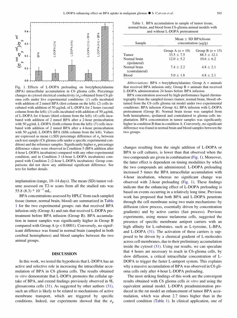
Experiment 1In Fig. 1, mean (# SD) percentage differences between sp
values measured in test and in reference samples are reportedfor each condition. The ANOVA analysis including the fiveexperimental conditions (described earlier) revealed thepresence of significant differences among them. SKN testdemonstrated significantly higher sp percentage differencevalues in Condition 5 (BPA addition after 4-hour L-DOPAincubation) compared with any other condition. Moreover,significantly higher sp percentage difference values werefound in Condition 3 (4-hour L-DOPA incubation) comparedwith Condition 2 (2-hour L-DOPA incubation). To excludethat the sp percentage difference observed in Condition 5was the result of an independent accumulation of L-DOPAand BPA at two different times, we performed the followingadditional comparison: [mean sp percentage difference ob-served in Condition 5] vs [(sp percentage difference observedin Condition 1) + (sp percentage difference observed in con-dition 3)]. The statistical result showed significantly highersp percentage values in Condition 5 than in Condition 1and Condition 3.
Experiment 2All rats survived the surgical implantation of C6 glioma
cells. As shown by MRI data collected 5 days after operation(baseline), all animals developed a brain tumor with an aver-age diameter of 1 mm. Longitudinal MRI monitoring demon-strated tumor growth in all rats. On average, tumor lesionsreached a diameter $2 mm 12 days after surgical
564 I. J. Radiation Oncology d Biology d Physics Volume 72, Number 2, 2008
implantation (range, 10–14 days). The mean (SD) tumor vol-ume assessed on T2-w scans from all the studied rats was55.8 (8.3) * 10$3 mL.
BPA concentrations assessed by HPLC from each sampledtissue (tumor, normal brain, blood) are summarized in Table1 for the two experimental groups: rats that received BPAinfusion only (Group A) and rats that received L-DOPA pre-treatment before BPA infusion (Group B). BPA accumula-tion in tumor samples was significantly higher in Group Bcompared with Group A (p < 0.0001). Conversely, no signif-icant difference was found in normal brain (sampled in bothcerebral hemispheres) and blood samples between the twoanimal groups.
DISCUSSION
In this work, we tested the hypothesis that L-DOPA has anactive and selective role in increasing the intracellular accu-mulation of BPA in C6 glioma cells. The results obtainedin vitro demonstrate that L-DOPA promotes the cellular up-take of BPA, and extend findings previously observed in 9Lgliosarcoma cells (31). As suggested by other authors (31),such an effect is likely to be related to mechanisms of activemembrane transport, which are triggered by specificconditions. Indeed, our experiments showed that the sp
changes resulting from the single addition of L-DOPA orBPA to cell cultures, is lower than that observed when thetwo compounds are given in combination (Fig. 1). Moreover,the latter effect is dependent on timing modalities by whichthe two compounds are administered. L-DOPA preloadingincreased 5 times the BPA intracellular accumulation with4-hour incubation, whereas no significant change wasobserved with 2-hour preloading (Fig. 1). These findingsindicate that the enhancing effect of L-DOPA preloading isbased on events occurring in a relatively long time. Previouswork has proposed that both BPA and L-DOPA penetratethrough the cell membrane using two main mechanisms: bydiffusion (slow process, essentially driven by concentrationgradients) and by active carries (fast process). Previousexperiments, using mouse melanoma cells, suggested thepresence of specific membrane antiport carriers with anhigh affinity for L-substrates, such as L-tyrosine, L-BPA,and L-DOPA (31). The activation of these carriers is sup-posed to be driven by a chemical gradient of L-moleculesacross cell membranes, due to their preliminary accumulationinside the cytosol (31). Using our results, we can speculatethat 4 hours are necessary to reach in C6-glioma cells, byslow diffusion, a critical intracellular concentration of L-DOPA to trigger the faster L-antiport system. This explainswhy a massive accumulation of BPA was observed in C6 gli-oma cells only after 4-hour L-DOPA preloading.
The most striking findings of this work are the convergentresults obtained with C6 glioma cells in vitro and using theequivalent animal model. L-DOPA preadministration pro-duced in the rat model an enhancement of tumor BPA accu-mulation, which was about 2.7 times higher than in thecontrol condition (Table 1). In clinical application, one of
Fig. 1. Effects of L-DOPA preloading on borylphenylalanine(BPA) intracellular accumulation in C6 glioma cells. Percentagechanges in cytosol electrical conductivity (sp) obtained from C6 gli-oma cells under five experimental conditions: (1) cells incubatedwith addition of 2 mmol BPA (first column on the left); (2) cells in-cubated with addition of 50 mg/mL of L-DOPA for 2 hours (secondcolumn from the left); (3) cells incubated with addition of 50 mg/mLof L-DOPA for 4 hours (third column from the left); (4) cells incu-bated with addition of 2 mmol BPA after a 2-hour preincubationwith 50 mg/mL L-DOPA (forth column from the left); (5) cells incu-bated with addition of 2 mmol BPA after a 4-hour preincubationwith 50 mg/mL L-DOPA BPA (fifth column from the left). Valuesare expressed as mean (#SD) percentage difference of sp betweeneach test sample (C6 glioma cells under a specific experimental con-dition) and the reference samples. Significantly higher sp percentagedifference values were observed in Condition 5 (BPA addition after4-hour L-DOPA incubation) compared with any other experimentalcondition, and in Condition 3 (4-hour L-DOPA incubation) com-pared with Condition 2 (2-hour L-DOPA incubation). Group com-parisons did not show any additional significant difference. Seetext for further details.
Table 1. BPA accumulation in sample of tumor tissue,normal brain, and blood from C6-glioma animal models with
and without L-DOPA pretreatment
SampleMean # SD BPA/tissueconcentration (mg/g)
Group A (n = 10) Group B (n = 15)Tumor 33.5 # 7.5 88.3 # 12.1Normal brain(ipsilateral)
12.0 # 5.2 10.6 # 6.2
Normal brain(contralateral)
7.4 # 2.2 4.8 # 2.1
Blood 5.0 # 1.8 4.8 # 2.1
Abbreviations: BPA = borylphenylalanine; Group A = animalsthat received BPA infusion only; Group B = animals that receivedL-DOPA administration 24 hours before BPA infusion.BPA concentration assessed by high-performance liquid chroma-
tography from the sampled tissues (tumor, normal brain, blood) ob-tained from the C6 cells glioma rat model under two experimentalconditions: BPA infusion (Group A); BPA infusion with L-DOPApretreatment (Group B). Normal brain tissue was sampled fromboth hemispheres, ipsilateral and contralateral to glioma cells im-plantation. BPA concentration in tumor samples was significantlyhigher in condition B than in condition A. Conversely, no significantdifference was found in normal brain and blood samples between thetwo groups.
L-DOPA enhancing effect on BPA uptake in malignant gliomas d S. CAPUANI et al. 565

the main limitations for BNCT effectiveness is the insuffi-cient accumulation of 10B carrier into the tumor cells. Inthis perspective, our results are particularly encouragingand might be considered for future BNCT clinical trials inhumans. When comparing BPA concentration in blood andnormal brain, there was no significant difference betweenrats that received L-DOPA and rats that did not. This makesthe potential use of L-DOPA in BNCT of brain tumors evenmore attractive. Indeed, these results show the potential abil-ity of L-DOPA to induce a significant increase of BNCTeffectiveness (i.e., tumor cells disruption) without remark-able side effects (i.e., normal brain tissue damage) associated.The selective effect of L-DOPA on tumor tissues is likely tobe due to genetic abnormalities in C6-glioma cells, condition-ing an overexpression of antiport membrane carriers for Lsubstrates.
Thus far, other strategies have been proposed to improveBNCT effectiveness by increasing BPA or BSH tumor accu-mulation. Some authors employed agents such as Cereport(RMP-7) (40) or mannitol (40, 41) to induce BBB disruptionin different tumor models. Other researches focused on dif-ferent modalities of BPA administration, demonstratinghigher BPA concentrations in the L9-glioblastoma modelafter a prolonged intracarotid BPA infusion (6 hours vs 2
hours) (42, 43). These alternative strategies showed anincrease of indexes such as tumor:brain and tumor:bloodBPA concentration, suggesting a potential usefulness forBNCT. However, their potential toxicity has not been fullyinvestigated. Our study showed a remarkable and selectiveincrease of BPA uptake in tumor tissues using L-DOPA,which is a well-tolerated medication associated with modestside effects and low toxicity.
To the best of our knowledge, there are no published stud-ies that have tested in vivo the effect of L-DOPA on BPA up-take in brain tumors. Our results appear promising, especiallyfor their potential application to clinical therapeutic proto-cols. Nevertheless, this study has several limitations, suchas the lack of determination of L-DOPA and BPA concentra-tions from blood, tumor, and normal brain samples at differ-ent time points and the absence of assessment of the radiationeffects. Further animal studies are therefore needed not onlyto investigate further the effect of L-DOPA pretreatment ontumor BPA accumulation but also to evaluate the final resultthat may be obtained after BNCT radiation. In particular, ap-propriate protocols should be defined in terms of modalitiesof L-DOPA and BPA administration (i.e., doses and timing).These insights might eventually promote pilot studies in pa-tients with malignant gliomas who are eligible for BNCT.
REFERENCES
1. Central Brain Tumor Registry of the United States. StatisticalReport: Primary Brain Tumors in the United States, 1998-–2002. Hinsdale, IL: Central Brain Tumor Registry of the UnitedStates; 2006.
2. Kleihues P, CaveneeWK. Pathology and Genetics of Tumors ofthe Nervous System (World Health Organisation Classificationof Tumours). Lyon, France: IARC Press; 2000. p.314.
3. SauerweinW. Principles and history of neutron capture therapy.Strahlenther Onkol 1993;169:1–6.
4. Barth RF, Soloway AH, Goodman JH, et al. Boron neutron cap-ture therapy of brain tumors: An emerging therapeutic modality.Neurosurgery 1999;44:433–450.
5. Barth RF, Coderre JA, Vicente MG, Blue TE. Boron neutroncapture therapy of cancer: current status and future prospects.Clin Cancer Res 2005;11:3987–4002.
6. Yamamoto T, Nakai K, Matsumura A. Boron neutron capturetherapy for glioblastoma. Cancer Lett 2008;262:143–152.
7. Capala J, Stenstam BH, Skold K, et al. Boron neutron capturetherapy for glioblastoma multiforme: Clinical studies in Swe-den. J Neurooncol 2003;62:135–144.
8. Busse PM, Harling OK, Palmer MR, et al. A critical examina-tion of the results from the Harvard-MIT NCT program PhaseI clinical trial of neutron capture therapy for intracranial disease.J Neurooncol 2003;62:111–121.
9. Joensuu H, Kankaanranta L, Seppala T, et al. Boron neutroncapture therapy of brain tumors: Clinical trials at the finnishfacility using boronophenylalanine. J Neurooncol 2003;62:123–134.
10. Diaz AZ. Assessment of the results from the Phase I/II boronneutron capture therapy trials at the Brookhaven National Lab-oratory from a clinician’s point of view. J Neurooncol 2003;62:101–109.
11. Palmer MR, Goorley JT, Kiger WS, et al. Treatment planningand dosimetry for the Harvard-MIT Phase I clinical trial of cra-nial neutron capture therapy. Int J Radiat Oncol Biol Phys 2002;53:1361–1379.
12. Stenstam BH, Pellettieri L, Sorteberg W, et al. BNCT for recur-rent intracranial meningeal tumours—case reports. Acta NeurolScand 2007;115:243–247.
13. Miyatake S, Tamura Y, Kawabata S, et al. Boron neutroncapture therapy for malignant tumors related to meningiomas.Neurosurgery 2007;61:82–90.
14. Coderre JA, Turcotte JC, Riley KJ, et al. Boron neutron capturetherapy: Cellular targeting of high linear energy transfer radia-tion. Technol Cancer Res Treat 2003;2:355–375.
15. Kato I, Ono K, Sakurai Y, et al. Effectiveness of BNCT forrecurrent head and neck malignancies. Appl Radiat Isot 2004;61:1069–1073.
16. Kankaanranta L, Seppala T, Koivunoro H, et al. Boron neutroncapture therapy in the treatment of locally recurred head andneck cancer. Int J Radiat Oncol Biol Phys 2007;69:475–482.
17. Coderre JA, Glass JD, Fairchild RG, et al. Selective targeting ofboronophenylalanine to melanoma in BALB/c mice for neutroncapture therapy. Cancer Res 1987;47:6377–6383.
18. Elowitz EH, Bergland RM, Coderre JA, et al. Biodistribution ofp-boronophenylalanine in patients with glioblastoma multi-forme for use in boron neutron capture therapy. Neurosurgery1998;42:463–468.
19. Chadha M, Capala J, Coderre JA. Boron neutron-capturetherapy (BNCT) for glioblastoma multiforme (GBM) usingthe epithermal neutron beam at the Brookhaven National Labo-ratory. Int J Radiat Oncol Biol Phys 1998;40:829–834.
20. Chanana AD, Capala J, Chadha M, et al. Boron neutron capturetherapy for glioblastoma multiforme: Interim results from thePhase I/II dose-escalation studies. Neurosurgery 1999;44:1182–1192.
21. Soloway AH, Hatanaka H, Davis MA. Penetration of brain andbrain tumor. VII. Tumor-binding sulfhydryl boron compounds.J Med Chem 1967;10:714–717.
22. Snyder HR, Reedy AJ, Lennarz WJ. Synthesis of aromaticboronic acids, aldehyde boronic acids and a boronic acid analogof tyrosine. J Am Chem Soc 1958;80:835–838.
566 I. J. Radiation Oncology d Biology d Physics Volume 72, Number 2, 2008

23. Ono K, Masunaga SI, Kinashi Y, et al. Radiobiological evi-dence suggesting heterogeneous microdistribution of boroncompounds in tumors: Its relation to quiescent cell populationand tumor cure in neutron capture therapy. Int J Radiat OncolBiol Phys 1996;34:1081–1086.
24. Ono K, Masunaga S, Suzuki M, et al. The combined effect ofboronophenylalanine and borocaptate in boron neutron capturetherapy for SCCVII tumors in mice. Int J Radiat Oncol BiolPhys 1999;43:431–436.
25. Fairchild RG, Slatkin DN, Coderre JA, et al. Optimization ofboron and neutron delivery for neutron capture therapy. Pig-ment Cell Res 1989;2:309–318.
26. Setiawan Y, Halliday GM, Harding AJ, et al. Effect of L-10B-pboronophenylalanine-fructose and the boron neutron capturereaction on mouse brain dopaminergic neurons. Cancer Res1995;55:874–877.
27. Capuani S, Gili T, Cametti C, et al. Radiowave dielectric inves-tigation of boron compounds distribution in cultured tumourcells: Relevance to boron neutron capture therapy. Chem PhysLett 2002;360:79–84.
28. Kageji T, Otersen B, Gabel D, et al. Interaction of mercaptoun-decahydrododecaborate (BSH) with phosphatidylcholine: Rele-vance to boron neutron capture therapy. Biochim Biophys Acta1998;1391:377–378.
29. Capala J, Makar MS, Coderre JA. Accumulation of boron inmalignant and normal cells incubated in vitro with boronophe-nylalanine, mercaptoborane or boric acid. Radiat Res 1996;146:554–560.
30. Papaspyrou M, Feinendegen EL, Muller-Gartner HW.Preloading with L.Tyrosine Increases the uptake of boronophe-nylalanine in mouse Melanoma Cells. Cancer Res 1994;54:6311–6314.
31. Wittig A, SauerweinWA, Coderre JA. Mechanisms of transportof p-borono-phenylalanine through the cell membrane in vitro.Radiat Res 2000;153:173–180.
32. Cotzias G. L-Dopa for Parkinsonism. N Engl J Med 1968;278:630.
33. Hornykiewicz O. L-DOPA: From a biologically inactive aminoacid to a successful therapeutic agent. Amino Acids 2002;23:65–70.
34. Bordi F, Cametti C, Paradossi G. High-frequency dielectricstudy of side-chain dynamics in poly(lysine) aqueous solutions.Biopolymers 2000;53:129–134.
35. Di Pierro D, Lazzarino G, Pastore FS, et al. Determination ofboronophenylalanine in biological samples using precolumno-phthalaldehyde derivatization and reversed-phase high-performance liquid chromatography. Anal Biochem 2000;284:301–306.
36. Looyenga H. Dielectric constants of heterogeneous mixtures.Physica 1965;31:401–406.
37. Bredberg E, Lennemas H, Paalzow L. Pharmacokinetics oflevodopa and carbidopa in rats following different routes ofadministration. Pharma Res 1994;11:549–555.
38. Kent AP, Stern GM, Webster RA. The effect of benserazide onthe peripheral and central distribution and metabolism oflevodopa after acute and chronic administration in the rat. BrJ Pharmacol 1990;100:743–748.
39. Hsieh CH, Chen YF, Chen FD, et al. Evaluation of pharmaco-kinetics of 4-borono-2-(18)F-fluoro-L-phenylalanine for boronneutron capture therapy in a glioma-bearing rat model withhyperosmolar blood-brain barrier disruption. J Nucl Med2005;46:1858–1865.
40. Barth RF, YangW, Bartus RT, et al. Neutron capture therapy ofintracerebral melanoma: Enhanced survival and cure afterblood-brain barrier opening to improve delivery of boronophe-nylalanine. Int J Radiat Oncol Biol Phys 2002;52:858–868.
41. Yang W, Barth RF, Rotaru JH, et al. Boron neutron capturetherapy of brain tumors: Enhanced survival followingintracarotid injection of sodium borocaptate with or withoutblood–brain barrier disruption. Int J Radiat Oncol Biol Phys1997;37:663–672.
42. Joel DD, Coderre JA, Micca PL, Nawrocky MM. Effect of doseand infusion time on the delivery of p-boronophenylalanine forneutron capture therapy. J Neurooncol 1999;41:213–221.
43. Morris GM, Micca PL, Nawrocky MM, Weissfloch LE,Coderre JA. Long-term infusions of p-boronophenylalaninefor boron neutron capture therapy: Evaluation using rat brain tu-mor and spinal cord models. Radiat Res 2002;158:743–752.
L-DOPA enhancing effect on BPA uptake in malignant gliomas d S. CAPUANI et al. 567
Related Documents


