Klinik und Poliklinik für Interdisziplinäre Endoskopie des Universitätsklinikum Hamburg-Eppendorf (Direktor: Prof. Dr. med. N. Soehendra) Technical Modifications for a One-Step Circumferential Endoscopic Mucosa Resection (CEMR) to Eradicate High-Grade Dysplasia and Mucosal Carcinoma in Barrett`s Esophagus. Preliminary Results D i s s e r t a t i o n Zur Erlangung des Grades eines Doktor der Medizin dem Fachbereich Medizin der Universität Hamburg vorgelegt von Yan Zhong From Xiamen, Fujian Province People’s Republic of China 1

Welcome message from author
This document is posted to help you gain knowledge. Please leave a comment to let me know what you think about it! Share it to your friends and learn new things together.
Transcript
Klinik und Poliklinik für Interdisziplinäre Endoskopie des Universitätsklinikum Hamburg-Eppendorf
(Direktor: Prof. Dr. med. N. Soehendra)
Technical Modifications for a One-Step Circumferential Endoscopic Mucosa
Resection (CEMR) to Eradicate High-Grade Dysplasia and Mucosal Carcinoma
in Barrett`s Esophagus. Preliminary Results
D i s s e r t a t i o n
Zur Erlangung des Grades eines Doktor der Medizin
dem Fachbereich Medizin der Universität Hamburg
vorgelegt von
Yan Zhong
From Xiamen, Fujian Province People’s Republic of China
1

Angenommen vom Fachbereich Medizin der Universität Hamburg am ……………….. Veröffentlicht mit Genehmigung des Fachbereichs Medizin der Universität Hamburg. Prüfungsausschuss, die/der Vorsitzender: Prüfungsausschuss: 2. Gutachter/in: Prüfungsausschuss: 3. Gutachter/in:
2

Dedicated to my parents
3

Table of Contents 1. General 7
1.1 History, Definitions, Terminology 7
1.2 Pathogenesis, Epidemiology 11
2. Diagnostic Procedures 18
2.1 Clinical Symptoms 18
2.2 Radiology 19
2.3 Endoscopy 20
2.4 Biopsy, Histopathology 21
2.5 Complementary optical Techniques 22
2.6 Stability of Histopathologic Tests 23
2.7 Staging 25
3. Options for Treatment 26
3.1 Conservative Treatment 26
3.2 Surgery 26
3.3 Endoscopic Treatment 27
3.3.1 Ablative Techniques 27
3.3.1.1 Photodynamic Therapy (PDT) 28
3.3.1.2 Electrocoagulation 28
3.3.1.3 Argonplasma Coagulation (APC) 29
3.3.1.4 Laser Ablation 29
3.3.2 Endoscopic Mucosa Resection (EMR) 30
3.3.3 Endoscopic Eradication of BE 34
4
4. Present Study 35
4.1 Aims of the Study 35
4.2 Study Design, Criteria for Enrolling and Exclusions 35
4.3 Treatment Procedure 36
4.3.1 Instruments 37
4.3.2 Circumferential EMR 38
4.3.3 Statistical Analysis 41
5. Results 42 6. Discussion 45 7. Summary 53 List of Literature 57 Curriculum Vitae 65 Appreciations 67 Declaration for Authenticity 68
5
List of Abbreviations used in the Text
APC = Argon plasma coagulation
BE = Barrett`s Esophagus
CEMR = Circumferential endoscopic mucosa resection
EMR = Endoscopic mucosa resection
EUS = Endoscopic ultrasound
EUS-FNA = EUS-guided fine-needle aspiration
GERD = Gastroesophageal reflux disease
HGD = High-grade dysplasia
LES = Lower esophageal sphincter
LGD = Low-grade dysplasia
MBL-CEMR = Multiband-Ligator-CEMR
PDT = Photodynamic therapy
PPI = Proton pump inhibitor
SCC = Squamous cell carcinoma
SCE = Squamous cell epithelium
6

Figure 1: Portrait of Sir Norman R. Barrett * *Norman Rupert Barrett (1903-1979), originally from Adelaide, Australia, sEngland and thereafter remained as a consultant surgeon at St. Thomas HLondon for the duration of his life. He developed novel concepts relating tof peptic esophagitis and the consideration of putative condition, which he reas a “short esophagus”. His reflections on the subject of ectopic gastric muco“dented and dogma” of mid 20th century concepts of esophagitis, asubsequently spawned a gastroenterological obsession that has led to the reof novel disease entity whose diagnosis and therapy has fixated contegastroenterologist, surgeons and pathologists. 1. General 1.1 History, Definitions, Terminology The first report about formal alterations and transformations of th
esophageal epithelium was published in 1950 by Norm
BARRETT(1) with complementary remarks by himself from 195
(Figure 1). The first publication did not find much echo, but dur
following years there was already a collection of observations, ma
surgeons, with different interpretations of what they found.
himself came to some conclusions in a lecture at Mayo Hospita
7
tudied in ospital in o subject ferred to sa (1957) nd have cognition mporary
e distal
an R.
6/1957
ing the
inly by
Barrett
l which

was published in 1957 where he summarized the situation of the last 6
years(2) . He started from observations of ulcers in the esophagus and
interpreted a metaplastic distal mucosa as result of mechanic dislocation
of gastric mucosa or as congenital short esophagus. A pathogenesis of
the ulcers from heterotopic islands of gastric mucosa seemed unrealistic
to him, because these dystopic lesions are mainly found in the proximal
esophagus without any ulceration. He concluded his text with this
statement: “I submit that most of these cases are in truth examples of
congenital short esophagus, in which there is neither general
inflammation nor stricture formation, but in which a part of the stomach
extends upwards into the mediastinum – or even to the neck – and that in
this stomach a typical chronic gastric ulcer can form.”
Obviously a period of confusions continued a further couple of years,
mainly because of inconsistent definitions and nomenclature. Barrett
described the situation and the problem of confusions in his lecture at the
Mayo Clinic, Rochester, in 1957: “This paper concerns a condition
whose existence is denied by some, misunderstood by others – and
ignored by the majority of surgeons. It has been called a variety of
names which have confused the story because they have suggested
incorrect etiologic explanations; congenital short esophagus, ectopic
gastric mucosa, short esophagus, and the lower esophagus lined by
gastric epithelium are but a few. At the present time the most accurate
description is that it is a state in which the lower end of the esophagus is
lined by columnar epithelium. This does not commit us to ideas which
could be wrong, but it carries certain implications which must be
clarified. The literature about esophageal disorders is confused because
common words have different meanings in the minds of different
writers.”
8

But also this verbal intervention could not stop the confusion of
definitions and nomenclatures. This is as more astonishing as Barrett
pointed out very clearly that this atypical mucosa of the distal esophagus
is not “Ectopic” from gastric mucosa but a completely different and new
nosologic entity: “These findings…suggest that the abnormal epithelium,
despite it looks, does not function exactly as stomach and probably
secretes little digestive juice.” And some lines further down: “Surgeons
who have studied the histology of specimen removed at operation have
found that the greater part of the unusual epithelium consists of simple
tubular glands which secrete mucus but which include few gastric
elements.”
He furthermore emphasizes that this must be an acquired phenomenon
and not embryonic resting tissue and he already assumed the correct
hypothesis about pathogenesis, which has been ascertained later: “One
of the facts which are difficult to explain is why this deformity always
involves the lower esophagus. No specimen has as yet been described in
which the whole of the gullet is lined by columnar cells. The explanation
could be that if the cardiac valve of a normal person were to become
incompetent and of the lower esophagus were, as a result, to be bathed
for a long time by digestive gastric juice, the squamous epithelium could
be eaten away and totally replaced by more quickly growing columnar
cells. This concept might explain the site of the deformity, the fact that
many cases occur in patients who have an incompetent cardia due to
sliding hiatal hernia, and the fact that many patients are elderly and
have history of heartburn dating back many years.”
But even this later statement of 1957 about his discovery from the year
1950 lists a couple of errors, e.g. his hypotheses to stricture development,
9

of ulcerogenesis and of formal typing of carcinoma within the
metaplastic segment.
During the following thirty years an intensified work about Barrett’s
phenomenon started and continued producing some more modifications
of definitions and nomenclature (3, 4). An important motivation for this
growing interest was based on an increasing prevalence of reflux
symptoms, reflux lesions and of adenocarcinoma of the esophagus (5, 6) .
Finally, Reid et al (7) made a proposal for implementation of a generally
accepted definition and classification. It is based on the presence of
specialized intestinal metaplasia of the distal esophagus and on the
extend of this changes of less than 3 cm (=short segment) or more than 3
cm (= long segment) proximal to the esophagogastric junction. A further
differentiation of an additional ultrashort-segment BE with only focal
metaplastic areas is not generally accepted and practiced (5).
Reflux- and time-associated consequences are observed as different
intensities of dysplasia of the Barrett-epithelium. According to a
proposal from Morson et al(8) they are differentiated to low-grade
dysplasia (LGD) and high-grade dysplasia (HGD). Some authors
supplemented an intermediate type, others tried to establish some sub-
classifications, e.g. epithelium of fundic type, of junctional type or
cardiac type(9, 10). These intentions for modification could not be found in
later publications and have obviously not been of general interest,
because all alterations of Barrett’s epithelium have their origin from
specialized intestinal metaplasia.
The most recent classification by expert consensus has been published
and named in 2001 according to the place of the conference: Vienna
Classification (Figure 2).
10

Figure2: The revised Vienna classification of gastrointestinal epithelial neoplasia
Category Diagnosis Clinically equivalent terms clinical management 1 Negative optional follow-up
for neoplasia 2 indefinite follow-up for neoplasia 3 Mucosal low-grade LGIN, low-grade Endoscopic resection
neoplasia adenoma/dysplasia or follow-up* 4 Mucosal high-grade Endoscopic or surgical neoplasia local resection* 4.1 HGIN, high-grade adenoma/dysplasia 4.2 HGIN, non-invasive carcinoma(CIS) 4.3 Suspicious for invasive carcinoma 4.4 intramucosal carcinoma 5 Submucosal or deeper Surgical resection*
invasion by carcinoma LGIN, low-grade intraepithelial neoplasia; HGIN, high-grade intraepithelial neoplasia,including both categories 4.1 and 4.2; CIS, carcinoma in situ; intramucosal, invading into the lamina propria or muscularis mucosae. *choice of treatment will depend on the size of the lesion, the depth of invasion as assessed endoscopically, radiologically, or ultrasonographically, the histological differentiation grade, and on general factors such as the patient’s age and co-morbid conditions. 1.2 Pathogenesis, Epidemiology
Metaplastic transformation of the epithelium correlates significantly with
an increased reflux of acid and biliary-intestinal secretion, including
intensity and duration of the exposition, as it has been supposed by
Barrett in 1950(1) and proved by Stein et al(5)using detailed experiments
(Figures 3,4).
11

Figure 3: BE and different complications correlated to acid exposition, calculated as percentage of 24-hours-period(5)
0
4
8
12
16
20
24
ControlSubjects
RefluxEsophagitis
EG-JunctionIntestinalMetaplasia
ShortSegmentBarrett
Long SegmentBarrett
EarlyBarrettCancer
Mea
n di
stal
eso
phag
eal A
cid
Expo
sure
Ti
me
on p
H m
onito
ring
(% o
f 24
Hou
rs)
** **
* *
*P<0.01 vs control subjects. **P<0.01 vs control subjects and patients with reflux esophagitis.
N=20 N=65 N=22 N=28 N=38 N=19
12

Figure 4: BE and different complications and their correlation to exposition
**P<0.01 vs control subjects and patients with reflux esophagitis
he influence of acid-free reflux on the development of metaplasia could
Barrett’s
rom observations of the progress to dysplasia it has been found that this
with biliary and intestinal reflux, calculated as percentage of a 24-hours-period(5)
0
4
8
12
16
20
24
28
ConSubjects
reesophagitis
EG-JuIntestinalMetaplasia
ShSegmentBarrett
Long Barrett
EBarrettCancer
N=20 N=65 N=22 N=28 N=38 N=19
me
** **
Mea
n D
ista
l Eso
phae
al B
ile E
xpos
ure
Tion
BIL
ITEC
Mon
itorin
g(%
of24
Hou
rs)
*
*
Segment arlyorttrol flux nction
*P<0.01 vs control subjects. .
.
T
be demonstrated very clearly by Meyer et al (11) in their report about
totally gastrectomized patients. The significance of functional barriers by
the lower esophageal sphincter, of cellular protection and of the
molecular background of these processes still remains unclear.
Observed familiar accumulations of BE and probably also of
carcinoma need further genetic and molecular biologic analyses(12) F
process is generally not fast and not obligatory. Schnell et al(13) observed
a sample of 1099 patients with BE out of a pool of 1125 totally for more
13

than 20 years. They found in 230 cases (=20.9%) constant metaplasia
without alterations, in 738 cases (= 67.2%) a constant low-graded
dysplasia and in 79 cases (= 7.2%) high-graded dysplasia. For the
primary status they revealed pre-existent carcinoma in 42 cases (= 3.8%).
During the observation period carcinoma developed from LGD in 10
patients (= 0.9%) and from HGD in 16 patients (= 1.5%). As result they
concluded, that for the vast majority of patients i.e. 63 of 79 (= 85%)
even HGD will not develop adenocarcinoma. This is different from the
conclusions of Hameeteman et al(10) who suppose a continuous
progression to different grades of dysplasia and out of an observation
from 5 patients with Barrett’s carcinoma they made risky extrapolations:
“Our data support the concept that a sequence of progression of
dysplasia to carcinoma exists. The time it takes such development shows
considerable variation, and high-grade dysplasia has been found to exist
far as long as 3.5 yr without evidence of carcinomatous degeneration.”
By analysis using multivariate regressions for their large sample of
he problem of potential progression from dysplasia to carcinoma has
patients with BE Schnell et al(13) found out, that there was only one
significant correlation, namely between the length of Barrett’s segment
and the risk of carcinoma. According to the rules of probability this
could be expected.
T
also been evaluated by Shaheen et al(14)using meta-analysis of 25
suitable publications out of a pool of 554 publications, the majority of
which with lacks of information. They evaluated a correlation between
cancer risk and several factors: size of the study, the definition of BE,
retrospective vs. prospective nature of the study, surveillance interval
14

and the effect of cancer detected in the first year of surveillance. In spite
of several further unsolved problems, they came to the following
conclusion: “In conclusion, in studies reporting the incidence of
adenocarcinoma of the esophagus in the setting of BE, there is a strong
inverse relationship between the seize of the study and the reported
cancer risk......Publication bias, such smaller studies are published only
if they feature high cancer risk, is a possible explanation for the
observation”
This problem of overestimating the risk to develop carcinoma was
able 1: Prevalence and estimated risk to develop carcinoma in different opulations(16)
Land PJ/Ca M (Ca)
already pointed out by Hameeteman et al(10) and later by Spechler (15) in
an editorial. The first group demonstrated the uncertainty for risk
calculations for prevalence of Barrett’s carcinoma showing large
variations from 0% to 46.5% with a mean of 10%. For follow-up studies
during longer periods they calculated incidences of carcinoma between 1
to 82 patient yrs and 1 to 441 patient years. This means a 30-40 fold risk
to develop carcinoma in BE compared to normal population (Table 1-3). Tp Author Cameron et al 1985 USA 440 2 Robertson et al 1988 EB 56 3 Van der Veen et al 1989 NL 170 4 Hameeteman et al 1989 NL 52 5 Ovaska et al 1989 SF 55 3 PJ = patient’s year
15

Table 2: Epidemiology and risk of carcinogenesis in BE calculated in incidences per patient year, showing a large range in observations(17) Author No. of patient-years No. of cancers Incidence (per
patient-year)
Spechler* 1984 350 2 1 in 175 Sprung 1984 162 2 1 in 81 Cameron* 1985 884 2 1 in 441 Sampliner 1985 92 1 1 in 92 Achkar 1988 166 1 1 in 166 Robertson 1988 218 3 1 in 56 Van der Veen* 1989 681 4 1 in 170
Ovaska 1989 166 3 1 in 55 Hameeteman 1989 269 5 1 in 52 Skinner 1989 145 3 1 in 48 Williamson 1991 497 5 1 in 99 *means “by postal inquiry”, all other studies are based on endoscopic and histopathologic examinations.
Table 3: Variations in calculated risk for carcinoma in BE during surveillance (10)
Study, year (ref) case/patient-year follow-up Skinner, 1989 1/48
Hameeteman, 1989 1/52 Robertson 1988 1/56 Present study 1/73 Williamson 1991 1/99 Van der Veen 1989 1/170 Spechler 1984 1/ 175 Mean 1/100 To find out the prevalence of BE for different ages, Cameron et al(18)
analyzed the data of 51.311 patients from Olmsted County (Minnes) who
contacted medical institutions for any reason. Their results show a close
correlation of BE to the male population, to people of older ages and
with longer history of reflux (Figure 5).
16

Further epidemiologic data form 9 counties of the USA from 1979 to
1987 have been collected by Blot et al(19). Analyzing the reports of 9406
patients with carcinoma of the esophagus, they found an increasing
importance of BE because the frequencies of adenocarcinoma of the
lower esophagus grew for more than 100% with special burden for the
white male population and the age of 55 yr and more. These results are
comparable to the conclusions of Falk(12) and the empiric results of
Wright et al(17).
Figure 5: Prevalence of BE depending on the age of analized population. Results of epidemiologic research in Olmsted County (Minnes). During endoscopic procedure of 51,311 people contacting health care institutions. Clinical prevalence: 18/100,000 and Autopsy-prevalence: 376/1000, 000(18) .
00.10.20.30.40.50.60.70.80.9
11.11.21.3
0-9
10--19
20-29
30-39
40-49
50-59
60-69
70-79
80-89
Age, yr
Barrett's esophagus %
Males
Males andfemales
Females
A similar reverse setting to evaluate the importance of BE for the
development of adenocarcinoma was tried by Dulai et al(20). They
analyzed the literature about operated adenocarcinoma of the esophagus
for references to BE. They found only 4.7% ± 2.9% documented
diagnoses of BE as precursors and they concluded that the situation is
17

still open using this access of research. Their final statement lines out a
different aspect for an explanation for this unexpected low rate of BE
found in their study: “These data thus provide a clear and compelling
rationale for the development of effective screening strategies to identify
patients with Barrett’s esophagus.”
Falk(12) points out that there is a close connection of prevalence of BE to
the strictness of definition: in patients with reflux symptoms he
described metaplasia in 12%, when the size of metaplastic area was not
precisely documented and only in 5% when the size was precisely
determined with 3 cm or more.
2. Diagnostic Procedures 2.1 Clinical Symptoms
There are no clear clinical criteria, symptoms or combinations of
symptoms that can guide to the diagnosis of BE. Chronic reflux disease
(GERD) and BE are similar in all essentials, (Table 4). Table 4: Clinical symptoms in patients with BE. There is a close coincidention to the symptoms of reflux esophagitis(21) Symptoms BE Esophagitis Total Heartburn 137 172 309 Dysplasia 8 3 11 Vomiting/nausea 4 4 8 Asymptoms 19 26 45
18
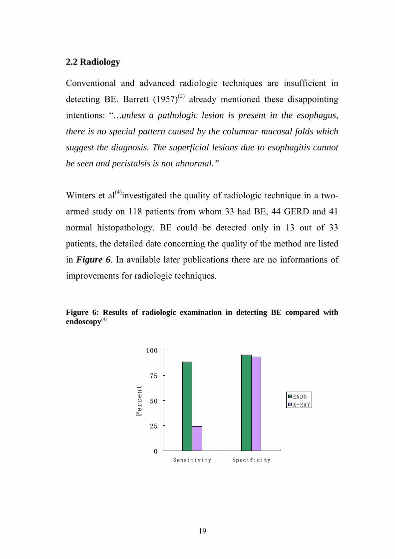
2.2 Radiology Conventional and advanced radiologic techniques are insufficient in
detecting BE. Barrett (1957)(2) already mentioned these disappointing
intentions: “…unless a pathologic lesion is present in the esophagus,
there is no special pattern caused by the columnar mucosal folds which
suggest the diagnosis. The superficial lesions due to esophagitis cannot
be seen and peristalsis is not abnormal.”
Winters et al(4)investigated the quality of radiologic technique in a two-
armed study on 118 patients from whom 33 had BE, 44 GERD and 41
normal histopathology. BE could be detected only in 13 out of 33
patients, the detailed date concerning the quality of the method are listed
in Figure 6. In available later publications there are no informations of
improvements for radiologic techniques.
Figure 6: Results of radiologic examination in detecting BE compared with endoscopy(4)
0
25
50
75
100
Sensitivity Specificity
Percent
E N DO
X-RAY
19

2.3 Endoscopy It was again Barrett (2) who obviously already had a high estimation of
endoscopy, despite the lack of modern instruments, when he stated:
“Esophagoscopy is an essential preoperative investigation in all patients
suffering from diseases in the esophagus. The diagnosis should be
suspected if the change-over of the mucous membrane is found at a high
level. As the epithelial transition is sharp, there should be no difficulty in
marking its point unless there is local inflammation. If there is a doubt,
pinch biopsies will settle the matter.”
This situation did not change essentially during the time following:
visible endoscopic criteria are not sufficient to identify BE from other
alteration, sometimes even from normal squamous epithelium. Larger
studies like that of Conio et al(21)about endoscopic macromorphology for
the diagnosis of BE cannot show sufficient quality of conventional
diagnostic criteria: sensitivity, specificity, accuracy and predictive values
(Table 5).
Table 5: Sensitivity and Specificity of endoscopic macroscopic diagnosis in BE. The results are not satisfying showing a sensitivity of 69.3%, a specificity of 63.1%, a pos. predictive value of 84%, a neg. predictive value of 62.7% and an accuracy of 66.5%(21) Endoscopic diagnosis Histological diagnosis Total Metaplasia (BE) Reflux esophagitis BE 142 62 (=30.4%) 204 Reflux grade Ⅱ 26 58 84 Reflux grade Ⅲ 10 26 36 Peptic stenosis 9 10 19 Peptic ulcers 6 4 10 More than 1 lesion 12 8 20 Total 205 168 373
20

The statement of Falk(12) is remarkable, when he cites observations from
Spechler et al(22) about their diagnoses of BE without any visible
alteration of the esophagus in 26 out of 142 patients (18%). Similar
experiences with BE diagnosis in normal appearing mucosa of the lower
esophagus were made by Nandukar et al(23), Johnston et al(24), Hirota et
al(25),Voutilainen et al(26), Pereira et al(27), Trudgill et al(28) and by
Ormsby et al(29)in autopsies.
By current definition BE is a specialized intestinal metaplasia of SCE of
the distal esophagus. That means the diagnosis of BE is based on the
results of histology of biopsy particles.
2.4 Biopsy, Histopathology
Biopsy does not show inherent reliability, even if all technical proposals
for good quality had been taken into consideration: four quadrant
biopsies in short distances, minimum 12 particles in suspected short
segment BE and more in suspected long-segment BE.
The diagnostic safety can be improved by double-checks after short
intervals. This design was used by Bonelli et al(16) in a multicenter study
analyzing the results of 157 followed up patients from a pool of 405
patients. Second biopsies at one year follow-up revealed that 13.4% of
the initial histologic findings were incorrect (Table 6).
21

Table 6: Initial histologic diagnosis compared to the second one after one year. The data showing the histologic findings of 157 followed up patients from a sample of 405. In this group of 21 newly detected cases of metaplasia, two adenocarcinomas were found (16)
First examination follow-up examination BE Negative BE 100 13 Negative 21 (=15.4%) 23
The value of technical details during biopsy taking has been
demonstrated by Reid et al(30) using a large-scaled study of
histopathologic examinations of 48 patients with adenocarcinoma and
123 with HGD in BE. They found out that accuracy of the diagnosis
could be improved by 100%, when they reduced the distances of biopsy-
sites from 2 cm to 1 cm. In visible altered mucosa the error rate for 2 cm
intervals was still 29%. It is also remarkable that even in resected
specimens early mucosal carcinoma (n=36) could be detected only in
39% of the patients using conventional techniques of histopathologic
sections. This error mainly (i.e. for 96%) concerned carcinoma restricted
to the mucosa (T1m).
2.5 Complementary Optical Techniques Additional staining techniques for targeted biopsies are obviously unable
to improve the diagnostic results. Egger et al(6) investigated vital staining
with methylene-blue and autofluorescence markers in 345 biopsy
particles of 35 patients with BE. They found high specificity for the two
techniques (91%) in differentiating metaplasia from HGD. But
22
the sensitivity rates of 21% for autofluorescence and 37% for methylene-
blue staining were rather disappointing. Comparable results were found
by Wo et al(31) whereas other groups saw some improvements using vital
staining (32, 33).
Some new endoscopy-associated complementary optical techniques like
fluorescence-spectroscopy or coherence-tomography are still in clinical
experimental stage. A combination of different complementary
techniques seems to show some improvement of diagnostic quality (34) .
It remains unclear whether the use of all these additional techniques is
more effective than improved technique of biopsy, particularly for the
investigation of larger areas of BE.
2.6 Accuracy of Histopathologic Diagnosis Differences in interpretations of pathologists regarding BE and dysplasia
grades represent an additional problems. Falk(12) mentioned in an
editorial that the interobserver agreement among 20 pathologists for the
diagnosis of metaplasia without dysplasia was only 35%.
Reid et al(30) reported on interobserver variations among 9 pathologists
from 4 institutions in classifying BE into five given subclasses. They
found an agreement for HGD and mucosal carcinoma in 85% of the
specimen and 87%, respectively, whereas the agreement for metaplaisa
and LGD was only 72% (Table 7).
23

Table 7: Interobserver variations in histologic diagnoses of BE, dysplasia and intramucosal carcinoma. The study was based on analysis of 70 biopsy specimens by 8 pathologists(35) Round 1 Round 2 High-grade dysplasia + intramucosal carcinomar vs other 87% 85% Negative for dysplasia vs other 71% 72% Negative + indefinite vs other 75% 77% Negative vs indefinite + low-grade vs high-grade + intramucosal carcinoma 58% 61% Ormsby et al(29) concluded from their experiences in autopsies: “Because
different pathologists made many of the endoscopic and surgical
pathology interpretations, it is also possible that interobserver variation
in the diagnosis of intramucosal carcinoma contributed to the
discrepancy.”
Schnell et al(13) pointed out that this interobserver variability for the
classification of metaplasia, LGD and HGD does really exist, but is not
mentioned in the majority of all studies published. The group came to
the following statement: “However the few studies that report
intraobserver and interobserver variation show the greatest agreement
for higher grades of dysplasia. Agreement approached 85% for HGD
and intramucosal adenocarcinoma.” Hameeteman et al(10) also found a
higher interobserver variability for specimens with low-grade and
intermediate dysplasia.
24
2.7 Staging The task of a reliable pretherapeutic staging is particularly regarding the
identification of intramucosal carcinoma (T1m), in order to allow
performing endoscopic mucosa resection (EMR) with lower mortality
and morbidity. Only in this early stage of T1m the probability of lymph
node involvement is negligible (36-38).
Endoscopic ultrasound (EUS) is currently the method of choice for
staging of gastrointestinal tumors. For superficial tumors, high resolution
radial EUS probes with 20 and 30 MHz transducers are used. Linear
EUS-scopes with fine-needle aspiration (FNA) and colour-Doppler
facilities are used for lymph node staging. However, the overall accuracy
rate of EUS for T-staging of esophageal cancer is only 84% according to
collected data from 21 studies recently reported by Shim et al(39).
Accuracy rate for T1 is 80.5%, for T2 76%, for T3 92% and for T4 86%.
The overall accuracy rate of the N-staging is 77%, with 69% for N0,
89% for N1(40).With the aid of EUS-guided FNA the result of N-staging
is significantly improved up to 87%(41).
Owing to the lack of reliable pretherapeutic tumor staging, EMR (see
3.3.2) has been recommended as a diagnostic tool. EMR provides better
diagnostic tissues than biopsy. Superficial mucosal lesions with lifting
sign after submucosal saline injection are resected endoscopically using
diathermic snare. If histology of the specimen confirms that resection
was complete and the tumor is confined to the mucosa, EMR can be
considered curative therapy(42, 43).
25
3. Therapeutic Options 3.1 Conservative treatment, surveillance For BE with and without LGD there is strong consensus for conservative
treatment combined with different intervals for surveillance. LGD
should be re-confirmed by expert pathologist within 6 months.
Conservative treatment means: elimination of gastroesophageal reflux by
reduction of body mass index (BMI) and administration of PPI. Under
conservative treatment regression from metaplasia to normal SCE has
been observed. But such improvement occurs rather infrequently.
Surveillance endoscopy with four-quadrant biopsies in one-year interval
is recommended for patients with BE and LGD. For BE without
dysplasia, endoscopy surveillance with biopsy in 3-years interval is
considered adequate. In cases with large hiatal hernia and incompetence
of the LES surgical repair (laparoscopic fundoplication) may be
considered. BE with HGD may be an indication for resective surgery.
3.2 Surgery The rational for recommending surgery in BE with HGD is the high
coincidence of cancer detected in the surgical specimens. The
coincidence rate has been reported in surgical literature to be as high as
40%. Some cancers had even already infiltrated the submucosal layer (44,
45)This problem particularly applies to long-segment BE because of the
high incidence of multifocal lesions and the risk of missing these lesions
endoscopically. Falk et al(46) reported that endoscopy and 4-quadrant
biopsies at 2 cm intervals even with jumbo biopsy forceps missed cancer
in 33% of the cases. Many of these lesions were invisible endoscopically.
26

The operative mortality of total esophagectomy is ranging between 1%
and 10% depending on the skill of the surgeon with postoperative
morbidity of 10% to 50% (44, 45).
Lack of knowledge about the risk and speed of progression of HGD to
cancer has led to conducting follow-up studies. Schnell et al(13) reported
that only 16% of a total of 75 patients with BE and HGD developed
cancer during a mean of 7.3 years follow-up. During this follow-up
period, endoscopy surveillance with biopsy was performed every three
months. Patients who developed cancer and were compliant could be
cured with surgical or ablative therapy.
Due to the relatively high risk of surgery, endoscopic interventional
modalities have increasingly become popular in the therapy of BE with
dysplasia. The spectrum includes different mucosal ablative and
resection methods.
3.3 Endoscopic Treatment 3.3.1 Ablative methods Endoscopic ablation of BE uses either thermal or photodynamic devices.
The aim is to destroy the metaplastic and dysplastic epithelium allowing
the restoration by SCE. Major drawback of this treatment is lack of
histologic confirmation of complete eradication of BE. APC is currently
the most commonly used method for ablation of BE.
27

3.3.1.1 Argon Plasma Coagulation (APC) APC is a non-contact monopolar coagulation using argon gas as
transmitter. The penetration depth of APC is less and the risk of
perforation is therefore lower as compared to monopolar coagulation and
Neodym-YAG Laser. The technique appears to be suitable for
destruction of larger mucosa surface. Eradication rates reported in the
literature range between 38% and 99%. Transient mild retrosternal
discomfort and odynophagia were observed in most of the patients.
Esophageal stricture and bleeding as more serious complications
occurred in 0-7%(47-52). Complication rate may increase if high power
APC is used (53).
3.3.1.2 Photodynamic Therapy (PDT) PDT is a physicochemical ablation treatment based on accumulation of
photosensitizer in tissue. The principle of PDT is selective sensitization
of precancerous or malignant lesions using a systemically applicable
photosensitizer with subsequent endoscopically controlled,
photochemically induced tissue ablation. Following exposure with light
of an adjusted wave-length, zytotoxic reagents develop, mainly singlet
oxygen, which then selectively destruct neoplastic tissue. The depth of
treatment depends on the penetration of the Laser light and localization
of the photosensitizer in the esophageal wall.
Photofrin, Porfimer sodium is the only photosensitizer that received US
Food and Drug Administration approval for use in the esophagus. The
light exposure is performed 2-3 days after intravenous application of the
sensitizer under endoscopic control using a 1.5 to 2.5 cm cylindrical
28

diffuser or a windowed centering esophageal balloon(54). The major
problems of PDT are post-therapeutic stenosis and long-term skin
photosensitization hypersensitivity for 60-90 days.
A new second-generation photosensitizer being used in trials is 5-
Aminolaevulinic acid (5-ALA). It is administrated orally only 4-6 hours
before light exposure and has the advantage of limiting skin
photosensitization to 2 days. This method seems to be effective only for
very superficial lesions up to 2 mm in depth(37).
3.3.1.3 Electrocoagulation This contact thermal modality can also be used for ablation of BE.
However, application through the probe is pinpointed and is therefore
rather cumbersome and time consuming especially in long-segment BE.
For mucosal ablation only bipolar electrocoagulation should be used
because monopolar electrocoagulation is associated with higher
perforation risk due to the relatively deep penetration. One multicenter
study including a total of 58 patients with BE and no dysplasia reported
a complete BE ablation of 78% at 6 months by using multipolar
electrocoagulation. Transient mild chest pain and odynophagia occurred
in 36% of the patients and esophageal stricture in 2% (55).
3.3.1.4 Laser Coagulation Laser has the same disadvantage as electrocoagulation, namely
pinpointed application and time consuming. The penetration depth of the
energy is greater as compared with that of APC. Laser has therefore been
completely replaced by APC in the gastrointestinal endoscopy. There
have been only a few reports on the use of Nd:YAG-Laser and KTP-
29

Laser for BE ablation in rather small numbers of patients. The results are
comparable with those of APC (56, 57).
3.3.2 Endoscopic Mucosa Resection (EMR) This technique was first introduced in the seventies by Ottenjann(58) and
Deyhle(59) as snare biopsy for obtaining large mucosa samples in the
stomach. In 1976, Martin(60) first described the lift and cut biopsy
technique using a double-channel endoscope for submucosal samplings.
Tada et al(61)introduced the technique of “strip biopsy” using a double-
channel endoscope. Since then, the technique of EMR has gained rapid
popularity in Japan. Several technical modifications have been
introduced mainly for the treatment of early malignant lesions in the
esophagus and stomach (Figure 7).
Figure 7: Techniques of EMR
Lift and Cut using a double-channel endoscope (Tada et al. Gastrointest Endosc 1984) (61) Suck and Cut using an overtube (Makuuchi et al. Jpn J Surg Gastroenterol 1991) (62) Suck and Cut using a cap (Inoue et al.Surg Endosc 1990) (63) Suck and Cut using band ligation device
(Chaves et al.Gastrointest Endosc 1994) (64)
Simple Snare Resection without any addtional tool (Soehendra et al. Endoscopy 1997) (65)
30

The principle of most EMR techniques is either “lift and cut” or “suck
and cut”. Submucosal injection to lift the mucosa which was introduced
by Deyhle et al(66) is generally considered as useful for reducing the risk
of perforation. The “lift and cut” technique using a double-channel
endoscope has been the most commonly used EMR method in Japan
until 1998 (Table 8) (67)
Table 8: EMR methods for early gastric cancer used in Japan until 1998(67)
Author n Method complete incomplete
2CS 1CS EMRC EMRL EMR
Tada 334 334 - - - 78% 22%
Takekoshi 308 308 - - - 74% 26%
O-izumi 256 256 - - - 91% 9%
Takahashi 140 140 - - - 77% 23%
Misaki 115 115 - - - 47% 53%
Atsumi 113 113 - - - 63% 37%
Honmyo 62 62 - - - 69% 31%
Hiki 48 48 - - - 71% 29%
Fujisaki 187 - - - 187 62% 38%
Chonan 123 46 31 46 - 70% 30%
Tani 86 - - 86 - 98% 2%
Abe 60 25 35 - - 62% 38%
Total 1832 79% 4% 7% 10% 74% 26%
2CS=two channel scope; 1CS=one channel scope; EMRC=EMR with cap; EMRL=EMR with
ligation
The “en bloc” resection method using IT- (insulation-tipped diathermic),
flex, triangle or hook knife has been recently proposed to achieve higher
rate of complete removal of gastric cancer, hence reduction of the
31

recurrence rate (Figure 8)(68-70). Gotoda et al had shown a significant
reduction of recurrence rate from 5 % to 0% as compared to previous
piece-meal resection technique(71).
Figure 8: Different instruments for “en bloc” resection: IT knife, TT knife, flex
knife and hook knife.
Hook knife IT-knife
TT knife Flex knife
However, “en bloc” EMR is associated with a higher complication rate.
Perforations occurred in 5% in a large series but were endoscopically
manageable in 98% of the cases. The en-bloc EMR technique(68) is best
suitable for the stomach. For the esophagus with smaller lumen, this
technique is rather cumbersome.
In Europe, EMR is playing an increasing role in the treatment of early
malignant mucosal changes (HGIN and T1m) in BE, as the number of
this type of cancer is rising rapidly. The most commonly practiced
method in the esophagus is the “suck and cut” technique which was
popularized by Inoue et al(72). A special transparent plastic cap is
32

mounted to the distal end of the endoscope. Submucosal injection of 20-
30 ml saline solution is performed prior to resection. A special
asymmetric soft snare made of braided wire is used as it can be properly
placed in the inner gutter at the distal end of the cap. The tumor bearing
mucosa is first sucked into the cap and then snared. The resected
specimen is then sucked into the cap and retrieved by withdrawing the
endoscope (Figure 9). The soft snare is easily deformed and therefore
usually suitable for single use only. Another “suck and cut” technique
uses the single rubber band ligator to create a pseudopolyp enabling
snare polypectomy of flat mucosal lesions(73, 74).
Figure 9: “Suck and cut” EMR technique using a cap(63)
EMR: Inoue-Technique
Injection Suction Snaring
C=cap; N=needle; L=lesion; I=injection; M=muscle propria; S=snare
33
3.3.3 Endoscopic eradication of BE EMR performed in BE has been localized resections restricted to the
mucosa bearing malignant changes. Depending on the length of follow-
up periods, recurrence rate of tumor rises from 14 % up to 30 %(37, 75).
Most of these recurrences occurred as metachronous lesions which
emerged from the remaining BE. However, multifocal early malignant
changes are known to exist especially in long-segment BE suggesting
that some of the recurrences may be from preexisting synchronous
lesions. In fact, HGD or HGIN in BE represents a diagnostic problem.
Endoscopic recognition of these lesions even with the aid of methylene
blue staining and other currently available imaging techniques has not
been perfected as yet. Four quadrant random biopsies are not sufficient
enough in detecting all the early malignant changes(6, 31, 46). To avoid
development of cancer, endoscopic treatment of BE has been proposed.
Although several studies have shown encouraging results of thermal
ablation of BE, there still remain some uncertainties due to incomplete
removal and evidence of buried subepithelial BE glands after treatment. (48-50, 76, 77). The Department of Interdisciplinary Endoscopy at the
University Hospital Hamburg-Eppendorf has therefore recommended
circumferential EMR for complete removal of BE containing HGD or
IMC. Simple snare resection technique was performed to completely
remove BE in “piece-meal” fashion(78). Since this technique and other
“suck and cut” techniques using a cap or band ligation device are quite
cumbersome in removing long-segment BE, we have recently modified
the multiband variceal ligator (MBL) to facilitate multiple, extensive
mucosal resections.
34
Conio et al(79) first published the feasibility of circumferential EMR in an
animal study. The group from the Department of Interdisciplinary
Endoscopy at the University Hospital of Hamburg-Eppendorf reported
the first clinical results of circumferential EMR in BE using the simple
snare technique. EMR in this small series (n=12) was performed in 3-4
week intervals.
4. Present Study 4.1. Aims of the Study The aim of this study is to evaluate the feasibility and safety of the novel
“band and cut” technique in BE using the modified variceal multiband
ligating (MBL) device and to report on our preliminary clinical
experience with the modified MBL device used for circumferential EMR
of BE containing HGD and/or IMC.
4.2 Study Design, Criteria for Enrolling and Exclusion In this uncontrolled prospective study, consecutive patients with BE and
HGD and/or IMC referred to EMR during the first 10 months of 2004
were included. The trial has a fixed start point and open end. It followed
the regulations of the Declaration of Helsinki (1964) for biomedical
research in human in the revised versions of 1975 and 1983 and the
Recommendations for Good Clinical Practice (GCP) of the FDA.
Proven that the indication is given, informed consent was obtained orally
and written prior to treatment.
35

Criteria for inclusion were:
- BE regardless the length;
- Verified HGD and IMC by two pathologist;
- No suspicious regional lymph node in EUS;
- Consent of the patient;
- Limited number of inclusions to 10-15 patients for the first
calculations regarding efficiency and safety.
Exclusion criteria were:
- BE without Dysplasia or with LGD;
- Indefinite EUS finding in excluding involvement of the
submucosal layer and regional lymph nodes;
- No consent of the patient;
- General clinical risk with critical parameters according to
NYHA III and higher preventing safe endoscopy under
propofol anesthesia.
4.3. Methods and Instruments
All patients were clinically examined to evaluate their individual risk
profile, especially to rule out coagulopathy.
If there were no calculable risks - i.e. for otherwise healthy patients-, the
treatment was planned as outpatient procedure with observation for 6-8
hours in the department. All other patients were hospitalized.
Endoscopy and EMR were performed under conscious sedation using i.v.
propofol according to the individual need given by an assisting physician
experienced in intensive care treatment. Oxygen saturation and pulse
36

rate were continuously monitored using pulse oxymeter during the
endoscopic treatment and the post treatment observation.
The entire EMR procedure was recorded on mini disk and important
images were additionally stored in the computer.
4.3.1. Instruments
Therapeutic electronic endoscopes with 3.7 mm working channel were
used (GIF-1T 140/160, Olympus Co. Tokyo, Japan). Banding for
creating pseudopolyp was performed with a modified Six Shooter MBL
(Wilson-Cook, Winston-Salem, NC, USA) which allows for six banding
procedures. The modification of MBL (Wilson-Cook, Winston-Salem,
NC, USA) consists simply in widening the threading channel of the
cranking device from 2 mm to 3.2 mm (Figure 10). This allows for the
insertion of a 7 French catheter through the threading channel of the
cranking device into the 3.7 mm working channel of the endoscope.
Band ligation can be performed with the polypectomy snare still within
the working channel without any increased friction during winding of
the thread. This enables sequential banding and snare resection of
esophageal mucosa without the need to change the endoscope. With this
modified MBL, extensive or circumferential EMR can be accomplished
usng only a single endoscope within a relatively short time. Other 7
French accessories, such as argon plasma coagulation (APC) probe,
clipping device or hot biopsy forceps can also be introduced if required
without the need to retrieve the endoscope and the MBL device.
For resection, mini hexagonal polypectomy snare sized 1.5 x 2.5 cm
made of braided wire (AcuSnare SASMH-1, Wilson-Cook, Winston-
37

Salem, NC, USA) is used. This snare can be reused in the same session
for several resections owing to its shape’s stability.
Figure 10: Design of the modified cranking device of the multiband-ligator
(MBL) used for performing one-step CEMR. The threading channel is widened
from 2.0 mm to 3.2 mm.
ID 2.0 mm
ID 3.3 mm
4.3.2. Circumferential EMR The Barrett’s mucosa is first sucked into the ligating barrel and the
rubber band is deployed in the same manner like variceal ligation
creating a pseudopolyp. No submucosal saline injection prior to ligation
is required. The polypoid bleb is then immediately resected using pure
coagulating current (output 60 watt, setting 3). It does not matter
whether the snare is placed above or below the band. In most of the
cases, however, the snare will automatically lie below the rubber band.
Following each resection, the specimen and the detached rubber band are
pushed into the stomach by using the tip of the snare’s catheter or
flushed down by a water jet from a pump machine connected to the
accessory channel of the endoscope. The second ligation is performed by
sucking the adjacent mucosa with a bit overlapping ensuring that no
Barrett’s remnant remains. The procedure is started from the gastro-
38

esophageal junction and accomplished circumferentially until the entire
Barrett’s mucosa is completely removed (Figure 11 a-h).
Figure 11a-h: Endoscopic images. Procedure and result of MBL-CEMR. a. 5 cm long-segment BE with a IMC (nodule). b. Six-shooter MBL is targeted to the nodule. c. The IMC bearing mucosa area is sucked into the barrel and a pseudopolyp is created by ligating it. d. The polypectomy snare is placed around the pseudopolyp. e. The first resection is performed. The next pseudopolyp is being created by sucking the adjacent mucosa area with slight overlapping. f. After two sequentially performed resections no remnant of BE is seen in the resected area. g. The final endoscopic image after the CEMR has been accomplished. h. Endoscopic image at 6 month follow-up showing complete restoration of the BE with no recurrence.
Figure 17a Figure 17b
Figure 17c Figure 17d
39

Figure 17e Figure 17f
Figure 17g Figure 17h
At the end of the procedure all resected specimens are collected in the
stomach by using the Roth’s retrieval net basket (US endoscopy, Mentor,
OH, USA), and spread over a cork plate. Each of the specimens is
measured individually prior to formalin fixation (Figure 12).
40

Figure 12: Disposable 2.5mm Roth net basket for collecting the resected mucosa specimens (left). Resected specimens and measuring on a cork plate (right).
Following EMR, patients were put on proton-pump inhibitor (at least 40
mg/day) and pureed diet was recommended. The first endoscopic
follow-up was performed three weeks later on out-patient basis.
Repeated EMR sessions for cases of extensive Barrett’s segment were
then carried out in 3-4 weeks interval. If dysphagia occurred, patients
were advised to come back immediately. In case of stricture, bougienage
was performed using 27-38 French Savary-Gilliard dilators (Wilson-
Cook) depending on the stricture’s grade. 4.3.3 Statistical Analysis The data management was descriptive. For the study of correlations the
following non-parametric tests were used: Fisher’s exact test for
independent and the McNemar test for dependent samples using 5%
significance levels.
41

5. Results During a period of ten months total of 14 consecutive patients were
treated with the new MBL-EMR technique. There were 12 men and 2
women with a median age of 64 years (range 43-82yrs). All patients had
circumferential Barrett’s segment with a length of 2-10 cm (median 4
cm). 9 patients had long-segment BE (≥ 3 cm) and 5 short-Segment BE
(< 3 cm). In the initial biopsy prior to EMR, IMC was found in 11 and
HGD in 3 cases, respectively. No multifocal lesions were diagnosed.
In 5 of 14 patients, complete circumferential EMR was accomplished in
one session using 3-18 bands (median: 6). Six patients required a total of
2 sessions, one patient 4 sessions, and one patient 5 sessions until the
entire BE was removed. One patient having multifocal HGD and/or IMC
in 24 of a total of 49 specimens was finally recommended to surgery
because of technical difficulties caused by mural thickening after 4
sessions. The median number of EMR sessions was 2 (range 1-5). The
mean size of EMR specimens measured prior to formalin fixation was
14.2 ± 4.1 mm (range 7-22 mm).
Histology of the EMR specimens confirmed IMC in 8 and HGD in 3
cases, LGD in 2. In one patient, no dysplasia in BE could be detected. In
5 patients with long-segment BE, multifocal lesions were found (In cases
with multifocal lesions, the highest degree of dysplasia or malignant
changes was selected). In a total of 217 EMR specimens, 40 IMC and 43
HGD were detected histologically.
Rubber bands were deployed successfully in 217 of a total 236 shootings.
Deployment failure occurred in patients with mural thickening or scar
formation from previous EMR due to lack of compliance of the tissue to
suction.
42
Minor bleeding occurred in 4 patients which were controlled in all at the
end of the EMR procedure, employing only 1-3 clip in each patient. In
one patient with liver cirrhosis, additional bleeding from a submucosal
collateral vein was controlled by immediate obliteration using a total of
2 cc of cyanoacrylate/Lipiodol mixture (ratio 0.5:0.8 cc). 0.5 cc of the
mixture was administered per injection. Detailed description of our
technique of cyanoacrylate glue injection has already been described21.
Hemorrhages occurred equally in 2 patients with short-segment and 2
with long-segment BE.
Esophageal strictures occurred in 10 patients (71%), all following the
first circumferential EMR after a median of 7 days (range: 5-10) which
concur with the onset of dysphagia symptoms. A median of 5 (range: 1-
11) sessions of weekly bougienage were performed for relief of
dysphagia. Stricture developed in 7 patients with long-segment BE and 3
with short-segment BE. This difference was statistically not significant
(p>0.2).
In one patient, deep tear of the esophageal wall occurred 4 weeks after
EMR during the fourth session of bougienage which was performed
incrementally with 33, 36 and 38 French Savary-Gilliard dilators. A
limited resection of the distal esophagus and esophagogastric junction
and reconstruction by interposition of isoperistaltic pedicled jejunal
segment according to Merendino et al(80) was performed. The operation
was suggested by the surgeon because CT-scan revealed free air in the
mediastinum although patient was symptom free. Histological
examination of the resected specimen showed no remnant of BE. No
perforation was found. The postoperative course was uneventful.
The procedure time of each EMR session ranged from 30 to 60 minutes
(median 30 minutes).
43
Re-grading of histologic diagnosis occurred in 5 of 14 patients (36%).
There was a preoperative overgrading in 4 cases and an undergrading in
one case. The difference of the pre-EMR diagnostic errors between long-
segment BE (n=2) and short-segment BE (N=3) was statistically not
significant (p > 0.3).
The aim of the trial, a complete resection (eradication) of the BE in one
session, was achieved in 5 of 14 patients (36%), and almost achieved in
another 6 cases. The total numbers of sessions needed did not reveal
statistical significant differences between short-segement and long-
segment BE (p > 0.5). For this statistical analysis, one patient who
finally underwent surgery was excluded.
A summery of the data is shown in the Table 9 containing informations
about demography of patients, histopathology, numbers of EMR
sessions, total number of specimens, and outcome/complications.
44

Table 9: List of all patients treated in present study with demographic, histopathologic, EMR and outcome data. LB = long-segment BE; SB = short-segment BE.
N. Sex Ages Length Segment Histology Treatment Pieces Complications Follow-up [cm] LB/SB pre post sessions (n) (n) 1 M 64 5 LB IMC IMC 1 15 Stenosis 4x Bougienage
2 M 76 2 SB IMC IMC 1 3 Bleeding 1 clip
3 M 62 3 LB IMC IMC 4 17
4 M 43 9 LB HGD HGD 1 18 Stenosis 4x Bougienage
Operation
5 M 67 9 LB IMC IMC 4 49 Operation
6 M 72 10 LB HGD HGD 5 42 Stenosis 6x Bougienage
7 M 67 4 LB IMC IMC 1 4 Bleeding 3 clips
Stenosis 3x Bougienage
8 M 49 3.5 LB IMC HGD 2 5 Bleeding 1 clip
Stenosis 6x Bougienage
9 M 63 2 SB IMC IMC 1 6 Stenosis 6x Bougenage
10 M 57 4 LB IMC IMC 2 6 Stenosis 11x Bougienage
11 F 82 2 SB IMC HGD 2 14 Stenosis 1x Bougienage
12 F 77 2 SB HGD IMC 2 14 Bleeding 2 clips
13 M 56 2 SB IMC LGD 2 11 Stenosis 6x Bougienage
14 M 68 7 LB IMC BE 2 13 Stenosis 1x Bougienage
6. Discussion It is well established that patients with BE have an increased risk of
developing esophageal adenocarcinoma. The presence of dysplasia
appears to predate the development of adenocarcinoma (metaplasia-
dysplasia-adenocarcinoma sequence) in these patients. This is based on
three observations: 1) a number of retrospective studies have shown that
dysplasia was present in surgical specimens of 35-91% of patients with
distal esophageal adenocarcinoma(81,82); 2) approximately 50% of
patients who had an esophageal resection for the diagnosis of HGD have
been found to harbour small foci of adenocarcinoma on detailed
45

histologic examination of the resected specimens(83-85); and 3)
longitudinal studies in the same patients have shown the sequential
progression of BE to LGD, HGD and adenocarcinoma.
Based on collected data, Falk(12) calculated that risk of progression from
HGD to IMC after two years is 20-25% (Table 10). The risk increases
with the length of follow-up and also with the size of BE and the
dysplastic area(43).
Table 10: Risk of developing adenocarcinoma in BE with HGD during 1-7 years surveillance(12) Study N Developing cancer (%) Follow-up interval (yr)
Reid 76 59% 5 Buttar 100 14% (focal high-grade dysplasia) 3 56% (diffuse high-grade dysplasia) 3 Schnell 79 5% 1
16% 7 The estimated incidence of adenocarcinoma in patients with BE is
ranging from 0.2 to 2.0%(14, 15, 21, 86)The risk of malignant degeneration
may be greater the longer the segment of BE(87) but even patients with
short-segment BE are also at risk(88). In this study, 3 of 5 patients with
short-segment BE had IMC and two LGD.
Apart from the higher risk of having malignant changes, long-segment
BE has been found to harbour multiple lesions. Heitmiller et
al(45)reported that in 13 of 30 patients (= 43%) who underwent
esophagectomy for HGD adenocarcinomas were found. The tumor
stages in detail were: T1N0M0 in 8, T2N0M0 in 2, T3N0M0 in 2 cases
46
and T3N1M0 in 1 case. Intramucosal carcinoma was not separately
mentioned.
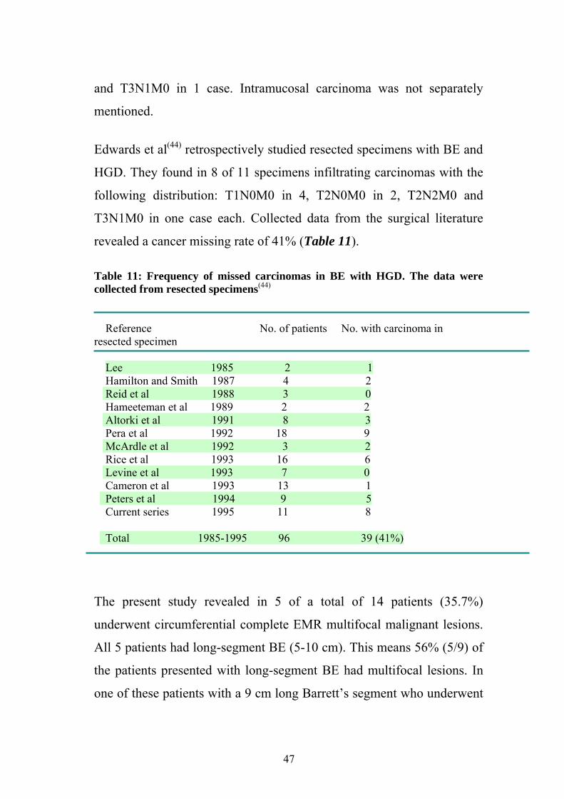
Edwards et al(44) retrospectively studied resected specimens with BE and
HGD. They found in 8 of 11 specimens infiltrating carcinomas with the
following distribution: T1N0M0 in 4, T2N0M0 in 2, T2N2M0 and
T3N1M0 in one case each. Collected data from the surgical literature
revealed a cancer missing rate of 41% (Table 11). Table 11: Frequency of missed carcinomas in BE with HGD. The data were collected from resected specimens(44)
Reference No. of patients No. with carcinoma in resected specimen
Lee 1985 2 1 Hamilton and Smith 1987 4 2 Reid et al 1988 3 0 Hameeteman et al 1989 2 2 Altorki et al 1991 8 3 Pera et al 1992 18 9 McArdle et al 1992 3 2 Rice et al 1993 16 6 Levine et al 1993 7 0 Cameron et al 1993 13 1 Peters et al 1994 9 5 Current series 1995 11 8 Total 1985-1995 96 39 (41%)
The present study revealed in 5 of a total of 14 patients (35.7%)
underwent circumferential complete EMR multifocal malignant lesions.
All 5 patients had long-segment BE (5-10 cm). This means 56% (5/9) of
the patients presented with long-segment BE had multifocal lesions. In
one of these patients with a 9 cm long Barrett’s segment who underwent
47
four sessions of MBL-EMR, HGD and/or IMC were found in 24 of a
total of 49 EMR specimens. This patient was finally referred to surgery
as EMR could not be accomplished due to technical difficulties caused
by mural thickening.
Another problem encountered in long-segment BE is that HGD and early
carcinoma in BE often occur in the absence of endoscopic abnormalities.
In a relatively high percentage of patients with BE, these early malignant
changes were detected incidentally. In 28 patients with HGD without
gross or microscopic evidence of carcinoma who underwent
esophagectomy, unsuspected cancer was found in 10 of 28 (36%) of
esophagectomy specimens. In 13 of these patients preoperative
endoscopy revealed only BE. Four-quadrant jumbo biopsies at 2-cm
intervals missed cancer in 33% of the patients (46). In this study, pre-
EMR diagnosis only detected a single malignant lesion in the 4 patients
in whom multifocal malignant lesions were found in the specimens after
circumferential complete EMR.
Local recurrence has been a major drawback of localized EMR. Long-
segment BE is especially prone to have a high recurrence rate after
localized EMR, although histological examination has shown complete
removal of the malignant lesion in the EMR specimen. The recurrence
rate rises with the length of the follow-up(37, 75). In the largest single
center series of EMR for HGD and early adenocarcinoma in BE,
recurrence rate has been as high as 33% during a mean follow-up of
34±10 months. The high recurrence rate may be explained by the
existence of multifocal malignant and premalignant lesions in BE which
48

have been overlooked in the initial diagnosis prior to EMR or by
metachronous development of new dysplastic tissue.
For these reasons, several authors have recommended ablative
treatments, such as APC or PDT. APC as the most commonly practiced
method for BE with or without LGD has been shown to be ineffective in
ablating BE. Greater length of BE is the major factor associated with a
high relapse rate. Buried glands were observed in up to 69% of the
cases(50).
PDT using photofrin as the most aggressive ablative treatment has been
used for treating BE with HGD and T1 cancer. On the intention-to-treat
basis, complete ablation was achieved in 53.8% of cases with HGD and
in only 33.3% of cases with cancer. Persistent BE was observed in
11.1% and 22.3%, respectively.
The major drawback of PDT and other thermal ablative treatments is the
lack of histologic confirmation.
Our group was the first to show that the problems of incomplete
treatment associated with multifocal lesions and high relapse rate might
be minimized by performing circumferential complete EMR. In our first
12 patients treated by the single snare resection technique, no residual
tumor tissue or metachronous recurrence during a follow-up of 9 months
was observed(78).
The present study is a continuation of this treatment concept, but
focusing on technical improvement.
The band and snare technique seems to be safe and more effective than
the cap technique(89) compared the two techniques for early esophageal
49

cancer in 72 patients. Hundred EMR were performed to resect mucosal
SCC and adenocarcinoma of the distal esophagus, 50 with each
technique in a randomized trial. The cap technique was carried out with
submucosal saline injection, and the band technique using the
euroligator (mandel + rupp medizintechnik gmbh, Erkrath, Germany).
No significant differences were found in size of the resected specimens
and size of ulcer measured 24 hours after EMR. Only one minor
bleeding occurred in each group. No perforation or other complications
were observed. Interestingly, the cap technique with submucosal saline
injection was associated with a higher technical failure rate as compared
to the band technique without submucosal injection (12% vs. 2%, p <
0.01).
Our long-term experience had proved that simple snare EMR technique
in the esophagus does not require prior submucosal injection. Neither
technical advantage nor prevention of perforation can be achieved by
submucosal injection. In this preliminary experience with the modified
MBL device, no involvement of the proper muscle layer was observed
histologically in the resected specimens. No perforation was also
documented.
The safety-profile of the circumferential EMR using the MBL device in
the present study is however tainted by an absolutely high rate of
strictures during the early postoperative week associated with clinical
symptoms of dysphagia. This problem has not been described after local
EMR(89) or seems to be of minor importance and infrequent (less than
3%) when local EMR is combined with PDT(37).
50
Stricture formation seems to be also a major problem in PDT when used
as a monotherapy. Overholt et al(77) reported a stricture rate of 30%, and
Falk et al(12) 36% after PDT with porfimer-sodium.
The only data about esophageal stricture formation following radical
circumferential EMR was from our group(78) with a stricture rate of
16.7% (2 in 12 cases).
In this present study, stricture occurred in 10 of 14 patients (71%), in
whom circumferential EMR was performed regardless the length of BE
and the total number of EMR sessions required for complete removal of
BE. Symptoms of dysphagia began 5-10 (median: 7) days after the
initial circumferential EMR. Bougienage was performed to dilate the
stricture using Savary-Gilliard dilators. Since the esophageal wall within
the first two weeks after EMR is still fragile, bougienage has to be
performed with extreme caution. In one patient with a 5 cm long BE
who underwent a circumferential EMR using a total of 18 rubber bands
in one session, deep tear of esophageal wall occurred during the fourth
bougienage although dilation was done incrementally and only up to 38
French.
Due to the very high stricture rate, EMR at the present time should not
be performed circumferentially in a single session. Complete endoscopic
removal of long-segment BE can be achieved by doing sequential EMR
accomplished in several sessions at 3-4 weeks interval. Systematic
longitudinal piece-meal resections of about 75% of the circumference up
to a maximal length of 4 cm of BE per session seems to be the most
appropriate approach to accomplish complete EMR. For this extent of
51
EMR, a six-shooter MBL is sufficient. Further experiences are warranted
to confirm this hypothesis.
Prevention of stricture formation must be the most important aim of any
EMR technique attempting for complete removal of BE. Following
proposals have been discussed: 1) Combination of different techniques
(multimodal therapy) as it has been practiced by(37) with local EMR plus
PDT or APC. 2) Use of stricture-preventing medication following EMR.
Radu(90)reported on significant reduction of stricture and perforation
rates by administrating Mitomycin C in an animal study.
Finally, procedure-related morbidity, long-term survival rate and quality
of life of patients after complete EMR need to be compared with those of
limited distal esophageal resection or radical esophagectomy. The risk of
regional lymph node metastasis in BE with HGD or IMC is negligible(91)
However, continuous PPI administration and regular endoscopic
surveillance are mandatory for patients treated with EMR to prevent
recurrent GERD, and to detect newly formed BE and eventually early
malignant changes as well. Long-term follow-up of larger number of
patients treated with complete EMR are therefore needed for a final and
firm conclusion.
The different technologies to treat HGD and intramucosal carcinoma in
BE have their specific advantages, drawbacks and risk profiles as shown
in Figure 13. Some of the drawbacks and risks are inherent to the
treatment procedures and cannot be significantly reduced by changing
e.g. the performance or protocol. Others are reducible to a certain extent
like the post-procedural stricture. In this context the technique of MBL-
52
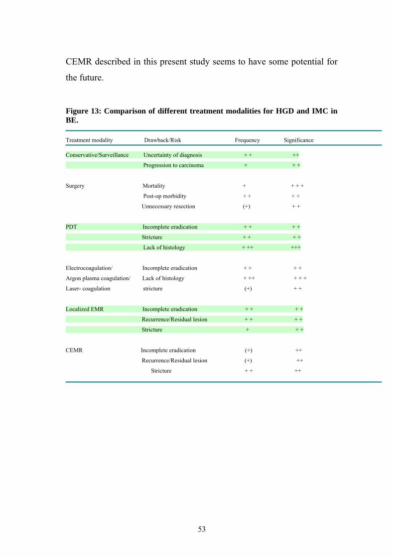
CEMR described in this present study seems to have some potential for
the future. Figure 13: Comparison of different treatment modalities for HGD and IMC in BE. Treatment modality Drawback/Risk Frequency Significance Conservative/Surveillance Uncertainty of diagnosis + + ++
Progression to carcinoma + + +
Surgery Mortality + + + +
Post-op morbidity + + + +
Unnecessary resection (+) + +
PDT Incomplete eradication + + + +
Stricture + + + +
Lack of histology + ++ +++
Electrocoagulation/ Incomplete eradication + + + +
Argon plasma coagulation/ Lack of histology + ++ + + +
Laser- coagulation stricture (+) + +
Localized EMR Incomplete eradication + + + +
Recurrence/Residual lesion + + + +
Stricture + + +
CEMR Incomplete eradication (+) ++
Recurrence/Residual lesion (+) ++
Stricture + + ++
53

7. Summary Various techniques are available for endoscopic mucosal resection
(EMR) in the upper and lower gastrointestinal tract. For early cancers of
the esophagus, “suck and cut” technique using a transparent cap or
variceal band ligator is the most commonly practiced method. To
facilitate multiple or circumferential EMR, a modified multiband
variceal ligator (MBL) is introduced which allows sequential banding
and snare resection without the need to withdraw the endoscope. To
enable band delivery with a snare inserted in the therapeutic endoscope,
the threading channel of the cranking device is enlarged from 2 mm to
3.2 mm. The six shooter MBL was used.
The aim of this study is to evaluate the feasibility and short-term results
of CEMR using this new device.
14 consecutive patients with BE containing HGD and/or IMC referred
during the first 10 months of 2004 to the Department of Interdisciplinary
Endoscopy, University Hospital Hamburg-Eppendorf were treated with
this new technique. There were 12 men and 2 women with a median age
of 64 years (range 43-82yrs). 9 patients had long-segment BE (≥ 3 cm)
and 5 short-Segment BE (< 3 cm). In the initial biopsy prior to EMR,
IMC was found in 11 and HGD in 3 cases, respectively. No multifocal
lesions were diagnosed. In 5 of 14 patients, complete circumferential
EMR was accomplished in one session using 3-18 bands (median: 6).
Six patients required a total of 2 sessions, one patient 4 sessions, and one
patient 5 sessions until the entire BE was removed. One patient having
multifocal HGD and/or IMC in 24 of a total of 49 specimens was finally
54

recommended to surgery because of technical difficulties caused by
mural thickening after 4 sessions. The median number of EMR sessions
was 2 (range 1-5). The procedure time of each EMR session ranged from
30 to 60 minutes (median 30 minutes).
The mean size of EMR specimens measured prior to formalin fixation
was 14.2 ± 4.1 mm (range 7-22 mm).
Histology of the EMR specimens confirmed IMC in 8 and HGD in 4
cases, LGD in 1. In one patient, no dysplasia in BE could be detected. In
5 patients with long-segment BE, multifocal lesions were found (56% of
patients with long-segment BE).
Minor bleeding occurred in 4 patients which were controlled in all at the
end of the EMR procedure. In one patient with liver cirrhosis who first
underwent TIPS, additional bleeding from a submucosal collateral vein
was controlled by immediate obliteration using a total of 2 cc of
cyanoacrylate/Lipiodol mixture. Hemorrhages occurred equally in 2
patients with short-segment and 2 with long-segment BE.
Esophageal strictures occurred in 10 patients (71%), all following the
first circumferential EMR after a median of 7 days (range: 5-10). A
median of 5 (range: 1-11) sessions of weekly bougienage were
performed for relief of dysphagia. Stricture developed in 7 patients with
long-segment BE and 3 with short-segment BE. This difference was
statistically not significant.
In one patient, deep tear of the esophageal wall occurred during the
fourth session of bougienage. A limited distal resection of the esophagus
was performed. Histological examination of the resected specimen
showed no remnant of BE. The postoperative course was uneventful.
55
The novel technique of MBL-EMR described here is a safe and effective
method which facilitates and simplifies circumferential removal of BE
containing HGD and/or IMC. However, the method is associated with a
very high stricture rate if circumferential EMR is performed in one
single session. Complete removal of BE should therefore be achieved by
repeated partial EMR. Long-term follow-up is needed to observe for late
recurrence and determining the clinical impact of this method in
comparison to surgery, especially to the limited distal esophageal
resection.
56
Literature 1. Barrett NR. Chronic peptic ulcer of the oesophagus and 'oesophagitis'. Br J Surg 1950;38(150):175-82. 2. Barrett NR. The lower esophagus lined by columnar epithelium. Surgery 1957;41(6):881-94. 3. Naef AP, Savary M, Ozzello L. Columnar-lined lower esophagus: an acquired lesion with malignant predisposition. Report on 140 cases of Barrett's esophagus with 12 adenocarcinomas. J Thorac Cardiovasc Surg 1975;70(5):826-35. 4. Winters C, Jr., Spurling TJ, Chobanian SJ, Curtis DJ, Esposito RL, Hacker JF, 3rd, et al. Barrett's esophagus. A prevalent, occult complication of gastroesophageal reflux disease. Gastroenterology 1987;92(1):118-24. 5. Stein HJ, Feith M, Feussner H. The relationship between gastroesophageal reflux, intestinal metaplasia and adenocarcinoma of the esophagus. Langenbecks Arch Surg 2000;385(5):309-16. 6. Egger K, Werner M, Meining A, Ott R, Allescher HD, Hofler H, et al. Biopsy surveillance is still necessary in patients with Barrett's oesophagus despite new endoscopic imaging techniques. Gut 2003;52(1):18-23. 7. Reid BJ. Barrett's esophagus and esophageal adenocarcinoma. Gastroenterol Clin North Am 1991;20:817-34. 8. Morson BC, Sobin LH, Grundmann E, Johansen A, Nagayo T, Serck-Hanssen A. Precancerous conditions and epithelial dysplasia in the stomach. J Clin Pathol 1980;33(8):711-21. 9. Paull A, Trier JS, Dalton MD, Camp RC, Loeb P, Goyal RK. The histologic spectrum of Barrett's esophagus. N Engl J Med 1976;295(9):476-80. 10. Hameeteman W, Tytgat GN, Houthoff HJ, van den Tweel JG. Barrett's esophagus: development of dysplasia and adenocarcinoma. Gastroenterology 1989;96(5 Pt 1):1249-56. 11. Meyer W, Vollmar F, Bar W. Barrett-esophagus following total gastrectomy. A contribution to it's pathogenesis. Endoscopy 1979;11(2):121-6. 12. Falk GW. Barrett's esophagus. Gastroenterology 2002;122(6):1569-91. 13. Schnell TG, Sontag SJ, Chejfec G, Aranha G, Metz A, O'Connell S, et al. Long-term nonsurgical management of Barrett's esophagus with high-grade dysplasia. Gastroenterology 2001;120(7):1607-19.
57
14. Shaheen NJ, Crosby MA, Bozymski EM, Sandler RS. Is there publication bias in the reporting of cancer risk in Barrett's esophagus? Gastroenterology 2000;119(2):333-8. 15. Spechler SJ. Barrett's esophagus: an overrated cancer risk factor. Gastroenterology 2000;119(2):587-9. 16. Bonelli L. Barrett's esophagus: results of a multicentric survey. G.O.S.P.E. (Gruppo Operativo per lo Studio delle Precancerosi Esofagee. Endoscopy 1993;25(9):652-4. 17. Wright TA, Gray MR, Morris AI, Gilmore IT, Ellis A, Smart HL, et al. Cost effectiveness of detecting Barrett's cancer. Gut 1996;39(4):574-9. 18. Cameron AJ. Epidemiologic studies and the development of Barrett's esophagus. Endoscopy 1993;25(9):635-6. 19. Blot WJ, Devesa SS, Kneller RW, Fraumeni JF, Jr. Rising incidence of adenocarcinoma of the esophagus and gastric cardia. Jama 1991;265(10):1287-9. 20. Dulai GS, Guha S, Kahn KL, Gornbein J, Weinstein WM. Preoperative prevalence of Barrett's esophagus in esophageal adenocarcinoma: a systematic review. Gastroenterology 2002;122(1):26-33. 21. Conio M. Endoscopic features of Barrett's esophagus. G.O.S.P.E. Gruppo Operativo per lo Studio delle Precancerosi Esofagee. Endoscopy 1993;25(9):642-4. 22. Spechler SJ, Zeroogian JM, Antonioli DA, Wang HH, Goyal RK. Prevalence of metaplasia at the gastro-oesophageal junction. Lancet 1994;344(8936):1533-6. 23. Nandurkar S, Talley NJ. Barrett's esophagus: the long and the short of it. Am J Gastroenterol 1999;94(1):30-40. 24. Johnston MH, Hammond AS, Laskin W, Jones DM. The prevalence and clinical characteristics of short segments of specialized intestinal metaplasia in the distal esophagus on routine endoscopy. Am J Gastroenterol 1996;91(8):1507-11. 25. Hirota WK, Loughney TM, Lazas DJ, Maydonovitch CL, Rholl V, Wong RK. Specialized intestinal metaplasia, dysplasia, and cancer of the esophagus and esophagogastric junction: prevalence and clinical data. Gastroenterology 1999;116(2):277-85. 26. Voutilainen M, Farkkila M, Juhola M, Nuorva K, Mauranen K, Mantynen T, et al. Specialized columnar epithelium of the esophagogastric junction: prevalence and associations. The Central Finland Endoscopy Study Group. Am J Gastroenterol 1999;94(4):913-8. 27. Pereira AD, Suspiro A, Chaves P, Saraiva A, Gloria L, de Almeida JC, et al. Short segments of Barrett's epithelium and intestinal metaplasia in normal appearing
58

oesophagogastric junctions: the same or two different entities? Gut 1998;42(5):659-62. 28. Trudgill NJ, Suvarna SK, Kapur KC, Riley SA. Intestinal metaplasia at the squamocolumnar junction in patients attending for diagnostic gastroscopy. Gut 1997;41(5):585-9. 29. Ormsby AH, Kilgore SP, Goldblum JR, Richter JE, Rice TW, Gramlich TL. The location and frequency of intestinal metaplasia at the esophagogastric junction in 223 consecutive autopsies: implications for patient treatment and preventive strategies in Barrett's esophagus. Mod Pathol 2000;13(6):614-20. 30. Reid BJ, Blount PL, Feng Z, Levine DS. Optimizing endoscopic biopsy detection of early cancers in Barrett's high-grade dysplasia. Am J Gastroenterol 2000;95(11):3089-96. 31. Wo JM, Ray MB, Mayfield-Stokes S, Al-Sabbagh G, Gebrail F, Slone SP, et al. Comparison of methylene blue-directed biopsies and conventional biopsies in the detection of intestinal metaplasia and dysplasia in Barrett's esophagus: a preliminary study. Gastrointest Endosc 2001;54(3):294-301. 32. Canto MI, Setrakian S, Petras RE, Blades E, Chak A, Sivak MV, Jr. Methylene blue selectively stains intestinal metaplasia in Barrett's esophagus. Gastrointest Endosc 1996;44(1):1-7. 33. Sharma P, Topalovski M, Mayo MS, Weston AP. Methylene blue chromoendoscopy for detection of short-segment Barrett's esophagus. Gastrointest Endosc 2001;54(3):289-93. 34. Georgakoudi I, Jacobson BC, Van Dam J, Backman V, Wallace MB, Muller MG, et al. Fluorescence, reflectance, and light-scattering spectroscopy for evaluating dysplasia in patients with Barrett's esophagus. Gastroenterology 2001;120(7):1620-9. 35. Reid BJ, Haggitt RC, Rubin CE, Roth G, Surawicz CM, Van Belle G, et al. Observer variation in the diagnosis of dysplasia in Barrett's esophagus. Hum Pathol 1988;19(2):166-78. 36. Natsugoe S, Aikou T, Yoshinaka H, Saihara T, Baba M, Takao S, et al. Lymph node metastasis of early stage carcinoma of the esophagus and of the stomach. J Clin Gastroenterol 1995;20(4):325-8. 37. May A, Gossner L, Pech O, Fritz A, Gunter E, Mayer G, et al. Local endoscopic therapy for intraepithelial high-grade neoplasia and early adenocarcinoma in Barrett's oesophagus: acute-phase and intermediate results of a new treatment approach. Eur J Gastroenterol Hepatol 2002;14(10):1085-91.
59

38. Vieth M, Ell C, Gossner L, May A, Stolte M. Histological analysis of endoscopic resection specimens from 326 patients with Barrett's esophagus and early neoplasia. Endoscopy 2004;36(9):776-81. 39. Shim CS. EUS staging of esophageal cancer. Digestive Endosc 2004;16:s153-6. 40. Rosch T. Endosonographic staging of esophageal cancer: a review of literature results. Gastrointest Endosc Clin N Am 1995;5(3):537-47. 41. Vazquez-Sequeiros E, Wiersema MJ, Clain JE, Norton ID, Levy MJ, Romero Y, et al. Impact of lymph node staging on therapy of esophageal carcinoma. Gastroenterology 2003;125(6):1626-35. 42. Nijhawan PK, Wang KK. Endoscopic mucosal resection for lesions with endoscopic features suggestive of malignancy and high-grade dysplasia within Barrett's esophagus. Gastrointest Endosc 2000;52(3):328-32. 43. Buttar NS, Wang KK, Sebo TJ, Riehle DM, Krishnadath KK, Lutzke LS, et al. Extent of high-grade dysplasia in Barrett's esophagus correlates with risk of adenocarcinoma. Gastroenterology 2001;120(7):1630-9. 44. Edwards MJ, Gable DR, Lentsch AB, Richardson JD. The rationale for esophagectomy as the optimal therapy for Barrett's esophagus with high-grade dysplasia. Ann Surg 1996;223(5):585-9; discussion 589-91. 45. Heitmiller RF, Redmond M, Hamilton SR. Barrett's esophagus with high-grade dysplasia. An indication for prophylactic esophagectomy. Ann Surg 1996;224(1):66-71. 46. Falk GW, Rice TW, Goldblum JR, Richter JE. Jumbo biopsy forceps protocol still misses unsuspected cancer in Barrett's esophagus with high-grade dysplasia. Gastrointest Endosc 1999;49(2):170-6. 47. Kahaleh M, Van Laethem JL, Nagy N, Cremer M, Deviere J. Long-term follow-up and factors predictive of recurrence in Barrett's esophagus treated by argon plasma coagulation and acid suppression. Endoscopy 2002;34(12):950-5. 48. Ackroyd R, Tam W, Schoeman M, Devitt PG, Watson DI. Prospective randomized controlled trial of argon plasma coagulation ablation vs. endoscopic surveillance of patients with Barrett's esophagus after antireflux surgery. Gastrointest Endosc 2004;59(1):1-7. 49. Basu KK, Pick B, Bale R, West KP, de Caestecker JS. Efficacy and one year follow up of argon plasma coagulation therapy for ablation of Barrett's oesophagus: factors determining persistence and recurrence of Barrett's epithelium. Gut 2002;51(6):776-80.
60

50. Van Laethem JL, Peny MO, Salmon I, Cremer M, Deviere J. Intramucosal adenocarcinoma arising under squamous re-epithelialisation of Barrett's oesophagus. Gut 2000;46(4):574-7. 51. Morris CD, Byrne JP, Armstrong GR, Attwood SE. Prevention of the neoplastic progression of Barrett's oesophagus by endoscopic argon beam plasma ablation. Br J Surg 2001;88(10):1357-62. 52. Schulz H, Miehlke S, Antos D, Schentke KU, Vieth M, Stolte M, et al. Ablation of Barrett's epithelium by endoscopic argon plasma coagulation in combination with high-dose omeprazole. Gastrointest Endosc 2000;51(6):659-63. 53. Pereira-Lima JC, Busnello JV, Saul C, Toneloto EB, Lopes CV, Rynkowski CB, et al. High power setting argon plasma coagulation for the eradication of Barrett's esophagus. Am J Gastroenterol 2000;95(7):1661-8. 54. Overholt BF, Panjehpour M, Haydek JM. Photodynamic therapy for Barrett's esophagus: follow-up in 100 patients. Gastrointest Endosc 1999;49(1):1-7. 55. Sampliner RE, Faigel D, Fennerty MB, Lieberman D, Ippoliti A, Lewin K, et al. Effective and safe endoscopic reversal of nondysplastic Barrett's esophagus with thermal electrocoagulation combined with high-dose acid inhibition: a multicenter study. Gastrointest Endosc 2001;53(6):554-8. 56. Salo JA, Salminen JT, Kiviluoto TA, Nemlander AT, Ramo OJ, Farkkila MA, et al. Treatment of Barrett's esophagus by endoscopic laser ablation and antireflux surgery. Ann Surg 1998;227(1):40-4 57. Barham CP, Jones RL, Biddlestone LR, Hardwick RH, Shepherd NA, Barr H. Photothermal laser ablation of Barrett's oesophagus: endoscopic and histological evidence of squamous re-epithelialisation. Gut 1997;41(3):281-4. 58. Ottenjann R, Lux G, Henke M, Strauch M. Big Particle Biopsy. Endoscopy 1973;5:139-43. 59. Deyhle P, Sulser H, Sauberli H. Endoscopic snare ectomy of an early gastric cancer: a therapeutical metnod? Endoscopy 1974;6:195-8. 60. Martin TR, Onstad GR, Silvis SE, Vennes JA. Lift and cut biopsy technique for submucosal sampling. Gastrointest Endosc 1976;23(1):29-30. 61. Tada M, Murata M, Murakami M. Development of the Strip-Off Biopsy. Gastroenterol. Endosc 1984;26:833-9. 62. Makuuchi H, Machimura T, Soh Y, et al. Endoscopic mucosectomy for mucosal carcinomas in the esophagus. Jpn J Surg Gastroenterol 1991;24:2599-603.
61
63. Inoue H, Endo M. Endoscopic esophageal mucosal resection using a transparent tube. Surg Endosc 1990;4(4):198-201. 64. Chaves DM, Sakai P, Mester M, Spinosa SR, Tomishige T, Ishioka S. A new endoscopic technique for the resection of flat polypoid lesions. Gastrointest Endosc 1994;40(2 Pt 1):224-6. 65. Soehendra N, Binmoeller KF, Bohnacker S, Seitz U, Brand B, Thonke F, et al. Endoscopic snare mucosectomy in the esophagus without any additional equipment: a simple technique for resection of flat early cancer. Endoscopy 1997;29(5):380-3. 66. Deyhle P, Largiader S, Jenny S, Fumagalli I. A Method for Endoscopic Electroresection of Sessile Colonic Polyps. Endoscopy 1973;5:38-40. 67. Kojima T, Parra-Blanco A, Takahashi H, Fujita R. Outcome of endoscopic mucosal resection for early gastric cancer: review of the Japanese literature. Gastrointest Endosc 1998;48(5):550-4; discussion 554-5. 68. Ono H, Kondo H, Gotoda T, Shirao K, Yamaguchi H, Saito D, et al. Endoscopic mucosal resection for treatment of early gastric cancer. Gut 2001;48(2):225-9. 69. Yahagi N, Fujishiro M, Kakushima N, Kobayashi K, Hashimoto T, Oka M. Endoscopic submucosal dissection for early gastric cancer using the tip of an electrosurgical snare (thin type). Digestive Endoscopy 2004;16:34-8. 70. Oyama T, Kikuchi Y. Aggressive endoscopic mucosal resection in the upper GI tract-Hook knife EMR method. Min Invas Ther & Allied Technol 2002;11:291-5. 71. Gotoda T, Ono H, Oda I, Hamanaka N, Eguchi T, Yamaguchi Y. The importance of histological evaluation and necessity of one-piece resection for endoscopic gastric mucosal resection. Stomach and Intestine 2002;37:1145-54. 72. Inoue H, Takeshita K, Hori H, Muraoka Y, Yoneshima H, Endo M. Endoscopic mucosal resection with a cap-fitted panendoscope for esophagus, stomach, and colon mucosal lesions. Gastrointest Endosc 1993;39(1):58-62. 73. Sakai P, Filho FM, Iryia K, Moura EGH, Tomishigue T, Scabbia A. An endoscopic technique for resection of small gastrointestinal carcinomas. Gastrointest Endosc 1996;44:65-7. 74. Fleischer DE, Wang GQ, Dawsey S, Tio TL, Newsome J, Kidwell J, et al. Tissue band ligation followed by snare resection (band and snare): a new technique for tissue acquisition in the esophagus. Gastrointest Endosc 1996;44(1):68-72. 75. Ell C, May A, Gossner L, Pech O, Gunter E, Mayer G, et al. Endoscopic mucosal resection of early cancer and high-grade dysplasia in Barrett's esophagus. Gastroenterology 2000;118(4):670-7.
62

76. Shand A, Dallal H, Palmer K, Ghosh S, MacIntyre M. Adenocarcinoma arising in columnar lined oesophagus following treatment with argon plasma coagulation. Gut 2001;48(4):580-1. 77. Overholt BF, Panjehpour M, Halberg DL. Photodynamic therapy for Barrett's esophagus with dysplasia and/or early stage carcinoma: long-term results. Gastrointest Endosc 2003;58(2):183-8. 78. Seewald S, Akaraviputh T, Seitz U, Brand B, Groth S, Mendoza G, et al. Circumferential EMR and complete removal of Barrett's epithelium: a new approach to management of Barrett's esophagus containing high-grade intraepithelial neoplasia and intramucosal carcinoma. Gastrointest Endosc 2003;57(7):854-9. 79. Conio M, Sorbi D, Batts KP, Gostout CJ. Endoscopic circumferential esophageal mucosectomy in a porcine model: an assessment of technical feasibility, safety, and outcome. Endoscopy 2001;33(9):791-4. 80. Merendino KA, Dillard DH. The concept of sphincter substitution by an interposed jejunal segment for anatomic and physiologic abnormalities at the esophagogastric junction; with special reference to reflux esophagitis, cardiospasm and esophageal varices. Ann Surg 1955;142(3):486-506. 81. Haggitt RC, Tryzelaar J, Ellis FH, Colcher H. Adenocarcinoma complicating columnar epithelium-lined (Barrett's) esophagus. Am J Clin Pathol 1978;70(1):1-5. 82. Skinner DB, Walther BC, Riddell RH, Schmidt H, Iascone C, DeMeester TR. Barrett's esophagus. Comparison of benign and malignant cases. Ann Surg 1983;198(4):554-65. 83. Altorki NK, Sunagawa M, Little AG, Skinner DB. High-grade dysplasia in the columnar-lined esophagus. Am J Surg 1991;161(1):97-9; discussion 99-100. 84. Rice TW, Falk GW, Achkar E, Petras RE. Surgical management of high-grade dysplasia in Barrett's esophagus. Am J Gastroenterol 1993;88(11):1832-6. 85. McArdle JE, Lewin KJ, Randall G, Weinstein W. Distribution of dysplasias and early invasive carcinoma in Barrett's esophagus. Hum Pathol 1992;23(5):479-82. 86. Eckardt VF, Kanzler G, Bernhard G. Life expectancy and cancer risk in patients with Barrett's esophagus: a prospective controlled investigation. Am J Med 2001;111(1):33-7. 87. Iftikhar SY, James PD, Steele RJ, Hardcastle JD, Atkinson M. Length of Barrett's oesophagus: an important factor in the development of dysplasia and adenocarcinoma. Gut 1992;33(9):1155-8. 88. Conio M, Blanchi S, Lapertosa G, Ferraris R, Sablich R, Marchi S, et al. Long-term endoscopic surveillance of patients with Barrett's esophagus. Incidence of
63

dysplasia and adenocarcinoma: a prospective study. Am J Gastroenterol 2003;98(9):1931-9. 89. May A, Gossner L, Behrens A, Kohnen R, Vieth M, Stolte M, et al. A prospective randomized trial of two different endoscopic resection techniques for early stage cancer of the esophagus. Gastrointest Endosc 2003;58(2):167-75. 90. Radu A, Grosjean P, Fontolliet C, Monnier P. Endoscopic mucosal resection in the esophagus with a new rigid device: an animal study. Endoscopy 2004;36(4):298-305. 91. Madisch A, Miehlke S, Bayerdorffer E, Wiedemann B, Antos D, Sievert A, et al. Long-term follow-up after complete ablation of Barrett's esophagus with argon plasma coagulation. World J Gastroenterol 2005;11(8):1182-6.
64
Curriculum Vitae YAN ZHONG M.D. PERSONAL INFORMATION Address: Division of Endoscopy, Dept. of Gastroenterology of Zhongshan Hospital, Xiamen University Xiamen City, Fujian 361004, P. R. China Position: Gastroenterologist, deputy director of the Dept. of gastroenterology Phone: +49-40-428035422/5423, +491737728246 Email: [email protected] Gender: Female Date of Birth: February 18, 1968 EDUCATION AND TRAINING 10. 2003-present Dept. of Interdisciplinary Endoscopy, UKE, 20246
Hamburg, Germany 03, 1996-04, 1996 Endoscopic Training about Stent Therapy for
esophageal malignant stricture, Changzhou Hospital 01, 1993-12, 1993 Digestive Endoscopy Training Program, Fujian
Province Hospital 09, 1985-06, 1990 M.D., Dept. of Clinical Medicine, Fujian Medical University HONOR AND AWARD 2000 Third class of national award for the scientific research, Xiamen,China 2001 Third class of national award for the scientific research, Xiamen,China RESEARCH INTEREST AND CLINICAL SKILLS I attended the Fujian Medical University in 1985, following five years studying, I received my Bachelor degree in 1990 and then started my career as a physician in Xiamen Zhongshan Hospital. Firstly, I finished three years internship and then received five years specialist training as a gastroenterologist. During this period, I learned almost all the diagnostic techniques and medical therapy as a physician. As a gastroenterologist, I
65

received the training of endoscopic diagnosis and therapy. I am experienced in general endoscopic diagnosis and therapy. In 2003, I became deputy director of the Dept. of gastroenterology at the Zhongshan Hospital. As a Gastroenterologist, my research interest is to investigate new ideas and to master new skills and techniques and to explore the novel approaches for diagnosis and treatment. EMPLOYMENT HISTORY 01.2003-present, Deputy director of the Dept. of gastroenterology
Xiamen Zhongshan Hospital, Xiamen University. 01, 1997-12, 2002 Consultant, Xiamen Zhongshan Hospital, Xiamen University. 06, 1990-12, 1996 Resident, Xiamen Zhongshan Hospital, Xiamen University. PUBLICATIONS 1. Zhong Y. 2003. Relationship Between Periampullary Diverticula And
Biliary Calculi. Fujian Medical Journal. 2. Zhong Y. 2002. The application of EUS in diagnosis and treatment of
upper gastroenterology. Fujian Medical Journal. 24 (6); 171-174 3. Zhong Y. 2001. Comparison of Self-expanding Stent Therapy in
Malignant Esophageal Strictures Following Radiotherapy. Chinese Journal of
Digestive Endoscopy. 18-4:216-218, 4. Zhong Y. 2001. The diagnosis of ERCP to obstructive jaundice.
Fujian Medicine 23-1, 13-14 5. Zhong Y. 1999. Stent dilation and intraluminal radiotherapy of
esophageal malignant stricture. Chinese Journal of Digestive Endoscopy, 16-4, 198-200
6. Zhong Y. 1998. Observation of plasma endothelin of patients with digestive ulcer. Labeled Immunoassays and clinical Medicine, 5-3, 161-162
7. Zhong Y. 1997. Serum level of gastrin and motilin in gastro-duodenal disease . Labeled Immunoassays and clinical Medicine, 4-1, 50-51
8. Zhong Y. 1997. Application of sutures in the operation of stomach and duodenum. Xiamen Medicine, 26-1, 22-23
9. Zhong Y. 1995. Sclerotherapy of esophageal variceal bleeding. New Journal of Digestive Diseases, 3 Suppl.4, 194
66

Appreciation I would like to thank Prof. Dr. Nib Soehendra for giving me the opportunity of writing this thesis in his Department and for his supports. Prof. Dr. Manfred Rehner deserves my heartiest thanks for his valuable advices and great help in accomplishing my thesis. Priv.-Doz. Dr. Stefan Seewald, Dr. Salem Omar, Dr. Stefan Groth and Dr. Uwe Seitz also deserve my sincere thanks for their supports.
67
Declaration of Authenticity Ich versichere ausdruecklich, dass ich die Arbeit selbstaendig und ohne fremde Hilfe verfasst, andere als die von mir angegebenen Quellen und Hilfsmittel nicht benutzt und die aus den benutzten Werken woertlich oder inhaltlich entnommenen Stellen einzeln nach Ausgabe (Auflage und Jahr des Erscheinens), Band und Seite des benutzten Werkes kenntlich gemacht habe. Ferner versichere ich, dass ich die Dissertation bisher nicht einem Fachvertreter an einer anderen Hochschule zur Ueberpruefung vorgelegt oder mich anderweitig um Zulassung zur Promotion beworben habe. Yan Zhong
68
Related Documents