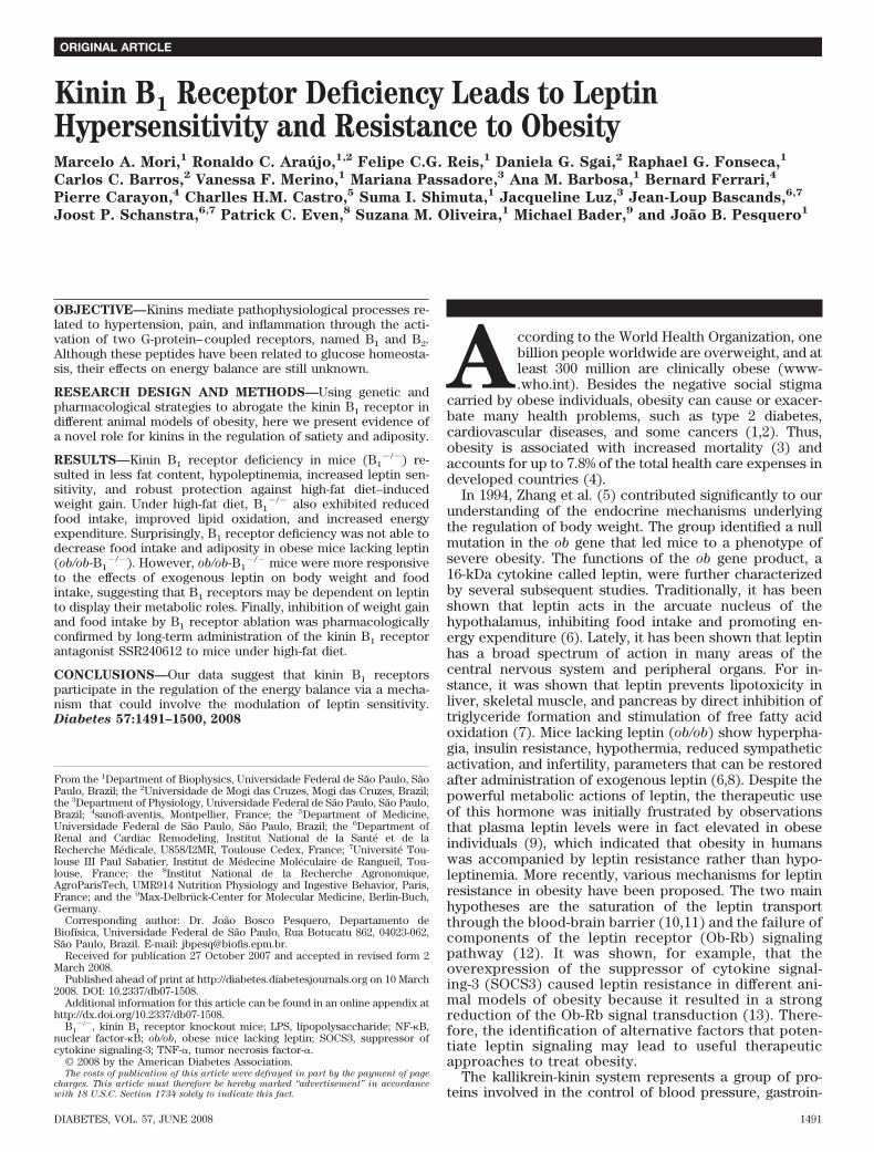
Kinin B 1 Receptor Deficiency Leads to Leptin Hypersensitivity and Resistance to Obesity Marcelo A. Mori, 1 Ronaldo C. Arau ´ jo, 1,2 Felipe C.G. Reis, 1 Daniela G. Sgai, 2 Raphael G. Fonseca, 1 Carlos C. Barros, 2 Vanessa F. Merino, 1 Mariana Passadore, 3 Ana M. Barbosa, 1 Bernard Ferrari, 4 Pierre Carayon, 4 Charlles H.M. Castro, 5 Suma I. Shimuta, 1 Jacqueline Luz, 3 Jean-Loup Bascands, 6,7 Joost P. Schanstra, 6,7 Patrick C. Even, 8 Suzana M. Oliveira, 1 Michael Bader, 9 and Joa ˜o B. Pesquero 1 OBJECTIVE—Kinins mediate pathophysiological processes re- lated to hypertension, pain, and inflammation through the acti- vation of two G-protein– coupled receptors, named B 1 and B 2 . Although these peptides have been related to glucose homeosta- sis, their effects on energy balance are still unknown. RESEARCH DESIGN AND METHODS—Using genetic and pharmacological strategies to abrogate the kinin B 1 receptor in different animal models of obesity, here we present evidence of a novel role for kinins in the regulation of satiety and adiposity. RESULTS—Kinin B 1 receptor deficiency in mice (B 1 / ) re- sulted in less fat content, hypoleptinemia, increased leptin sen- sitivity, and robust protection against high-fat diet–induced weight gain. Under high-fat diet, B 1 / also exhibited reduced food intake, improved lipid oxidation, and increased energy expenditure. Surprisingly, B 1 receptor deficiency was not able to decrease food intake and adiposity in obese mice lacking leptin (ob/ob-B 1 / ). However, ob/ob-B 1 / mice were more responsive to the effects of exogenous leptin on body weight and food intake, suggesting that B 1 receptors may be dependent on leptin to display their metabolic roles. Finally, inhibition of weight gain and food intake by B 1 receptor ablation was pharmacologically confirmed by long-term administration of the kinin B 1 receptor antagonist SSR240612 to mice under high-fat diet. CONCLUSIONS—Our data suggest that kinin B 1 receptors participate in the regulation of the energy balance via a mecha- nism that could involve the modulation of leptin sensitivity. Diabetes 57:1491–1500, 2008 A ccording to the World Health Organization, one billion people worldwide are overweight, and at least 300 million are clinically obese (www- .who.int). Besides the negative social stigma carried by obese individuals, obesity can cause or exacer- bate many health problems, such as type 2 diabetes, cardiovascular diseases, and some cancers (1,2). Thus, obesity is associated with increased mortality (3) and accounts for up to 7.8% of the total health care expenses in developed countries (4). In 1994, Zhang et al. (5) contributed significantly to our understanding of the endocrine mechanisms underlying the regulation of body weight. The group identified a null mutation in the ob gene that led mice to a phenotype of severe obesity. The functions of the ob gene product, a 16-kDa cytokine called leptin, were further characterized by several subsequent studies. Traditionally, it has been shown that leptin acts in the arcuate nucleus of the hypothalamus, inhibiting food intake and promoting en- ergy expenditure (6). Lately, it has been shown that leptin has a broad spectrum of action in many areas of the central nervous system and peripheral organs. For in- stance, it was shown that leptin prevents lipotoxicity in liver, skeletal muscle, and pancreas by direct inhibition of triglyceride formation and stimulation of free fatty acid oxidation (7). Mice lacking leptin (ob/ob) show hyperpha- gia, insulin resistance, hypothermia, reduced sympathetic activation, and infertility, parameters that can be restored after administration of exogenous leptin (6,8). Despite the powerful metabolic actions of leptin, the therapeutic use of this hormone was initially frustrated by observations that plasma leptin levels were in fact elevated in obese individuals (9), which indicated that obesity in humans was accompanied by leptin resistance rather than hypo- leptinemia. More recently, various mechanisms for leptin resistance in obesity have been proposed. The two main hypotheses are the saturation of the leptin transport through the blood-brain barrier (10,11) and the failure of components of the leptin receptor (Ob-Rb) signaling pathway (12). It was shown, for example, that the overexpression of the suppressor of cytokine signal- ing-3 (SOCS3) caused leptin resistance in different ani- mal models of obesity because it resulted in a strong reduction of the Ob-Rb signal transduction (13). There- fore, the identification of alternative factors that poten- tiate leptin signaling may lead to useful therapeutic approaches to treat obesity. The kallikrein-kinin system represents a group of pro- teins involved in the control of blood pressure, gastroin- From the 1 Department of Biophysics, Universidade Federal de Sa ˜ o Paulo, Sa ˜o Paulo, Brazil; the 2 Universidade de Mogi das Cruzes, Mogi das Cruzes, Brazil; the 3 Department of Physiology, Universidade Federal de Sa ˜ o Paulo, Sa ˜ o Paulo, Brazil; 4 sanofi-aventis, Montpellier, France; the 5 Department of Medicine, Universidade Federal de Sa ˜o Paulo, Sa ˜ o Paulo, Brazil; the 6 Department of Renal and Cardiac Remodeling, Institut National de la Sante ´ et de la Recherche Me ´ dicale, U858/I2MR, Toulouse Cedex, France; 7 Universite ´ Tou- louse III Paul Sabatier, Institut de Me ´ decine Mole ´ culaire de Rangueil, Tou- louse, France; the 8 Institut National de la Recherche Agronomique, AgroParisTech, UMR914 Nutrition Physiology and Ingestive Behavior, Paris, France; and the 9 Max-Delbru ¨ ck-Center for Molecular Medicine, Berlin-Buch, Germany. Corresponding author: Dr. Joa ˜o Bosco Pesquero, Departamento de Biofı ´sica, Universidade Federal de Sa ˜ o Paulo, Rua Botucatu 862, 04023-062, Sa ˜ o Paulo, Brazil. E-mail: jbpesq@biofis.epm.br. Received for publication 27 October 2007 and accepted in revised form 2 March 2008. Published ahead of print at http://diabetes.diabetesjournals.org on 10 March 2008. DOI: 10.2337/db07-1508. Additional information for this article can be found in an online appendix at http://dx.doi.org/10.2337/db07-1508. B 1 / , kinin B 1 receptor knockout mice; LPS, lipopolysaccharide; NF-B, nuclear factor-B; ob/ob, obese mice lacking leptin; SOCS3, suppressor of cytokine signaling-3; TNF-, tumor necrosis factor-. © 2008 by the American Diabetes Association. The costs of publication of this article were defrayed in part by the payment of page charges. This article must therefore be hereby marked “advertisement” in accordance with 18 U.S.C. Section 1734 solely to indicate this fact. ORIGINAL ARTICLE DIABETES, VOL. 57, JUNE 2008 1491

Welcome message from author
This document is posted to help you gain knowledge. Please leave a comment to let me know what you think about it! Share it to your friends and learn new things together.
Transcript
Kinin B1 Receptor Deficiency Leads to LeptinHypersensitivity and Resistance to ObesityMarcelo A. Mori,
1Ronaldo C. Araujo,
1,2Felipe C.G. Reis,
1Daniela G. Sgai,
2Raphael G. Fonseca,
1
Carlos C. Barros,2
Vanessa F. Merino,1
Mariana Passadore,3
Ana M. Barbosa,1
Bernard Ferrari,4
Pierre Carayon,4
Charlles H.M. Castro,5
Suma I. Shimuta,1
Jacqueline Luz,3
Jean-Loup Bascands,6,7
Joost P. Schanstra,6,7
Patrick C. Even,8
Suzana M. Oliveira,1
Michael Bader,9
and Joao B. Pesquero1
OBJECTIVE—Kinins mediate pathophysiological processes re-lated to hypertension, pain, and inflammation through the acti-vation of two G-protein–coupled receptors, named B1 and B2.Although these peptides have been related to glucose homeosta-sis, their effects on energy balance are still unknown.
RESEARCH DESIGN AND METHODS—Using genetic andpharmacological strategies to abrogate the kinin B1 receptor indifferent animal models of obesity, here we present evidence ofa novel role for kinins in the regulation of satiety and adiposity.
RESULTS—Kinin B1 receptor deficiency in mice (B1�/�) re-
sulted in less fat content, hypoleptinemia, increased leptin sen-sitivity, and robust protection against high-fat diet–inducedweight gain. Under high-fat diet, B1
�/� also exhibited reducedfood intake, improved lipid oxidation, and increased energyexpenditure. Surprisingly, B1 receptor deficiency was not able todecrease food intake and adiposity in obese mice lacking leptin(ob/ob-B1
�/�). However, ob/ob-B1�/� mice were more responsive
to the effects of exogenous leptin on body weight and foodintake, suggesting that B1 receptors may be dependent on leptinto display their metabolic roles. Finally, inhibition of weight gainand food intake by B1 receptor ablation was pharmacologicallyconfirmed by long-term administration of the kinin B1 receptorantagonist SSR240612 to mice under high-fat diet.
CONCLUSIONS—Our data suggest that kinin B1 receptorsparticipate in the regulation of the energy balance via a mecha-nism that could involve the modulation of leptin sensitivity.Diabetes 57:1491–1500, 2008
According to the World Health Organization, onebillion people worldwide are overweight, and atleast 300 million are clinically obese (www-.who.int). Besides the negative social stigma
carried by obese individuals, obesity can cause or exacer-bate many health problems, such as type 2 diabetes,cardiovascular diseases, and some cancers (1,2). Thus,obesity is associated with increased mortality (3) andaccounts for up to 7.8% of the total health care expenses indeveloped countries (4).
In 1994, Zhang et al. (5) contributed significantly to ourunderstanding of the endocrine mechanisms underlyingthe regulation of body weight. The group identified a nullmutation in the ob gene that led mice to a phenotype ofsevere obesity. The functions of the ob gene product, a16-kDa cytokine called leptin, were further characterizedby several subsequent studies. Traditionally, it has beenshown that leptin acts in the arcuate nucleus of thehypothalamus, inhibiting food intake and promoting en-ergy expenditure (6). Lately, it has been shown that leptinhas a broad spectrum of action in many areas of thecentral nervous system and peripheral organs. For in-stance, it was shown that leptin prevents lipotoxicity inliver, skeletal muscle, and pancreas by direct inhibition oftriglyceride formation and stimulation of free fatty acidoxidation (7). Mice lacking leptin (ob/ob) show hyperpha-gia, insulin resistance, hypothermia, reduced sympatheticactivation, and infertility, parameters that can be restoredafter administration of exogenous leptin (6,8). Despite thepowerful metabolic actions of leptin, the therapeutic useof this hormone was initially frustrated by observationsthat plasma leptin levels were in fact elevated in obeseindividuals (9), which indicated that obesity in humanswas accompanied by leptin resistance rather than hypo-leptinemia. More recently, various mechanisms for leptinresistance in obesity have been proposed. The two mainhypotheses are the saturation of the leptin transportthrough the blood-brain barrier (10,11) and the failure ofcomponents of the leptin receptor (Ob-Rb) signalingpathway (12). It was shown, for example, that theoverexpression of the suppressor of cytokine signal-ing-3 (SOCS3) caused leptin resistance in different ani-mal models of obesity because it resulted in a strongreduction of the Ob-Rb signal transduction (13). There-fore, the identification of alternative factors that poten-tiate leptin signaling may lead to useful therapeuticapproaches to treat obesity.
The kallikrein-kinin system represents a group of pro-teins involved in the control of blood pressure, gastroin-
From the 1Department of Biophysics, Universidade Federal de Sao Paulo, SaoPaulo, Brazil; the 2Universidade de Mogi das Cruzes, Mogi das Cruzes, Brazil;the 3Department of Physiology, Universidade Federal de Sao Paulo, Sao Paulo,Brazil; 4sanofi-aventis, Montpellier, France; the 5Department of Medicine,Universidade Federal de Sao Paulo, Sao Paulo, Brazil; the 6Department ofRenal and Cardiac Remodeling, Institut National de la Sante et de laRecherche Medicale, U858/I2MR, Toulouse Cedex, France; 7Universite Tou-louse III Paul Sabatier, Institut de Medecine Moleculaire de Rangueil, Tou-louse, France; the 8Institut National de la Recherche Agronomique,AgroParisTech, UMR914 Nutrition Physiology and Ingestive Behavior, Paris,France; and the 9Max-Delbruck-Center for Molecular Medicine, Berlin-Buch,Germany.
Corresponding author: Dr. Joao Bosco Pesquero, Departamento deBiofısica, Universidade Federal de Sao Paulo, Rua Botucatu 862, 04023-062,Sao Paulo, Brazil. E-mail: [email protected].
Received for publication 27 October 2007 and accepted in revised form 2March 2008.
Published ahead of print at http://diabetes.diabetesjournals.org on 10 March2008. DOI: 10.2337/db07-1508.
Additional information for this article can be found in an online appendix athttp://dx.doi.org/10.2337/db07-1508.
B1�/�, kinin B1 receptor knockout mice; LPS, lipopolysaccharide; NF-�B,
nuclear factor-�B; ob/ob, obese mice lacking leptin; SOCS3, suppressor ofcytokine signaling-3; TNF-�, tumor necrosis factor-�.
© 2008 by the American Diabetes Association.The costs of publication of this article were defrayed in part by the payment of page
charges. This article must therefore be hereby marked “advertisement” in accordance
with 18 U.S.C. Section 1734 solely to indicate this fact.
ORIGINAL ARTICLE
DIABETES, VOL. 57, JUNE 2008 1491
testinal tract mobility, inflammation, and pain induction(14,15). In this system, precursor proteins called kinino-gens are cleaved by kallikreins to release kinins, whichcan further be cleaved by carboxypeptidases to give rise todes-Arg-kinins. The main active kinins are bradykinin andits metabolite des-Arg9-bradykinin. Bradykinin and des-Arg9-bradykinin use distinct G-protein–coupled receptorsto exert their effects. Whereas bradykinin interacts withthe B2 receptor subtype, des-Arg9-bradykinin has higheraffinity for the B1 receptor (14). Kinin B2 receptors areconstitutively expressed in most tissues and exert mostfunctions attributed to the kallikrein-kinin system, includ-ing vasodilation and salt excretion (14,16). Conversely, thekinin B1 receptor is thought to be strongly induced byinflammatory stimuli and to mediate processes of injuryand pain (14,15,17). Accordingly, kinin B1 receptor knock-out mice (B1
�/�) are hypoalgesic, are less responsive tolipopolysaccharide (LPS)-induced hypotension (18), andpresent inhibition of neutrophil migration into inflamedtissue (19).
Evidence that kinins induce insulin sensitivity in adipo-cytes (20) and control insulin released by pancreatic cells(21,22) indicates that the kallikrein-kinin system alsoparticipates in metabolic processes. However, the physio-logical relevance of this system to energy homeostasis andthe contributions of the kinin B1 receptor to these pro-cesses are still not well understood. Recently, our groupshowed that the B1 receptor mRNA is upregulated in thewhite adipose tissue, liver, and hypothalamus of ob/obmice (23), suggesting for the first time a correlationbetween obesity and the kinin B1 receptor. Furthermore,we observed that des-Arg9-bradykinin administration inmice is able to increase blood leptin levels (24). In thepresent report, we examined the consequences of B1receptor ablation on the etiopathology of obesity usingtwo animal models, the high-fat diet–induced obesity andthe obese ob/ob mice. In addition, we evaluated the effectof pharmacological inhibition of B1 receptors on weightgain and food intake of mice under high-fat diet. Theresults of these studies strongly support a role for thekinin B1 receptor in controlling body weight and fataccumulation through a mechanism that may involve themodulation of leptin signaling.
RESEARCH DESIGN AND METHODS
B1�/� mice (18) used in the experiments were originated from 10 generations’
backcrossing of an initially mixed genetic background (129/Sv and C57Bl/6)with C57Bl/6 mice (Taconic, Germantown, NY). Therefore, C57Bl/6 animalswere used as their controls. The ob/ob-B1
�/� strain was generated by cross-breeding C57Bl/6 ob�/� mice (The Jackson Laboratories, Bar Harbor, ME)with B1
�/� mice. Animals have been obtained from the Universidade Federalde Sao Paulo (Brazil), from the Max-Delbruck-Center for Molecular Medicine(Berlin-Buch, Germany), and from the Animal House (Toulouse, France). Allexperiments reported have been conducted as stated in the National Institutesof Health Guide for the Care and Use of Laboratory Animals (Institute ofLaboratory Animal Resources, National Academy Press, Washington, DC,1996) and approved by a local committee. Animals were maintained onstandard mouse chow at 22°C on a 12-h light-dark cycle with ad libitum accessto food and tap water. Food consumption and body weight were monitoredweekly in individualized animals. In all experiments, 12- to 18-week-old maleswere used.High-fat diet treatment. Mice were fed either standard diet (10% kcal fat) orhigh-fat diet (45% kcal fat) (Research Diets, New Brunswick, NJ) for 9 weeks.After the treatment, mice were killed for blood and tissue collection. The serawere separated for leptin and insulin quantification with ELISA kits (R&DSystems [Minneapolis, MN] and Millipore [Billerica, MA], respectively). Tis-sues explants were weighed and used for adipocyte isolation, or frozen forprotein, RNA, or triglycerides extraction.
Body composition analysis. Total body fat was estimated by dual-energyX-ray absorptiometry using a Hologic QDR 4500 scanner (Hologic, Waltham,MA) as described previously (25).Adipose tissue cellularity. Isolation of fat cells and calculation of adipocytevolume and number in epididymal fat depots were performed as previouslydescribed (26). Briefly, images of isolated adipocytes were acquired from alight microscope fitted with a camera, and the average cellular volume wasestimated by measuring the diameter of at least 100 adipocytes per animalusing the software ImageJ (http://rsb.info.nih.gov/ij/). Fat cell number wascalculated by dividing the adipose tissue triglyceride content by the averageadipocyte lipid content, obtained by the multiplication of the average fat cellvolume by the triolein density (0.92).Gene and protein expression. Please refer to the online appendix (availableat http://dx.doi.org/10.2337/db07-1508).Energy expenditure. In vivo indirect open circuit calorimetry was per-formed as previously described (26). Briefly, respiratory exchanges andspontaneous activity signals of freely moving mice were computed at 10-sintervals using a computer-assisted data acquisition program (ADDENFImetabolic chamber prototype). Mice were housed individually in the meta-bolic cages at 10:00 A.M. without food, and the postabsorptive resting energyexpenditure (�basal metabolism) was measured between 4:00 and 6:00 P.M. At6:00 P.M., we gave a 1-g meal of either control or high-fat diet to the mice andcomputed the increase in energy expenditure in response to feeding (metab-olism and respiratory quotient).Leptin sensitivity. To determine leptin sensitivity, we administered recom-binant mouse leptin (40 �g � day�1 � mouse�1 i.p.; R&D Systems) during 4 days(at 5:00 P.M.) and evaluated food intake and/or weight loss. Food intake during5 days before leptin injection was considered as the basal consumption(100%). An independent group of fasted animals was killed 45 min after thefirst leptin bolus for hypothalamus collection, protein extraction, and Westernblotting quantification of pSTAT3 and STAT3.SSR240612 chronic administration. ALZET micro-osmotic pumps (Alza,Palo Alto, CA), set to deliver for 28 days either SSR240612 (3 mg � kg�1 bodywt � day�1; Sanofi-Aventis, Paris, France) or vehicle (2% DMSO), weresubcutaneously implanted in anesthetized C56Bl/6 mice under sterile condi-tions. Seven days after the implant, mice were submitted to high-fat diet for 3weeks. Body weight and food intake were assessed weekly in the period of 1month before and after the implant. At the end of the treatment, pumps werechecked for their contents, and animals with filled ones were not consideredin the data analysis.Statistical analysis. All values were expressed as means � SE. Statisticalanalyses were carried out using two-tailed Student’s unpaired t test tocompare two independent groups or ANOVA followed by Bonferroni’s test tocompare more than two. Significance was rejected at P � 0.05.
RESULTS
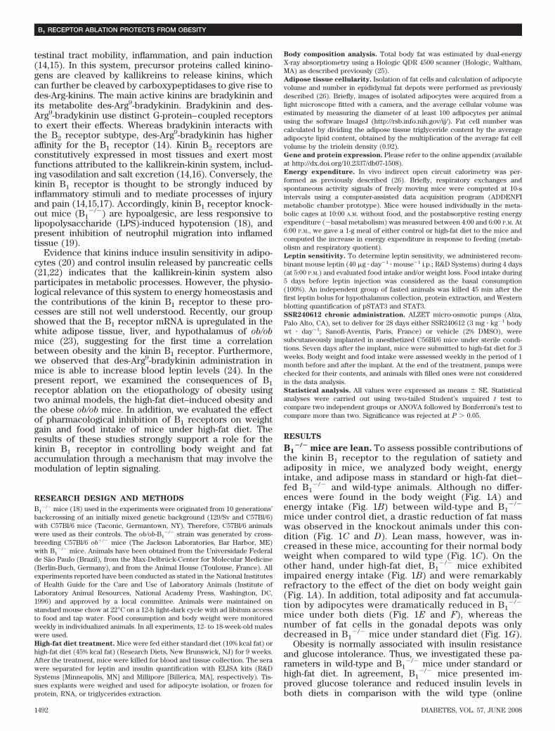
B1�/� mice are lean. To assess possible contributions of
the kinin B1 receptor to the regulation of satiety andadiposity in mice, we analyzed body weight, energyintake, and adipose mass in standard or high-fat diet–fed B1
�/� and wild-type animals. Although no differ-ences were found in the body weight (Fig. 1A) andenergy intake (Fig. 1B) between wild-type and B1
�/�
mice under control diet, a drastic reduction of fat masswas observed in the knockout animals under this con-dition (Fig. 1C and D). Lean mass, however, was in-creased in these mice, accounting for their normal bodyweight when compared to wild type (Fig. 1C). On theother hand, under high-fat diet, B1
�/� mice exhibitedimpaired energy intake (Fig. 1B) and were remarkablyrefractory to the effect of the diet on body weight gain(Fig. 1A). In addition, total adiposity and fat accumula-tion by adipocytes were dramatically reduced in B1
�/�
mice under both diets (Fig. 1E and F), whereas thenumber of fat cells in the gonadal depots was onlydecreased in B1
�/� mice under standard diet (Fig. 1G).Obesity is normally associated with insulin resistance
and glucose intolerance. Thus, we investigated these pa-rameters in wild-type and B1
�/� mice under standard orhigh-fat diet. In agreement, B1
�/� mice presented im-proved glucose tolerance and reduced insulin levels inboth diets in comparison with the wild type (online
B1 RECEPTOR ABLATION PROTECTS FROM OBESITY
1492 DIABETES, VOL. 57, JUNE 2008
appendix), confirming our previous data showing in-creased insulin sensitivity in these mice (21).
Next, we measured serum leptin levels (Fig. 2A) and theexpression of leptin mRNA (Fig. 2B) and protein (Fig. 2C)in the epididymal white adipose tissue of wild-type andB1
�/� mice. In all cases, we observed a significant de-crease in leptin content in B1
�/� mice when comparedwith wild-type mice. Notably, high-fat diet did not lead toan increase of leptin levels in B1
�/� mice.Taken together, these results demonstrate that B1
�/�
mice have reduced adiposity and are strongly resistant todiet-induced hyperleptinemia and body weight gain.Improved energy expenditure with spontaneous ac-tivity and lipid oxidation in B1
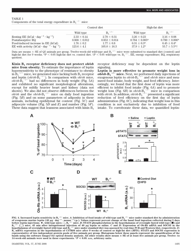
�/� mice under high-fatdiet. Analysis of feed efficiency (body weight gain/energyconsumed) (Fig. 3A) revealed that inhibition of weightgain observed in B1
�/� mice under high-fat diet could notbe totally explained by decreased energy intake (Fig. 1B).Therefore, we quantified the components of total energyexpenditure under normal and high-fat feeding in B1
�/�
and wild-type mice. No differences in resting energy ex-penditure, increase in energy expenditure in response tofeeding, or differences in respiratory quotient (respiratoryquotient, VCO2/VO2) in the postabsorptive state (4 h beforemeal and 8–12 h after meal) were observed between B1
�/�
and wild-type mice (Table 1). Under standard diet, respi-ratory quotient levels after feeding were �1.0 in bothgroups, suggesting that de novo lipogenesis in response tocarbohydrate ingestion is occurring in these animals at asimilar extent. In contrast, under high-fat diet, B1
�/� miceexhibited a meal-induced increase in respiratory quotientthat lasted 4 h less than in wild-type mice (Fig. 3B and C),indicating that postprandial inhibition of lipid oxidation issignificantly lower in these animals. In addition, B1
�/�
mice under high-fat regimen expended significantly moreenergy with activity (�46.6%, P 0.002; Table 1). To-gether, these data suggest that B1
�/� mice can resist
high-fat diet–induced weight gain in part due to increasedenergy expenditure with spontaneous activity and de-creased inhibition of lipid oxidation in response to a fatmeal.Hypersensitivity to leptin in B1
�/� mice. Together,hypoleptinemia, protection against high-fat diet–inducedhyperleptinemia, elevated energy expenditure, and re-duced food intake under high-fat diet suggest increasedleptin responsiveness in B1
�/� mice. To test this hypothe-sis, we administered exogenous leptin to B1
�/� and wild-type mice and quantified daily food intake andphosphorylation of STAT3, a downstream component ofthe Ob-Rb signaling pathway, in the hypothalamus of theseanimals. A stronger inhibitory effect of exogenous leptinon food intake in B1
�/� mice was observed when com-pared with wild-type mice (Fig. 4A). Furthermore, thelevels of phosphorylated STAT3 after leptin injection weresignificantly higher in the hypothalamus of B1
�/� in com-parison with wild-type mice (Fig. 4B). In addition, wemeasured the expression of Ob-Rb mRNA in hypothala-mus of B1
�/� and wild-type mice and found upregulationof this transcript in B1
�/� (Fig. 4C). Moreover, the expres-sion of SOCS3, a potent inhibitor of the Ob-Rb signaling,was decreased in hypothalamus of B1
�/� mice (Fig. 4D).These data suggest that Ob-Rb signaling is facilitated inB1
�/� mice, which could contribute to the inhibition offood intake and enhanced energy expenditure in theseanimals under high-fat diet. Another aspect of leptinsensitivity, the passage of leptin through the blood-brainbarrier, was addressed in B1
�/� and wild-type mice bymeasuring brain accumulation of intravenously injected[125I]-labeled leptin. Although B1
�/� mice had a tendencyto an increased leptin passage to the brain, no significantdifferences were observed in relation to the wild-type mice(data not shown).
We also assessed kinin B1 receptor mRNA expression inthe hypothalamus of wild-type mice after 9 weeks of high-fat
Bo
dy
wei
gh
t(g
)A
Time (weeks)
†
*
* * * * ** *
† ††
†† † †
WT ControlB1
-/- ControlWT HFDB1
-/- HFD
20
25
30
35
40
45
0 2 4 6 8 10
B
En
erg
y in
take
(k
Cal
/wee
k)
Time (weeks)
*†
WT ControlB1
-/- ControlWT HFDB1
-/- HFD
70
80
90
100
110
120
130
0 2 4 6 8 10
†**
B1-/-
100 µµm
E
WT
Control diet HFDD
0
0.5
1
1.5
2
2.5WT
B1-/-
Ep
idid
ymal
fat
(g)
*
***
**
ControlDiet
HFD
G
WTB1
-/-
0
0.5
1
1.5
2
2.5
3
Ad
ipo
cyte
nu
mb
er (
x10
6 )
***
ControlDiet
HFD
F
*
*
Ad
ipo
cyte
vo
lum
e (c
m3 x
10-6
)
*
ControlDiet
HFD0
0.2
0.4
0.6
0.8
1
1.2
1.4
WTB1
-/-
C
ControlDiet
Bo
dy
com
po
siti
on
(g
) Fat massLean mass
*
HFD
WT B1-/- WT B1
-/-0
1020304050
*
††
FIG. 1. Reduced fat mass in B1�/� mice. Twelve-week-old wild-type and B1
�/� mice were submitted to standard (control) or high-fat diet (HFD)for 9 weeks and body weight (A), energy intake (B), total fat mass (C), epididymal fat weight (D), isolated adipocyte morphology (E), and averageadipocyte volume (F) and number (G) were measured. �, wild type under control diet; �, B1
�/� under control diet; Œ, wild type under HFD; E,B1
�/� under HFD. Results are means � SE. A and B: *P < 0.05 B1�/� HFD versus wild-type HFD. †P < 0.05 wild-type control diet versus wild-type
HFD; n � 11 per group. C: *P < 0.05 B1�/� fat mass versus wild-type fat mass. †P < 0.05 B1
�/� lean mass versus wild-type lean mass; n � 5 pergroup. D, F, and G: *P < 0.05. **P < 0.01 between indicated groups; n � 6 per group. Microscopy images are representative examples of six animalsper group.
M.A. MORI AND ASSOCIATES
DIABETES, VOL. 57, JUNE 2008 1493

regimen. Interestingly, we found higher levels of this mRNAin high-fat diet–fed mice, in comparison with animals understandard diet (Fig. 4E). Taken together, these results indicatethat the kinin B1 receptor may participate in the mechanismof leptin resistance in obesity.
A
Ser
um
Lep
tin
(n
g/m
l)
0
10
20
30
40
50
60
70
ControlDiet
HFD
***
*
** WT B1
-/-
Lep
tin
mR
NA
(a.
u.)
0
5
10
15
20
25
*
****B
C
Control
HFDLep
tin
WT B1-/-
0.2
0.4
0.6
0.8
1
1.2
1.4 *****
Lep
tin
(a.
u.)
ControlDiet
HFD
WT B1
-/-
ControlDiet
HFD
WT B1
-/-
FIG. 2. Decreased leptin levels in B1�/� mice. A: Serum leptin concen-
tration was determined in 20 1-week-old wild-type and B1�/� mice after
9 weeks under standard diet (control) and high-fat diet (HFD). B andC: Leptin mRNA and protein were quantified in the epididymal whiteadipose tissue of these mice, respectively. Values are means � SE of sixanimals per group. Protein expression is representative of two inde-pendent pools of three animals per group. Quantification of the bandswas also assessed using the software ImageJ for optical densityanalysis. *P < 0.05; **P < 0.01; ***P < 0.001 between indicated groups.a.u., arbitrary units.
A
Fee
d E
ffic
ien
cyg
/kC
al)
* **
ControlDiet
HFD
WT B1
-/-
0
0.004
0.008
0.012
0.016
0.2
0.6
1.0
1.4
1.8
Area UnderCurveTime vs Meal Onset (h)
0.7
0.8
0.9
1.0
1.1
1.2
-4 -2 0 2 4 6 8 10 12R
Q
B Control Diet
C
**
0.2
0.6
1.0
1.4
-4 -2 0 2 4 6 8 10 12
WT B1-/-
0.7
0.8
0.9
1.0
1.1
1.2
RQ
HFD
WT B1
-/-
WT B1
-/-
WT B1-/-
Time vs Meal Onset (h)Area Under
Curve
FIG. 3. Improved energy expenditure in B1�/� mice. Twelve-week-old
wild-type and B1�/� mice were submitted to 9 weeks of standard diet
(control) and high-fat diet (HFD). A: Feed efficiency (body weightgain/energy consumed) was calculated during this period (n � 11 pergroup). Next, mice were housed in a calorimeter for 8 h without accessto food, and 1 g pellet of the corresponding diet was introduced intothe metabolic cage (time 0 on the x-axis, 6:00 P.M. clock time) formeasurement of the meal-induced changes in respiratory quotient(RQ) under standard diet (B) and HFD (C). F, wild-type mice; E, B1
�/�
mice. Calorimetry results show means � SE of five animals per group.*P < 0.05; **P < 0.01 between indicated groups.
B1 RECEPTOR ABLATION PROTECTS FROM OBESITY
1494 DIABETES, VOL. 57, JUNE 2008
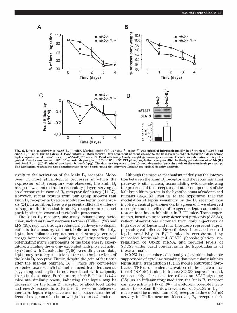
Kinin B1 receptor deficiency does not protect ob/obmice from obesity. To estimate the importance of leptinhypersensitivity to the phenotype of resistance to obesityin B1
�/� mice, we generated mice lacking both B1 receptorand leptin (ob/ob-B1
�/�). In comparison with ob/ob mice,ob/ob-B1
�/� had no differences in body weight (Fig. 5A)and exhibited no significant morphological alterations,except for mildly heavier heart and kidney (data notshown). We also did not observe differences between theob/ob and the ob/ob-B1
�/� mice on daily food ingestion(Fig. 5B) and on some parameters of adiposity in theseanimals, including epididymal fat content (Fig. 5C) andadipocyte volume (Fig. 5D and E) and number (Fig. 5F).These data suggest that leanness associated with kinin B1
receptor deficiency may be dependent on the leptinsignaling.Leptin is more effective to promote weight loss inob/ob-B1
�/� mice. Next, we performed daily injections ofexogenous leptin to ob/ob-B1
�/� and ob/ob mice and mea-sured food intake, body weight, and feed efficiency. Inter-estingly, we found that the first dose of leptin was moreefficient to inhibit food intake (Fig. 6A) and to promoteweight loss (Fig. 6B) in ob/ob-B1
�/� mice in comparisonwith ob/ob. In addition, ob/ob-B1
�/� presented a significantreduction of feed efficiency on the first day of leptinadministration (Fig. 6C), indicating that weight loss in thiscondition is not exclusively due to inhibition of foodintake. To corroborate these data, we quantified leptin-
0
1
2
3
4*
Ob
-Rb
mR
NA
(a.u
.)
WTB1
-/-
CControl DietHFD
B1
mR
NA
(a.
u.)
0
30
60 *
E
A
Time (days)
% o
f b
asal
ing
esti
on
70
80
90
100
0 1 2 3 4
*
*
WTB1
-/-
D
WT B1-/-
SOCS3
0
123
WT B1-/-
*
SO
CS
3(a
.u.)
B
WT
B1-/
-
+ Leptin
pSTAT3
STAT3
WT
B1-/
-
+ Salinep
ST
AT
3/S
TA
T3
(a.u
.) *
0
0.5
1
1.5
2
2.5 *
FIG. 4. Increased leptin sensitivity in B1�/� mice. A: Inhibition of food intake of wild-type and B1
�/� mice under standard diet by administrationof exogenous murine leptin (40 �g � day�1 � mouse�1 i.p.). Values represent percent change of the basal food ingestion collected during 5 daysbefore leptin injections. �, wild-type mice; �, B1
�/� mice. B: STAT3 phosphorylation in the hypothalamus of overnight fasted wild-type (f) andB1
�/� (�) mice, 45 min after intrapritoneal injection of 40 �g leptin or saline. C and D: Expression of Ob-Rb mRNA and SOCS3 in thehypothalamus of overnight fasted wild-type and B1
�/� mice under standard diet was assessed by real-time PCR and Western blot, respectively. E:B1 mRNA expression in the hypothalamus of C57Bl/6 mice after 9 weeks of control or high-fat diet (HFD). STAT3 and SOCS3 expression isrepresentative of two independent protein pools of five animals per group. Histograms below these panels represent the quantification of thebands using the software ImageJ for optical density analysis. All other results show means � SE of at least five animals per group. Twelve- to14-week-old animals were used in these experiments. *P < 0.05. a.u., arbitrary units.
TABLE 1Components of the total energy expenditure in B1
�/� mice
Control diet High-fat diet
Wild type B1�/� Wild type B1
�/�
Resting EE (kCal � day�1 � kg�1) 2.33 � 0.14 2.70 � 0.31 3.26 � 0.23 3.16 � 0.08Postabsorptive RQ 0.868 � 0.012 0.853 � 0.014 0.784 � 0.005* 0.780 � 0.006*Food-induced increase in EE (kCal) 1.76 � 0.2 1.77 � 0.8 0.31 � 0.3* 0.45 � 0.4*EE with activity (kCal � day�1 � kg�1) 123.6 � 4.1 105.0 � 16.3 37.9 � 1.2* 55.7 � 5.5*†
Data are means � SE of Q5 animals per group. Twelve-week-old wild-type and B1�/� mice were submitted to standard diet (control) and
high-fat diet for 9 weeks. *P 0.05 high-fat diet vs. control diet. †P 0.05 wild-type vs. B1�/�. EE, energy expenditure; RQ, respiratory
quotient.
M.A. MORI AND ASSOCIATES
DIABETES, VOL. 57, JUNE 2008 1495
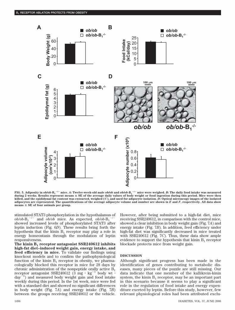
stimulated STAT3 phosphorylation in the hypothalamus ofob/ob-B1
�/� and ob/ob mice. As expected, ob/ob-B1�/�
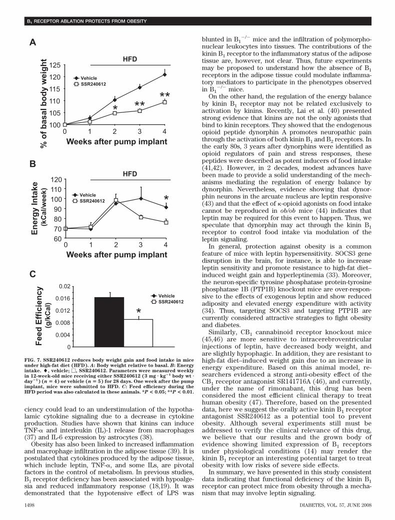
showed increased levels of phosphorylated STAT3 afterleptin induction (Fig. 6D). These results bring forth thehypothesis that the kinin B1 receptor may play a role inenergy homeostasis through the modulation of leptinresponsiveness.The kinin B1 receptor antagonist SSR240612 inhibitshigh-fat diet–induced weight gain, energy intake, andfeed efficiency in mice. To validate our findings usingknockout models and to confirm the pathophysiologicalfunction of the kinin B1 receptor in obesity, we pharma-cologically blocked this receptor in mice for 28 days bychronic administration of the nonpeptide orally active B1receptor antagonist SSR240612 (3 mg � kg�1 body wt �day�1) and measured body weight gain and food intakeweekly during this period. In the 1st week, mice were fedwith a standard diet and showed no significant differencesin body weight (Fig. 7A) and energy intake (Fig. 7B)between the groups receiving SSR240612 or the vehicle.
However, after being submitted to a high-fat diet, micereceiving SSR240612, in comparison with the control mice,showed a clear inhibition in body weight gain (Fig. 7A) andenergy intake (Fig. 7B). In addition, feed efficiency underhigh-fat diet was significantly decreased in mice treatedwith SSR240612 (Fig. 7C). Thus, these data show ampleevidence to support the hypothesis that kinin B1 receptorblockade protects mice from weight gain.
DISCUSSION
Although significant progress has been made in theidentification of genes contributing to metabolic dis-eases, many pieces of the puzzle are still missing. Ourdata indicate that one member of the kallikrein-kininsystem, the kinin B1 receptor, may be an important partin this scenario because it seems to play a significantrole in the regulation of food intake and energy expen-diture exerted by leptin. Before this study, however, fewrelevant physiological roles had been attributed exclu-
05
10152025
Fo
od
In
take
(k
Cal
/day
)
ob/obob/ob-B1
-/-
B
0
20
40
60
Bo
dy
Wei
gh
t (g
)
ob/obob/ob-B1
-/-
A
C
Ep
idid
ymal
fat
(g
) ob/obob/ob-B1
-/-
0123456
Ad
ipo
cyte
vo
lum
e (c
m3 x
10-6
)
0
1
2
3
4
ob/obob/ob-B1
-/-
E
0
0.2
0.4
0.6
0.8
ob/obob/ob-B1
-/-
Ad
ipo
cyte
nu
mb
er (
x10
6 )F
D100 µm
ob/ob-B1-/-ob/ob
100 µm
FIG. 5. Adiposity in ob/ob-B1�/� mice. A: Twelve-week-old male ob/ob and ob/ob-B1
�/� mice were weighed. B: The daily food intake was measuredduring 2 weeks. Results represent means � SE of the average daily values of body weight or food ingestion during this period. Mice were thenkilled, and the epididymal fat content was extracted, weighed (C), and used for adipocyte isolation. D: Optical microscopy images of the isolatedadipocytes are represented. The quantifications of the average adipocyte volume and number are shown in E and F, respectively. All data showmeans � SE of four animals per group.
B1 RECEPTOR ABLATION PROTECTS FROM OBESITY
1496 DIABETES, VOL. 57, JUNE 2008
sively to the activation of the kinin B1 receptor. More-over, in most physiological processes in which theexpression of B1 receptors was observed, the kinin B1receptor was considered a secondary player, serving asan alternative in case of B2 receptor deficiency (14,27).However, recent results from our group showed thatkinin B1 receptor activation modulates leptin homeosta-sis (24). In addition, here we present sufficient evidenceto support the idea that kinin B1 receptors are in factparticipating in essential metabolic processes.
The kinin B1 receptor, like many inflammatory mole-cules, including tumor necrosis factor-� (TNF-�) (28) andLPS (29), may act through redundant pathways to displayboth its inflammatory and metabolic actions. Similarly,leptin has inflammatory actions and strongly controlsenergy homeostasis (6), mainly by regulating satiety andpotentiating many components of the total energy expen-diture, including the energy expended with physical activ-ity (8) and with fat oxidation (7,30). According to our data,leptin may be a key mediator of the metabolic actions ofthe kinin B1 receptor. Firstly, despite the gain of fat tissueafter the high-fat regimen, B1
�/� mice are completelyprotected against high-fat diet–induced hyperleptinemia,suggesting that leptin is not correlated with adipositylevels in these mice. Furthermore, ob/ob-B1
�/� and ob/obmice are similarly obese, indicating that leptin may benecessary for the kinin B1 receptor to affect food intakeand energy expenditure. Finally, B1 receptor deficiencyincreases leptin responsiveness and exacerbates the ef-fects of exogenous leptin on weight loss in ob/ob mice.
Although the precise mechanism underlying the interac-tion between the kinin B1 receptor and the leptin signalingpathway is still unclear, accumulating evidence showingthe presence of this receptor and other components of thekallikrein-kinin system in the hypothalamus of rodents andhumans (23,31,32) lead us to the hypothesis that themodulation of leptin sensitivity by the B1 receptor mayinvolve a central phenomenon. In agreement, we observedmore pronounced effects of exogenous leptin administra-tion on food intake inhibition in B1
�/� mice. These exper-iments, based on previously described protocols (8,33,34),reflect observations obtained from daily injections ofhigh doses of leptin and may therefore represent supra-physiological effects. Nevertheless, increased centralleptin sensitivity in B1
�/� mice is corroborated byincreased leptin-induced STAT3 phosphorylation, up-regulation of Ob-Rb mRNA, and reduced levels ofSOCS3 under basal conditions in the hypothalamus ofthese animals.
SOCS3 is a member of a family of cytokine-induciblesuppressors of cytokine signaling that particularly inhibitsleptin signal transduction (13). In mouse embryonic fibro-blasts, TNF-�–dependent activation of the nuclear fac-tor-�B (NF-�B) is able to induce SOCS3 expression and,consequently, elicit negative effects on STAT signaling(35). As an inflammatory mediator, the kinin B1 receptorcan also activate NF-�B (36). Therefore, a possible mech-anism to explain the downregulation of SOCS3 in B1
�/�
mice would be a reduction of B1 receptor–induced NF-�Bactivity in Ob-Rb neurons. Moreover, B1 receptor defi-
Time (days)
% o
f b
asal
ing
esti
on
*
ob/obob/ob-B1
-/-
30
50
70
90
110
0 1 2 3 4
A
889092949698
100102
0 1 2 3 4
*
ob/obob/ob-B1
-/-
% o
f b
asal
bo
dy
wei
gh
t
Time (days)
B
-0.3
-0.2
-0.1
0
0 1 2 3 4
ob/obob/ob-B1
-/-
*
Fee
d E
ffic
ien
cy
(g/k
Cal
)
Time (days)
ob/ob
ob/ob
-B1-/
-
pSTAT3
STAT3
pS
TA
T3/
ST
AT
3 (a
.u.)
0
0.2
0.4
0.6
0.8 *
C D
FIG. 6. Leptin sensitivity in ob/ob-B1�/� mice. Murine leptin (40 �g � day�1 � mice�1) was injected intraperitoneally in 18-week-old ob/ob and
ob/ob-B1�/� mice during 4 days. A: Food intake. B: Body weight. Data represent percent change to the basal values collected during 5 days before
leptin injections. �, ob/ob mice; �, ob/ob-B1�/� mice. C: Feed efficiency (body weight gain/energy consumed) was also calculated during this
period. Results are means � SE of four animals per group. *P < 0.05. D: STAT3 phosphorylation was quantified in the hypothalamus of ob/ob (f)and ob/ob-B1
�/� (�) 45 min after a leptin bolus (40 �g). The data are representative of two independent protein pools of three animals per group.The histogram represents the quantification of the bands using the software ImageJ for optical density analysis.
M.A. MORI AND ASSOCIATES
DIABETES, VOL. 57, JUNE 2008 1497
ciency could lead to an understimulation of the hypotha-lamic cytokine signaling due to a decrease in cytokineproduction. Studies have shown that kinins can induceTNF-� and interleukin (IL)-1 release from macrophages(37) and IL-6 expression by astrocytes (38).
Obesity has also been linked to increased inflammationand macrophage infiltration in the adipose tissue (39). It ispostulated that cytokines produced by the adipose tissue,which include leptin, TNF-�, and some ILs, are pivotalfactors in the control of metabolism. In previous studies,B1 receptor deficiency has been associated with hypoalge-sia and reduced inflammatory response (18,19). It wasdemonstrated that the hypotensive effect of LPS was
blunted in B1�/� mice and the infiltration of polymorpho-
nuclear leukocytes into tissues. The contributions of thekinin B1 receptor to the inflammatory status of the adiposetissue are, however, not clear. Thus, future experimentsmay be proposed to understand how the absence of B1receptors in the adipose tissue could modulate inflamma-tory mediators to participate in the phenotypes observedin B1
�/� mice.On the other hand, the regulation of the energy balance
by kinin B1 receptor may not be related exclusively toactivation by kinins. Recently, Lai et al. (40) presentedstrong evidence that kinins are not the only agonists thatbind to kinin receptors. They showed that the endogenousopioid peptide dynorphin A promotes neuropathic painthrough the activation of both kinin B1 and B2 receptors. Inthe early 80s, 3 years after dynorphins were identified asopioid regulators of pain and stress responses, thesepeptides were described as potent inducers of food intake(41,42). However, in 2 decades, modest advances havebeen made to provide a solid understanding of the mech-anisms mediating the regulation of energy balance bydynorphin. Nevertheless, evidence showing that dynor-phin neurons in the arcuate nucleus are leptin responsive(43) and that the effect of �-opioid agonists on food intakecannot be reproduced in ob/ob mice (44) indicates thatleptin may be required for this event to happen. Thus, wespeculate that dynorphin may act through the kinin B1receptor to control food intake via modulation of theleptin signaling.
In general, protection against obesity is a commonfeature of mice with leptin hypersensitivity. SOCS3 genedisruption in the brain, for instance, is able to increaseleptin sensitivity and promote resistance to high-fat diet–induced weight gain and hyperleptinemia (33). Moreover,the neuron-specific tyrosine phosphatase protein-tyrosinephosphatase 1B (PTP1B) knockout mice are over-respon-sive to the effects of exogenous leptin and show reducedadiposity and elevated energy expenditure with activity(34). Thus, targeting SOCS3 and targeting PTP1B arecurrently considered attractive strategies to fight obesityand diabetes.
Similarly, CB1 cannabinoid receptor knockout mice(45,46) are more sensitive to intracerebroventricularinjections of leptin, have decreased body weight, andare slightly hypophagic. In addition, they are resistant tohigh-fat diet–induced weight gain due to an increase inenergy expenditure. Based on this animal model, re-searchers evidenced a strong anti-obesity effect of theCB1 receptor antagonist SR141716A (46), and currently,under the name of rimonabant, this drug has beenconsidered the most efficient clinical therapy to treathuman obesity (47). Therefore, based on the presenteddata, here we suggest the orally active kinin B1 receptorantagonist SSR240612 as a potential tool to preventobesity. Although several experiments still must beaddressed to verify the clinical relevance of this drug,we believe that our results and the grown body ofevidence showing limited expression of B1 receptorsunder physiological conditions (14) may render thekinin B1 receptor an interesting potential target to treatobesity with low risks of severe side effects.
In summary, we have presented in this study consistentdata indicating that functional deficiency of the kinin B1receptor can protect mice from obesity through a mecha-nism that may involve leptin signaling.
B
En
erg
y In
take
(kC
al/w
eek)
Weeks after pump implant
100
110
120HFD
*VehicleSSR240612
60
70
80
90
0 1 2 3 4
0
0.004
0.008
0.012
0.016
0.02
VehicleSSR240612
*
Fee
d E
ffic
ien
cy
(g/k
Cal
)
C
A
Weeks after pump implant% o
f b
asal
bo
dy
wei
gh
t
**
VehicleSSR240612
HFD
100
105
110
115
120
125
0 1 2 3 4
***
FIG. 7. SSR240612 reduces body weight gain and food intake in miceunder high-fat diet (HFD). A: Body weight relative to basal. B: Energyintake. �, vehicle; �, SSR240612. Parameters were measured weeklyin 12-week-old mice receiving either SSR240612 (3 mg � kg�1 body wt �day�1) (n � 4) or vehicle (n � 5) for 28 days. One week after the pumpimplant, mice were submitted to HFD. C: Feed efficiency during theHFD period was also calculated in these animals. *P < 0.05; **P < 0.01.
B1 RECEPTOR ABLATION PROTECTS FROM OBESITY
1498 DIABETES, VOL. 57, JUNE 2008
ACKNOWLEDGMENTS
This work was supported by grants from the Fundacao deAmparo a Pesquisa do Estado de Sao Paulo, ConselhoNacional de Desenvolvimento Cientıfico e Tecnologico,Coordenacao de Aperfeicoamento de Pessoal de NıvelSuperior, and the Institut National de la Sante et de laRecherche Medicale.
We thank Elton D. da Silva, Juliana G. Pessoa, MartinWurtele, and Vicencia M.T. Sales for technical or intellec-tual assistance.
REFERENCES
1. Friedman JM: Modern science versus the stigma of obesity. Nat Med
10:563–569, 20042. Kopelman PG: Obesity as a medical problem. Nature 404:635–643, 20003. Flegal KM, Graubard BI, Williamson DF, Gail MH: Excess deaths associ-
ated with underweight, overweight, and obesity. JAMA 293:1861–1867,2005
4. Hill JO, Wyatt HR, Reed GW, Peters JC: Obesity and the environment:where do we go from here? Science 299:853–855, 2003
5. Zhang Y, Proenca R, Maffei M, Barone M, Leopold L, Friedman JM:Positional cloning of the mouse obese gene and its human homologue.Nature 372:425–432, 1994
6. Friedman JM, Halaas JL: Leptin and the regulation of body weight inmammals. Nature 395:763–770, 1998
7. Shimabukuro M, Koyama K, Chen G, Wang MY, Trieu F, Lee Y, NewgardCB, Unger RH: Direct antidiabetic effect of leptin through triglyceridedepletion of tissues. Proc Natl Acad Sci U S A 94:4637–4641, 1997
8. Pelleymounter MA, Cullen MJ, Baker MB, Hecht R, Winters D, Boone T,Collins F: Effects of the obese gene product on body weight regulation inob/ob mice. Science 269:540–543, 1995
9. Considine RV, Sinha MK, Heiman ML, Kriauciunas A, Stephens TW, NyceMR, Ohannesian JP, Marco CC, McKee LJ, Bauer TL, Caro JF: Serumimmunoreactive-leptin concentrations in normal-weight and obese hu-mans. N Engl J Med 334:292–295, 1996
10. Schwartz MW, Peskind E, Raskind M, Boyko EJ, Porte D Jr: Cerebrospinalfluid leptin levels: relationship to plasma levels and to adiposity in humans.Nat Med 2:589–593, 1996
11. Banks WA, Farrell CL: Impaired transport of leptin across the blood-brainbarrier in obesity is acquired and reversible. Am J Physiol Endocrinol
Metab 285:E10–E15, 200312. Munzberg H, Myers MG Jr: Molecular and anatomical determinants of
central leptin resistance. Nat Neurosci 8:566–570, 200513. Bjorbaek C, Elmquist JK, Frantz JD, Shoelson SE, Flier JS: Identification of
SOCS-3 as a potential mediator of central leptin resistance. Mol Cell
1:619–625, 199814. Leeb-Lundberg LM, Marceau F, Muller-Esterl W, Pettibone DJ, Zuraw BL:
International union of pharmacology. XLV. Classification of the kininreceptor family: from molecular mechanisms to pathophysiological con-sequences. Pharmacol Rev 57:27–77, 2005
15. Calixto JB, Medeiros R, Fernandes ES, Ferreira J, Cabrini DA, CamposMM: Kinin B1 receptors: key G-protein-coupled receptors and their rolein inflammatory and painful processes. Br J Pharmacol 143:803– 818,2004
16. Emanueli C, Maestri R, Corradi D, Marchione R, Minasi A, Tozzi MG, SalisMB, Straino S, Capogrossi MC, Olivetti G, Madeddu P: Dilated and failingcardiomyopathy in bradykinin B(2) receptor knockout mice. Circulation
100:2359–2365, 199917. Medeiros R, Cabrini DA, Ferreira J, Fernandes ES, Mori MA, Pesquero JB,
Bader M, Avellar MC, Campos MM, Calixto JB: Bradykinin B1 receptorexpression induced by tissue damage in the rat portal vein: a critical rolefor mitogen-activated protein kinase and nuclear factor-kappaB signalingpathways. Circ Res 94:1375–1382, 2004
18. Pesquero JB, Araujo RC, Heppenstall PA, Stucky CL, Silva JA Jr, WaltherT, Oliveira SM, Pesquero JL, Paiva AC, Calixto JB, Lewin GR, Bader M:Hypoalgesia and altered inflammatory responses in mice lacking kinin B1receptors. Proc Natl Acad Sci U S A 97:8140–8145, 2000
19. Araujo RC, Kettritz R, Fichtner I, Paiva AC, Pesquero JB, Bader M: Alteredneutrophil homeostasis in kinin B1 receptor-deficient mice. Biol Chem
382:91–95, 200120. Beard KM, Lu H, Ho K, Fantus IG: Bradykinin augments insulin-stimulated
glucose transport in rat adipocytes via endothelial nitric oxide synthase-
mediated inhibition of Jun NH2-terminal kinase. Diabetes 55:2678–2687,2006
21. Araujo RC, Mori MA, Merino VF, Bascands JL, Schanstra JP, Zollner RL,Villela CA, Nakaie CR, Paiva AC, Pesquero JL, Bader M, Pesquero JB: Roleof the kinin B1 receptor in insulin homeostasis and pancreatic isletfunction. Biol Chem 387:431–436, 2006
22. Metz S, VanRollins M, Strife R, Fujimoto W, Robertson RP: Lipoxygenasepathway in islet endocrine cells: oxidative metabolism of arachidonic acidpromotes insulin release. J Clin Invest 71:1191–1205, 1983
23. Abe KC, Mori MA, Pesquero JB: Leptin deficiency leads to the regulation ofkinin receptors expression in mice. Regul Pept 138:56–58, 2007
24. Mori MA, Araujo RC, Pesquero JB: Kinin B1 receptor stimulation modu-lates leptin homeostasis: evidence for an insulin-dependent mechanism.Int Immunopharmacol 8:242–246, 2008
25. Castro CH, Shin CS, Stains JP, Cheng SL, Sheikh S, Mbalaviele G, SzejnfeldVL, Civitelli R: Targeted expression of a dominant-negative N-cadherin invivo delays peak bone mass and increases adipogenesis. J Cell Sci
117:2853–2864, 200426. Yvan-Charvet L, Even P, Bloch-Faure M, Guerre-Millo M, Moustaid-Moussa
N, Ferre P, Quignard-Boulange A: Deletion of the angiotensin type 2receptor (AT2R) reduces adipose cell size and protects from diet-inducedobesity and insulin resistance. Diabetes 54:991–999, 2005
27. Duka I, Kintsurashvili E, Gavras I, Johns C, Bresnahan M, Gavras H:Vasoactive potential of the b(1) bradykinin receptor in normotension andhypertension. Circ Res 88:275–281, 2001
28. Hotamisligil GS: Inflammation and metabolic disorders. Nature 444:860–867, 2006
29. Shi H, Kokoeva MV, Inouye K, Tzameli I, Yin H, Flier JS: TLR4 links innateimmunity and fatty acid-induced insulin resistance. J Clin Invest 116:3015–3025, 2006
30. Chen G, Koyama K, Yuan X, Lee Y, Zhou YT, O’Doherty R, Newgard CB,Unger RH: Disappearance of body fat in normal rats induced by adenovi-rus-mediated leptin gene therapy. Proc Natl Acad Sci U S A 93:14795–14799, 1996
31. Mahabeer R, Naidoo S, Raidoo DM: Detection of tissue kallikrein and kininB1 and B2 receptor mRNAs in human brain by in situ RT-PCR. Metab
Brain Dis 15:325–335, 200032. Shi B, Mahesh VB, Bhat GK, Ping L, Brann DW: Evidence for a role of
bradykinin neurons in the control of gonadotropin-releasing hormonesecretion. Neuroendocrinology 67:209–218, 1998
33. Mori H, Hanada R, Hanada T, Aki D, Mashima R, Nishinakamura H, TorisuT, Chien KR, Yasukawa H, Yoshimura A: Socs3 deficiency in the brainelevates leptin sensitivity and confers resistance to diet-induced obesity.Nat Med 10:739–743, 2004
34. Bence KK, Delibegovic M, Xue B, Gorgun CZ, Hotamisligil GS, Neel BG,Kahn BB: Neuronal PTP1B regulates body weight, adiposity and leptinaction. Nat Med 12:917–924, 2006
35. Li X, Massa PE, Hanidu A, Peet GW, Aro P, Savitt A, Mische S, Li J, MarcuKB: IKKalpha, IKKbeta, and NEMO/IKKgamma are each required for theNF-kappa B-mediated inflammatory response program. J Biol Chem
277:45129–45140, 200236. Schanstra JP, Bataille E, Marin Castano ME, Barascud Y, Hirtz C, Pesquero
JB, Pecher C, Gauthier F, Girolami JP, Bascands JL: The B1-agonist[des-Arg10]-kallidin activates transcription factor NF-kappaB and induceshomologous upregulation of the bradykinin B1-receptor in cultured humanlung fibroblasts. J Clin Invest 101:2080–2091, 1998
37. Tiffany CW, Burch RM: Bradykinin stimulates tumor necrosis factor andinterleukin-1 release from macrophages. FEBS Lett 247:189–192, 1989
38. Schwaninger M, Sallmann S, Petersen N, Schneider A, Prinz S, LibermannTA, Spranger M: Bradykinin induces interleukin-6 expression in astrocytesthrough activation of nuclear factor-kappaB. J Neurochem 73:1461–1466,1999
39. Tilg H, Moschen AR: Adipocytokines: mediators linking adipose tissue,inflammation and immunity. Nat Rev Immunol 6:772–783, 2006
40. Lai J, Luo MC, Chen Q, Ma S, Gardell LR, Ossipov MH, Porreca F:Dynorphin A activates bradykinin receptors to maintain neuropathic pain.Nat Neurosci 9:1534–1540, 2006
41. Chang GQ, Karatayev O, Ahsan R, Gaysinskaya V, Marwil Z, Leibowitz SF:Dietary fat stimulates endogenous enkephalin and dynorphin in theparaventricular nucleus: role of circulating triglycerides. Am J Physiol
Endocrinol Metab 292:E561–E570, 200742. Sainsbury A, Lin S, McNamara K, Slack K, Enriquez R, Lee NJ, Boey D,
Smythe GA, Schwarzer C, Baldock P, Karl T, Lin EJ, Couzens M, Herzog H:Dynorphin knockout reduces fat mass and increases weight loss duringfasting in mice. Mol Endocrinol 21:1722–1735, 2007
43. Elias CF, Kelly JF, Lee CE, Ahima RS, Drucker DJ, Saper CB, Elmquist JK:
M.A. MORI AND ASSOCIATES
DIABETES, VOL. 57, JUNE 2008 1499

Chemical characterization of leptin-activated neurons in the rat brain.J Comp Neurol 423:261–281, 2000
44. Morley JE, Levine AS, Gosnell BA, Kneip J, Grace M: The kappa opioidreceptor, ingestive behaviors and the obese mouse (ob/ob). Physiol Behav
31:603–606, 198345. Ravinet Trillou C, Delgorge C, Menet C, Arnone M, Soubrie P: CB1
cannabinoid receptor knockout in mice leads to leanness, resistance to
diet-induced obesity and enhanced leptin sensitivity. Int J Obes Relat
Metab Disord 28:640–648, 200446. Di Marzo V, Goparaju SK, Wang L, Liu J, Batkai S, Jarai Z, Fezza F, Miura
GI, Palmiter RD, Sugiura T, Kunos G: Leptin-regulated endocannabinoidsare involved in maintaining food intake. Nature 410:822–825, 2001
47. Wadman M: Rimonabant adds appetizing choice to slim obesity market.Nat Med 12:27, 2006
B1 RECEPTOR ABLATION PROTECTS FROM OBESITY
1500 DIABETES, VOL. 57, JUNE 2008
Related Documents